a major role for rcan1 in atherosclerosis...
TRANSCRIPT
Research Article TRANSPARENTPROCESS
OPENACCESSRCAN1 mediates atherosclerosis
A major role for RCAN1 in atherosclerosisprogression
Nerea Mendez-Barbero1y, Vanesa Esteban1y, Silvia Villahoz1, Amelia Escolano1, Katia Urso1,Arantzazu Alfranca2, Cristina Rodrıguez3, Susana A. Sanchez4§, Tsuyoshi Osawa5, Vicente Andres6,Jose Martınez-Gonzalez3, Takashi Minami5, Juan Miguel Redondo1*,z, Miguel R. Campanero7**,z
Keywords: atherosclerosis;
hypercholesterolemia; inflammation;
macrophage; RCAN1
DOI 10.1002/emmm.201302842
Received April 04, 2013
Revised August 08, 2013
Accepted September 03, 2013
(1) Department of Vascular Biology and Inflammation,
Investigaciones Cardiovasculares, Madrid, Spain
(2) Human Genetics Department, Institute for Rare Dise
III Health Institute, Madrid, Spain
(3) Centro de Investigacion Cardiovascular (CSIC-IC
Barcelona, Spain
(4) Microscopy and Dynamic Imaging Unit, Centro Na
ciones Cardiovasculares, Madrid, Spain
(5) Division of Vascular Biology, The Research Center f
and Technology (RCAST), The University of Tokyo, T
(6) Department of Epidemiology, Atherothrombosis a
Nacional de Investigaciones Cardiovasculares, Madr
� 2013 The Authors. Published by John Wiley and Sons,the terms of the Creative Commons Attribution License, wprovided the original work is properly cited.
Atherosclerosis is a complex inflammatory disease involving extensive vascular
vessel remodelling and migration of vascular cells. As RCAN1 is implicated in cell
migration, we investigated its contribution to atherosclerosis. We show RCAN1
induction in atherosclerotic human and mouse tissues. Rcan1 was expressed in
lesional macrophages, endothelial cells and vascular smooth muscle cells and
was induced by treatment of these cells with oxidized LDLs (oxLDLs). Rcan1
regulates CD36 expression and its genetic inactivation reduced atherosclerosis
extension and severity in Apoe�/� mice. This effect was mechanistically linked to
diminished oxLDL uptake, resistance to oxLDL-mediated inhibition of macro-
phage migration and increased lesional IL-10 and mannose receptor expression.
Moreover, Apoe�/�Rcan1�/� macrophages expressed higher-than-Apoe�/� levels
of anti-inflammatory markers. We previously showed that Rcan1 mediates
aneurysm development and that its expression is not required in haematopoietic
cells for this process. However, transplantation of Apoe�/�Rcan1�/� bone-
marrow (BM) cells into Apoe�/� recipients confers atherosclerosis resistance. Our
data define amajor role for haematopoietic Rcan1 in atherosclerosis and suggest
that therapies aimed at inhibiting RCAN1 expression or function might
significantly reduce atherosclerosis burden.
INTRODUCTION
Atherosclerosis, the underlying cause of myocardial infarction,stroke and peripheral vascular disease, is the major cause ofmorbidity and mortality in the developed world. The initial stepsof atherosclerosis are characterized by the subendothelialaccumulation of apolipoprotein B‐containing low‐density lipo-
Centro Nacional de
ases Research, Carlos
CC), IIB Sant Pau,
cional de Investiga-
or Advanced Science
okyo, Japan
nd Imaging, Centro
id, Spain
Ltd on behalf of EMBO. Thich permits use, distribut
proteins (LDLs) in the artery wall. The oxidative modificationof these lipoproteins [oxidized LDL (oxLDL)] triggers theactivation of the vascular endothelium and drives an influx ofmonocytes to the vascular intima, where they differentiateinto macrophages and phagocytose oxLDL (Hansson &Hermansson, 2011). Although in other contexts macrophagesegress from the inflammation site after engulfing unwanted
(7) Department of Cancer Biology, Instituto de Investigaciones Biomedicas
Alberto Sols, CSIC-UAM, Madrid, Spain
*Corresponding author: Tel: þ34 91 4531200, Ext. 1150;
Fax: þ34 91 4531265;
E-mail: [email protected]
**Corresponding author: Tel: þ34 91 5854490; Fax: þ34 91 5854401;
E-mail: [email protected]
yThese authors contributed equally to this work.zJMR and MRC contributed equally as corresponding and senior authors.§Present address: Department of polymers, Facultad de Ciencias Quimicas,
Universidad de Concepcion, Concepcion, Chile
his is an open access article underion and reproduction in any medium,
EMBO Mol Med (2013) 5, 1901–1917 1901

Research Article www.embomolmed.orgRCAN1 mediates atherosclerosis
1902
material, in an atherosclerotic plaque the loading of oxLDL intomacrophages shifts them to a more sessile, foam‐cell phenotype,and these foam cells do not leave the lesion after clearing thelipids (Angeli et al, 2004; Randolph, 2008). The trapping ofcholesterol‐engorged foam cells causes the plaque to expandthrough the recruitment of additional leukocytes and vascularsmooth muscle cells (VSMCs). As these lesions mature theycontinue to accumulate extracellular lipids, and the central coreof the mature plaque becomes necrotic. Rupture of plaquesproduce acute coronary syndromes, unstable angina, myocardi-al infarction and sudden death (Libby, 2002).
Monocytes/macrophages are a relatively heterogeneouspopulation, and the existence of at least two broad classes ofmacrophage phenotype has been proposed: proinflammatorymacrophages (classically activated or M1) and those involved inresolution and repair (alternatively activated or M2) (Gordon &Taylor, 2005). M2 macrophages produce low levels of pro‐inflammatory cytokines but high levels of arginase1 (Arg1),mannose receptor (Mrc1 or CD206) and IL10, and have a higherphagocytic capacity and a lower antigen presentation capacitythanM1macrophages (Gordon & Taylor, 2005). The existence ofother macrophage phenotypes has been proposed that fit neitherthe classical nor the alternative activation pattern (Mosser &Edwards, 2008). The heterogeneity of atherosclerotic plaquemacrophages has been recognized for many years and severaltypes of macrophages have been found in atherosclerotic lesions(Bouhlel et al, 2007; Khallou‐Laschet et al, 2010).
Regulator of calcineurin 1 (RCAN1) belongs to a family ofendogenous regulators of calcineurin activity (RCAN; previouslyknown as DSCR/MCIP/calcipressin/Adapt78 in mammals)(Davies et al, 2007). The RCAN1 protein is highly conserved(Davies et al, 2007), displaying 96% identity between humanandmouse (Strippoli et al, 2000). The human andmouse RCAN1genes are expressed as two isoforms, RCAN1‐1 and RCAN1‐4,that differ at their N terminus as a consequence of alternativepromoter usage and first exon usage (Davies et al, 2007; Fuenteset al, 1997). RCAN1‐1 and RCAN1‐4 have different expressionpatterns and different regulation mechanisms control theirexpression. While RCAN1‐1 seems to be constitutively expressedin most tissues, transcription of the RCAN1‐4 variant is inducedde novo by several stimuli that activate the calcineurin‐NFATpathway (Cano et al, 2005; Crawford et al, 1997; Ermaket al, 2002; Esteban et al, 2011; Minami et al, 2004; Wanget al, 2002; Yang et al, 2000). RCAN1 has been implicated inimportant physiological and pathological processes, includingtumour growth and angiogenesis, sepsis, cardiac hypertrophy,mast‐cell function, T‐cell survival, and synaptic plasticity andmemory (Baek et al, 2009; Harris et al, 2005; Hoeffer et al, 2007;Ryeom et al, 2008; Yang et al, 2009). Rcan1 additionally plays anessential role in the migration of VSMCs in response toangiotensin II stimulation; moreover, Rcan1 genetic ablationin the mouse confers resistance to abdominal aortic aneurysmand to neointima formation in a restenosis model (Estebanet al, 2011). Rcan1 in endothelial cells inhibits VEGF‐inducedmigration and in vitro tube formation (Iizuka et al, 2004; Minamiet al, 2004). In contrast, Rcan1 knockdown in cancer cell linesincreases motility while its forced expression reduces their
� 2013 The Authors. Published by John Wiley and Sons, Ltd on behalf of EMBO.
motility and CN activity (Espinosa et al, 2009). Rcan1 thusappears to have opposite roles in cell migration in differentsettings. Since most studies have involved the simultaneousinactivation of Rcan1‐1 and Rcan1‐4 or have examined the effectof over‐expressing or knocking‐down only one of these isoforms,it has not yet been possible to ascribe specific roles to one Rcan1isoform and not the other.
Here, we investigated the contribution of Rcan1 to athero-sclerosis development. We show that RCAN1 is induced inhuman and mouse atherosclerotic tissues and, using a mousemodel of atherosclerosis and bone‐marrow (BM) transplantationassays, we demonstrate that Rcan1 in haematopoietic cellspromotes atherosclerosis. We also present evidence that the pro‐atherogenic action of Rcan1 is mediated by oxLDL‐uptake andthat macrophage polarization and trapping are central to its pro‐atherogenic role.
RESULTS
RCAN1‐4 is upregulated in human and mouse atherosclerosislesionsTo assess RCAN1 expression in human atherosclerotic lesions,we compared human atherosclerotic coronary arteries with non‐atherosclerotic coronary arteries and internal mammary arteries,a vessel that does not develop atherosclerosis. RCAN1‐4 proteinexpression was markedly higher in atherosclerotic vessels thanin non‐atherosclerotic coronary arteries and internal mammaryarteries (Fig 1A). Although RCAN1‐1 expression is usuallyconstitutive, its level also appeared to be higher in atheroscle-rotic arteries, but the difference was less marked than forRCAN1‐4 (Fig 1A). The protein expression differences wereaccompanied by correspondingly higher expression of RCAN1‐1and RCAN1‐4 mRNA in atherosclerotic arteries (Fig S1 ofSupporting Information).
To investigate the role of RCAN1 in atherogenesis, we usedtheApoe�/�mousemodel (Plump et al, 1992; Zhang et al, 1992).These mice develop atherosclerosis spontaneously, and theappearance of lesions is accelerated by feeding them acholesterol‐rich diet. Like human familial hypercholesterolemiapatients, these mice develop lesions in the aortic valves (Getz &Reardon, 2012). We fed 3‐month old Apoe�/� mice a high‐fatdiet (HFD) for 6 weeks and compared Rcan1 expression in theaortic valves with that in wild‐type and Apoe�/� mice fed astandard chow diet. Aortic valves of Apoe�/� mice fed an HFDshowed marked Rcan1 staining in cells close to areas of lipiddeposition, while staining was much weaker in the non‐atherosclerotic valves of mice fed the control diet (Fig 1B, toppanels). More intense Rcan1 expression in the valves of HFD‐fedmice was also evident upon analysis by confocal immunofluo-rescence (Fig 1B, lower panels). Immunofluorescent staining ofplaques for markers of endothelial cells, VSMCs and macro-phages revealed elevated Rcan1 expression in all three cell types(Fig 1C). Quantitative PCR analysis of the aortic arch, which isalso predisposed to lesion formation in mice, revealed higherRcan1 expression in chow‐fed Apoe�/�mice than in wt C57BL/6mice, and expression was higher still in Apoe�/� mice fed an
EMBO Mol Med (2013) 5, 1901–1917
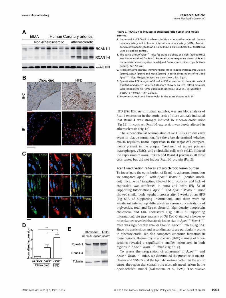
Figure 1. RCAN1‐4 is induced in atherosclerotic human and mouse
arteries.
A. Immunoblot of RCAN1 in atherosclerotic and non-atherosclerotic human
coronary artery and in human internal mammary artery (hIMA). Protein
bands corresponding to RCAN1-1 and RCAN1-4 are indicated. a-ACTINwas
used as loading control.
B. The aortic sinus of Apoe�/�mice fed standard chow or a high-fat diet (HFD)
was immunostained for Rcan1. Representative images are shown of Rcan1
immunohistochemistry (top panels) and fluorescence microscopy (bottom
panels). Bar, 50mm.
C. Representative confocal immunofluorescence images of Rcan1 (red), lectin
(green), aSMA (green) and Mac3 (green) in aortic sinus lesions of HFD-fed
Apoe�/� mice. Merged images are also shown. Bar, 5mm.
D. Quantitative PCR analysis of Rcan1 mRNA expression in the aortic arch of
C57BL/6 and Apoe�/� mice fed standard chow or an HFD. mRNA amounts
were normalized to Hprt1 expression (means� SEM; n¼3). Student’s
t-test, �p¼0.013, ��p¼0.0019.
E. Representative Rcan1 immunoblot in the same tissues as in D.
Research Articlewww.embomolmed.orgNerea Mendez-Barbero et al.
EMBO Mol Med (2013) 5, 1901–1917 �
HFD (Fig 1D). As in human samples, western blot analysis ofRcan1 expression in the aortic arch of these animals indicatedthat Rcan1‐4 was strongly induced in atherosclerotic mice(Fig 1E). In contrast, Rcan1‐1 expression was barely affected inatherosclerosis (Fig 1E).
The subendothelial accumulation of oxLDLs is a crucial earlyevent in plaque formation. We therefore determined whetheroxLDL regulates Rcan1 expression in the major cell compart-ments present in the plaque. Treatment of mouse primarymacrophages, VSMCs, and endothelial cells with oxLDL inducedthe expression of Rcan1 mRNA and Rcan1‐4 protein in all threecells types, but did not induce Rcan1‐1 protein (Fig 2).
Rcan1 inactivation reduces atherosclerotic lesion burdenTo investigate the contribution of Rcan1 to atheroma formationwe compared Apoe�/� with Apoe�/�Rcan1�/� (double knock-out) mice. Rcan1 targeting affected both isoforms and lack ofexpression was confirmed in aorta and heart (Fig S2 ofSupporting Information). Apoe�/� and Apoe�/�Rcan1�/� miceshowed similar body weight increases after 6 weeks on an HFD(Fig S3A of Supporting Information), and there were nosignificant inter‐group differences in serum concentrations oftriglyceride, total and free cholesterol, high‐density lipoproteincholesterol and LDL cholesterol (Fig S3B–C of SupportingInformation). En face analysis of Oil Red O stained atheroscle-rotic plaques revealed that aortic lesion size inApoe�/�Rcan1�/�
mice was significantly smaller than in Apoe�/� mice (Fig 3A).Since the aortic sinus and ascending aorta are particularly proneto atherosclerosis, we also compared atheroma formation inthese regions. Haematoxylin and eosin (H&E) staining of cross‐sections revealed a significantly smaller lesion area in bothregions in Apoe�/�Rcan1�/� mice (Fig 3B–C).
To assess the progression of atheromas in Apoe�/� andApoe�/�Rcan1�/� mice, we determined the presence of macro-phages and VSMCs and the lipid deposition pattern in the aorticcusps, the region that contains the most advanced lesions in theApoe‐deficient model (Nakashima et al, 1994). The relative
2013 The Authors. Published by John Wiley and Sons, Ltd on behalf of EMBO. 1903

Figure 2. Induction of Rcan1‐4 expression by oxLDL. Quantitative PCR and
representative immunoblot analysis of Rcan1 expression in cells isolated from
Apoe�/� mice and treated with 50mg/ml oxLDL:
A. VSMCs (n¼3),
B. Peritoneal macrophages (n¼3) and
C. Endothelial cells (n¼6). mRNA amounts were normalized to Hprt1
expression (means� SEM). Student’s t-test, �p¼0.007, ��p¼0.001,���p¼0.0003.
Research Article www.embomolmed.orgRCAN1 mediates atherosclerosis
1904
content ofmacrophages was lower inApoe�/�Rcan1�/� plaques,whereas numbers of VSMCs were similar in the two genotypes(Fig 4A). To confirm that Apoe�/�Rcan1�/� mice had less‐advanced plaques, we classified lesions according to the Starymethod (Stary et al, 1994) into early plaques (grade I) containingonly macrophages; grade II lesions containing macrophages,VSMCs and a few scattered cholesterol clefts; grade IIIlesions containing macrophages, VSMCs and numerouscholesterol clefts and advanced plaques (grade IV) containingmacrophages, VSMCs and a large lipid core (Fig 4B). After6 weeks of HFD, � 42% plaques in Apoe�/� mice were grade IVand only � 14% were grade I (Fig 4C). In contrast,the proportion of grade IV plaques in Apoe�/�Rcan1�/� micewas � 28% and that of grade I plaques was � 31% (Fig 4C).These results therefore indicate that Rcan1 plays a key role inatherosclerosis progression.
� 2013 The Authors. Published by John Wiley and Sons, Ltd on behalf of EMBO.
While Rcan1 targeting generates less advanced atheroscleroticplaques, it might as well potentially decrease their stability.Although theApoe�/�mouse is not particularly prone to developunstable plaques in the aorta (Getz & Reardon, 2012), it wasformally possible that Rcan1 deficiency might increase plaquevulnerability. We therefore determined the effect of Rcan1targeting on plaque stability. A key feature of unstable plaques isthinning of the fibrous cap, usually accompanied by a reductionin collagen content. The extent and thickness of the fibrous cap,relative to plaque size, were larger in Apoe�/�Rcan1�/� micethan in Apoe�/� mice (Fig 5A and Fig S4 of SupportingInformation), whereas the collagen content of the fibrous capwas similar in both genotypes (Fig 5B–C). Moreover, lipidcontent, determined by Oil Red staining, was lower in plaques ofApoe�/�Rcan1�/� mice (Fig 5B) and these plaques had a higherstability score (Fig 5D). Expression of metalloproteases MMP2and MMP9, another index of plaque instability, was almostidentical in Apoe�/� and Apoe�/�Rcan1�/� plaques (Fig S5 ofSupporting Information) and no plaques displayed evidence ofhaemorrhage (Fig S6 of Supporting Information). These data,together with the lower macrophage content of Rcan1‐deficientplaques, thus indicate that Rcan1 targeting does not inducecharacteristics of unstable atherosclerotic plaques, and suggestinstead that Rcan1 inactivation might increase plaque stability.
Rcan1 regulates CD36 expression and mediates foam‐cellformationOil Red O staining of intracellular lipids indicated that whilemacrophages isolated from Apoe�/� and Apoe�/�Rcan1�/� miceengulfed unmodified LDL similarly (Fig 6A), fewer Apoe�/
�Rcan1�/� macrophages engulfed oxLDL, and Apoe�/�Rcan1�/
� macrophages also appeared to load less oxLDL per cell(Fig 6B). We measured intracellular oxLDL particles by Laurdangeneralized polarization (Laurdan GP) (Sanchez et al, 2007;Sanchez et al, 2010), a confocal technique that measures watercontent inside lipid compartments. Intracellular oxLDLs aretightly packed and shift Laurdan GP towards red frequencies(Ferretti et al, 2002), whereas other lipid compartments,including intracellular membranes, contain more water andgive a yellow or green signal (Sanchez et al, 2007; Sanchezet al, 2010). Macrophages exposed to oxLDL thus show itspresence in the cytosol as red dots (Fig 6C). Quantification of thearea occupied by oxLDL revealed that Apoe�/� macrophagestook up � 2.5 times more oxLDL than Apoe�/�Rcan1�/� cells(Fig 6C).
OxLDL uptake and foam‐cell formation are mediated by theclass A and B scavenger receptors SR‐A and CD36 (Febbraioet al, 2000; Suzuki et al, 1997). Real‐time PCR analysis detectedsignificantly higher levels of CD36 in the aortic arches ofApoe�/�
mice compared with Apoe�/�Rcan1�/� mice (Fig 6D), whereasexpression of SR‐A was similar in the two genotypes (Fig S7A ofSupporting Information). Accordingly, flow cytometry analysisrevealed that CD36 levels, but not those of SR‐A, weresignificantly downregulated in Apoe�/�Rcan1�/� macrophages(Fig 6E and Fig S7B of Supporting Information). Lentiviral re‐expression of Rcan1‐1 and Rcan1‐4 in Apoe�/�Rcan1�/�
peritoneal macrophages (Fig 7A) increased cell surface
EMBO Mol Med (2013) 5, 1901–1917

Figure 3. Rcan1 deficiency decreases atherogenesis burden.
A. Representative Oil red O staining and quantification of lesion area in the aortas of Apoe�/� (n¼11) and Apoe�/�Rcan1�/�mice (n¼18) fed an HFD for 6 weeks.
Each data point denotes an individual mouse, and the horizontal bars denote the mean (long bar) and the SEM.
B. Representative H&E staining of the aortic sinus and quantification of the lesion area in this region (mean� SEM).
C. Representative H&E staining of the ascending aorta and quantification of lesion measured as intima/media ratio in this region in the same cohort of animals
(mean� SEM). Bars, 200mm. Student’s t-test, �p¼0.02, ��p¼0.0025, ���p¼0.0001.
Research Articlewww.embomolmed.orgNerea Mendez-Barbero et al.
expression of CD36 (Fig 7B) and concomitantly increased thenumbers of oxLDL particles taken up by Apoe�/�Rcan1�/�
peritoneal macrophages (Fig 7C–D). These results thus supportthat Rcan1 contributes to foam‐cell formation by regulatingCD36 expression.
Lipid accumulation by lesional macrophages can also reflectaltered cholesterol efflux. To investigate the contribution of Rcan1to cholesterol efflux, foam‐cell formation was induced inApoe�/�
and Apoe�/�Rcan1�/� peritoneal macrophages by incubatingthem with particles of acetylated LDL (acLDL), a non‐atheroscle-rotic modified form of LDL, in the presence of 3H‐cholesterol.Apoe�/�Rcan1�/� macrophages accumulated less 3H‐cholesterol(Fig S8A of Supporting Information). Addition of HDL to the foam‐
cell cultures promoted cholesterol efflux, and this effect wasmodestly stronger in Rcan1‐deficient cells (Fig S8B of SupportingInformation). Consistent with this effect, ABC transporterexpression was slightly higher in Apoe�/�Rcan1�/� peritonealmacrophages and in atherosclerotic lesions in the aortic arch ofthese animals (Fig S8C–D of Supporting Information).
EMBO Mol Med (2013) 5, 1901–1917 �
Rcan1 regulates oxLDL‐mediated inhibition of macrophagemigrationThe presence of macrophages in atheroma plaques depends notonly on their recruitment, but also on their capacity to exit theplaque, a process strongly impaired by oxLDL (Angeli et al, 2004;Randolph, 2008). Since Rcan1 can either promote or repress cellmigration (Espinosa et al, 2009; Iizuka et al, 2004; Minamiet al, 2004), we first determined whether Rcan1 wasrequired for chemotactic macrophage migration. Apoe�/� andApoe�/�Rcan1�/� macrophages migrated similarly in BoydenTranswell chambers in response to MCP‐1 or a combination ofMCP‐1 and fetal bovine serum (FBS) (Fig S9 of SupportingInformation). We next investigated whether Rcan1 participatedin oxLDL‐elicited inhibition of macrophage chemotaxis. Whileexposure of Apoe�/� macrophages to oxLDL sharply reducedtheir migration towards the chemotactic stimulus, Rcan1‐deficient macrophages were not affected (Fig 8A–B). Accord-ingly, in wound‐healing assays oxLDL inhibited randommigration of MCP1‐treated Apoe�/� macrophages, but not that
2013 The Authors. Published by John Wiley and Sons, Ltd on behalf of EMBO. 1905
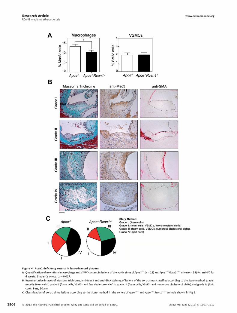
Figure 4. Rcan1 deficiency results in less‐advanced plaques.
A. Quantification of neointimal macrophage and VSMC content in lesions of the aortic sinus of Apoe�/� (n¼11) and Apoe�/�Rcan1�/�mice (n¼18) fed an HFD for
6 weeks. Student’s t-test, �p¼0.017.
B. Representative images of Masson’s trichrome, anti-Mac3 and anti-SMA staining of lesions of the aortic sinus classified according to the Stary method: grade I
(mostly foam cells), grade II (foam cells, VSMCs and few cholesterol clefts), grade III (foam cells, VSMCs and numerous cholesterol clefts) and grade IV (lipid
core). Bars, 50mm.
C. Classification of aortic sinus lesions according to the Stary method in the cohort of Apoe�/� and Apoe�/�Rcan1�/� animals shown in Fig 3.
Research Article www.embomolmed.orgRCAN1 mediates atherosclerosis
1906 � 2013 The Authors. Published by John Wiley and Sons, Ltd on behalf of EMBO. EMBO Mol Med (2013) 5, 1901–1917
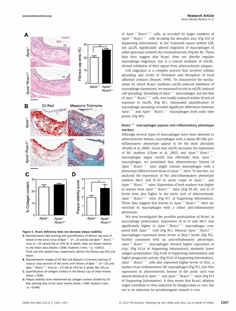
Figure 5. Rcan1 deficiency does not decrease plaque stability.
A. Representative H&E staining and quantification of fibrous cap area of
lesions in the aortic sinus of Apoe�/� (n¼25 valves) and Apoe�/�Rcan1�/�
mice (n¼45 valves) fed an HFD for 6 weeks. Data are shown relative
to the lesion area (means� SEM). Student’s t-test, ��p¼0.0012.
Thick and thin dotted lines, respectively, delimit the fibrous cap (FC) and
lesion.
B. Representative images of Oil Red and Masson’s trichrome staining of
cryocut cross-sections of the aortic arch lesions of Apoe�/� (n¼15) and
Apoe�/�Rcan1�/� mice (n¼17) fed an HFD for 6 weeks. Bar, 50mm.
C. Quantification of collagen content in the fibrous cap of these lesions
(mean� SEM).
D. Plaque stability score determined by collagen content divided by Oil
Red staining area of the same lesions (mean� SEM; Student t-test,�p¼0.048).
Research Articlewww.embomolmed.orgNerea Mendez-Barbero et al.
EMBO Mol Med (2013) 5, 1901–1917 �
of Apoe�/�Rcan1�/� cells, as revealed by larger numbers ofApoe�/�Rcan1�/� cells invading the denuded area (Fig S10 ofSupporting Information). In the Transwell assays neither LDLnor acLDL significantly altered migration of macrophages ofeither genotype towards the chemoattractant (Fig 8A–B). Thesedata thus suggest that Rcan1 does not directly regulatemacrophage migration, but is a central mediator of oxLDL‐elicited inhibition of their egress from atherosclerotic plaques.
Cell migration is a complex process that involves cellularspreading and cycles of formation and disruption of focaladhesion contacts (Stossel, 1994). To characterize the mecha-nisms by which Rcan1 mediates oxLDL‐induced inhibition ofmacrophage chemotaxis, wemeasured its role in oxLDL‐inducedcell spreading. Spreading of Apoe�/� macrophages, but not thatof Apoe�/�Rcan1�/� cells, was readily induced within 30min ofexposure to oxLDL (Fig 8C). Automated quantification ofmacrophage spreading revealed significant differences betweenApoe�/� and Apoe�/�Rcan1�/� macrophages from early timepoints (Fig 8D).
Rcan1�/� macrophages express anti‐inflammatory phenotypemarkersAlthough several types of macrophages have been detected inatherosclerotic lesions, macrophages with a classic M1‐like pro‐inflammatory phenotype appear to be the most abundant(Waldo et al, 2008). Given that oxLDL increases the expressionof M1 markers (Chase et al, 2002) and Apoe�/�Rcan1�/�
macrophages ingest oxLDL less efficiently than Apoe�/�
macrophages, we postulated that atherosclerotic lesions ofApoe�/�Rcan1�/� mice might contain macrophages with aphenotype different from those of Apoe�/�mice. To test this, weanalysed the expression of the anti‐inflammatory phenotypemarkers Mrc1 and IL‐10 in aortic cusps of Apoe�/� andApoe�/�Rcan1�/� mice. Expression of both markers was higherin lesions from Apoe�/�Rcan1�/� mice (Fig 9A–B), and IL‐10levels were also higher in the aortic arch of atheroscleroticApoe�/�Rcan1�/� mice (Fig S11 of Supporting Information).These data suggest that lesions in Apoe�/�Rcan1�/� mice areenriched in macrophages with a rather anti‐inflammatoryphenotype.
We next investigated the possible participation of Rcan1 inmacrophage polarization. Expression of IL‐10 and Mrc1 wassignificantly higher in Apoe�/�Rcan1�/� macrophages com-pared with Apoe�/� cells (Fig 9C), whereas Apoe�/�Rcan1�/�
macrophages expressed lower levels of Mcp‐1 levels (Fig 9C).Further consistent with an anti‐inflammatory phenotype,Apoe�/�Rcan1�/� macrophages showed higher expression ofArg1 (Fig S12A of Supporting Information), modestly lowerantigen presentation (Fig S12B of Supporting Information) andhigher phagocytic activity (Fig S12C of Supporting Information).Apoe�/�Rcan1�/� cells also expressed higher levels of iNos, amarker of pro‐inflammatory M1 macrophages (Fig 9C), but iNosexpression in atherosclerotic lesions of the aortic arch wasalmost identical in Apoe�/� and Apoe�/�Rcan1�/� mice (Fig S13of Supporting Information). It thus seems that Rcan1 ablationmight contribute to iNos induction by thioglycolate ex vivo, butnot to its induction by proatherogenic stimuli in vivo.
2013 The Authors. Published by John Wiley and Sons, Ltd on behalf of EMBO. 1907
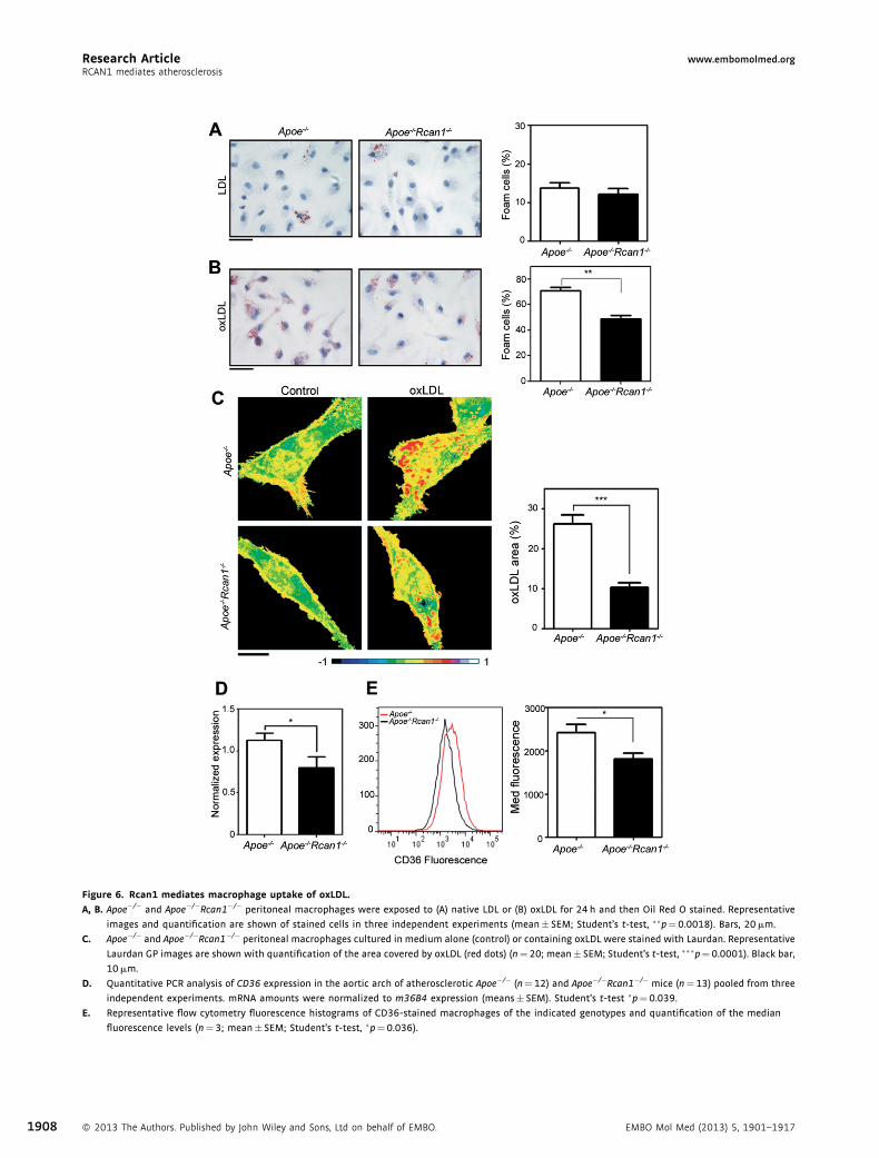
Figure 6. Rcan1 mediates macrophage uptake of oxLDL.
A, B. Apoe�/� and Apoe�/�Rcan1�/� peritoneal macrophages were exposed to (A) native LDL or (B) oxLDL for 24 h and then Oil Red O stained. Representative
images and quantification are shown of stained cells in three independent experiments (mean� SEM; Student’s t-test, ��p¼0.0018). Bars, 20mm.
C. Apoe�/� and Apoe�/�Rcan1�/� peritoneal macrophages cultured in medium alone (control) or containing oxLDL were stained with Laurdan. Representative
Laurdan GP images are shown with quantification of the area covered by oxLDL (red dots) (n¼20; mean� SEM; Student’s t-test, ���p¼0.0001). Black bar,
10mm.
D. Quantitative PCR analysis of CD36 expression in the aortic arch of atherosclerotic Apoe�/� (n¼12) and Apoe�/�Rcan1�/� mice (n¼13) pooled from three
independent experiments. mRNA amounts were normalized to m36B4 expression (means� SEM). Student’s t-test �p¼0.039.
E. Representative flow cytometry fluorescence histograms of CD36-stained macrophages of the indicated genotypes and quantification of the median
fluorescence levels (n¼3; mean� SEM; Student’s t-test, �p¼0.036).
Research Article www.embomolmed.orgRCAN1 mediates atherosclerosis
1908 � 2013 The Authors. Published by John Wiley and Sons, Ltd on behalf of EMBO. EMBO Mol Med (2013) 5, 1901–1917

Figure 7. Rcan1 re‐expression in Apoe�/�Rcan1�/�macrophages increases
CD36 expression and the uptake of oxLDL. Apoe�/�Rcan1�/� peritoneal
macrophages were transduced with lentiviruses encoding GFP or Rcan1-1-
IRES-GFP plus Rcan1-4-IRES-GFP.
A. Representative immunoblot analysis of Rcan1 expression in these cells.
B. Quantification of the normalized median fluorescence levels of CD36
determined by flow cytometry in the GFPþ population (n¼4; mean
� SEM; Student’s t-test �p¼0.02).
C, D. (C) Representative Laurdan GP images and (D) quantification of the area
covered by oxLDL (red dots) in GFP-F4/80 double-positive cells after
culture in the presence of oxLDL (n¼3; mean� SEM; Student’s t-test,�p¼0.01). Scale bar, 10mm.
Research Articlewww.embomolmed.orgNerea Mendez-Barbero et al.
Transplantation of Rcan1�/� BM cells confers resistance toatherosclerosisTo test the possible promotion of atherosclerosis by macro-phage‐expressed Rcan1, we reconstituted the BM of 2‐month oldlethally irradiated Apoe�/� mice with BM‐derived cells fromApoe�/�Rcan1�/� or Apoe�/� mice, thereby generatingApoe�/� mice with either Apoe�/�Rcan1�/� macrophages(Rcan1�/�!Apoe�/�) or Apoe�/�Rcan1þ/þ macrophages(Rcan1þ/þ!Apoe�/�). Since RCAN1 has been reported toinduce GSK3b expression (Ermak et al, 2006) and this proteinmodulates haematopoietic progenitor cell function (Lapidet al, 2013), it is formally possible that Rcan1 ablation influencesthe nature of BM reconstitution. However, chimericRcan1�/�!Apoe�/� mice showed no significant reduction in
EMBO Mol Med (2013) 5, 1901–1917 �
Gsk3b expression (Fig S14A of Supporting Information), andblood cell populations of reconstituted Rcan1þ/þ!Apoe�/� andRcan1�/�!Apoe�/� mice were indistinguishable (Fig S14B–Dof Supporting Information).
After reconstitution for 4 week, mice were fed an HFDfor 6 weeks. Chimeric Rcan1�/�!Apoe�/� and Rcan1þ/þ!Apoe�/� mice showed no significant differences in the bodyweight gain or in the serum concentrations of triglyceride, totalcholesterol, HDL or LDL (Fig S15 of Supporting Information). Enface analysis of Oil Red O aorta staining revealed markedlysmaller lesion area in mice lacking expression of Rcan1 in BM‐
derived cells (Fig 10A). Cross‐sectional analysis by H&E stainingindicated that lesion size in the aortic sinus and the intima/media ratio in the ascending aorta were significantly smaller inRcan1�/�!Apoe�/� mice than in Rcan1þ/þ!Apoe�/� mice(Fig 10B–C). These data demonstrate that Rcan1 expression inthe haematopoietic cell compartment plays a major role inatherosclerosis progression.
DISCUSSION
Atherosclerosis is a complex disease involving lipid accumula-tion and the central participation of endothelial cells, VSMCs andmonocyte‐derived macrophages. Our results demonstrate thatRCAN1 is induced in human and mouse atherosclerosis andstrongly suggest that Rcan1 promotes disease progression. Inaddition, we have identified several mechanisms underlyingRcan1‐dependent atherosclerosis development. In particular, wehave shown that Rcan1 mediates CD36 expression, foam‐cellformation and oxLDL‐elicited inhibition of macrophage migra-tion. Moreover, Rcan1 ablation promotes expression of IL‐10 andother anti‐inflammatory markers, including Mrc1, in macro-phages and lesions.
Rcan1 plays a critical role in vascular wall remodellingassociated with aneurysm and neointima formation afterangioplasty; and notably, BM transplantation experiments showedthat haematopoietic‐cell expression of Rcan1 is not required foraneurysm (Esteban et al, 2011). In contrast, our current findingsshow that Rcan1 expression in BM‐cells is critical for atherogene-sis. Our results do not however exclude a proatherogenic role ofRcan1 in VSMCs or endothelial cells. The eventual production oftissue‐specific Rcan1‐targeted mice will help to resolve thisquestion. The different requirements for BM‐cell expression ofRcan1 in atherosclerosis and aneurysmmight be attributable to thepivotal role of Rcan1 in foam‐cell formation, a central feature ofatherosclerosis but not of aneurysm.
Although human and mouse atherosclerosis differ in severalaspects, critical features are shared, and our results indicate thatone such feature is RCAN1 induction. Moreover, we demon-strate that oxLDL, one of main stimuli of atherosclerosis,strongly induces Rcan1‐4 not only in macrophages and VSMCs,but also in endothelial cells. Althoughmost leukocytes in humanand mouse atherosclerotic lesions are macrophages, plaquesalso contain lymphocytes, neutrophils and mast cells (Woollard& Geissmann, 2010). Whether Rcan1 is also induced in thesecells remains to be determined.
2013 The Authors. Published by John Wiley and Sons, Ltd on behalf of EMBO. 1909
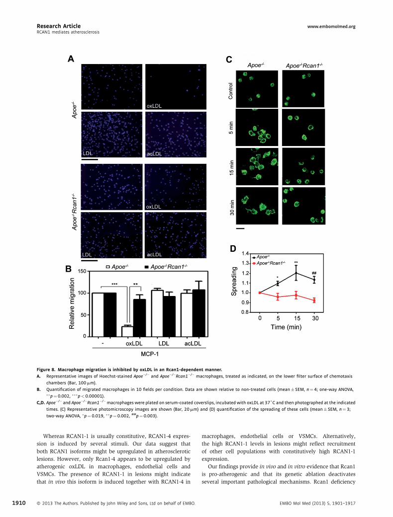
Figure 8. Macrophage migration is inhibited by oxLDL in an Rcan1‐dependent manner.
A. Representative images of Hoechst-stained Apoe�/� and Apoe�/�Rcan1�/� macrophages, treated as indicated, on the lower filter surface of chemotaxis
chambers (Bar, 100mm).
B. Quantification of migrated macrophages in 10 fields per condition. Data are shown relative to non-treated cells (mean� SEM, n¼4; one-way ANOVA,��p¼0.002, ���p<0.00001).
C,D. Apoe�/� and Apoe�/�Rcan1�/�macrophages were plated on serum-coated coverslips, incubated with oxLDL at 37˚C and then photographed at the indicated
times. (C) Representative photomicroscopy images are shown (Bar, 20mm) and (D) quantification of the spreading of these cells (mean� SEM, n¼3;
two-way ANOVA, �p¼0.019, ��p¼0.002, ##p¼0.003).
Research Article www.embomolmed.orgRCAN1 mediates atherosclerosis
1910
Whereas RCAN1‐1 is usually constitutive, RCAN1‐4 expres-sion is induced by several stimuli. Our data suggest thatboth RCAN1 isoforms might be upregulated in atheroscleroticlesions. However, only Rcan1‐4 appears to be upregulated byatherogenic oxLDL in macrophages, endothelial cells andVSMCs. The presence of RCAN1‐1 in lesions might indicatethat in vivo this isoform is induced together with RCAN1‐4 in
� 2013 The Authors. Published by John Wiley and Sons, Ltd on behalf of EMBO.
macrophages, endothelial cells or VSMCs. Alternatively,the high RCAN1‐1 levels in lesions might reflect recruitmentof other cell populations with constitutively high RCAN1‐1expression.
Our findings provide in vivo and in vitro evidence that Rcan1is pro‐atherogenic and that its genetic ablation deactivatesseveral important pathological mechanisms. Rcan1 deficiency
EMBO Mol Med (2013) 5, 1901–1917

Figure 9. Increased expression of alternative macrophage polarization
markers in Apoe�/�Rcan1�/� lesions and isolated macrophages.
A, B. Representative immunofluorescence staining of (A) IL-10 and (B) Mrc1
in aortic sinus lesions of Apoe�/� and Apoe�/�Rcan1�/�mice fed an HFD.
Bars, 20mm.
C. Quantitative PCR analysis of IL-10, Mrc1, Mcp-1 and iNOS mRNA
expression in Apoe�/� and Apoe�/�Rcan1�/� peritoneal macrophages.
mRNA amounts were normalized to m36B4 expression (mean� SEM;
n¼3). Student’s t-test, ��p¼0.0078, ##p¼0.0058, ���p¼0.0007,###p¼0.0009.
Research Articlewww.embomolmed.orgNerea Mendez-Barbero et al.
inhibits macrophage engulfment of oxLDL and hence foam‐cellformation, and Rcan1�/� macrophages are resistent to oxLDL‐mediated inhibition of migration towards chemotactic stimuli, afeature related to foam‐cell egress from the plaque in vivo.Finally, in the absence of Rcan1, macrophages appear toacquire features of a predominantly anti‐inflammatory pheno-type. Consistent with this view, Rcan1 inactivation in the Apoe‐deficient mouse atherosclerosis model attenuates atheroscleroticlesion burden in terms both of lesion area and severity.While most lesions of Apoe�/� mice were advanced(grade IV), Apoe�/�Rcan1�/� mice exhibited a greater number
EMBO Mol Med (2013) 5, 1901–1917 �
of early lesions (grade I) without showing any evidence ofincreased plaque instability.
The lower Oil Red O surface staining and the lowercontent of extracellular lipids in atherosclerotic lesions ofApoe�/�Rcan1�/� mice are consistent with the relatively lowuptake of oxLDL by Apoe�/�Rcan1�/� macrophages. Interest-ingly, these macrophages have a similar capacity to engulf nativeLDL. Given that endocytic uptake of native LDL and oxLDLoccurs via different receptors (Greaves & Gordon, 2009), ourdata suggest that Rcan1 promotes the expression or activity ofreceptors specific for chemically modified forms of LDL. Thisconclusion is supported by our findings that CD36, a scavengerreceptor involved in the uptake of modified forms of LDL andfoam‐cell formation (Febbraio et al, 2000), is downregulated inRcan1 deficient macrophages, while re‐expression of Rcan1‐1and Rcan1‐4 in these cells increased CD36 expression and foam‐
cell formation. Cholesterol efflux was weakly increased inApoe�/�Rcan1�/� macrophages and the levels of the cellulartransporters involved in this process were not significantlyincreased in these cells or in atherosclerotic lesions. Thus, thecontribution of cholesterol efflux to Rcan1‐mediated regulationof foam‐cell formation remains uncertain.
The inflammatory nature of atherosclerotic disease is widelyaccepted. During the resolution of inflammation, macrophagesegress from the inflamed site after engulfing pathogens, toxins orapoptotic cells. However, cholesterol‐engorgedmacrophages failto egress after clearing lipids and hence fail to resolve theinflammatory process (Angeli et al, 2004). Egress of macro-phages from plaques is actively inhibited during hypercholes-terolemia and this inhibition has been attributed, at least in part,to oxLDL loading into macrophages (Park et al, 2009). Our datasuggest that Rcan1 might facilitate the trapping of lipid‐ladenmacrophages in atherosclerotic lesions by mediating oxLDLuptake, macrophage spreading and inhibition of their egress.Our data thus suggest that Rcan1 might participate in a positivefeedback loop in which Rcan1‐mediated accumulation of oxLDLparticles inhibits foam‐cell egress from the plaque, resulting inincreased exposure of the trapped foam cells to oxLDL andincreased engulfment of these particles.
Most lesional macrophages in advanced plaques display aproinflammatory M1 phenotype, whereas alternatively activatedM2 macrophages are more abundant in early lesions (Khallou‐Laschet et al, 2010; Waldo et al, 2008). An additionalmacrophage subtype, whose gene expression program differsfrom that of M1 and M2 cells and which shows enhancedphagocytotic and chemotactic capacities, has also been reported
2013 The Authors. Published by John Wiley and Sons, Ltd on behalf of EMBO. 1911

Figure 10. Atherosclerosis requires Rcan1 expression in BM‐derived cells.
A. Representative Oil red O staining and quantification of the lesion area in the aortas of Apoe�/� mice transplanted with BM from Apoe�/� (n¼16) or
Apoe�/�Rcan1�/� (n¼15) mice after 6 weeks on an HFD. Each data point denotes an individual mouse, and the horizontal bars denote the mean (long bar) and
the SEM.
B. Representative H&E images and lesion area quantification in aortic sinus sections of these mice. Quantification is shown as mean� SEM (n¼13). Bar, 200mm.
C. Representative H&E images and quantification of lesion measured as intima/media ratio. Data are means� SEM (n¼12). Bar, 200mm. Student’s t-test,�p¼0.02, #p¼0.024, ���p¼0.0001.
Research Article www.embomolmed.orgRCAN1 mediates atherosclerosis
1912
in advanced plaques (Kadl et al, 2010), and analysis of CD163and CD206 (MRC1) distribution in human carotid plaquemacrophages also suggested that there may be at least threemacrophage phenotypes present in human plaques (Bouhlelet al, 2007). Our results show elevated expression of IL‐10 andMrc1 in lesions of Rcan1 deficient mice and no increase of iNos.Since IL‐10 is a powerful anti‐inflammatory cytokine and its
� 2013 The Authors. Published by John Wiley and Sons, Ltd on behalf of EMBO.
genetic ablation in mice promotes atherosclerotic lesionformation (Mallat et al, 1999), these results suggest thatRcan1 deficiency promotes the presence of macrophages withanti‐inflammatory properties in the atheroma plaque, aconclusion consistent with the retarded progression of lesionsin Apoe�/�Rcan1�/� mice. Whether these IL‐10‐producing cellsare M2‐like macrophages or an additional macrophage type
EMBO Mol Med (2013) 5, 1901–1917
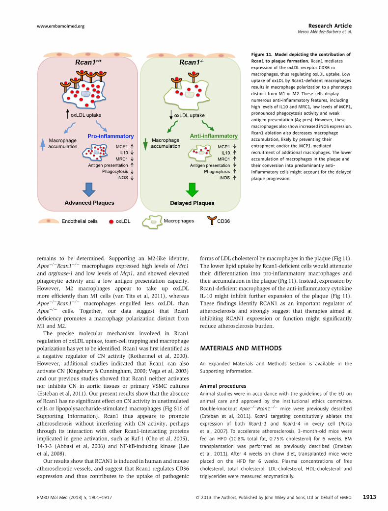
Figure 11. Model depicting the contribution of
Rcan1 to plaque formation. Rcan1 mediates
expression of the oxLDL receptor CD36 in
macrophages, thus regulating oxLDL uptake. Low
uptake of oxLDL by Rcan1-deficient macrophages
results in macrophage polarization to a phenotype
distinct from M1 or M2. These cells display
numerous anti-inflammatory features, including
high levels of IL10 and MRC1, low levels of MCP1,
pronounced phagocytosis activity and weak
antigen presentation (Ag pres). However, these
macrophages also show increased iNOS expression.
Rcan1 ablation also decreases macrophage
accumulation, likely by preventing their
entrapment and/or the MCP1-mediated
recruitment of additional macrophages. The lower
accumulation of macrophages in the plaque and
their conversion into predominantly anti-
inflammatory cells might account for the delayed
plaque progression.
Research Articlewww.embomolmed.orgNerea Mendez-Barbero et al.
remains to be determined. Supporting an M2‐like identity,Apoe�/�Rcan1�/� macrophages expressed high levels of Mrc1and arginase‐1 and low levels of Mcp1, and showed elevatedphagocytic activity and a low antigen presentation capacity.However, M2 macrophages appear to take up oxLDLmore efficiently than M1 cells (van Tits et al, 2011), whereasApoe�/�Rcan1�/� macrophages engulfed less oxLDL thanApoe�/� cells. Together, our data suggest that Rcan1deficiency promotes a macrophage polarization distinct fromM1 and M2.
The precise molecular mechanism involved in Rcan1regulation of oxLDL uptake, foam‐cell trapping and macrophagepolarization has yet to be identified. Rcan1 was first identified asa negative regulator of CN activity (Rothermel et al, 2000).However, additional studies indicated that Rcan1 can alsoactivate CN (Kingsbury & Cunningham, 2000; Vega et al, 2003)and our previous studies showed that Rcan1 neither activatesnor inhibits CN in aortic tissues or primary VSMC cultures(Esteban et al, 2011). Our present results show that the absenceof Rcan1 has no significant effect on CN activity in unstimulatedcells or lipopolysaccharide‐stimulated macrophages (Fig S16 ofSupporting Information). Rcan1 thus appears to promoteatherosclerosis without interfering with CN activity, perhapsthrough its interaction with other Rcan1‐interacting proteinsimplicated in gene activation, such as Raf‐1 (Cho et al, 2005),14‐3‐3 (Abbasi et al, 2006) and NF‐kB‐inducing kinase (Leeet al, 2008).
Our results show that RCAN1 is induced in human andmouseatherosclerotic vessels, and suggest that Rcan1 regulates CD36expression and thus contributes to the uptake of pathogenic
EMBO Mol Med (2013) 5, 1901–1917 �
forms of LDL cholesterol by macrophages in the plaque (Fig 11).The lower lipid uptake by Rcan1‐deficient cells would attenuatetheir differentiation into pro‐inflammatory macrophages andtheir accumulation in the plaque (Fig 11). Instead, expression byRcan1‐deficient macrophages of the anti‐inflammatory cytokineIL‐10 might inhibit further expansion of the plaque (Fig 11).These findings identify RCAN1 as an important regulator ofatherosclerosis and strongly suggest that therapies aimed atinhibiting RCAN1 expression or function might significantlyreduce atherosclerosis burden.
MATERIALS AND METHODS
An expanded Materials and Methods Section is available in the
Supporting Information.
Animal proceduresAnimal studies were in accordance with the guidelines of the EU on
animal care and approved by the institutional ethics committee.
Double‐knockout Apoe�/�Rcan1�/� mice were previously described
(Esteban et al, 2011). Rcan1 targeting constitutively ablates the
expression of both Rcan1-1 and Rcan1-4 in every cell (Porta
et al, 2007). To accelerate atherosclerosis, 3‐month‐old mice were
fed an HFD (10.8% total fat, 0.75% cholesterol) for 6 weeks. BM
transplantation was performed as previously described (Esteban
et al, 2011). After 4 weeks on chow diet, transplanted mice were
placed on the HFD for 6 weeks. Plasma concentrations of free
cholesterol, total cholesterol, LDL‐cholesterol, HDL‐cholesterol and
triglycerides were measured enzymatically.
2013 The Authors. Published by John Wiley and Sons, Ltd on behalf of EMBO. 1913
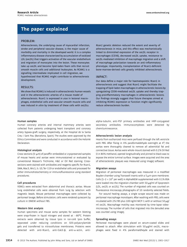
The paper explained
PROBLEM:
Atherosclerosis, the underlying cause of myocardial infarction,
stroke and peripheral vascular disease, is the major cause of
morbidity and mortality in the developed world. It is a complex
inflammatory disease characterized by accumulation of oxidized
LDL (oxLDL) that triggers activation of the vascular endothelium
and migration of monocytes into the lesion. These monocytes
take up oxLDL and become lipid-laden foam cells that recruit
smooth muscle cells and additional leukocytes. As RCAN1 is a
signalling intermediate implicated in cell migration, we
hypothesized that RCAN1 might contribute to atherosclerosis
development.
RESULTS:
We show that RCAN1 is induced in atherosclerotic human vessels
and in the atherosclerotic arteries of a mouse model of
atherosclerosis. Rcan1 is expressed in vivo in lesional macro-
phages, endothelial cells and vascular smooth muscle cells and
was induced in vitro by treatment of these cells with oxLDLs.
Rcan1 genetic deletion reduced the extent and severity of
atherosclerosis in mice, and this effect was mechanistically
linked to diminished expression of the oxLDL receptor in
macrophages (CD36), decreased oxLDL uptake, resistance to
oxLDL-mediated inhibition of macrophage migration and a shift
of macrophage polarization towards an anti-inflammatory
phenotype. Importantly, transplantation of Rcan1-deficient
bone-marrow-derived cells greatly inhibited atherosclerosis.
IMPACT:
Our data define a major role for haematopoietic Rcan1 in
atherosclerosis and suggest that Rcan1 might facilitate the
trapping of lipid-ladenmacrophages in atherosclerotic lesions by
upregulating CD36-mediated oxLDL uptake and thereby trap-
ping proinflammatory macrophages in atherosclerotic lesions.
Our findings strongly suggest that future therapies aimed at
inhibiting RCAN1 expression or function might significantly
reduce atherosclerosis burden.
Research Article www.embomolmed.orgRCAN1 mediates atherosclerosis
1914
Human samplesHuman coronary arteries and internal mammary arteries were
collected from patients undergoing heart transplant and coronary
artery bypass‐graft surgery, respectively, at the Hospital de la Santa
Creu i Sant Pau (Barcelona, Spain). The studies were approved by the
Ethics Committee and were conducted in accordance with the Helsinki
Declaration.
Histological analysisCross‐sections (5‐mm) of paraffin‐embedded or cryopreserved samples
of mouse hearts and aortae were immunostained or evaluated by
conventional Masson’s Trichrome, H&E or Oil Red staining. Cross‐
sections were stained with antibodies and reagents specific for Rcan1,
SMA, Mac3, Mrc1, IL‐10, Ter‐119 or endothelial cells and processed for
either immunohistochemistry or immunofluorescence using standard
procedures.
Cell proceduresVSMCs were extracted from abdominal and thoracic aortas. Mouse
lung endothelial cells were obtained from lung by selection with
magnetic beads. Mouse peritoneal macrophages were collected by
peritoneal lavage. Before stimulation, cells were rendered quiescent by
culture in DMEM without FBS.
Western blot analysisHuman specimens and mouse aorta samples for western blotting
were snap‐frozen in liquid nitrogen and stored at �80°C. Protein
extracts were obtained by tissue lysis in ice‐cold lysis buffer,
separated under reducing conditions on SDS‐polyacrylamide
gels and transferred to nitrocellulose membranes. Proteins were
detected with anti‐Rcan1, anti‐Gsk3‐b, anti‐a‐actin, anti‐
� 2013 The Authors. Published by John Wiley and Sons, Ltd on behalf of EMBO.
alpha‐tubulin, anti‐PSF primary antibodies and HRP‐conjugated
secondary antibodies. Immunocomplexes were detected by
chemiluminescence.
Atherosclerotic lesion analysisHearts from euthanized mice were perfused through the left ventricle
with PBS. After fixing in 4% paraformaldehyde overnight at 4°, the
aortas were thoroughly cleaned to remove all adventitial fat and
connective tissue. Aortas were whole‐mount stained with 0.2% Oil Red
O in 80% methanol, opened longitudinally and pinned to black wax to
expose the entire luminal surface. Images were acquired and the area
of atherosclerotic plaques was measured using ImageJ software.
Migration assaysMigration of peritoneal macrophages was measured in a modified
Boyden chamber using Transwell inserts with a 5mm‐pore membrane.
Cells (1–2�105 per well) in AlphaMEM supplemented with 0.1% BSA
were loaded into the migration chamber with 50mg/ml of lipoprotein
(LDL, oxLDL or acLDL). The number of migrated cells was counted on
fluorescence microscopy photographs of 10 randomly selected fields.
For wound healing assays, a single scrape wound was made on
peritoneal macrophage monolayers. After washing with PBS, cells were
incubated with 2% FBS plus 100ng/ml MCP‐1 with or without 50mg/
ml oxLDL. Macrophage motility was monitored by time‐lapse video-
microscopy. The number of cells that migrated into the denuded area
was counted using ImageJ.
Spreading assaysPeritoneal macrophages were placed on serum‐coated slides and
allowed to attach. After stimulation with 50mg/ml oxLDL, macro-
phages were fixed in 4% paraformaldheyde and stained with
EMBO Mol Med (2013) 5, 1901–1917

Research Articlewww.embomolmed.orgNerea Mendez-Barbero et al.
fluorescein‐conjugated phalloidin. Cell perimeter and surface area
were measured to determine cell spreading according to the formula
Spreading ¼ perimeter2= 4� p� areað Þ:
Foam‐cell formationPeritoneal macrophages were plated on coverslips, incubated with
50mg/ml LDL or oxLDL for 24h, fixed in 4% paraformaldehyde and
stained with Oil red O and counterstained with haematoxylin.
Laurdan GP microscopyLaurdan GP microscopy has been described previously (Bagatolli
et al, 2003; Sanchez et al, 2007). Peritoneal macrophages were
cultured in the presence of 1mM Laurdan in serum‐free medium.
Laurdan fluorescence was excited with a mode‐locked titanium‐
sapphire laser set at 780nm and its emission collected at 445–465nm
and 474–514 nm. GP images were obtained with an ALBA microscope
equipped with a 63� water objective and analysed using Image‐J
software.
Quantitative PCRReal‐time quantitative RT‐PCR was performed using a Prime Time
qPCR assay specific for human GAPDH and TaqMan Gene Expression
assays specific for human RCAN1-1, RCAN1-4 and mouse Rcan1 and
Hprt1. SYBR Green was used for RT‐PCR detection of mouse CD-36, SR-
A, ABCA, ABCG, IL-10, Mrc1, Arg1, Mcp-1, iNos and m36B4. Calculations
were made from measurements of 3 replicates of each sample. The
amount of target mRNA in samples was estimated by the 2CT
relative quantification method using GAPDH, m36B4 or Hprt1 for
normalization.
Statistical analysisAll values are expressed as means� SEM. Differences were evaluated
using one‐way or two‐way analysis of variance (ANOVA) and
Bonferroni’s post hoc test (experiments with �3 groups) or the
Student’s t‐test, as appropriate for the data. Statistical significance was
assigned at p<0.05.
Author contributionsThe study was conceived by JMR and MRC. NMB, VE, JMR andMRC designed the study and analysed the data. NMB and VEperformed most of the experiments with contributions from SV,AE, KU and AA. CR and JMG assessed RCAN1 expression inhuman samples. SAS performed Laurdan GP microscopy andanalysed the data obtained. VA, TO and TM providedexperimental support and ideas for the project. JMR and MRCwrote the manuscript with contributions of NMB, SAS and JMG.All authors read and approved the manuscript.
AcknowledgementsWe thank Dr. S. de la Luna for kindly providing pHA‐CALP1Land pHA‐CALP1S plasmids, Dr. S. Bartlett for English editing andR. Alberca for technical support. The Spanish Ministry ofEconomy and Competitiveness (Ministerio de Economía yCompetitividad) supports MRC, JMR and JM‐G with grants
EMBO Mol Med (2013) 5, 1901–1917 �
SAF2010‐15126, SAF2009‐10708 and SAF2012‐40127, respec-tively; MRC is also supported by the Spanish Council forScientific Research (CSIC); JMR is also supported by FundaciónLa Marató TV3 (080731), and Fundación Genoma España(GENOMA). The Red de Investigacion Cardiovascular (RIC) ofthe Spanish Ministry of Health (Ministerio de Sanidad) supportsthe research of JMR, VA and JM‐Gwith grants RD12/0042/0022,RD12/0042/0028 and RD12/0042/0053, respectively. The Cen-tro Nacional de Investigaciones Cardiovasculares (CNIC) issupported by the Spanish Ministry of Economy and Competi-tiveness and the Pro‐CNIC Foundation. TM was supported inpart by the Leading‐edge Research Promotion Fund (LS038,TM). VE was an investigator of the Sara Borrell Program (CD06/00232), and NM‐B holds an FPU fellowship (FPU2008‐1500).
Supporting Information is available at EMBO MolecularMedicine Online.
The authors declare that they have no conflict of interest.
ReferencesAbbasi S, Lee JD, Su B, Chen X, Alcon JL, Yang J, Kellems RE, Xia Y (2006) Protein
kinase-mediated regulation of calcineurin through the phosphorylation of
modulatory calcineurin-interacting protein 1. J Biol Chem 281: 7717-7726
Angeli V, Llodra J, Rong JX, Satoh K, Ishii S, Shimizu T, Fisher EA, Randolph GJ
(2004) Dyslipidemia associated with atherosclerotic disease systemically
alters dendritic cell mobilization. Immunity 21: 561-574
Baek KH, Zaslavsky A, Lynch RC, Britt C, Okada Y, Siarey RJ, Lensch MW, Park
IH, Yoon SS, Minami T et al (2009) Down’s syndrome suppression of tumour
growth and the role of the calcineurin inhibitor DSCR1. Nature 459: 1126-
1130
Bagatolli LA, Sanchez SA, Hazlett T, Gratton E (2003) Giant vesicles, Laurdan,
and two-photon fluorescence microscopy: Evidence of lipid lateral
separation in bilayers. Methods Enzymol 360: 481-500
Bouhlel MA, Derudas B, Rigamonti E, Dievart R, Brozek J, Haulon S, Zawadzki C,
Jude B, Torpier G, Marx N et al (2007) PPARgamma activation primes human
monocytes into alternative M2 macrophages with anti-inflammatory
properties. Cell Metab 6: 137-143
Cano E, Canellada A, Minami T, Iglesias T, Redondo JM (2005) Depolarization
of neural cells induces transcription of the Down syndrome critical region 1
isoform 4 via a calcineurin/nuclear factor of activated T cells-dependent
pathway. J Biol Chem 280: 29435-29443
Chase AJ, Bond M, Crook MF, Newby AC (2002) Role of nuclear factor-kappa B
activation in metalloproteinase-1, -3, and -9 secretion by human
macrophages in vitro and rabbit foam cells produced in vivo. Arterioscler
Thromb Vasc Biol 22: 765-771
Cho YJ, Abe M, Kim SY, Sato Y (2005) Raf-1 is a binding partner of DSCR1. Arch
Biochem Biophys 439: 121-128
Crawford DR, Leahy KP, Abramova N, Lan L, Wang Y, Davies KJ (1997) Hamster
adapt78 mRNA is a Down syndrome critical region homologue that is
inducible by oxidative stress. Arch Biochem Biophys 342: 6-12
Davies KJ, Ermak G, Rothermel BA, Pritchard M, Heitman J, Ahnn J, Henrique-
Silva F, Crawford D, Canaider S, Strippoli P et al (2007) Renaming the
DSCR1/Adapt78 gene family as RCAN: Regulators of calcineurin. FASEB J 21:
3023-3028
Ermak G, Harris CD, Battocchio D, Davies KJ (2006) RCAN1 (DSCR1 or Adapt78)
stimulates expression of GSK-3beta. FEBS J 273: 2100-2109
Ermak G, Harris CD, Davies KJ (2002) The DSCR1 (Adapt78) isoform 1 protein
calcipressin 1 inhibits calcineurin and protects against acute calcium-
mediated stress damage, including transient oxidative stress. FASEB J 16:
814-824
2013 The Authors. Published by John Wiley and Sons, Ltd on behalf of EMBO. 1915

Research Article www.embomolmed.orgRCAN1 mediates atherosclerosis
1916
Espinosa AV, Shinohara M, Porchia LM, Chung YJ, McCarty S, Saji M, Ringel MD
(2009) Regulator of calcineurin 1 modulates cancer cell migration in vitro.
Clin Exp Metastasis 26: 517-526
Esteban V, Mendez-Barbero N, Jimenez-Borreguero LJ, Roque M, Novensa L,
Garcia-Redondo AB, Salaices M, Vila L, Arbones ML, Campanero MR et al
(2011) Regulator of calcineurin 1 mediates pathological vascular wall
remodeling. J Exp Med 208: 2125-2139
Febbraio M, Podrez EA, Smith JD, Hajjar DP, Hazen SL, Hoff HF, Sharma K,
Silverstein RL (2000) Targeted disruption of the class B scavenger receptor
CD36 protects against atherosclerotic lesion development in mice. J Clin
Invest 105: 1049-1056
Ferretti G, Rabini RA, Bacchetti T, Vignini A, Salvolini E, Ravaglia F, Curatola G,
Mazzanti L (2002) Glycated low density lipoproteins modify platelet
properties: A compositional and functional study. J Clin Endocrinol Metab
87: 2180-2184
Fuentes JJ, Pritchard MA, Estivill X (1997) Genomic organization, alternative
splicing, and expression patterns of the DSCR1 (Down syndrome candidate
region 1) gene. Genomics 44: 358-361
Getz GS, Reardon CA (2012) Animal models of atherosclerosis. Arterioscler
Thromb Vasc Biol 32: 1104-1115
Gordon S, Taylor PR (2005)Monocyte andmacrophage heterogeneity. Nat Rev
Immunol 5: 953-964
Greaves DR, Gordon S (2009) The macrophage scavenger receptor at 30 years
of age: Current knowledge and future challenges. J Lipid Res 50: S282-286
Hansson GK, Hermansson A (2011) The immune system in atherosclerosis.
Nat Immunol 12: 204-212
Harris CD, Ermak G, Davies KJ (2005) Multiple roles of the DSCR1 (Adapt78 or
RCAN1) gene and its protein product calcipressin 1 (or RCAN1) in disease.
Cell Mol Life Sci 62: 2477-2486
Hoeffer CA, Dey A, Sachan N, Wong H, Patterson RJ, Shelton JM, Richardson JA,
Klann E, Rothermel BA (2007) The Down syndrome critical region protein
RCAN1 regulates long-term potentiation and memory via inhibition of
phosphatase signaling. J Neurosci 27: 13161-13172
Iizuka M, Abe M, Shiiba K, Sasaki I, Sato Y (2004) Down syndrome candidate
region 1,a downstream target of VEGF, participates in endothelial cell
migration and angiogenesis. J Vasc Res 41: 334-344
Kadl A, Meher AK, Sharma PR, Lee MY, Doran AC, Johnstone SR, Elliott MR,
Gruber F, Han J, Chen W et al (2010) Identification of a novel macrophage
phenotype that develops in response to atherogenic phospholipids via Nrf2.
Circ Res 107: 737-746
Khallou-Laschet J, Varthaman A, Fornasa G, Compain C, Gaston AT, ClementM,
Dussiot M, Levillain O, Graff-Dubois S, Nicoletti A et al (2010) Macrophage
plasticity in experimental atherosclerosis. PLoS One 5: e8852
Kingsbury TJ, Cunningham KW (2000) A conserved family of calcineurin
regulators. Genes Dev 14: 1595-1604
Lapid K, Itkin T, D’Uva G, Ovadya Y, Ludin A, Caglio G, Kalinkovich A, Golan K,
Porat Z, Zollo M et al (2013) GSK3beta regulates physiological migration of
stem/progenitor cells via cytoskeletal rearrangement. J Clin Invest 123:
1705-1717
Lee EJ, Seo SR, Um JW, Park J, Oh Y, Chung KC (2008) NF-kappaB-inducing
kinase phosphorylates and blocks the degradation of Down syndrome
candidate region 1. J Biol Chem 283: 3392-3400
Libby P (2002) Inflammation in atherosclerosis. Nature 420: 868-874
Mallat Z, Besnard S, Duriez M, Deleuze V, Emmanuel F, Bureau MF, Soubrier F,
Esposito B, Duez H, Fievet C et al (1999) Protective role of interleukin-10 in
atherosclerosis. Circ Res 85: e17-24
Minami T, Horiuchi K, Miura M, Abid MR, Takabe W, Noguchi N, Kohro T,
Ge X, Aburatani H, Hamakubo T et al (2004) Vascular endothelial growth
factor- and thrombin-induced termination factor, Down syndrome critical
region-1, attenuates endothelial cell proliferation and angiogenesis. J Biol
Chem 279: 50537-50554
Mosser DM, Edwards JP (2008) Exploring the full spectrum of macrophage
activation. Nat Rev Immunol 8: 958-969
� 2013 The Authors. Published by John Wiley and Sons, Ltd on behalf of EMBO.
Nakashima Y, Plump AS, Raines EW, Breslow JL, Ross R (1994) ApoE-deficient
mice develop lesions of all phases of atherosclerosis throughout the arterial
tree. Arterioscler Thromb 14: 133-140
Park YM, Febbraio M, Silverstein RL (2009) CD36 modulates migration of
mouse and human macrophages in response to oxidized LDL and may
contribute to macrophage trapping in the arterial intima. J Clin Invest 119:
136-145
Plump AS, Smith JD, Hayek T, Aalto-Setala K, Walsh A, Verstuyft JG, Rubin EM,
Breslow JL (1992) Severe hypercholesterolemia and atherosclerosis in
apolipoprotein E-deficient mice created by homologous recombination in
ES cells. Cell 71: 343-353
Porta S, Serra SA, Huch M, Valverde MA, Llorens F, Estivill X, Arbones ML, Marti
E (2007) RCAN1 (DSCR1) increases neuronal susceptibility to oxidative
stress: A potential pathogenic process in neurodegeneration. Hum Mol
Genet 16: 1039-1050
Randolph GJ (2008) Emigration of monocyte-derived cells to lymph nodes
during resolution of inflammation and its failure in atherosclerosis. Curr
Opin Lipidol 19: 462-468
Rothermel B, Vega RB, Yang J, Wu H, Bassel-Duby R, Williams RS (2000) A
protein encoded within the Down syndrome critical region is enriched
in striated muscles and inhibits calcineurin signaling. J Biol Chem 275:
8719-8725
Ryeom S, Baek KH, Rioth MJ, Lynch RC, Zaslavsky A, Birsner A, Yoon SS, McKeon
F (2008) Targeted deletion of the calcineurin inhibitor DSCR1 suppresses
tumor growth. Cancer Cell 13: 420-431
Sanchez SA, Tricerri MA, Gunther G, Gratton E, (2007) Laurdan generalized
polarization: From cuvette to microscope. In Modern Research and
Educational Topics inMicroscopy: Applications in Physical/Chemical Sciences,
Mendez-Vilas A, Diaz J (eds), pp 1007-1014. Badajoz, Spain: Formatex
Sanchez S. A, Tricerri MA, Ossato G, Gratton E (2010) Lipid packing determines
protein-membrane interactions: Challenges for apolipoprotein A-I and high
density lipoproteins. Biochim Biophys Acta 1798: 1399-1408
Stary HC, Chandler AB, Glagov S, Guyton JR, Insull W, Jr., Rosenfeld ME,
Schaffer SA, Schwartz CJ, Wagner WD, Wissler RW (1994) A
definition of initial, fatty streak, and intermediate lesions of atherosclerosis.
A report from the Committee on Vascular Lesions of the Council on
Arteriosclerosis, American Heart Association. Arterioscler Thromb 14:
840-856
Stossel TP (1994) The machinery of cell crawling. Sci Am 271: 54-55,
58-63
Strippoli P, Petrini M, Lenzi L, Carinci P, Zannotti M (2000) Themurine DSCR1-
like (Down syndrome candidate region 1) gene family: Conserved synteny
with the human orthologous genes. Gene 257: 223-232
Suzuki H, Kurihara Y, Takeya M, Kamada N, Kataoka M, Jishage K, Ueda O,
Sakaguchi H, Higashi T, Suzuki T et al (1997) A role for macrophage
scavenger receptors in atherosclerosis and susceptibility to infection.
Nature 386: 292-296
van Tits LJ, Stienstra R, van Lent PL, Netea MG, Joosten LA, Stalenhoef AF
(2011) Oxidized LDL enhances pro-inflammatory responses of alternatively
activated M2 macrophages: A crucial role for Kruppel-like factor 2.
Atherosclerosis 214: 345-349
Vega RB, Rothermel BA, Weinheimer CJ, Kovacs A, Naseem RH, Bassel-Duby R,
Williams RS, Olson EN (2003) Dual roles of modulatory calcineurin-
interacting protein 1 in cardiac hypertrophy. Proc Natl Acad Sci USA 100:
669-674
Waldo SW, Li Y, Buono C, Zhao B, Billings EM, Chang J, Kruth HS (2008)
Heterogeneity of human macrophages in culture and in atherosclerotic
plaques. Am J Pathol 172: 1112-1126
Wang Y, De Keulenaer GW, Weinberg EO, Muangman S, Gualberto A,
Landschulz KT, Turi TG, Thompson JF, Lee RT (2002) Direct biomechanical
induction of endogenous calcineurin inhibitor Down Syndrome Critical
Region-1 in cardiac myocytes. Am J Physiol Heart Circ Physiol. 283: H533-
539
EMBO Mol Med (2013) 5, 1901–1917

Research Articlewww.embomolmed.orgNerea Mendez-Barbero et al.
Woollard KJ, Geissmann F (2010) Monocytes in atherosclerosis: Subsets and
functions. Nat Rev Cardiol 7: 77-86
Yang J, Rothermel B, Vega RB, Frey N, McKinsey TA, Olson EN, Bassel-Duby R,
Williams RS (2000) Independent signals control expression of the
calcineurin inhibitory proteins MCIP1 and MCIP2 in striated muscles. Circ
Res 87: E61-68
EMBO Mol Med (2013) 5, 1901–1917 �
Yang YJ, Chen W, Edgar A, Li B, Molkentin JD, Berman JN, Lin TJ (2009) Rcan1
negatively regulates Fc epsilonRI-mediated signaling and mast cell
function. J Exp Med 206: 195-207
Zhang SH, Reddick RL, Piedrahita JA, Maeda N (1992) Spontaneous
hypercholesterolemia and arterial lesions in mice lacking apolipoprotein E.
Science 258: 468-471
2013 The Authors. Published by John Wiley and Sons, Ltd on behalf of EMBO. 1917