a lange medical book clinical · pdf filea lange medical book clinical neuroanatomy ... 1 2 3...
TRANSCRIPT
a LANGE medical book
ClinicalNeuroanatomy
Twenty-Seventh Edition
New York Chicago San Francisco Lisbon London Madrid Mexico CityMilan New Delhi San Juan Seoul Singapore Sydney Toronto
Stephen G. Waxman, MD, PhDBridget Marie Flaherty Professor of Neurology, Neurobiology, & PharmacologyDirector, Center for Neuroscience & Regeneration ResearchYale University School of MedicineNew Haven, Connecticut
MCGH267-FM_i-xii.qxd 4/6/13 9:31 AM Page i
Clinical Neuroanatomy, Twenty-Seventh Edition
Copyright © 2013 by McGraw-Hill Education. All rights reserved. Printed in China. Except as permitted under the United States Copyright Actof 1976, no part of this publication may be reproduced or distributed in any form or by any means, or stored in a data base or retrieval system,without the prior written permission of the publisher.
Previous editions copyright © 2010, 2003, 2000 by The McGraw-Hill Companies, Inc.
1 2 3 4 5 6 7 8 9 0 CTP/CTP 18 17 16 15 14 13
ISBN 978-0-07-179797-9MHID 0-07-179797-1ISSN 0892-1237
Notice
Medicine is an ever-changing science. As new research and clinical experience broaden our knowledge, changes in treatment and drug ther-apy are required. The author and the publisher of this work have checked with sources believed to be reliable in their efforts to provideinformation that is complete and generally in accord with the standards accepted at the time of publication. However, in view of the pos-sibility of human error or changes in medical sciences, neither the author nor the publisher nor any other party who has been involved inthe preparation or publication of this work warrants that the information contained herein is in every respect accurate or complete, andthey disclaim all responsibility for any errors or omissions or for the results obtained from use of the information contained in this work.Readers are encouraged to confirm the information contained herein with other sources. For example and in particular, readers are advisedto check the product information sheet included in the package of each drug they plan to administer to be certain that the informationcontained in this work is accurate and that changes have not been made in the recommended dose or in the contraindications for admin-istration. This recommendation is of particular importance in connection with new or infrequently used drugs.
This book was set in Minion Pro by Aptara, Inc.The editors were Michael Weitz and Robert Pancotti.The production supervisor was Jeffrey Herzich.Project management was provided by Abhishan Sharma, Aptara, Inc.The text designer was Elise Lansdon.China Translation & Printing Services, Ltd. was printer and binder.
McGraw-Hill Education books are available at special quantity discounts to use as premiums and sales promotions, or for use in corporate trainingprograms. To contact a representative, please e-mail us at [email protected].
MCGH267-FM_i-xii.qxd 4/6/13 9:31 AM Page ii

For Wendy and Rosalie, new lights in my life.
MCGH267-FM_i-xii.qxd 4/6/13 9:31 AM Page iii
MCGH267-FM_i-xii.qxd 4/6/13 9:31 AM Page iv

MCGH267-FM_i-xii.qxd 4/6/13 9:31 AM Page v

MCGH267-FM_i-xii.qxd 4/6/13 9:31 AM Page vi
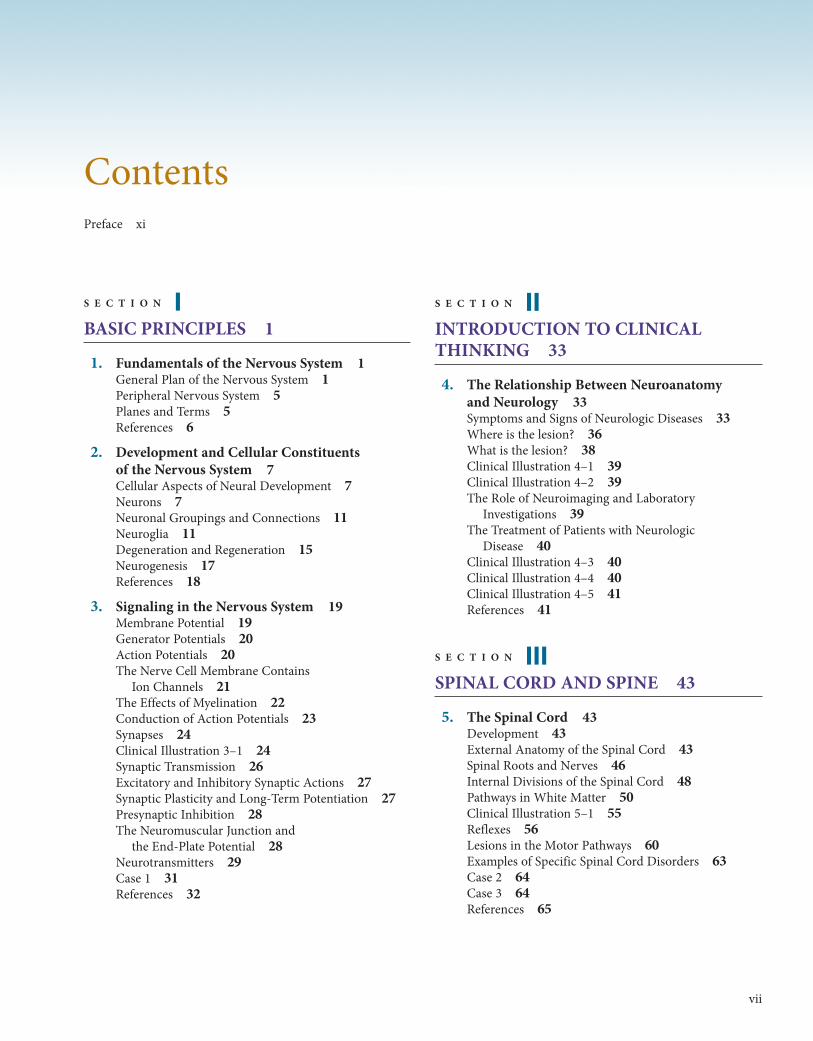
ContentsPreface xi
S E C T I O N IBASIC PRINCIPLES 1
1. Fundamentals of the Nervous System 1General Plan of the Nervous System 1Peripheral Nervous System 5Planes and Terms 5References 6
2. Development and Cellular Constituents of the Nervous System 7Cellular Aspects of Neural Development 7Neurons 7Neuronal Groupings and Connections 11Neuroglia 11Degeneration and Regeneration 15Neurogenesis 17References 18
3. Signaling in the Nervous System 19Membrane Potential 19Generator Potentials 20Action Potentials 20The Nerve Cell Membrane Contains
Ion Channels 21The Effects of Myelination 22Conduction of Action Potentials 23Synapses 24Clinical Illustration 3–1 24Synaptic Transmission 26Excitatory and Inhibitory Synaptic Actions 27Synaptic Plasticity and Long-Term Potentiation 27Presynaptic Inhibition 28The Neuromuscular Junction and
the End-Plate Potential 28Neurotransmitters 29Case 1 31References 32
S E C T I O N IIINTRODUCTION TO CLINICALTHINKING 33
4. The Relationship Between Neuroanatomy and Neurology 33Symptoms and Signs of Neurologic Diseases 33Where is the lesion? 36What is the lesion? 38Clinical Illustration 4–1 39Clinical Illustration 4–2 39The Role of Neuroimaging and Laboratory
Investigations 39The Treatment of Patients with Neurologic
Disease 40Clinical Illustration 4–3 40Clinical Illustration 4–4 40Clinical Illustration 4–5 41References 41
S E C T I O N IIISPINAL CORD AND SPINE 43
5. The Spinal Cord 43Development 43External Anatomy of the Spinal Cord 43Spinal Roots and Nerves 46Internal Divisions of the Spinal Cord 48Pathways in White Matter 50Clinical Illustration 5–1 55Reflexes 56Lesions in the Motor Pathways 60Examples of Specific Spinal Cord Disorders 63Case 2 64Case 3 64References 65
vii
MCGH267-FM_i-xii.qxd 4/6/13 9:31 AM Page vii

viii Contents
6. The Vertebral Column and Other StructuresSurrounding the Spinal Cord 67Investing Membranes 67Spinal Cord Circulation 68The Vertebral Column 69Clinical Illustration 6–1 69Clinical Illustration 6–2 71Lumbar Puncture 71Imaging of the Spine and Spinal Cord 73Case 4 73Case 5 74References 77
S E C T I O N IVANATOMY OF THE BRAIN 79
7. The Brain Stem and Cerebellum 79Development of the Brain Stem
and Cranial Nerves 79Brain Stem Organization 79Cranial Nerve Nuclei in the Brain Stem 82Medulla 82Pons 87Midbrain 88Vascularization 89Clinical Illustration 7–1 90Cerebellum 91Clinical Illustration 7–2 92Clinical Illustration 7–3 92Clinical Illustration 7–4 96Case 6 98Case 7 98References 98
8. Cranial Nerves and Pathways 99Origin of Cranial Nerve Fibers 99Functional Components of the Cranial Nerves 99Anatomic Relationships of the Cranial Nerves 102Case 8 116Case 9 116References 118
9. Diencephalon 119Thalamus 119Hypothalamus 121Subthalamus 126Epithalamus 127Circumventricular Organs 128Case 10 129References 129
10. Cerebral Hemispheres/Telencephalon 131Development 131Anatomy of the Cerebral Hemispheres 131Microscopic Structure of the Cortex 136
Clinical Illustration 10–1 140Physiology of Specialized Cortical Regions 142Basal Ganglia 143Internal Capsule 144Case 11 147Case 12 147References 147
11. Ventricles and Coverings of the Brain 149Ventricular System 149Meninges and Submeningeal Spaces 150CSF 152Barriers in the Nervous System 154Skull 156Case 13 160Case 14 161References 162
12. Vascular Supply of the Brain 163Arterial Supply of the Brain 163Venous Drainage 165Cerebrovascular Disorders 169Clinical Illustration 12–1 175Case 15 177Case 16 178References 181
S E C T I O N VFUNCTIONAL SYSTEMS 183
13. Control of Movement 183Control of Movement 183Major Motor Systems 183Motor Disturbances 189Case 17 193Case 18 194References 194
14. Somatosensory Systems 195Receptors 195Connections 195Sensory Pathways 195Cortical Areas 196Pain 196Case 19 199Case 20 200References 200
15. The Visual System 201The Eye 201Visual Pathways 205The Visual Cortex 209Clinical Illustration 15–1 210Case 21 214References 214
MCGH267-FM_i-xii.qxd 4/6/13 9:31 AM Page viii
Contents ix
16. The Auditory System 215Anatomy and Function 215Auditory Pathways 215Case 22 218References 219
17. The Vestibular System 221Anatomy 221Vestibular Pathways 221Functions 221Case 23 224References 224
18. The Reticular Formation 225Anatomy 225Functions 225References 228
19. The Limbic System 229The Limbic Lobe and Limbic System 229Olfactory System 229Hippocampal Formation 230Clinical Illustration 19–1 232Functions and Disorders 236Septal Area 236Case 24 239References 239
20. The Autonomic Nervous System 241Autonomic Outflow 241Autonomic Innervation of the Head 247Visceral Afferent Pathways 248Hierarchical Organization of the Autonomic
Nervous System 249Transmitter Substances 251Case 25 255References 255
21. Higher Cortical Functions 257Frontal Lobe Functions 257Language and Speech 257Cerebral Dominance 262Memory and Learning 262Epilepsy 262Clinical Illustration 21–1 264Case 26 265Case 27 266References 266
S E C T I O N VIDIAGNOSTIC AIDS 267
22. Imaging of the Brain 267Skull X-Ray Films 267Angiography 267Computed Tomography 268Magnetic Resonance Imaging 270Magnetic Resonance Spectroscopy 273Diffusion-Weighted Imaging 273Functional MRI 274Positron Emission Tomography 275Single Photon Emission CT 276References 276
23. Electrodiagnostic Tests 277Electroencephalography 277Evoked Potentials 278Transcranial Motor Cortical Stimulation 280Electromyography 280Nerve Conduction Studies 283References 284
24. Cerebrospinal Fluid Examination 285Indications 285Contraindications 285Analysis of the CSF 285Reference 286
S E C T I O N VIIDISCUSSION OF CASES 287
25. Discussion of Cases 287The Location of Lesions 287The Nature of Lesions 288Cases 289References 303
Appendix A: The Neurologic Examination 305Appendix B: Testing Muscle Function 313Appendix C: Spinal Nerves and Plexuses 329Appendix D: Questions and Answers 347
Index 355
MCGH267-FM_i-xii.qxd 4/6/13 9:31 AM Page ix

MCGH267-FM_i-xii.qxd 4/6/13 9:31 AM Page x
xi
Preface
Very few organ systems, if any, present as fascinating an arrayof structures and mechanisms as the human brain and spinalcord. Furthermore, it is hard to think of a clinical field thatdoes not encompass at least some aspect of the neurosciences,from molecular and cellular neurobiology through motor,sensory, and cognitive neuroscience, to human behavior andeven social interactions. It is the brain, in fact, that makes usuniquely human. No surprise, then, that neuroscience hasemerged as one of the most exciting fields of research and nowoccupies a central role as a substrate for clinical medicine.
One of the unique things about the nervous system is itsexquisite architecture. The nervous system contains more celltypes than any other organ or organ system, and its con-stituent nerve cells—more than 100,000,000,000 of them—and an even larger number of supportive glial cells arearranged in a complex but orderly, and functionally crucial,way. Many disease processes affect, in a direct or indirect way,the nervous system. Thus, every clinician, and every basic sci-entist with an interest in clinical disease, needs an understand-ing of neuroanatomy. Stroke remains the most frequent causeof death in most industrialized societies; mood disorders suchas depression affect more than 1 person in 10; and clinicaldysfunction of the nervous system occurs in 25% of patientsin most general hospital settings at some time during theirhospital stay. An understanding of neuroanatomy is crucialnot only for neurologists, neurosurgeons, and psychiatristsbut also for clinicians in all subspecialties, since patients ofevery stripe will present situations that require an understand-ing of the nervous system, its structure, and its function.
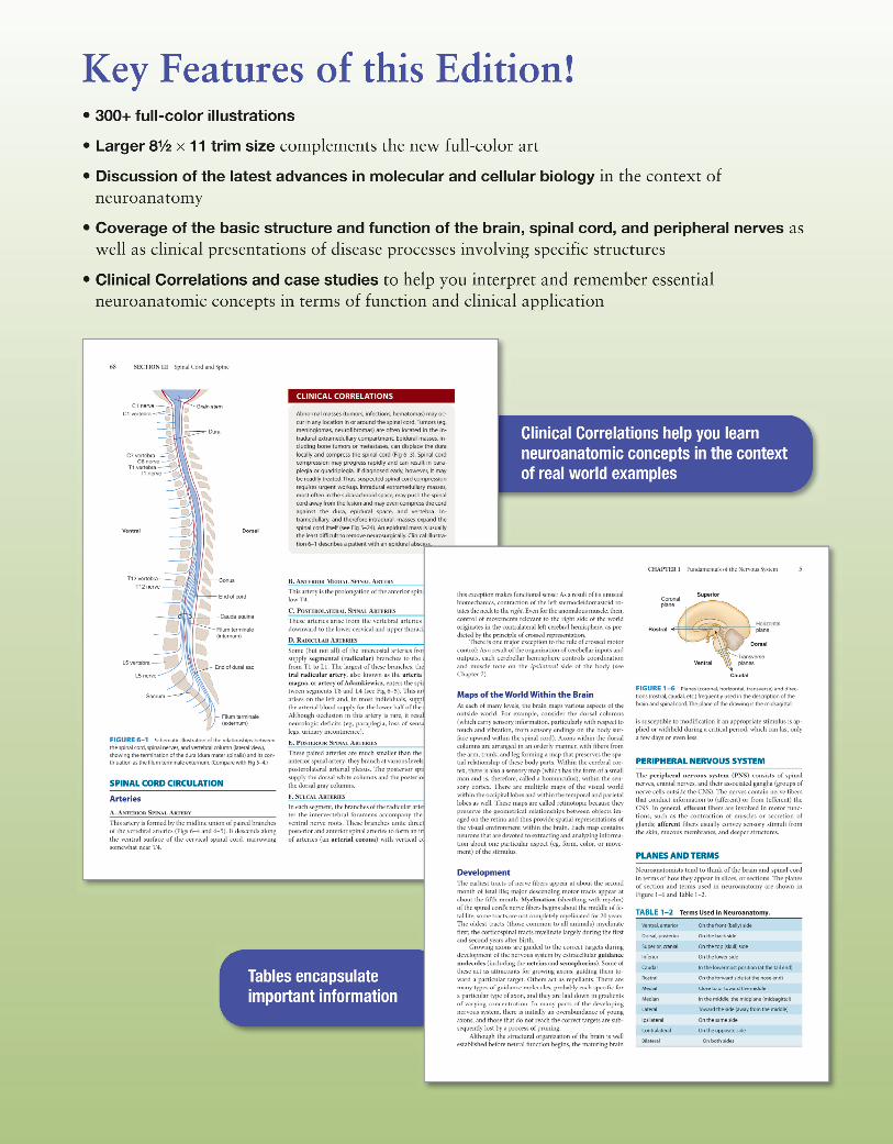
This book, now in its 27th edition, is designed as anaccessible, easy-to-remember synopsis of neuroanatomy andits functional and clinical implications. Since many of us learnand remember better when material is presented visually, thisbook is well illustrated not only with clinical material such asbrain scans and pathological specimens but also with hun-dreds of diagrams and tables that are designed to be clear,
explicative, and memorable. This book is not meant to sup-plant longer, comprehensive handbooks on neuroscience andneuroanatomy. On the contrary, it has been designed to pro-vide a manageable and concise overview for busy medical stu-dents and residents, as well as trainees in health-related fieldssuch as physical therapy; graduate students and postdoctoralfellows with an interest in neuroanatomy and its functionalunderpinnings; and clinicians in practice, for whom minutesare precious.
This book is unique in containing a section entitled“Introduction to Clinical Thinking,” which introduces thereader, early in the text, to the logical processes involved inusing neuroanatomy as a basis for thinking about patients.Since some trainees remember patients better than isolatedfacts, I have included discussions of clinical correlates andclinical illustrations that synthesize the most important char-acteristics of patients selected from an extensive clinical expe-rience. Also included are illustrative clinical images includingcomputer tomography (CT) and magnetic resonance imaging(MRI), both of normal brain and spinal cord, and of commonclinical entities that trainees will likely encounter.
As with past editions, I owe a debt of gratitude to manycolleagues and friends, especially members of the Departmentof Neurology at Yale Medical School. Joachim Baehring, MD,and Joseph Schindler, MD, of Yale, as well as Catharina Faber,MD, at the University of Maastricht contributed invaluableclinical illustrations. Over the years, these colleagues andfriends have helped to create an environment where learning isfun, a motif that I have woven into this book. I hope that read-ers will join me in finding that neuroanatomy, which providesmuch of the foundation for both neuroscience and clinicalmedicine, can be enjoyable, memorable, and easily learned.
Stephen G. Waxman, MD, PhDNew Haven, ConnecticutApril 2013
MCGH267-FM_i-xii.qxd 4/6/13 9:31 AM Page xi

MCGH267-FM_i-xii.qxd 4/6/13 9:31 AM Page xii