a human beta cell line with drug inducible excision of
TRANSCRIPT

HAL Id: inserm-01298792https://www.hal.inserm.fr/inserm-01298792
Submitted on 6 Apr 2016
HAL is a multi-disciplinary open accessarchive for the deposit and dissemination of sci-entific research documents, whether they are pub-lished or not. The documents may come fromteaching and research institutions in France orabroad, or from public or private research centers.
L’archive ouverte pluridisciplinaire HAL, estdestinée au dépôt et à la diffusion de documentsscientifiques de niveau recherche, publiés ou non,émanant des établissements d’enseignement et derecherche français ou étrangers, des laboratoirespublics ou privés.
A human beta cell line with drug inducible excision ofimmortalizing transgenes.
Marion Benazra, Marie-José Lecomte, Claire Colace, Andreas Müller, CécileMachado, Séverine Pechberty, Emilie Bricout-Neveu, Maud Grenier-Godard,
Michèle Solimena, Raphaël Scharfmann, et al.
To cite this version:Marion Benazra, Marie-José Lecomte, Claire Colace, Andreas Müller, Cécile Machado, et al.. Ahuman beta cell line with drug inducible excision of immortalizing transgenes.. Molecular metabolism,Elsevier, 2015, 4 (12), pp.916-925. �10.1016/j.molmet.2015.09.008�. �inserm-01298792�

Original article
A human beta cell line with drug inducibleexcision of immortalizing transgenes
Marion Benazra 1,2,3,4, Marie-José Lecomte 5, Claire Colace 1,2,3,4, Andreas Müller 6,7, Cécile Machado 5,Severine Pechberty 5, Emilie Bricout-Neveu 5, Maud Grenier-Godard 5, Michele Solimena 6,7,8,Raphaël Scharfmann 9, Paul Czernichow 5, Philippe Ravassard 1,2,3,4,*
ABSTRACT
Objectives: Access to immortalized human pancreatic beta cell lines that are phenotypically close to genuine adult beta cells, represent a majortool to better understand human beta cell physiology and develop new therapeutics for Diabetes. Here we derived a new conditionallyimmortalized human beta cell line, EndoC-bH3 in which immortalizing transgene can be efficiently removed by simple addition of tamoxifen.Methods: We used lentiviral mediated gene transfer to stably integrate a tamoxifen inducible form of CRE (CRE-ERT2) into the recently developedconditionally immortalized EndoC bH2 line. The resulting EndoC-bH3 line was characterized before and after tamoxifen treatment for cellproliferation, insulin content and insulin secretion.Results: We showed that EndoC-bH3 expressing CRE-ERT2 can be massively amplified in culture. We established an optimized tamoxifentreatment to efficiently excise the immortalizing transgenes resulting in proliferation arrest. In addition, insulin expression raised by 12 fold andinsulin content increased by 23 fold reaching 2 mg of insulin per million cells. Such massive increase was accompanied by enhanced insulinsecretion upon glucose stimulation. We further observed that tamoxifen treated cells maintained a stable function for 5 weeks in culture.Conclusions: EndoC bH3 cell line represents a powerful tool that allows, using a simple and efficient procedure, the massive production offunctional non-proliferative human beta cells. Such cells are close to genuine human beta cells and maintain a stable phenotype for 5 weeks inculture.
� 2015 The Authors. Published by Elsevier GmbH. This is an open access article under the CC BY license (http://creativecommons.org/licenses/by/4.0/).
Keywords Cell engineering; Human pancreatic beta cell line; Conditional immortalization; Tamoxifen inducible CRE; Human beta cell function
1. INTRODUCTION
Type 1 diabetes results from the destruction of insulin-producingpancreatic beta cells by a beta cell-specific autoimmune processwhile type 2 diabetes results from the combination of insulin resistanceand inadequate insulin secretion. Thus, for both types of diabetes,functional beta cell mass is not sufficient for appropriate glycemiccontrol. Therefore understanding beta cell physiology and function is acritical issue for understanding diabetes and developing innovativetherapeutic solutions. Rodent beta cells have been used so far andwere instrumental for acquiring important basic knowledge of beta cellfunction. However, data generated with such cells cannot easily betranslated to humans since major species differences have been re-ported [1,2]. Thus, access to human beta cells is crucial to progress inunderstanding human specific beta cell function and, unfortunately,scarcity of organ donors makes it necessary to search for othersources [3].
1Institut du cerveau et de la moelle (ICM), Biotechnology & Biotherapy Team, 75013 PFrance 4Université Pierre et Marie Curie, 75013 Paris, France 5Endocells, Pépinière d’enInstitute of the Helmholtz Center Munich at the University Hospital and Faculty of Medic(DZD e.V), 85764 Neuherberg, Germany 8Max Planck Institute of Molecular Cell BiologyMédecine, Université Paris Descartes, Sorbonne Paris Cité, 75014 Paris, France
*Corresponding author. ICM Biotechnology & Biotherapy Team, Hôpital Pitié SalpêtrièrE-mail: [email protected] (P. Ravassard).
Received June 26, 2015 � Revision received September 17, 2015 � Accepted Septem
http://dx.doi.org/10.1016/j.molmet.2015.09.008
916 MOLECULAR METABOLISM 4 (2015) 916e925 � 2015 The Authors. Published by E
To develop such alternative sources, large efforts have been under-taken to differentiate human embryonic or induced pluripotent stemcells (hESCs/iPSCs) towards pancreatic mature endocrine cells. Sincethe important original contribution of the Viacyte group to generateendocrine cells from hESCs [4,5], recent advances have been made inthis field to obtain in vitro more fully mature pancreatic endocrine cells[6,7]. Still, both the production yield and the robustness of the processneed to be further improved. Using an approach based on targetedoncogenesis in human fetal pancreas, we generated the first immor-talized human beta cell line, referred as EndoC-bH1, giving access tounlimited number of functional human beta cells [8]. Although, this lineis similar to primary adult human beta cells, it is continuously prolif-erating, which represents a major difference with mature beta cellsthat that have a low proliferation rate [9]. We recently reported theproduction of the second generation of human beta cell line, referred toas EndoC-bH2 that was conditionally immortalized. In this cell line,both large T antigen of SV40 (SV40LT) and human telomerase reverse
aris, France 2CNRS UMR7225, 75013 Paris, France 3INSERM U1127, 75013 Paris,treprises Institut du Cerveau et de la Moelle, 75007 Paris, France 6Paul Langerhansine, TU Dresden, 01307 Dresden, Germany 7German Center for Diabetes Researchand Genetics, 01307 Dresden, Germany 9INSERM, U1016, Institut Cochin, Faculté de
e, 47 Bd. De l’Hôpital, 75013 Paris, France. Tel./fax: þ33 157274575.
ber 22, 2015 � Available online 20 October 2015
lsevier GmbH. This is an open access article under the CC BY license (http://creativecommons.org/licenses/by/4.0/).www.molecularmetabolism.com

transcriptase (hTERT), used as immortalizing transgenes, can beremoved by CRE mediated excision [10]. We have shown thatconstitutive expression of CRE in EndoC-bH2 cells resulted in drasticproliferation arrest and enhancement of beta cell function both at thelevel of insulin content and secretion upon glucose stimulation. Thus,excised EndoC-bH2 cells are highly representative of human primarybeta cells.In previous studies [10], we transduced EndoC-bH2 cells with a len-tiviral vector expressing CRE that efficiently excised immortalizingtransgenes in more than 95% of cells. Although such an approach isefficient, mass production of excised cells that would require massiveamounts of viral vectors cannot be easily achieved. Therefore, tocircumvent this limitation, we devised a drug-activated excisionstrategy coupled with antibiotic selection.Many drug-inducible systems have been used to control geneexpression both in vitro and in vivo [11e14]. We selected here the onebased on CRE-ERT2 fusion protein [15]. CRE-ERT2 has high affinity forthe 4-hydroxytamoxifen (TAM) but not for the endogenous estradiol.Therefore, the recombinase activity of CRE-ERT2 is dependent on theaddition of this compound to the culture medium. After its exposure tothe specific inducer TAM, CRE-ERT2 is translocated from the cyto-plasm into the nucleus and excises loxP-flanked DNA regions.In the present study, we stably modified EndoC-bH2 excisable line bylentiviral vector-mediated gene transfer to integrate both CRE-ERT2and a constitutive puromycin selection marker. The resulting line,EndoC-bH3, was selected and massively expanded in vitro in thepresence of puromycin. TAM dose and duration of treatment wereoptimized to achieve efficient immortalizing transgene excision. TAMmediated excision resulted in a sharp decrease of EndoC-bH3 cellproliferation and pronounced enhancement of beta-cell specific fea-tures such as insulin expression, storage in secretory granules andglucose stimulated secretion. These properties were maintained inculture for several weeks. Importantly, by opposition to the previousEndoC-b2 cells, the massive production of this cell line in its excisedstate is simple, giving access to large-scale drug discovery, prolifer-ation studies and development of preclinical models.
2. MATERIALS AND METHODS
2.1. Lentiviral vectors and cell transductionA tamoxifen inducible form of CRE (CRE-ERT2) was cloned down-stream of the CMV promoter in a lentiviral backbone. Briefly, LR clo-nase II recombination was performed using pTrip CMV rfa-GatewayDelta U3 destination [16] vector and pENTR/D/TOPOeCre-ERT2 entryclone. The Cre-ERT2 fragment was amplified by PCR from a plasmidkindly provided by Guilan Vodjdani (INSERM UMR1141) using theforward primer 50CACCGGTACCCTCGAGATCGAT30 and reverse primer50TCAAGCTGTGGCAGGGAAACC30, and the resulting PCR product wascloned into the pENTR/D/TOPO plasmid to generate the Cre-ERT2 entryclone.The pTrip PGK puro polyA/CMV CRE-ERT2 Delta U3 was generatedusing pTrip CMV CRE-ERT2 Delta U3 as backbone in which a PGKpuromycin resistance polyA was inserted in the reverse orientation onthe 50 side of the triplex sequence. Briefly, a linker containing EcoRI-compatible SacII, SalI BamHI MluI and EcoRI restriction sites wasfirst inserted in the EcoRI site of pTrip CMV CRE-ERT2 Delta U3. Next,the polyA signal from human beta globin was amplified by PCR frompCDNA3.0 vector (Invitrogen) with primers containing SacII and SalIoverhanging ends, and the resulting PCR product was cloned in theintegrated linker sequence between SacII and SalI sites. The PGKpromoter sequence was digested from pTrip PGK eGFP Delta U3 vector
MOLECULAR METABOLISM 4 (2015) 916e925 � 2015 The Authors. Published by Elsevier GmbH. This is an owww.molecularmetabolism.com
[17] using MluI and BamHI and cloned in the polyA containing vector.Finally, the puromycin resistance gene was amplified by PCR frompLKO puro vector (Addgene) with primers containing BamHI and SalIoverhanging ends and the resulting PCR product was cloned betweenthe PGK promoter and the polyA sequences in the corresponding re-striction sites. Lentiviral vector stocks were produced by transienttransfection of 293T cells with the p8.91 encapsidation plasmid,pHCMV-G, encoding the vesicular stomatitis virus (VSV) glycoprotein-Gand the pTRIP DU3 as previously described [18].EndoC-bH2 cells were transduced with pTrip PGK puro polyA/CMVCRE-ERT2 Delta U3 to generate EndoC-bH3 cells using a total amountof viral particles of 30 ng of p24 capside protein per 105 cells in thepresence of 10 mg/ml DEAE-dextran as described elsewhere [18].
2.2. Cell line culture and excision processEndoC-bH3 cells were cultured in DMEM containing 5.6 mM glucose,2% BSA fraction V, 50 mM 2-mercaptoethanol, 10 mM nicotinamide,5.5 mg/ml transferrin, 6.7 ng/ml sodium selenite, Penicillin (100 units/ml)/Streptomycin (100 mg/ml). Ten mg/ml of puromycin (selectiveantibiotic) were added extemporaneously in the complete medium.Cells were seeded onto matrigel- and fibronectin-coated culture platesat 4 � 106 cells/plate. Passage was performed every week. Inducibleexcision of CRE mediated immortalizing transgenes was performedwith addition of TAM, 1 mM unless specify in the text.Cells were counted according to manufacturer instructions using theADAM-MC automatic cell counter instrument (NanoEnTek Inc. SeoulKorea).
2.3. ImmunostainingFor immunocytochemistry, EndoC-bH3 cells were treated with TAM for14 days. They were next seeded on 12-mm matrigel/fibronectin glasscoated coverslips and further cultured with TAM for 7 days. Next, thecells were fixed for 1 h in 4% paraformaldehyde. The following anti-bodies were used for immunostaining: guinea pig anti-insulin antibody(1/500, DakoCytomation, A0564) and mouse anti-SV40LT (1/50, Cal-biochem Merck Biosciences, DP-02). The secondary antibodies werefluorescein anti-mouse antibody (1/200, Immunotech, IM0819) andTexas-red anti-guinea pig antibody (1/200; Jackson ImmunoresearchLaboratories, 706-076-148). Nuclei were stained with Hoechst 33342fluorescent stain (Life Technologies). Digital images of cells werecaptured using an Olympus Fluoview FV1000 confocal microscope.
2.4. Cell proliferation assaysCell proliferation analysis was conducted using Click-iT EdU AlexaFluor 647 Flow Cytometry Assay Kit (Life Technology). Briefly, EdU wasadded into cell-culture medium to a final concentration of 10 mmol/lone hour before the endpoint of the experiments. Cells were collectedafter trypsin treatment, washed once with 2 ml of 1% BSA in PBS, fixedusing Click-iT fixative, and incubated for 15 min in saponin-basedpermeabilization solution. Cells were then treated with Click-iT reac-tion cocktail, according to manufacturer’s instruction, for 30 minbefore flow cytometry analysis. DAPI (1 mM) (FxCycle Violet Stain; LifeTechnologies) was added directly before FACS analysis. Data wereacquired on an LSRFortessa (BD Biosciences) and analyzed with FACSDiva software (BD). A total of 50,000 events were collected for the cellproliferation analysis.
2.5. RNA isolation, reverse transcription and RT-PCRTotal RNA was isolated from EndoC-bH3 cell line using the RNeasymicrokit (Qiagen), as described previously [10]. First strand cDNA wasprepared using Superscript reagents (Invitrogen). Quantitative RT-PCR
pen access article under the CC BY license (http://creativecommons.org/licenses/by/4.0/). 917

Original article
was performed using LightCycler 1536 DNA Green Master mix (Roche)and analyzed on a 1536 LightCycler system (Roche), according to themanufacturer’s instructions. The list of primers used is presented insupporting Table 1. Qualitative RT-PCR for CRE expression analysiswas performed using CRE-F AAAATTTGCCTGCATTACCG and CRE-RATGTTTAGCTGGCCCAAATG primer pairs generating a 262 nucleo-tides amplification product.
2.6. Insulin secretion and contentEndoC-bH3 cells were seeded onto matrigel- and fibronectin-coated96 well plates at 7 � 104 cells/well for un-excised and excisedcells. Three days later, cells were incubated overnight in culture me-dium that contained 2.8 mM glucose, followed by 60 min incubation inHEPES-buffered KrebseRinger Buffer (KRB) (115 mmol/l NaCl,5 mmol/l KCl, 1 mmol/l CaCl2, 1 mmol/l MgCl2, 24 mmol/l NaHCO3,10 mmol/l HEPES pH 7.4, and 0.2% BSA) that contained 0.5 mMglucose. At the end of this incubation, stimulated insulin secretion wasmeasured by static incubation in KRB that contained varying glucoseconcentrations for 60 min. Glucose stimulation was performed in thepresence or absence of 500 mM IBMX.For insulin content measurement, cells were lysed directly in theculture wells with TETG solution 20 mM Tris pH 8.0; 0.1% Triton X-100; 1% Glycerol; 137 mM NaCl; 2 mM EGTA and anti-protease tablet(Roche) for 5 min on ice. The lysate was next centrifuged at 3,000 rpmfor 5 min and stored at �20 �C until insulin ELISA assay.Insulin secretion and intracellular content were measured in duplicateby ELISA according to manufacturer’s instructions using the humaninsulin kit (Mercodia), which does not cross-react with proinsulin.
2.7. Electron microscopyCells were fixed with 2.5% Glutaraldehyde and 4% Paraformaldehydein Sörensen’s phosphate buffer (pH 7.4) over night. After fixation, cellswere processed for standard Epon embedding as previously described[19]. Ultrathin sections with a thickness of 70 nm were cut on a LeicaEM6 ultramicrotome and imaged using a Tecnai 12 Biotwin Trans-mission Electron Microscope (FEI Company, Hillsboro, OR, USA) with abottom-mount 2�Aw2K F214 CCD camera (TVIPS, Gauting, Germany).Sixty random images with a size of 8.92 mm � 8.92 mm were takenper condition at a resolution of 4.35 nm/pixel. TEM images wereviewed using FIJI software and insulin secretory granules (SGs) werescored manually on a Wacom Cintiq 15X LCD tablet (Wacom) tocalculate their number normalized for b-cell cytosolic area.
2.8. Oligonucleotides-based array comparative genomichybridization (CGH array)CGH array was performed using SurePrint G3 Human CGH Bundle(4 � 180K) (Agilent) according to manufacturer instructions. Briefly,1 mg of genomic DNA corresponding to either a human male control[20], EndoC-bH2 or EndoC-bH3 cells both at passage 40 was frag-mented by heating at 95 �C for 30 min. Fragmented DNAs werelabeled with Cy3 (control DNA) and Cy5 (EndoC-bH2/EndoC-bH3 DNA)fluorescent dUTP, respectively, using Genomic DNA Enzymatic La-beling Kit (Agilent Technology). Microcon YM 30 spin columns (Milli-pore) were used to remove the unincorporated nucleotides and dyes.Hybridizations of labeled DNA to SurePrint G3 Human CGH Bundle(4� 180K) array (Agilent) were performed in a hybridization oven at 42C at 20 rpm for 40 h. Hybridized arrays were then washed following themanufacturer’s instructions. Microarray slides were scanned on aNimblegen MS200 Microarray Scanner at a 2 mm resolution. Featureextraction was done with Cytogenomics Software (Agilent). Extracteddata were imported and analyzed using Nexus 7.0 (Biodiscovery).
918 MOLECULAR METABOLISM 4 (2015) 916e925 � 2015 The Authors. Published by E
3. RESULTS
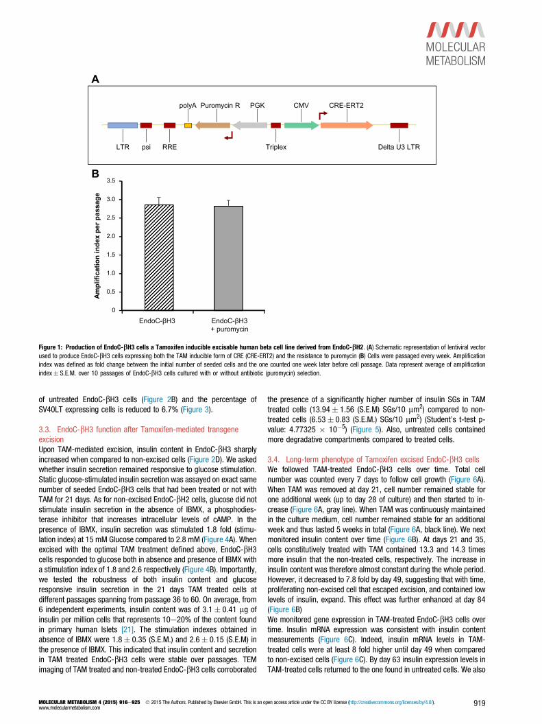
3.1. Production of EndoC-bH3, an excisable human beta cell linethat integrated CRE-ERT2Our objective was to stably integrate the coding sequence of CRE-ERT2fusion protein into the genome of EndoC-bH2 cells and to couple suchintegration with an antibiotic resistance to select CRE-ERT2 positivecells. First, EndoC-bH2 sensitivity to neomycin, puromycin orhygromycin antibiotics was tested. Both puromycin and hygromycinefficiently killed EndoC-bH2 cells within two weeks at 10 mg/ml and150 mg/ml respectively (not shown). We next constructed a lentiviralvector expressing both CRE-ERT2 and a Puromycin resistance geneunder the control of CMV and PGK promoters, respectively (Figure 1A).The vector was used to transduce EndoC-bH2 cells, and we named theresulting puromycin-selected cells EndoC-bH3. Importantly, the pro-liferation rate of EndoC-bH3 cells was similar in the presence orabsence of antibiotic (Figure 1B), indicating that the selection does nothamper the overall growth of the cells. EndoC-bH3 can therefore bemassively amplified in culture. We next analyzed the chromosomalstability of EndoC-bH3 cells at passage 40 in comparison to EndoC-bH2 cells. CGH array profiles are almost identical between the twolines (Figure S1) indicating chromosomal stability following integrationof CRE-ERT2 and antibiotic selection.
3.2. Tamoxifen treatment to achieve efficient activation ofrecombinase CRE and excision of SV40LTOur previous data obtained with EndoC-bH2 indicate that constitutiveCRE expression results in excision of SV40LT [10]. Under such con-ditions, 21 days after transduction with CRE-expressing lentiviralvector, proliferation was massively reduced and insulin contentincreased. To follow excision efficacy in EndoC-bH3 cells that expressCRE-ERT2, we used 3 readouts: SV40LT expression, incorporation ofEdU for proliferation assessment and insulin content measurement.Both the concentration of TAM and the duration of treatment wereinvestigated. Excision efficacy was monitored 21 days after TAMaddition using increasing doses from 1 to 10 mM. TAM was maintainedin the medium for 7 or 21 days (Figure 2A). Importantly, the decreasein SV40LT expression and EdU incorporation were parallel (Figure 2BeC). With 7 days treatment, EndoC-bH3 proliferation decreases in adose-dependent manner. 11.5% of control untreated cells incorpo-rated EdU whereas EdU incorporation decreased to 5.1% and 2.2%with 1 and 10 mM TAM, respectively. With 21 days TAM treatment, alltested concentrations were equivalent and, on average, only 0.75% ofcells incorporated EdU (Figure 2C). EndoC-bH2 cells that do not containan integrated TAM inducible CRE were treated for 7 or 21 days witheither 1 or 10 mM TAM. Such treatments had no effect on cell pro-liferation (Figure 2D) demonstrating that the massive reduction ofproliferation observed with EndoC-bH3 treated with TAM is due to theinduction of CRE-ERT2 dependent excision.Following a 7 days TAM treatment, insulin content increased by 12.9and 17.5 fold when treated with 1e10 mM TAM compared to controluntreated cells (Figure 2E). Such induction was lower than the 23.3fold induction obtained with 21 days 1 mM TAM treatment (Figure 2E).Similar high inductions were obtained with 2.5 and 5 mM TAM with 21days TAM treatment protocol whereas higher doses were less efficient(Figure 2E). Therefore 1 mM TAM treatment for 21 days representedthe optimal dose that gave the highest outcome to decrease SV40LTexpression, to decrease proliferation and to enhance insulin content.Finally, the percentage of cells positive for SV40LT protein expressionsharply decreased upon 21 days TAM treatment. Indeed with theoptimal TAM treatment, SV40LT gene expression represents only 8.6%
lsevier GmbH. This is an open access article under the CC BY license (http://creativecommons.org/licenses/by/4.0/).www.molecularmetabolism.com
A
CRE-ERT2
LTR Delta U3 LTRTriplexpsi
CMV
RRE
PGKpolyA Puromycin R
B
EndoC-βH3 EndoC-βH3 + puromycin
0
0.5
1.0
1.5
2.0
2.5
3.0
3.5
Ampl
ifica
tion
inde
x pe
r pas
sage
Figure 1: Production of EndoC-bH3 cells a Tamoxifen inducible excisable human beta cell line derived from EndoC-bH2. (A) Schematic representation of lentiviral vectorused to produce EndoC-bH3 cells expressing both the TAM inducible form of CRE (CRE-ERT2) and the resistance to puromycin (B) Cells were passaged every week. Amplificationindex was defined as fold change between the initial number of seeded cells and the one counted one week later before cell passage. Data represent average of amplificationindex� S.E.M. over 10 passages of EndoC-bH3 cells cultured with or without antibiotic (puromycin) selection.
of untreated EndoC-bH3 cells (Figure 2B) and the percentage ofSV40LT expressing cells is reduced to 6.7% (Figure 3).
3.3. EndoC-bH3 function after Tamoxifen-mediated transgeneexcisionUpon TAM-mediated excision, insulin content in EndoC-bH3 sharplyincreased when compared to non-excised cells (Figure 2D). We askedwhether insulin secretion remained responsive to glucose stimulation.Static glucose-stimulated insulin secretion was assayed on exact samenumber of seeded EndoC-bH3 cells that had been treated or not withTAM for 21 days. As for non-excised EndoC-bH2 cells, glucose did notstimulate insulin secretion in the absence of IBMX, a phosphodies-terase inhibitor that increases intracellular levels of cAMP. In thepresence of IBMX, insulin secretion was stimulated 1.8 fold (stimu-lation index) at 15 mM Glucose compared to 2.8 mM (Figure 4A). Whenexcised with the optimal TAM treatment defined above, EndoC-bH3cells responded to glucose both in absence and presence of IBMX witha stimulation index of 1.8 and 2.6 respectively (Figure 4B). Importantly,we tested the robustness of both insulin content and glucoseresponsive insulin secretion in the 21 days TAM treated cells atdifferent passages spanning from passage 36 to 60. On average, from6 independent experiments, insulin content was of 3.1� 0.41 mg ofinsulin per million cells that represents 10e20% of the content foundin primary human Islets [21]. The stimulation indexes obtained inabsence of IBMX were 1.8� 0.35 (S.E.M.) and 2.6� 0.15 (S.E.M) inthe presence of IBMX. This indicated that insulin content and secretionin TAM treated EndoC-bH3 cells were stable over passages. TEMimaging of TAM treated and non-treated EndoC-bH3 cells corroborated
MOLECULAR METABOLISM 4 (2015) 916e925 � 2015 The Authors. Published by Elsevier GmbH. This is an owww.molecularmetabolism.com
the presence of a significantly higher number of insulin SGs in TAMtreated cells (13.94� 1.56 (S.E.M) SGs/10 mm2) compared to non-treated cells (6.53� 0.83 (S.E.M.) SGs/10 mm2) (Student’s t-test p-value: 4.77325 � 10�5) (Figure 5). Also, untreated cells containedmore degradative compartments compared to treated cells.
3.4. Long-term phenotype of Tamoxifen excised EndoC-bH3 cellsWe followed TAM-treated EndoC-bH3 cells over time. Total cellnumber was counted every 7 days to follow cell growth (Figure 6A).When TAM was removed at day 21, cell number remained stable forone additional week (up to day 28 of culture) and then started to in-crease (Figure 6A, gray line). When TAM was continuously maintainedin the culture medium, cell number remained stable for an additionalweek and thus lasted 5 weeks in total (Figure 6A, black line). We nextmonitored insulin content over time (Figure 6B). At days 21 and 35,cells constitutively treated with TAM contained 13.3 and 14.3 timesmore insulin that the non-treated cells, respectively. The increase ininsulin content was therefore almost constant during the whole period.However, it decreased to 7.8 fold by day 49, suggesting that with time,proliferating non-excised cell that escaped excision, and contained lowlevels of insulin, expand. This effect was further enhanced at day 84(Figure 6B)We monitored gene expression in TAM-treated EndoC-bH3 cells overtime. Insulin mRNA expression was consistent with insulin contentmeasurements (Figure 6C). Indeed, insulin mRNA levels in TAM-treated cells were at least 8 fold higher until day 49 when comparedto non-excised cells (Figure 6C). By day 63 insulin expression levels inTAM-treated cells returned to the one found in untreated cells. We also
pen access article under the CC BY license (http://creativecommons.org/licenses/by/4.0/). 919
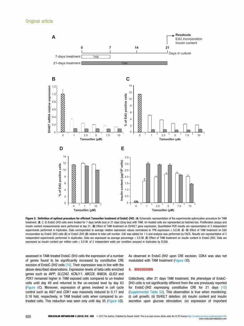
TAM
TAM
7-days treatment
21-days treatment
0 7 14 21
ReadoutsEdU incorporationInsulin content
Days in culture
A
2
4
6
8
10
12
14
01 100
% o
f EdU
posi
tive
cells
Tamoxifen (μM)
D E
Insu
linco
nten
t (μg
/106
cells
)
1 2.5 5 7.5 1000
0.5
1.0
1.5
2.0
2.5
3.0
3.5
Tamoxifen (μM)
Tamoxifen (μM)
C
1 2.5 5 7.5 100
% o
f EdU
posi
tive
cells
2
4
6
8
10
12
14
0
Tamoxifen (μM)
0
0.2
0.4
0.6
0.8
1.0
1.2
0 1 2.5 5 7.5 10
SV40
T m
RN
Are
lativ
e ex
pres
sion
B
Figure 2: Definition of optimal procedure for efficient Tamoxifen treatment of EndoC-bH3. (A) Schematic representation of the experimental optimization procedure for TAMtreatment. (B, C, E) EndoC-bH3 cells were treated for 7 days (white box) or 21 days (Gray box) with TAM. Un-treated cells are represented as hatched box. Proliferation assays andinsulin content measurements were performed at day 21. (B) Effect of TAM treatment on SV40LT gene expression. Quantitative PCR results are representative of 2 independentexperiments performed in triplicates. Data corresponded to average relative expression values normalized to TPB expression� S.E.M. (CeD) Effect of TAM treatment on EdUincorporation by EndoC-bH3 cells (C) or EndoC-bH2 (D) relative to total cell number. EdU was added for 1 h and analysis was performed by FACS. Results are representative of 2independent experiments performed in duplicates. Data are expressed as average percentage� S.E.M. (E) Effect of TAM treatment on insulin content in EndoC-bH3. Data areexpressed as insulin content per million cells� S.E.M. of 3 independent wells per condition assayed in triplicates by ELISA.
Original article
assessed in TAM-treated EndoC-bH3 cells the expression of a numberof genes found to be significantly increased by constitutive CREexcision of EndoC-bH2 cells [10]. Their expression was in line with theabove-described observations. Expression levels of beta cells enrichedgenes such as IAPP, SLC2A2, KCNJ11, ABCC8, RAB3A, GLIS3 andPDX1 remained higher in TAM exposed cells compared to un-treatedcells until day 49 and returned to the un-excised level by day 63(Figure 6C). Moreover, expression of genes involved in cell cyclecontrol such as Ki67 and CDK1 was massively reduced to 0.17 and0.16 fold, respectively, in TAM treated cells when compared to un-treated cells. This reduction was seen only until day 35 (Figure 6D).
920 MOLECULAR METABOLISM 4 (2015) 916e925 � 2015 The Authors. Published by E
As observed in EndoC-bH2 upon CRE excision, CDK4 was also notmodulated with TAM treatment (Figure 6D).
4. DISCUSSION
Collectively, after 21 days TAM treatment, the phenotype of EndoC-bH3 cells is not significantly different from the one previously reportedfor EndoC-bH2 expressing constitutive CRE for 21 days [10](Supplemental Table S2). This observation is true when monitoring:(i) cell growth; (ii) SV40LT deletion; (iii) insulin content and insulinsecretion upon glucose stimulation; (iv) expression of important
lsevier GmbH. This is an open access article under the CC BY license (http://creativecommons.org/licenses/by/4.0/).www.molecularmetabolism.com

TAM 1μM TAM 2.5 μM TAM 5 μM TAM 7.5 μM TAM 10 μM
7 days treatment
Control
INS
-SV
40LT
-nuc
lei
INS
-SV
40LT
21 days treatment
INS
-SV
40LT
- nuc
lei
INS
-SV
40LT
23.3 % +/- 4.9 14.3 % +/- 1.324.1 % +/- 4.8 22.2 % +/- 1.6 14.5 % +/- 1.5
7.0% +/- 0.7 7.4% +/- 0.96.7% +/- 1.4 7.4% +/- 0.9 6.9% +/- 0.8
Figure 3: SV40LT and insulin immunostaining following Tamoxifen treatment. SV40LT (green), Insulin (red) Nuclei (blue) immunofluorescent staining of control or excisedcells analyzed 21 days after a 7 or 21 days TAM treatment. Percent of SV40LT positive nuclei relative to total number of nuclei is indicated below each conditions. Confocalacquisition settings were identical for all conditions. Scale bar ¼ 50 mm.
BA
0,0
0,1
0,2
0,3
0,4
0,5
Insu
linse
cret
ed(n
g/h)
2.8 15 2.8+ IBMX
15+ IBMX
Glucose (mM)
0,0
1,0
2,0
3,0
4,0
5,0
6,0
Insu
linse
cret
ed(n
g/h)
2.8 15 2.8+ IBMX
15+ IBMX
Glucose (mM)
Figure 4: Glucose responsive insulin secretion in EndoC-bH3 following treatment with Tamoxifen. EndoC-bH3 were treated or not for 21 days with 1 mM TAM. (A) Glucosestimulated insulin secretion (GSIS) on un-treated EndoC-bH3 in presence or absence of IBMX. (B) GSIS on TAM-treated EndoC-bH3 in presence or absence of IBMX. (AeB) GSISdata are expressed as ng of secreted insulin per hour� S.E.M. of 3 independent wells seeded with 7 � 104 cells per condition assayed in triplicates by ELISA.
MOLECULAR METABOLISM 4 (2015) 916e925 � 2015 The Authors. Published by Elsevier GmbH. This is an open access article under the CC BY license (http://creativecommons.org/licenses/by/4.0/).www.molecularmetabolism.com
921

Figure 5: TEM analysis of treated and untreated EndoC-bH3. Non-treated and treated EndoC-bH3 cells were analyzed after Epon embedding and ultrathin sectioning. (A) Non-treated EndoC-bH3, inset: detail of insulin SGs, (B) TAM-treated EndoC-bH3, inset: detail of insulin SGs. Scale bar: 2 mm. Labeling key: Nuc, nucleus; Mito, mitochondria; SG,secretory granules; arrowheads, degradative compartments.
Original article
functional beta cell and proliferation markers. Thus, by developing thissimple and efficient TAM treatment procedure, non-proliferativefunctional human beta cells can now be produced in large quantity.It has been reported that TAM could have proapoptotic effect in vivo onmouse beta cells [22]. We addressed cell survival upon continuousexposure to TAM (1 mM and 10 mM) for 21 days in EndoC-bH2, the cellline from which EndoC-bH3 was derived, and in which the CRE-ERT2transgene is absent. We did not observe any difference in cell growthbetween treated and non-treated cells (Supplemental Figure S2) nor atthe level of EdU incorporation (Figure 2C) even at the high dose of10 mM. Our results thus indicate that continuous treatment with TAMdoes not interfere with cell survival. Morphometry of TEM imagesrevealed a 2.15 fold increase in the number of insulin SGs in TAMtreated vs. non-treated cells. Treated cells contained instead fewerdegradative compartments compared to non-treated cells, possiblydue to the switch from proliferation to SG production.In previous work, when CRE was constitutively expressed in EndoC-bH2 cells, immortalizing transgenes were excised in the vast majorityof cells [10]. We hypothesized that the remaining non-excised cellshad escaped initial transduction with CRE-expressing lentiviral vector.Therefore, it was reasonable to envisage that adding an antibioticselection system in the integrative vector expressing CRE-ERT2 wouldallow selection of 100% CRE-ERT2 positive cells and increase effi-ciency of immortalizing transgene excision. When dose and durationof TAM treatment were optimized, we observed that, although cellswere grown under continuous puromycin selection pressure, efficacyof excision was not complete since some cells remained in a prolif-erative state (Figure 2A) and scattered cells still expressed SV40LT(Figure 3). Two main reasons could explain this counterintuitiveobservation of incomplete CRE mediated recombination. First, TAM-inducible CRE recombination has been extensively used in vivo intransgenic models and efficacy of recombination is highly variabledepending on recombinase expression levels [23,24] and genomeavailability of LoxP sites [25e27]. Since, EndoC-bH3 cells weregenerated from lentiviral mediated transduction of EndoC-bH2 cellswithout further clonal selection, the variability of CRE-ERT2 integrationsites may be responsible for inter-cellular variability of recombinaseexpression. Such heterogeneity could sustain the fact that a small cell
922 MOLECULAR METABOLISM 4 (2015) 916e925 � 2015 The Authors. Published by E
population was unable to achieve efficient excision of immortalizingtransgenes. Second, the lentiviral construct used to generate EndoC-bH3 cells is composed of two separate transcription units. Specif-ically, in the same integrated fragment, CRE-ERT2 is expressed underthe control of the CMV promoter and the resistance to puromycinunder a different promoter (PGK). Thus, with time in culture, CRE-ERT2 transcription could be reduced or extinguished, while puromy-cin resistance remains selected by the antibiotic in the culture me-dium. Of note, CMV promoters are known to be subject to extinctionwith time [17,28e30].While the vast majority of the cells efficiently recombine the loxP sitesand excise the immortalizing transgenes, rare cells escape this pro-cess. Our data indicate that the overall composition of the cell pop-ulations (unexcised vs excised cells) is maintained relatively constantfor 49 days with the majority of cells baring a non-proliferative statewith high insulin content. However, with time, the proliferativeadvantage of non-excised cells will result in a time dependent modi-fication of the relative proportion of both cell populations and by day66, the cell population is mainly composed of cells that behave likeproliferating, untreated EndoC-bH3. Interestingly, although such cellswere proliferating in the presence of TAM, after 66 and 119 days ofTAM treatment, they still expressed CRE-ERT2 mRNA (Figure S3),suggesting that they were unable to activate loxP recombination toefficiently remove immortalizing transgenes. The above-describedexperiments were performed on non-clonal cell populations. Ampli-fying cell subclones could represent a way to select lines with furtherincreased loxP recombination efficacy. Furthermore, complete removalof proliferating SV40LT positive cells could be achieved throughcounter-selection using a suicide gene approach such as thymidinekinase in the presence of Gancyclovir [31].
5. CONCLUSION
Altogether, we have produced a novel human beta cell line derivedfrom EndoC-bH2 cells [10] that contains floxed immortalizing trans-genes and an integrated TAM inducible form of CRE recombinase.Such line can be massively amplified followed by immortalizingtransgenes removal by simple addition of TAM, giving rise to non-
lsevier GmbH. This is an open access article under the CC BY license (http://creativecommons.org/licenses/by/4.0/).www.molecularmetabolism.com

ki67 CDK1 CDK4
B
0
2
4
6
8
10
12
14
16
18
20
21 35 49 63 84Days
Fold
chan
ge in
sulin
cont
ent
TAMTAM
A
0
50
100
150
200
250
300
cell
num
ber(
x106
)
0 20 40 60 Days
D
0
0.5
1.0
1.5
2.0
2.5
3.0
mR
NA
expr
essi
on fo
ldch
ange
D21D35D49D63D84
Time point
C
0
2
4
6
8
10
12
14
16
18
20
22
INS IAPP SLC2A2 ABCC8 KCNJ11 RAB3A GLIS3 PDX1
mR
NA
expr
essi
on fo
ldch
ange
D21D35D49D63D84
Time point
Figure 6: Long-term phenotype of Tamoxifen treated EndoC-bH3. (A) Cell growth monitoring over time (every 7 days) during 63 days. TAM treatment was either stopped atday 21 (in gray), or continued during the whole experiment (in black). (B) Insulin content was determined in EndoC-bH3 cells continuously treated with TAM and compared tountreated cells. Analysis was performed every second week, starting 21 days after initial TAM treatment. Data are expressed as fold change over untreated cells (dotted line) ofinsulin content� S.E.M. of 3 independent wells per condition assayed in replicates by ELISA. (CeD) Gene expression by quantitative RT-PCR was determined in EndoC-bH3 cellsthat were continuously treated with TAM and compared to untreated cells. Analysis was performed every second week, starting 21 days after initial TAM treatment. Data areexpressed as fold change of mRNA expression relative to TBP� S.E.M. of 3 independent RNA preparations assayed in triplicates.
MOLECULAR METABOLISM 4 (2015) 916e925 � 2015 The Authors. Published by Elsevier GmbH. This is an open access article under the CC BY license (http://creativecommons.org/licenses/by/4.0/).www.molecularmetabolism.com
923

Original article
proliferating functional human beta cells. This new line offers simpleand efficient procedure to mass produce non-proliferative human betacells that can maintain a stable phenotype for 5 weeks in culture thusproviding a powerful tool for large-scale drug discovery.
ACKNOWLEDGMENTS
We thank Marine Giry from the Genotyping & Sequencing platform of the Institut du
Cerveau et de la Moelle (ICM) for technical assistance in performing CGH array and
the iVector platform of the ICM for technical assistance in lentiviral vector production.
This work was supported by grants from the 7th Framework Program of the Euro-
pean Union under grant agreements no. 241883 (BetaCellTherapy), from the Inno-
vative Medicines Initiative Joint Undertaking under grant agreement no. 155005
(IMIDIA), resources of which are composed of financial contribution from the Euro-
pean Union’s Seventh Framework Programme (FP7/2007-2013) and EFPIA com-
panies’ in kind contribution e for this specific work with additional financial
contribution from Sanofi as part of their in kind contribution. R.S.’s laboratory belongs
to the Laboratoire d’Excellence consortium Revive and to the Departement Hospitalo-
Universitaire (DHU) Autoimmune and Hormonal diseases and is supported by the
Foundation Bettencourt Schueller. P.R.’s laboratory is supported by the Institut
Hospitalo-Universitaire de Neurosciences Translationnelles de Paris, IHU-A-ICM,
Investissements d’Avenir ANR-10-IAIHU-06. Work in the Solimena group is sup-
ported in part with funds from the German Ministry of Education and Research to the
German Center for Diabetes Research (DZD e.V.).
CONFLICT OF INTEREST
R. Scharfmann, P. Czernichow and P. Ravassard are shareholders and consultants
for Endocells.
APPENDIX A. SUPPLEMENTARY DATA
Supplementary data related to this article can be found at http://dx.doi.org/10.1016/j.
molmet.2015.09.008.
REFERENCES
[1] Scharfmann, R., Rachdi, L., Ravassard, P., 2013. Concise review: in search of
unlimited sources of functional human pancreatic beta cells. Stem Cells
Translational Medicine 2(1):61e67.
[2] Nair, G., Hebrok, M., 2015. Islet formation in mice and men: lessons for the
generation of functional insulin-producing beta-cells from human pluripotent
stem cells. Current Opinion In Genetics & Development 32:171e180.
[3] Newby, B.N., Terada, N., Mathews, C.E., 2014. In search of a surrogate:
engineering human beta cell lines for therapy. Trends in Endocrinology and
Metabolism 25(8):378e380.
[4] Kroon, E., Martinson, L.A., Kadoya, K., Bang, A.G., Kelly, O.G., Eliazer, S.,
et al., 2008. Pancreatic endoderm derived from human embryonic stem cells
generates glucose-responsive insulin-secreting cells in vivo. Nature Biotech-
nology 26(4):443e452.
[5] D’Amour, K.A., Bang, A.G., Eliazer, S., Kelly, O.G., Agulnick, A.D., Smart, N.G.,
et al., 2006. Production of pancreatic hormone-expressing endocrine cells
from human embryonic stem cells. Nature Biotechnology 24(11):1392e1401.
[6] Pagliuca, F.W., Millman, J.R., Gurtler, M., Segel, M., Van Dervort, A., Ryu, J.H.,
et al., 2014. Generation of functional human pancreatic beta cells in vitro. Cell
159(2):428e439.
[7] Rezania, A., Bruin, J.E., Arora, P., Rubin, A., Batushansky, I., Asadi, A., et al.,
2014. Reversal of diabetes with insulin-producing cells derived in vitro from
human pluripotent stem cells. Nature Biotechnology 32(11):1121e1133.
[8] Ravassard, P., Hazhouz, Y., Pechberty, S., Bricout-Neveu, E., Armanet, M.,
Czernichow, P., et al., 2011. A genetically engineered human pancreatic beta
924 MOLECULAR METABOLISM 4 (2015) 916e925 � 2015 The Authors. Published by E
cell line exhibiting glucose-inducible insulin secretion. Journal of Clinical
Investigation 121(9):3589e3597.
[9] Kulkarni, R.N., Mizrachi, E.B., Ocana, A.G., Stewart, A.F., 2012. Human beta-
cell proliferation and intracellular signaling: driving in the dark without a road
map. Diabetes 61(9):2205e2213.
[10] Scharfmann, R., Pechberty, S., Hazhouz, Y., von Bulow, M., Bricout-Neveu, E.,
Grenier-Godard, M., et al., 2014. Development of a conditionally immortalized
human pancreatic beta cell line. Journal of Clinical Investigation 124(5):2087e
2098.
[11] Rivera, V.M., Clackson, T., Natesan, S., Pollock, R., Amara, J.F., Keenan, T.,
et al., 1996. A humanized system for pharmacologic control of gene
expression. Nature Medicine 2(9):1028e1032.
[12] Vogel, R., Mammeri, H., Mallet, J., 2008. Lentiviral vectors mediate non-
immunosuppressive rapamycin analog-induced production of secreted thera-
peutic factors in the brain: regulation at the level of transcription and
exocytosis. Human Gene Therapy 19(2):167e178.
[13] No, D., Yao, T.P., Evans, R.M., 1996. Ecdysone-inducible gene expression in
mammalian cells and transgenic mice. Proceedings of the National Academy
of Sciences of the United States of America 93(8):3346e3351.
[14] Gossen, M., Bujard, H., 1992. Tight control of gene expression in mammalian
cells by tetracycline-responsive promoters. Proceedings of the National
Academy of Sciences of the United States of America 89(12):5547e5551.
[15] Feil, R., Brocard, J., Mascrez, B., LeMeur, M., Metzger, D., Chambon, P.,
1996. Ligand-activated site-specific recombination in mice. Proceedings of the
National Academy of Sciences of the United States of America 93(20):10887e
10890.
[16] Russ, H.A., Bar, Y., Ravassard, P., Efrat, S., 2008. In vitro proliferation of cells
derived from adult human beta-cells revealed by cell-lineage tracing. Diabetes
57(6):1575e1583.
[17] Norrman, K., Fischer, Y., Bonnamy, B., Wolfhagen Sand, F., Ravassard, P.,
Semb, H., 2010. Quantitative comparison of constitutive promoters in human
ES cells. PLoS One 5(8):e12413.
[18] Castaing, M., Guerci, A., Mallet, J., Czernichow, P., Ravassard, P.,
Scharfmann, R., 2005. Efficient restricted gene expression in beta cells by
lentivirus-mediated gene transfer into pancreatic stem/progenitor cells. Dia-
betologia 48(4):709e719.
[19] Knoch, K.P., Bergert, H., Borgonovo, B., Saeger, H.D., Altkruger, A.,
Verkade, P., et al., 2004. Polypyrimidine tract-binding protein promotes insulin
secretory granule biogenesis. Nature Cell Biology 6(3):207e214.
[20] Reyes-Botero, G., Giry, M., Mokhtari, K., Labussiere, M., Idbaih, A.,
Delattre, J.Y., et al., 2013. Molecular analysis of diffuse intrinsic brainstem
gliomas in adults. Journal of Neuro-Oncology.
[21] Ling, Z., Pipeleers, D., 1996. Prolonged exposure of human beta cells to
elevated glucose levels results in sustained cellular activation leading to a loss
of glucose regulation. Journal of Clinical Investigation 98:2805e2812.
[22] Le May, C., Chu, K., Hu, M., Ortega, C.S., Simpson, E.R., Korach, K.S., et al.,
2006. Estrogens protect pancreatic beta-cells from apoptosis and
prevent insulin-deficient diabetes mellitus in mice. Proceedings of the
National Academy of Sciences of the United States of America 103(24):9232e
9237.
[23] Metzger, D., Chambon, P., 2001. Site- and time-specific gene targeting in the
mouse. Methods 24(1):71e80.
[24] Buelow, B., Scharenberg, A.M., 2008. Characterization of parameters required
for effective use of tamoxifen-regulated recombination. PLoS One 3(9):e3264.
[25] Guo, C., Yang, W., Lobe, C.G.A., 2002. Cre recombinase transgene with
mosaic, widespread tamoxifen-inducible action. Genesis 32(1):8e18.
[26] Long, M.A., Rossi, F.M., 2009. Silencing inhibits Cre-mediated recombination
of the Z/AP and Z/EG reporters in adult cells. PLoS One 4(5):e5435.
[27] Vooijs, M., Jonkers, J., Berns, A., 2001. A highly efficient ligand-regulated Cre
recombinase mouse line shows that LoxP recombination is position depen-
dent. EMBO Reports 2(4):292e297.
lsevier GmbH. This is an open access article under the CC BY license (http://creativecommons.org/licenses/by/4.0/).www.molecularmetabolism.com

[28] Hong, S., Hwang, D.Y., Yoon, S., Isacson, O., Ramezani, A., Hawley, R.G.,
et al., 2007. Functional analysis of various promoters in lentiviral vectors at
different stages of in vitro differentiation of mouse embryonic stem cells.
Molecular Therapy 15(9):1630e1639.
[29] Liew, C.G., Draper, J.S., Walsh, J., Moore, H., Andrews, P.W., 2007. Transient
and stable transgene expression in human embryonic stem cells. Stem Cells
25(6):1521e1528.
MOLECULAR METABOLISM 4 (2015) 916e925 � 2015 The Authors. Published by Elsevier GmbH. This is an owww.molecularmetabolism.com
[30] Scharfmann, R., Axelrod, J.H., Verma, I.M., 1991. Long-term in vivo expres-
sion of retrovirus-mediated gene transfer in mouse fibroblast implants. Pro-
ceedings of the National Academy of Sciences of the United States of America
88(11):4626e4630.
[31] Fillat, C., Carrio, M., Cascante, A., Sangro, B., 2003. Suicide gene therapy
mediated by the Herpes Simplex virus thymidine kinase gene/Ganciclovir
system: fifteen years of application. Current Gene Therapy 3(1):13e26.
pen access article under the CC BY license (http://creativecommons.org/licenses/by/4.0/). 925