a gnotobiotic model to examine plant and microbiome
TRANSCRIPT

microorganisms
Article
A Gnotobiotic Model to Examine Plant and MicrobiomeContributions to Survival under Arsenic Stress
María del Carmen Molina 1,* , James F. White 2 , Sara García-Salgado 3 , M. Ángeles Quijano 3 andNatalia González-Benítez 1
�����������������
Citation: Molina, M.d.C.; White, J.F.;
García-Salgado, S.; Quijano, M.Á.;
González-Benítez, N. A Gnotobiotic
Model to Examine Plant and
Microbiome Contributions to
Survival under Arsenic Stress.
Microorganisms 2021, 9, 45.
https://dx.doi.org/10.3390/
microorganisms9010045
Received: 11 November 2020
Accepted: 23 December 2020
Published: 26 December 2020
Publisher’s Note: MDPI stays neu-
tral with regard to jurisdictional claims
in published maps and institutional
affiliations.
Copyright: © 2020 by the authors. Li-
censee MDPI, Basel, Switzerland. This
article is an open access article distributed
under the terms and conditions of the
Creative Commons Attribution (CC BY)
license (https://creativecommons.org/
licenses/by/4.0/).
1 Área de Biodiversidad y Conservación, Departamento de Biología, Geología, Física y Química Inorgánica,Universidad Rey Juan Carlos, 28933 Móstoles, Spain; [email protected]
2 Department of Plant Biology, Rutgers University, New Brunswick, NJ 08901-8520, USA; [email protected] Departamento de Ingeniería Civil: Hidráulica y Ordenación del Territorio, Escuela Técnica Superior de
Ingeniería Civil, Universidad Politécnica de Madrid, 28014 Madrid, Spain; [email protected] (S.G.-S.);[email protected] (M.Á.Q.)
* Correspondence: [email protected]
Abstract: So far, the relative importance of the plant and its microbiome in the development ofearly stages of plant seedling growth under arsenic stress has not been studied. To test the role ofendophytic bacteria in increasing plant success under arsenic stress, gnotobiotic seeds of J. montanawere inoculated with two endophytic bacteria: Pantoea conspicua MC-K1 (PGPB and As resistantbacteria) and Arthrobacter sp. MC-D3A (non-helper and non-As resistant bacteria) and an endobac-teria mixture. In holobiotic seedlings (with seed-vectored microbes intact), neither the capacity ofgermination nor development of roots and lateral hairs was affected at 125 µM As(V). However, ingnotobiotic seedlings, the plants are negatively impacted by absence of a microbiome and presenceof arsenic, resulting in reduced growth of roots and root hairs. The inoculation of a single PGPB(P. conspicua-MCK1) shows a tendency to the recovery of the plant, both in arsenic enriched andarsenic-free media, while the inoculation with Arthrobacter sp. does not help in the recovery of theplants. Inoculation with a bacterial mixture allows recovery of plants in arsenic free media; however,plants did not recover under arsenic stress, probably because of a bacterial interaction in the mixture.
Keywords: gnotobiotic; biome; endophytic bacteria; arsenic; healthy plant
1. Introduction
Arsenic is a natural metalloid of the earth’s crust, often with an anthropogenic origin:insecticides, mining and smelting, pesticides or fertilizers, industrial processes, coal com-bustion, etc. [1]. It is considered to be a non-essential metalloid for plants and animals [2].However, it can be accumulated in plants to toxic levels with important pernicious ef-fects, altering physiological processes, growth and modifying their morphology [3]. Theconversion of As(V) to As(III) inside plants can generates free reactive oxygen species(ROS), such as superoxide radicals (O2−), hydroxyl radicals (OH−), and hydrogen peroxide(H2O2). ROS can cause unrepairable damage to important macromolecules, includinglipids, proteins, carbohydrates, and DNA [4,5]. Furthermore, arsenic provokes pronouncedreductions in gas exchange attributes (photosynthesis, stomatal conductance, transpirationrate, and intercellular CO2) and a significant reduction in chlorophyll content [6]. As aconsequence of these physiological alterations, important morphological effects are ob-served, such as reduced leaf numbers, reduced leaf area, chlorotic appearance of leaves,reduced stem diameter, reduced dry weight, etc. [3,6–8]. Some plants have developedseveral strategies to survive this metalloid, for instance through chelation processes, trans-formation, accumulation in membranous organelles, extrusion [9–11] or translocation byxylem to shoot tissues [12]. Other plant strategies have shown the reduction of As influxby suppressing phosphate/arsenate uptake systems and/or increasing the antioxidants
Microorganisms 2021, 9, 45. https://dx.doi.org/10.3390/microorganisms9010045 https://www.mdpi.com/journal/microorganisms

Microorganisms 2021, 9, 45 2 of 18
against ROS produced in response to arsenic [13]. However, more recently, the role of themicrobiomes of plants are also being studied since many bacteria have the ability to respireand metabolize As. Bacteria from soil, rhizosphere and endophytic bacteria are being usedin applications to increase crop productivity and decontamination of soil [14]. In the sameway, studies have suggested that plant-associated microbes have an exceptional abilityto reduce contaminant phytotoxicity by immobilizing them in roots, or through binding,accumulation, transformation or dilution in the host plant [15,16]. In particular, endophyticbacteria are able to assist plants in As(V) reduction and As(III) oxidation favoring theextrusion of As(III) to the soil or the translocation to the leaves [17–19], decreasing thetranslocation to the grain [20] and activating detoxification mechanisms [21,22]. Thus,microbes are able to transform As(III) to non-volatile organic forms, such as monomethy-larsonic acid (MMA), dimethylarsinic acid (DMA) and trimethylarsine oxide (TMAO),which could be excreted to the soil [23,24] or volatile forms, such as trimethylarsine (TMA),which are easily removed by diffusion [10,25,26]. On the other hand, As can be vacuolatedwithin the bacteria and even some bacteria can substitute arsenic for phosphorus to sustaingrowth [27]. Some bacteria can reduce oxidative stress and show down-regulation of Siand P transporters that eventually favor the entry of As [28]. Endophytic actinobacteria arecapable of producing siderophores that bind arsenic, as a mechanism for detoxification [29].It is possible that some of these metabolic capacities may have been acquired by horizontaltransfer [30].
Inoculated plants with these bacteria or bacterial consortia improve arsenic seques-tration efficiency with an evident advantage in phytoremediation [21,22,31,32]. However,there are open questions from an ecophysiological point of view. What is the role of themicrobiome in plant resistance to As in early stages of seedling growth? Does remedia-tion depend on the microbiome and all the interspecific interactions that are establishedbetween microbiome, plant and inoculum [33,34]?
Jasione montana L. is a biennial, rarely annual, species in family Campanulaceae. Itgrows on heaths and moors at high elevations in rocky districts, coastal and cliffs, quarriesand natural escarpments, where the soil is thin and acid [35]. It is widely distributed andhighly polymorphic with many ecological variants [36]. It can complete its developmentand reproductive cycle in environments that highly contaminated by arsenic [37,38], behav-ing as a tolerant plant [39,40]. Like all macroorganisms, J. montana can be considered as acomplex multi-genomic organism (plant-metaorganism or plant-holobiont) constituted bythe plant and its associated microbiota [41,42]. According to previous works [43], the useof gnotobiotic model plants can provide the knowledge of relative contributions of eachbiological component of the metaorganism. The aim of the present work is to develop anunderstanding of the role of the plant microbiome in plant resistance to As stress in earlystages of plant growth. In order to meet this goal, we use a partial gnotobiotic functional(without streptomycin-sensitive biome) of J. montana to evaluate the physiological responseof J. montana under As stress conditions and to analyze the relative roles of the plant andthe associated microbiome in resistance to As stress.
2. Materials and Methods2.1. Seeds Selection and Plants Collection
J. montana seeds were obtained from two different locations. To obtain a gnotobioticmodel of J. montana, seeds were collected from Saint-Georges, Cantal (France) and providedby germoplasme bank (Muséum National d’Histoire Naturelle, Paris). These seeds werecollected in 2010 from plants grown on acid soil without arsenic contamination. To obtainPPGB endophytic bacteria resistant to As, J. montana adult plants collected in 2016 froman arsenic mine in Bustarviejo (Madrid, Spain), in sandy and acid soil, with high arsenicconcentrations (0.3–30 g·Kg−1) [38,39] were collected. After collecting, these plants werewashed to remove the remains of soil, dust and other organic remains. Then, they weredried and stored in the refrigerator, which is used for the isolation of endophytic bacteria.

Microorganisms 2021, 9, 45 3 of 18
2.2. Screening of Arsenic Effect on the Germination and Development of Seedlings
J. montana seeds were washed with 2% (v/v) sodium hypochlorite for 5 min withslow rotation in a sterilized 50-mL conical centrifuge tube and germinated in 0.7% agarose(low melting point, Sigma-Aldrich) plates [44]. Germinated seeds (average of 20 per eachplate) were grown for 5 days at 25 ◦C with increasing concentrations of As(V) from 0,125, 250, 500, 1000, 1500, 2000 µM (HAsNa2O4·7H2O, Sigma-Aldrich). Two replicates ateach concentration were prepared to estimate the percentages of germination, plants withdeveloped roots (more than 5 mm) and plants whose roots developed lateral hairs (at leastten lateral hairs). During seed germination, seeds were discarded if a halo of microbesappeared around the seeds being considered such as microbial contamination.
Anatomical and oxidative deterioration in cotyledons and incipient root tumors werealso tested in seedling plants germinated in non-arsenic 0.7% agarose (control). Eachplate (x 3) contained 10 seeds that were irrigated after 10 days with water or with watersupplemented with 1 mM As(V) for two days.
2.3. Seed Sterilization Effects on Plant Development
To test the role of the microbiome in J. montana under arsenic stress, 4 treatmentswere made. In the control treatment (T1), seeds were not treated with antibiotics to retainholobiotic seedlings (with seed-vectored microbes intact). To obtain gnotobiotic seedings,endophytic bacteria were eliminated by several sterilization treatments. Treatment 2 (T2)seeds were washed with streptomycin sulphate (100 g l-1, Sigma-Aldrich) 24 h beforegermination and rinsed with miliQ water (three times) to remove the antibiotic (Verma et al.2017). To produce a severe sterilization process, seeds were placed in germination media(0.7% agarose) with 5 µL of streptomycin (100 g L−1) for the duration of the experiments(10 days; T3). Treatment 4 (T4) was the combination of T2 and T3. Each treatment wasdeveloped under As stress (125 µM As[V]) and no As stress. In addition, to test the role ofepiphytic microbiota, each treatment was developed with and without seeds, being washedin 2% (v/v) sodium hypochlorite for 5 min with slow rotation and rinsed three times withmiliQ water to remove the sodium hypochlorite solution. A total of 16 treatments; antibioticeffect (×4), arsenic effect (×2) and hypochlorite effect (×2). Each treatment was done intriplicate, and 20 seeds were placed on each plate (Figure 1).
Microorganisms 2021, 9, x FOR PEER REVIEW 3 of 17
were washed to remove the remains of soil, dust and other organic remains. Then, they
were dried and stored in the refrigerator, which is used for the isolation of endophytic
bacteria.
2.2. Screening of Arsenic Effect on the Germination and Development of Seedlings
J. montana seeds were washed with 2% (v/v) sodium hypochlorite for 5 min with slow
rotation in a sterilized 50‐mL conical centrifuge tube and germinated in 0.7% agarose (low
melting point, Sigma‐Aldrich) plates [44]. Germinated seeds (average of 20 per each plate)
were grown for 5 days at 25 °C with increasing concentrations of As (V) from 0, 125, 250,
500, 1000, 1500, 2000 μM (HAsNa2O4∙7H2O, Sigma‐Aldrich). Two replicates at each con‐
centration were prepared to estimate the percentages of germination, plants with devel‐
oped roots (more than 5 mm) and plants whose roots developed lateral hairs (at least ten
lateral hairs). During seed germination, seeds were discarded if a halo of microbes ap‐
peared around the seeds being considered such as microbial contamination.
Anatomical and oxidative deterioration in cotyledons and incipient root tumors were
also tested in seedling plants germinated in non‐arsenic 0.7% agarose (control). Each plate
(x 3) contained 10 seeds that were irrigated after 10 days with water or with water sup‐
plemented with 1 mM As(V) for two days.
2.3. Seed Sterilization Effects on Plant Development
To test the role of the microbiome in J. montana under arsenic stress, 4 treatments
were made. In the control treatment (T1), seeds were not treated with antibiotics to retain
holobiotic seedlings (with seed‐vectored microbes intact). To obtain gnotobiotic seedings,
endophytic bacteria were eliminated by several sterilization treatments. Treatment 2 (T2)
seeds were washed with streptomycin sulphate (100 g l‐1, Sigma‐Aldrich) 24 h before ger‐
mination and rinsed with miliQ water (three times) to remove the antibiotic (Verma et al.
2017). To produce a severe sterilization process, seeds were placed in germination media
(0.7% agarose) with 5 μL of streptomycin (100 g L−1) for the duration of the experiments
(10 days; T3). Treatment 4 (T4) was the combination of T2 and T3. Each treatment was
developed under As stress (125 μM As[V]) and no As stress. In addition, to test the role of
epiphytic microbiota, each treatment was developed with and without seeds, being
washed in 2% (v/v) sodium hypochlorite for 5 min with slow rotation and rinsed three
times with miliQ water to remove the sodium hypochlorite solution. A total of 16 treat‐
ments; antibiotic effect (x 4), arsenic effect (x 2) and hypochlorite effect (x 2). Each treat‐
ment was done in triplicate, and 20 seeds were placed on each plate (Figure 1).
Figure 1. (1) Effect of hypochlorite with 2 levels (surface sterilization to eliminate epiphytes). (2)
Effect of antibiotic with 4 levels. Seeds with no antibiotic (T1), seeds with antibiotic 24h before
germination (T2), seeds with antibiotic during germination (T3), seeds with antibiotic before and
during germination (T4 = T2 + T3). Levels of sterilization (non, light, severe and extreme). The anti‐
biotic was streptomycin for the elimination of endophytes. (3) Effect of As with 2 levels. A total 25
seeds in each treatment (16 treatments). 3) Germination and development was carried out on 0.7%
agarose plates.
Figure 1. (1) Effect of hypochlorite with 2 levels (surface sterilization to eliminate epiphytes). (2) Ef-fect of antibiotic with 4 levels. Seeds with no antibiotic (T1), seeds with antibiotic 24 h beforegermination (T2), seeds with antibiotic during germination (T3), seeds with antibiotic before andduring germination (T4 = T2 + T3). Levels of sterilization (non, light, severe and extreme). Theantibiotic was streptomycin for the elimination of endophytes. (3) Effect of As with 2 levels. A total25 seeds in each treatment (16 treatments). (3) Germination and development was carried out on0.7% agarose plates.

Microorganisms 2021, 9, 45 4 of 18
2.4. Microscopic Microbiome Detection
A colorimetric test using 3, 3, diaminobenzidine (DAB) stain allows to detection ofH2O2 (reddish-brown coloration in tissues) as a consequence of the detoxification of freereactive oxygen species (ROS) activated by superoxide dismutase in plant tissue [45]. Inour experiments, use of DAB allowed visualization of reactive oxygen associated withbacteria in/on seeding roots [46]. Because plant cells secrete reactive oxygen onto bacteriathat penetrate plant cells and come into contact with the root cell plasma membranes,intracellular penetration of root cells may be indicated by dark red or brown staining(H2O2) over or within root hairs and/or parenchyma cells. To increase capacity to visualizebacteria, we used aniline blue (0.01%, aqueous) as a counterstain. Slides were examinedusing bright field microscopy on a Zeiss Axioskope with a Spot InsightTM 4 megapixeldigital camera.
2.5. Isolation of Endophytic Bacteria Resistant to As(V)
Endophytic bacteria were isolated from the stems of J. montana collected on arseniccontaminated soil [38]. To remove bacterial and fungal epiphytes, stems were treated with2% (v/v) sodium hypochlorite for 20 min with slow rotation and rinsed three times withmilliQ water to remove the sodium hypochlorite solution. Stems were cut in in fragments(5 cm) placed on agar plates (LB-YES-PDA +10 mM As(V)). The plates were incubatedat room temperature until bacteria became visible and could be isolated. The emergingbacteria were isolated and maintained in LB broth (Sigma-Aldrich) at room temperature.All bacteria were sensitive to streptomycin (100 g L−1)
2.6. Molecular Identification and Characterization of Endophytic Bacteria
All bacteria were identified by 16S rDNA sequencing according to Molina et al. [47].Total genomic DNA was extracted by use of a DNA extraction kit (MoBio Laboratories,Solano Beach, CA, USA) and the 16s rDNA sequence was amplified using universal primers16sF (5′-AGAGTTTGATCCTGGCTCAG-3′) and 16sR (5′-CTACGGCTACCTTGTTACGA-3′). The PCR products were purified using a PCR purification kit (Qiagen) and sent toGenewiz Inc. (South Plainfield, NJ, USA) for sequencing. The sequences were BLASTsearched on the NCBI GenBank database to find the closest matches.
Bacterial As(V) Minimum Inhibitory Concentrations (AMIC) was determined as thelowest concentrations of arsenate that inhibit visible growth of the isolates. To test theAMIC, bacteria were grown in LB or PDA agar plates with concentrations from 125 µMto 450 µM. Bacteria were grown on LB plates supplemented with 10 mM As(V). After10 days, bacteria were harvested and washed with sterile water, three times, at 13,000 rpmfor 5 min each. The last pellet was resuspended in 1 mL of sterile and deionized water andused to determinate total arsenic and their species according to García-Salgado et al. [48].Speciation studies were performed by high performance liquid chromatography-photo-oxidation-hydride generation-atomic fluorescence spectrometry (HPLC-(UV)-HG-AFS),using both anion and cation exchange chromatography; while total arsenic concentrationswere determined by inductively coupled plasma atomic emission spectrometry (ICP-AES).
Production of indole acetic acid (IAA) by bacteria in broth cultures was assessed bythe colorimetric method [49] using Salkowski reagent [44]. The test to evaluate inhibition ofpotentially pathogenic fungi by endophytic bacteria was done on LB plates using the dualculture technique according to Verma et al. [44]. Alternaria sp., a potential pathogen [50,51]isolated from the surface of the Jasione seeds, was used as a test fungus. Finally, bacteriawere screened for phosphate solubilization by a plate assay method using Pikovskayaagar media [52]. Those bacteria with a clear zone around colonies were considered to bephosphate solubilizers.
Microorganisms 2021, 9, 45 5 of 18
2.7. Inoculation of Single Bacteria and Bacterial Mixture onto Gnotobiotic Seeds
To test the effects of the horizontal transfer of bacteria on the response of the gnotobi-otic seedling to As stress, bacteria were added to the gnotobiotic seeds of J. montana. Thegnotobiotic plant was achieved by washing the hypochlorite treatment and T2.
One treatment was achieved by not adding bacteria to the gnotobiotic seeds, anothertreatment was adding Pantoea conspicua MC-K1 (the best PGPB and As resistant bacterium),adding Arthrobacter sp. MC-D3A (non-helper and non-As resistant bacterium), and the lastone treatment was adding an artificial mixture prepared with Pantoea conspicua MC-K1,Kocuria rosea MC-D2, Kocuria sp. MC-K2, Rodococcus rhodochorus MC-D1 and Arthrobacter sp.MC-D3A. The inoculum was prepared by adding bacteria to a glass flask with 50 mL of LBand incubating at laboratory ambient temperature in a continuous orbital shaker for 5 days.After that, 1 mL of each culture (single bacterium or mixture culture) was centrifuged at2000 rpm for 5 min. Pellets were suspended in phosphate-buffer to obtain aliquots of eachbacterium and the mixture (5 bacteria together) with a cell density value of 700, at 600 nm ina spectrophotometer (Spectronic Genesys; Thermo Electron Corp.). A total of 5 µL of eachsingle bacterium or bacterial mixture was inoculated on the gnotobiotic seedlings whichwere kept at room temperature under fluorescent lights for 15 days. We analyzed severalphysiological parameters, including germination, hypocotyls, lateral hairs, cotyledons andhealthy seedling percentage.
Each treatment was replicated 5 times and each plate contained 30 seeds planted on0.7% agarose media with 125 µM As(V) in media (or control) and inoculated (or not) withtwo single bacteria or bacterial mixture.
A total of 10 treatments; inoculum effect (×5), arsenic effect (×2). Each treatment wasreplicated 5 times and 30 seeds were plating on 0.7% agarose media. Those treatments withAs in agarose contained 125 µM of As(V).
2.8. Statistical Analysis
All data were analyzed using R software. Germination, development of roots, andhealthy plants were compared between different treatments with GLMA binomial functionwas used because parameters were binomial.
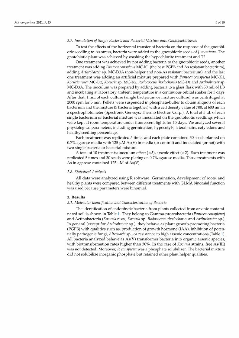
3. Results3.1. Molecular Identification and Characterization of Bacteria
The identification of endophytic bacteria from plants collected from arsenic contami-nated soil is shown in Table 1. They belong to Gamma-proteobacteria (Pantoea conspicua)and Actinobacteria (Kocuria rosea, Kocuria sp. Rodococcus rhodochorus and Arthrobacter sp.).In general (except for Arthrobacter sp.), they behave as plant growth-promoting bacteria(PGPB) with qualities such as, production of growth hormone (IAA), inhibition of poten-tially pathogenic fungi, Alternaria sp., or resistance to high arsenic concentrations (Table 1).All bacteria analyzed behave as As(V) transformer bacteria into organic arsenic species,with biotransformation rates higher than 30%. In the case of Kocuria strains, free As(III)was not detected. Moreover, P. conspicua was a phosphate solubilizer. The bacterial mixturedid not solubilize inorganic phosphate but retained other plant helper qualities.
Microorganisms 2021, 9, 45 6 of 18
Table 1. Molecular identification (similarity > 99.5%) and characterization of five endophytic bacteria (and a mixture of all of them) from J. montana growing on arsenic contaminated soil:ability to solubilize inorganic phosphate, to produce IAA, to inhibit the growth of Alternaria sp., and arsenic species and total arsenic concentrations within bacteria growing at 10 mMarsenate.
Molecular Approach * P IAA Production % Fugal Inh. AMIC As(V) a As(III) a Total As b As(V) Biot. c
Strains (µg mL−1
DO−1)LB mM mgL−1 mgL−1 mgL−1 %
MC-K1 P.conspicua T + 1.96 ± 0.07 40 450 0.125 ± 0.3 10−3 0.091 ± 0.001 0.63 ± 0.03 66MC-K2 Kocuria sp. − 0.46 ± 0.10 53 450 0.561 ± 0.008 n.d. 0.88 ± 0.02 36MC-D1 R. rhodochorus T − 0.76 ± 0.10 40 450 0.266 ± 0.005 0.022 ± 0.001 0.67 ± 0.03 57MC-D2 K. rosea T − 1.05 ± 0.07 46.7 450 0.152 ± 0.001 n.d. 0.54 ± 0.04 72
MC-D3A Arthrobacter sp. − n.d. 73 10 n.a. n.a. n.a.Mixture bacteria − 4.46 ± 0.01 20 200 n.a. n.a. n.a.
LB: Luria Bertani medium, n.d.: non-detected, n.a.: non-analyzed, As(V)MIC: As(V) minimal inhibition concentration. a Determined by anion exchange HPLC-(UV)-HG-AFS. b Determined by ICP-AES. c Estimatedas the difference, expressed as a percentage, between total As and the sum of As species found in samples. * Whenever possible, molecular identification was carried out with the type strain T.

Microorganisms 2021, 9, 45 7 of 18
3.2. Anatomical and Physiological Effects of Arsenic on Seedlings
When seedlings were germinated on media with different arsenate concentrations(Figure 2) morphological changes were observed. Germination was significantly affected byarsenic concentration above 500 µM As(V). The percentage of well-developed roots and lat-eral hair abundance also decreased, with arsenic concentration being drastically decreasedwith arsenic concentrations above 125 µM As(V). A total of 1 mM arsenate concentrationwas thus used to evaluate anatomical changes provoked by arsenic. Because of this prelimi-nary screening, 125 µM arsenate concentration was selected for inoculation experiments onthe gnotobiont, given that germination, root system and hypocotyl development was notaffected but parameters such as cotyledon production and plant viability were variable andeasily measurable parameters. Seedlings of J. montana not treated with As(V) showed devel-opment of well-developed root systems with abundant lateral hairs (Figure 3A) and healthycotyledons with oxidation reactions only in the vascular system (Figure 3C). A matrix wasobserved around them, occupied by aniline blue stained epiphytic bacteria (Figure 3A,B).In addition, brown-stained intracellular bacteria exhibiting elevated H2O2 presence werealso observed in root cells (Figure 3B). Coccoidal bacteria were visualized in apoplasticspaces of root cells (Figure 3D) and bacterial rods were frequently seen within parenchymacells (Figure 3E). Seedling plants watered with 1 mM arsenate solution showed anatomicalchanges with strong signs of oxidative stress in the cotyledons, reduction of chlorophylls(Figure 4C) and incipient tumors in the root (Figure 4D). In addition, frequently collapsedroot cells were observed (Figure 4A). However, coccoidal bacteria (Figure 4A) and bacterialrods within parenchyma cells (Figure 4B) were detected in control seedlings (Figure 3).
Microorganisms 2021, 9, x FOR PEER REVIEW 6 of 17
Table 1. Molecular identification (similarity > 99.5%) and characterization of five endophytic bacteria (and a mixture of all
of them) from J. montana growing on arsenic contaminated soil: ability to solubilize inorganic phosphate, to produce IAA,
to inhibit the growth of Alternaria sp., and arsenic species and total arsenic concentrations within bacteria growing at 10
mM arsenate.
Molecular ap‐
proach * P IAA production % Fugal Inh. AMIC As (V) a As (III) a Total As b As (V) Biot. c
Strains (μgmL−1DO−1) LB mM mgL−1 mgL−1 mgL−1 %
MC‐K1 P.conspicua T + 1.96 ± 0.07 40 450 0.125 ± 0.3 10−3 0.091 ± 0.001 0.63 ± 0.03 66
MC‐K2 Kocuria sp. ‐ 0.46 ± 0.10 53 450 0.561 ± 0.008 n.d. 0.88 ± 0.02 36
MC‐D1 R. rhodochorus T ‐ 0.76 ± 0.10 40 450 0.266 ± 0.005 0.022 ± 0.001 0.67 ± 0.03 57
MC‐D2 K. rosea T ‐ 1.05 ± 0.07 46.7 450 0.152 ± 0.001 n.d. 0.54 ± 0.04 72
MC‐D3A Arthrobacter sp. ‐ n.d. 73 10 n.a. n.a. n.a.
Mixture bacteria ‐ 4.46 ± 0.01 20 200 n.a. n.a. n.a.
LB: Luria Bertani medium, n.d.: non‐detected, n.a.: non‐analyzed, As(V)MIC: As(V) minimal inhibition concentration. a De‐
termined by anion exchange HPLC‐(UV)‐HG‐AFS. b Determined by ICP‐AES. c Estimated as the difference, expressed as a
percentage, between total As and the sum of As species found in samples. * Whenever possible, molecular identification
was carried out with the type strain T.
3.2. Anatomical and Physiological Effects of Arsenic on Seedlings
When seedlings were germinated on media with different arsenate concentrations
(Figure 2) morphological changes were observed. Germination was significantly affected
by arsenic concentration above 500 μM As (V). The percentage of well‐developed roots
and lateral hair abundance also decreased, with arsenic concentration being drastically
decreased with arsenic concentrations above 125 μM As (V). A total of 1 mM arsenate
concentration was thus used to evaluate anatomical changes provoked by arsenic. Because
of this preliminary screening, 125 μM arsenate concentration was selected for inoculation
experiments on the gnotobiont, given that germination, root system and hypocotyl devel‐
opment was not affected but parameters such as cotyledon production and plant viability
were variable and easily measurable parameters. Seedlings of J. montana not treated with
As (V) showed development of well‐developed root systems with abundant lateral hairs
(Figure 3A) and healthy cotyledons with oxidation reactions only in the vascular system
(Figure 3C). A matrix was observed around them, occupied by aniline blue stained epi‐
phytic bacteria (Figure 3A,B). In addition, brown‐stained intracellular bacteria exhibiting
elevated H2O2 presence were also observed in root cells (Figure 3B). Coccoidal bacteria
were visualized in apoplastic spaces of root cells (Figure 3D) and bacterial rods were fre‐
quently seen within parenchyma cells (Figure 3E). Seedling plants watered with 1 mM
arsenate solution showed anatomical changes with strong signs of oxidative stress in the
cotyledons, reduction of chlorophylls (Figure 4C) and incipient tumors in the root (Figure
4D). In addition, frequently collapsed root cells were observed (Figure 4A). However, coc‐
coidal bacteria (Figure 4A) and bacterial rods within parenchyma cells (Figure 4B) were
detected in control seedlings (Figure 3).
Figure 2. J. montana planted on different concentration of arsenate. Percentage of germination (blacksquares), percentage of seedling plants that develop roots (gray squares) and percentage of seedlingplants with lateral hairs (white squares).

Microorganisms 2021, 9, 45 8 of 18
Microorganisms 2021, 9, x FOR PEER REVIEW 7 of 17
Figure 2. J. montana planted on different concentration of arsenate. Percentage of germi‐
nation (black squares), percentage of seedling plants that develop roots (gray squares)
and percentage of seedling plants with lateral hairs (white squares).
Figure 3. J. montana seedling plant control. (A). Root surrounded by a bacterium commu‐
nity (red arrow). (B). Detail of the matrix that surrounds the root hairs where aniline blue
stained bacteria can visualize (red arrow). Brown bacteria stained with DAB/peroxidase
for reactive oxygen (black arrows) within lateral hairs. (C). Healthy cotyledons without
signs of deterioration due to oxidative stress. Defection of H2O2 accumulation in circula‐
tory system (black arrows). (D). Aniline blue coccoidal bacteria in apoplastic space of root
(black arrow). (E). Bacteria rods stained with aniline blue inside hypocotylous paren‐
chyma (black arrows).
Figure 4. J. montana seedling plant control. (A). Root surrounded by a bacterium commu‐
nity (red arrow). (B). Detail of the matrix that surrounds the root hairs where aniline blue
Figure 3. J. montana seedling plant control. (A). Root surrounded by a bacterium community (redarrow). (B). Detail of the matrix that surrounds the root hairs where aniline blue stained bacteriacan visualize (red arrow). Brown bacteria stained with DAB/peroxidase for reactive oxygen (blackarrows) within lateral hairs. (C). Healthy cotyledons without signs of deterioration due to oxidativestress. Defection of H2O2 accumulation in circulatory system (black arrows). (D). Aniline bluecoccoidal bacteria in apoplastic space of root (black arrow). (E). Bacteria rods stained with anilineblue inside hypocotylous parenchyma (black arrows).
Microorganisms 2021, 9, x FOR PEER REVIEW 7 of 17
Figure 2. J. montana planted on different concentration of arsenate. Percentage of germi‐
nation (black squares), percentage of seedling plants that develop roots (gray squares)
and percentage of seedling plants with lateral hairs (white squares).
Figure 3. J. montana seedling plant control. (A). Root surrounded by a bacterium commu‐
nity (red arrow). (B). Detail of the matrix that surrounds the root hairs where aniline blue
stained bacteria can visualize (red arrow). Brown bacteria stained with DAB/peroxidase
for reactive oxygen (black arrows) within lateral hairs. (C). Healthy cotyledons without
signs of deterioration due to oxidative stress. Defection of H2O2 accumulation in circula‐
tory system (black arrows). (D). Aniline blue coccoidal bacteria in apoplastic space of root
(black arrow). (E). Bacteria rods stained with aniline blue inside hypocotylous paren‐
chyma (black arrows).
Figure 4. J. montana seedling plant control. (A). Root surrounded by a bacterium commu‐
nity (red arrow). (B). Detail of the matrix that surrounds the root hairs where aniline blue
Figure 4. J. montana seedling plants germinated on agarose plates and watered with 1 mM As(V).A. Coccoidal bacteria staining brown on cell wall surface in some root cells (black arrows) withmeristemic cells damaged (red arrow). B. Bacteria rods (arrows) with capacity to stain with anilineblue (arrows) inside hypocotylous parenchyma. C. Oxidative deterioration (arrows) in cotyledonswith H2O2 accumulation. D. Incipient root tumor caused by arsenic oxidative stress.

Microorganisms 2021, 9, 45 9 of 18
3.3. Anatomical and Physiological Effects on Gnotobiotic Jasione (1 mM Arsenate)
The effect of the [As], removing the epiphytic bacteria and the antibiotic treatments in-teraction on germination was not significantly different (Table 2). However, the interactionbetween treatments with [As] and treatment with hypochlorite were significantly difference(Table 2; Figure 5A,B). Regardless of the elimination of epiphytic bacteria with hypochloritesolution, the germination rate was significantly lower in all sterilization treatments (T2, T3,T4), compared to the control (T1), this reduction was more drastic under arsenic conditions.The lowest germination rate was always in T4, when the antibiotic was added before andafter planting (Figure 5A,B). The treatment T4 was so strong that seedlings did not showany effect of the As (Figure 5B). Removal epiphytic bacteria (washing seeds in 2% NaClO),significantly improved germination on 1 mM As(V) agarose (Figure 5A). When we testedthe success of removal of epiphytic bacteria in the seed plated onto different bacterialculture media, no bacterial presence was observed in any of the plates (data not shown).
Table 2. GLM analyses to test the effect of arsenic and hypochlorite washes on germination processof functional gnotobiotic J. montana obtained by different levels of sterilization (T1, T1, T3 and T4).Degrees of freedom (df), Deviance Residual (DR) and the probability associated to the estimation(Pr). The zero concentration of arsenate is the value of reference.
Parameter Factor df DR Pr (>/Chi/)
Germination
Treatment 3 365.08 <0.001Hypochloryte 1 16.05 <0.001[As] 1 258.75 <0.001Treatment:Hypochloryte 3 84.21 <0.001Treatment:As 3 21.61 <0.001Hypochloryte:As 1 3.07 nsTreatment:Hypochloryte:As 3 6.77 ns
Hypocotiledons
Treatment 3 1379.28 <0.001Hypochloryte 1 3.83 ns[As] 1 225.45 <0.001Treatment:Hypochloryte 3 46.84 <0.001Treatment:As 3 4.49 nsHypochloryte:As 1 0.11 nsTreatment:Hypochloryte:As 3 11.01 0.011
The capacity of J. montana to generate hypocotyls was reduced with any antibiotictreatment (T2, T3 and T4) and was significantly reduced when the sterilization treatmentbecame more drastic, a phenomenon that was independent of the presence or absence ofarsenic in the media and all other factors (Figure 5C). In those seeds germinated in theabsence of arsenic, the preliminary washing with sodium hypochlorite did not affect theirability to generate hypocotyls; however, when the seeds were subjected to As stress, theelimination of epiphytes favored the development of hypocotyls (Figure 5C).
The visualization, under an optical microscope, of the development of the seeds in oneof the sterilization treatments (light treatment, T2), after germinating with non-stressfulconditions, meristematic cells showed deterioration and roots were collapsed (Figure 6A).In this treatment, we visualized many lateral root hair primordia that did not elongate(Figure 6D). Frequently, the embryos appeared contorted (Figure 6F). Fewer bacteria on themeristem cell wall or endophytes in parenchyma cells were observed (Figure 6B,C).

Microorganisms 2021, 9, 45 10 of 18
Microorganisms 2021, 9, x FOR PEER REVIEW 9 of 17
[As]
Treatment:Hypochloryte
Treatment:As
Hypochloryte:As
Treatment:Hypochloryte:As
1
3
3
1
3
225.45
46.84
4.49
0.11
11.01
<0.001
<0.001
ns
ns
0.011
Figure 5. Experiment to analyze the effect of arsenic and hypochlorite washes on functional gnoto‐
biotic J. montana obtained by different levels of sterilization. T1, no sterilization; T2, washing the
seeds with streptomycin 24 hours before planting; T3, germination for ten days in streptomycin
media; T4, combination of T2 and T3. A and B show the effect of interaction treatment with As and
treatment with hypochlorite on germination. C and D, the percentage of hypocotyls developed. Dif‐
ferent letters mean statistically significant differences.
Figure 5. Experiment to analyze the effect of arsenic and hypochlorite washes on functional gnotobiotic J. montana obtainedby different levels of sterilization. T1, no sterilization; T2, washing the seeds with streptomycin 24 h before planting; T3,germination for ten days in streptomycin media; T4, combination of T2 and T3. (A) and (B) show the effect of interactiontreatment with As and treatment with hypochlorite on germination. (C), the percentage of hypocotyls developed. Differentletters mean statistically significant differences.

Microorganisms 2021, 9, 45 11 of 18Microorganisms 2021, 9, x FOR PEER REVIEW 10 of 17
Figure 6. Gnotibiotic J. montana (endophytic and epiphytic bacteria removal) without As.
(A). Root collapsed and intensely stained with DAB/peroxidase for reactive oxygen.
Root (B) and shoot (C) tissue without bacteria. (D). Early and non‐developed root hair
initial. (E). Dead or non‐cultivable bacteria around the root appendix. (F). Embryo
aborted before development.
3.4. Seedling Growth Promotion Experiments with Single Bacterium or Bacterial Mixture (125
μM Arsenate)
When holobiotic seedlings were developed on arsenic culture media (125 μM As (V)),
the percentage of germination, development of hypocotyls, roots, lateral hairs and healthy
plants were not affected (Figure 2). However, in gnotobiotic‐plants, the percentage of
healthy plants was determined by the two factors: intact biome and stress by arsenic (Fig‐
ure 7, Table 3), causing both parameters, a statistically significant reduction in the number
of healthy plants. The inoculation of a single PGPB (P. conspicua‐MCK1) shows a tendency
for the recovery of the plant, both in arsenic‐enriched and control media, while bioaug‐
mentation with Arthrobacter sp. does not help in the recovery of the plants either in stress
situation and control. Inoculation with the bacterial mixture (containing all five endo‐
phytic bacteria) allows a clear recovery of the plants in control conditions. However, the
interactions generated in the consortium under stress condition, probably as a conse‐
quence of Arthrobacter sp (non‐helper and non‐As resistant bacteria), prevent the recovery
of plants.
Figure 6. Gnotibiotic J. montana (endophytic and epiphytic bacteria removal) without As. (A). Rootcollapsed and intensely stained with DAB/peroxidase for reactive oxygen. Root (B) and shoot (C)tissue without bacteria. (D). Early and non-developed root hair initial. (E). Dead or non-cultivablebacteria around the root appendix. (F). Embryo aborted before development.
3.4. Seedling Growth Promotion Experiments with Single Bacterium or Bacterial Mixture(125 µM Arsenate)
When holobiotic seedlings were developed on arsenic culture media (125 µM As(V)),the percentage of germination, development of hypocotyls, roots, lateral hairs and healthyplants were not affected (Figure 2). However, in gnotobiotic-plants, the percentage ofhealthy plants was determined by the two factors: intact biome and stress by arsenic(Figure 7, Table 3), causing both parameters, a statistically significant reduction in thenumber of healthy plants. The inoculation of a single PGPB (P. conspicua-MCK1) showsa tendency for the recovery of the plant, both in arsenic-enriched and control media,while bioaugmentation with Arthrobacter sp. does not help in the recovery of the plantseither in stress situation and control. Inoculation with the bacterial mixture (containingall five endophytic bacteria) allows a clear recovery of the plants in control conditions.However, the interactions generated in the consortium under stress condition, probably asa consequence of Arthrobacter sp (non-helper and non-As resistant bacteria), prevent therecovery of plants.

Microorganisms 2021, 9, 45 12 of 18Microorganisms 2021, 9, x FOR PEER REVIEW 11 of 17
Figure 7. Gnotobiotic J.montana response to As (125 uM As (V))under different treatments
inoculated with helper bacteria, a non‐helper bacterium or a mixture of bacteria. Natural
control plants, gnotobiotic plants treated with streptomycin 24 hours before planting and
not inoculated, inoculated with Artlrobacter sp (non‐helper bacteria), inoculated with a
Pantoea conspicua (helper bacteria) and inoculated with a mixture of endophytic bacteria
(Pantoeaconspicua, Kocuriarosea, Kocuria sp.Rodococcus rhodochorus,Arthrobacter sp.).
Table 3. GLM analyses to test the effect of arsenic (125uM As[V]) on germination and healthy de‐
velopment of J. montcma obtained by different treatments. Degrees of freedom (df),Deviance
Residual (DR) and the probability associated to the estimation (Pr). Natural control plants (T1),
gnotobiotic plants treated with streptomycin 24 hours before planting and no inoculated (T2), in‐
oculated with Arthrobacter sp (non‐helper bacteria; T3), inoculated with a Pantoea. conspicua (helper
bacteria; T4) andinoculated with a mixture of bacteria (T5).
4. Discussion
Our results have shown how J. montana can support 125 μM arsenate concentration
without significant changes to germination, hypocotyls and root development, although
only 50% of the seeds developed healthy plants. This threshold concentration has also
been described in other As‐tolerant species, such as Betula celtiberica [21]. The percent of
germination and root development were drastically reduced at 500 and 250 μM As (V),
respectively. These physiological parameters are sensitive to arsenic pollution because
early roots are the first contact points with this toxic compound [13,53]. Liu et al. [54] sug‐
gest that some of the defense mechanisms have not yet developed in this early stage of
seedling development. Harminder et al. [55] conclude that arsenic causes a reduction in
root elongation by inducing oxidative stress, which is related to enhanced lipid peroxida‐
tion, but not to H2O2 accumulation. In agreement with previous works [5,53,56] our results
Parameter Factor df DR Pr(>/Chi/)
Germination Treatment
[As]
Treatment: [As]
4
1
4
7.33
0.074
2.661
0.11
0.78
0.61
Healthy plants Treatment
[As]
Treatment: [As]
4
1
4
170.315
46.072
27.251
<0.001
<0.001
<0.001
Figure 7. Gnotobiotic J.montana response to As (125 uM As(V)) under different treatments inoculated with helper bacteria, anon-helper bacterium or a mixture of bacteria. Natural control plants, gnotobiotic plants treated with streptomycin 24 hbefore planting and not inoculated, inoculated with Artlrobacter sp (non-helper bacteria), inoculated with a Pantoea conspicua(helper bacteria) and inoculated with a mixture of endophytic bacteria (Pantoeaconspicua, Kocuriarosea, Kocuria sp.Rodococcusrhodochorus,Arthrobacter sp.).
Table 3. GLM analyses to test the effect of arsenic (125uM As[V]) on germination and healthy devel-opment of J. montcma obtained by different treatments. Degrees of freedom (df), Deviance Residual(DR) and the probability associated to the estimation (Pr). Natural control plants (T1), gnotobioticplants treated with streptomycin 24 hours before planting and no inoculated (T2), inoculated withArthrobacter sp (non-helper bacteria; T3), inoculated with a Pantoea. conspicua (helper bacteria; T4)andinoculated with a mixture of bacteria (T5).
Parameter Factor df DR Pr(>/Chi/)
GerminationTreatment 4 7.33 0.11[As] 1 0.074 0.78Treatment: [As] 4 2.661 0.61
Healthy plantsTreatment 4 170.315 <0.001[As] 1 46.072 <0.001Treatment: [As] 4 27.251 <0.001
4. Discussion
Our results have shown how J. montana can support 125 µM arsenate concentrationwithout significant changes to germination, hypocotyls and root development, althoughonly 50% of the seeds developed healthy plants. This threshold concentration has alsobeen described in other As-tolerant species, such as Betula celtiberica [21]. The percent ofgermination and root development were drastically reduced at 500 and 250 µM As(V),respectively. These physiological parameters are sensitive to arsenic pollution becauseearly roots are the first contact points with this toxic compound [13,53]. Liu et al. [54]suggest that some of the defense mechanisms have not yet developed in this early stage ofseedling development. Harminder et al. [55] conclude that arsenic causes a reduction in rootelongation by inducing oxidative stress, which is related to enhanced lipid peroxidation,but not to H2O2 accumulation. In agreement with previous works [5,53,56] our resultsconfirm the hypothesis that plants stressed with arsenate (1 mM As(V)) suffer free ROSthat can be detoxified because of the activation of superoxide dismutase (with an increasein the production of H2O2). Coleoptiles showed cellular oxidative damage with chloroticappearance. Reduction in chlorophyll content because of arsenic has previously been
Microorganisms 2021, 9, 45 13 of 18
described [6] and has been related to decrease in leaf gas-exchange [57] and photosynthesisdamage [58]. The presence of root tumors can also be an effect of the oxidative damagecaused to the cells [59]. Broken cells in the outer layer cortex as an arsenic consequencehas been previously observed in other plants [8]. Endophytes and epiphytic bacteria inroot and shoot tissues, which are seemingly healthy, points to a possible implication ofmicrobiome in the acclimatization to this stressful situation.
The sterilization methods to achieve a gnotobiotic Jasione showed that the washing ofthe seeds with 2% NaClO did not completely remove the bacteria (Figure 6E,F), but thosethat were observed microscopically after treatment, either were dead or were non-cultivablebacteria, because they did not emerge when plated on suitable media for bacterial growth.This sterilization protocol resulted in an increase in germination and early development,which may be a consequence of the reduction of phytopathogenic microorganisms associ-ated with the seed surface [60,61]. The treatment of the seeds with streptomycin, duringthe 24 h prior to being plated on germination media (Figure 5), resulted in a significantreduction in the early development of seedlings [44]. Verma et al. [44] demonstrated howplant–gnotobiont model achieves, given that the plant tissues stay free of bacteria.
The most drastic treatment of sterilization using streptomycin throughout the develop-ment period resulted in a greater reduction in germination and total annulation of furtherdevelopment. These results could be explained because of the pernicious effect of the an-tibiotic on organelles, such as mitochondria [62], that would result in irreversible damageto seedling development. Minder et al. [63] describes how antibiotics in agricultural soilsdamage plants and soil microbial communities. However, other possible explanationscould be that microbiota is intimately involved in the development of seedlings [44,64–66]and antibiotics, by decreasing the internal microbial potential, lead to significant damageto plants. In fact, inoculation experiments on the gnobiotic system confirm that indige-nous endophytic bacteria from J. montana play important roles in modulating seedlingdevelopment [44], especially under stress conditions. In fact, the inoculation with onlyone PGP microbe, such as strain P. conspicua MC-K1, allowed the partial recovery of de-velopment, both in the presence and in the absence of arsenic. P. conspicua MC-K1 hasan ability to protect the host plant from arsenic stress, apparently through several plantgrowth-promoting mechanisms. It is able to synthesize IAA that activates root growth,nodule production and development of lateral hairs [65]. It also has antifungal capacity thatcan defend the seeds in these early stages of development from attack of pathogenic fungi.In fact, P. agglomerans (genetically close to P. conspicua) had antagonistic properties towardsAlternaria sp. [67] and is a diazotrophic bacterium [68]. P. conspicua MC-K1 also behaves asan inorganic phosphate solubilizer. Elevated levels of arsenic in soils also interfere with Puptake, but bacteria can compensate for this deficiency by solubilizing P, which may beparticularly important in the early stages of seedling development [69,70]. Nevertheless,the most important helping qualities are a higher As(V) MIC (450 mM) and the capacityto accumulate As and reduce As(V) to As(III), metabolizing the latter towards less toxicarsenic organic forms (Table 1). Arsenic detoxification mechanisms reported include eithermethylation and subsequent transformation into volatile forms [25], or strong bindingof As(III) to –SH groups of cytosolic proteins [71]. We did not detect methylation forms,but it is evident that complex organic forms (perhaps protein complexes, phytochelatins,etc.) were produced and these probably had reduced toxicity. Other Pantoea strains havealso been shown to have high As(V) MIC [72]. Wu et al. [73] reported Pantoea sp. as anAs(V) reducer for the first time and, recently, Tian and Jing [74] sequenced its genome tocharacterize the molecular mechanism of arsenate reduction. Given that P. conspicua is aPGP bacterium then transforms As(V) into As(III) and mainly into organic forms, and alsofavors the growth of J. montana under arsenic stress, it is possible that it is involved in theresistance of J. montana to arsenic stress.
The behavior of Arthrobacter strains against arsenic is variable. Some strains are ableto survive at high concentrations [75] but others only support low concentrations [76],like Arthrobacter sp. MC-D3A. Arthrobacter species are widely present in soils and environ-
Microorganisms 2021, 9, 45 14 of 18
mental polluted with chemicals and heavy metals [75] and in water As contaminated [77].It has been described as an endophytic PGP bacterium [78], but some strains have beenrecognized as opportunistic pathogens [79]. Arthrobacter MC-D3A does not behave like aPGPB and is not tolerant to As concentrations higher than 10 mM, at least, in aposymbioticconditions. Use of a complex inoculum, rather than single strains, improved arsenic seques-tration efficiency of hyper-accumulator plants [22,31]. Our bacterial mixture behaved as ahelper inoculum in control conditions but did not show positive effects under arsenic stress,even though four of the five species had plant growth promotional features. Bacterialmixture can evolve very rapidly [80], generating antagonism or competitive interactionsbetween species and causing community collapse and subsequent loss of plant protec-tion [81]. The presence of Arthrobacter sp. MC-D3A in the bacterial mixture may be themain reason that this complex inoculum did not help J. montana in physiological recoveryunder stress conditions since this bacterium appeared to negatively affect seedlings.
The plant-metaorganism (or plant-holobiont) bio-components show different contri-butions against arsenic stress. The seedling by itself is able to modulate the perniciouseffect of 125 mM arsenic in early stages of development (germination, hypocotyl and lateralhair production) but in absence of the microbiome the ability to develop healthy plants isdrastically reduced. The inoculation with a P. conspicua MC-K1 in gnotobiotic seedlingsseems to improve arsenic acclimatization capacity. Possibly the interaction of this microbe,together with other components of the microbiome, and the plant itself, are responsible forthe survival of the metaorganism under arsenic stress conditions. Since P. conspicua wasisolated from adult plants but beneficial in early germination, it is possible that seedingsrecruit it from the soil in the early stages of development (horizontal transfer) or it maybe vectored with the seed (vertical transfer). Additional research is needed to evaluatethese hypotheses. Arthrobacter strains can have negative effects for the plant under stressfulconditions. The relationship of this bacterium to others through competition–facilitatorinteractions can also drive the response of the plant to these conditions. Therefore, thetradeoff between helper and non-helper bacteria, together with the plant, would determinethe plant-metaorganism response to As contamination [82].
5. Conclusions
The physiological damage in plants is greater the more drastic the sterilization processis. When we use light sterilization conditions, J. montana seeds without your streptomycin-sensitive microbiome may germinate but they show significantly reduced ability forseedling development, especially under As stress. The type of bacteria (helper or non-helper) to the gnotobiotic plants shows different responses, from a tendency to recovery ofdevelopment to a reduction in this capacity. The interactions between bacteria determinethe response of the plant that varies significantly in environments contaminated by arsenic.
Author Contributions: Conceptualization, M.d.C.M., N.G.-B. and J.F.W.; methodology, M.d.C.M.,S.G.-S.; software, N.G.-B.; validation, N.G.-B., M.Á.Q. and J.F.W.; formal analysis, N.G.-B.; investiga-tion, M.d.C.M.; N.G.-B.; resources, J.F.W.; writing—original draft preparation, M.d.C.M.; writing—review and editing, N.G.-B., M.Á.Q.; project administration, M.d.C.M.; funding acquisition, J.F.W. Allauthors have read and agreed to the published version of the manuscript.
Funding: This work was financially supported by Ministerio de Educación y Ciencia (ProjectCTM2007-66432), Universidad Politécnica de Madrid (Project GI115815203) and Universidad ReyJuan Carlos (Project Driades).
Institutional Review Board Statement: Not applicable.
Informed Consent Statement: Informed consent was obtained from all subjects involved in the study.
Data Availability Statement: MDPI Research Data Policies” at https://www.mdpi.com/ethics.
Acknowledgments: The authors would like to thank Jaime López (Universidad Rey Juan Carlos),K. L. Kingsley (Rutgers University), M. S. Bergen (Rutgers University) and S. K. Verma (BanarasHindu University) for his invaluable help and collaboration. M.C. Molina thanks Rutgers University

Microorganisms 2021, 9, 45 15 of 18
for her time there as a Visiting Scientist. J. F. White is grateful for support from the USDA-NIFAMultistate Project W3147, and the New Jersey Agricultural Experiment Station.
Conflicts of Interest: The authors declare no conflict of interest.
References1. Singh, R.; Singh, S.; Parihar, P.; Singh, V.P.; Prasad, S.M. Arsenic contamination, consequences and remediation techniques:
A review. Ecotoxicol. Environ. Saf. 2015, 112, 247–270. [CrossRef] [PubMed]2. Mishra, S.; Mattusch, J.; Wennrich, R. Accumulation and transformation of inorganic and organic arsenic in rice and role of
thiol-complexation to restrict their translocation to shoot. Sci. Rep. 2017, 7, 40522. [CrossRef] [PubMed]3. Abbas, G.; Murtaza, B.; Bibi, I.; Shahid, M.; Niazi, N.K.; Khan, M.I.; Amjad, M.; Hussain, M. Arsenic uptake, toxicity, detoxification,
and speciation in plants: Physiological, biochemical, and molecular aspects. Int. J. Environ. Res. Public Health 2018, 15, 59.[CrossRef] [PubMed]
4. Srivastava, M.; Ma, L.Q.; Singh, N.; Singh, S. Antioxidant responses of hyper-accumulator and sensitive fern species to arsenic.J. Exp. Bot. 2005, 56, 1335–1342. [CrossRef]
5. Rafiq, M.; Shahid, M.; Abbas, G.; Shamshad, S.; Khalid, S.; Niazi, N.K.; Dumat, C. Comparative effect of calcium and EDTA onarsenic uptake and physiological attributes of Pisum sativum. Int. J. Phytoremediat. 2017, 19, 662–669. [CrossRef]
6. Anjum, S.A.; Xie, X.-Y.; Wang, L.-C.; Saleem, M.F.; Man, C.; Lei, W. Morphological, physiological and biochemical responses ofplants to drought stress. Afr. J. Agric. Res. 2011, 6, 2026–2032.
7. Talukdar, D. Arsenic-induced changes in growth and antioxidant metabolism of fenugreek. Russ. J. Plant Physiol. 2013, 60,652–660. [CrossRef]
8. Armendariz, A.L.; Talano, M.A.; Travaglia, C.; Reinoso, H.; Oller, A.L.W.; Agostini, E. Arsenic toxicity in soybean seedlings andtheir attenuation mechanisms. Plant Physiol. Biochem. 2016, 98, 119–127. [CrossRef]
9. Liu, W.-J.; Zhu, Y.-G.; Smith, F.A.; Smith, S.E. Do phosphorus nutrition and iron plaque alter arsenate (As) uptake by rice seedlingsin hydroponic culture? New Phytol. 2004, 162, 481–488. [CrossRef]
10. Tripathi, R.D.; Srivastava, S.; Mishra, S.; Singh, N.; Tuli, R.; Gupta, D.K.; Maathuis, F.J.M. Arsenic hazards: Strategies for toleranceand remediation by plants. Trends Biotechnol. 2017, 25, 158–165. [CrossRef]
11. Chen, Y.; Han, Y.-H.; Cao, Y.; Zhu, Y.-G.; Rathinasabapathi, B.; Ma, L.Q. Arsenic transport in rice and biological solutions toreduce arsenic risk from rice. Front. Plant Sci. 2017, 8, 268. [CrossRef] [PubMed]
12. Farooq, M.A.; Islam, F.; Ali, B.; Najeeb, U.; Mao, B.; Gill, R.A.; Yan, G.; Siddique, K.H.M.; Zhou, W. Arsenic toxicity in plants:Cellular and molecular mechanisms of its transport and metabolism. Environ. Exp. Bot. 2016, 132, 42–52. [CrossRef]
13. Abedin, M.J.; Meharg, A.A. Relative toxicity of arsenite and arsenate on germination and early seedling growth of rice (Oryzasativa L.). Plant Soil 2002, 243, 57–66. [CrossRef]
14. Finkel, O.M.; Castrillo, G.; Paredes, S.H.; González, I.S.; Dangl, J.L. Understanding and exploiting plant beneficial microbes. Curr.Opin. Plant Biol. 2017, 38, 155–163. [CrossRef]
15. Rajkumar, M.; Sandhya, S.; Prasad, M.N.V.; Freitas, H. Perspectives of plant-associated microbes in heavy metal phytoremediation.Biotechnol. Adv. 2012, 30, 1562–1574. [CrossRef]
16. Babu, A.G.; Reddy, M.S. Dual inoculation of arbuscular mycorrhizal and phosphate solubilizing fungi contributes in sustainablemaintenance of plant health in fly ash ponds. Water Air. Soil Pollut. 2011, 219, 3–10. [CrossRef]
17. Tiwari, S.; Sarangi, B.K.; Thul, S.T. Identification of arsenic resistant endophytic bacteria from Pteris vittata roots and characteriza-tion for arsenic remediation application. J. Environ. Manag. 2016, 180, 359–365. [CrossRef]
18. Han, Y.H.; Fu, J.W.; Chen, Y.; Rathinasabapathi, B.; Ma, L.Q. Arsenic uptake, arsenite efflux and plant growth in hyperaccumulatorPteris vittata: Role of arsenic-resistant bacteria. Chemosphere 2016, 144, 1937–1942. [CrossRef]
19. Salducci, M.-D.; Folzer, H.; Issartel, J.; Rabier, J.; Masotti, V.; Prudent, P.; Affre, L.; Hardion, L.; Tatoni, T.; Laffont-Schwob,I. How can a rare protected plant cope with the metal and metalloid soil pollution resulting from past industrial activities?Phytometabolites, antioxidant activities and root symbiosis involved in the metal tolerance of Astragalus tragacantha. Chemosphere2019, 217, 887–896. [CrossRef]
20. Cheng, C.; Nie, Z.W.; He, L.Y.; Sheng, X.F. Rice-derived facultative endophytic Serratia liquefaciens F2 decreases rice grain arsenicaccumulation in arsenic-polluted soil. Environ. Pollut. 2020, 259, 113832. [CrossRef]
21. Mesa, V.; Navazas, A.; González-Gil, R.; González, A.; Weyens, N.; Lauga, B.; Gallego, J.L.R.; Sánchez, J.; Peláez, A.I. Use ofendophytic and rhizosphere bacteria to improve phytoremediation of arsenic-contaminated industrial soils by autochthonousBetula celtiberica. Appl. Environ. Microbiol. 2017, 83, e03411-16. [CrossRef] [PubMed]
22. Mukherjee, G.; Saha, C.; Naskar, N.; Mukherjee, A.; Mukherjee, A.; Lahiri, S.; Majumder, A.L.; Seal, A. An endophytic bacterialconsortium modulates multiple strategies to improve arsenic phytoremediation efficacy in Solanum nigrum. Sci. Rep. 2018,8, 6979. [CrossRef] [PubMed]
23. Maeda, S.; Ohki, A.; Miyahara, K.; Takeshita, T.; Higashi, S. Growth characteristics and arsenic metabolism of two species ofarsenic-tolerant bacteria. Appl. Organomet. Chem. 1990, 4, 245–250. [CrossRef]
24. Bentley, R.; Chasteen, T.G. Microbial methylation of metalloids: Arsenic, antimony, and bismuth. Microbiol. Mol. Biol. Rev. 2002,66, 250–271. [CrossRef]
Microorganisms 2021, 9, 45 16 of 18
25. Qin, J.; Rosen, B.P.; Zhang, Y.; Wang, G.J.; Franke, S.; Rensing, C. Arsenic detoxification and evolution of trimethylarsine gas by amicrobial arsenite S-adenosylmethionine methyltransferase. Proc. Natl. Acad. Sci. USA 2006, 103, 2075–2080. [CrossRef]
26. Páez-Espino, D.; Tamames, J.; de Lorenzo, V.; Cánovas, D. Microbial responses to environmental arsenic. Biometals 2009, 22,117–130. [CrossRef]
27. Wolfe-Simon, F.; Blum, J.S.; Kulp, T.R.; Gordon, G.W.; Hoeft, S.E.; Pett-Ridge, J.; Stolz, J.F.; Webb, S.M.; Weber, P.K.; Davies,P.C.W.; et al. A bacterium that can grow by using arsenic instead of phosphorus. Science 2011, 332, 1163–1166. [CrossRef]
28. Dolphen, R.; Thiravetyan, P. Reducing arsenic in rice grains by leonardite and arsenic–resistant endophytic bacteria. Chemosphere2019, 223, 448–454. [CrossRef]
29. Retamal-Morales, G.; Mehnert, M.; Schwabe, R.; Tischler, D.; Zapata, C.; Chávez, R.; Schlömann, M.; Levicán, G. Detection ofarsenic-binding siderophores in arsenic-tolerating Actinobacteria by a modified CAS assay. Ecotoxicol. Environ. Saf. 2018, 157,176–181. [CrossRef]
30. Vital, T.Z.; Román-Ponce, B.; Orduña, F.N.R.; de los Santos, P.E.; Vásquez-Murrieta, M.S.; Deng, Y.; Wang, E.T. An endophyticKocuria palustris strain harboring multiple arsenate reductase genes. Arch. Microbiol. 2019, 201, 1285–1293. [CrossRef]
31. Lampis, S.; Santi, C.; Ciurli, A.; Andreolli, M.; Vallini, G. Promotion of arsenic phytoextraction efficiency in the fern Pteris vittataby the inoculation of As-resistant bacteria: A soil bioremediation perspective. Front. Plant Sci. 2015, 6, 80. [CrossRef] [PubMed]
32. Verma, S.K.; White, J.F. Indigenous endophytic seed bacteria promote seedling development and defend against fungal disease inbrown top millet (Urochloa ramosa L.). J. Appl. Microbiol. 2018, 124, 764–778. [CrossRef] [PubMed]
33. Callaway, R.M.; Brooker, R.W.; Choler, P.; Kikvidze, Z.; Lortie, C.J.; Michalet, R.; Paolini, L.; Pugnaire, F.I.; Newingham, B.;Aschehoug, E.T.; et al. Positive interactions among alpine plants increase with stress. Nature 2002, 417, 844–848. [CrossRef][PubMed]
34. Butterfield, B.J. Effects of facilitation on community stability and dynamics: Synthesis and future directions. J. Ecol. 2009, 97,1192–1201. [CrossRef]
35. Parnell, J. Variation in Jasione montana L. (Campanulaceae) and related species in Europe and North Africa. J. Watsonia 1987, 16,249–267.
36. Pérez-Espona, S.; Sales, F.; Hedge, I.; Möller, M. Phylogeny and species relationships in Jasione (Campanulaceae) with emphasison the ‘Montana-Complex’. Edinb. J. Bot. 2005, 62, 29–51. [CrossRef]
37. García-Salgado, S.; García-Casillas, D.; Quijano-Nieto, M.A.; Bonilla-Simón, M.M. Arsenic and heavy metal uptake and accumula-tion in native plant species from soils polluted by mining activities. Water Air Soil Pollut. 2012, 223, 559–572. [CrossRef]
38. Gutiérrez-Ginés, M.J.; Pastor, J.; Hernández, A.J. Heavy metals in native mediterranean grassland species growing at abandonedmine sites: Ecotoxicological Assessment and Phytoremediation of Polluted Soils. In Soil Biology 44, Heavy Metal Contamination ofSoils; Sherameti, I., Varma, A., Eds.; Springer: Berlin/Heidelberg, Germany, 2015; pp. 159–178.
39. Porter, E.K.; Peterson, P.J. Arsenic accumulation by plants on mine waste (United Kingdom). Sci. Total Environ. 1975, 4, 356–371.[CrossRef]
40. Benson, L.M.; Porter, E.K.; Peterson, P.J. Arsenic accumulation, tolerance and genotypic variation in plants on arsenical minewastes in S.W. England. J. Plant Nutr. 1981, 3, 655–666. [CrossRef]
41. Bosch, T.C.G.; McFall-Ngai, M.J. Metaorganisms as the new frontier. Zoology 2011, 114, 185–190. [CrossRef]42. Vandenkoornhuyse, P.; Quaiser, A.; Duhamel, M.; Le Van, A.; Dufresne, A. The importance of the microbiome of the plant
holobiont. New Phytol. 2015, 206, 1196–1206. [CrossRef] [PubMed]43. Kurzbaum, E.; Kirzhner, F.; Armon, R. A hydroponic system for growing gnotobiotic vs. sterile plants to study phytoremediation
processes. Int. J. Phytoremediat. 2014, 16, 267–274. [CrossRef] [PubMed]44. Verma, S.K.; Kingsley, K.; Irizarry, I.; Bergen, M.; Kharwar, R.N.; White, J.F. Seed-vectored endophytic bacteria modulate
development of rice seedlings. J. Appl. Microbiol. 2017, 122, 1680–1691. [CrossRef] [PubMed]45. Herzog, V.; Fahimi, H.D. A new sensitive colorimetric assay for peroxidase using 3,3′-diaminobenzidine as hydrogen donor. Anal.
Biochem. 1973, 55, 554–562. [CrossRef]46. White, J.F.; Torres, M.S.; Somu, M.P.; Johnson, H.; Irizarry, I.; Chen, Q.; Zhang, N.; Walsh, E.; Tadych, M.; Bergen, M. Hydrogen
peroxide staining to visualize intracellular bacterial infections of seedling root cells. Microsc. Res. Technol. 2014, 77, 566–573.[CrossRef] [PubMed]
47. Molina, M.C.; González, N.; Bautista, L.F.; Sanz, R.; Simarro, R.; Sánchez, I.; Sanz, J.L. Isolation and genetic identification of PAHdegrading bacteria from a microbial consortium. Biodegradation 2009, 20, 789–800. [CrossRef] [PubMed]
48. García-Salgado, S.; Quijano, M.A.; Bonilla, M.M. Arsenic speciation in edible alga samples by microwave-assisted extractionand high-performance liquid chromatography coupled to atomic fluorescence spectrometry. Anal. Chim. Acta 2012, 714, 38–46.[CrossRef]
49. Gordon, S.A.; Weber, R.P. Colorimetric estimation of indoleacetic acid. Plant Physiol. 1951, 26, 192–195. [CrossRef]50. Kusaba, M.; Tsuge, T. Phylogeny of Alternaria fungi known to produce host-specific toxins on the basis of variation in internal
transcribed spacers of ribosomal DNA. Curr. Genet. 1995, 28, 491–498. [CrossRef]51. Soares, M.A.; Li, H.-Y.; Bergen, M.; da Silva, J.M.; Kowalski, K.P.; White, J.F. Functional role of an endophytic Bacillus amyloliq-
uefaciens in enhancing growth and disease protection of invasive English ivy (Hedera helix L.). Plant Soil 2016, 405, 107–123.[CrossRef]

Microorganisms 2021, 9, 45 17 of 18
52. Pikovskaya, R.I. Mobilization of phosphorus in soil in connection with vital activity of some microbial species. Mikrobiologiya1948, 17, 362–370.
53. Hartley-Whitaker, J.; Ainsworth, G.; Meharg, A.A. Copper and arsenate induced oxidative stress in Holcus lanatus L. clones withdifferential sensitivity. Plant Cell Environ. 2001, 24, 713–722. [CrossRef]
54. Liu, X.; Zhang, S.; Shan, X.; Zhu, Y.-G. Toxicity of arsenate and arsenite on germination, seedling growth and amylolytic activityof wheat. Chemosphere 2005, 61, 293–301. [CrossRef] [PubMed]
55. Harminder, P.S.; Daizy, R.B.; Ravinder, K.K.; Komal, A. Arsenic-induced root growth inhibition in mung bean (Phaseolus aureusRoxb.) is due to oxidative stress resulting from enhanced lipid peroxidation. Plant Growth Regul. 2007, 53, 65–73.
56. Gupta, M.; Sharma, P.; Sarin, N.B.; Sinha, A.L. Differential response of arsenic stress in two varieties of Brassica juncea L.Chemosphere 2009, 74, 1201–1208. [CrossRef] [PubMed]
57. Stoeva, N.; Berova, M.; Zlatev, Z. Effect of arsenic on some physiological parameters in bean plants. Biol. Plantarum. 2005, 49,293–296. [CrossRef]
58. Farnese, F.S.; Oliveira, J.T.A.; Farnese, M.S.; Gusman, G.S.; Silveira, N.M.; Siman, L.I. Uptake arsenic by plants: Effects on mineralnutrition, growth and antioxidant capacity. Idesia 2014, 32, 99–106. [CrossRef]
59. Bolhassani, A.; Khavari, S.; Bathaie, S.Z. Saffron and natural carotenoids: Biochemical activities and anti-tumor effects. Biochim.Biophys. Acta 2014, 1845, 20–30. [CrossRef]
60. Young, S.B.; Setlow, P. Mechanisms of killing of Bacillus subtilis spores by hypochlorite and chlorine dioxide. J. Appl. Microbiol.2003, 95, 54–67. [CrossRef]
61. Verma, S.; Yadav, K.; Singh, N. Optimization of the protocols for surface sterilization, regeneration and acclimatization of Steviarebaudiana Bertoni. Am. Eurasian J. Agric. Environ. Sci. 2011, 11, 221–227.
62. Robert, F.; Hevor, T.K. Abnormal organelles in cultured astrocytes are largely enhanced by streptomycin and intensively bygentamicin. Neuroscience 2007, 144, 191–197. [CrossRef] [PubMed]
63. Minden, V.; Deloy, A.; Volkert, A.M.; Leonhardt, S.D.; Pufal, G. Antibiotics impact plant traits, even at small concentrations.AoB Plants 2017, 9, plx010. [CrossRef] [PubMed]
64. Tsavkelova, E.A.; Cherdyntseva, T.A.; Klimova, S.Y.; Shestakov, A.I.; Botina, S.G.; Netrusov, A.I. Orchid-associated bacteriaproduce indole-3-acetic acid, promote seed germination, and increase their microbial yield in response to exogenous auxin.Arch. Microbiol. 2007, 188, 655–664. [CrossRef] [PubMed]
65. Cassán, F.; Perrig, D.; Sgroy, V.; Masciarelli, O.; Penna, C.; Luna, V. Azospirillum brasilense Az39 and Bradyrhizobium japonicumE109, inoculated singly or in combination, promote seed germination and early seedling growth in corn (Zea mays L.) andsoybean (Glycine max L.). Eur. J. Soil Biol. 2009, 45, 28–35. [CrossRef]
66. White, J.F.; Kingsley, K.I.; Kowalski, K.P.; Irizarry, I.; Micci, A.; Soares, M.A.; Bergen, M.S. Disease protection and allelopathicinteractions of seed-transmitted endophytic pseudomonads of invasive reed grass (Phragmites australis). Plant Soil 2018, 422,195–208. [CrossRef]
67. Links, M.G.; Demeke, T.; Gräfenhan, T.; Hill, J.E.; Hemmingsen, S.M.; Dumonceaux, T.J. Simultaneous profiling of seed-associatedbacteria and fungi reveals antagonistic interactions between microorganisms within a shared epiphytic microbiome on Triticumand Brassica seeds. New Phytol. 2014, 202, 542–553. [CrossRef]
68. White, J.F.; Crawford, H.; Torres, M.S.; Mattera, R.; Irizarry, I.; Bergen, M.A. A proposed mechanism for nitrogen acquisition bygrass seedlings through oxidation of symbiotic bacteria. Symbiosis 2012, 57, 161–171. [CrossRef]
69. Kuklinsky-Sobral, J.; Araújo, W.L.; Mendes, R.; Geraldi, I.O.; Pizzirani-Kleiner, A.A.; Azevedo, J.L. Isolation and characterizationof soybean-associated bacteria and their potential for plant growth promotion. Environ. Microbiol. 2004, 6, 1244–1251. [CrossRef]
70. Ma, Y.; Rajkumar, M.; Zhang, C.; Freitas, H. Beneficial role of bacterial endophytes in heavy metal phytoremediation. J. Environ.Manag. 2016, 174, 14–25. [CrossRef]
71. Rahman, F.; Chen, Z.; Naidu, R. A comparative study of the extractability of arsenic species from silverbeet and amaranthvegetables. Environ. Geochem. Health 2009, 31, 103–113. [CrossRef]
72. Escalante, G.; Campos, V.L.; Valenzuela, C.; Yañez, J.; Zaror, C.; Mondaca, M.A. Arsenic resistant bacteria isolated from arseniccontaminated river in the Atacama Desert (Chile). Bull. Environ. Contam. Toxicol. 2009, 83, 657–661. [CrossRef] [PubMed]
73. Wu, Q.; Du, J.; Zhuang, G.; Jing, C. Bacillus sp. SXB and Pantoea sp. IMH, aerobic As(V)-reducing bacteria isolated fromarsenic-contaminated soil. J. Appl. Microbiol. 2013, 114, 713–721. [CrossRef] [PubMed]
74. Tian, H.; Jing, C. Genome sequence of the aerobic arsenate-reducing bacterium Pantoea sp. strain IMH. Genome Announc. 2014, 2,e00267-14. [CrossRef] [PubMed]
75. Xu, L.; Shi, W.; Zeng, X.-C.; Yang, Y.; Zhou, L.; Mu, Y.; Liu, Y. Draft genome sequence of Arthrobacter sp. strain B6 isolated fromthe high-arsenic sediments in Datong Basin, China. Stand. Genom. Sci. 2017, 12, 11. [CrossRef]
76. Chopra, B.K.; Bhat, S.; Mikheenko, I.P.; Xu, Z.; Yang, Y.; Luo, X.; Chen, H.; van Zwieten, L.; Lilley, R.M.; Zhang, R. Thecharacteristics of rhizosphere microbes associated with plants in arsenic-contaminated soils from cattle dip sites. Sci. Total Environ.2007, 378, 331–342. [CrossRef]
77. Prasad, K.S.; Ramanathan, A.L.; Paul, J.; Subramanian, V.; Prasad, R. Biosorption of arsenite (As+ 3) and arsenate (As+ 5) fromaqueous solution by Arthrobacter sp. biomass. Environl. Technol. 2013, 34, 2701–2708. [CrossRef]
78. Palaniappan, P.; Chauhan, P.S.; Saravanan, V.S.; Anandham, R.; Sa, T. Isolation and characterization of plant growth promotingendophytic bacterial isolates from root nodule of Lespedeza sp. Biol. Fertil. Soils 2010, 46, 807–816. [CrossRef]

Microorganisms 2021, 9, 45 18 of 18
79. Huang, Y.; Zhao, N.; He, L.; Wang, L.; Liu, Z.; You, M.; Guan, F. Arthrobacter scleromae sp. nov. isolated from human clinicalspecimens. J. Clin. Microbiol. 2005, 43, 1451–1455. [CrossRef]
80. González, N.; Simarro, R.; Molina, M.C.; Bautista, L.F.; Delgado, L.; Villa, J.A. Effects of surfactants on PAH biodegradation by abacterial consortium and on the dynamics of the bacterial community during the process. Bioresour. Technol. 2011, 102, 9438–9446.[CrossRef]
81. Becker, J.; Eisenhauer, N.; Scheu, S.; Jousset, A. Increasing antagonistic interactions cause bacterial communities to collapse athigh diversity. Ecol. Lett. 2012, 15, 468–474. [CrossRef]
82. Thijs, S.; Sillen, W.; Rineau, F.; Weyens, N.; Vangronsveld, J. Towards an enhanced understanding of plant–microbiome interactionsto improve phytoremediation: Engineering the metaorganism. Front. Microbiol. 2016, 7, 341. [CrossRef] [PubMed]