a glassy-winged sharpshooter cell line supports replication of
TRANSCRIPT

Journal of Invertebrate Pathology 94 (2007) 130–139
www.elsevier.com/locate/yjipa
A glassy-winged sharpshooter cell line supports replication of Rhopalosiphum padi virus (Dicistroviridae)
Sandhya Boyapalle a,1, Narinder Pal a, W. Allen Miller b, Bryony C. Bonning a,¤
a Department of Entomology, Iowa State University, 418 Science II, Ames, IA 50011-3222, USAb Department of Plant Pathology, Iowa State University, Ames, IA 50011, USA
Received 27 July 2006; accepted 26 September 2006Available online 17 November 2006
Abstract
Rhopalosiphum padi virus (RhPV) (family Dicistroviridae; genus Cripavirus) is an icosahedral aphid virus with a 10 kb positive-senseRNA genome. To study the molecular biology of RhPV, identiWcation of a cell line that supports replication of the virus is essential. Wescreened nine cell lines derived from species within the Lepidoptera, Diptera and Hemiptera for susceptibility to RhPV following RNAtransfection. We observed cytopathic eVects (CPE) only in cell lines derived from hemipterans, speciWcally GWSS-Z10 cells derived fromthe glassy winged sharp shooter, Homalodisca coagulata and DMII-AM cells derived from the corn leaf hopper, Dalbulus maidis. Transla-tion and appropriate processing of viral gene products, RNA replication and packaging of virus particles in the cytoplasm of GWSS-Z10cells were examined by Western blot analysis, Northern blot hybridization and electron microscopy. Infectivity of the GWSS-Z10 cellderived-virus particles to the bird cherry-oat aphid, R. padi, was conWrmed by RT-PCR and Western blot. The GWSS-Z10 cell line pro-vides a valuable tool to investigate replication, structure and assembly of RhPV.© 2006 Elsevier Inc. All rights reserved.
Keywords: Insect cell line; Dicistrovirus; Aphid virus
1. Introduction
Rhopalosiphum padi virus (RhPV), which was Wrst iso-lated from the bird cherry-oat aphid, R. padi, belongs to thefamily Dicistroviridae (Fauquet et al., 2005). The dicistrovi-ruses are restricted to invertebrate hosts and possess a posi-tive-sense RNA genome with a characteristic dicistronicarrangement (Christian and Scotti, 1998). Other membersof the family include Drosophila C virus (DCV) (Johnsonand Christian, 1998), and Cricket paralysis virus(CrPV)(Reinganum et al., 1970). The dicistrovirus genomehas two open reading frames (ORF); the 5� ORF encodes apolyprotein that includes the non-structural proteins
* Corresponding author. Fax: +1 515 294 5957.E-mail address: [email protected] (B.C. Bonning).
1 Present address: Interdisciplinary Oncology Department, H. LeeMoYtt Cancer Center & Research Institute, University of South Florida,Tampa, FL 33612, USA.
0022-2011/$ - see front matter © 2006 Elsevier Inc. All rights reserved.doi:10.1016/j.jip.2006.09.010
including RdRp, while the 3� ORF encodes the structuralpolyprotein. Proteins from both ORFs are translated viainternal initiation of ribosomes directly on the genomicRNA at internal ribosome entry sites (IRES) (Domieret al., 2000; Wilson et al., 2000; Royall et al., 2004). Activityof the RhPV 5� IRES has been demonstrated in mamma-lian, Drosophila, and wheat germ in vitro translation sys-tems, and also in Spodoptera frugiperda (Sf21) cells, whichare commonly used for baculovirus expression of recombi-nant proteins (Royall et al., 2004). Structural proteins accu-mulate in vast excess of non-structural proteins in infectedcells (Wilson et al., 2000).
RhPV is known to infect seven species of aphid: R. padi,Schizaphis graminum, R. ruWabdominalis (D’Arcy et al.,1981; Gildow and D’Arcy, 1988), R. maidis, Metopolophiumdirhodum, Diuraphis noxia and Sitobion avenae (Wechmarand Rybicki, 1981; Williamson et al., 1989). In contrast toRhPV which has been isolated only from hemipteran spe-cies, and DCV which has been isolated only from dipteran

S. Boyapalle et al. / Journal of Invertebrate Pathology 94 (2007) 130–139 131
species, CrPV has been isolated from members of theOrthoptera, Hymenoptera, Lepidoptera, Hemiptera, andDiptera (Christian and Scotti, 1998). CrPV and DCV repli-cate readily in several Drosophila cell lines, and CrPV repli-cates in a number of other insect cell lines (Scotti et al.,1996; Christian and Scotti, 1998). Cell lines that supportreplication of dicistroviruses other than CrPV and DCVare unknown. Unfortunately, with respect to Wnding celllines that support replication of RhPV, it has not been pos-sible to establish continuous cell lines derived from aphids(Peters and Black, 1970; Hirumi and Maramorosch, 1971;Christian and Scotti, 1998). Primary cell lines cultured fromAcrythosiphon pisum (Hirumi and Maramorosch,1971)(Boyapalle, unpublished observation) and Hyper-omyzus lactucae (Peters and Black, 1970; Hirumi and Mar-amorosch, 1971) were viable only for a few weeks.
A cell culture system that allows replication of RhPVwould be an asset for further study of the biology of thisvirus. Nine cell lines derived from species within the Lep-idoptera, Diptera and Hemiptera were tested for suscep-tibility to infection by RhPV. Here, we reportidentiWcation of one hemipteran cell line that supportsthe replication and packaging of infectious RhPV, and asecond hemipteran cell line that is likely to support repli-cation of RhPV.
2. Materials and methods
2.1. Viruses and cells
RhPV was puriWed from infected colonies of R. padimaintained at Iowa State University as described previ-ously (D’Arcy et al., 1981). CrPV and DCV were propa-gated in cultured Drosophila melanogaster cells (Schneider’sline 2: S2)(Schneider, 1972). The viruses were puriWed frominfected S2 cells as described previously (Krishna et al.,2003).
Sf21 cells derived from the fall armyworm, Spodopterafrugiperda (Vaughn et al., 1977) were maintained in TC-100insect cell medium (Sigma Chemicals, St. Louis, MO, USA)supplemented with fetal bovine serum (FBS; Gibco-BRL)to a Wnal concentration of 10%. The Sf9 cell line, which wasderived from the Sf21 cell line, was maintained in Hink’sTNM (JRH Biosciences, Lenexa, KA, USA) containing 3%FBS. Tn5B1-4 “High-Five” cells derived from the cabbagelooper Trichoplusia ni (Wickham et al., 1992) were main-tained in Excel 405 (JRH Biosciences) serum free medium.OnE cells derived from the European corn borer, Ostrinianubialis were maintained in Excel 405 supplemented withFBS to a Wnal concentration of 3%. S2 cells derived fromvinegar Xy D. melanogaster were maintained in Schneider’smedium (Sigma Chemicals, St. Louis, MO, USA) with 10%FBS. Cell lines derived from the leaf hopper Agallia con-stricta (AC-20) (McIntosh et al., 1973), the velvetbean cat-erpillar Anticarsia gemmatalis (BCIRL-AG-AM1)(McIntosh and IgonoV, 1989), corn leaf hopper Dalbulusmaidis (DMII-AM; A. McIntosh, University of Missouri,
CO), glassy-winged sharpshooter Homalodisca coagulata(GWSS-Z10) (Kamita et al., 2005) were maintained inExcel 405 with 10% FBS. All cell culture media were sup-plemented with penicillin–streptomycin (Sigma) to a Wnalconcentration of 1% with the exception of Schneider’smedium which was supplemented with penicillin–strepto-mycin–amphotericin B (Wnal concentration 1%: Invitro-gen).
2.2. Isolation and transfection of viral RNA
Viral RNA was extracted using the Absolutely RNA®
RT-PCR MiniPrep kit (Stratagene) according to the manu-facturer’s speciWcations. The RNA concentration was deter-mined by measurement of absorbance at 260 nm. To test theability of the cell lines to support replication of RhPV, DCVand CrPV, the following procedure was employed (Maso-umi et al., 2003). Cells were seeded into four wells of a 6-wellcell culture plate (Fisher ScientiWc) at a density of 2£105
cells per well for Sf9 and AC-20 cells; 1£105 for Sf21, High-Five, OnE and AG-AM1 cells; 6£105 for S2 cells. GWSS-Z10 and DMII-AM cells were seeded such that they werenear conXuent. The cells were allowed to attach to the sur-face for 4 h at room temperature. Media and Xoating cellswere then removed and the cells washed with serum-free medium. One well was treated with 0.5�g DOTAP(N-[1-(2,3-dioleoyloxy)propyl]-N,N,N-trimethylammoniummethyl-sulfate (Alexis) in 1 ml of serum-free medium. Thesecond, third and fourth wells were treated with 0.5�gDOTAP in 1 ml serum-free medium along with 4�g ofRhPV RNA, 2�g DCV RNA and 2 �g CrPV RNA, respec-tively. All treatments were maintained at 28 °C for 6 h, cellswere gently washed twice with serum-containing mediaexcept High-Five cells which were washed in serum-freemedium. The cells were then incubated in 1.5 ml of theirrespective media at 28 °C. Cells were examined daily forcytopathic eVects (CPE) and harvested after 4–8 days.
For production of virus for aphid infectivity experi-ments, GWSS-Z10 cells in two 60 mm dishes (2.5£ 106 cellsper dish) were transfected with 30�g of RhPV RNA asdescribed above. Infected cells were harvested 4 days post-transfection (dpt).
2.3. Western blot analysis of proteins
Harvested cells were washed twice in phosphate-buVeredsaline (PBS) and resuspended in 100�l of PBS. Aliquots wereboiled for 5min in 100�l of 2£ protein dissociation buVer(2.3% SDS, 10% glycerol, 5% 2-mercaptoethanol, 62.5mMTris–HCl and 0.01% bromophenol blue, pH 6.8) and proteinsresolved by SDS–PAGE. Approximately 150ng of puriWedvirus alone was run as a positive control in each gel. Tenmicroliter samples were resolved by electrophoresis on 4–12% SDS–PAGE gels (Bio-Rad Laboratories), after whicheach gel was treated for 30min with Towbin buVer (10 mMTris base, 96mM glycine in 10% methanol). Proteins weretransferred to Hybond-P membrane (Amersham Pharmacia)

132 S. Boyapalle et al. / Journal of Invertebrate Pathology 94 (2007) 130–139
in Towbin buVer using an electro blotter apparatus (Bio-Rad) for 1h at 100 V. The membrane was placed for 2 h atroom temperature in a blocking solution consisting of 5%skimmed milk and 0.1% Tween 20 in Tris-buVered saline(TBS). Rabbit polyclonal anti-RhPV antiserum was puriWedas described previously (Bassham and Raikhel, 1998), diluted100-fold in blocking solution, and incubated with the mem-brane for 1 h at room temperature. Polyclonal anti-DCV andanti-CrPV antisera (Christian and Scotti, 1994) were diluted2000-fold in blocking solution, and incubated with the mem-brane for 1 h at room temperature. Membranes were washedtwice with 0.1% Tween 20 in TBS before reaction with goatanti-rabbit immunoglobulin conjugated with horseradishperoxidase (Sigma). After washing for 1h, the bound enzymewas detected using the ECL detection system (AmershamPharmacia).
2.4. Plasmids
Full length double stranded cDNA of RhPV was clonedbetween the EcoRI–KpnI sites of pGEM-3ZF (Promega) toproduce the plasmid pGEM-3ZF.RhPV (Boyapalle and R.Beckett, unpublished data). The T7 promoter was posi-tioned upstream of the 5� end of the RhPV sequence andthe SP6 promoter downstream of the 3� end of the RhPVsequence.
2.5. Preparation of in vitro transcripts
The plasmid pGEM-3ZF.RhPV was linearized withAcc651, and used as template for in vitro transcription offull length positive-strand RNA from the T7 promoter atthe 5� end using the T7 MegaScript kit (Ambion). Fulllength negative-sense RNA was transcribed by linearizingthe plasmid with EcoRI, and using it as template for in vitrotranscription from the SP6 promoter using the SP6 Mega-script kit (Ambion). Strand-speciWc 32P-labeled RNAprobes were prepared from the T7 and SP6 promoters inthe vector using T7 or SP6 RNA polymerase, respectively,following digestion of the plasmid with HpaI.
2.6. Northern blot hybridization
To analyze the production of viral RNA over the courseof infection, GWSS-Z10 cells were transfected with RhPVRNA or full-length negative-sense RhPV transcript with0.5 �g DOTAP as described above. Transfected cells wereharvested at various hours post-transfection (hpt) and totalcellular RNA was extracted using Trizol (Invitrogen)according to the manufacturer’s speciWcations. RNA wasanalyzed by Northern blot as described previously (Koevet al., 1999). Approximately 100 ng of viral RNA spikedwith total RNA from mock infected cells, or full-lengthnegative-sense RhPV transcript were run as positive con-trols. A 32P-labeled probe complementary to the 1.9 kb 3�-terminal sequence of RhPV was used to detect productionof positive-strand RNA in the cells. A 32P-labeled probe
complementary to the 1.2 kb 5� end sequence of RhPV wasused to detect negative-sense RhPV RNA in the cells.
2.7. PuriWcation of virus particles from infected GWSS-Z10 cells
GWSS-Z10 cells were harvested when extensive CPEwere observed at 4 dpt and the virus particles were puriWedas described previously (Krishna et al., 2003). PuriWed virusparticles were negatively stained with 2% uranyl acetateand examined by TEM, or used to test for infectivity toaphids (Section 2.9).
2.8. Immunogold electron microscopy
GWSS-Z10 cells and DMII cells were grown to nearconXuence in T-25 Xasks (Fisher ScientiWc), two Xasks foreach cell line. The cells were transfected with 1 �g ofDOTAP and 10 �g of RhPV RNA. GWSS-Z10 and DM11cells were harvested at 4 or 10 dpt, respectively. The cellswere pelleted and Wxed in 1% paraformaldehyde–0.5% glut-eraldehyde–0.05% sodium cacodylate, pH 7.1, for 10 min at4 °C, then in 2% paraformaldehyde–2.5% gluteraldehyde–0.05% sodium cacodylate, pH 7.1, for 30 min at 4 °C. Afterwashing the cells three times (10 min each time) with 0.05 Msodium cacodylate, the cells were dehydrated with a seriesof ethanol concentrations (50%, 70%, 85%, 95%, 3£ 100%)for 30 min for each step at 4 °C. Cells were then inWltratedwith ethanol: LR White Resin using ratios of 1:1 (2 h at4 °C), and 1:3 (overnight at 4 °C) followed by pure LRWhite (24 h at 4°C, with a resin change after 8 h). The cellswere then embedded in gelatin capsules, and resin was poly-merized at 4 °C for 48 h under UV light.
Gold labeling of the grids was carried out as describedby Erickson (Erickson et al., 1993). Grids were treated with25 �l of TBS-supplemented buVer (0.05 M Tris, 0.85%NaCl, pH 8.3–8.5, 0.5% normal goat serum, 0.5% normalpig serum and 0.5% BSA) with 3% non-fat dry milk for 2 hat room temperature. Grids were then immersed in 50 �ldrops of 1:10 puriWed rabbit polyclonal anti-RhPV antise-rum diluted in TBS-supplemented buVer with 3% non-fatdry milk for 5 h at 37 °C. After washing the grids three timesin TBS-supplemented buVer, the grids were treated with25 �l drops of 1:100 dilution of secondary goat anti-rabbitantibody conjugated with 10 nm gold particles (Ted PellaInc.) for 1 h at room temperature. After stream washing andthree washes in drops of distilled water (10 min each wash),grids were dried and stained with 2% aqueous uranyl ace-tate for 5 min and examined on a JOEL 1200 EX scanning/transmission electron microscope at 80 kV.
2.9. Infectivity of virus particles to aphids
Clones of uninfected R. padi were initiated parthenoge-netically from single apterous adults. The vertical transmis-sion rate of RhPV in R. padi is approximately 28% (D’Arcyet al., 1981). Progeny of each family were tested for the
S. Boyapalle et al. / Journal of Invertebrate Pathology 94 (2007) 130–139 133
presence of RhPV by Western blot and RT-PCR. Virus-freefamilies were pooled for production of the virus-free stock.For Western blot analysis, at least six aphids from eachtreatment were ground in a microfuge tube in 200 �l proteindissociation buVer (2.3% SDS, 10% glycerol, 5% 2-mercap-toethanol, 62.5 mM-Tris–HCl, 0.01% bromophenol blue,pH 6.8), boiled for 5 min and proteins resolved by SDS–PAGE. Approximately 150 ng of puriWed virus was run as apositive control. Ten microliter samples were resolvedby electrophoresis on 4–12% SDS–PAGE gels (Bio-RadLaboratories). Western blotting with puriWed polyclonalanti-RhPV antiserum was as described in Section 2.3. ForRT-PCR, total RNA from at least 10 aphids per replicatewas extracted using Trizol (Invitrogen) according to themanufacturer’s speciWcations. RT-PCR was carried outusing the primers 5�-TTAATTTCGAACCCCGTCAG-3�(from RhPV nt 848) and 5�-CTCAGTTTCGGGCTCTCTTG-3� (from RhPV nt 2387). PCR conditions used were94 °C for 1 min, 58 °C for 1 min, 72 °C for 2 min for 30 cycleswith a Wnal extension at 72 °C for 10 min. PCR productswere visualized in ethidium bromide-stained 1% agarosegels. Virus-free aphid clones were maintained in a separatebuilding from the RhPV-infected colony.
To test for replication and infectivity of GWSS-Z10 cell-derived virus particles to aphids, R. padi (40 aphids for eachof two replicate experiments) were allowed to feed for 16 h onParaWlm sachets containing puriWed virus particles in 25%sucrose in 0.01 M phosphate buVer, pH 7 (Mittler, 1988; Ras-ochova and Miller, 1996). Control aphids were fed on 25%sucrose alone. After the acquisition period, aphids weretransferred to cages and maintained on oats for 17 days. Todetermine whether aphids were infected with RhPV we usedRT-PCR and Western blot analysis for detection of RhPVRNA and proteins respectively as described above. Thein vitro-transcribed positive-sense transcript (Section 2.5) wasused as a positive control for RT-PCR.
3. Results
3.1. CrPV and DCV infection of cultured cell lines
The ability of RhPV to replicate in various insect celllines was tested by transfecting the cells with viral RNA
and DOTAP as transfection agent. DCV and CrPV RNAswere used as positive controls for RhPV transfection exper-iments. A summary of the transfection results is presentedin Table 1. The Drosophila (S2) cell line was permissive toCrPV and DCV as described previously, and exhibited dis-tinct CPE 2–3 dpt (Plus et al., 1975; Reinganum, 1975; Pluset al., 1978). By 5 dpt there were no living S2 cells in wellstransfected with CrPV or DCV. The cells treated withDOTAP alone (negative control) were healthy at 5 dpt andcontinued to divide. AG-AM1 cells transfected with CrPVor DCV showed signs of swelling and hypotrophy, withsome evidence of cell lysis and blebbing, and detached fromthe surface (Plus et al., 1975; Plus et al., 1978; Masoumiet al., 2003). CPE were more pronounced in CrPV RNA-transfected cells (3–4 dpt) compared to those transfectedwith DCV RNA (5 dpt).
For the GWSS-Z10 cell line, cells showed signs of CPEby 3–4 days for both CrPV and DCV. The cells lost theircharacteristic Wbroblast-like extensions, became roundedand were of uniform size. They detached from the substratewith approximately 90% of the cells Xoating. On examina-tion with a phase contrast microscope, the cells had a gran-ular appearance with some evidence of cell lysis. Incontrast, cells in the control wells were dividing and healthywith characteristic Wbroblast-like extensions. There were nogranular inclusions in the cytoplasm of the control cells.There were only a few Xoating cells in the control wells,which in contrast to the infected cells, varied in size andshape.
For the DMII cell line, the cells transfected with CrPVand DCV RNA exhibited CPE in 4–6 dpt. The cells wererounded and looked strikingly diVerent from cells in thecontrol wells. Infected cells lost their surface extensions, butdid not Xoat. The cells were clumped and the cytoplasmappeared granular. None of the other Wve cell lines testedwere permissive for CrPV or DCV replication followingtransfection with viral RNA.
3.2. RhPV replication in cultured cell lines
Of the nine cell lines tested, only the GWSS-Z10 andDMII cell lines appeared to be permissive to infection bytransfection with RhPV RNA. CPE were observed in
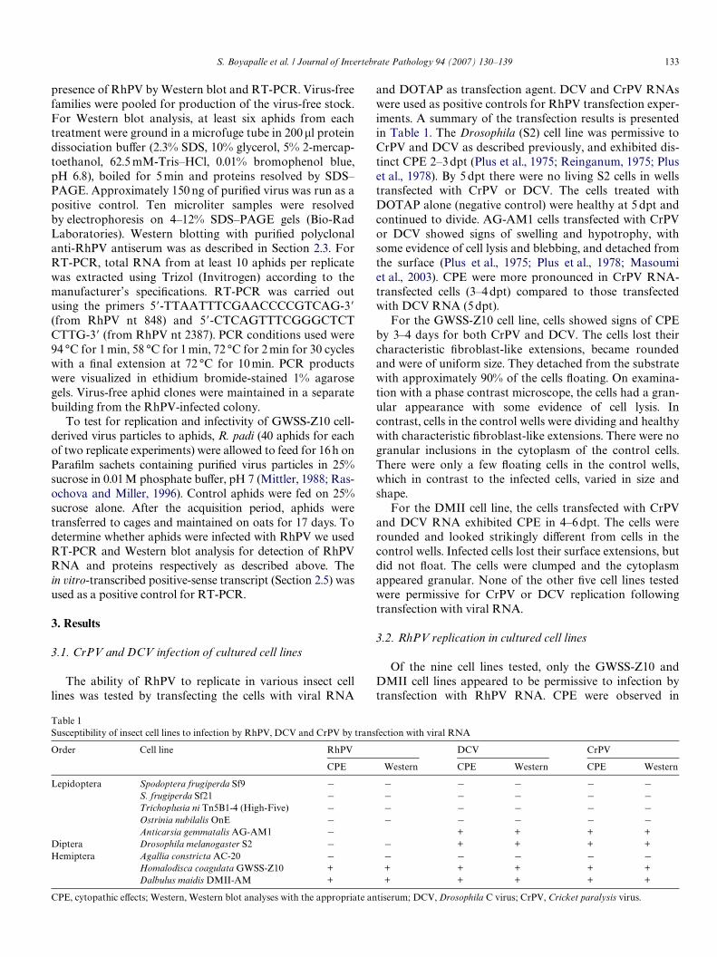
Table 1Susceptibility of insect cell lines to infection by RhPV, DCV and CrPV by transfection with viral RNA
CPE, cytopathic eVects; Western, Western blot analyses with the appropriate antiserum; DCV, Drosophila C virus; CrPV, Cricket paralysis virus.
Order Cell line RhPV DCV CrPV
CPE Western CPE Western CPE Western
Lepidoptera Spodoptera frugiperda Sf9 ¡ ¡ ¡ ¡ ¡ ¡S. frugiperda Sf21 ¡ ¡ ¡ ¡ ¡ ¡Trichoplusia ni Tn5B1-4 (High-Five) ¡ ¡ ¡ ¡ ¡ ¡Ostrinia nubilalis OnE ¡ ¡ ¡ ¡ ¡ ¡Anticarsia gemmatalis AG-AM1 ¡ + + + +
Diptera Drosophila melanogaster S2 ¡ ¡ + + + +Hemiptera Agallia constricta AC-20 ¡ ¡ ¡ ¡ ¡ ¡
Homalodisca coagulata GWSS-Z10 + + + + + +Dalbulus maidis DMII-AM + + + + + +

134 S. Boyapalle et al. / Journal of Invertebrate Pathology 94 (2007) 130–139
GWSS-Z10 cells at 4 dpt (Fig. 1B), while the control cellsappeared healthy, with Wbroblast-like extensions (Fig. 1A).DMII cells showed CPE 8–10 dpt that were similar to thosedescribed above (Fig. 1E). To determine whether CPEcould be caused by non-infectious viral RNA, GWSS-Z10cells were transfected with the in vitro synthesized full-length negative-sense RhPV RNA transcript. There was nodiVerence between these cells and control cells (treated withDOTAP only) at 4 dpt (Fig. 1A and C). This result showsthat the CPE observed resulted from infectious viral RNA.
3.3. Detection of coat proteins following viral RNA transfections
To determine whether transfection of cells with viral RNAresulted in synthesis of viral coat proteins, we performedimmunoblots using antiserum against puriWed virus prepara-tions of RhPV, DCV and CrPV (Table 1). RhPV has threemajor virion proteins of 30, 29 and 28kDa (Williamson et al.,1988). The fourth coat protein (VP4) has not been detected byWestern blot, although it can be detected in protein gels (Gil-dow and D’Arcy, 1990). The capsid proteins for DCV are37.7, 33.3 and 29.2kDa (Jousset et al., 1972), and for CrPV are35, 34 and 30kDa (Moore et al., 1985). Viral structural pro-teins were detected in all cell lines that exhibited CPE follow-ing transfection with viral RNA (Fig. 2; Table 1).
3.4. Synthesis of viral RNA following viral RNA transfection
We next examined viral RNA accumulation in GWSS-Z10 cells transfected with RhPV RNA. Northern blot
hybridization was used to detect accumulation of positiveand negative-sense genomic RNA molecules (Fig. 3A andC). Input positive-sense viral RNA and negative-sensein vitro transcript were detected at 12 and 24 hpt (Fig. 3Aand B). Blots revealed that substantial amounts of positive-sense viral RNA were present at least up to 96 hpt(Fig. 3A). While a striking increase is not apparent, the lev-els must be compared against the large amount of inoculum
Fig. 2. Detection of dicistrovirus coat proteins. (A) Sucrose gradient-puri-Wed virus particles were run on an SDS–polyacrylamide gel and coat pro-teins visualized by Coomassie blue staining. RhPV was puriWed from R.padi; DCV and CrPV were puriWed from D. melanogaster S2 cells. Molec-ular mass markers are indicated. (B) Western blot analysis of RhPVderived from R. padi and puriWed by sucrose gradient centrifugation (R.padi; positive control), of GWSS-Z10 cells (Z10; negative control), and ofGWSS-Z10 cells transfected with RhPV RNA (Z10-RhPV) at 4 dpt.
Fig. 1. Transfection of GWSS-Z10 cells (A–C) and DMII cells (D–E) with RhPV RNA. (A) GWSS-Z10 cells treated with DOTAP alone (negative con-trol). (B) GWSS-Z10 cells transfected with 10 �g of RhPV RNA showing CPE at 4 days post-transfection (dpt). Note the rounding of the cells, andreduction in Wbroblast-like extensions and cell number. Ninety percent of the cells detached from the Xask. (C) GWSS-Z10 cells transfected with 10 �g neg-ative-sense full length in vitro transcript of RhPV (negative control). CPE were not apparent 4 days post-transfection. The characteristic Wbroblast-likeextensions (arrows) were clearly visible in control treatments (A and C). (D) DMII cells treated with DOTAP alone (negative control). Note the Wbroblast-like extensions. (E) DMII cells transfected with 10 �g of RhPV RNA showing CPE at 8 days post-transfection. Infected cells (E) became rounded (arrows),were of uniform size, and detached from the Xask in clumps. Cell division was reduced.

S. Boyapalle et al. / Journal of Invertebrate Pathology 94 (2007) 130–139 135
present at early time points, much of which degraded by12 hpt (low molecular weight smear). For comparison, non-replicatable RNA (negative-sense inoculum) levelsdecreased rapidly after 24 h, (Fig. 3B).
To conWrm replication of the RhPV RNA, we tried todetect negative-strand RhPV RNA in cells inoculated withinfectious viral RNA. Negative-strand RNA was indeeddetected in cells inoculated with RhPV RNA as in Fig. 3A,beyond 96 hpt, (Fig. 3C). The negative-sense RNA was notdetected until 48 hpt. Between 48 and 96 hpt these negative-sense RNAs appeared to have reached peak levels. Bandsare not well deWned because the negative-strand accumu-lates at a many fold lower level than the corresponding pos-itive-sense species (Fig. 3C).
3.5. Detection of virus particles in infected cells
To determine whether virions could be observed in theinfected cells, GWSS-Z10 cells were transfected as described
above and the cells harvested at 4dpt, when CPE were pro-nounced, for puriWcation of virus particles (Krishna et al.,2003). PuriWed virus particles were negatively stained with 2%uranyl acetate and examined by TEM (Fig. 4B). RhPV parti-cles puriWed from aphids (Fig. 4A) were examined for com-parison. In both cases, the size of the particles was 26–27nmas observed by Rybicki (Rybicki and Wechmar, 1984). Theapparent size of virus particles puriWed from GWSS-Z10 cells4dpt with DCV RNA was 29–30nm as previously described(Jousset et al., 1972; Reinganum, 1975.
3.6. Immunolocalization of RhPV in GWSS-Z10 and DMII cells
To determine the intracellular localization of the virusparticles, GWSS-Z10 and DMII cells were transfected asdescribed above, harvested 4 dpt and sectioned for exami-nation by TEM. AYnity puriWed RhPV antiserum was usedfor immunolocalization of virus proteins. Infected cells
Fig. 3. Northern blot hybridization of RhPV RNA from transfected GWSS-Z10 cells. (A) Detection of positive-sense genomic RNA. Total RNAextracted at indicated hours after transfection of GWSS-Z10 cells with viral RNA. Hybridization was carried out using a 32P-labeled 3� end-speciWc, nega-tive-strand riboprobe. Controls are virion RNA extracted from virus particles spiked with total RNA from mock infected cell (+); mock infected GWSS-Z10 cells (¡). (B) Control treatments for detection of positive-sense genomic RNA. Total RNA from GWSS-Z10 cells transfected with full-length, in vitrotranscribed negative-strand RhPV RNA. Hybridization was carried out using a 32P-labeled 5� end-speciWc, positive-stranded riboprobe. Controls arein vitro synthesized full-length negative-sense RhPV6 RNA (¡), in vitro synthesized full-length positive-sense RhPV6 RNA (+; 100 ng). (C) Detection ofnegative-sense RhPV RNA in GWSS-Z10 cells. Total RNA was extracted at 12, 48, 96, 120 and 144 after transfection with virion RNA. Hybridization wascarried out with a 5� end positive-strand RNA-speciWc riboprobe. In vitro synthesized full-length negative-sense RhPV6 RNA (¡; 200 ng) was used as pos-itive control. Lower panels show ethidium bromide-stained ribosomal RNA that indicate total amount of RNA loaded.
Fig. 4. Virus particles derived from R. padi and GWSS-Z10 cells. (A) R. padi-derived virus particles of RhPV. Virus particles were puriWed from R. padi by10–40% sucrose gradient centrifugation and negatively stained with 2% uranyl acetate for analysis by TEM. (B) GWSS-Z10 cell-derived virus particles ofRhPV. Virus particles were puriWed from GWSS-Z10 cells 4 days post-transfection with RhPV RNA. The particles in A and B are 26–27 nm in diameter.(C) GWSS-Z10 cell-derived virus particles of DCV. Virus particles (27–29 nm) were puriWed from GWSS-Z10 cells 4 days post-transfection with DCVRNA. Virus particles were negatively stained and analyzed by TEM. Bars D 200 nm.

136 S. Boyapalle et al. / Journal of Invertebrate Pathology 94 (2007) 130–139
were compared to mock-infected GWSS-Z10 (Fig. 5A andB) and DMII cells (Fig. 5C and D). In the infected cells,large electron-dense amorphous aggregates (up to 2 �m indiameter) and oval-shaped inclusions were labeled by anti-RhPV antiserum in the cytoplasm (Fig. 5A and C). Thesecytoplasmic structures appeared to be enclosed by a mem-brane. Almost every DMII cell (8–10 dpt), had at least one
virus-induced cytoplasmic structure (Fig. 5C), and mostcells had numerous structures.
Moreover, viral antigen was concentrated in electron-dense patches in the cytoplasm, probably consisting ofsmall vesicles (Fig. 5F, arrow). Mock infected GWSS-Z10or DMII cells did not exhibit any of these cytoplasmicinclusions or labeling (Fig. 5B and D).
Fig. 5. Immunogold labeling of RhPV proteins in GWSS-Z10 and DMII cells. (A) GWSS-Z10 cells 4 dpt with 10 �g RhPV RNA. (B) Mock-infectedGWSS-Z10 cells. (C) DMII cells, 8 dpt with 10 �g RhPV RNA. (D) Mock-infected DMII cells. Note the oval, electron dense inclusion bodies in the cyto-plasm in A and C. (E) and (F) detail of infected GWSS-Z10 cells 4 dpt showing labeling of cell membrane (E) and electron dense regions in the cytoplasm(F). Size bars, 200 nm.

S. Boyapalle et al. / Journal of Invertebrate Pathology 94 (2007) 130–139 137
3.7. Infectivity of GWSS-Z10 cell-derived virus particles to GWSS-Z10 cells and aphids
Fig. 6B shows infection of GWSS-Z10 cells followingtreatment with supernatant harvested at 6 dpt from cellstransfected with RhPV RNA. Characteristic CPE wereobserved, with rounding of the cells and detachment fromthe surface of the Xask. Importantly, supernatant fromtransfected GWSS-Z10 cells contained infectious virus at6 dpt.
To test for production of infectious RhPV in GWSS-Z10cells, aphids were fed on virus particles puriWed from RhPVRNA-transfected GWSS-Z10 cells. Aphids were then main-tained on oats for 17 days, during which time their numbersincreased at least 5-fold. RT-PCR and Western blot analy-sis of aphids in two separate replicate experiments, showedthe presence of RhPV RNA and immunoreactive structuralproteins (Fig. 7).
4. Discussion
We have identiWed one insect cell line that supports rep-lication of RhPV, and a second cell line that is likely to sup-port replication of RhPV. We also identiWed new cell linescapable of being infected by CrPV and DCV. AlthoughRhPV, CrPV and DCV are in the same family, cellsresponded diVerently to infection. CrPV and DCV causedCPE in GWSS-Z10 and DMII cells in almost half the timerequired for RhPV to do so. Extensive cell lysis wasobserved with CrPV and DCV infection while RhPVinfected cells became rounded and detached from the sur-face with limited cell lysis at 4 dpt. Twice the amount ofRhPV RNA inoculum was required to elicit CPE compara-ble to those of DCV and CrPV infection. Scotti et al.observed that DL1 (D. melanogaster) cells appeared to sup-port growth of DCV but without the distinctive CPEobserved for CrPV infections, although the infected cells
Fig. 6. Reinfection of GWSS-Z10 cells with supernatant from RhPV RNA transfected GWSS-Z10 cells. (A) Mock-infected GWSS-Z10 cells. (B) Reinfec-tion of GWSS-Z10 monolayer with the supernatant from the GWSS-Z10 cells 6 days after transfection with RhPV RNA. CPE were observed 6–7 dayspost-infection with rounding of the cells (arrows), loss of Wbroblast-like extensions and detachment of the cells from the Xask.
Fig. 7. Detection of RhPV in aphids fed on GWSS-Z10-derived virus particles. (A) Detection of positive-strand RhPV RNA. RT-PCR of in vitro-tran-scribed positive-sense transcript was used as a positive control (+; 1539 nt product). RT-PCR of RNA extracted from aphids fed on sucrose solution (¡;negative control) or GWSS-Z10 cell-derived virus particles (two replicates; 1, 2). L, 1 kb ladder. (B) Detection of RhPV structural proteins by Western blotin infected and RhPV-free aphid colonies. RhPV virions (W, 0.1 �g); RhPV-positive R. padi colony (+); virus-free R. padi colony (¡); Acyrthosiphon pisumnegative control. Protein extracts from six aphids were examined by Western blot for each colony. (C) Western blot analysis of aphids fed on GWSS-Z10cell-derived virus particles. RhPV structural proteins were detected in all four replicates (lanes 1–4; six aphids per lane). Control aphids were fed onsucrose alone (¡, negative control).
BA
C

138 S. Boyapalle et al. / Journal of Invertebrate Pathology 94 (2007) 130–139
did clump and detached from the surface of the cultureXask several days after inoculation (Scotti et al., 1981).
Western blot analysis (Table 1 and Fig. 2) revealed thatviral RNA transfected into GWSS-Z10 and DMII cells wastranslated and that the ORF2 polyprotein was eYcientlyprocessed into structural proteins. These data demonstratethat the two IRES (5� IRES and IGR-IRES) of RhPV areactive in these cell lines. This result is not surprising giventhat the 5� IRES functions in wheat and HeLa cell lines.Detection of structural proteins is not in itself indicative ofa cell line being permissive for virus replication however.Masoumi et al. (2003) observed that CrPV was unable toinfect and/or replicate in either T. ni or A. aegypti cells eventhough both IRES elements were functional in these celllines. These Wndings were in general agreement with theWndings of Shaw-Jackson and Michiels (1999) for Theiler’svirus, that IRES elements may be active in cell types thatare not derived from natural hosts of the virus.
While there was no striking increase in the amount ofviral RNA detected in GWSS-Z10 cells (Fig. 3A), consider-ation of the ratio of Northern blot signal to the amountloaded on the gel and comparison with cells transfectedwith the non-replicatable negative-sense RNA (Fig. 3B)suggests that viral RNA replication occurred in these cells.The large amounts of RNA detected at early time pointsrepresent inoculum. It is unlikely that inoculum RNAwould not decrease by 48 or 72 hpt. The low molecularweight smear at 12 h shows degradation of the inoculumRNA. Thus, the RNA at the later time points is likely toresult from replication. Consistent with this, low amountsof negative strand were detected (Fig. 3C). The apparentdetection of negative-strand RNA suggests that RhPV rep-licates in GWSS-Z10 cells. The virus particles puriWed fromthe infected cells were morphologically indistinguishablefrom the virus particles puriWed from aphids. These resultsindicate that cellular factors required for the processing ofviral gene products and assembly of virus particles wereavailable in the GWSS-Z10 cell line.
RhPV immunoreactive proteins appear to localize in thecytoplasm of the infected cell (Fig. 5A and C). In host spec-iWcity studies of RhPV in aphids, RhPV particles occurredfree in the cytoplasm and also packed in crystalline arraysin large membrane vesicles (Gildow and D’Arcy, 1990).Virions were never observed in the nucleus, and the nuclearenvelope and mitochondria of the gut cells remained intactfollowing virus replication and development of severe cyto-pathic symptoms. The authors also observed that clustersof single membrane vesicles were associated with the viri-ons during early stages of infection. Occasionally, a clumpof label was observed associated with the cell membrane(Fig. 5E), which may indicate a site of viral endocytosis orrelease.
DCV and CrPV virions are also found in crystallinearrays in the cytoplasm of infected cells, as for vertebratepicornaviruses (Reinganum et al., 1970; Jousset et al., 1972;Jousset and Plus, 1975. Our EM observations (Fig. 5A andC) also revealed formation of electron dense amorphous
cytoplasmic structures that were strongly labeled by anti-RhPV antibodies. These observations are consistent withproduction of similar structures by other viruses: Similarstructures were observed within the cytoplasm of the gutcells of the pea aphid infected with A. pisum virus (van denHeuvel et al., 1997) and abundant cytoplasmic structuresloaded with virions were seen when a midgut cell linederived from Helicoverpa zea was infected with Providencevirus (family Tetraviridae) (Pringle et al., 2003).
The apparent demonstration that RhPV virions pro-duced in GWSS-Z10 cells following transfection withRhPV RNA, are infectious to GWSS-Z10 cells (Fig. 6) sug-gests that RNA was packaged within virus particles andthat any receptor required for infection by virus particles ispresent in these cells. Western blot and PCR analyses arerequired to conWrm this result. However, infection of virus-free aphids with the GWSS-Z10 cell-derived virionsconWrms that infectious virus is produced following trans-fection of GWSS-Z10 cells with RhPV RNA and that thisvirus was transmitted between aphids. The infectivity ofGWSS-Z10 cell-derived virus particles to aphids conWrmsthat RhPV does indeed replicate in GWSS-Z10 cells.
We have shown that one hemipteran cell line (GWSS-Z10) is permissive to RhPV infection, and that a second cellline (DMII) is likely to be permissive to RhPV infection. TheGWSS-Z10 cells allow for translation and appropriate pro-cessing of viral gene products, RNA replication and packag-ing of virus particles, thereby producing infectious RhPV.The GWSS-Z10 cell line provides a valuable tool to investi-gate the replication, assembly and structure of RhPV.
Acknowledgments
The authors thank Dr. Art McIntosh, USDA-ARS,Columbia, MO, for providing the DMII, AC-20 and AG-AM1 cell lines, and Dr. Shizuo G. Kamita, University ofCalifornia, Davis for providing GWSS-Z10 cells. Theauthors thank Dr. Peter Christian, National Institute forBiological Standards and Control, UK for providing CrPV,DCV and the respective antisera, and Randy Beckett forassistance with constructing the full-length clone of theRhPV genome. This material is based upon work supportedby an Iowa State University Carver Trust grant as well asHatch Act and State of Iowa funds.
References
Bassham, D.C., Raikhel, N.V., 1998. An Arabidopsis VPS45p homolog impli-cated in protein transport to the vacuole. Plant Physiol. 117, 407–415.
Christian, P.D., Scotti, P., 1994. A suggested taxonomy and nomenclaturefor the cricket paralysis and Drosophila C virus complex. J. Invertebr.Pathol. 63, 157–162.
Christian, P.D., Scotti, P.D., 1998. Picornalike viruses of insects. In: L.K.Miller, L.A. Ball (Eds.), The Insect Viruses. Plenum Press, New York,pp. 301–336.
D’Arcy, C.J., Burnett, P.A., Hewings, A.D., 1981. Detection, biologicaleVects and transmission of a virus of the aphid Rhopalosiphum padi.Virology 114, 268–272.

S. Boyapalle et al. / Journal of Invertebrate Pathology 94 (2007) 130–139 139
Domier, L.L., McCoppin, N.K., D’Arcy, C.J., 2000. Sequence requirementsfor translation initiation of Rhopalosiphum padi virus ORF2. Virology268, 264–271.
Erickson, P.A., Lewis, G.P., Fisher, S.K., 1993. Postembedding immunocy-tochemical techniques for light and electron microscopy. Methods CellBiol. 37, 283–310.
Fauquet, C.M., Mayo, M.A., ManiloV, J., Desselberger, U., Ball, L.A.,2005. Proceedings of the 8th Report of the International Committee onTaxonomy of Viruses. Academic Press, New York.
Gildow, F.E., D’Arcy, C.J., 1988. Barley and oats as reservoirs for an aphidvirus and the inXuence on barley yellow dwarf virus transmission. Phy-topathology 78 (6), 811–816.
Gildow, F.E., D’Arcy, C.J., 1990. Cytopathology and experimental hostrange of Rhopalosiphum padi virus, a small isometric RNA virus infect-ing cereal grain aphids. J. Invertebr. Pathol. 55, 245–257.
Hirumi, H., Maramorosch, K., 1971. Cell culture of Hemiptera. Invertebr.Tissue Cult. 1, 307–339.
Johnson, K.N., Christian, P.D., 1998. The novel genome organization ofthe insect picorna-like virus Drosophila C virus suggests this virusbelongs to a previously undescribed virus family. J. Gen. Virol. 79, 1.
Jousset, F.-X., Plus, N., 1975. Etude de la transmission verticale des picor-naviruses de Drosophila melanogaster et de Drosophila immigrans.Annu. Microbiol. B (Paris) 126, 231–249.
Jousset, F.X., Plus, N., Croizier, G., Thomas, M., 1972. Existence chez Dro-sophila de deux groupes de picornaviruaea de propietes serologiques etbiologiques diVerentes. C. R. Acad. Sci. (Paris) 275, 3043–3046.
Kamita, S.G., Do, Z., Samra, A., Hagler, J., Hammock, B.D., 2005. Charac-terization of cell lines developed from the glassy-winged sharpshooter,Homalodisca coagulata (Hemiptera: Cicadellidae). In Vitro Cell. Dev.Biol.-Animal 41 (5–6), 149–153.
Koev, G., Mohan, B.R., Miller, W.A., 1999. Primary and secondary struc-tural elements required for synthesis of barley yellow dwarf virus sub-genomic RNA1. J. Virol. 73 (4), 2885–2896.
Krishna, N.K., Marshall, D., Scheemann, A., 2003. Analysis of RNA pack-aging in wild-type and mosaic protein capsids of Xock house virususing recombinant baculovirus vectors. Virology 305 (1), 10–24.
Masoumi, A., Hanzlik, T.N., Christian, P., 2003. Functionality of the 5�-and intergenic IRES elements of cricket paralysis virus in a range ofinsect cell lines, and its relationship with viral activities. Virus Res. 94(2), 113–120.
McIntosh, A.H., IgonoV, C.M., 1989. Replication of Autographa califor-nica nuclear polyhedrosis virus in Wve lepidopteran cell lines. J. Inver-tebr. Pathol. 54, 97–102.
McIntosh, A.H., Maramorosch, K., Rechtoris, C., 1973. Adaptation of aninsect cell line (Agallia constricta) in a mammalian cell culture medium.In Vitro 8 (5), 375–378.
Mittler, T.E., 1988. Applications of artiWcial feeding techniques for aphids.In: A.K. Minks, P. Harrewijn (Eds.), Aphids, Their Biology, NaturalEnemies and Control. Elsevier, Amsterdam, pp. 145–170.
Moore, N.F., Reavy, B., King, L.A., 1985. General characteristics, geneorganization and expression of small RNA viruses in insects. J. Gen.Virol. 66, 647–654.
Peters, D., Black, L.M., 1970. Infection of primary cultures of aphid cellswith a plant virus. Virology 40, 847–853.
Plus, N., Croizier, G., Jousset, F.X., David, J., 1975. Picornaviruses oflaboratory and wild Drosophila melanogaster: Geographical distribu-tion and serotypic composition. Ann. Microbiol. A (Inst. Pasteur)126, 107–117.
Plus, N., Croizier, G., Reinganum, C., Scotti, P.D., 1978. Cricket paralysisvirus and Drosophila C virus: Serological analysis and comparison ofcapsid polypeptides and host range. J. Invertebr. Pathol. 31, 296–302.
Pringle, F.M., Johnson, k.N., Goodman, C.L., McIntosh, A.H., Ball, L.A.,2003. Providence virus: a new member of the Tetraviridae that infectscultured insect cells. Virology 306 (2), 359–370.
Rasochova, L., Miller, W.A., 1996. Satellite RNA of barley yellow dwarf-RPV virus reduces accumulation of RPV helper virus RNA and atten-uates RPV symptoms in oats. Mol. Plant-Microbe Interact. 9, 646–650.
Reinganum, C., 1975. The isolation of cricket paralysis virus from theemperor gum moth, Antheraea eucalypti Scott, and its infectivitytowards a range of insect species. Intervirology 5 (1–2), 97–102.
Reinganum, C., O’Loughlin, G.T., Hogan, T.W., 1970. A non-occludedvirus of the Weld crickets Teleogryllus aceanicus and T. commodus(Orthoptera: Gryllidae). J. Invertebr. Pathol. 16, 220–314.
Royall, E., Woolaway, K.E., Schacherl, J., Kubick, S., Belsham, G.J., Rob-erts, L.O., 2004. The Rhopalosiphum padi virus 5� internal ribosomeentry site is functional in Spodoptera frugiperda 21 cells and in theircell-free lysates: implications for the baculovirus expression system. J.Gen. Virol. 85 (6), 1565–1569.
Rybicki, E.P., von Wechmar, M.B., 1984. Serological, biophysical andbiochemical investigations of aphid-transmitted viruses of smallgrains. Progress in Russian wheat aphid, Diuraphis noxia Mordw.research in the Republic of South Africa. M.C. Walters, Departmentof Agriculture, Republic of South Africa, Technical Communication191. pp. 42–43.
Schneider, I., 1972. Cell lines derived from late embryonic stages of Dro-sophila melanogaster. J. Embryol. Exp. Morph. 27, 353–365.
Scotti, P.D., Hoefakker, P., Dearing, S., 1996. The production of cricketparalysis virus in suspension cultures of insect cell lines. J. Invertebr.Pathol. 68, 109–112.
Scotti, P.D., Longworth, J.L., Plus, N., Crozier, G., Reinganum, C., 1981.The biology and ecology of strains of an insect small RNA virus com-plex. Adv. Virus Res. 26, 117–143.
Shaw-Jackson, C., Michiels, T., 1999. Absence of internal ribosome entrysite-mediated tissue speciWcity in the translation of bicistronic trans-genes. J. Virol. 73, 2729–2738.
van den Heuvel, J.F., Hummelen, H., Verbeek, M., Dullemans, A.M., van derWilk, F., 1997. Characteristics of Acyrthosipon pisum virus, a newly iden-tiWed virus infecting the pea aphid. J. Invertebr. Pathol. 70, 169–176.
Vaughn, J.L., Goodwin, R.H., Tompkins, G.J., McCawley, P., 1977. Theestablishment of two cell lines from the insect Spodoptera frugiperda(Lepidoptera: Noctuidae). In Vitro 13, 213–217.
Wechmar, M.B.V., Rybicki, E.P., 1981. Aphid transmission of three virusescauses Free State streak disease. S. Afr. J. Sci. 77, 488–492.
Wickham, T.J., Davis, T., Granados, R.R., Shuler, M.L., Wood, H.A., 1992.Screening of insect cell lines for the production of recombinant pro-teins and infectious virus in the baculovirus system. Biotechnol. Prog.8, 391–396.
Williamson, C. von Wechmar, M.B., Rybicki, E.P., 1989. Further charac-terization of Rhopalosiphum padi virus of aphids and comparison ofisolates from South Africa and Illinois. J. Invertebr. Pathol. 54, 85–96.
Williamson, G., Rybicki, E.P., Kasdorf, G.G.F., von Wechmar, M.B., 1988.Characterization of a new picorna-like virus isolated from aphids. J.Gen. Virol. 69, 787–795.
Wilson, J.E., Powell, M.J., Hoover, S.E., Sarnow, P., 2000. Naturally occur-ring dicistronic cricket paralysis virus RNA is regulated by two inter-nal ribosome entry sites. Mol. Cell. Biol. 20 (14), 4990–4999.