a functioning chimera of the cyclic nucleotide-binding domain from the bovine retinal rod ion...
TRANSCRIPT
A Functioning Chimera of the Cyclic Nucleotide-Binding Domain from the BovineRetinal Rod Ion Channel and the DNA-Binding Domain from Catabolite
Gene-Activating Protein†
Sean-Patrick Scott,‡ Irene T. Weber,‡ Robert W. Harrison,§ Jannette Carey,| and Jacqueline C. Tanaka*,⊥
Departments of Biology, Chemistry, and Computer Science, Georgia State UniVersity, Atlanta, Georgia 30303, Department ofChemistry, Princeton UniVersity, Princeton, New Jersey 08544, and Department of Biology, School of Science and Technology,
Temple UniVersity, Philadelphia, PennsylVania 19122
ReceiVed December 11, 2000; ReVised Manuscript ReceiVed May 3, 2001
ABSTRACT: The eukaryotic cyclic nucleotide-gated (CNG) ion channels are a family of large membraneproteins activated by cytoplasmic cGMP or cAMP. Their cyclic nucleotide-binding domain is structurallyhomologous with that of the catabolite gene-activator protein (CAP), a solubleEscherichia colitranscriptionfactor. Differences in ligand activation among sensory channels suggest differences in the underlyingmolecular mechanisms of signal readout. To study the structural, functional, and conformationalconsequences of nucleotide binding, we fused the cyclic nucleotide-binding domain from the bovine retinalrod CNG channelR subunit (BrR) to the DNA-binding domain from CAP. The chimera forms a solubledimer that binds both cGMP and cAMP with association constants of 3.7× 10⁴ M-1 for [3H]cGMP and3.1 × 104 M-1 for [3H]cAMP. The binding of cAMP, but not cGMP, exposes a chymotrypsin cleavagesite in the chimera at a position similar to the site in the CAP exposed by cAMP binding. At high cAMPconcentrations, a biphasic pattern of cleavage is seen, suggesting that the low-affinity cAMP bindingsites are also occupied. Cyclic AMP promotes specific binding to a DNA fragment encoding the lacoperator region; theKd for the protein-DNA binding is∼200 nM, which is 2-fold higher than theKd forCAP under identical conditions. A 7 Å crystal structure shows that the overall secondary and tertiarystructure of BrR/CAP is the same as that of CAP with two cAMP molecules bound per dimer. Thebiochemical characterization of the chimera suggests it will be a useful system for testing hypothesesabout channel activation, providing further insight into channel function.
Cyclic nucleotide-gated (CNG)1 ion channels were firstidentified in the primary sensory neurons of the visual (1)and olfactory systems (2), where they are an integral part ofthe cascade transducing external stimuli into voltage signals.The phototransduction cascade results in light-induceddecreases in cGMP levels in the photoreceptor outer seg-ments (3), while cAMP is likely the signal molecule inolfactory cells (4, 5). CNG channels are also expressed in anumber of nonsensory cells, including retinal cone terminals(6) and ganglion cells (7, 8), cardiac pacemaker cells (9),sperm (10), and hippocampus (11, 12). The physiological
roles of the channels in nonsensory cells, as well as theirnucleotide signaling cascades, have yet to be delineated.
Within the CNG channel family, channels respond dif-ferently to cGMP and cAMP, which differ only at the C2
and C6 positions on the purine ring. Typically, ionic currentsare measured from inside-out, excised, patches to determinethe nucleotide concentration required for activation of 50%of the current, theK0.5, as well as the fraction of currentactivated at saturation for those nucleotides that are partialagonists. Homotetrameric CNG channels expressed fromcDNA encoding the bovine retinal rodR (BrR) subunit arefully activated by cGMP (13), while cAMP is a partialagonist (14). The olfactory CNG channels, on the other hand,are fully activated by both cGMP and cAMP (2, 15). Sincethe K0.5 values reflect both nucleotide binding and channelgating, it has been difficult to address the molecular basisof nucleotide discrimination in CNG channels. Attempts tomeasure the direct nucleotide binding to CNG channels havebeen hampered by the relatively low-affinity binding interac-tion and the relatively low abundance of the channelprotein.
In previous studies, the activation properties of a seriesof nucleotide analogues were compared (16, 17) and three-dimensional models of the cyclic nucleotide-binding domainwere constructed using the coordinates of the cAMP binding
† This work was supported by NIH Grants EY-06640 (J.C.T.) andGM19736 (NRSA to S.-P.S.).
* To whom correspondence should be addressed: Department ofBiology, School of Science and Technology, Temple University,Philadelphia, PA 19122. Phone: (215) 204-8868. Fax: (215) 204-6646.E-mail: [email protected].
‡ Departments of Biology and Chemistry, Georgia State University.§ Department of Computer Science, Georgia State University.| Princeton University.⊥ Temple University.1 Abbreviations: CNG, cyclic nucleotide-gated; cAMP, 3′,5′-cyclic
adenosine monophosphate; cGMP, 3′,5′-cyclic guanosine monophos-phate; CRP, cAMP receptor protein; CAP, catabolite gene-activatorprotein; BrR, bovine rodR subunit CNG channel; BrR/CAP, chimerawith the bovine rodR CNG channel binding domain (residues 489-604) replacing residues 14-127 of CAP, a S128A mutation, and ahistidine tag; SEM, standard error of the mean.
7464 Biochemistry2001,40, 7464-7473
10.1021/bi002804x CCC: $20.00 © 2001 American Chemical SocietyPublished on Web 05/30/2001

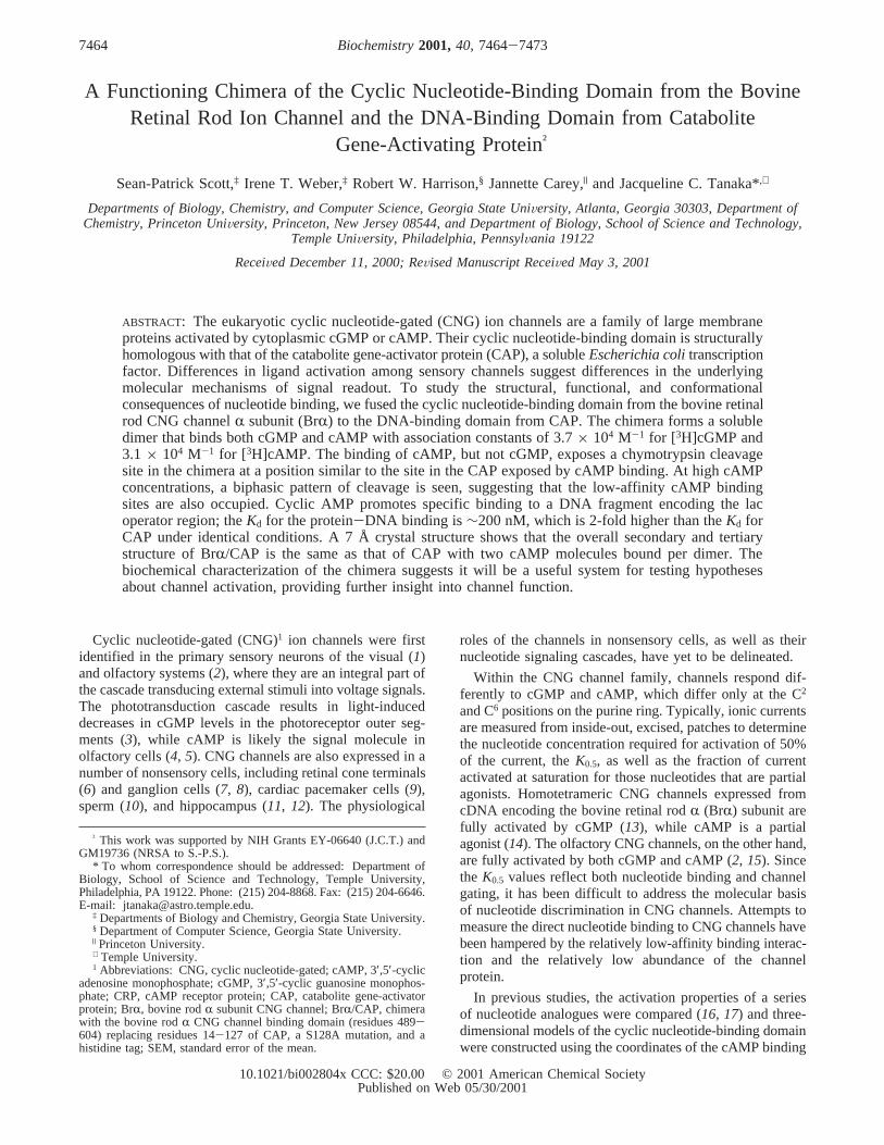
domain of the cAMP receptor protein (CAP, also called thecAMP receptor protein or CRP) (18, 19). CAP is a soluble,dimeric, Escherichia colitranscription factor activated bythe binding of cAMP (20, 21). CAP binds cGMP nearly aswell as cAMP, but cGMP does not promote transcription(22). Each subunit of 209 residues (23-25) contains anN-terminal cyclic nucleotide-binding domain and a C-terminal DNA-binding domain (Figure 1B) (26). The eight-stranded â barrel of the nucleotide-binding domain isfollowed by helices B and C, a hallmark of this family ofcyclic nucleotide-binding domains. Three conserved glycines(33, 45, and 71), shown in Figure 1C, are important formaintaining the fold of the barrel (27). The conserved E72and R82 interact with the ribofuranose of the cyclic nucle-otide (28) and are important for binding of the ligand (29).
The goal of the work presented here was to express asoluble protein containing the cyclic nucleotide-bindingdomain of the BrR CNG channel in order to study its ligandactivation and signaling properties. We sought an approachthat would be capable of providing information about howligand identity is recognized by the binding domain of theprotein and transmitted to a different domain or neighboringsubunit. The chimera, BrR/CAP, was constructed from thecyclic nucleotide-binding domain of the BrR CNG channeland the DNA-binding domain of CAP. Our approach wasdesigned to take advantage of the native CAP solubility tominimize the problems of protein folding. The chimericprotein was overproduced by induction inE. coli and purifiedusing a histidine tag. No refolding was required. BrR/CAPis a dimer, binds both cAMP and cGMP, and binds DNAspecifically at the lac operator in the presence of cAMP. Atlow resolution, the crystal structure is essentially the sameas that of CAP in its overall structure. The chimera undergoesa conformational change exposing a chymotrypsin site uponbinding of cAMP but not cGMP. The results demonstratethe utility of this model system for understanding the readoutof the ligand signal from the cyclic nucleotide-bindingdomain of the CNG channel.
MATERIALS AND METHODS
Mutation and PCR Protocols.The cDNAs for CAP andthe BrR CNG channel were subcloned into the pAlter-1vector (Promega) for single-stranded DNA mutagenesis,according to the protocol of Promega. Single-strandedmutagenesis was used to generate aSalI restriction site atthe N-terminus of both CAP and the BrR CNG channel cyclicnucleotide-binding domain to swap the domains. Single-stranded mutagenesis was also used to add anNdeI restrictionsite at the start site of the CAP cDNA. The PCR methods ofgene splicing by overlap extension (30) were used to addthe DNA-binding domain of CAP to the cyclic nucleotide-binding domain of BrR and mutate Ser 128 to Ala. Theconstructs were sequenced after cloning.
The first round of PCR used the cDNA in pAlter-1plasmids, a 5′ probe for the BrR cDNA of 5′-AGC-AAT-ACA-TGC-ATT-TTC-GAA-ATG-TAA-GCA-AAG-A-3 ′, anda 3′ CAP probe of 5′-GGT-GAT-AAA-TCA-GTC-TGC-G-3′. The internal probes were 5′-CTT-TCT-CTG-CAT-CTT-TCA-TCA-AAA-TTT-GCT-TCC-3′ for BrR and 5′-TGA-TGA-AAG-ATG-CAG-AGA-AAG-TGG-GCA-ACC-3′ forCAP. The products of the first round were combined with
the 5′ BrR probe and the 3′ CAP probe for a second roundof PCR. This product was restricted withSalI and BssHIIenzymes and the product placed into pAlter-1 with CAP/NdeI/SalI similarly restricted. After sequencing had beencarried out, theNdeI/EcoRI fragment of the pAlter-1 CAP/NdeI/SalI/PCR fragment was placed into theNdeI/EcoRIrestriction sites of the pET 28a vector for expression.
Protein Expression and Purification.Constructs of BrR/CAP and CAP containing His tags were expressed in theIPTG-inducible vector pET 28a in the BL21(DE3) Lys Scells at 37°C from Novagen. Four liters of LB medium wasinoculated with bacteria, and the bacteria were allowed togrow to an OD600 of ∼0.4. At this point, the temperaturewas decreased to 25°C. At an OD600 of ∼0.6, 0.24 g/L IPTGwas added. The cells were allowed to grow for 5 h at 25°Cbefore harvesting. Cells were spun at 6000 rpm for 15 minin a Sorvall RC-5b refrigerated superspeed centrifuge usinga GSA rotor. The pellet was resuspended in binding buffer[20 mM Tris (pH 7.9), 0.5 M NaCl, and 5 mM imidazole]and then sonicated for 1 min on and 1 min off six times ata 30% duty cycle with a power setting of 3 on a Bransonmodel 250 sonifier. The sonicate was centrifuged at 14 000rpm for 30 min in an SS34 rotor in a Sorvall centrifuge.The resultant supernatant was centrifuged at 35 000 rpm for90 min in a Ti 60 rotor in a Beckman L8-M ultracentrifuge.For the expression of CAP without a His tag, we followedthe previously established procedures (29).
The purification of His-tagged BrR/CAP and CAP wasaccomplished using a Ni2+ agarose column from Novagen.After the column was charged and prewashed, the proteinwas batch loaded onto the column. Following loading, thecolumn was washed with a volume of binding buffer 10 timesgreater than the bed volume followed by a volume of washbuffer [20 mM Tris (pH 7.9), 0.5 M NaCl, and 60 mMimidazole] 8 times greater than the bed volume. The proteinwas released from the column in a step with elution buffer[20 mM Tris (pH 7.9), 0.5 M NaCl, and 1 M imidazole].Protein purification was followed by SDS-PAGE anddetermination ofA280. The fractions containing the purifiedprotein were combined and dialyzed into one of the followingbuffers and stored at 4°C: storage buffer [20 mM Tris (pH7.8), 500 mM NaCl, 0.1 mM EDTA, 0.1 mM DTT, and 50%glycerol], X-ray buffer [50 mM sodium phosphate (pH 8.0),500 mM NaCl, and 1 mM EDTA], or functional buffer [50mM Tris (pH 8.0), 500 mM NaCl, and 1 mM EDTA].Dialysis was performed using a volume that was at least 300times greater than the sample volume in Pierce Slide-A-Lyzerdialysis cassettes, with a MW cutoff of 10 000. The externalbuffer was changed at least twice: the first time after 1 hand the second after 4 h. Dialysis was stopped within 16 h.Amicon 10 000 MW cutoff concentrators were used toconcentrate the sample. All dialysis and concentration of theprotein was carried out at 4°C. The protein concentrationwas determined using Bio-Rad protein assay reagent andBSA as a standard. The purification of CAP without a Histag was performed using two steps, following previouslypublished methods (31). The first step was ion exchangechromatography using DEAE followed by a BioRex 70column. The protein was immediately dialyzed into one ofthe buffers described above.
Cyclic Nucleotide-Binding Assays.Equilibrium bindingassays for [3H]cGMP and [3H]cAMP were carried out at
Functional CAP Chimera with Rod CNG Channel Binding Domain Biochemistry, Vol. 40, No. 25, 20017465
room temperature (20-22 °C) using small Amicon filtrationdevices. Typically, varying amounts of protein were mixed
with 3H-labeled cyclic nucleotides in a total volume of 200µL containing 50 mM Tris (pH 7.8), 100 mM NaCl, and 1
A B
C
FIGURE 1: (A) Schematic of the BrR/CAP chimera. (Top) The BrR CNG channel consists of cytoplasmic N-terminal and C-terminalregions separated by six transmembrane helices. The pore region is located between transmembrane helices 5 and 6. The C-linker regionlies between the final transmembrane segment and the cyclic nucleotide-binding domain (b). (Middle) CAP has an N-terminal cyclicnucleotide-binding domain (O) and a C-terminal DNA-binding domain (0). (Bottom) BrR/CAP protein contains N-terminal residues 1-10of CAP followed by the BrR CNG channel cyclic nucleotide-binding domain and the DNA-binding domain of CAP. (B) Structure of theCAP monomer. The model was obtained from PDB entry 3GAP. The eight-strandedâ barrel cyclic nucleotide-binding domain of CAP(light gray ribbon) is shown at the bottom in this orientation; helix C is labeled. Cyclic AMP is shown as a skeletal model. The DNA-binding domain (dark gray ribbon) lies at the top of this orientation with the protease cleavage site and the S128A mutation indicated byarrows. This picture was generated using RasMol. (C) Sequence of BrR/CAP and CAP. The numbers on the top denote the numbering ofBrR/CAP with the histidine tag included. The numbers below the sequence denote CAP residues. The sequence alignment of the BrRportion of BrR/CAP with CAP was described previously (18). Identical residues in both CAP and BrR/CAP are boxed. The secondarystructure, determined from the CAP-cAMP crystal structure, is shown below the sequences (26). A dash denotes the lack of a correspondingamino acid. Amino acids preceding and following the BrR binding domain that do not correspond to either the CAP or CNG channelbinding domain are denoted with an asterisk. The slant at position 137 denotes the chymotrypsin cleavage site.
7466 Biochemistry, Vol. 40, No. 25, 2001 Scott et al.
mM EDTA and allowed to equilibrate for 30 min. Equilibra-tion times were initially based on assays with CAP using 5min to equilibrate (32). Samples were placed into an Amiconcentrifugal filter device, with a MW cutoff of 10 000. After30 min, the sample was spun at 14 000 rpm in a microcen-trifuge for 35 s. This allowed approximately 20µL (10%)of the volume to pass through. Triplicate samples of 5µLwere taken from the top (bound and free ligand) and bottom(free ligand) after centrifugation. Concentration-responsedata were fitted with a nonlinear algorithm in Tablecurve 4(Jandel). The binding data shown in Figure 3 were fit witheq 1
where BrR/CAP is the concentration of the free chimericprotein, cNMP is the concentration of the free cyclicnucleotide, andKA is the equilibrium association constantfor ligand binding, according to Scheme 1, which assumesone binding site for each monomer of BrR/CAP.
Analytical Ultracentrifugation Experiments.Measurementswere taken using a BeckmanXL-I analytical ultracentrifuge.Protein samples were dissolved at approximately 40µM(monomer) in buffer containing 50 mM Tris, 500 mM NaCl,and 1 mM EDTA (pH 7.8). Sedimentation equilibriumexperiments were carried out at 48 000 rpm in six-channel,carbon-epoxy composite centerpieces supplied by Beckman.Equilibrium was assessed by the absence of a significantchange in radial concentration gradients in scans separatedby a few hours. Data were analyzed by curve fitting to theequation for a single ideal species using Igor-ProÆ (Wave-
metrics, Lake Oswego, OR) programs developed from aprevious version (33). Partial specific volumes and solventdensities were calculated using the program Sedinterp (34).We estimate an uncertainty of(4% in the calculatedmolecular weight of the protein (35). This arises largely fromuncertainty in the partial specific volume that is calculatedfrom a weight average of individual amino acids. This degreeof accuracy is sufficient to distinguish monomers fromaggregates. An alternative model fixed the molecular weightto the value calculated from its sequence and fitted the datato a monomer-dimer equilibrium model.
Chymotrypsin Proteolytic Assays.The proteolytic assayswere performed in the absence of ligand or in the presenceof cAMP, cGMP, or other cyclic nucleotides. All assays wereperformed, following previously described procedures (36,37), at 37°C for 30 min in a total volume of 10µL of thefollowing buffer: 50 mM Tris (pH 7.8), 100 mM NaCl, and1 mM EDTA. The protein concentration ranged from 0.2 to0.35 mg/mL, and the chymotrypsin concentration was 12µg/mL. The proteolysis was stopped by adding the gel
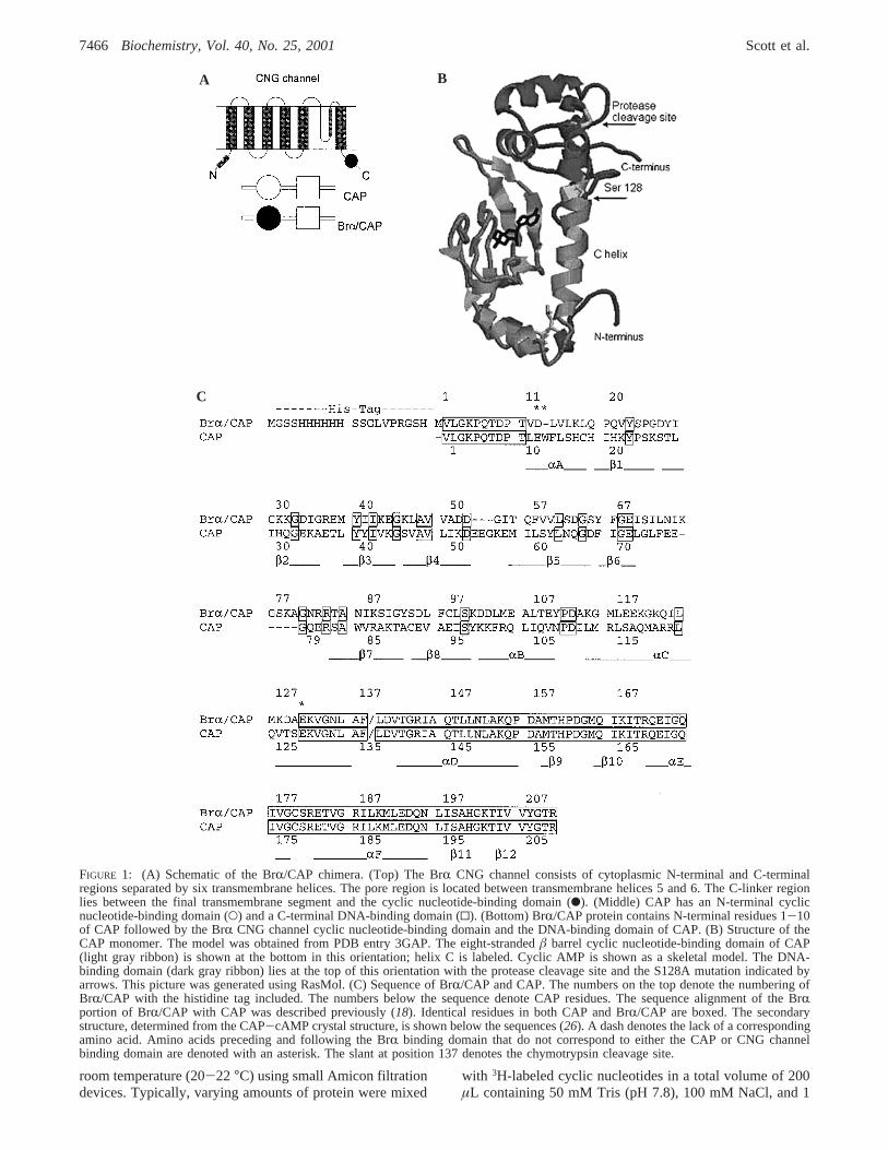
FIGURE 2: Expression of BrR/CAP protein. The Coomassie-stainedgel lanes show proteins collected during a typical purification: lane1, standard MW markers; lane 2, before IPTG induction, at 37°C;lane 3, after induction for 5 h at 25°C; lane 4, pellet after 14 000rpm centrifugation; lane 5, supernatant after 14 000 rpm centrifuga-tion; and lane 6, purified protein following elution from the affinitycolumn.
KA )[complex]
[BrR/CAP][cNMP](1)
Scheme1
BrR/CAP + cNMP 798KA
complex
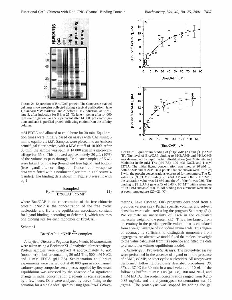
FIGURE 3: Equilibrium binding of [3H]cGMP (A) and [3H]cAMP(B). The level of BrR/CAP binding to [3H]cAMP and [3H]cGMPwas determined by rapid partial ultrafiltration (see Materials andMethods) in 50 mM Tris (pH 7.8), 100 mM NaCl, and 1 mMEDTA. The initial ligand concentration was fixed at 20µM forboth cAMP and cGMP. Data points that are shown were fit to eq1 with the protein concentrations expressed for monomers. TheKAvalue for [3H]cGMP binding to BrR/CAP was 2.87× 10⁴ M-1;the saturation value was 24µM, and ther2 of the fit was 0.96. Thebinding to [3H]cAMP gave aKA of 3.48× 10⁴ M-1 with a saturationof 19.5µM and anr2 of 0.96. All binding measurements were madeat room temperature (20-21 °C).
Functional CAP Chimera with Rod CNG Channel Binding Domain Biochemistry, Vol. 40, No. 25, 20017467
loading dye to the sample and heating the samples to>80°C. If the samples were not immediately loaded onto thegel, they were frozen at-20 °C. All samples were electro-phoresed on 10 to 20% SDS-PAGE gels from OwlSeparation Systems. Samples were loaded in SDS buffercontaining 100 mM Tris (pH 8.8), 200 mM glycine, and 0.1%SDS. Gels were run at∼60 mA for 90 min.
DNA Binding Assay.The lac promoter/operator regionDNA was obtained from a pBluescriptII KS plasmid (Strat-agene) containing cDNA encoding the catfish olfactory CNGchannel (15). The plasmid was digested with thePVuIIrestriction enzyme, and the unresolved fragment mixture wasused without further purification. DNA binding was followedthe method described by Crothers and co-workers (38) withminor modifications. Final solution conditions in the bindingreactions were 30 mM sodium acetate, 10 mM Tris-HCl (pH7.5), 0.1 M NaCl, 10 mM MgCl2, 1 mM DTT, and 10%glycerol. Reaction mixtures 25µL in volume contained∼3nM DNA and the indicated concentrations of BrR/CAP orCAP protein. Binding reaction mixtures were incubated atroom temperature for 30 min in the presence of 100µMcAMP, or no nucleotide. Gels of approximately 15 cm×15 cm × 0.1 cm were composed of 10% (w/v) totalacrylamide (29:1 acrylamide:bisacrylamide ratio) and 0.5×TBE buffer (39) and were run in 0.5× TBE until a constantcurrent was reached prior to sample loading. Gels initiallywere run with 20µM cyclic nucleotide in the gel andrecirculating in the running buffer, but identical results wereobtained without these additions, as in Figure 4. Sampleswere loaded while gels were running at 200 V, and then thevoltage was reduced to 120 V until the xylene cyanol trackingdye (which comigrates with∼100 bp DNA in this system)had run nearly to the bottom of the gel (∼150 min). Gelswere stained by soaking in ethidium bromide and photo-graphed under UV illumination.
X-ray Crystallography.BrR/CAP was maintained at aconcentration of 4.5 mg/mL in buffer containing 50 mM
sodium phosphate, 0.5 M NaCl, 1 mM EDTA (pH 7.8), and0.5 mM cAMP. This solution was mixed 1:1 with wellsolution containing 200 mM MES (pH 6.0) and 15%polyethelene glycol 4000 in a hanging drop experiment.X-ray diffraction data were collected on a crystal withdimensions of approximately 0.05 mm× 0.05 mm× 0.1mm using an R-Axis IIc imaging plate detector mounted onan RU200 Rigaku rotating anode X-ray generator operatedat 50 kV and 100 mA with a 0.5 mm collimator. Diffractiondata were collected at room temperature with 1.5° oscilla-tions. Each oscillation frame was exposed for 45 min. Thedistance to the detector plate was set at 100 mm. Thediffraction data extended to 7 Å resolution. The data set wasmerged using the HKL suite (40). The space group wasdetermined by symmetry and systematic absences. Thediffraction data were reduced in space groupP212121 withthe following unit cell dimensions:a ) 105.58 Å,b ) 98.08Å, andc ) 46.70 Å. The structure was solved by molecularreplacement using CCP4 (41) with the crystal structure ofCAP with cAMP (3GAP) (28) as the model. XPLOR (42)was used to generate the 2Fo - Fc andFo - Fc maps. Theligand was removed from the 3GAP structure to generatethe (Fo - Fc)omit map. The resultant model and maps werevisualized using the program CHAIN (43).
RESULTS
Construction of BrR/CAP.The CNG channel BrR subunithas 690 amino acids with large cytoplasmic domains on boththe N-terminus and C-terminus; six transmembrane helicesare positioned between the termini (44). Native rod outersegment CNG channels are tetrameric, containing subunitsof two types,R andâ (16, 45). A schematic of theR subunitis shown in Figure 1A. The pore region lies between thefifth and sixth transmembrane domains. The cyclic nucle-otide-binding domain of 120 residues follows the finaltransmembrane helix (46). The BrR/CAP chimera wasgenerated from the cDNA of theR subunit of the bovinerod CNG channel and CAP as shown schematically in Figure1A. Residues 489-604 of the BrR channel (BrR/CAPresidues 14-129 in Figure 1C) replaced residues 14-127of CAP. The N-terminal start site and the first 10 aminoacids of CAP, which lie outside the nucleotide-bindingdomain, were retained. The swap of BrR for CAP residueswas accomplished by introducing aSalI restriction site inthe cDNA for both proteins, which changed the DNAsequence of residue L11 in CAP to Val in the chimera, andresidue E488 of the BrR binding domain to D12 in thechimera. Following residue D604 in BrR (BrR/CAP residue129), the native CAP residue S128 at the chimera junctionwas changed to Ala. Finally, a His tag was added at theN-terminus to facilitate purification.
Expression and Purification of Soluble BrR/CAP. BrR/CAP (BrR/CAP with a His tag) was expressed as a solubleprotein following induction with IPTG at 25°C. In initialexperiments with temperature induction, which requiresgrowth at 42 °C (29), the majority of the protein wasinsoluble (data not shown). Typically, IPTG inductionfollowing a shift to 25°C was continued for 5 h. Figure 2shows the protein gel from a typical purification. Most ofthe BrR/CAP was present in the soluble fraction (lane 5),although some was present in the insoluble pellet (lane 4)after a 30 min spin at 14 000 rpm. The amount and
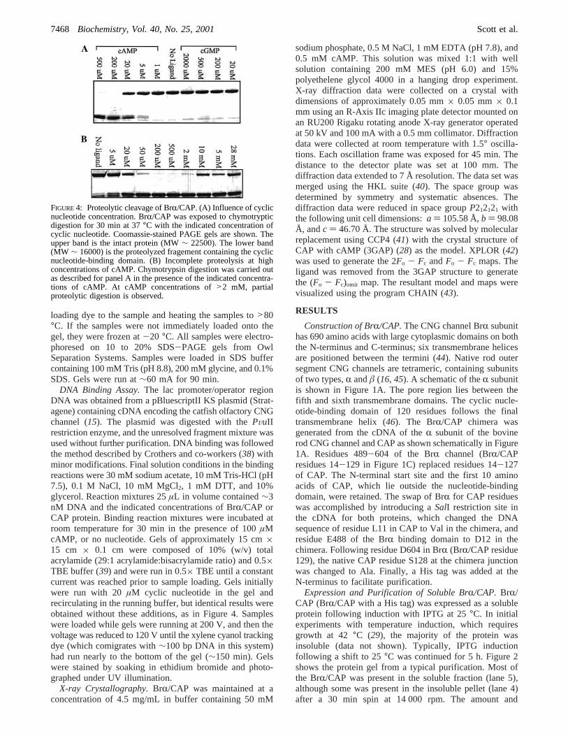
FIGURE 4: Proteolytic cleavage of BrR/CAP. (A) Influence of cyclicnucleotide concentration. BrR/CAP was exposed to chymotrypticdigestion for 30 min at 37°C with the indicated concentration ofcyclic nucleotide. Coomassie-stained PAGE gels are shown. Theupper band is the intact protein (MW∼ 22500). The lower band(MW ∼ 16000) is the proteolyzed fragement containing the cyclicnucleotide-binding domain. (B) Incomplete proteolysis at highconcentrations of cAMP. Chymotrypsin digestion was carried outas described for panel A in the presence of the indicated concentra-tions of cAMP. At cAMP concentrations of>2 mM, partialproteolytic digestion is observed.
7468 Biochemistry, Vol. 40, No. 25, 2001 Scott et al.
distribution of BrR/CAP expressed is roughly equivalent tothe amount of His-tagged CAP produced under the sameconditions (data not shown). Using a one-step purificationwith Ni2+ agarose, approximately 5 mg of BrR/CAP mono-mer was obtained per liter of cultured bacteria. The purifiedprotein (lane 6, Figure 2) was∼95% pure.
Both [3H]cAMP and [3H]cGMP Bind to Brr/CAP. Ini-tially, [3H]cGMP and [3H]cAMP binding assays wereperformed by increasing the concentration of the cyclicnucleotide with a fixed amount of BrR/CAP. Complexeswere formed in solution containing 100 mM NaCl, 50 mMTris (pH 7.8), and 1 mM EDTA at room temperature (20-22 °C) and equilibrated for 30 min. Bound and freenucleotides were partially separated by fast centrifugalfiltration. The method modifies that reported previously for[3H]cAMP binding to CAP (47). In our assay, only a smallfraction of the total incubation volume was allowed to passthrough the filter, to minimize the potential disturbance ofthe preexisting equilibrium. The low affinity of the BrR/CAP chimera for cGMP and cAMP resulted in a low signal-to-noise ratio in this assay, especially at high ligandconcentrations. The signal-to-noise ratio was improved byusing a constant ligand concentration and increasing amountsof protein. Results from such an assay are shown in Figure3. Fitting of eq 1 assumed one binding site per monomer.More complex models may also fit the binding data, butgiven the limitations of this assay, we confined our analysisto the simplest model that provided an adequate fit (see thelegend of Figure 3). The mean equilibrium associationconstants from three measurements were 3.72× 10⁴ M-1 (8.0× 10³ (SEM) for [3H]cGMP in panel A and 3.09× 10⁴
M-1 ( 2.0× 10³ (SEM) for [3H]cAMP in panel B, indicatingthat the chimera binds both nucleotides with similar affinities.The limitations of this assay, including its sources of errors,suggest that these values may represent lower limits of theaffinities.
BrR/CAP Is a Dimer.Intersubunit interactions in the CAPdimer largely involve residues in the nucleotide-bindingdomain (26). To examine the subunit assembly state of BrR/CAP, sedimentation equilibrium experiments were performedin the analytical ultracentrifuge in the presence and absenceof cAMP or cGMP. At a loading concentration of ap-proximately 40µM, the molecular weight calculated fromfitting of a single-species model to the data gives an averagemolecular weight of 49 000( 2000 for BrR/CAP. CAP rununder the same conditions without the His tag yielded anaverage molecular weight of 43 000( 2000 (polypeptidemolecular weight, 23 509). The observed molecular weightfor BrR/CAP corresponds well to twice the calculatedmolecular weight of the polypeptide (25 315), indicating thatthe smallest kinetic unit in solution is a dimer in buffercontaining 500 mM NaCl, 1 mM EDTA, and 50 mM Tris(pH 7.8) at 25°C. The ultracentrifugation results also showthat the subunit assembly of the chimera does not changeupon the binding of ligand. Although the uncertainty in thederived molecular weight is relatively large, the data are notbetter described by a monomer-dimer equilibrium. Fittingof such a model to the data indicated no more than 5% BrR/CAP monomer at the loading concentration that was used(data not shown). Preliminary analysis of the anisotropy offluorescently labeled BrR/CAP confirms the presence ofdimers even at substantially lower protein concentrations (D.
Jameson, S.-P. Scott, and J. Tanaka, unpublished observa-tions).
Proteolysis ReVeals cAMP-Induced ConformationalChanges. Upon binding cAMP, CAP undergoes conforma-tional changes that expose a chymotrypsin proteolysis site(labeled in panels B and C of Figure 1) between residues136 and 137 (36) to generate two fragments, the cyclicnucleotide-binding domain and the DNA-binding domain.Despite similar binding affinities for cAMP and cGMP, CAPis not cleaved in the presence of cGMP (37). In Figure 4A,BrR/CAP was exposed to chymotrypsin in the presence ofcGMP and cAMP at different concentrations. At 200 and500 µM cAMP, the chimera is cleaved in 30 min at 37°Ccompletely and quantitatively to yield the cyclic nucleotide-binding domain fragment at a molecular weight of ap-proximately 15 000; the smaller DNA-binding domain is notretained on the gels under the running conditions that areused. No cleavage is seen in the absence of ligand or in thepresence of cGMP concentrations as high as 2000µM.
Chymotrypsin digestions were performed at high concen-trations of cAMP (Figure 4B). The results show that at cAMPconcentrations ofg5 mM, the conformation of BrR/CAP ispartially resistant to proteolysis. Interpretation of this resultin light of proteolysis experiments with CAP suggests thatthe chimera retains the low-affinity cAMP binding sites firstseen in the crystal structure of CAP bound to DNA (48).These sites are located at the interface of the DNA-bindingdomain and the nucleotide-binding domain.
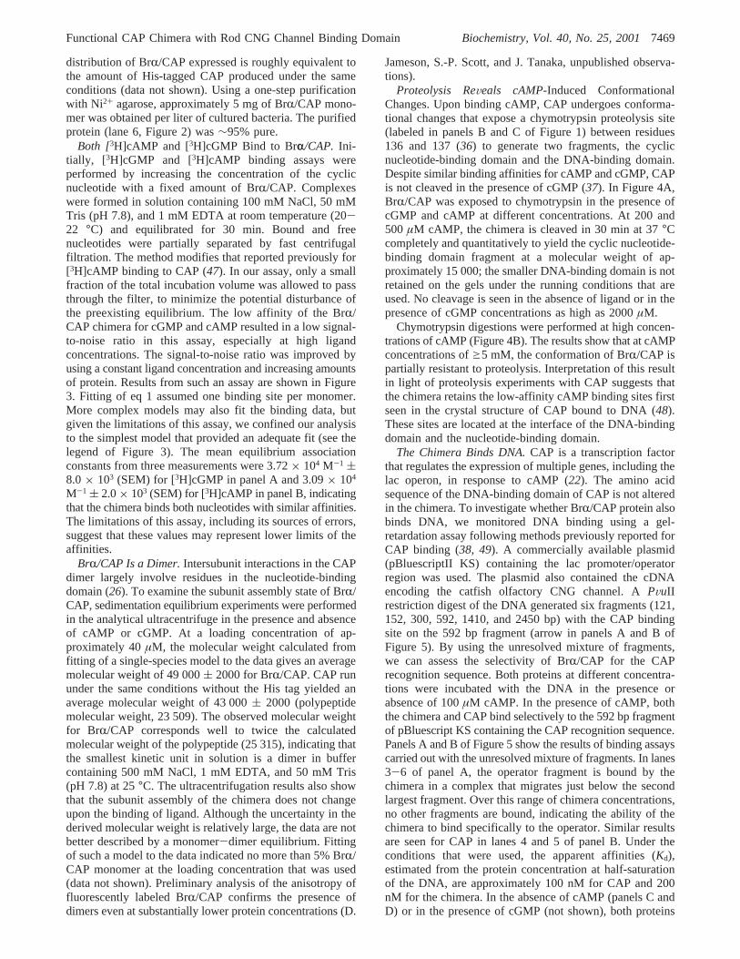
The Chimera Binds DNA.CAP is a transcription factorthat regulates the expression of multiple genes, including thelac operon, in response to cAMP (22). The amino acidsequence of the DNA-binding domain of CAP is not alteredin the chimera. To investigate whether BrR/CAP protein alsobinds DNA, we monitored DNA binding using a gel-retardation assay following methods previously reported forCAP binding (38, 49). A commercially available plasmid(pBluescriptII KS) containing the lac promoter/operatorregion was used. The plasmid also contained the cDNAencoding the catfish olfactory CNG channel. APVuIIrestriction digest of the DNA generated six fragments (121,152, 300, 592, 1410, and 2450 bp) with the CAP bindingsite on the 592 bp fragment (arrow in panels A and B ofFigure 5). By using the unresolved mixture of fragments,we can assess the selectivity of BrR/CAP for the CAPrecognition sequence. Both proteins at different concentra-tions were incubated with the DNA in the presence orabsence of 100µM cAMP. In the presence of cAMP, boththe chimera and CAP bind selectively to the 592 bp fragmentof pBluescript KS containing the CAP recognition sequence.Panels A and B of Figure 5 show the results of binding assayscarried out with the unresolved mixture of fragments. In lanes3-6 of panel A, the operator fragment is bound by thechimera in a complex that migrates just below the secondlargest fragment. Over this range of chimera concentrations,no other fragments are bound, indicating the ability of thechimera to bind specifically to the operator. Similar resultsare seen for CAP in lanes 4 and 5 of panel B. Under theconditions that were used, the apparent affinities (Kd),estimated from the protein concentration at half-saturationof the DNA, are approximately 100 nM for CAP and 200nM for the chimera. In the absence of cAMP (panels C andD) or in the presence of cGMP (not shown), both proteins
Functional CAP Chimera with Rod CNG Channel Binding Domain Biochemistry, Vol. 40, No. 25, 20017469
bind only nonspecifically, with longer fragments bound first.The DNA binding experiments show that BrR/CAP bindsto the lac operator sequence of DNA in a cAMP-dependentmanner and that the protein-DNA complexes formed withCAP and BrR/CAP have essentially identical mobilities. Thisresult suggests that both bend the DNA to a similar extent.The results also suggest that much of the DNA specificityresides in the DNA-binding domain as expected.
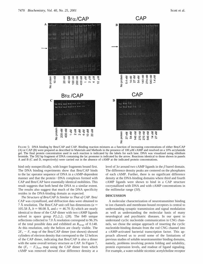
The Structure of BrR/CAP Is Similar to That of CAP.BrR/CAP was crystallized, and diffraction data were obtained to7 Å resolution. The BrR/CAP unit cell has dimensions (a )105.58 Å,b ) 98.08 Å, andc ) 46.70 Å) which are nearlyidentical to those of the CAP dimer with two cAMP ligandssolved in space groupP212121 (28). The 849 uniquereflections collected to 7.0 Å resolution correspond to 96.3%of the total possible data and exhibited anRmerge of 0.148.At this resolution, only the helices are clearly visible. The2Fo - Fc map of the BrR/CAP dimer (not shown) showedcylinders of electron density that correspond to the 12 helicesof the CAP dimer, indicating that BrR/CAP is also a dimerwith the same overall tertiary structure as CAP. In Figure 7,the (Fo - Fc)omit map using the CAP dimer from whichcAMP was removed showed clear difference density at a
level of 3σ around two cAMP ligands in theâ barrel domain.The difference density peaks are centered on the phosphatesof each cAMP. Further, there is no significant differencedensity at the DNA-binding domains where third and fourthcAMP ligands were shown to bind in a CAP structurecocrystallized with DNA and with cAMP concentrations inthe millimolar range (50).
DISCUSSION
A molecular characterization of neurotransmitter bindingto ion channels and membrane-bound receptors is central tounderstanding synaptic transmission and signal modulationas well as understanding the molecular basis of manyneurological and psychiatric diseases. In our quest tounderstand cyclic nucleotide communication in CNG chan-nels, we chose the unique approach of inserting the cyclicnucleotide-binding domain from the rod CNG channel intoa cAMP-activated bacterial transcription factor. This ap-proach allowed us to avoid some of the limitations ofprevious studies of soluble neurotransmitter binding domains,namely, problems involving protein folding and solubility,protein expression levels, and readout of ligand signaling.For example, a water-soluble nicotinic acetylcholine receptor
FIGURE 5: DNA binding by BrR/CAP and CAP. Binding reaction mixtures as a function of increasing concentrations of either BrR/CAP(A) or CAP (B) were prepared as described in Materials and Methods in the presence of 100µM cAMP and resolved on a 10% acrylamidegel. The final protein concentration used in each reaction is indicated by the labels for each lane. DNA was visualized using ethidiumbromide. The 592 bp fragment of DNA containing the lac promoter is indicated by the arrow. Reactions identical to those shown in panelsA and B (C and D, respectively) were carried out in the absence of cAMP at the indicated protein concentrations.
7470 Biochemistry, Vol. 40, No. 25, 2001 Scott et al.
retaining theR-bungarotoxin binding properties of the nativeR7 receptor was expressed inXenopusoocytes (51). Unfor-tunately, the yield of the soluble protein was 8 fmol/oocyte,limiting the number of biochemical and biophysical studiesthan can be done with the protein. In another example, asoluble glutamate receptor, which bound ligands with thecharacteristic high affinities of the intact receptor, wasexpressed in bacteria (52). The yields were high, but theprotein required refolding (53, 54). This construct was usedto obtain a high-resolution crystal structure of the ionotrophicglutamate receptor GluR2 (55). In comparison, the BrR/CAPchimera does not require refolding, and our purificationmethods give sufficient yields to study ligand-activatedsignaling as well as the biochemical and structural propertiesof the soluble binding domain.
The general assumption of homology modeling is that thethree-dimensional folding of functionally related proteindomains will be conserved more strongly than the aminoacid sequence (56). Homology models of several cyclicnucleotide-binding domains based on the structure of CAPprovided insight into how cyclic nucleotides interact withthe binding domain (13, 19). The structure of the chimerapresented here shows that the molecular models based onthe 17% level of sequence identity between CAP and BrRnucleotide-binding domains are correct in the overall tertiaryfold of the binding domain, although a detailed analysis ofthe nucleotide interactions must await a high-resolutioncrystal structure. The ultracentrifugation data and the crystalstructure of BrR/CAP offer the first biochemical evidencethat the nucleotide-binding domains of two CNG channelsubunits interact directly via the long C helices as observedfor CAP. This result supports the idea that tetrameric CNGchannel assemblies function as a pair of dimers (57). The
chimera provides a system with which to test directlypredictions about nucleotide interactions at the structural andbiochemical level, and to correlate the results with theelectrophysiology.
The BrR/CAP chimera and CAP share the unusual andunexpected property of binding to cGMP and cAMP withsimilar affinities despite signaling properties that report thepresence of one, but not the other, cyclic nucleotide. CAPbinds cAMP with aKd of ∼5 µM and binds cGMP with a2-6-fold weaker affinity (22, 32, 58); the binding of cAMPinitiates transcription, whereas cGMP has no effect. Thechimera binds [3H]cGMP with an affinity of 27µM and [3H]-cAMP with an affinity of 32 µM. In homomeric BrRchannels, cGMP activates maximal currents with aK0.5 of∼80µM, whereas saturating concentrations of cAMP activate∼1% of the maximal current withK0.5 values ranging from800 to 2000µM (46). TheK0.5 values provide little insightinto the relative cyclic nucleotide binding affinities, however,since they reflect the conformational changes involved inchannel opening as well as ligand binding (46).
Several functional properties of the BrR/CAP chimeraexamined in this report show clear differences betweencGMP and cAMP interactions with the BrR nucleotide-binding domain. In the chymotrypsin assay, cAMP promotescleavage between the N-terminal nucleotide-binding domainand the C-terminal DNA-binding domain. No proteolyticdigestion is seen in the absence of cAMP or in the presenceof cGMP. Proteolytic digestion of the chimera as a functionof cAMP concentration demonstrates biphasic behavior. Thisbiphasic behavior was reported in chymotrypsin studies withCAP (36, 47), and to understand the behavior, it is necessaryto review recent work on CAP which has long been regardedas a paradigm for allosterically activated DNA-bindingregulatory proteins (59). Exhaustive analysis of its cAMPand DNA binding equilibria aimed at providing a quantitativeunderstanding of allosteric activation led to a widely acceptedmodel that accounts for the biphasic dependence of CAPproperties on cAMP concentration (47, 58). In this model,binding of 1 equivalent of cAMP to the CAP dimersuppresses binding of the second cAMP equivalent bynegative cooperativity, and activates DNA binding by long-range conformational changes propagated through helix C.Binding of the second cAMP equivalent at much highercAMP concentrations reduces DNA binding activity. Twentyyears of biochemical and biophysical studies on CAP andits mutants were interpreted in terms of this model, andconditions for studying the active form of CAP weredetermined from it. Then in 1997, a previously undetectedsecond pair of cAMP binding sites was observed in crystalsof DNA-bound CAP dimers (50). Their location at thecrevices between the nucleotide- and DNA-binding domainsstrongly suggests that cAMP binding to these secondary sitescould play a functional role in transcription regulation.Occupancy of the secondary cAMP binding sites at highconcentrations of cAMP in CAP has been postulated toaccount for the biphasic behavior in the chymotrypsin digest(48). The cAMP-dependent biphasic pattern seen with BrR/CAP is essentially the same as that seen for CAP, and mayhave the same underlying explanation for both proteins. Thebinding pocket for the secondary cAMP sites in the CAPcrystal structure of Passner and Steitz (1997) is formed bythe bound DNA and by residues of the DNA-binding domain,
FIGURE 6: Low-resolution structure of BrR/CAP. The X-ray crystalstructure of BrR/CAP with cAMP was solved at 7 Å resolutionusing molecular replacement with PDB entry 3GAP (CAP withtwo cAMP ligands bound) as the search model. The figure displaysthe backbone structure of the 3GAP model with the protein in greenand the ligands in blue. Superimposed on the model is the (Fo -Fc)omit map density, contoured at the 3σ level (red), obtained using3GAP with the two cAMP ligands removed.
Functional CAP Chimera with Rod CNG Channel Binding Domain Biochemistry, Vol. 40, No. 25, 20017471
principally Q170, Q174, G177, C178, S179, and R180, whichare preserved in the chimera, and by residues of the hairpinloop region of CAP, which differ in the chimera. Furtherstudies of both proteins are required to elucidate the basisfor their biphasic behaviors.
Differences in cAMP- and cGMP-dependent communica-tion between the BrR nucleotide-binding domain and theDNA-binding domain are also seen in the DNA bindingstudies. The chimera retains the cAMP-dependent recognitionof the lac operator, while cGMP does not promote specificDNA binding. The failure of cGMP to activate transcriptionin CAP is not understood. To date, all of the CAP structureswere obtained with cAMP bound in the cyclic nucleotide-binding domain (26, 28, 50, 60-63), providing little insightinto the cGMP-bound form of CAP or the apo form. Thefunctional assays that have been applied to the chimera todate have been based on assays used for cAMP-activatedCAP and thus have not evaluated possible effects of cGMP.Recent studies of CAP show structural and conformationaldifferences upon binding of cGMP versus cAMP (64, 65).
CAP activation by cAMP binding is thought to involveintersubunit communication through the C helices, mediatedby the C6 amino group of cAMP, and rearrangement of theDNA and nucleotide-binding domains via theâ4-â5 hairpinloop which lies over the cAMP (62). By comparison of thestructures of CAP and the CO sensing transcription activatorCooA (66, 67), a related CAP family member, the mostimportant regions for allosteric communication were deter-mined to be the C helices and the hairpin loop or switchregion corresponding to residues 51-59 in CAP. Theseresidues comprise the loop betweenâ4 andâ5 and part ofthe â5 strand in the BrR channel. Studies of channelactivation have generally focused on the interaction betweenthe C helices in the dimer; however, theâ4-â5 hairpinregion is also likely to be important for transmission of ligandsignals as suggested by ligand analogue studies and muta-genesis (13, 17). Although we can only speculate about therole of this critical region in channel activation, futurebiochemical and biophysical investigations of BrR/CAP willbe aimed at resolving ligand-dependent conformationalchanges in the switch region of BrR.
ACKNOWLEDGMENT
We greatly appreciate the assistance and expertise of Dr.Jim Lear for the ultracentrifugation. We also thank Dr. JamesHarman for the CAP cDNA clone and Dr. William Zagottafor the cDNA of theR bovine rod CNG channel. We thankYuan-Fang Wang and Dr. Bhuvaneshwari Mahalingam forassistance and discussions on X-ray experiments. StacyStewart and Jason Thomas, undergraduate students in thelaboratory, produced the gels shown in Figures 2 and 4.
REFERENCES
1. Fesenko, E. E., Kolesnikov, S. S., and Lyubarsky, A. L. (1985)Nature 313, 310-313.
2. Nakamura, T., and Gold, G. H. (1987)Nature 325, 442-444.3. Pugh, E. N., and Lamb, T. D. (1993)Biochim. Biophys. Acta
1141, 111-149.4. Zufall, F., Firestein, S., and Shepherd, G. M. (1994)Annu.
ReV. Biophys. Biomol. Struct. 23, 577-607.5. Biel, M., Sautter, A., Ludwig, A., Hofmann, F., and Zong, X.
(1998)Naunyn-Schmiedebergs Arch. Pharmacol. 358, 140-144.
6. Savchenko, A., Barnes, S., and Kramer, R. H. (1997)Nature390, 694-698.
7. Kawai, F., and Sterling, P. (1999)J. Neurosci. 19, 2954-2959.
8. Ahmad, I., Leinders-Zufall, T., Docsis, J. D., Shepherd, G.M., Zufall, F., and Barnstable, C. J. (1994)Neuron 12, 155-165.
9. Ludwig, A., Zong, X., Stiebert, J., Hullin, R., Hofmann, F.,and Biel, M. (1999)EMBO J. 18, 2323-2329.
10. Biel, M., Zong, X., Distler, M., Bosse, E., Klugbauer, N.,Murakami, M., Flockerzi, V., and Hofmann, F. (1994)Proc.Natl. Acad. Sci. U.S.A. 91, 3505-3509.
11. Kingston, P. A., Zufall, F., and Barnstable, C. J. (1996)Proc.Natl. Acad. Sci. U.S.A. 93, 10440-10445.
12. Moosmang, S., Biel, M., Hofmann, F., and Ludwig, A. (1999)Biol. Chem. 380, 975-980.
13. Scott, S.-P., and Tanaka, J. C. (1998)Biochemistry 37, 17239-17252.
14. Varnum, M. D., Black, K. D., and Zagotta, W. N. (1995)Neuron 15, 619-625.
15. Goulding, E. H., Ngai, J., Kramer, R. H., Colicos, S., Axel,R., Siegelbaum, S. A., and Chess, A. (1992)Neuron 8, 45-58.
16. Tanaka, J. C., Eccleston, J. F., and Furman, R. E. (1989)Biochemistry 28, 2776-2784.
17. Scott, S.-P., and Tanaka, J. C. (1995)Biochemistry 34, 2338-2347.
18. Kumar, V. D., and Weber, I. T. (1992)Biochemistry 31, 4643-4649.
19. Scott, S.-P., Harrison, R. W., Weber, I. T., and Tanaka, J. C.(1996)Protein Eng. 9, 333-344.
20. Zubay, G., Schwatz, D., and Beckwith, J. (1970)Proc. Natl.Acad. Sci. U.S.A. 66, 104-110.
21. Emmer, M., de Crombrugghe, B., Pastan, I., and Perlman, R.(1970)Proc. Natl. Acad. Sci. U.S.A. 66, 480-487.
22. Anderson, W. B., Perlman, R. L., and Pastan, I. (1972)J. Biol.Chem. 247, 2717-2722.
23. Aiba, H., Fujimoto, S., and Ozaki, N. (1982)Nucleic AcidsRes. 10, 1345-1361.
24. Anderson, W. B., Schneider, A. B., Emmer, M., Perlman, R.L., and Pastan, I. (1971)J. Biol. Chem. 246, 5929-5937.
25. Cossart, P., Gicquel-Sanzey, B., and Adhya, S. (1982)NucleicAcids Res. 10, 1363-1378.
26. McKay, D. B., Weber, I. T., and Steitz, T. A. (1982)J. Biol.Chem. 257, 9518.
27. Weber, I. T., Steitz, T. A., Bubis, J., and Taylor, S. S. (1987)Biochemistry 26, 343-351.
28. Weber, I. T., and Steitz, T. A. (1987)J. Mol. Biol. 198, 311-326.
29. Belduz, A. O., Lee, E. J., and Harman, J. G. (1993)NucleicAcids Res. 21, 1827-1835.
30. Horton, R. M., Ho, S. N., Pullen, J. K., Hunt, H. D., Cai, Z.,and Pease, L. R. (1993)Methods Enzymol. 217, 271-279.
31. Harman, J. G., KcKenney, K., and Peterkofsky, A. (1986)J.Biol. Chem. 261, 16332-16339.
32. Donoso-Pardo, J. L., Turner, P. C., and King, R. W. (1987)Eur. J. Biochem. 168, 687-694.
33. Brooks, I., Weitzel, R., Chan, W., Lee, G., Watts, D. G.,Soneson, K. K., and Hensley, P. (1994) inModern AnalyticalUltracentrifugation(Schuster, T. M., and Laue, T. M., Eds.)pp 15-36, Birkhauser, Boston.
34. Laue, T., Shaw, B. D., Ridgeway, T. M., and Pelletier, S. L.(1992) inAnalytical ultracentrifugation in biochemistry andpolymer science(Harding, S. E., Rowe, A. J., and Horton, J.C., Eds.), The Royal Society of Chemistry, Cambridge, U.K.
35. Kharakoz, D. P. (1997)Biochemistry 36, 10276-10285.36. Krakow, J. S., and Pastan, I. (1973)Proc. Natl. Acad. Sci.
U.S.A. 70, 2529-2533.37. Ebright, R. H., Le Grice, S. F. J., Miller, J. P., and Krakow,
J. S. (1985)Mol. Biol. 182, 91-107.38. Liu-Johnson, H.-N., Gartenberg, M. R., and Crothers, D. M.
(1986)Cell 47, 995-1005.
7472 Biochemistry, Vol. 40, No. 25, 2001 Scott et al.
39. Maniatis, T., Fritsch, E. F., and Sambrook, J. (1982) inMolecular cloning. A laboratory manual, Cold Spring HarborLaboratory Press, Plainview, NY.
40. Otwinowski, Z., and Minor, W. (1997) inMethods inEnzymology(Carter, C. W., and Sweet, R. M., Eds.) AcademicPress, San Diego.
41. Collaborative Computational Project Number 4 (1994)ActaCrystallogr. D50, 760-763.
42. Brunger, A. T. (1992)XPLOR, Yale University Press, NewHaven, CT.
43. Sack, J. S. (1988)J. Mol. Graphics 6, 224-225.44. Kaupp, U. B., Niidome, T., Tanabe, T., Terada, S., Bonigk,
W., Stuhmer, W., Cook, N. J., Kangawa, K., Matsuo, H.,Hirose, T., Miyata, T., and Numa, S. (1989)Nature 342, 762-766.
45. Chen, T. Y., Peng, T. W., Dhallan, R. S., Ahamed, B., Reed,R. R., and Yau, K.-W. (1993)Nature 362, 764-767.
46. Zagotta, W. N., and Siegelbaum, S. A. (1996)Annu. ReV.Neurosci. 19, 235-263.
47. Heyduk, T., and Lee, J. C. (1989)Biochemistry 28, 6914-6924.
48. Mukhopadhyay, J., Sur, R., and Parrack, P. (1999)FEBS Lett.453, 215-218.
49. Straney, D. C., Straney, S. B., and Crothers, D. M. (1989)J.Mol. Biol. 206, 41-57.
50. Passner, J. M., and Steitz, T. A. (1997)Proc. Natl. Acad. Sci.U.S.A. 94, 2843-2847.
51. Wells, G. B., Anand, R., Wang, F., and Lindstrom, J. (1998)J. Biol. Chem. 273, 964-973.
52. Arvola, M., and Keina¨nen, K. (1996)J. Biol. Chem. 271,15527-15532.
53. Chen, G.-Q., and Gouaux, E. (1997)Proc. Natl. Acad. Sci.U.S.A. 94, 13431-13436.
54. Chen, G. Q., Sun, Y., Rongsheng, J., and Gouaux, E. (1998)Protein Sci. 7, 2623-2630.
55. Armstrong, N., Sun, Y., Chen, G. Q., and Gouaux, E. (2000)Nature 395, 913-917.
56. Weber, I. T. (1990)Proteins: Struct., Funct., Genet. 7, 172-184.
57. Liu, D. T., Tibbs, G. R., Paoletti, P., and Siegelbaum, S. A.(1998)Neuron 21, 235-248.
58. Takahashi, M., Blazy, B., and Baudras, A. (1980)Biochemistry19, 5124-5130.
59. Kolb, A., Busby, S., Buc, H., Garges, S., and Adhya, S. (1993)Annu. ReV. Biochem. 62, 749-795.
60. Parkinson, G., Wilson, C., Gunasekera, A., Ebright, Y. W.,Ebright, R., and Berman, H. M. (1996)J. Mol. Biol. 260, 395.
61. Weber, I. T., Gilliland, G., Harman, J. G., and Peterkofsky,A. (1987)J. Biol. Chem. 262, 5630-5636.
62. Passner, J. M., Schulz, S. C., and Steitz, T. A. (2000)J. Mol.Biol. 304, 847-859.
63. Schultz, S. C., Shields, G. C., and Steitz, T. A. (1991)Science253, 1001-1007.
64. Won, H. S., Yaazaki, T., Lee, T. W., Yoon, M. K., Park, S.H., Kyogoku, Y., and Lee, B. J. (2000)Biochemistry 39,13953-13962.
65. Baichoo, N., and Heyduk, T. (1999)Protein Sci. 8, 518-528.66. Lanzilotta, W. N., Schuller, D. J., Thorsteinsson, M. V., Kerby,
R. L., Roberts, G. P., and Poulos, T. L. (2000)Nat. Struct.Biol. 7, 876-880.
67. Chan, M. K. (2000)Nat. Struct. Biol. 7, 822.
BI002804X
Functional CAP Chimera with Rod CNG Channel Binding Domain Biochemistry, Vol. 40, No. 25, 20017473