a custom virtual reality training solution for
TRANSCRIPT
INNOVATION Open Access
A custom virtual reality training solution forophthalmologic surgical clinical trialsFelix Heimann1* , Giulio Barteselli2, André Brand1, Andreas Dingeldey1, Laszlo Godard1, Hendrik Hochstetter1,Michael Schneider1, Alexander Rothkegel1, Clemens Wagner1, Joshua Horvath2 and Shrirang Ranade2
Abstract
We present a summary of the development and clinical use of two custom designed high-fidelity virtual-realitysimulator training platforms. This simulator development program began in 2016 to support the phase III clinicaltrial Archway (ClinicalTrials.gov identifier, NCT03677934) intended to evaluate the Port Delivery System (PDS)developed by Genentech Inc. and has also been used to support additional clinical trials. The two simulatorsaddress two specific ophthalmic surgical procedures required for the successful use of PDS and provide state-of-the-art physical simulation models and graphics. The simulators incorporate customized active haptic feedbackinput devices that approximate different hand pieces including a custom hand piece specifically designed for PDSimplantation. We further describe the specific challenges of the procedure and the development of correspondingtraining strategies realized within the simulation platform.
IntroductionThe industry development of simulators in healthcare isgenerally focused on such surgical, procedural, or diag-nostic challenges that occur with high volume and aremostly performed according to well established and pos-sibly international standards. This combination has thebest chance of producing a profitable business casewhich can carry the extensive costs usually associatedwith the development of a new high-fidelity simulationsolution, especially when virtual reality (VR) technolo-gies are involved. Consequently, the bulk of availablesimulator products target a rather small set of medicalprocedures which is particularly true for surgical simula-tors [1]. Custom development commissioned by industrypartners from pharmaceutical and medical device indus-tries constitute a segment of the surgical simulationmarket that extends the range of simulator solutions toproduct-specific and non-standardized procedures but
remains limited to high revenue products to justify theinvestment.The partnership formed between Genentech Inc.
and VRmagic GmbH in the autumn 2016 deviatedfrom this pattern in that it was formed to produce atraining device for a product that was still in develop-ment and had not yet been commercialized. Genen-tech was about to start a phase II study for a newand innovative product, the Port Delivery System withranibizumab (PDS), which is a new permanent andrefillable eye implant, approximately the size of agrain of rice, that releases drug continuously into thevitreous of the human eye. The phase II Ladder study(ClinicalTrials.gov identifier, NCT02510794), specific-ally evaluated the effects of a specialized formulationof ranibizumab delivery for the treatment of neovas-cular (wet) age-related macular degeneration (see [2]).The initial clinical experience with the implant hadindicated that the implant insertion can be performedsafely as an outpatient surgical procedure. However,the implant insertion surgery involves a number ofsurgical steps not commonly performed by vitreo-retinal surgeons. Furthermore, the refill-exchange
© The Author(s). 2021 Open Access This article is licensed under a Creative Commons Attribution 4.0 International License,which permits use, sharing, adaptation, distribution and reproduction in any medium or format, as long as you giveappropriate credit to the original author(s) and the source, provide a link to the Creative Commons licence, and indicate ifchanges were made. The images or other third party material in this article are included in the article's Creative Commonslicence, unless indicated otherwise in a credit line to the material. If material is not included in the article's Creative Commonslicence and your intended use is not permitted by statutory regulation or exceeds the permitted use, you will need to obtainpermission directly from the copyright holder. To view a copy of this licence, visit http://creativecommons.org/licenses/by/4.0/.The Creative Commons Public Domain Dedication waiver (http://creativecommons.org/publicdomain/zero/1.0/) applies to thedata made available in this article, unless otherwise stated in a credit line to the data.
* Correspondence: [email protected] GmbH, Mannheim, GermanyFull list of author information is available at the end of the article
Heimann et al. Advances in Simulation (2021) 6:12 https://doi.org/10.1186/s41077-021-00167-z
procedure that exchanges the implant content withfresh drug only resembles the commonly performedintravitreal injections but requires a more precise exe-cution. To ensure patient safety and minimize the riskof surgical errors to the overall outcome of the trial,Genentech’s development team started looking for thebest possible way to train the study investigators on thePDS procedures. After 2 years of intense development ef-forts, in the spring of 2018, Genentech and VRmagicrolled out two completely new, state-of-the-art virtualreality simulator platforms enabling its users to practicethe implant insertion and the refill-exchange of the PDS,not in support of training and marketing for an existingproduct but as a training aid for surgeons involved in on-going PDS clinical development.While some training data has already been collected,
including performance metrics and training meta-data,previous training sessions were focused on providing thebest possible training of the trial investigators. However,protocols that would ensure an unbiased evaluation ofthe simulator itself were not a priority. As it is currentlyunclear when statistically relevant data suitable for thevalidation of the simulator construct might be available,this article intends to provide a concise description ofthe hardware and software that constitutes the simulatorplatform.The list of distinct AR/VR surgery simulator systems
which are consistently employed in real-life training pro-grams is short ([3] lists 70 types of simulators over allmedical fields that were developed during the last 20years) and grows shorter if we consider only systemswith active haptic feedback [4].
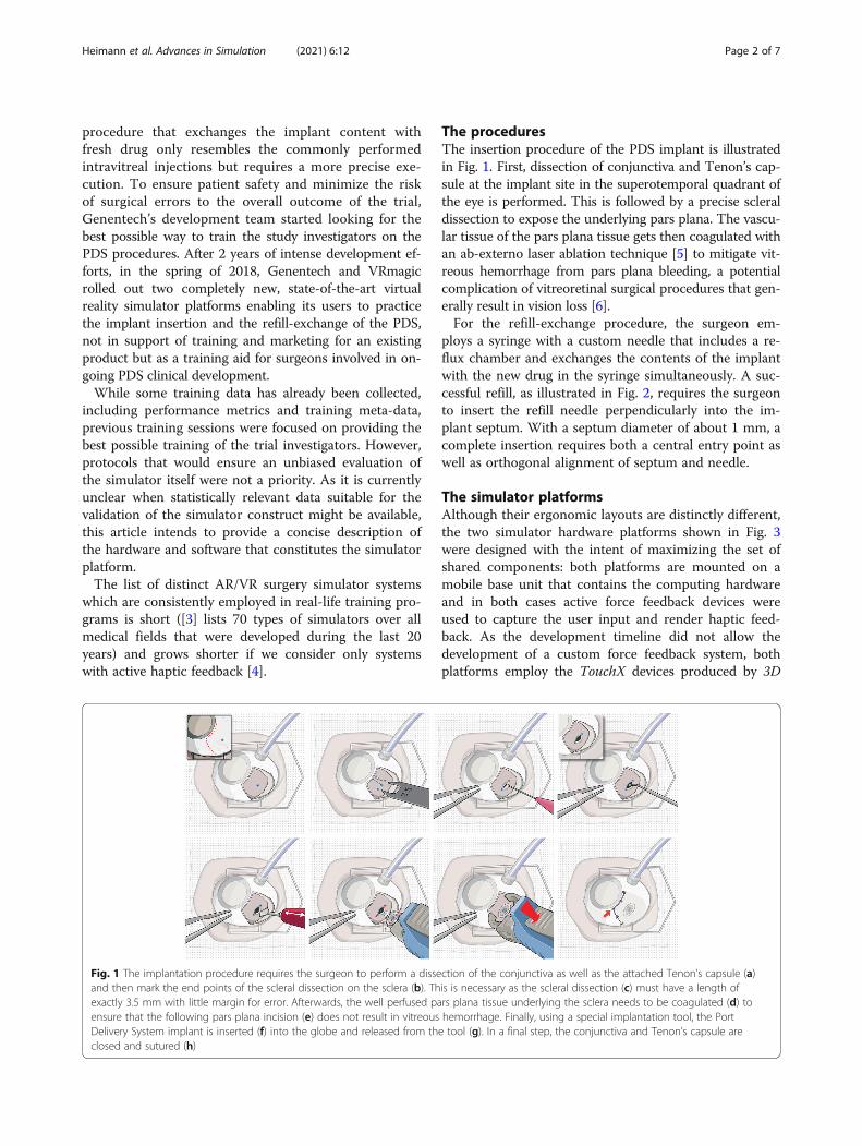
The proceduresThe insertion procedure of the PDS implant is illustratedin Fig. 1. First, dissection of conjunctiva and Tenon’s cap-sule at the implant site in the superotemporal quadrant ofthe eye is performed. This is followed by a precise scleraldissection to expose the underlying pars plana. The vascu-lar tissue of the pars plana tissue gets then coagulated withan ab-externo laser ablation technique [5] to mitigate vit-reous hemorrhage from pars plana bleeding, a potentialcomplication of vitreoretinal surgical procedures that gen-erally result in vision loss [6].For the refill-exchange procedure, the surgeon em-
ploys a syringe with a custom needle that includes a re-flux chamber and exchanges the contents of the implantwith the new drug in the syringe simultaneously. A suc-cessful refill, as illustrated in Fig. 2, requires the surgeonto insert the refill needle perpendicularly into the im-plant septum. With a septum diameter of about 1 mm, acomplete insertion requires both a central entry point aswell as orthogonal alignment of septum and needle.
The simulator platformsAlthough their ergonomic layouts are distinctly different,the two simulator hardware platforms shown in Fig. 3were designed with the intent of maximizing the set ofshared components: both platforms are mounted on amobile base unit that contains the computing hardwareand in both cases active force feedback devices wereused to capture the user input and render haptic feed-back. As the development timeline did not allow thedevelopment of a custom force feedback system, bothplatforms employ the TouchX devices produced by 3D
Fig. 1 The implantation procedure requires the surgeon to perform a dissection of the conjunctiva as well as the attached Tenon’s capsule (a)and then mark the end points of the scleral dissection on the sclera (b). This is necessary as the scleral dissection (c) must have a length ofexactly 3.5 mm with little margin for error. Afterwards, the well perfused pars plana tissue underlying the sclera needs to be coagulated (d) toensure that the following pars plana incision (e) does not result in vitreous hemorrhage. Finally, using a special implantation tool, the PortDelivery System implant is inserted (f) into the globe and released from the tool (g). In a final step, the conjunctiva and Tenon’s capsule areclosed and sutured (h)
Heimann et al. Advances in Simulation (2021) 6:12 Page 2 of 7

Systems. When bought off the shelf, the device comeswith a fixed stylus that is held by the user like a pen andattached to the actuated device arms via a gimbal. How-ever, the design does not enable the user to exchangethe stylus with a custom handpiece and the extent of thegimbal mechanics prohibit the usage of two devices insufficient proximity to simulate user input during theimplant insertion procedure. This specific procedure re-quires bimanual instrument interaction on a surgicalfield spanning less than one octant of a human eye. To
avoid device collisions during the simulation, the gimbaland stylus parts of the device were substituted by cus-tom components designed by the VRmagic developmentteam with a geometric configuration optimized for oph-thalmic surgery training. As the procedures require theuse of different instruments that cannot be accuratelyrepresented by the same physical handpiece, the newgimbal construction was augmented to allow exchanginghandpieces that can be fastened via a magnetic lock sys-tem and communicate internal degrees of freedom, e.g.,
Fig. 2 The pictures to the left and in the middle illustrate the concept of the refill-exchange procedure. A correct execution requires the fullpenetration of the special vented refill needle into the self-sealing septum of the PDS implant. This can only be achieved if the entry point of theneedle is centralized within the septum surface and the needle is perpendicularly aligned with the implant. The picture to the right shows asimulator screenshot and illustrates the use of abstract guidance elements to help the user understand the geometric challenges ofthe procedure
Fig. 3 The simulator platform for the implant insertion procedure (left picture) approximates a setup in which the user sits in an OR environmentand performs surgery on the virtual patient from a frontal position. Only about one quadrant of the virtual patient’s head is actually realized as aphysical model, roughly modeling the part from the brow ridge to the top, which is necessary as a hand rest for the user. The simulator platformfor the refill-exchange procedure (right picture) enables the user to stand next to the virtual patient who is positioned sitting in an inclinedophthalmic chair
Heimann et al. Advances in Simulation (2021) 6:12 Page 3 of 7

the opening angle of a forceps, via spring loaded connec-tors. In total, four different handpieces were developed.Their shapes and internal degrees of freedom were mod-eled to emulate (i) forceps, (ii) a syringe, (iii) a numberof simple straight instruments (e.g., scalpel or cotton tip)and (iv) a special tool designed by Genentech for thesole purpose of placing and releasing the implant.The salient differences between the two platforms pre-
sented in Fig. 3 arise from the distinctly different con-texts in which both procedures are performed. Whileinsertion of the implant requires the surgeon to operatein an adequately prepared and staffed OR environment,the refill-exchange can be performed by a single practi-tioner with minimal preparation in a clinical office set-ting. The former requires the surgeon to sit and observethe situs through a microscope, the latter is to be per-formed in a standing position, slightly bent over the pa-tient sitting in an ophthalmic chair and using onlyloupes for magnification. The differences in the ergo-nomic setup translates into different work space require-ments on the haptic input devices and correspondinglydifferent positions and orientations of these devices rela-tive to the virtual situs which constitute the predomin-ant constraints for the platform design.For the implant insertion procedure, all visual feed-
back from the virtual scene is generated by a specialstereoscopic viewing system. It was developed byVRmagic in cooperation with Haag-Streit Diagnosticsand combines an actual microscope lens tube withtwo micro displays providing a resolution of 1080 ×1080 pixels per eye.The development timeline prohibited any attempt of
developing a custom head mounted display that wouldoptimally simulate the loupes provided to the investiga-tors for the refill-exchange procedure. However, bymodeling the optical effects of the magnifying lenseswithin the graphic rendering pipeline, the requirementson the actual hardware displays could be sufficiently re-duced to allow the integration of a consumer product.After weighing the benefits of the various products avail-able by the beginning of 2018, the Oculus Rift CV1 wasselected due to the high fidelity and stability of its track-ing system.
Simulator training benefitsToday, there are various methods for simulation-basedsurgical training in ophthalmology including traditionalwet lab training with animal models or hybrid animal/synthetic models, dry lab training on synthetic models aswell as virtual reality training simulators [7]. While clin-ical development is still ongoing and there is not yet anydata that would allow for a validation study of the simu-lator training, the following descriptions of some aspectsof the training modules provide examples to illustrate
the potential of virtual reality simulators to teach theseprocedures efficiently.
Modularized learningFor the implant insertion procedure, the simulator cur-riculum covers only the parts with the highest impact onthe clinical outcome starting with the scleral dissectionand ending with the actual insertion and placement ofthe PDS implant. This partial procedure is split into fourseparate training modules comprising (i) the scleral dis-section, (ii) the laser ablation of the pars plana, (iii) thepars plana incision, and (iv) the actual implant insertion.For the refill-exchange procedure, a separation into
more than one training module is not appropriate. In-stead, the procedure can be performed on different vir-tual patients representing varying levels of difficultyessentially controlled by the visibility of the implantthrough the conjunctiva and the haptic resistance duringinsertion.Each training module can be started directly thus en-
abling the user to train procedural steps out of orderand focus their efforts on those tasks considered mostchallenging. Furthermore, the user can restart a moduleat any point and thus reset the scene for a new attemptinstantaneously. Out of order training and immediate re-starts provide a significant benefit that cannot beattained when training on animal models and constitutecrucial features considering that training sessions for thetrial investigators may last only 15–30 min.
Scleral dissectionThe PDS implant needs to be seated within a scleral in-cision of a very specific target length that corresponds tothe long axis of the implant, with a minimal margin forerror. Furthermore, the incision needs to be createdwithout harming the underlying pars plana tissue. Con-sidering that the average thickness of the scleral tissue isonly ~ 0.7 mm [8], the surgeon needs to navigate hisblade within a very narrow corridor while compensatingfor the erratic forces resulting from the toughness of thescleral tissue.To enable a real-time simulation of this process, the
simulator employs a very fine and dynamic sub-triangulation of the scleral tissue adjacent to the expand-ing cut. The resulting resolution is sufficient for anaccurate physical simulation model and the rendering ofconsistent haptic feedback. A significant effort was madeto obtain an accurate graphical rendering of the fibrousscleral tissue allowing the user to learn the correct inter-pretation of visual cues that indicate the incision depthwithin the scleral tissue.Compared with other conceivable training methods, a
major benefit of this approach is the ability of the VRsimulator to compute and report the relevant metrics in
Heimann et al. Advances in Simulation (2021) 6:12 Page 4 of 7

real-time as well as in a posteriori analysis. The formeris provided as a heads-up display (HUD) that allows theuser to directly associate subtle visual cues with objectivedepth information and thus improve their ability to assessthe incision geometry (see the first picture in Fig. 4). TheHUD further allows the user to recognize common mis-takes like rounded depth profiles at the incision cornerswhich are hard to recognize even by expert observers. Aposteriori evaluation of user performance includes infor-mation about tissue treatment as well as the latitudinaland longitudinal placement of the incision.
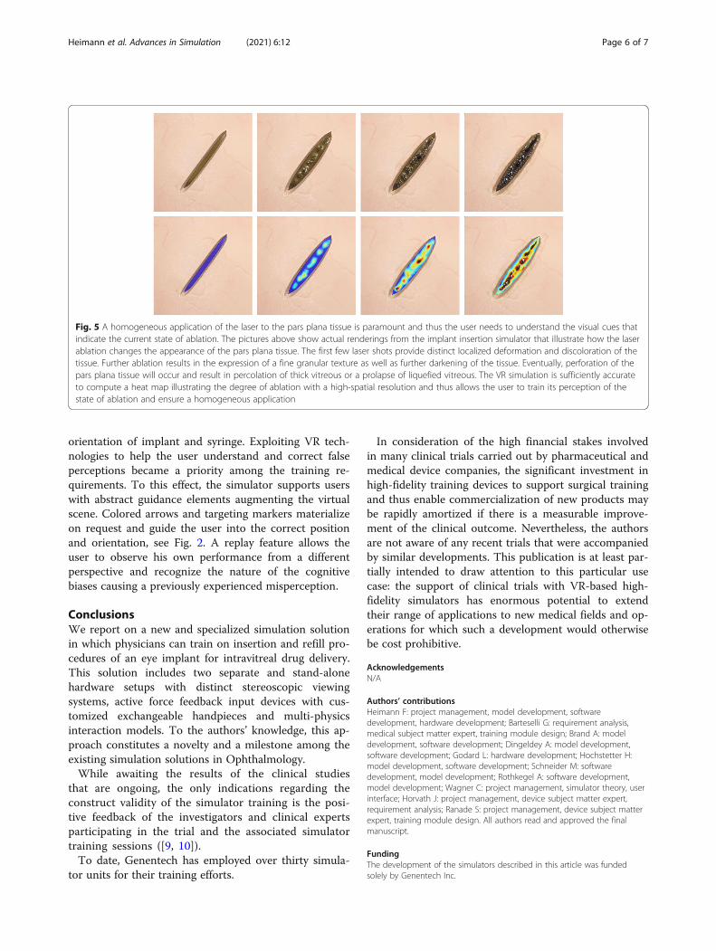
Laser ablation of the pars planaThe fundamental challenge of this procedure is the cor-rect application of the laser probe which is used in acontext and configuration not usually employed invitreoretinal surgery. The ablation of the pars plana tis-sue needs to be applied as homogeneously as possiblebecause the perforation of the tissue, which is achievedat the end-point of the procedure, may result in the pro-lapse of liquefied vitreous that will impede further treat-ment of yet insufficiently ablated parts of the tissue.The VR simulator renders a highly accurate represen-
tation of the various visual indicators that reveal thecurrent ablation state of the tissue. Using the
instantaneous restart feature of the simulator, the usercan experiment with different laser settings and quicklyobtain an intuitive understanding of the coagulationtechnique. A further benefit of the VR environment isthe simulator’s ability to display a heat map of the ex-posed pars plana tissue either as a HUD or as an overlayelement (see Fig. 5).
Refill-exchangePotential complications during this procedure originatemostly from poor alignment of the syringe needle withthe implant septum. Implementing a sufficiently accur-ate simulation model was a challenge due to the com-plex coupling of the relevant scene objects: the implantorientation is coupled to its adjacent sclera tissue andthe external forces that drive this interaction result fromthe penetration of the septum by the syringe needle orits collision with the implant flange. As the needle isflexible, the overall system has a distinct non-linear dy-namic. Hence, significant development efforts were re-quired to render the characteristic haptic effects thatguide the user during needle insertion.Early training sessions with the simulator quickly re-
vealed that the users had a tendency to be overconfidentabout their ability to visually estimate the relative
Fig. 4 The pictures above show graphical renderings from all four training modules of the implant insertion simulator. All relevant physical effectsare simulated in real time including the fluid dynamics of blood, supra choroidal fluids, and vitreous, the elastic instrument interaction with thesclera tissue, the ablation effects of the laser, and the dissection of sclera and pars plana tissue. The pictures also illustrate some of the VRguidance elements that instruct the user during training. During scleral dissection, a heads-up display (HUD) shows a profile illustrating the cutdepth and length (top-left picture). During pars plana ablation, a heat map can be shown either in the periphery of the field-of-view (top-rightpicture) or directly over the pars plana tissue (see Fig. 5). During pars plana incision, a cross section of the supra-temporal eye quadrant illustratesthe penetration depth and angle of the slit knife and its proximity to intra-ocular structures (bottom-left picture)
Heimann et al. Advances in Simulation (2021) 6:12 Page 5 of 7
orientation of implant and syringe. Exploiting VR tech-nologies to help the user understand and correct falseperceptions became a priority among the training re-quirements. To this effect, the simulator supports userswith abstract guidance elements augmenting the virtualscene. Colored arrows and targeting markers materializeon request and guide the user into the correct positionand orientation, see Fig. 2. A replay feature allows theuser to observe his own performance from a differentperspective and recognize the nature of the cognitivebiases causing a previously experienced misperception.
ConclusionsWe report on a new and specialized simulation solutionin which physicians can train on insertion and refill pro-cedures of an eye implant for intravitreal drug delivery.This solution includes two separate and stand-alonehardware setups with distinct stereoscopic viewingsystems, active force feedback input devices with cus-tomized exchangeable handpieces and multi-physicsinteraction models. To the authors’ knowledge, this ap-proach constitutes a novelty and a milestone among theexisting simulation solutions in Ophthalmology.While awaiting the results of the clinical studies
that are ongoing, the only indications regarding theconstruct validity of the simulator training is the posi-tive feedback of the investigators and clinical expertsparticipating in the trial and the associated simulatortraining sessions ([9, 10]).To date, Genentech has employed over thirty simula-
tor units for their training efforts.
In consideration of the high financial stakes involvedin many clinical trials carried out by pharmaceutical andmedical device companies, the significant investment inhigh-fidelity training devices to support surgical trainingand thus enable commercialization of new products maybe rapidly amortized if there is a measurable improve-ment of the clinical outcome. Nevertheless, the authorsare not aware of any recent trials that were accompaniedby similar developments. This publication is at least par-tially intended to draw attention to this particular usecase: the support of clinical trials with VR-based high-fidelity simulators has enormous potential to extendtheir range of applications to new medical fields and op-erations for which such a development would otherwisebe cost prohibitive.
AcknowledgementsN/A
Authors’ contributionsHeimann F: project management, model development, softwaredevelopment, hardware development; Barteselli G: requirement analysis,medical subject matter expert, training module design; Brand A: modeldevelopment, software development; Dingeldey A: model development,software development; Godard L: hardware development; Hochstetter H:model development, software development; Schneider M: softwaredevelopment, model development; Rothkegel A: software development,model development; Wagner C: project management, simulator theory, userinterface; Horvath J: project management, device subject matter expert,requirement analysis; Ranade S: project management, device subject matterexpert, training module design. All authors read and approved the finalmanuscript.
FundingThe development of the simulators described in this article was fundedsolely by Genentech Inc.
Fig. 5 A homogeneous application of the laser to the pars plana tissue is paramount and thus the user needs to understand the visual cues thatindicate the current state of ablation. The pictures above show actual renderings from the implant insertion simulator that illustrate how the laserablation changes the appearance of the pars plana tissue. The first few laser shots provide distinct localized deformation and discoloration of thetissue. Further ablation results in the expression of a fine granular texture as well as further darkening of the tissue. Eventually, perforation of thepars plana tissue will occur and result in percolation of thick vitreous or a prolapse of liquefied vitreous. The VR simulation is sufficiently accurateto compute a heat map illustrating the degree of ablation with a high-spatial resolution and thus allows the user to train its perception of thestate of ablation and ensure a homogeneous application
Heimann et al. Advances in Simulation (2021) 6:12 Page 6 of 7
Availability of data and materialsN/A
Declarations
Competing interestsThe custom simulator development described was a collaborative effort ofGenentech Inc. and VRmagic GmbH. All authors are employees of eitherVRmagic GmbH or Genentech Inc.
Author details1VRmagic GmbH, Mannheim, Germany. 2Genentech, Inc., South SanFrancisco, CA, USA.
Received: 15 September 2020 Accepted: 30 March 2021
References1. Prescient & Strategic Intelligence: Section 5.1 - By Offering. Surgical
Simulation Market Research Report, LS11131. 2020. p. 55–60.2. Campochiaro PA, Marcus DM, Awh CC, Regillo C, Adamis AP, Bantseev V,
et al. The port delivery system with ranibizumab for neovascular age-relatedmacular degeneration: results from the randomized phase 2 ladder clinicaltrial. Ophthalmology. 2019;126(8):1141–54. https://doi.org/10.1016/j.ophtha.2019.03.036.
3. Panteleimon P, Chorti A, Papagiouvanni I, Paparoidamis G, Drosos C,Panagiotakopoulos T, et al. Virtual and augmented reality in medicaleducation. Intechopen. 2018. https://doi.org/10.5772/intechopen.71963.
4. Rangarajan K, Davis H, Pucher PH. Systematic review of virtual haptics insurgical simulation: a valid educational tool? J Surg Educ. 2020;77(2):337–47.https://doi.org/10.1016/j.jsurg.2019.09.006.
5. Bantseev V, Schuetz C, Booler HS, Horvath J, Hovaten K, Erickson S, BentleyE, Nork TM, Freeman WR, Stewart JM, Barteselli G. Evaluation of surgicalfactors affecting vitreous hemorrhage following Port Delivery System withranibizumab implant insertion in a minipig model. Retina. 2020;40(8):1520–8. https://doi.org/10.1097/IAE.0000000000002614.
6. Spraul CW, Grossniklaus HE. Vitreous hemorrhage. Surv Ophthalmol.1997;42(1):3–39. https://doi.org/10.1016/s0039-6257(97)84041-6.
7. Lee R, Raison N, Lau WY, Aydin A, Dasgupta P, Ahmed K, Haldar S. Asystematic review of simulation-based training tools for technical and non-technical skills in ophthalmology. Eye. 2020;34(10):1737–59. https://doi.org/10.1038/s41433-020-0832-1.
8. Norman RE, Flanagan JG, Rausch SMK, Sigal IA, Tertinegg I, Eilaghi A, et al.Dimensions of the human sclera: thickness measurement and regionalchanges with axial length. Exp Eye Res. 2010;90(2):277–84. https://doi.org/10.1016/j.exer.2009.11.001.
9. Castellanos S. Genentech uses virtual reality to train eye surgeons. The WallStreet Journal, February 6th 2019. https://www.wsj.com/articles/genentech-uses-virtual-reality-to-train-eye-surgeons-11549495028.
10. Pieramici DJ, Heimann F, Brassard R, Barteselli G, Ranade S. Virtual realitybecomes a reality for ophthalmologic surgical clinical trials. Trans Vis SciTech. 2020;9–7:1.
Publisher’s NoteSpringer Nature remains neutral with regard to jurisdictional claims inpublished maps and institutional affiliations.
Heimann et al. Advances in Simulation (2021) 6:12 Page 7 of 7