a conserved motif in region v of the large polymerase ...jvi.asm.org/content/82/2/775.full.pdf ·...
TRANSCRIPT

JOURNAL OF VIROLOGY, Jan. 2008, p. 775–784 Vol. 82, No. 20022-538X/08/$08.00�0 doi:10.1128/JVI.02107-07Copyright © 2008, American Society for Microbiology. All Rights Reserved.
A Conserved Motif in Region V of the Large Polymerase Proteins ofNonsegmented Negative-Sense RNA Viruses That Is Essential
for mRNA Capping�
Jianrong Li,† Amal Rahmeh,† Marco Morelli, and Sean P. J. Whelan*Department of Microbiology and Molecular Genetics, Harvard Medical School, 200 Longwood Ave., Boston, Massachusetts 02115
Received 22 September 2007/Accepted 26 October 2007
Nonsegmented negative-sense (NNS) RNA viruses cap their mRNA by an unconventional mechanism.Specifically, 5� monophosphate mRNA is transferred to GDP derived from GTP through a reaction thatinvolves a covalent intermediate between the large polymerase protein L and mRNA. This polyribonucleoti-dyltransferase activity contrasts with all other capping reactions, which are catalyzed by an RNA triphos-phatase and guanylyltransferase. In these reactions, a 5� diphosphate mRNA is capped by transfer of GMP viaa covalent enzyme-GMP intermediate. RNA guanylyltransferases typically have a KxDG motif in which thelysine forms this covalent intermediate. Consistent with the distinct mechanism of capping employed by NNSRNA viruses, such a motif is absent from L. To determine the residues of L protein required for capping, wereconstituted the capping reaction of the prototype NNS RNA virus, vesicular stomatitis virus, from highlypurified components. Using a panel of L proteins with single-amino-acid substitutions to residues universallyconserved among NNS RNA virus L proteins, we define a new motif, GxxT[n]HR, present within conservedregion V of L protein that is essential for this unconventional mechanism of mRNA cap formation.
The 5� terminus of eukaryotic mRNA is modified by theaddition of a 7mGpppN cap structure. The cap structure isessential for mRNA stability, mRNA transportation, splicingof pre-mRNAs, and translation (13, 28). Formation of thisstructure requires a series of enzymatic reactions. An RNAtriphosphatase (RTPase) hydrolyzes the 5� triphosphate(pppN) end of mRNA to yield a 5� diphosphate (ppN). This iscapped by an RNA guanylyltransferase (GTase) which trans-fers Gp derived from GTP to form the cap structure. The capis subsequently methylated by guanine-N-7 (G-N-7) methyl-transferase (MTase) to yield 7mGpppN, which can be furthermethylated by ribose-2�-O (2�-O) MTase to yield 7mGpppNm
(14, 36).Cap formation in nonsegmented negative-strand (NNS)
RNA viruses involves a different reaction mechanism. For ve-sicular stomatitis virus (VSV) (2), spring viremia of carp virus(16), and respiratory syncytial virus (RSV) (5), the underlinedphosphates of the 5� GpppN triphosphate bridge were shownto be derived from GDP rather than GMP. Recent studies withVSV demonstrated that this reaction does not involve transferof guanylate onto the mRNA, but rather involves a polyribo-nucleotidyltransferase activity (29). Here, the mRNA cappingreaction proceeds via a covalent intermediate between the241-kDa viral polymerase protein L and the 5� monophosphatemRNA. This monophosphate mRNA is transferred onto GDPderived from GTP to yield the GpppA mRNA cap. Conse-quently, this mechanism of cap formation is in marked contrastwith those catalyzed by conventional GTases. The crystal struc-
tures of representative GTases have been solved and theirreaction mechanisms studied in biochemical detail (17).GTases typically contain a KxDG motif, in which the lysineforms the covalent intermediate with GMP, and consistentwith the distinct mechanism such a motif is absent from theVSV L protein. In contrast to our knowledge of how RNAGTases catalyze formation of the mRNA cap, cap formation bypolyribonucleotidyltransferases is not well understood and theactive-site residues have not been mapped. Consequently, thelocation of the capping activity within the 2,109-amino-acidVSV L protein has not been determined.
The NNS RNA viruses share a common strategy for geneexpression. The template for RNA synthesis is a protein-RNAcomplex in which the genomic RNA is encapsidated by theviral nucleocapsid (N) protein and associated with the RNA-dependent RNA polymerase (RdRP). The minimal viral com-ponents of the RdRP were shown for VSV to comprise the Lprotein and an accessory phosphoprotein (P) (11). The RdRPsequentially copies the genomic RNA into five capped andpolyadenylated mRNAs (1, 4). The mRNAs acquire their 5�mRNA cap structure and 3� poly(A) tail during their synthesis,and the enzymatic activities that catalyze these reactions areprovided by L protein.
Sequence alignments between representative NNS RNA vi-ruses identified six regions of conservation in the L protein(CRI to -VI) separated by regions of lower sequence homology(33). For Sendai virus (SeV), CRI is implicated in recruitmentof N protein during replication and interaction with P protein(8) and CRII appears to play a role in template binding (27,38). Whether these regions of L mediate the same function inVSV has not been determined. However, it is clear for all NNSRNA virus L proteins that CRIII contains the polymeraseactive-site motif, and consistent with this, modification of VSVL protein residue D714, which is predicted to coordinate a
* Corresponding author. Mailing address: Department of Microbi-ology and Molecular Genetics, Harvard Medical School, 200 Long-wood Ave., Boston, MA 02115. Phone: (617) 432-1923. Fax: (617)738-7664. E-mail: [email protected].
† J.L. and A.R. made equal contributions to this article.� Published ahead of print on 14 November 2007.
775
on July 7, 2018 by guesthttp://jvi.asm
.org/D
ownloaded from

catalytically essential Mg2� ion, inhibits all RdRP activity (37).In addition, CRVI of SeV and VSV L protein has been shownto function as the mRNA cap methylase (15, 21, 22, 30). Basedon these assignments, we suspected that either CRIV or -Vmight serve as the mRNA capping enzyme. This idea is con-sistent with a prior study that described a small-molecule in-hibitor of RSV RNA synthesis in vitro (23). This inhibitorresulted in the synthesis of short uncapped viral RNAs. Viralmutants resistant to this inhibitor were selected, and the resis-tance mutations were mapped to CRV, suggesting that CRV ofL plays a role in either mRNA cap formation or polymeraseprocessivity.
In this study, we established a system to study cap formationin vitro using purified recombinant L protein. Guided by phy-logenetic comparisons among NNS RNA virus L proteins,we generated 19 L protein variants with single-amino-acidchanges to residues in CRV of L that are highly conservedamong the NNS RNA viruses. We analyzed the ability of theseL proteins to support RNA synthesis and cap formation invitro. These studies define a new motif that is conserved amongthe NNS RNA virus L proteins that are essential for mRNAcap formation.
MATERIALS AND METHODS
Purification of N RNA template and RNP complex. Nucleocapsid RNA (NRNA) template was purified from recombinant VSV essentially as describedpreviously (31). Briefly, 4 mg purified virus was disrupted on ice for 1 h in 20 mMTris-HCl (pH 8.0), 0.1% Triton X-100, 5% glycerol, 5 mM EDTA, 3.5 mMdithioerythritol, 20% dimethyl sulfoxide, and 1.0 M LiCl. Template was recov-ered by centrifugation (190,000 � g, 3.5 h) through a glycerol step gradient of0.25 ml each of 40, 45, and 50% glycerol in TED (20 mM Tris-Cl [pH 8.0], 1 mMEDTA, 2 mM dithioerythritol) supplemented with 0.1 M NaCl. The pellet wasresuspended in 0.3 ml of TED plus 10% glycerol and disrupted on ice again,except Triton and EDTA concentrations were reduced to 0.05% and 1 mM,respectively. The N RNA template was isolated by banding in a 3.6-ml 20 to 40%(wt/wt) CsCl gradient (150,000 � g, 2.5 h), recovered by side puncture, anddiluted fourfold with 10 mM Tris-Cl (pH 8.0) plus 0.1 mM EDTA. Template wasrecovered following centrifugation (150,000 � g, 1.5 h) through a 0.5-ml cushion(50% glycerol, TED buffer, 0.1 M NaCl).
The RNP complex was purified as described previously (6). Briefly, 1 mg ofpurified VSV was disrupted (0.4 M NaCl, 0.2% Triton X-100) for 1 h on ice. TheRNP complex was recovered as pellet following centrifugation (160,000 � g, 1 h)through a 4-ml 10% sucrose NTE (0.1 M NaCl, 1 mM EDTA, 0.01 M Tris-Cl [pH7.4]) cushion. Both N RNA and RNP pellets were resuspended in 0.3 ml NTEand stored in aliquots at �80°C.
Expression and purification of recombinant VSV polymerase. Plasmids carry-ing functional VSV L and P genes were described previously (32). To facilitateprotein purification, hexahistidine tags were introduced at the N terminus ofVSV L and P and were separately inserted into pFASTBAC DUAL (Invitrogen)under control of the polyhedrin promoter. A third plasmid, designed to coex-press L and P, was generated by insertion of N His-L under control of thepolyhedrin promoter and an untagged version of P under control of the P10promoter. Recombinant baculoviruses (BV) were recovered following transfec-tion of bacmid DNA into Spodoptera frugiperda Sf21 cells using Cellfectin (In-vitrogen). Mutagenesis of the L gene was performed using QuikChange (Strat-agene, La Jolla, CA). A 2-kb fragment containing the desired mutations wassequenced prior to subcloning into pFASTBAC DUAL. This confirmed that noother mutations were introduced. For expression of polymerase components,Sf21 cells were infected at a multiplicity of infection of 10 with recombinant BV.At 72 h postinfection, cells were scraped into media and washed twice withice-cold phosphate-buffered saline, and the cells were recovered by centrifuga-tion. Cells were suspended in lysis buffer (50 mM NaH2PO4, 10% glycerol, 0.2%NP-40, 300 mM NaCl, 10 mM imidazole [pH 8.0]) supplemented with EDTA-free protease inhibitor cocktail (Roche) and 1 mM phenylmethylsulfonyl fluorideand disrupted by sonication. The cell lysates were clarified by centrifugation(30,000 � g, 40 min) and incubated with Ni-nitrilotriacetic acid (NTA)-agarosebeads (Qiagen) for 2 h. The resin was then washed with 20 bed volumes of wash
buffer (50 mM NaH2PO4, 10% glycerol, 300 mM NaCl, 20 mM imidazole [pH8.0]), and the His-tagged proteins were eluted in elution buffer (50 mMNaH2PO4, 10% glycerol, 300 mM NaCl, 250 mM imidazole [pH 7.0]).
Where indicated, the His-L and His-P proteins were purified further by ion-exchange chromatography through either a Mono S HR 5/5 or a Mono Q HR 5/5column (GE Healthcare), respectively. Prior to loading, the fractions eluted fromNi-NTA-agarose were diluted twofold in buffer A (50 mM Tris-HCl [pH 7], 0.1M NaCl, 10% glycerol, 1 mM dithiothreitol [DTT]), which had also been used toequilibrate the columns. Columns were washed with 5 column volumes of bufferA, and the proteins were eluted with a 20-column-volume gradient of 0.1 to 1 MNaCl in buffer A. Peak fractions were pooled and dialyzed against 50 mMTris-HCl (pH 7.4), 200 mM NaCl, 15% glycerol, and 1 mM DTT. The purifiedproteins were separated by 10% sodium dodecyl sulfate-polyacrylamide gel elec-trophoresis (SDS-PAGE) and visualized by Coomassie blue stain. Proteinamounts were determined by Bradford assay.
Reconstitution of viral RNA synthesis in vitro. Viral RNA was synthesized invitro either using purified RNP complex from virus particles or using purifiedpolymerase from insect cells. Reconstituted reactions were typically performedusing 1 �g of N RNA template, 1 �g of purified L, and 0.5 �g of purified P,nucleoside triphosphates (1 mM ATP and 0.5 mM each CTP, GTP, and UTP),and 30% (vol/vol) rabbit reticulocyte lysate (Promega). Where indicated, reac-tion mixtures were supplemented with 15 �Ci of [�-32P]GTP (3,000 Ci mmol�1)(Perkin-Elmer, Wellesley, MA). After 5 h of incubation at 30°C, RNA waspurified, resuspended in 60 �l of water, and analyzed by electrophoresis in 6%acid-agarose-urea gels (20).
Primer extension assay. Minus-sense oligonucleotides, corresponding to nu-cleotides (nt) 27 to 47 and 130 to 115 of the complete VSV genome sequence,were used in primer extensions as described previously (43). Products wereanalyzed by electrophoresis on 6% polyacrylamide gels and detected byPhosphorImager. Where indicated, products of in vitro transcription reactionswere treated with tobacco acid pyrophosphatase (TAP) to remove the cap struc-ture, prior to reverse transcription.
Analysis of the mRNA cap structure. To directly examine the cap structure,RNA was digested with TAP (Epicenter, Madison, WI), as previously described(21, 22). Products were analyzed by thin-layer chromatography (TLC) on poly-ethyleneimine-F cellulose sheets (EM Biosciences). Plates were developed using1.2 M LiCl and dried, and the spots were visualized using a PhosphorImager.Markers 7mGp and Gp were visualized by UV shadowing at 254 nm.
Characterization of the 5� termini by terminator exonuclease. Purified RNAswere treated with TAP and alkaline phosphatase (AP), followed by T4 polynu-cleotide kinase (PNK) and exonuclease (Exo). Reaction mixtures were thentreated with proteinase K in presence of 0.5% SDS, followed by phenol-chloro-form extraction, and precipitated with ethanol and glycogen. RNA samples wereanalyzed by electrophoresis on acid-agarose gels, and products were detected byusing a PhosphorImager.
Synthesis of 5�-triphosphorylated oligo-RNAs. RNA (pppApApCpApG orpppApCpGpApA) was synthesized using partially double-stranded syntheticDNA templates containing a T7 polymerase promoter as described previously(29). Transcription reaction mixtures contained 100 nM of annealed template;0.2 U �l�1 of T7 polymerase (NEB); and 1 mM of ATP, CTP, and GTP in T7reaction buffer (40 mM Tris-HCl [pH 7.9], 6 mM MgCl2, 10 mM DTT, and 2 mMspermidine). Where indicated, reaction mixtures contained 20 cpm pmol�1 or3 � 104 cpm pmol�1 of [�-32P]GTP. Following 4 h of incubation, DNA wasdigested with 3 U of RQ1 DNase (Promega) and the RNA was recovered byphenol-chloroform-isoamyl alcohol (25:24:1) extraction and ethanol precipita-tion. RNA was washed with 70% ethanol, dried, and resuspended in H2O.Following addition of formamide stop solution, samples were boiled for 3 minand then resolved by electrophoresis on 20% polyacrylamide–7 M urea gels.Transcripts were visualized by autoradiography, and the 5-nt products wereexcised, crushed, and soaked in 0.3 M ammonium acetate (pH 5.2). Eluted RNAwas recovered by ethanol precipitation, and the sequence was confirmed bydigestion with RNase T1.
RNA trans capping assay. The RNA trans capping assay was performed es-sentially as described previously (29). Briefly, 5 �M of 5�-triphosphorylated 5-ntoligo-RNAs was incubated with 0.5 �M of [�-32P]GTP or [�-32P]dGTP (400 cpmfmol�1) and 300 ng of His-L in capping buffer (50 mM MOPS [morpholinepro-panesulfonic acid; pH 5.8], 1 mM MnCl2, 2 mM DTT, 5% glycerol, and 0.1 mg/mlbovine serum albumin), at 30°C for 2 h. Reactions were stopped by addition ofTris-HCl (pH 8) to 100 mM, followed by treatment with AP and subsequentdigestion with proteinase K. Reaction products were extracted by phenol-chlo-roform-isoamyl alcohol (25:24:1), precipitated with ethanol, washed, dried, andresuspended in H2O.
776 LI ET AL. J. VIROL.
on July 7, 2018 by guesthttp://jvi.asm
.org/D
ownloaded from
Quantitative analysis. Quantitative analysis was performed by using aPhosphorImager (GE Healthcare, Typhoon) and ImageQuant TL software (GEHealthcare, Piscataway, NJ). Statistical analysis was performed on three to fiveindependent experiments. The significance of the values was determined by apaired Student’s t test.
RESULTS
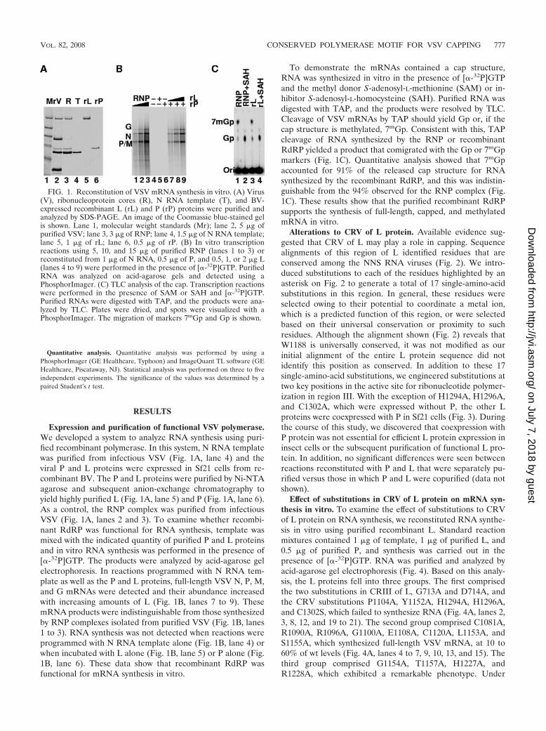
Expression and purification of functional VSV polymerase.We developed a system to analyze RNA synthesis using puri-fied recombinant polymerase. In this system, N RNA templatewas purified from infectious VSV (Fig. 1A, lane 4) and theviral P and L proteins were expressed in Sf21 cells from re-combinant BV. The P and L proteins were purified by Ni-NTAagarose and subsequent anion-exchange chromatography toyield highly purified L (Fig. 1A, lane 5) and P (Fig. 1A, lane 6).As a control, the RNP complex was purified from infectiousVSV (Fig. 1A, lanes 2 and 3). To examine whether recombi-nant RdRP was functional for RNA synthesis, template wasmixed with the indicated quantity of purified P and L proteinsand in vitro RNA synthesis was performed in the presence of[�-32P]GTP. The products were analyzed by acid-agarose gelelectrophoresis. In reactions programmed with N RNA tem-plate as well as the P and L proteins, full-length VSV N, P, M,and G mRNAs were detected and their abundance increasedwith increasing amounts of L (Fig. 1B, lanes 7 to 9). ThesemRNA products were indistinguishable from those synthesizedby RNP complexes isolated from purified VSV (Fig. 1B, lanes1 to 3). RNA synthesis was not detected when reactions wereprogrammed with N RNA template alone (Fig. 1B, lane 4) orwhen incubated with L alone (Fig. 1B, lane 5) or P alone (Fig.1B, lane 6). These data show that recombinant RdRP wasfunctional for mRNA synthesis in vitro.
To demonstrate the mRNAs contained a cap structure,RNA was synthesized in vitro in the presence of [�-32P]GTPand the methyl donor S-adenosyl-L-methionine (SAM) or in-hibitor S-adenosyl-L-homocysteine (SAH). Purified RNA wasdigested with TAP, and the products were resolved by TLC.Cleavage of VSV mRNAs by TAP should yield Gp or, if thecap structure is methylated, 7mGp. Consistent with this, TAPcleavage of RNA synthesized by the RNP or recombinantRdRP yielded a product that comigrated with the Gp or 7mGpmarkers (Fig. 1C). Quantitative analysis showed that 7mGpaccounted for 91% of the released cap structure for RNAsynthesized by the recombinant RdRP, and this was indistin-guishable from the 94% observed for the RNP complex (Fig.1C). These results show that the purified recombinant RdRPsupports the synthesis of full-length, capped, and methylatedmRNA in vitro.
Alterations to CRV of L protein. Available evidence sug-gested that CRV of L may play a role in capping. Sequencealignments of this region of L identified residues that areconserved among the NNS RNA viruses (Fig. 2). We intro-duced substitutions to each of the residues highlighted by anasterisk on Fig. 2 to generate a total of 17 single-amino-acidsubstitutions in this region. In general, these residues wereselected owing to their potential to coordinate a metal ion,which is a predicted function of this region, or were selectedbased on their universal conservation or proximity to suchresidues. Although the alignment shown (Fig. 2) reveals thatW1188 is universally conserved, it was not modified as ourinitial alignment of the entire L protein sequence did notidentify this position as conserved. In addition to these 17single-amino-acid substitutions, we engineered substitutions attwo key positions in the active site for ribonucleotide polymer-ization in region III. With the exception of H1294A, H1296A,and C1302A, which were expressed without P, the other Lproteins were coexpressed with P in Sf21 cells (Fig. 3). Duringthe course of this study, we discovered that coexpression withP protein was not essential for efficient L protein expression ininsect cells or the subsequent purification of functional L pro-tein. In addition, no significant differences were seen betweenreactions reconstituted with P and L that were separately pu-rified versus those in which P and L were copurified (data notshown).
Effect of substitutions in CRV of L protein on mRNA syn-thesis in vitro. To examine the effect of substitutions to CRVof L protein on RNA synthesis, we reconstituted RNA synthe-sis in vitro using purified recombinant L. Standard reactionmixtures contained 1 �g of template, 1 �g of purified L, and0.5 �g of purified P, and synthesis was carried out in thepresence of [�-32P]GTP. RNA was purified and analyzed byacid-agarose gel electrophoresis (Fig. 4). Based on this analy-sis, the L proteins fell into three groups. The first comprisedthe two substitutions in CRIII of L, G713A and D714A, andthe CRV substitutions P1104A, Y1152A, H1294A, H1296A,and C1302S, which failed to synthesize RNA (Fig. 4A, lanes 2,3, 8, 12, and 19 to 21). The second group comprised C1081A,R1090A, R1096A, G1100A, E1108A, C1120A, L1153A, andS1155A, which synthesized full-length VSV mRNA, at 10 to60% of wt levels (Fig. 4A, lanes 4 to 7, 9, 10, 13, and 15). Thethird group comprised G1154A, T1157A, H1227A, andR1228A, which exhibited a remarkable phenotype. Under
FIG. 1. Reconstitution of VSV mRNA synthesis in vitro. (A) Virus(V), ribonucleoprotein cores (R), N RNA template (T), and BV-expressed recombinant L (rL) and P (rP) proteins were purified andanalyzed by SDS-PAGE. An image of the Coomassie blue-stained gelis shown. Lane 1, molecular weight standards (Mr); lane 2, 5 �g ofpurified VSV; lane 3, 3 �g of RNP; lane 4, 1.5 �g of N RNA template;lane 5, 1 �g of rL; lane 6, 0.5 �g of rP. (B) In vitro transcriptionreactions using 5, 10, and 15 �g of purified RNP (lanes 1 to 3) orreconstituted from 1 �g of N RNA, 0.5 �g of P, and 0.5, 1, or 2 �g L(lanes 4 to 9) were performed in the presence of [�-32P]GTP. PurifiedRNA was analyzed on acid-agarose gels and detected using aPhosphorImager. (C) TLC analysis of the cap. Transcription reactionswere performed in the presence of SAM or SAH and [�-32P]GTP.Purified RNAs were digested with TAP, and the products were ana-lyzed by TLC. Plates were dried, and spots were visualized with aPhosphorImager. The migration of markers 7mGp and Gp is shown.
VOL. 82, 2008 CONSERVED POLYMERASE MOTIF FOR VSV CAPPING 777
on July 7, 2018 by guesthttp://jvi.asm
.org/D
ownloaded from

standard conditions, full-length mRNA was not detected forG1154A and T1157A; rather short transcripts were observed(Fig. 4A, lanes 14 and 16). Similarly, H1227A and R1228Ayielded short transcripts, although some RNA comigrated withfull-length VSV N mRNA (Fig. 4A, lanes 17 and 18). In this
system, the quantity of mRNA synthesized is dependent uponthe amount of L protein (Fig. 1B). Therefore, for L proteins ingroups 1 and 3, we increased the amount of polymerase up to3 �g and reexamined RNA synthesis in vitro. Under theseconditions, RNA synthesis was still undetectable for D714A,P1104A, Y1152A, H1294A and C1302S (data not shown andFig. 4B, lane 2). In contrast, some full-length N, P, M, and GmRNA was observed for H1296A (Fig. 4B, lane 7). Full-lengthN mRNA was also observed for G1154A, T1157A, H1227A,and R1228A, although the abundance was less than 1% of thatof the wt. Consistent with the reconstitutions using 1 �g of Lprotein, the major products observed were short (Fig. 4B, lanes3 to 6). These ranged from 100 nt up to the full-length mRNA,although the majority were �400 nt (Fig. 4B, lane 1).
Changes to CRV of L protein inhibit mRNA cap formation.To determine the effect of mutations to CRV of L on theposition at which polymerase initiated RNA synthesis and toexamine whether mRNAs were capped, we used a primerextension assay. A schematic of the 3� end of the VSV genome
FIG. 2. Alignment of CRV of the L protein of NNS RNA viruses. The alignment was generated using CLUSTALW, and conserved residueswere emphasized by BOXSHADE. Residues modified in the present study are highlighted by asterisks. The consensus sequence is shown. Lproteins and their accession numbers are as follows: VSIV, VSV Indiana (P03523); VSNJV, VSV New Jersey (P16379); RABV, rabies virus(P16289); SYNV, Sonchus yellow net virus (P31332); MEASV, measles virus (P12576); SENDV, SeV (Q06996); PIV2, human parainfluenza virus2 (P26676); PIV3, human parainfluenza virus 3 (P12577); PIV5, parainfluenza virus 5 (Q03396); NDV, Newcastle disease virus (P11205); HRSV,human RSV (P28887); EBOZ, Ebola virus Zaıre (Q05318).
FIG. 3. Purification of L protein with substitutions in CRV. Re-combinant BV coexpressing the indicated L and P were used to infectSf21 cells. Purified protein was analyzed by SDS-PAGE. Markers of 3�g of RNP or recombinant VSV (VSV) are shown.
778 LI ET AL. J. VIROL.
on July 7, 2018 by guesthttp://jvi.asm
.org/D
ownloaded from

and the products detected by this assay are shown (Fig. 5A).Two primers were used: one designed to anneal to positions 47to 34 of the positive-sense leader RNA, Le�, and a seconddesigned to anneal to positions 80 to 55 within the N mRNA.Extension of the leader primer by reverse transcriptase (RT)on RNA synthesized by wt L protein yielded a 47-nt product,which corresponds to Le� (Fig. 5B, lanes 3 and 23). Thisproduct was absent when L was omitted from the reaction (Fig.5B, lane 1). Extension of the N primer yielded an 81-nt productwhich corresponds to RT extending onto the cap structure(Fig. 5B, lanes 3 and 23). To confirm this, RNAs were treatedwith TAP to cleave the cap prior to reverse transcription. Thisresulted in detection of an 80-nt product that mapped to po-sition U1 of the N gene start (Fig. 5B, lanes 4 and 24). Ascontrol, TAP cleavage had no effect on the size of the RTproducts from the uncapped leader RNA (Fig. 5B, lanes 3, 4,23, and 24).
The RNA products synthesized by each polymerase wereexamined in an identical manner. Consistent with the failure toobserve RNA synthesis (Fig. 4), no products were detectedfor G713A, D714A, P1104A, Y1152A, H1294A, and C1302S.Reactions reconstituted with C1081A, R1090A, R1096A,G1100A, E1108A, C1120A, L1153A, S1155A, and H1296Ayielded N mRNA products whose migration and sensitivity toTAP were indistinguishable from those to wt L (Fig. 5B, lanes9 to 16, 19 to 22, 27, 28, 31, 32, 43, and 44), indicating that theywere capped. In contrast, extension of the N primer by RT on
RNA synthesized by G1154A, T1157A, H1227A, and R1228Ayielded only the 80-nt RT product, even in the absence of TAP.This suggested that the RNA lacked a cap structure (Fig. 5B,lanes 29, 30, and 33 to 38). Each of these L proteins synthe-sized the 47-nt uncapped leader RNA (Fig. 5B). Taken to-gether, these data show that the N mRNA synthesized byG1154A, T1157A, H1227A, and R1228A lacked a cap struc-ture.
To directly show that the products were uncapped, transcrip-tion reactions were carried out in the presence of [�-32P]GTPand 1 mM SAM or SAH. RNA was purified and digested withTAP, and the products were resolved by TLC. For wt L, tran-scription reactions performed in the presence of SAM yieldeda single product that comigrated with a 7mGp marker, whichconfirmed that the RNA was capped and methylated (Fig. 5C,lane 1). Cleavage of RNA synthesized in the presence of SAHyielded Gp, which represents the unmethylated cap structure(Fig. 5C, lane 2). In contrast, when reactions were reconsti-tuted with G1154A, T1157A, H1227A, and R1228A, 7mGp wasbelow the limit of detection (Fig. 5C, lanes 3 to 6). A low level(�5%) of Gp was observed (Fig. 5C, lanes 3 to 6), indicative ofa defective capping activity. Consistent with the data of theprimer extension assay, TAP cleavage of RNA synthesized byG1100A, E1108A, and C1120A yielded 7mGp, which con-firmed that their RNAs were capped and methylated (Fig. 5C,lanes 7 to 9). These data directly show that RNA synthesized
FIG. 4. Substitutions in CRV of L protein are accompanied by defects in mRNA synthesis in vitro. (A) Transcription reactions performed invitro in the presence of 1 �g of N RNA template, 0.5 �g of recombinant P (rP), and 1 �g of recombinant L (rL) and [�-32P]GTP were analyzedby electrophoresis on acid-agarose gels. The identity of the mRNAs is shown on the left. Note that 1/4 the total reaction was analyzed for wt L,whereas 1/2 the reaction was analyzed for each mutant. (B) As in panel A, except with 3 �g of L. Lane 1, 50-bp DNA size ladder, labeled by T4PNK in the presence of [�-32P]ATP. (C) Quantitative analysis. For each mRNA, the mean standard deviation was expressed as a percentageof that observed for recombinant VSV (rVSV) for three independent experiments.
VOL. 82, 2008 CONSERVED POLYMERASE MOTIF FOR VSV CAPPING 779
on July 7, 2018 by guesthttp://jvi.asm
.org/D
ownloaded from

by G1154A, T1157A, H1227A, and R1228A lacked a 5� capstructure.
Cap-defective L proteins generate transcripts with 5�triphosphate termini. The direct cap analysis demonstratedthat G1154A, T1157A, H1227A, and R1228A were defective inmRNA cap formation. To determine whether the RNA con-tained a 5� triphosphate (5� pppRNA) or had been processedto a monophosphate (5� pRNA), we used an exonuclease sen-sitivity assay. Terminator Exo is a 5�-to-3� processive enzymethat degrades 5� pRNA. In contrast, 5� pppRNA, or RNA thatcontains a 5� cap, is resistant. To render all uncapped RNAsensitive to Exo, RNA was treated with AP to remove theterminal phosphates, followed by T4 PNK to generate a 5�pRNA. This treatment will not affect capped mRNAs (Fig.6A). To render capped RNA sensitive to Exo digestion, RNAwas first treated with TAP to remove the cap and generate 5�pRNA (Fig. 6A).
RNA was synthesized in vitro in the presence of [�-32P]GTP,purified, subjected to the indicated treatment, and analyzed on
acid-agarose gels (Fig. 6B). Treatment with TAP and Exo ledto complete degradation of all mRNA synthesized by wt Lprotein, whereas either enzyme alone had no effect (Fig. 6B,lanes 1 to 4). This indicated that the mRNA synthesized by wtL was fully capped. Consistent with this, treatment of RNAwith AP followed by T4 PNK prior to Exo treatment had nodetectable effect (Fig. 6B, lane 6), indicating that these werenot 5� pppRNA. The RNA synthesized by C1120A displayed asimilar sensitivity, confirming that these mRNAs were alsofully capped (Fig. 6B, lanes 7 to 12). In contrast, RNA synthe-sized by G1154A, T1157A, H1227A, and R1228A showed analtered sensitivity profile. In each case, RNA was resistant toExo digestion (Fig. 6B, lanes 14, 20, 26, and 32), indicating thatthey were not 5� pRNA. Treatment of RNA with TAP, fol-lowed by AP, T4 PNK, and Exo resulted in the total degrada-tion of all RNA (Fig. 6B, lanes 16, 22, 28, and 34), indicatingthat they were either capped or 5� pppRNA. Omission of TAPfrom these reactions led to degradation of the majority of theRNA produced by G1154A, T1157A, H1227A, and R1228A
FIG. 5. Substitutions in CRV of L protein result in defects in mRNA cap formation. (A) Schematic of the 3� end of the VSV genome and theproducts of in vitro RNA synthesis. Leader RNA (Le�) and N mRNAs were detected by primer extension using primers denoted x and y,respectively. (B) Products were analyzed on denaturing 6% polyacrylamide gels and detected using a PhosphorImager. The size of the RT productis shown. Where indicated, samples were treated with TAP to hydrolyze the cap structure. Under these conditions, RT cannot extend onto the cap,thus yielding a 1-nt-shorter product. rL, recombinant L. (C) Viral mRNA was synthesized as for panel A, except 15 �Ci of [�-32P]GTP wasincorporated into the reaction. Purified RNAs were digested with TAP and analyzed by TLC. Plates were dried, and spots were visualized usinga PhosphorImager. Migration of markers 7mGp and Gp and the chromatographic origin are shown. (D) Quantitative analysis performed on threeindependent experiments. Released Gp (mean standard deviation) was expressed as a percentage of the total released Gp from wt L.
780 LI ET AL. J. VIROL.
on July 7, 2018 by guesthttp://jvi.asm
.org/D
ownloaded from

(Fig. 6B, lanes 18, 24, 30, and 36), consistent with the presenceof a 5� pppRNA. However, we cannot formally eliminate thepossibility that some of these transcripts lacked 5� phosphatesas they too would be resistant to exonuclease cleavage. Theseexperiments indicate that these polymerases are unable to pro-cess the 5� end of the RNA. Notably, a product that comigrateswith full-length N mRNA was faintly visible for each of thesemutants, consistent with a low level of synthesis of a cappedfull-length transcript (Fig. 6B, lanes 18, 24, 30, and 36).
Dissection of mRNA capping and polymerase processivitydefects. The above experiments show that some polymeraseswith substitutions in CRV were defective in cap formation and
polymerase processivity. Thus, it was formally possible thattranscripts were uncapped because they failed to reach thenecessary length during their synthesis. To discriminate be-tween these possibilities, we adapted a trans capping assay. Forthis assay, 5-nt-long triphosphate-initiated transcripts thatcorrespond in sequence to either the VSV leader RNApppApCpGpApA or the VSV mRNA pppApApCpApG weregenerated by T7 RNA polymerase in the presence of[�-32P]GTP. These transcripts were analyzed by electrophore-sis on 20% polyacrylamide gels, the 5-nt products were excisedand purified, and their identities were confirmed by cleavagewith RNase T1 (Fig. 7A, lanes 1 to 6). RNase T1 cleaves 3� ofG residues and cleaves the leader RNA oligonucleotide into aradioactive 3-mer and an unlabeled 2-mer (Fig. 7A, lanes 2 and3). In contrast, the mRNA oligonucleotide was resistant tocleavage, indicating that it is the correct size and sequence(Fig. 7A, lanes 5 and 6). Unlabeled RNA, synthesized in anidentical fashion, was incubated with highly purified wt L pro-tein and [�-32P]GTP. This resulted in the trans capping of themRNA transcript (Fig. 7B, lanes 3 and 4). To confirm that thisRNA was capped, the products were treated with TAP, whichspecifically removed the mRNA cap, resulting in the appear-ance of a Gp product (Fig. 7B, lane 5). Consistent with thepresence of a 5� cap, the RNA was resistant to Exo degradation(Fig. 7B, lane 6). In contrast to the ability of wt L to cap 5-nttranscripts corresponding to the mRNA, the leader transcriptwas not capped (Fig. 7B, lanes 10 and 11). This is consistentwith the reported sequence-specific requirement for cap for-mation (29, 42). Using this assay, we examined whether poly-merases with defects in processivity as well as cap formationwere able to cap in trans. Consistent with their inability togenerate capped RNA during in vitro transcription reactions,G1154A, T1157A, H1227A, and R1228A were defective intrans cap formation (Fig. 7C, lanes 1 to 6). A low level of transcapping activity was observed for G1154A and T1157A, whichis consistent with the Exo digestion experiments. These datasupport and extend the results of direct cap analysis of theproducts of in vitro transcription reactions by demonstratingthat G1154A, T1157A, H1227A, and R1228A are defective incap formation independent of their defects in processivity.
Earlier work demonstrated that the VSV capping enzymecould efficiently use dGTP during cap formation (35). We thusalso examined the ability of purified L to transfer RNA onto
FIG. 6. Prematurely terminated transcripts contain 5� triphos-phate. (A) Schematic of the strategy to determine the status of the 5�termini following sequential treatments with AP, T4 PNK, terminatorExo, and TAP. (B) Transcription reactions performed as in Fig. 4B,treated with the indicated enzymes, analyzed on acid-agarose gels, anddetected by PhosphorImager. Migration of the viral N, P, M, and GmRNAs is shown.
FIG. 7. Defects in cap formation are independent of defects in processivity. (A) Five nucleotides of RNA corresponding to the 5� end of Leor mRNA was synthesized by T7 RNA polymerase and purified by 20% PAGE. Purified RNAs and their T1 digestion products are shown alongwith size markers. (B) Unlabeled 5-nt RNAs were incubated with purified L and [�-32P]GTP prior to electrophoresis by 20% PAGE. Whereindicated, products of this reaction were treated with TAP or Exo. rL, recombinant L. (C) As for panel B, except using the indicated mutants.(D) As for panel C, except using dGTP as an acceptor.
VOL. 82, 2008 CONSERVED POLYMERASE MOTIF FOR VSV CAPPING 781
on July 7, 2018 by guesthttp://jvi.asm
.org/D
ownloaded from

dGDP as an acceptor. Consistent with this earlier study, andconfirming that the activity studied here reflects VSV mRNAcapping, wt L efficiently used dGTP in this reaction (Fig. 7D,lane 2), whereas G1154A, T1157A, H1227A, and R1228Awere unable to use dGTP (Fig. 7D, lanes 3 to 6). These datathus show that a motif, GXXT[n]HR, present within CRV of Lprotein plays a central role in mRNA cap formation.
DISCUSSION
Using genetic and biochemical approaches, we determinedthe role of CRV of the L protein of VSV in RNA synthesis. Wereconstituted RNA synthesis in vitro using highly purified poly-merase components and found that all amino acid changes toCRV of L either abolished or diminished RNA synthesis. Themajority of the polymerases that synthesized RNA producedtranscripts that initiated at the correct position, were cappedand appeared full length. However, four substitutions,G1154A, T1157A, H1227A, and R1228A, resulted in the syn-thesis of uncapped RNAs that terminated prematurely. Thesedata suggest a model for RNA synthesis in which addition ofthe mRNA cap plays a key role in controlling polymeraseprocessivity. Coupled with the fact that the mRNA cappingmachinery requires specific signals in the RNA (29, 42) andthat mRNA cap methylation is accomplished by two activitiesthat share a common SAM binding site (22), these data alsoshow that the entire capping apparatus of these viruses isdistinct to that employed by other systems. Furthermore, theseexperiments define a motif, GxxT[n]HR, that is essential forVSV mRNA cap formation. These residues are strictly con-served among the L proteins of all NNS RNA viruses, suggest-ing that they cap their mRNAs via a polyribonucleotidyltrans-ferase.
Identification of CRV of L protein as the capping enzyme.Prior to the present study, little information was availableregarding the function of CRV of L protein. This region wassuggested as a potential metal binding region based on theconservation of Cys and His residues and was suggested to playa critical structural role for L (33). In this report, we demon-strated that substitutions to CRV diminished or preventedRNA synthesis (Fig. 4). Despite this, polymerases that synthe-sized RNA still responded correctly to the cis-acting signalsthat control polymerase initiation (Fig. 5). The most strikingeffect of substitutions to CRV was on mRNA cap formation(Fig. 5). Four polymerases were found to be defective in capformation (Fig. 5) and synthesized transcripts with 5� triphos-phate (Fig. 6). These transcripts were truncated ranging in sizefrom 100 nt up to the full-length N mRNA (Fig. 4 and 6). Thedefect in cap formation was not simply a consequence of adefect in polymerase processivity (Fig. 7). SubstitutionsG1154A, T1157A, H1227A, and R1228A resulted in poly-merases that were unable to cap short VSV RNAs in trans.Further analysis showed that these defects in cap formationwere accompanied by defective RTPase activities of polymer-ase (Fig. 6). These data show that CRV plays a critical role forfull function of the L protein and was required for cap forma-tion.
For two of the Paramyxoviridae, SeV and human RSV, theeffect of amino acid changes in this region of L has beenexamined. For SeV, clusters of charged-to-alanine substitu-
tions were introduced in CRV of L, which resulted in defectsin RNA synthesis (10). Some of these changes differentiallyaffected transcription versus replication, specifically inhibitingtranscription. However, the effect of these alterations onmRNA cap formation was not analyzed. For human RSV, aninhibitor that resulted in the synthesis of short uncapped tran-scripts was used to select for resistant mutants (23). Resistantmutants contained substitutions in CRV of L, specifically atE1269D, I1381S, and L1421F. These mutants were four- toeightfold less sensitive to the inhibitor. Although these data areconsistent with CRV functioning as capping enzyme, they arealso consistent with this region of L serving a critical proces-sivity function. A sequence alignment between a portion of thisregion of L protein and 41 amino acids of a nucleoside diphos-phate (NDP) kinase led these investigators to suggest that thisregion of L might play a role in nucleotide binding.
An unusual motif in CRV of L protein essential for mRNAcap formation. Sequence alignments of CRV of NNS RNAvirus L proteins identified conserved residues (Fig. 2). In thisstudy, we found that the four underlined residues presentwithin the conserved sequence GSxTxe[X27–28]W[X37–39]HRwere essential for mRNA cap formation. The mechanism ofcap formation in VSV was recently shown to involve a novelpolyribonucleotidyltransferase activity (29). This activity in-volves the sequence specific recognition of RNA by L proteinand its transfer onto a GDP acceptor. Given the apparentconservation of the residues essential for this activity acrossNNS RNA virus L proteins, we suggest that this represents asignature motif for this polyribonucleotidyltransferase activity.Intriguingly, this motif is even present in the polymerase ofBorna disease virus, although the conservation does not extendto include H1294. A search of the current protein sequencedatabase using a hidden Markov model based upon this motifdid not reveal other members of this family of enzymes.
Relationship of NNS RNA virus capping enzyme to othercapping enzymes. Conventional mRNA cap structures areformed by the action of two enzymes, an RTPase and an RNAGTase. RTPases fall into two groups that differ in their re-quirement for a divalent cation. The metal dependent enzymesfound in protozoa, fungi and viruses have been shown to re-move the � phosphate from both RNA and NTPs (19). Theseenzymes were recently recognized as being part of a triphos-phate tunnel metalloenzyme superfamily, reflecting the struc-tural properties of these enzymes and their dependence upondivalent cations (19). A signature of these enzymes are twoglutamate-containing motifs that serve as a metal binding siteand are essential for catalysis. One interpretation of the effectsof CRV substitutions is that the VSV L protein behaves in asimilar way to the triphosphate tunnel metalloenzyme super-family, although there are no obvious counterparts to the glu-tamate motifs present in L. However, this region shares fea-tures common to many metal ion binding proteins, including alarge number of Cys and His residues that might fulfill thisrole. Metal-independent RTPase enzymes are found in plantsand metazoans and employ a cysteine phosphatase-like mech-anism for catalysis (9, 40). These enzymes contain a signatureHCXXXXXR[S/T] motif and are typically incapable of hydro-lyzing NTP as a substrate. A number of viral RTPases havebeen described and many serve as metal-dependent enzymescapable of hydrolyzing � phosphates from NTP and RNA.
782 LI ET AL. J. VIROL.
on July 7, 2018 by guesthttp://jvi.asm
.org/D
ownloaded from

However, several of these enzymes are distinctly different tothe triphosphate tunnel metalloenzymes. These include theNS3 protein of Kunjin virus, which contains signature WalkerA and B motifs that play key roles in its RTPase activities (26),and the rotavirus NSP2 protein which employs a histidine triadfor � phosphate hydrolysis (41). Inspection of the VSV Lprotein sequence indicates that the sequence lacks any suchmotifs.
RNA GTases typically contain a signature Kx[D/N]G motifthat plays a central role in the reaction. The lysine residueforms the covalent adduct with Gp, prior to its transfer ontothe diphosphate RNA acceptor (17, 18). As with RTPases,viruses have evolved GTases that use distinct motifs, such asthe HxH motif present in alphavirus capping machines thattransfer 7mGp through a covalent histidine Gp intermediate(3). In addition, Kx[V/L/I]S has been suggested to serve as theGTase active site for reovirus and rotavirus (24, 25). Consistentwith the novel mechanism involved in cap formation for VSV,none of these motifs are present or required in L.
Evolution of the NNS RNA virus capping machinery. Theevolutionary origin of a polyribonucleotidyltransferase thatmediates cap formation is unknown. In contrast, CRIII, whichcatalyzes ribonucleotide polymerization, shares homology withall other template-dependent polynucleotide polymerases (34).Although there are differences between the MTase domainand other known MTases, it shares significant homology withknown 2�-O MTases (7, 12, 15, 21). Thus, the polymerase andmethylase domains share a relationship to other enzymes thatcatalyze similar activities, whereas the capping enzyme has noapparent homologues. Distinct flavors of cap formation havebeen defined in other parasites of eukaryotes; however, they allinvolve transfer of guanylate onto the RNA chain, rather thanvice versa. The evolutionary origin of the polyribonucleotidyltransferase activity required for formation of the cap structureis thus unclear. It is striking that this enzyme functions in asequence-specific manner (29, 42), and the constraints associ-ated with this may have led to selection against this type ofcap-forming activity, leaving this activity confined to the NNSRNA virus L proteins. Alternatively, the enzyme could perhapshave evolved through adapting an existing activity.
A model linking cap formation to polymerase processivity.In this study, we have shown that polymerases defective inmRNA cap formation synthesized short transcripts in vitro.Prior work showed that mutation of the conserved elements ofa VSV gene start sequence led to synthesis of short transcriptsthat were not reactive with an antibody against trimethyl-guanosine (39). This led to the development of a model inwhich 5� processing is linked to polymerase processivity. How-ever, it was formally possible that these elements serve as aprocessivity signal independent of cap formation and that tran-scripts must be a minimum length in order to gain access to thecapping machinery. Here we separated these events to studycap formation independent of processivity through the use of atrans capping assay. Our data provide strong support for sucha model (Fig. 8) in which cap formation leads to polymerasebecoming fully processive so that it now elongates until itencounters a gene-end termination and polyadenylation signal.Failure of polymerase to add the cap leads to frequent intra-genic termination and accumulation of uncapped RNA.
In summary, we show that a key function of CRV of L
protein is to cap viral mRNA and we define a motif that isabsolutely essential for this activity. Given that this reaction iscatalyzed by a novel polyribonucleotidyltransferase activity,our data suggest that such enzymes are characterized by aGxxT[n]64HR motif. Our data also support that this activity isconserved among all NNS RNA viruses for which L genesequence is available. By directly defining the critical and con-served amino acids required for the essential polyribonucleoti-dyltransferase, these data support the notion that the mRNAcapping enzymes of NNS RNA viruses are attractive targets forantiviral therapy.
ACKNOWLEDGMENTS
This work was supported by NIAID grant AI01857 to S.P.J.W.We thank Lee Gehrke, David Cureton, Kevin Mitchell, and Rebeca
Burdeinick-Kerr for reviews of the manuscript; Max Nibert and hislaboratory for guidance on baculovirus experiments; and Bob Freemanfor guidance on Markov models.
REFERENCES
1. Abraham, G., and A. K. Banerjee. 1976. Sequential transcription of the genesof vesicular stomatitis virus. Proc. Natl. Acad. Sci. USA 73:1504–1508.
2. Abraham, G., D. P. Rhodes, and A. K. Banerjee. 1975. The 5� terminalstructure of the methylated mRNA synthesized in vitro by vesicular stoma-titis virus. Cell 5:51–58.
3. Ahola, T., and L. Kaariainen. 1995. Reaction in alphavirus mRNA capping:formation of a covalent complex of nonstructural protein nsP1 with 7-methyl-GMP. Proc. Natl. Acad. Sci. USA 92:507–511.
4. Ball, L. A., and C. N. White. 1976. Order of transcription of genes ofvesicular stomatitis virus. Proc. Natl. Acad. Sci. USA 73:442–446.
5. Barik, S. 1993. The structure of the 5� terminal cap of the respiratorysyncytial virus mRNA. J. Gen. Virol. 74:485–490.
6. Breindl, M., and J. J. Holland. 1975. Coupled in vitro transcription andtranslation of vesicular stomatitis virus messenger RNA. Proc. Natl. Acad.Sci. USA 72:2545–2549.
7. Bujnicki, J. M., and L. Rychlewski. 2002. In silico identification, structureprediction and phylogenetic analysis of the 2�-O-ribose (cap 1) methyltrans-ferase domain in the large structural protein of ssRNA negative-strandviruses. Protein Eng. 15:101–108.
8. Chandrika, R., S. M. Horikami, S. Smallwood, and S. A. Moyer. 1995.Mutations in conserved domain I of the Sendai virus L polymerase proteinuncouple transcription and replication. Virology 213:352–363.
9. Changela, A., C. K. Ho, A. Martins, S. Shuman, and A. Mondragon. 2001.Structure and mechanism of the RNA triphosphatase component of mam-malian mRNA capping enzyme. EMBO J. 20:2575–2586.
FIG. 8. Model for mRNA synthesis in NNS RNA viruses. See textfor details.
VOL. 82, 2008 CONSERVED POLYMERASE MOTIF FOR VSV CAPPING 783
on July 7, 2018 by guesthttp://jvi.asm
.org/D
ownloaded from

10. Cortese, C. K., J. A. Feller, and S. A. Moyer. 2000. Mutations in domain Vof the Sendai virus L polymerase protein uncouple transcription and repli-cation and differentially affect replication in vitro and in vivo. Virology277:387–396.
11. Emerson, S. U., and Y.-H. Yu. 1975. Both NS and L proteins are required forin vitro RNA synthesis by vesicular stomatitis virus. J. Virol. 15:1348–1356.
12. Ferron, F., S. Longhi, B. Henrissat, and B. Canard. 2002. Viral RNA-polymerases—a predicted 2�-O-ribose methyltransferase domain shared byall Mononegavirales. Trends Biochem. Sci. 27:222–224.
13. Furuichi, Y., A. LaFiandra, and A. J. Shatkin. 1977. 5�-terminal structureand mRNA stability. Nature 266:235–239.
14. Furuichi, Y., and A. J. Shatkin. 2000. Viral and cellular mRNA capping: pastand prospects. Adv. Virus Res. 55:135–184.
15. Grdzelishvili, V. Z., S. Smallwood, D. Tower, R. L. Hall, D. M. Hunt, andS. A. Moyer. 2005. A single amino acid change in the L-polymerase proteinof vesicular stomatitis virus completely abolishes viral mRNA cap methyl-ation. J. Virol. 79:7327–7337.
16. Gupta, K. C., and P. Roy. 1980. Alternate capping mechanisms for transcrip-tion of spring viremia of carp virus: evidence for independent mRNA initi-ation. J. Virol. 33:292–303.
17. Hakansson, K., A. J. Doherty, S. Shuman, and D. B. Wigley. 1997. X-raycrystallography reveals a large conformational change during guanyl transferby mRNA capping enzymes. Cell 89:545–553.
18. Hakansson, K., and D. B. Wigley. 1998. Structure of a complex between acap analogue and mRNA guanylyl transferase demonstrates the structuralchemistry of RNA capping. Proc. Natl. Acad. Sci. USA 95:1505–1510.
19. Ho, C. K., Y. Pei, and S. Shuman. 1998. Yeast and viral RNA 5� triphos-phatases comprise a new nucleoside triphosphatase family. J. Biol. Chem.273:34151–34156.
20. Lehrach, H., D. Diamond, J. M. Wozney, and H. Boedtker. 1977. RNAmolecular weight determinations by gel electrophoresis under denaturingconditions, a critical reexamination. Biochemistry 16:4743–4751.
21. Li, J., E. C. Fontaine-Rodriguez, and S. P. J. Whelan. 2005. Amino acidresidues within conserved domain VI of the vesicular stomatitis virus largepolymerase protein essential for mRNA cap methyltransferase activity. J. Vi-rol. 79:13373–13384.
22. Li, J., J. T. Wang, and S. P. Whelan. 2006. A unique strategy for mRNA capmethylation used by vesicular stomatitis virus. Proc. Natl. Acad. Sci. USA103:8493–8498.
23. Liuzzi, M., S. W. Mason, M. Cartier, C. Lawetz, R. S. McCollum, N. Danse-reau, G. Bolger, N. Lapeyre, Y. Gaudette, L. Lagace, M. J. Massariol, F. Do,P. Whitehead, L. Lamarre, E. Scouten, J. Bordeleau, S. Landry, J. Rancourt,G. Fazal, and B. Simoneau. 2005. Inhibitors of respiratory syncytial virusreplication target cotranscriptional mRNA guanylylation by viral RNA-de-pendent RNA polymerase. J. Virol. 79:13105–13115.
24. Luongo, C. L. 2002. Mutational analysis of a mammalian reovirus mRNAcapping enzyme. Biochem. Biophys. Res. Commun. 291:932–938.
25. Luongo, C. L., K. M. Reinisch, S. C. Harrison, and M. L. Nibert. 2000.Identification of the guanylyltransferase region and active site in reovirusmRNA capping protein lambda2. J. Biol. Chem. 275:2804–2810.
26. Mastrangelo, E., M. Milani, M. Bollati, B. Selisko, F. Peyrane, V. Pandini,G. Sorrentino, B. Canard, P. V. Konarev, D. I. Svergun, X. de Lamballerie,B. Coutard, A. A. Khromykh, and M. Bolognesi. 2007. Crystal structure andactivity of Kunjin virus NS3 helicase: protease and helicase domain assemblyin the full length NS3 protein. J. Mol. Biol. 372:444–455.
27. Muller, R., O. Poch, M. Delarue, D. H. Bishop, and M. Bouloy. 1994. RiftValley fever virus L segment: correction of the sequence and possible func-tional role of newly identified regions conserved in RNA-dependent poly-merases. J. Gen. Virol. 75:1345–1352.
28. Muthukrishnan, S., G. W. Both, Y. Furuichi, and A. J. Shatkin. 1975.5�-terminal 7-methylguanosine in eukaryotic mRNA is required for transla-tion. Nature 255:33–37.
29. Ogino, T., and A. K. Banerjee. 2007. Unconventional mechanism of mRNAcapping by the RNA-dependent RNA polymerase of vesicular stomatitisvirus. Mol. Cell 25:85–97.
30. Ogino, T., M. Kobayashi, M. Iwama, and K. Mizumoto. 2005. Sendai virusRNA-dependent RNA polymerase L protein catalyzes cap methylation ofvirus-specific mRNA. J. Biol. Chem. 280:4429–4435.
31. Ongradi, J., C. Cunningham, and J. F. Szilagyi. 1985. The role of polypep-tides L and NS in the transcription process of vesicular stomatitis virus NewJersey using the temperature-sensitive mutant tsE1. J. Gen. Virol. 66:1011–1023.
32. Pattnaik, A. K., and G. W. Wertz. 1990. Replication and amplification ofdefective interfering particle RNAs of vesicular stomatitis virus in cellsexpressing viral proteins from vectors containing cloned cDNAs. J. Virol.64:2948–2957.
33. Poch, O., B. M. Blumberg, L. Bougueleret, and N. Tordo. 1990. Sequencecomparison of five polymerases (L proteins) of unsegmented negative-strandRNA viruses: theoretical assignment of functional domains. J. Gen. Virol.71:1153–1162.
34. Poch, O., I. Sauvaget, M. Delarue, and N. Tordo. 1989. Identification of fourconserved motifs among the RNA-dependent polymerase encoding ele-ments. EMBO J. 8:3867–3874.
35. Schubert, M., and R. A. Lazzarini. 1982. In vitro transcription of vesicularstomatitis virus. Incorporation of deoxyguanosine and deoxycytidine, andformation of deoxyguanosine caps. J. Biol. Chem. 257:2968–2973.
36. Shuman, S. 2001. Structure, mechanism, and evolution of the mRNA cap-ping apparatus. Prog. Nucleic Acid Res. Mol. Biol. 66:1–40.
37. Sleat, D. E., and A. K. Banerjee. 1993. Transcriptional activity and muta-tional analysis of recombinant vesicular stomatitis virus RNA polymerase.J. Virol. 67:1334–1339.
38. Smallwood, S., C. D. Easson, J. A. Feller, S. M. Horikami, and S. A. Moyer.1999. Mutations in conserved domain II of the large (L) subunit of theSendai virus RNA polymerase abolish RNA synthesis. Virology 262:375–383.
39. Stillman, E. A., and M. A. Whitt. 1999. Transcript initiation and 5�-endmodifications are separable events during vesicular stomatitis virus transcrip-tion. J. Virol. 73:7199–7209.
40. Takagi, T., C. R. Moore, F. Diehn, and S. Buratowski. 1997. An RNA5�-triphosphatase related to the protein tyrosine phosphatases. Cell 89:867–873.
41. Vasquez-Del Carpio, R., F. D. Gonzalez-Nilo, G. Riadi, Z. F. Taraporewala,and J. T. Patton. 2006. Histidine triad-like motif of the rotavirus NSP2octamer mediates both RTPase and NTPase activities. J. Mol. Biol. 362:539–554.
42. Wang, J. T., L. E. McElvain, and S. P. J. Whelan. 2007. The vesicularstomatitis virus mRNA capping machinery requires specific cis-acting signalsin the RNA. J. Virol. 81:11499–11506.
43. Whelan, S. P., and G. W. Wertz. 2002. Transcription and replication initiateat separate sites on the vesicular stomatitis virus genome. Proc. Natl. Acad.Sci. USA 99:9178–9183.
784 LI ET AL. J. VIROL.
on July 7, 2018 by guesthttp://jvi.asm
.org/D
ownloaded from