a comprehensive prospective study to understand
TRANSCRIPT

American Journal of Microbiological Research, 2020, Vol. 8, No. 4, 117-135 Available online at http://pubs.sciepub.com/ajmr/8/4/2 Published by Science and Education Publishing DOI:10.12691/ajmr-8-4-2
A Comprehensive Prospective Study to Understand Chikungunya Infection in Delhi Region
during 2010-2011
Singh Pradeep Kumar*, Dar Lalit, Broor Shobha
Department of Microbiology, All India Institute of Medical Sciences, New Delhi, India *Corresponding author:
Received September 23, 2020; Revised October 25, 2020; Accepted November 02, 2020
Abstract INTRODUCTION: Chikungunya is an arbovirus causing febrile illness with high strike rate and is known for outbreaks in more than 60 countries globally. During 2010-2011 unusual high number of suspected Chikungunya infection patients attended AIIMS OPD, Delhi, India. The NCR region with its semiarid geography restricts arbovirus outbreaks to monsoon months and is previously known for Dengue outbreaks although Chikungunya outbreaks were previously unknown to the region. The surge in suspected Chikungunya infection cases indicates a possible outbreak in the NCR region. This comprehensive prospective and follow-up study was designed to understand the Chikungunya infection among patients attending AIIMS OPD during 2010-11. METHOD: During June 2010 to Dec 2011, 2346 blood samples were collected from outpatients and inpatients attending AIIMS OPD, New Delhi. Samples were grouped in acute and chronic cases. Some patients were also enrolled for year-long follow-up study. Serum samples were tested for CHIKV using PCR and for IgM antibodies to chikungunya virus by IgM-capture ELISA. Real-time PCR was performed targeting the E1 gene for viral load determination in patient sera. RESULT: CHIKV positivity of 35% (746/2112) in acute and 67% (156/234) in chronic suspected cases were found by ELISA. most affected patients belonged to the age group >30-45 yrs. and above in both genders. Acute confirmed cases included 383 (51.3%) females and 363 (48.7%) males and chronic includes 92 (58.9%) females and 64 (41.1%) males. Clinical symptoms include polyarthralgia, fever, and rashes. Maximum positivity was seen in Oct-Nov of both years. During follow-up study, 118 patients enrolled, persisting polyarthralgia and anti-CHIKV IgM was detected up to 2 years while circulating CHIKV was detected by PCR up to 3 months in few patients. CONCLUSION: Chikungunya virus has emerged in Delhi during 2010 and contributed to about 30-40% of fever and arthralgia. CHIKV prevalence is highest in post monsoon month of October. The virus can remain in blood circulation for weeks, while anti-CHIKV IgM can persist for more than a year with complaints of periodic polyarthralgia.
Keywords: Chikungunya, India, polyarthralgia, followup
Cite This Article: Singh Pradeep Kumar, and Dar Lalit, Broor Shobha, “A Comprehensive Prospective Study to Understand Chikungunya Infection in Delhi Region during 2010-2011.” American Journal of Microbiological Research, vol. 8, no. 4 (2020): 117-135. doi: 10.12691/ajmr-8-4-2.
1. Introduction
1.1. Background In 1955 first published report of Chikungunya fever
was reported from Makonde Plateau in the Southern Province of Tanganyika of Africa by Robinson MC [1]. Due to its clinical features a local name was rapidly used by natives to define the disease i.e. Chikungunya meaning “that which bends up”. In 1956 R. W. Ross isolated the Chikungunya Virus (CHIKV) from serum samples obtained from acute phase Chikungunya patients from Newala district of Tanganyika. CHIKV has RNA genome consisting of a linear, positive sense; single-stranded RNA molecule of approximately 11.8 kilobases [2,3]. In Africa
CHIKV is maintained in a Sylvatic cycle among Aedes species mosquitoes, wild primates, squirrels, birds and rodents [4]. In Asia including India the disease is transmitted by A. aegypti and A. albopictus [5,6].
Since 1953 then many other epidemics of CHIKV have been reported from different countries of Africa, Asia, America and Europe. Many countries also reported re-emergence of CHIKV with large proportion of population affected [7,8]. First Chikungunya outbreak in Asia was reported from the Philippines in 1954, with reemergence in 1956 and 1968. During the 1970s, frequent outbreaks occurred in southern and south-east Asia but subsequently decreased in incidence, and virus activity seemed to have ceased in many areas, with small localized outbreaks only. Similar outbreaks and sporadic cases have been confirmed in Thailand, Sri Lanka, Vietnam, Pakistan, Cambodia, Laos, Burma, Philippines and India. Between 1982 and
American Journal of Microbiological Research 118
1985, the virus spread into Indonesia and was identified in south Sumatra, Java, and other nearby Islands. In 1998 the first outbreak in Malaysia was recorded [7,8,9,10,11,12].
The second documented CHIKV emergence began in coastal Kenya in 2004 [13] that spreads via air or sea route to Islands in the Indian oceans including La Reunion and other nearby countries including India. Chikungunya spreading via infected air traveler was later documented from USA, Germany and France [14,15,16]. During period of March 2005 to December 2006 Reunion Island witness the largest outbreak of CHIKV with more than 266,000 people infected (36% of total population). This outbreak was later linked to case reports in other countries of Europe, America and Asia [17,18].
After 2005, till 2018 Chikungunya infection was reported from various countries of Asia, Africa, Europe and America [19-43].
The classic clinical symptoms after infection by CHIKV are abrupt febrile illness (temperature usually >38.9°C), polyarthralgia and maculopapular rash (a form of micro-vasculitis, more than 1/3rd of cases). Typically, CHIKV causes in human an abrupt onset of fever (85-100%), incapacitating arthralgia (87-99%) [44], myalgia (60-93%) and sometimes a rash (35%). Rashes involve extremities and trunk of patient [45]. Other symptoms, such as headache, asthenia, nausea, vomiting etc. have also been described. Fevers lasts from 1-8 days while rashes typically resolved in 3-5 days [46,47,48,49,50,51]. The acute signs and symptoms usually resolve in less than 2 weeks, but arthralgia may linger for weeks, months or even years and this is a clinical sign that may distinguish CHIKV from DV infection.
Unusual complications such as hepatitis, pneumonia, mild hemorrhage, pre-renal failure, cardiologic manifestations (heart failure, myocarditis) and neurologic diseases are also seen in Chikungunya patients. Of critical note, these severe cases were reported from hospitalized cohorts of patients and may be related to several underlying medical conditions; most commonly with hypertension, respiratory conditions and diabetes mellitus [17,49,52,53].
Moreover, while CHIKV-associated fatalities had not been reported prior to this outbreak, at least 260 persons infected with CHIKV died in La Réunion Island [54]. Finally, although never described before, cases of severe neonatal CHIKV infection were observed in La Réunion Island, associated with severe disease and encephalopathy, and suggested a possible mother-to-child CHIKV transmission, which has since been confirmed [55].
Chikungunya was first reported in 1963 from Kolkata (Previously Calcutta) [12], following that there are several reports of Chikungunya virus infection from different parts of India, mainly southern states [56,57,58,59]. In 1973 the last outbreak of Chikungunya was reported from Barsi Maharashtra India and after that it was thought that Chikungunya has disappeared from India. In 2005 when large outbreaks of Chikungunya were reported from countries in Indian Ocean like Reunion Island, Maldives Mauritius etc., at almost the same time by end of 2005 outbreaks of Chikungunya started in India in coastal Andhra Pradesh and Karnataka. From February 2006, to Oct 10, 2006, the WHO regional office for south-east Asia and the National Vector Borne Disease Control Programme of India reported that 151 districts located in
ten states/provinces of India had been hit by Chikungunya fever. About 1·36 million suspected cases had been reported in south India, where 539 million people live. In some states attack rates as high as 45% were reported. Andhra Pradesh was the first Indian state to report suspected cases in December 2005, and was also one of the worst affected states (more than 80 000 suspected cases) [60]. Subsequently, movement of Chikungunya was seen to Central Western Indian states (Gujarat, Madhya Pradesh and Rajasthan) [47,61]. By the end of 2006, Chikungunya reaches northern parts of India evidenced by reported sporadic cases of CHIKV infection along with massive dengue outbreak in Delhi [62,63], although only few cases were also reported from Haryana and Uttar Pradesh (NVBDCP). After 2007 nearly all states of India were reported with Chikungunya infection [64].
From 2006 to 2009 total of 1,666,412 (~1.6 million cases of Chikungunya from nearly all states of India were reported by NVBDCP, India, of which only 795 cases were reported from Delhi [64]. Delhi and NCR have been reporting occasional Chikungunya cases but no prospective study has been carried out to estimate the prevalence of Chikungunya infection in suspected cases.
Present study was designed as comprehensive prospective study to understand Chikungunya infection in Delhi region during 2010-2011 that provides a detailed account of seasonality and magnitude of Chikungunya infection in this region.
2. Methods and Materials
2.1. Study Laboratory investigation for diagnosis of Chikungunya
infection in suspected patients.
2.1.1. Patients Enrolled in the Study Blood samples were collected from outpatients and
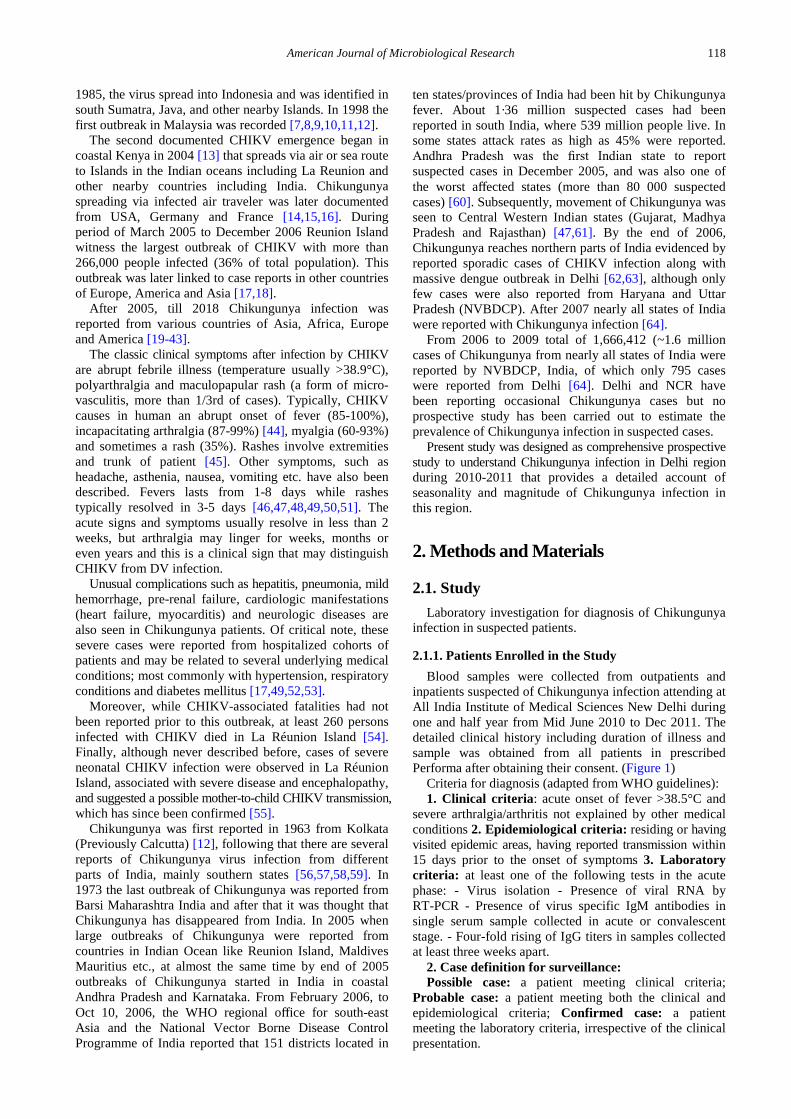
inpatients suspected of Chikungunya infection attending at All India Institute of Medical Sciences New Delhi during one and half year from Mid June 2010 to Dec 2011. The detailed clinical history including duration of illness and sample was obtained from all patients in prescribed Performa after obtaining their consent. (Figure 1)
Criteria for diagnosis (adapted from WHO guidelines): 1. Clinical criteria: acute onset of fever >38.5°C and
severe arthralgia/arthritis not explained by other medical conditions 2. Epidemiological criteria: residing or having visited epidemic areas, having reported transmission within 15 days prior to the onset of symptoms 3. Laboratory criteria: at least one of the following tests in the acute phase: - Virus isolation - Presence of viral RNA by RT-PCR - Presence of virus specific IgM antibodies in single serum sample collected in acute or convalescent stage. - Four-fold rising of IgG titers in samples collected at least three weeks apart.
2. Case definition for surveillance: Possible case: a patient meeting clinical criteria;
Probable case: a patient meeting both the clinical and epidemiological criteria; Confirmed case: a patient meeting the laboratory criteria, irrespective of the clinical presentation.
119 American Journal of Microbiological Research
Figure 1. Flow Chart of study methodology
2.1.2. Inclusion and Exclusion Criteria All patients presented with fever were including, acute
illness was defined within <15 days of onset of illness with presence of fever, and/or polyarthralgia, and/or rash. Chronic/persisting illness was defined as patients with persisting polyarthralgia and past history of fever. Patients who refuse to give consent were excluded from the study.
2.1.3. Controls Forty (40) unrelated healthy controls (20 male and 20
female) of different age group with no previous history of polyarthralgia were enrolled in the control group. Consent was also obtained from the control group.
2.1.4. Follow-up of Confirmed Cases of Chikungunya Infection
To understand the CHIKV infection progression, 75 patients were followed up for 6 months during which serum samples were collected at 15 day, 1 month, 3 month and 6-month intervals.
2.2. Sample Processing Blood was collected in plain collection vials and sent to
laboratory for investigation within 1-2 hours. Serum was separated from the blood using standard centrifugation at 5000 rpm for 15 minutes. Four aliquots were prepared for storage at -70°C until further testing was carried out.
2.3. Diagnostic Assays CHIKV infection was diagnosed by IgM capture ELISA
assay using serum samples from all suspected CHIK patients. Serum sample from suspected CHIK patients in 0-10 days duration of illness were also tested by Reverse transcriptase PCR (RT-PCR) targeting E1 gene.
2.3.1. CHIKV Specific IgM Capture ELISA All Serum samples were tested using CHIKV specific
IgM capture ELISA kit supplied by National Institute of Virology (NIV), Pune, India. Performance of ELISA and calculation of results were done by following manufacturer instructions.

American Journal of Microbiological Research 120
Table 1. Primers used for RT-PCR amplification of E1
Primer Name Sequence of primer Length of amplicon RT-PCR [65] CHIK/E1-S TACCCATTCATGTGGGGC 294 bp CHIK/E1-C GCCTTTGTACACCACGATT PCR E1 gene primer (for Cloning) [66] 10445F ACCATGCCGTCACAGTTA 804 bp 11296R ACACGCATAGCACCACGATTAGAA Real time PCR primer probe [67] CHIK E1 F TCGACGCGCCCTCTTTAA 10,865-10,882 CHIK E1 R ATCGAATGCACCGCACACT 10,973-10,991 CHIK E1 P ACCAGCCTGCACCCATTCCTCAGAC 10,902-10,926
2.3.2. RNA Extraction and PCR
RNA from serum samples was isolated using Qiagen Viral RNA extraction kit as per manufacturer’s instruction. cDNA was prepared by using avian myeloblastosis virus reverse transcriptase (Promega Inc.). Partial E1 gene of CHIKV was amplified using published primers, Taq DNA Polymerase (Banglore Biogene India)with amplicon size of 294bp ([65]; Table 1). Amplification was visualized at 2% agarose gel using ethidium bromide under Gel Doc™ XR+ System (Biorad Inc.). Standard Ross strain was used as positive control in reactions. The standard strain of Chikungunya Virus (CHIKV) strain S27-African (Ross Strain) prototype was kindly provided by national Institute of Virology (NIV), Pune, India.
2.3.3. Quantitative real-time PCR (qRT-PCR)
2.3.3.1. Cloning of E1 Gene E1 gene from RNA isolated from C6/36 cell line
cultured standard Chikungunya African strain (S-27) was amplified using primers and Pfx polymerase enzyme (Invitrogen Life Technologies, Carlsbad CA, USA) ([68]; Table 1) that encompassed the real-time primer probe binding regions (Figure 2). RNA was extracted from standard (African strain) of Chikungunya virus and cDNA was prepared with random primers and Avian myoblast virus Reverse transcriptase.
2.3.3.2. Purification of the PCR Product The PCR product was resolved on a 1.5% agarose gel
containing 0.5 µg/ml of ethidium bromide (EtBr), and the expected sized band was excised from the gel and purified using Gel extraction kit (QIAGEN GmbH, Germany), as per the manufacturer’s instruction.
2.3.3.3. A-tailing of the Gel Purified DNA and TA-cloning
The gel purified PCR product was A-tailed for TA-cloning using Taq DNA polymerase (Invitrogen Inc, USA). Ligation reaction was set up using the pGEM®T Easy vector kit (Promega Corp., Madison, WI, USA).
2.3.3.4. Competent Cell Preparation and Transformation in E.coli
E. coli DH5α competent cells were prepared by CaCl2 and transformation was done by heat shock method as described earlier [69]. Transformed cells were selected using Blue-white screening method. The white colonies
were screened for the presence of insert by colony PCR using M13 universal primer followed by DNA sequencing.
2.3.3.5. Isolation of the Recombinant Plasmid Positive colony was inoculated in 10 ml of Luria-Bertani
broth with 100 µg/ml of ampicillin and grown overnight at 37°C. Plasmid DNA was isolated using the QIAprep Spin Miniprep Kit (Qiagen Inc., Gmbh, Germany) following the manufacturer’s instructions. The recombinant plasmids were sequenced using M13F and M13R universal primers commercially.
2.3.3.6. RNA Standards for Real Time PCR InVitro Transcription, Purification and transcription:
Riboprobe system (Promega Corporation, USA) was used for in vitro transcription. The in vitro transcription reaction purification and estimation of RNA transcript was done in accordance with manufacturer’s instructions.
2.3.3.7. Real Time RT-PCR The amplification target was 127 bp of E1 gene, which
codes for the structural envelope protein E1 using published Primers and probe based on Taqman chemistry (Carolyn J. Edwards et al.2007), (Table 1; Figure 2).
The 18s rRNA internal control kit the “Ribosomal RNA control reagents” (Cat. No. 4308329) was obtained from Applied Biosystem Inc (Foster City CA, USA) and used as an endogenous/internal control for the Real Time PCR assay.
The real-time RT-PCR was carried out using the 7500 real-time PCR system (ABI, USA). A 20 μl reaction volume was used with the Superscript III Platinum one-step qRT-PCR system (Invitrogen).
The instrument by default is set to neglect any fluorescence value above baseline to be omitted between 3-15 cycles of real time RT-PCR. The ΔRn value can be translated into a quantitative result by constructing a standard curve.
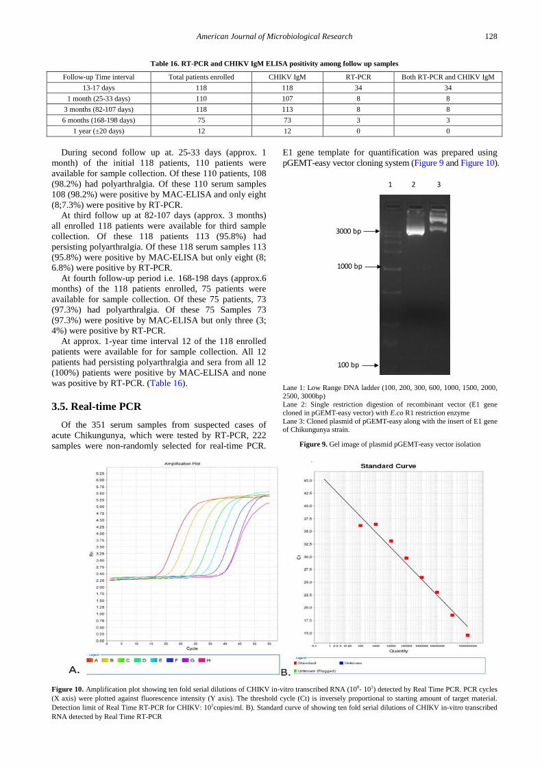
Serial RNA dilutions containing 101 to 108 copies/ml were used to generate standard curve in real-time PCR.
2.4. Data Analysis Variables were analyzed using percentage, mean and
median. Chi-square tests were used to determine statistically significant differences between groups and p-value of less than 0.05 was considered as significant.

121 American Journal of Microbiological Research
Figure 2. PCR primers binding sites used to amplify E1 gene region encompassing primer probes of real-time PCR. Nucleotide positions are given by numbers with corresponding position of standard CHIKV genome (Ross strain)
2.5. Ethical Approval Ethics approval was obtained for this study from Ethics
Committee of All India Institute of Medical Sciences, New Delhi, India (Ref. No IESC/T-343/02.09.2011 & RT-10/29.06.2012) (6-8-12).
3. Results
3.1. Socio Demographic and Clinical Data
All the patients belonged to NCR region of Delhi, India including Noida, Greater Noida, Ghaziabad and Faridabad.
Patients with acute illness (<5 days duration) During June 2010-December 2011, serum sample was
collected from total of 2112 suspected Chikungunya patients were enrolled with less than/equal to 15 days of onset of symptoms. All sera were tested using IgM
capture-ELISA (MAC-ELISA) assay. Blood samples that were received in laboratory on ice, were also tested for Chikungunya virus in serum by RT- PCR and in some samples viral load was also determined by real-time PCR assay.
Patients enrolled (2112) includes 964 (45.6%) females with age range of 6 months to 85 years (mean 33.4 years; median = 33 years) and 1148 (54.4%) males with age range of 6 months to 85 years (mean 32.1 years; median = 30 years) (Table 2; Figure 3).
Table 2. Demographics of patients enrolled with suspected Chikungunya acute illness (≤15 days duration)
Acute phase patients (n=2112)
Female (n=964) Male (n=1148)
Age range 6 mo - 85 years 6 mo - 85 years
Mean 33.4 years 32.1 years
Median 33 years 30 years
Figure 3. Age group and gender of patients enrolled with acute illness (<15 days duration)

American Journal of Microbiological Research 122
Three hundred and twenty five (325; 15%) patients were from age group of 6 months to 15 years, 720 (34%) patients belonged to age group of 16 years to 30 years, 625 (30%) patients were of age between 31 years to 45 years and 442 (21%) patients belonged to age group of 46 years to 85 years (Table 3).
Table 3. Age and gender wise distribution of suspected Chikungunya patients with acute illness (≤15 days duration)
Chikungunya Acute illness patients
Age group Total Male Female ODD ratio
6 months-15 years 325 205 120 1.7 16-30 years 720 411 309 1.3 31-45 years 625 283 342 0.8 >45 years 442 249 193 1.3
Total 2112 1148 (54.4%) 964 (45.6%) 1.2 Six hundred thirty three (633;30%) serum samples were
collected from patients with 0-5 days duration of illness, 1287 (61%) serum samples were collected from patients with 6-10 days duration of illness and 192 (9%) serum samples were collected from patients with 11-15 days duration of illness (Table 4).
Table 4. Distribution by duration of illness of suspected Chikungunya patients enrolled with acute illness (<15 days duration)
Acute illness Duration of illness Total Male Female ODD Ratio 0-5 days 633 352 281 1.3 6-10 days 1287 703 584 1.2 11-15 days 192 93 99 0.9 Total 2112 1148 964 1.2
Patients with Chronic illness Serum samples were collected from 234 patients having
16 day to 12 months duration of polyarthralgia. All samples were tested by MAC-ELISA and RT- PCR was done on samples collected on ice.
Total of two hundred and thirty four (234) patients were enrolled with chronic illness enrolled. These includes 138 (59%) females with age range of 3 years to 78 years (mean 38.1 years; median = 38.5 years) and 96 (41%) males with age range of 2 years to 80 years (mean 38 years; median = 38 years) (Table 5; Figure 4).
Table 5. Demographics of patients enrolled with persisting polyarthralgia (Chronic illness; >15 days duration)
Chronic phase patients (n=234)
Female (n=138) Male (n=96)
Age range 3 years - 78 years 2 years - 80 years
Mean 38.1 years 38 years
Median 38.5 years 38 years
Fourteen (14; 6%) patients belonged to age group of 2
years - 15 years, 60 (26%) patients belonged to age group of 16 years - 30 years, 94 (40%) patients were of age between 31 years - 45 years and 66 (28%) patients belonged to age group of 46 years - 80 years (Table 6 and Table 6).
Table 6. Age and gender wise distribution of patients with persisting polyarthralgia (Chronic illness; >15 days duration)
Chikungunya Chronic illness patients Age group Total Female Male ODD ratio 3 -15 years 14 6 8 0.75 16-30 years 60 35 25 1.4 31-45 years 94 62 32 1.9 >45 years 66 35 31 1.1
Total 234 138 (59%) 96 (41%) 1.4 One hundred thirty four (134) serum samples were
collected from patients with 16 days - 1 month duration of illness, 73 serum samples were collected from patients with 1 month- 3 months duration of illness, 14 serum samples were collected from patients with 3 months - 6 months duration of illness and 13 serum samples were collected from patients with 6 months - 1 year duration of illness (Table 7).
Table 7. Distribution by duration of illness of patients enrolled with persisting polyarthralgia (Chronic phase; >15 days duration)
Chronic illness Duration of illness Total Male Female ODD Ratio
16-1mo 134 58 76 0.8 1 mo -3 mo 73 29 44 0.7 3 mo- 6 mo 14 5 9 0.6 6 mo -1 yr 13 4 9 0.4
Total 234 96 138 0.7
Figure 4. Age group and gender of patients enrolled with chronic illness (>15 days duration)
123 American Journal of Microbiological Research
Follow-up patients Seventy five (75) patients were followed up for 6
months during which blood samples were collected at 15 day, 1 month, 3 month and 6-month time intervals. All follow-up samples were screened for persistence of CHIKV specific IgM by MAC-ELISA and Chikungunya Virus in serum by reverse transcriptase PCR.
3.2. MAC ELISA
3.2.1. Patients with Acute Illness (<5 Days Duration) Results of MAC ELISA in suspected Chikungunya patients with acute illness (<5 days duration)
All 2112 suspected serum samples from Chikungunya cases were tested for presence of CHIKV specific IgM in
serum using MAC ELISA assay. Of these, 746 (35%) were found to be positive by ELISA. This included 383 (51.3%) females and 363 (48.7%) males Most affected patients belonged to age group >30-45 yrs. and above in both genders (Table 8).
Total patients enrolled (2112) included 964 (45.6%) females of which 383 (40%) tested positive by ELISA and 1148 (54.4%) males of which 363 (32%) tested positive by ELISA (Table 8).
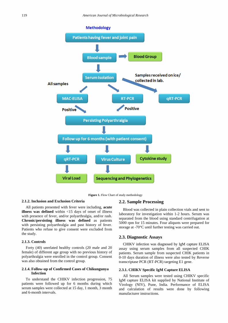
Of the 633 Samples collected from 0-5 days duration of illness, 177 (28%)% positive by ELISA, Of the 1287 Samples collected from 6-10 days duration of illness, 468 (36%) tested positive by ELISA and out of 192 Samples collected from 11-15 days duration of illness, 101 (53%) were positive. (Table 9; Figure 5).
Table 8. Age group and gender wise distribution of suspected and MAC-ELISA positive Chikungunya patients with acute illness (<15 days duration)
anti-CHIKV - ELISA results for Acute illness samples
Age group Total No. of Positive/total (%)
Female No. of Positive/total no. tested (%)
Male No. of Positive/total no. tested (%)
0-15 years 64/325 (20%) 16/120 (13%) 48/205 (23%)
16-30 years 169/720 (23%) 97/309 (31%) 72/411 (18%)
31-45 years 295/625 (47%) 168/342 (49%) 127/283 (45%)
>45 years 218/442 (49%) 102/193 (53%) 116/249 (47%)
Total 746/2112 (35%) 383/964 (40%) 363/1148 (32%)
Table 9. CHIKV IgM positivity by duration of illness in Chikungunya patients with acute illness (<15 days duration)
Duration of illness and CHIKV specific IgM persistence (Acute Phase)
Duration of illness Total samples collected MAC-ELISA positive Percentage Positivity
0-5 days 633 177 28%
6-10 days 1287 468 36%
11-15 days 192 101 53% *
Total 2112 746 35%
*p<0.001.
Figure 5. anti-CHIKV IgM positivity by duration of illness in patients with acute illness (<15 days duration)
633
1287
192177
468
101
28%
36%
53%
0
10
20
30
40
50
60
0
200
400
600
800
1000
1200
1400
0-5 days 6-10 days 11-15 days
Perc
enta
ge P
ositi
vity
(MAC
-ELI
SA)
No.
of s
ampl
es
Duration of illness
anti-CHIKV IgM positivity by duration of illness (Acute illness)
Total ELISA positive Percentage Positivity

American Journal of Microbiological Research 124
Clinical features of suspected and confirmed (by anti-CHIKV IgM ELISA) patients of Chikungunya with acute illness (<5 days duration)
All patients (2112) enrolled in study had history of fever, 908 (42.9%) patients had polyarthralgia and 76 (3.6%) had rashes on whole body or parts like face, trunk, limbs and extensors. All Patients enrolled were having 1-15 days of illness duration with mean of 7 days (median = 7 days).
All 746 patients detected positive for CHIKV specific IgM had history of fever while 559 (75%) also had polyarthralgia and 29 (3.9%) gave history of rashes on different parts of body (Table 10).
Of the 363 ELISA positive male patients 100 % complained of fever, 265 (73%) had polyarthralgia and 16 (4%) gave history of rash on body parts. Of the 383 female ELISA positive all (100%) had fever, 294 (77%) had polyarthralgia and 13 (3%) had rashes on various
body parts (Table 10). Comparison of clinical features between anti-CHIKV
IgM positive and anti-CHIKV IgM negative revealed that polyarthralgia had a high odds ratio (8.7) in IgM positive patients (Table 10).
Month wise distribution of acute Chikungunya cases (June 2010-Dec 2011)
Month wise distribution of total number of suspected acute Chikungunya patients enrolled and number positive by CHIKV specific IgM capture ELISA during the study period (June 2010- December 2011) is shown in Figure 6 and Table 11.
Maximum number of suspected cases (1679) were seen from August 2010 to November 2010. However % positivity remained high during most of the study period with maximum % positivity (59%-71%) from October 2010-January 2011.
Table 10. Comparison of clinical features among CHIKV IgM positive and CHIKV IgM negative patients of Chikungunya in acute phase (<15 days duration)
Clinical feature of confirmed (CHIKV specific IgM positive) patients and CHIKV specific IgM negative patients ODD ratio
IgM positive (n=746) IgM negative (n=1366)
Male
(n=363) Female (n=383)
Total 746
Male (n=785)
Female (n=581)
Total 1366 -
Features
Fever 363 (100%) 383 (100%) 746 785 (100%) 581 (100%) 1366 -
Polyarthralgia 265 (73%) 294 (77%) 559/746 (75%) 173 (22%) 177(30%) 350/1366 (25.6) 8.6773*
Rashes 16 (4%) 13 (3%) 29 (3.9%) 31 (4%) 16(3%) 47/1366 (3.4%) 1.1351
*p<0.001.
Table 11. Monthly distribution of suspected and confirmed by ELISA Chikungunya cases from suspected Chikungunya patients during the study period
Month of sample collection No of suspected cases enrolled No of positive by anti-CHIKV IgM ELISA Percentage positivity
Jun-10 21 1 5%
Jul-10 57 2 4%
Aug-10 492 40 8%
Sep-10 305 50 16%
Oct-10 459 271 59%
Nov-10 323 228 71%
Dec-10 36 22 61%
Jan-11 23 14 61%
Feb-11 10 2 20%
Mar-11 9 1 11%
Apr-11 11 4 36%
May-11 11 2 18%
Jun-11 11 4 36%
Jul-11 25 5 20%
Aug-11 21 6 29%
Sep-11 61 18 30%
Oct-11 121 28 23%
Nov-11 95 40 42%
Dec-11 21 8 38%
Total 2112 746 35%

125 American Journal of Microbiological Research
Figure 6. Month wise distributions of suspected and confirmed by MAC-ELISA Chikungunya cases with acute illness
3.3. Patients with Chronic Phase of Illness Results of MAC-ELISA in samples collected from patients with chronic illness
All patients (234) enrolled had past history of fever and persisting polyarthralgia, eight patients also had history of rash on body parts.
All 234 suspected cases were tested for presence of CHIKV specific IgM in serum using MAC ELISA assay. Of these, 156 (67%) were found to be positive by MAC-ELISA. This includes 92 (58.9%) females and 64
(41.1%) males Most affected patients belonged to age group >30-45 yrs. and above in both genders (Table 12).
Of the 134 Samples collected from 16 days-1month duration of illness, 84 (63%) tested positive by ELISA, Of the 73 Samples collected from more than 1month-3 months duration of illness, 58 (79%) were positive, while out the 14 Samples collected from more than 3 months-6 months duration of illness, 8 were positive and Of the 13 Samples collected from more than 6 month - 1 year duration of illness 6 (46%) tested positive. (Table 13; Figure 7).
Table 12. Age group and gender wise distribution of patients with chronic polyarthralgia and MAC-ELISA positive Chikungunya patients. (Chronic illness; >15 days duration)
anti-CHIKV IgM - ELISA results for Chronic illness samples
Age group Total No. of Positive/total no. tested (%)
Male No. of Positive/total no. tested (%)
Female No. of Positive/total no. tested (%)
3-15 years 4/14 (29%) 3/8 (38%) 1/6 (17%)
16-30 years 34/60 (57%) 13/25 (52%) 21/35 (60%)
31-45 years 67/94(71%) 26/32 (81%) 41/62 (66%)
>45 years 51/66 (77%) 22/31 (71%) 29/35 (83%)
Total 156/234 (67%) 64/96 (67%) 92/138 (67%)
Table13. anti-CHIKV IgM positivity by duration of illness in Chikungunya patients with chronic illness (>15 days duration)
Duration of illness and anti-CHIKV specific IgM persistence (Chronic Phase)
Duration of illness Total samples collected MAC-ELISA positive Percentage Positivity
16-1mo 134 84 63%
1 mo -3 mo 73 58 79%
3 mo- 6 mo 14 8 57%
6 mo -1 yr 13 6 46%
Total 234 156 67%

American Journal of Microbiological Research 126
Figure 7. CHIKV IgM positivity by duration of illness in patients with chronic illness (>15 days duration)
Clinical features of suspected and confirmed (by anti-CHIKV IgM ELISA) patients with persisting polyarthralgia (chronic illness)
All patients (234) enrolled had past history of fever and persisting polyarthralgia, eight patients also had history of rash on body parts.
All 156 patients detected positive for CHIKV specific IgM had past history of fever and persisting polyarthralgia, while 5 (3%) reported history of rashes on their body. statistical analysis
We performed paired two-tail Pearson Chi2 t-test to detect any significant correlation. We analyzed all pairs of results during acute and chronic illness. We found significant correlation among acute phase results including, ELISA (p<0.001) and PCR positivity (p<0.01) in females, ELISA positivity in patients with 11-15 days duration of illness (p<0.001), ELISA positivity in patients with
polyarthralgia (p<0.001) and PCR positivity in ELISA positive samples (p<0.001).
3.3. RT - PCR for E1 Gene in Chikungunya Patients
3.3.1. Samples from Acute Illness We tested 290 serum samples that were received on ice
for CHIKV RNA by RT-PCR. One hundred and thirty one samples (131; 45%) tested positive by RT-PCR. (Gel image of RT-PCR results is given in Figure 8).
Of the 144 Samples collected from 1-5 days duration of illness, 51 (35%)% were positive by RT-PCR, while from the 146 Samples collected from 6-10 days duration of illness, 62 (42.5%) tested positive by RT-PCR and out of 30 Samples collected from 11-15 days duration of illness, 18 (60%) were positive (Table 14).
Figure 8. Gel image of E1 gene PCR done on clinical samples. Samples 1,2,3, 4,5,6,7,8,9,10,11,13,14 are positive (Band Size 294 bp). Legends: M, 100bp marker; P =positive control; n=Negative control; 1-18= samples 1-10 = patient’s sample (1-8 are positive and 9-10 are negative).
134
73
14 13
84
58
8 6
63% 79%57%
46%
0
10
20
30
40
50
60
70
80
90
0
20
40
60
80
100
120
140
160
16-1mo 1 mo -3 mo 3 mo- 6 mo 6 mo -1 yr
Perc
enta
ge P
ositi
vity
(MAC
-ELI
SA)
No
of S
ampl
es
Duration of Illness
CHIKV IgM positivity by duartion of illness (Chronic illness)
total ELISA positive Percentage Positivity

127 American Journal of Microbiological Research
Table 14. RT-PCR and IgM positivity among acute phase (<15 days duration) samples by duration of illness
Result RT-PCR and anti-CHIKV IgM-ELISA (acute phase)
Duration of illness RT-PCR results IgM positive IgM negative Total
1-5 day PCR positive 20 (17%) 31(27%) 51
PCR Negative 18(16%) 45(40%) 63
Total 38 76 114
6-10 day PCR positive 44(30%) 18(12%) 62
PCR Negative 24(16%) 60(41%) 84
Total 68 78 146
11-15 day PCR positive 11(37%) 7(23%) 18
PCR Negative 5(17%) 7(23%) 12
Total 16 14 30
Total PCR positive = 131 Total IgM positive=122 Both (Total) IgM and RT-PCR positive =75 * Total = 290
*p<0.05.
Table 15. RT-PCR and IgM positivity among chronic phase (>15 days duration) samples by duration of illness
Result of RT-PCR and MAC-ELISA (Chronic phase)
Duration of illness RT-PCR results IgM positive IgM negative Total
16day-1mo PCR positive 11 (40%) 4 (16%) 15
PCR Negative 6(22%) 6(22%) 12
Total 17 10 27
1-mo-3mo PCR positive 14 (50) 4 (15%) 18
PCR Negative 8 (28) 2 (7%) 10
Total 22 6 28
3mo-8 mo. PCR positive 2 (33%) 0 2
PCR Negative 2(33%) 2(33%) 4
Total 4 2 6
Total PCR positive = 35 # Total IgM positive=43 # Both (Total) IgM and RT-PCR positive =27 # Total = 61
#p>0.05. Of the 114 samples collected with 0-5 days duration of
illness, 20 samples tested positive by both RT-PCR and ELISA, 31 by RT-PCR only, 18 by ELISA only and 45 tested negative by both assays.
Of the 146 samples collected with 6-10 days duration of illness, 44 samples tested positive by both RT-PCR and ELISA, 18 by RT-PCR only, 24 by ELISA only and 60 tested negative by both assays. Of 30 samples collected with 11-15 days duration of illness, 11 samples tested positive by both RT-PCR and ELISA, 7 by RT-PCR only, 5 by ELISA only and 7 tested negative by both assays (Table 15). Samples from Chronic illness
Total of sixty one (61) serum samples were tested for CHIKV RNA by RT-PCR and 35 (57%) tested positive by RT-PCR. All patients (61) had previous history of fever, and polyarthralgia, three patients also had history of rashes on body parts.
These samples included, 27 collected from patients with 16 days to 1month duration of illness, of which 15 tested positive by RT-PCR, 28 samples were collected from patients with more than one month to 3 months duration of illness and 18 of these were found positive by RT-PCR. From the samples (6) collected from patients with more than three months to 8 months duration of illness, two (2) tested positive by RT-PCR (Table 15).
Of the total thirty five (35) samples positive by RT-PCR, 27 were also positive by MAC-ELISA, and eight were negative by MAC-ELISA.
3.4. Follow up Patients During this study we enrolled total of 118 confirmed
patients who were followed up for 6 months to 1 year. One hundred eighteen (118) serum samples were collected at 15 days, 110 serum samples collected at 1 month, 118 serum sample collected at 3 months, 75 samples collected at 6 months and 12 samples were collected at 1 year duration of illness from these patients. (Table 16)
One hundred and eighteen (118) patients include 62 (52.5%) males and 56 (47.5%) females. Patients belonged to age group of 11 yrs. to 73 yrs. (mean 39.6 years; Median 40 years). MAC-ELISA and RT-PCR testing on follow-up patients samples
Follow-up samples from 118 confirmed Chikungunya patients with 13-17 days (approx.15 days) duration of illness were collected, all 118 patients had polyarthralgia. Of these 118 serum samples all were positive by MAC-ELISA but only 34 (28.8%) were positive by RT-PCR (Table 16).
American Journal of Microbiological Research 128
Table 16. RT-PCR and CHIKV IgM ELISA positivity among follow up samples
Follow-up Time interval Total patients enrolled CHIKV IgM RT-PCR Both RT-PCR and CHIKV IgM 13-17 days 118 118 34 34
1 month (25-33 days) 110 107 8 8 3 months (82-107 days) 118 113 8 8
6 months (168-198 days) 75 73 3 3 1 year (±20 days) 12 12 0 0
During second follow up at. 25-33 days (approx. 1
month) of the initial 118 patients, 110 patients were available for sample collection. Of these 110 patients, 108 (98.2%) had polyarthralgia. Of these 110 serum samples 108 (98.2%) were positive by MAC-ELISA and only eight (8;7.3%) were positive by RT-PCR.
At third follow up at 82-107 days (approx. 3 months) all enrolled 118 patients were available for third sample collection. Of these 118 patients 113 (95.8%) had persisting polyarthralgia. Of these 118 serum samples 113 (95.8%) were positive by MAC-ELISA but only eight (8; 6.8%) were positive by RT-PCR.
At fourth follow-up period i.e. 168-198 days (approx.6 months) of the 118 patients enrolled, 75 patients were available for sample collection. Of these 75 patients, 73 (97.3%) had polyarthralgia. Of these 75 Samples 73 (97.3%) were positive by MAC-ELISA but only three (3; 4%) were positive by RT-PCR.
At approx. 1-year time interval 12 of the 118 enrolled patients were available for for sample collection. All 12 patients had persisting polyarthralgia and sera from all 12 (100%) patients were positive by MAC-ELISA and none was positive by RT-PCR. (Table 16).
3.5. Real-time PCR Of the 351 serum samples from suspected cases of
acute Chikungunya, which were tested by RT-PCR, 222 samples were non-randomly selected for real-time PCR.
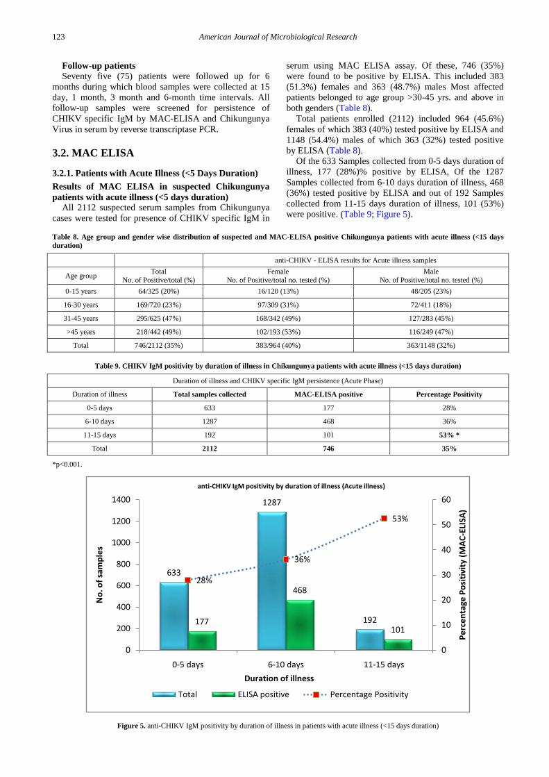
E1 gene template for quantification was prepared using pGEMT-easy vector cloning system (Figure 9 and Figure 10).
Lane 1: Low Range DNA ladder (100, 200, 300, 600, 1000, 1500, 2000, 2500, 3000bp) Lane 2: Single restriction digestion of recombinant vector (E1 gene cloned in pGEMT-easy vector) with E.co R1 restriction enzyme Lane 3: Cloned plasmid of pGEMT-easy along with the insert of E1 gene of Chikungunya strain.
Figure 9. Gel image of plasmid pGEMT-easy vector isolation
Figure 10. Amplification plot showing ten fold serial dilutions of CHIKV in-vitro transcribed RNA (108- 101) detected by Real Time PCR. PCR cycles (X axis) were plotted against fluorescence intensity (Y axis). The threshold cycle (Ct) is inversely proportional to starting amount of target material. Detection limit of Real Time RT-PCR for CHIKV: 101copies/ml. B). Standard curve of showing ten fold serial dilutions of CHIKV in-vitro transcribed RNA detected by Real Time RT-PCR
100 bp
1000 bp
3000 bp
1 2 3
129 American Journal of Microbiological Research
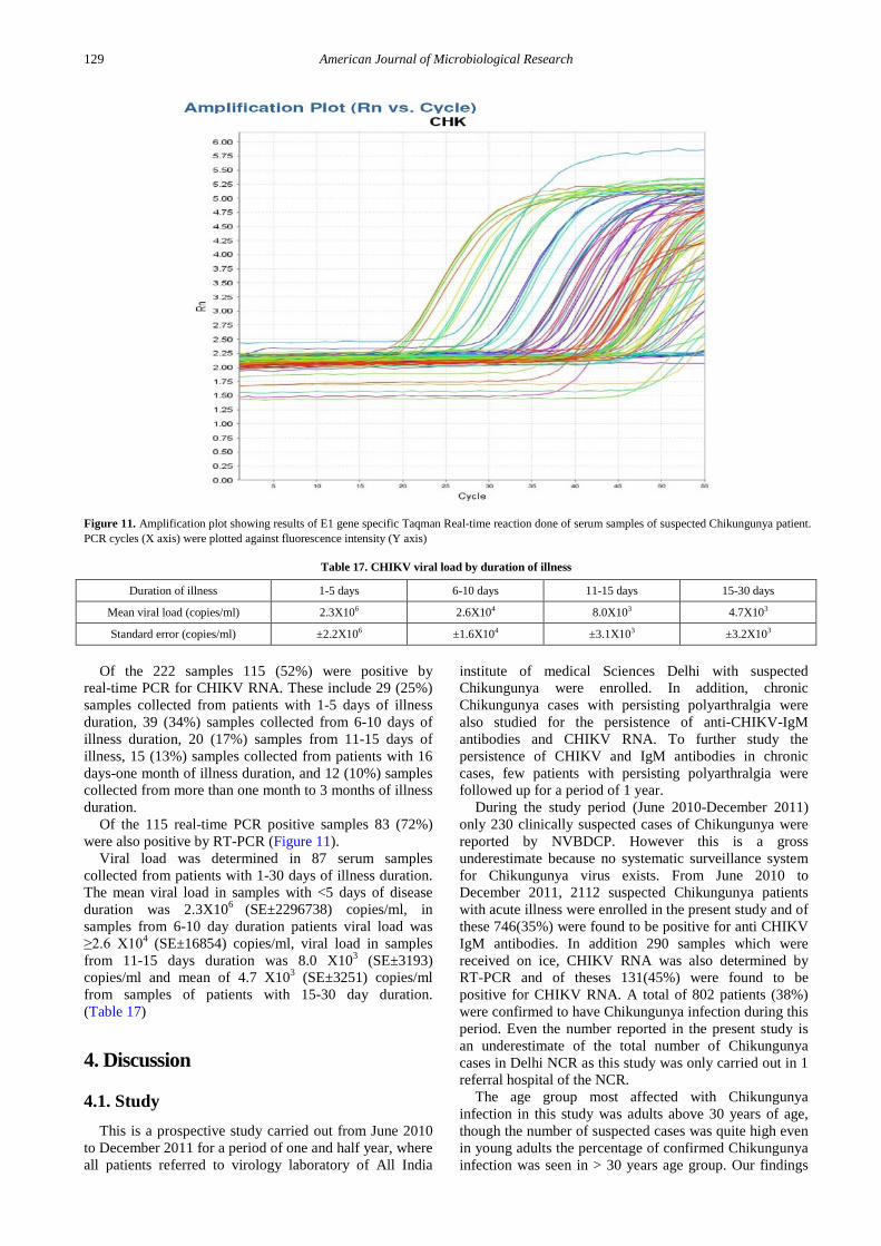
Figure 11. Amplification plot showing results of E1 gene specific Taqman Real-time reaction done of serum samples of suspected Chikungunya patient. PCR cycles (X axis) were plotted against fluorescence intensity (Y axis)
Table 17. CHIKV viral load by duration of illness
Duration of illness 1-5 days 6-10 days 11-15 days 15-30 days
Mean viral load (copies/ml) 2.3X10⁶ 2.6X10⁴ 8.0X10³ 4.7X10³
Standard error (copies/ml) ±2.2X10⁶ ±1.6X10⁴ ±3.1X10³ ±3.2X10³
Of the 222 samples 115 (52%) were positive by
real-time PCR for CHIKV RNA. These include 29 (25%) samples collected from patients with 1-5 days of illness duration, 39 (34%) samples collected from 6-10 days of illness duration, 20 (17%) samples from 11-15 days of illness, 15 (13%) samples collected from patients with 16 days-one month of illness duration, and 12 (10%) samples collected from more than one month to 3 months of illness duration.
Of the 115 real-time PCR positive samples 83 (72%) were also positive by RT-PCR (Figure 11).
Viral load was determined in 87 serum samples collected from patients with 1-30 days of illness duration. The mean viral load in samples with <5 days of disease duration was 2.3X10⁶ (SE±2296738) copies/ml, in samples from 6-10 day duration patients viral load was ≥2.6 X10⁴ (SE±16854) copies/ml, viral load in samples from 11-15 days duration was 8.0 X10³ (SE±3193) copies/ml and mean of 4.7 X10³ (SE±3251) copies/ml from samples of patients with 15-30 day duration. (Table 17)
4. Discussion
4.1. Study This is a prospective study carried out from June 2010
to December 2011 for a period of one and half year, where all patients referred to virology laboratory of All India
institute of medical Sciences Delhi with suspected Chikungunya were enrolled. In addition, chronic Chikungunya cases with persisting polyarthralgia were also studied for the persistence of anti-CHIKV-IgM antibodies and CHIKV RNA. To further study the persistence of CHIKV and IgM antibodies in chronic cases, few patients with persisting polyarthralgia were followed up for a period of 1 year.
During the study period (June 2010-December 2011) only 230 clinically suspected cases of Chikungunya were reported by NVBDCP. However this is a gross underestimate because no systematic surveillance system for Chikungunya virus exists. From June 2010 to December 2011, 2112 suspected Chikungunya patients with acute illness were enrolled in the present study and of these 746(35%) were found to be positive for anti CHIKV IgM antibodies. In addition 290 samples which were received on ice, CHIKV RNA was also determined by RT-PCR and of theses 131(45%) were found to be positive for CHIKV RNA. A total of 802 patients (38%) were confirmed to have Chikungunya infection during this period. Even the number reported in the present study is an underestimate of the total number of Chikungunya cases in Delhi NCR as this study was only carried out in 1 referral hospital of the NCR.
The age group most affected with Chikungunya infection in this study was adults above 30 years of age, though the number of suspected cases was quite high even in young adults the percentage of confirmed Chikungunya infection was seen in > 30 years age group. Our findings

American Journal of Microbiological Research 130
are similar to other studies showing that adults are more affected by Chikungunya [1,17,70,71,72,73].
Higher percentage of women (8% more) was found to have confirmed Chikungunya infection as compared to males in the present study. Similar findings have been reported in other studies from Malaysia, Reunion Island and India [17,31,74,75], which may be due to higher exposure of women to the vector. This study is first report from Delhi reporting high frequency Chikungunya among female patients.
Laboratory diagnosis for Chikungunya rests on testing serum or plasma to detect virus, viral nucleic acid, or virus-specific immunoglobulin (Ig) M and neutralizing antibodies. Viral culture may detect virus (by isolation in mosquito cells, mammalian cells or mosquitoes or mice) in the first 3 days of illness; during the first 8 days of illness, Chikungunya viral RNA can often be identified in serum (CDC). Chikungunya virus antibodies normally develop toward the end of the first week of illness (>4days) but can persist for several months even up to 1 year. Thus although detection of anti CHIKV IgM antibodies in acute phase is used for diagnosis of Chikungunya it can be present in patients even a year after the onset of disease. To definitively rule out the diagnosis, convalescent-phase samples should be obtained from patients whose acute-phase samples test negative . In the present study, all 2112 sera from suspected patients were tested for Chikungunya specific IgM antibodies using mu capture ELISA, and samples received on ice were also tested by RT-PCR for CHIKV RNA. Anti CHIKV IgM antibodies were detected in 35% of all suspected cases and CHIKV RNA was present in 45% of 290 samples tested. Anti-CHIKV positivity in suspected Chikungunya cases of 13%-80% was reported from India [70,71,73,76] and anti-CHIKV IgM positivity of 22%-80% was reported in patients from Thailand [31], and in travelers returning to France [77] and Germany [78].
CHIKV RNA is usually detected during first 8 days of illness with maximum positivity (up to 100%) in first 4-5 days of illness [31], however in the present study during first 5 days of illness 44.7% of suspected patients were positive for CHIKV RNA and in the same set of patients 33% also had CHIKV specific IgM antibodies. In patients with 6-10 days of duration of illness CHIKV RNA was seen in 42.5% of patients and CHIKV IgM antibodies were seen in 46.6% of patients. Interestingly CHIKV RNA was also detected in 60% of patients with 11-15 days duration of illness. These findings are somewhat different than other studies which have shown CHIKV RT-PCR positivity of 100% and ant-CHIKV IgM antibodies in only 10% of patients during first 4 days of illness [31]. This may be due to better patient selection in their study. Further we had to depend on patient’s history for duration of illness, which may not always be correct.
In the present study in patients with 0-5 days duration of illness CHIKV IgM antibodies were positive in 28%, in patients with 6-10 days duration of illness in 36% and in patients with 11-15 days duration of illness in 53%. Thus there was an increase in IgM positivity with increasing duration of illness. Similar increasing trends of Anti-CHIKV IgM positivity were shown in Chikungunya patients in a study from India where IgM positivity increased from 10% to 95% from day 1 to 7 post onset of
illness. Similarly in a study from Thailand IgM positivity in confirmed patients increased from day 1 and became 100% by day 7-8 and then remained at 100% till Day >17 [31].
In acute phase common clinical features seen in Chikungunya patients are fever, polyarthralgia, and rashes these can also differentiate Chikungunya from other similar infections [1,39,79]. Other atypical symptoms including neurological manifestation were reported in Indian Patients [71,80]. Chikungunya associated deaths were also reported in Chikungunya confirmed patients in La reunion [17]. In the present study of 746 anti-CHIKV IgM positive patients all gave history of fever, 75% complained of polyarthralgia, while only 3.9% complained of rashes on body parts. No case of encephalitis or mortality was seen in this study. In other studies from India 95-100% of Chikungunya patients had fever, 62%-80% had joint pains and 10-36% had rashes [70,74,81,82].
In this study maximum number of Chikungunya cases were seen during September 2010 to November 2010 and then during September to November of 2011 that is during and post monsoon season in North India. Chikungunya is known to be transmitted by mosquito vector belonging to Aedes species with most common agents are Aedes aegypti and A. albopictus mosquitoes [83]. Although both Aedes aegypti and A. albopictus mosquitoes are prevalent in India, the former is the main vector of Chikungunya virus and is prevalent in tropical areas. Both mosquitoes require high humidity and high temperature for the breeding [68]. During our study period, in 2010 National Capital territory of India witnessed a heavy rainfall during monsoon season, which provided the ideal conditions for the breeding of mosquitoes and for transmission of Chikungunya Virus that results in large number of Chikungunya infections. It also explains that although Delhi was considered to be a low prevalence zone for Chikungunya, but during the study period Delhi emerged as a major center of Chikungunya infection.
To further assess the presence of CHIKV RNA and IgM antibodies over time, 118 Chikungunya confirmed patients with persistent polyarthralgia were followed up with 75 patients being followed up till 6 months and 12 till 1 year. Of these 118 patients positive for anti-CHIKV IgM antibodies persistence of anti-CHIKV IgM was seen till 3 months in 95.7% of patients where as at 6 months follow up only 75 samples were available of which 73 were positive for anti-CHIKV IgM antibodies. CHIKV RNA was present in 34 of these patients at 2 weeks and could be detected in 3 patients even at 6 months. At 1 year only 12 samples were available all were anti-CHIKV IgM positive and none were positive for CHIKV RNA. This is a the only study as far as we know where anti-CHIKV IgM antibodies, RNA and information on persistence of polyarthralgia was collected in follow up samples. In a previous study from India anti-CHIKV IgM antibodies have been shown to persist for 2-4 months in 52% of patients with persistent polyarthralgia and in only 1.2% patients at 6-8 months. In both recovered and chronic disease group ant-CHIKV IgM antibodies were shown to persist up to 12 months in a study from la reunion island [46]. However no studies are available on the persistence of CHIKV RNA in blood for a long time in chronic disease.

131 American Journal of Microbiological Research
In experimental studies in mice persistence of CHIKV RNA in joint-associated tissues has been shown for at least 16 weeks. Inoculation of Rag1−/− mice, which lack T and B cells, resulted in higher viral levels in a variety of tissues, suggesting that adaptive immunity controls the tissue specificity and persistence of CHIKV infection. The presence of CHIKV RNA in tissues of wild-type and Rag1−/− mice was associated with histopathological evidence of synovitis, arthritis, and tendonitis. Further these authors showed that CHIKV-induced persistent arthritis is not mediated primarily by adaptive immune responses but musculoskeletal tissue pathology is caused by persistent CHIKV infection and controlled by adaptive immune responses [84]. In experimental study using immunocompetent adult nonhuman primates, CHIKV persists was seen in lymphoid tissues, liver, joints, and muscles for up to 3 months and is able to replicate in spleen, liver, and muscle for extended periods. Furthermore, results of in situ hybridization assays suggested that the virus persists mainly in mononuclear cells and, to a lesser extent, in endothelial cells [85].
However, human studies where CHIKV RNA can be demonstrated in joint tissues in persistent polyarthralgia are ethically difficult to carry out but even persistence of viral RNA in blood in this study points towards continued viral replication at low level in body may be joint tissues.
Chikungunya virus confirmed by RT-PCR (10-40%) in serum samples from acute illness (<7 days) had been reported in studies from India, Thailand, La reunion, Italy, and Germany [31,43,45,70,78,82,86]. It has been reported that during first 4 days of illness CHIKV RNA can be detected in 80-100% of patients where as IgM antibody is detected in only 10% of patients [31]. In the present study during first 5 days of illness CHIKV RNA was detected in 44.7% of 114 patients and IgM antibodies in 28% of 633 patients Chikungunya viral RNA can often be identified in serum of first 4 days illness duration patients (CDC, USA). In the present study presence of CHIKV RNA in patients sera of acute and chronic illness (1 day-3 months) was detected by RT-PCR for E1 gene. Sixty eight percent (68%) of the RT-PCR positive serum samples were from patients with 1- 10 days of illness duration, RT-PCR percentage positivity was seen to decrease with increased duration of illness. Persistent arthralgia
There are very few studies on long term clinical outcome of CHIKV infection and earlier studies showed lower prevalence of persistent arthralgia. However studies carried out after the Indian Ocean epidemic showed a much higher rate of persistent arthralgia. Persisting polyarthralgia in chronic phase of disease has been previously reported for 1 year to 3 years in different studies from south Africa, Singapore, France, Germany, La Reunion, Colombia, USA and India [15,45,48,74,87,88,89,90,91,92,93,94,95,96].
Persisting polyarthralgia has been seen in Chikungunya patients in studies from India and La Reunion [45,97], however in some studies presence of viral markers including anti CHIKV IgM was reported but CHIKV RNA in chronic illness has not been studied. In the present study 234 patients with persisting polyarthralgia during chronic illness, were tested for presence of viral markers
i.e. anti CHIKV IgM antibodies and CHIKV RNA. Persisting polyarthralgia was complained by all patients
during >16 days-1 year. Thirty three (33)% patients complained of Polyarthralgia but were negative for anti-CHIKV IgM antibodies. Polyarthralgia is a common feature of Chikungunya infection but it is also associated with other diseases. In a study from La Reunion, DC Andrade et al. 2010 reported persisting polyarthralgia in 16% chronic Chikungunya patients with 1 month duration of illness, in 33% patients with 3 months duration, and in 53% with 10 months duration of illness. In previous studies persisting polyarthralgia for 1 month - 30 months was reported among Chikungunya patients from Indian states including Kerala, Tamil Nadu, Karnataka, Assam, Maharashtra [73,97,98,99,100] and other countries including France south Africa, Singapore, France, Germany and La Reunion [15,48,92,93,94,95,96,101,102].
4.2. Anti-CHIKV IgM Anti-CHIKV IgM antibodies was seen with 63%-79%
positivity during 1month to 3 months duration of illness and positivity was declined with increasing duration of illness with 57% by 6 months and 46% by 1 year. In previous reports presence of anti-CHIKV IgM for 1 month to 18 months in chronic illness patients was reported from India, Thailand, Germany, La Reunion and Singapore CHIKV specific IgM [15,31,45,48,70,94,98,99].
Similarly, CHIKV RNA was detected in 55-64% of samples with 1 month - 3 months illness duration; CHIKV RNA was also detected in two patients at 8 months duration. Hoarau et al. [45] also reported presence of CHIKV in synovial macrophage in chronic Chikungunya patients with 18 months of illness duration.
4.3. Viral Load High Chikungunya viral load (108copies/ml) was seen
in patients with <7 days duration of illness in serum from acute phase Chikungunya patients from La reunion Island [93,103]. In the present study Chikungunya viral loads were estimated in serum of Chikungunya patients with 1-30 days duration of illness. Maximum viral load (106 copies/ml) was seen in serum samples taken from <5 days of illness, following that viral load exponentially reduced with increasing duration of illness with minimum of 103 copies/ml in serum of patients with 15-30 days of illness. In previous studies from India, CHIKV Viral load of 107-108copies/ml has been seen in Chikungunya patients with acute illness (<7 days) from Madhya Pradesh, Andhra Pradesh and Karnataka [81,104]. Similar reduction in CHIKV load with increasing duration of illness were previous reports from La Reunion, Italy, Germany and Singapore [78,86,92,93,103,105]. Furthermore, presence of Chikungunya virus for 30 days duration also confirm the idea of long term Chikungunya persistence by Laurent et al. [106] who previously reported 103 copies of CHIKV in 12 day illness duration samples. In experimental studies CHIKV persistence was seen for 15 days and 2 months post CHIKV inoculation in mouse model and nonhuman primate macaques, respectively [85,107].

American Journal of Microbiological Research 132
5. Conclusion
Thus, this is the first comprehensive prospective study of Chikungunya infection in Delhi NCR region, which describes the epidemiology and prevalence of disease. This study suggests higher percentage of affected women as compared to male. This study also suggest the need of well-organized surveillance for Chikungunya infection to report accurate number of cases in NCR region.
Funding Agency Study was carry out by the funds provided by
Department of biotechnology MHRD, Government of India under Indo Australia collaborative project funding scheme.
References [1] Robinson, Marion C. “An epidemic of virus disease in Southern
Province, Tanganyika territory, in 1952-1953.” Transactions of the Royal Society of Tropical Medicine and Hygiene. 49, no. 1 (1955): 28-32.
[2] Strauss, James H., and Ellen G. Strauss. “The alphaviruses: gene expression, replication, and evolution.” Microbiological reviews 58, no. 3 (1994): 491.
[3] Simizu, B., K. Yamamoto, K. Hashimoto, and T. Ogata. “Structural proteins of Chikungunya virus.” Journal of virology. 51, no. 1 (1984): 254-258.
[4] Diallo, Mawlouth, Joselyn Thonnon, Mumouni Traore-Lamizana, and Didier Fontenille. “Vectors of Chikungunya virus in Senegal: current data and transmission cycles.” The American journal of tropical medicine and hygiene60, no. 2 (1999): 281-286.
[5] Kumar, N. Pradeep, Rajan Joseph, T. Kamaraj, and P. Jambulingam. “A226V mutation in virus during the 2007 chikungunya outbreak in Kerala, India.”Journal of General Virology 89, no. 8 (2008): 1945-1948.
[6] McIntosh, B. M., H. E. Paterson, J. M. Donaldson, and J. De Sousa. “Chikungunya virus: viral susceptibility and transmission studies with some vertebrates and mosquitoes.” S Afr J Med Sci. 28 (1963): 45-52.
[7] Powers, Ann M., and Christopher H. Logue. “Changing patterns of chikungunya virus: re-emergence of a zoonotic arbovirus.” Journal of General Virology. 88, no. 9 (2007): 2363-2377.
[8] Mackenzie, J. S., K. B. Chua, P. W. Daniels, B. T. Eaton, H. E. Field, R. A. Hall, K. Halpin et al. “Emerging viral diseases of Southeast Asia and the Western Pacific.” Emerging infectious diseases. 7, no. 3 Suppl (2001): 497.
[9] Burt, Felicity J., Micheal S. Rolph, Nestor E. Rulli, Suresh Mahalingam, and Mark T. Heise. “Chikungunya: a re-emerging virus.” The Lancet. 379, no. 9816 (2012): 662-671.
[10] Lahariya, Chandrakant, and S. K. Pradhan. “Emergence of chikungunya virus in Indian subcontinent after 32 years: a review.” Journal of vector borne diseases. 43, no. 4 (2006): 151.
[11] Rao, T. Ramachandra. “Immunological surveys of arbovirus infections in South-East Asia, with special reference to dengue, chikungunya, and Kyasanur Forest disease.” Bulletin of the World Health Organization. 44, no. 5 (1971): 585.
[12] Sarkar, J. K., K. M. Pavri, S. N. Chatterjee, S. K. Chakravarty, and C. R. Anderson. “Virological and Serological Studies of Cases of Haemorrhagic Fever in Calcutta. Material Collected by the Calcutta School of Tropical Medicine.”The Indian journal of medical research. 52 (1964): 684-691.
[13] Chretien, Jean-Paul, Assaf Anyamba, Sheryl A. Bedno, Robert F. Breiman, Rosemary Sang, Kibet Sergon, Ann M. Powers et al. “Drought-associated chikungunya emergence along coastal East Africa.” The American journal of tropical medicine and hygiene. 76, no. 3 (2007): 405-407.
[14] Lanciotti, Robert S., Olga L. Kosoy, Janeen J. Laven, Amanda J. Panella, Jason O. Velez, Amy J. Lambert, and Grant L. Campbell.
“Chikungunya virus in US travelers returning from India, 2006.” Emerging infectious diseases. 13, no. 5 (2007): 764.
[15] Taubitz, Winfried, Jakob P. Cramer, Anette Kapaun, Martin Pfeffer, Christian Drosten, Gerhard Dobler, Gerd D. Burchard, and Thomas Löscher. “Chikungunya fever in travelers: clinical presentation and course.” Clinical Infectious Diseases. 45, no. 1 (2007): e1-e4.
[16] Hochedez, P., P. Hausfater, S. Jaureguiberry, F. Gay, A. Datry, M. Danis, F. Bricaire, and P. Bossi. “Cases of chikungunya fever imported from the islands of the South West Indian Ocean to Paris, France.” Euro surveillance: bulletin Europeen sur les maladies transmissibles European communicable disease bulletin. 12, no. 1 (2007).
[17] Renault, Philippe, Jean-Louis Solet, Daouda Sissoko, Elsa Balleydier, Sophie Larrieu, Laurent Filleul, Christian Lassalle et al. “A major epidemic of chikungunya virus infection on Reunion Island, France, 2005-2006.” The American journal of tropical medicine and hygiene 77, no. 4 (2007): 727-731.
[18] Borgherini, Gianandrea, Patrice Poubeau, Frederik Staikowsky, Manuella Lory, Nathalie Le Moullec, Jean Philippe Becquart, Catherine Wengling, Alain Michault, and Fabrice Paganin. “Outbreak of chikungunya on Reunion Island: early clinical and laboratory features in 157 adult patients.” Clinical infectious diseases. 44, no. 11 (2007): 1401-1407.
[19] Sanchez, F., Barboza, L.A., Burton, D. and Cintrón-Arias, A., 2018. Comparative analysis of dengue versus chikungunya outbreaks in Costa Rica. Ricerche di Matematica, 67(1), pp.163-174.
[20] Paul, B.J. and Sadanand, S., 2018. Chikungunya Infection: A Re-emerging Epidemic. Rheumatology and therapy, pp.1-10.
[21] Calba, C., Guerbois-Galla, M., Franke, F., Jeannin, C., Auzet-Caillaud, M., Grard, G., Pigaglio, L., Cadiou, B., de Lamballerie, X., Paty, M.C. and Leparc-Goffart, I., 2018. Investigation of an autochthonous chikungunya outbreak, July-September 2017, France. Revue d'Épidémiologie et de Santé Publique, 66, pp.S387-S388.
[22] Venturi, G., Di Luca, M., Fortuna, C., Remoli, M.E., Riccardo, F., Severini, F., Toma, L., Del Manso, M., Benedetti, E., Caporali, M.G. and Amendola, A., 2017. Detection of a chikungunya outbreak in Central Italy, August to September 2017. Eurosurveillance, 22(39).
[23] Delisle, E., Rousseau, C., Broche, B., Leparc-Goffart, I., L’ambert, G., Cochet, A., Prat, C., Foulongne, V., Ferre, J.B., Catelinois, O. and Flusin, O., 2015. Chikungunya outbreak in montpellier, France, September to October 2014. Eurosurveillance, 20(17), p.21108.
[24] Weaver, Scott C. “Arrival of Chikungunya Virus in the New World: Prospects for Spread and Impact on Public Health.” PLOS Neglected Tropical Diseases 8, no. 6 (2014): e2921.
[25] Leparc-Goffart, Isabelle, Antoine Nougairede, Sylvie Cassadou, Christine Prat, and Xavier de Lamballerie. “Chikungunya in the Americas.” Lancet 383, no. 9916 (2014): 514.
[26] Wangchuk, Sonam, Piyawan Chinnawirotpisan, Tshering Dorji, Tashi Tobgay, Tandin Dorji, In-Kyu Yoon, and Stefan Fernandez. “Chikungunya fever outbreak, Bhutan, 2012.” Emerging infectious diseases 19, no. 10 (2013): 1681.
[27] Wu, De, Jie Wu, Qiaoli Zhang, Haojie Zhong, Changwen Ke, and Xiaoling Deng. “Chikungunya outbreak in Guangdong province, China, 2010.” Emerging infectious diseases 18, no. 3 (2012): 493.
[28] Duong, Veasna, Anne-Claire Andries, Chantha Ngan, Touch Sok, Beat Richner, Nima Asgari-Jirhandeh, Steve Bjorge et al. “Reemergence of Chikungunya virus in Cambodia.” Emerging infectious diseases 18, no. 12 (2012): 2066.
[29] Grandadam, Marc, Valérie Caro, Sébastien Plumet, Jean-Michel Thiberge, Yvan Souarès, Anna-Bella Failloux, Hugues J. Tolou et al. “Chikungunya virus, southeastern France.” Emerging infectious diseases 17, no. 5 (2011): 910.
[30] Gould, E. A., P. Gallian, X. De Lamballerie, and R. N. Charrel. “First cases of autochthonous dengue fever and chikungunya fever in France: from bad dream to reality!.” Clinical microbiology and infection 16, no. 12 (2010): 1702-1704.
[31] Rianthavorn, Pornpimol, Kesmanee Prianantathavorn, Norra Wuttirattanakowit, Apiradee Theamboonlers, and Yong Poovorawan. “An outbreak of chikungunya in southern Thailand from 2008 to 2009 caused by African strains with A226V mutation.” International Journal of Infectious Diseases 14 (2010): e161-e165.

133 American Journal of Microbiological Research
[32] Paupy, Christophe, Benjamin Ollomo, Basile Kamgang, Sara Moutailler, Dominique Rousset, Maurice Demanou, Jean-Pierre Hervé, Eric Leroy, and Frédéric Simard. “Comparative role of Aedes albopictus and Aedes aegypti in the emergence of dengue and chikungunya in Central Africa.” Vector-Borne and Zoonotic Diseases 10, no. 3 (2010): 259-266.
[33] Leo, Yee S., Angela LP Chow, Li Kiang Tan, David C. Lye, Li Lin, and Lee C. Ng. “Chikungunya outbreak, Singapore, 2008.” Emerging infectious diseases15, no. 5 (2009): 836.
[34] Ng, Lisa FP, Angela Chow, Yong-Jiang Sun, Dyan JC Kwek, Poh-Lian Lim, Frederico Dimatatac, Lee-Ching Ng et al. “IL-1β, IL-6, and RANTES as biomarkers of Chikungunya severity.” PloS one 4, no. 1 (2009): e4261.
[35] Leroy, Eric M., Dieudoné Nkoghe, Benjamin Ollomo, Chimène Nze-Nkogue, Pierre Becquart, Gilda Grard, Xavier Pourrut et al. “Concurrent chikungunya and dengue virus infections during simultaneous outbreaks, Gabon, 2007.”Emerging infectious diseases 15, no. 4 (2009): 591.
[36] Sam, I., Yoke Fun Chan, Shie Yien Chan, Shih Keng Loong, Hock Khim Chin, Poh Sim Hooi, Rajasekaram Ganeswrie, and Sazaly AbuBakar. “Chikungunya virus of Asian and central/east African genotypes in Malaysia.” Journal of Clinical Virology 46, no. 2 (2009): 180-183.
[37] Schuffenecker, Isabelle, Isabelle Iteman, Alain Michault, Séverine Murri, Lionel Frangeul, Marie-Christine Vaney, Rachel Lavenir et al. “Genome microevolution of chikungunya viruses causing the Indian Ocean outbreak.” PLoS medicine 3, no. 7 (2006): e263.
[38] Charrel, Remi N., Xavier de Lamballerie, and Didier Raoult. “Chikungunya outbreaks-the globalization of vectorborne diseases.” New England Journal of Medicine. 356, no. 8 (2007): 769.
[39] WHO http://www.who.int/csr/don/2006_10_17/en/ [40] Mavalankar, Dileep, Priya Shastri, and Parvathy Raman.
“Chikungunya epidemic in India: a major public-health disaster.” The Lancet infectious diseases 7, no. 5 (2007): 306-307.
[41] AbuBakar, Sazaly, I-Ching Sam, Pooi-Fong Wong, NorAziyah MatRahim, Poh-Sim Hooi, and Nuruliza Roslan. “Reemergence of endemic chikungunya, Malaysia.” Emerging infectious diseases 13, no. 1 (2007): 147.
[42] Peyrefitte, Christophe N., Maël Bessaud, Boris AM Pastorino, Patrick Gravier, Sébastien Plumet, Olivier L. Merle, Isabelle Moltini et al. “Circulation of Chikungunya virus in Gabon, 2006-2007.” Journal of medical virology 80, no. 3 (2008): 430-433.
[43] Rezza, G., L. Nicoletti, R. Angelini, R. Romi, A. C. Finarelli, M. Panning, P. Cordioli et al. “Infection with chikungunya virus in Italy: an outbreak in a temperate region.” The Lancet 370, no. 9602 (2007): 1840-1846.
[44] Mizuno, Yasutaka, et al. “Clinical and radiological features of imported chikungunya fever in Japan: a study of six cases at the National Center for Global Health and Medicine.” Journal of Infection and Chemotherapy 17.3 (2011): 419-423.
[45] Hoarau, Jean-Jacques, Marie-Christine Jaffar Bandjee, Pascale Krejbich Trotot, Trina Das, Ghislaine Li-Pat-Yuen, Bérengère Dassa, Mélanie Denizot et al. “Persistent chronic inflammation and infection by Chikungunya arthritogenic alphavirus in spite of a robust host immune response.” The Journal of Immunology 184, no. 10 (2010): 5914-5927.
[46] Sissoko, Daouda, Khaled Ezzedine, Amrat Moendandzé, Claude Giry, Philippe Renault, and Denis Malvy. “Field evaluation of clinical features during chikungunya outbreak in Mayotte, 2005-2006.” Tropical Medicine & International Health 15, no. 5 (2010): 600-607.
[47] Chhabra, M., V. Mittal, D. Bhattacharya, U. V. S. Rana, and S. Lal. “Chikungunya fever: a re-emerging viral infection.” Indian Journal of Medical Microbiology 26, no. 1 (2008): 5.
[48] Win, M. K., A. Chow, F. Dimatatac, C. J. Go, and Y. S. Leo. “Chikungunya fever in Singapore: acute clinical and laboratory features, and factors associated with persistent arthralgia.” Journal of Clinical Virology 49, no. 2 (2010): 111-114.
[49] Pistone, Thierry, Khaled Ezzedine, Marie Boisvert, Marie‐Catherine Receveur, Isabelle Schuffenecker, Hervé Zeller, Marie‐Edith Lafon, Hervé Fleury, and Denis Malvy. “Cluster of chikungunya virus infection in travelers returning from Senegal, 2006.” Journal of travel medicine 16, no. 4 (2009): 286-288.
[50] Chopra, Arvind, V. Anuradha, V. Lagoo-Joshi, V. Kunjir, S. Salvi, and M. Saluja. “Chikungunya virus aches and pains: an emerging challenge.” Arthritis & Rheumatism 58, no. 9 (2008): 2921-2922.
[51] Rolle, A., Schepers, K., Cassadou, S., Curlier, E., Madeux, B., Hermann-Storck, C., Fabre, I., Lamaury, I., Tressieres, B., Thiery, G. and Hoen, B., 2016. Severe sepsis and septic shock associated with chikungunya virus infection, Guadeloupe, 2014. Emerging infectious diseases, 22(5), p.891.
[52] Powers, Ann M. “Chikungunya.” Clinics in laboratory medicine30, no. 1 (2010): 209-219.
[53] Staples, J. Erin, Robert F. Breiman, and Ann M. Powers. “Chikungunya fever: an epidemiological review of a re-emerging infectious disease.” Clinical Infectious Diseases 49, no. 6 (2009): 942-948.
[54] Josseran, Loïc, Christophe Paquet, Abdelkrim Zehgnoun, Nadège Caillere, Alain Le Tertre, Jean-Louis Solet, and Martine Ledrans. “Chikungunya disease outbreak, Reunion Island.” Emerging infectious diseases 12, no. 12 (2006): 1994.
[55] Ramful, Duksha, Magali Carbonnier, Marlene Pasquet, Brahim Bouhmani, Jamal Ghazouani, Tahir Noormahomed, Gilles Beullier et al. “Mother-to-child transmission of Chikungunya virus infection.” The Pediatric infectious disease journal 26, no. 9 (2007): 811-815.
[56] Ravi, V. “Re-emergence of chikungunya virus in India.” Indian journal of medical microbiology 24, no. 2 (2006): 83.
[57] Dandawate, C. N., K. V. Thiruvengadam, V. Kalyanasundaram, J. Rajagopal, and T. R. Rao. “Serological survey in Madras city with special reference to chikungunya.” The Indian journal of medical research 53, no. 8 (1965): 707-714.
[58] Jadhav, M., M. Namboodripad, R. H. Carman, D. E. Carey, and R. M. Myers. “Chikungunya disease in infants and children in Vellore: a report of clinical and haematological features of virologically proved cases.” The Indian journal of medical research. 53, no. 8 (1965): 764.
[59] Thiruvengadam, K. V., V. Kalyanasundaram, and J. Rajgopal. “Clinical and pathological studies on chikungunya fever in Madras city.” (1965).
[60] Pialoux, Gilles, Bernard-Alex Gaüzère, Stéphane Jauréguiberry, and Michel Strobel. “Chikungunya, an epidemic arbovirosis.” The Lancet infectious diseases 7, no. 5 (2007): 319-327.
[61] Dash, P. K., et al. “East Central South African genotype as the causative agent in reemergence of Chikungunya outbreak in India.” Vector-Borne and Zoonotic Diseases 7.4 (2007): 519-528.
[62] Chahar, Harendra S., Preeti Bharaj, Lalit Dar, Randeep Guleria, Sushil K. Kabra, and Shobha Broor. “Co-infections with chikungunya virus and dengue virus in Delhi, India.” Emerging infectious diseases 15, no. 7 (2009): 1077.
[63] Kukreti, Himani, Artee Chaudhary, R. S. Rautela, Ranjana Anand, Veena Mittal, Mala Chhabra, D. Bhattacharya, Shiv Lal, and Arvind Rai. “Emergence of an independent lineage of dengue virus type 1 (DENV-1) and its co-circulation with predominant DENV-3 during the 2006 dengue fever outbreak in Delhi.” International journal of infectious diseases 12, no. 5 (2008): 542-549.
[64] NVBDCP http://www.nvbdcp.gov.in/Chikun-main.html. [65] Hasebe, F., M. C. Parquet, B. D. Pandey, E. G. M. Mathenge, K.
Morita, V. Balasubramaniam, Z. Saat et al. “Combined detection and genotyping of Chikungunya virus by a specific reverse transcription-polymerase chain reaction.” Journal of medical virology 67, no. 3 (2002): 370-374.
[66] Santhosh, S. R., et al. “Comparative full genome analysis revealed E1: A226V shift in 2007 Indian Chikungunya virus isolates.” Virus Research 135.1 (2008): 36-41.
[67] Edwards, Carolyn J., et al. “Molecular diagnosis and analysis of Chikungunya virus.” Journal of clinical virology 39.4 (2007): 271-275.
[68] Santhosh, S. R., P. K. Dash, M. M. Parida, M. Khan, M. Tiwari, and P. V. Lakshmana Rao. “Comparative full genome analysis revealed E1: A226V shift in 2007 Indian Chikungunya virus isolates.” Virus research 135, no. 1 (2008): 36-41.
[69] Sambrook, Joseph, Edward F. Fritsch, and Tom Maniatis. Molecular cloning: a laboratory manual. No. Ed. 2. Cold spring harbor laboratory press, 1989.
[70] Ray, Pratima, Vinod H. Ratagiri, Sushil K. Kabra, Rakesh Lodha, Sumit Sharma, B. S. Sharma, Mani Kalaivani, and Naveet Wig.

American Journal of Microbiological Research 134
“Chikungunya infection in India: results of a prospective hospital based multi-centric study.”PloS one 7, no. 2 (2012): e30025.
[71] Suryawanshi, S. D., A. H. Dube, R. K. Khadse, S. V. Jalgaonkar, P. S. Sathe, S. D. Zawar, and M. P. Holay. “Clinical profile of chikungunya fever in patients in a tertiary care centre in Maharashtra, India.” (2009).
[72] Kaur, Prabhdeep, Manickam Ponniah, Manoj V. Murhekar, Vidya Ramachandran, Ramakrishnan Ramachandran, Hari Kishan Raju, Vanamail Perumal, Akhilesh C. Mishra, and Mohan D. Gupte. “Chikungunya outbreak, south India, 2006.” Emerging infectious diseases 14, no. 10 (2008): 1623-1625.
[73] Yergolkar, Prasanna N., Babasaheb V. Tandale, Vidya A. Arankalle, Padmakar S. Sathe, Sudeep AB, Swati S. Gandhe, Mangesh D. Gokhle, George P. Jacob, Supriya L. Hundekar, and Akhilesh C. Mishra. “Chikungunya outbreaks caused by African genotype, India.” Emerging infectious diseases 12, no. 10 (2006): 1580.
[74] Chopra, Arvind, and Anuradha Venugopalan. “Persistent rheumatic musculoskeletal pain and disorders at one year post-chikungunya epidemic in south Maharashtra—a rural community based observational study with special focus on naïve persistent rheumatic musculoskeletal cases and selected cytokine expression.” Indian Journal of Rheumatology 6, no. 1 (2011): 5-11.
[75] Selvavinayagam, T. S. “Chikungunya fever outbreak in Vellore, south India.”Indian Journal of community medicine 32, no. 4 (2007): 286.
[76] Shrinet, Jatin, Shanu Jain, Anil Sharma, Shashi Shekhar Staikowsky Staikowsky, Kalika Mathur, Vandita Rana, Raj K. Bhatnagar et al. “Genetic characterization of Chikungunya virus from New Delhi reveal emergence of a new molecular signature in Indian isolates.” Virol J 9 (2012): 100.
[77] Win, M. K., et al. “Chikungunya fever in Singapore: acute clinical and laboratory features, and factors associated with persistent arthralgia.” Journal of Clinical Virology 49.2 (2010): 111-114.
[78] Panning, Marcus, Klaus Grywna, Marjan Van Esbroeck, Petra Emmerich, and Christian Drosten. “Chikungunya fever in travelers returning to Europe from the Indian Ocean region, 2006.” Emerging infectious diseases 14, no. 3 (2008): 416.
[79] CDC http://wwwnc.cdc.gov/eid/article/14/10/07-0569_article. [80] Lewthwaite, Penny, Ravi Vasanthapuram, Jane C. Osborne, Ashia
Begum, Jenna LM Plank, M. Veera Shankar, Roger Hewson et al. “Chikungunya virus and central nervous system infections in children, India.” Emerging infectious diseases 15, no. 2 (2009): 329.
[81] Soni, Manisha, Anil Kumar Singh, Shashi Sharma, Ankita Agarwal, Natarajan Gopalan, P. V. Rao, Manmohan Parida, and Paban Kumar Dash. “Molecular and Virological Investigation of a Focal Chikungunya Outbreak in Northern India.” The Scientific World Journal 2013 (2013).
[82] Singh, Priyanka, Veena Mittal, Moshahid A. Rizvi, Dipesh Bhattacharya, Mala Chhabra, Devendra S. Rawat, Rattan L. Ichhpujani, Lakhvir S. Chauhan, and Arvind Rai. “Northward movement of East Central South African genotype of Chikungunya virus causing an epidemic between 2006-2010 in India.” The Journal of Infection in Developing Countries 6, no. 07 (2012): 563-571.
[83] Lambrechts, Louis, Thomas W. Scott, and Duane J. Gubler. “Consequences of the expanding global distribution of Aedes albopictus for dengue virus transmission.” PLoS neglected tropical diseases 4, no. 5 (2010): e646.
[84] Hawman, David W., Kristina A. Stoermer, Stephanie A. Montgomery, Pankaj Pal, Lauren Oko, Michael S. Diamond, and Thomas E. Morrison. “Chronic joint disease caused by persistent Chikungunya virus infection is controlled by the adaptive immune response.” Journal of virology 87, no. 24 (2013): 13878-13888.
[85] Labadie, Karine, Thibaut Larcher, Christophe Joubert, Abdelkrim Mannioui, Benoit Delache, Patricia Brochard, Lydie Guigand et al. “Chikungunya disease in nonhuman primates involves long-term viral persistence in macrophages.”The Journal of clinical investigation 120, no. 3 (2010): 894-906.
[86] Staikowsky, Frederik, François Talarmin, Philippe Grivard, Abdel Souab, Isabelle Schuffenecker, Karin Le Roux, Marc Lecuit, and Alain Michault. “Prospective study of Chikungunya virus acute infection in the Island of La Réunion during the 2005-2006 outbreak.” PLoS One 4, no. 10 (2009): e7603.
[87] Bouquillard, E., Fianu, A., Bangil, M., Charlette, N., Ribéra, A., Michault, A., Favier, F., Simon, F. and Flipo, R.M.,
2018. Rheumatic manifestations associated with Chikungunya virus infection: A study of 307 patients with 32-month follow-up (RHUMATOCHIK study). Joint Bone Spine, 85(2), pp.207-210.
[88] Bonifay, T., Lienne, J.F., Bagoée, C., Santa, F., Vesin, G., Walter, G., Nacher, M., Vaserman, N., Djossou, F. and Epelboin, L., 2018. Prevalence and risk factors of post chikungunya rheumatic musculoskeletal disorders: a prospective follow-up study in French Guiana. European Journal of Clinical Microbiology & Infectious Diseases, pp.1-6.
[89] Rodriguez-Morales, A.J., Restrepo-Posada, V.M., Acevedo-Escalante, N., Rodríguez-Muñoz, E.D., Valencia-Marín, M., Castrillón-Spitia, J.D., Londoño, J.J., Bedoya-Rendón, H.D., de Jesús Cárdenas-Pérez, J., Cardona-Ospina, J.A. and Lagos-Grisales, G.J., 2017. Impaired quality of life after chikungunya virus infection: a 12-month follow-up study of its chronic inflammatory rheumatism in La Virginia, Risaralda, Colombia. Rheumatology international, 37(10), pp.1757-1758.
[90] Rodríguez-Morales, A.J., Calvache-Benavides, C.E., Giraldo-Gómez, J., Hurtado-Hurtado, N., Yepes-Echeverri, M.C., García-Loaiza, C.J., Patiño-Barbosa, A.M., Sabogal-Roman, J.A., Patiño-Valencia, S., Hidalgo-Zambrano, D.M. and Vásquez-Serna, H., 2016. Post-chikungunya chronic arthralgia: results from a retrospective follow-up study of 131 cases in Tolima, Colombia. Travel medicine and infectious disease, 14(1), pp.58-59.
[91] Feldstein, L.R., Rowhani-Rahbar, A., Staples, J.E., Weaver, M.R., Halloran, M.E. and Ellis, E.M., 2017. Persistent arthralgia associated with chikungunya virus outbreak, US Virgin Islands, December 2014-February 2016. Emerging infectious diseases, 23(4), p.673.
[92] Chow, Angela, Zhisheng Her, Edward KS Ong, Jin-miao Chen, Frederico Dimatatac, Dyan JC Kwek, Timothy Barkham et al. “Persistent arthralgia induced by Chikungunya virus infection is associated with interleukin-6 and granulocyte macrophage colony-stimulating factor.” Journal of Infectious Diseases 203, no. 2 (2011): 149-157.
[93] Larrieu, Sophie, Nicolas Pouderoux, Thierry Pistone, Laurent Filleul, Marie-Catherine Receveur, Daouda Sissoko, Khaled Ezzedine, and Denis Malvy. “Factors associated with persistence of arthralgia among Chikungunya virus-infected travellers: report of 42 French cases.” Journal of Clinical Virology 47, no. 1 (2010): 85-88.
[94] Borgherini, Gianandrea, Patrice Poubeau, Annie Jossaume, Arnaud Gouix, Liliane Cotte, Alain Michault, Claude Arvin-Berod, and Fabrice Paganin. “Persistent arthralgia associated with chikungunya virus: a study of 88 adult patients on reunion island.” Clinical Infectious Diseases 47, no. 4 (2008): 469-475.
[95] Simon, Fabrice, Philippe Parola, Marc Grandadam, Sabrina Fourcade, Manuela Oliver, Philippe Brouqui, Pierre Hance et al. “Chikungunya infection: an emerging rheumatism among travelers returned from Indian Ocean islands. Report of 47 cases.” Medicine 86, no. 3 (2007): 123-137.
[96] Brighton, S. W., O. W. Prozesky, and A. L. De La Harpe. “Chikungunya virus infection.” A retrospective study of 107 (1983): 313-315.
[97] Ramachandran, V., P. Kaur, K. Kanagasabai, S. Vadivoo, and M. V. Murhekar. “Persistent arthralgia among Chikungunya patients and associated risk factors in Chennai, South India.” Journal of postgraduate medicine 60, no. 1 (2014): 3.
[98] Vijayakumar, Krishna Pillai, Thekkumkara Surendran Nair Anish, Biju George, Tony Lawrence, Sujina C. Muthukkutty, and Reshmi Ramachandran. “Clinical profile of chikungunya patients during the epidemic of 2007 in Kerala, India.”Journal of global infectious diseases 3, no. 3 (2011): 221.
[99] Mohan, Alladi, D. H. N. Kiran, I. Chiranjeevi Manohar, and D. Prabath Kumar. “Epidemiology, clinical manifestations, and diagnosis of Chikungunya fever: lessons learned from the re-emerging epidemic.” Indian journal of dermatology55, no. 1 (2010): 54.
[100] Ganu, Mohini A., and A. S. Ganu. “Post-chikungunya chronic arthritis—our experience with DMARDs over two year follow up.” J Assoc Physicians India 59 (2011): 83-6.
[101] Dupuis-Maguiraga, Laurence, Marion Noret, Sonia Brun, Roger Le Grand, Gabriel Gras, and Pierre Roques. “Chikungunya disease: infection-associated markers from the acute to the chronic phase of arbovirus-induced arthralgia.”PLoS neglected tropical diseases 6, no. 3 (2012): e1446.

135 American Journal of Microbiological Research
[102] de Andrade, Daniel C., Sylvain Jean, Pierre Clavelou, Radhouane Dallel, and Didier Bouhassira. “Chronic pain associated with the Chikungunya Fever: long lasting burden of an acute illness.” BMC infectious diseases 10, no. 1 (2010): 31.
[103] Parola, Philippe, Xavier De Lamballerie, Jacques Jourdan, Clarisse Rovery, Véronique Vaillant, Philippe Minodier, Philippe Brouqui, Antoine Flahault, Didier Raoult, and Rémi N. Charrel. “Novel chikungunya virus variant in travelers returning from Indian Ocean islands.” Emerging infectious diseases. 12, no. 10 (2006): 1493.
[104] Parida, M. M., S. R. Santhosh, P. K. Dash, N. K. Tripathi, V. Lakshmi, N. Mamidi, A. Shrivastva et al. “Rapid and real-time detection of Chikungunya virus by reverse transcription loop-mediated isothermal amplification assay.” Journal of clinical microbiology. 45, no. 2 (2007): 351-357.
[105] Carletti, Fabrizio, Licia Bordi, Roberta Chiappini, Giuseppe Ippolito, Maria R. Sciarrone, Maria R. Capobianchi, Antonino Di Caro, and Concetta Castilletti. “Rapid detection and quantification
of Chikungunya virus by a one-step reverse transcription-polymerase chain reaction real-time assay.” The American journal of tropical medicine and hygiene 77, no. 3 (2007): 521-524.
[106] Laurent, Philippe, Karin Le Roux, Philippe Grivard, Gérard Bertil, Florence Naze, Miguel Picard, Frédérik Staikowsky, Georges Barau, Isabelle Schuffenecker, and Alain Michault. “Development of a sensitive real-time reverse transcriptase PCR assay with an internal control to detect and quantify chikungunya virus.” Clinical chemistry. 53, no. 8 (2007): 1408-1414.
[107] Dhanwani, Rekha, Mohsin Khan, Vinay Lomash, Putcha Venkata Lakshmana Rao, Hinh Ly, and Manmohan Parida. “Characterization of Chikungunya Virus Induced Host Response in a Mouse Model of Viral Myositis.” PloS one. 9, no. 3 (2014): e92813.
[108] Inamadar, Arun C., Aparna Palit, V. V. Sampagavi, S. Raghunath, and N. S. Deshmukh. “Cutaneous manifestations of chikungunya fever: observations made during a recent outbreak in south India.” International journal of dermatology47, no. 2 (2008): 154-159.
© The Author(s) 2020. This article is an open access article distributed under the terms and conditions of the Creative Commons Attribution (CC BY) license (http://creativecommons.org/licenses/by/4.0/).