a comprehensive fate map by intracellular injection … › nmeyer › files › 2011 › 11 ›...
TRANSCRIPT

Meyer et al. EvoDevo 2010, 1:8http://www.evodevojournal.com/content/1/1/8
Open AccessRESEARC H
© 2010 Meyer et al; licensee BioMed Central Ltd. This is an Open Access article distributed under the terms of the Creative CommonsAttribution License (http://creativecommons.org/licenses/by/2.0), which permits unrestricted use, distribution, and reproduction inany medium, provided the original work is properly cited.
ResearchA comprehensive fate map by intracellular injection of identified blastomeres in the marine polychaete Capitella teletaNéva P Meyer1, Michael J Boyle2, Mark Q Martindale1 and Elaine C Seaver*1
AbstractBackground: The polychaete annelid Capitella teleta (formerly Capitella sp. I) develops by spiral cleavage and has been the focus of several recent developmental studies aided by a fully sequenced genome. Fate mapping in polychaetes has lagged behind other spiralian taxa, because of technical limitations.
Results: To generate a modern fate map for C. teleta, we injected 1,1'-dioctadecyl-3,3,3'3'-tetramethylindocarbocyanine perchlorate (DiI) into individual identified blastomeres through fourth-quartet micromere formation. Confocal laser scanning microscopy at single-cell resolution was used to characterize blastomere fates during larval stages. Our results corroborate previous observations from classic studies, and show a number of similarities with other spiralian fate maps, including unique and stereotypic fates for individual blastomeres, presence of four discrete body domains arising from the A, B, C and D cell quadrants, generation of anterior ectoderm from first quartet micromeres, and contributions to trunk ectoderm and ventral nerve cord by the 2d somatoblast. Of particular interest are several instances in which the C. teleta fate map deviates from other spiralian fate maps. For example, we identified four to seven distinct origins of mesoderm, all ectomesodermal. In addition, the left and right mesodermal bands arise from 3d and 3c, respectively, whereas 4d generates a small number of trunk muscle cells, the primordial germ cells and the anus. We identified a complex set of blastomere contributions to the posterior gut in C. teleta, which establishes the most complete map of posterior gut territories to date.
Conclusions: Our detailed cellular descriptions reveal previously underappreciated complexity in the ontogenetic contributions to several spiralian larval tissues, including the mesoderm, nervous system and gut. The formation of the mesodermal bands by 3c and 3d is in stark contrast to other spiralians, in which 4d generates the mesodermal bands. The results of this study provide a framework for future phylogenetic comparisons and functional analyses of cell-fate specification.
BackgroundMany metazoan embryos develop via highly stereotypedcleavage programs that enable the identification of indi-vidual blastomeres during early development. Embryonicfeatures that aid identification include differences in cellsize or pigmentation, or in spindle orientation relative tothe primary egg axis. Such embryos are amenable to cell-lineage and fate-mapping studies, which establish thedevelopmental origins of definitive regions, tissues andorgans in larval and adult animals, and provide the
groundwork for functional studies. Cell-lineage and fate-mapping studies were among the first rigorous attemptsat characterizing embryogenesis in the late 19th centuryin embryos as diverse as parasitic nematodes, ascidians,ctenophores, annelids, mollusks and various othermarine invertebrates. The ability to follow descendants ofidentified cells has increased dramatically in recent yearswith the advent of improved fluorescent reagents forintracellular labeling and advanced imaging techniques.
Early cell-lineage and fate-mapping studies revealedthat animals with dissimilar adult body plans probablyshared a common evolutionary ancestor, based on similardevelopmental features. A spectacular example is a groupof animals that displays a pattern of early development
* Correspondence: [email protected] Kewalo Marine Laboratory, Pacific Biosciences Research Center, University of Hawaii, 41 Ahui Street, Honolulu, Hawaii 96813, USAFull list of author information is available at the end of the article

Meyer et al. EvoDevo 2010, 1:8http://www.evodevojournal.com/content/1/1/8
Page 2 of 27
called spiral cleavage. This pattern of development is rec-ognizable by the timing, orientation and/or size of indi-vidual cell divisions, and is found in a large number ofdiverse animal groups including mollusks, annelids,sipunculans, echiurans, nemerteans, myzostomids, ecto-procts, polyclad flatworms and potentially gnathostomu-lids. Molecular phylogenomic analyses [1-4] haveindicated that the spiral cleavage program was probablyan ancestral characteristic of all non-ecdysozoan protos-tomes (lophotrochozoans) that was subsequently lost inselect taxa including lophophorates (brachiopods,phoronids), gastrotrichs, rotifers, parasitic (non-poly-clad) platyhelminthes and cephalopod mollusks. How-ever, a better understanding of the exact relationshipsamong lophotrochozoans, particularly between membersof the Platyzoa (for example, gnathostomulids, gas-trotrichs, rotifers and platyhelminthes), is needed todetermine whether spiral cleavage was an ancestral char-acter for all lophotrochozoans (Spiralia) or just a subset(Trochozoa).
During spiral cleavage, the cleavage spindles of the firsttwo divisions are oriented perpendicular to the animal-vegetal axis, and divide the zygote into four quadrants.The cells born from these first divisions are denoted theA, B, C and D blastomeres (Figure 1A). In animals withunequal spiral cleavage, the first two divisions areunequal in size, allowing for unambiguous identificationof each blastomere. Starting with the third cleavage, thefour macromeres generate tiers of often smaller, animaldaughters (micromeres) in alternating orientation (Figure1A). In the majority of spiralians, the first tier of microm-eres is cleaved in a clockwise (dexiotropic) directionwhen viewed from the animal pole. This is followed by acounterclockwise cleavage (laeotropic) of the second tierof micromeres. These divisions result from the alternat-ing 90 degree orientation of the mitotic spindles, and pro-duce a 'spiral' pattern of micromeres, the hallmark of thespiral cleavage program. Blastomere nomenclature fol-lows that of Conklin [5]. Macromeres are denoted by anupper case letter and micromeres by a lower case letter,corresponding to their quadrant of origin (Figure 1A).Each blastomere has a number corresponding to its birthorder. For example, after the first spiral cleavage (eight-cell stage), the macromeres are named 1A, 1B, 1C and 1D,and the micromeres are named 1a, 1b, 1c and 1d; after thesecond spiral cleavage (16-cell stage), the macromeres are2A, 2B, 2C and 2D and the micromeres are 2a, 2b, 2c and2d (Figure 1A). Subsequent micromere divisions aredenoted by numerical superscripts, a '1' for the animal-most daughter and a '2' for the vegetal daughter. Forexample, first quartet micromeres divide to give rise tofour animal micromere cells (1a1, 1b1, 1c1 and 1d1) andfour vegetal micromere cells (1a2, 1b2, 1c2 and 1d2).
In addition to the conserved pattern of spiral cleavage,blastomere fates are also largely conserved. Generally,animal micromeres give rise to ectoderm, whereas vege-tal macromeres give rise to endoderm. Another fre-quently conserved fate is that of the left and right larvaleyes, which are generated by micromeres 1a and 1c,respectively. Of particular importance in spiralian devel-opment are two cells derived from the D quadrant. Inannelids, the 2d micromere, called the primary somato-blast, gives rise to the majority of trunk ectoderm poste-rior to the mouth. The 4d cell, called the mesentoblast, isthe only micromere in any spiralian embryo that gener-ates both mesoderm and endoderm [6,7].
Although early descriptive work on spiralian embryosemphasized similarities in the fates of identified cells,modern intracellular studies have identified a number ofspecies-specific differences. For example, the larval eyesof chitons (polyplacophoran mollusks) are generatedfrom the second quartet micromeres 2a and 2c, ratherthan from 1a and 1c as observed in all other speciesexamined [8]. Likewise, the origin of ectomesoderm,which is mesoderm derived from the first three quartetsof ectodermal micromeres, varies across species and con-trasts with the highly conserved origin of endomesodermderived from 4d [7,9-11]. Differences in blastomere fateamong species probably reflect meaningful phylogeneticvariation in the development of homologous cells overevolutionary time, and provide a foundation for molecu-lar investigations of the causal basis of cell-fate determi-nation.
Polychaete annelids are a widely diverse group of primar-ily marine segmented worms. Although there are classiccell-lineage descriptions from the late 19th century, gen-eration of fate maps for polychaetes using intracellularlineage tracers has lagged behind those of other taxa. Theonly published intracellular fate-mapping study of a poly-chaete annelid is for the ragworm Platynereis dumerilii,in which the first quartet micromeres (1a to 1d) and mac-romeres (1A to 1D), 2d112, 4d and 4d1 were directly filled[12]. Although there are substantial detailed lineage datafor clitellid annelids (leeches and oligochetes), these ani-mals have a modified spiral cleavage program that givesrise to a specialized set of ectodermal and mesodermalteloblast cells not described in polychaetes. To generate amodern fate map for the polychaete annelid Capitellateleta [13], previously known as Capitella sp. I, weinjected the fluorescent dye 1,1'-dioctadecyl-3,3,3'3'-tetramethylindocarbocyanine perchlorate (DiI) intracel-lularly into identified blastomeres, and examined theirfate at larval stages using confocal laser scanning micros-copy. We determined the fates of each blastomerethrough formation of the fourth quartet of micromeres.The results of this study are compared with fate maps of

Meyer et al. EvoDevo 2010, 1:8http://www.evodevojournal.com/content/1/1/8
Page 3 of 27
other spiralian embryos, with particular emphasis onthose fate maps generated using intracellular injections.
ResultsC. teleta development and fate map overviewC. teleta embryos develop by unequal spiral cleavage,thus individual blastomeres are easily identifiable. Start-ing at the two-cell stage, divisions occur approximatelyevery hour and are roughly synchronous between quad-rants, although the D quadrant generally begins dividingfirst. At the four-cell stage, the D macromere is the largestcell, and shares a vegetal cross furrow with B. In thispaper, we use a lower case 'q' to refer to each micromerequartet and an upper case 'Q' to refer to each macromerequartet. The first quartet micromeres (1q) are born dex-iotropically (clockwise) with respect to the macromereswhen viewed from the animal pole; second quartetmicromeres (2q) are born laeotropically (counterclock-wise), and subsequent macromere cleavages alternatebetween dexiotropic and laeotropic. Our observations ofthe early cleavages of C. teleta are very similar to thedescriptions of Capitella capitata cleavages by Eisig in
1898 [14]. One notable exception is the size of 4d: Eisigdescribes 4d as much larger than other fourth quartetmicromeres, whereas in C. teleta, we found 4d to be thesame size as other 4q cells.
A standard embryonic and larval staging system hasbeen described previously for C. teleta [15]. In general,after 5 days of development at 19°C, the majority of larvaland adult structures are discernable. At this stage (latestage 6, early stage 7), the larva consists of an anteriorhead region, a segmented trunk and a posterior pygid-ium. The trunk is bounded by two ciliary bands: the pro-totroch (pt) anteriorly, and the telotroch (tt) posteriorly(Figure 1B). A third ciliary band, the neurotroch (nt),runs along the ventral midline (Figure 1B). There are alsorows of cilia in the pygidium called the pygidial ciliaryband (not shown in diagram) [16]. The larva has a cen-tralized nervous system consisting of an anterior brain orcerebral ganglion (br) and a ventral nerve cord (vn) con-sisting of up to 13 segmentally reiterated ganglia (Figure1B). The cerebral commissure (cm) and pair of larval eyes(ey) are also visible (Figure 1B). The mesoderm (ms) ispositioned between the ectoderm (ec) and endoderm (en)
Figure 1 Spiral cleavage and Capitella teleta larval body plan. (A) Diagram of unequal spiral cleavage. (B) Diagram of a late stage 6/early stage 7 Capitella teleta larva. Mesoderm is shown in red. an = anus, br = brain, cm = commissure, ec = ectoderm, en = endoderm, es = esophagus, ey = eye, fg = foregut, gl = gut lumen, mg = midgut, mo = mouth, ms = mesoderm, nt = neurotroch, ph = pharynx, pt = prototroch, rc = rectum, tt = telotroch, vn = ventral nerve cord.

Meyer et al. EvoDevo 2010, 1:8http://www.evodevojournal.com/content/1/1/8
Page 4 of 27
(Figure 1B), and many differentiated circular and longitu-dinal muscle fibers are present by this stage. The gut isregionalized along the anterioposterior axis into a foregut(fg), midgut (mg) and hindgut. The foregut is further sub-divided into a buccal cavity, pharynx (ph) and esophagus(es), and we used the term 'mouth' (mo) to refer to thecells lining the opening of the buccal cavity (Figure 1B).At mid to late larval stages, the mouth is continuous withthe presumptive pharynx and esophagus [17]The midgutin C teleta comprises an intestine that extends from theesophagus to the rectum. Traditionally, the 'hindgut' inpolychaetes is described as a proctodeal invagination ofectoderm [18,19]. To more accurately interpret and com-pare the C. teleta fate map with other spiralian fate maps,we used the terms 'rectum' and 'anus' when referring tothe posteriormost end (hindgut) of the alimentary canal.In the larva of C. teleta, the rectum (rc) is a short regionin the pygidium that connects the intestine with a termi-nal anus (an) (Figure 1B). By late stage 6, a lumen (gl) isvisible within the developing midgut and rectum (Figure1B). Late stage 6 C. teleta larvae are competent to meta-morphose after another 3 to 4 days of development at19AC (stage 9).
The injection of individual blastomeres resulted inclones of labeled descendant cells that were highly repro-ducible, enabling us to generate a stereotypic fate map forC. teleta. Blastomeres 1q, 1q1, 1q2, 2q, 3q, 4d, 2Q, 3Q and4D were injected with DiI (Table 1), allowed to develop tostages 5 to 8, and scored as alive or fixed. Most animalswere scored between stages 6 and 7 because visualizationof DiI at later stages (8 and 9) is difficult in large clonesbecause of the dilution of DiI. In brief, first quartetmicromeres generate the anterior ectoderm including thebrain and prototroch (Figure 2A-D). The second quartetmicromeres generate the ectoderm posterior to the pro-totroch, the ventral nerve cord, portions of the mouth,the majority of the foregut, a single posterior row of pro-totrochal cells, the telotroch and rectum (Figure 2E-H).The third quartet of micromeres give rise to portions of
the foregut and mouth, anterior mesoderm, cells sur-rounding the anus, and the left and right mesodermalbands in the trunk (Figure 2I-L). 4d forms a few musclecells, the anus and the primordial germ cells (Figure 2M).Finally, macromeres 3A, 3B, 3C and 4D generate endo-derm (Figure 2N-R). This fate map is largely consistentwith the fate map of other spiralians, especially withrespect to the ectodermal fates. The main deviation isthat 4d does not generate the mesodermal bands; this fateis divided between 3c and 3d.
First quartet micromeresIn C. teleta, descendents of the first quartet micromeresare subdivided between the left-right and dorsal-ventralquadrants of the anterior ectoderm (1a, left-ventral; 1b,right-ventral; 1c, right-dorsal; 1d, left-dorsal) (Figure 3A-D). Micromeres 1a to 1d give rise to the anterior ecto-derm, brain (or cerebral ganglion), larval eyes and theprototroch (Figure 3).Micromere 1aDescendants of the 1a micromere form left-ventral headectoderm and the left-ventral prototroch (Figure 3A, E).In C. teleta, there are two larval eyes, each consisting ofthree cells: a sensory cell, a pigment cell and a supportingcell [20]. Micromere 1a clearly forms the pigment cell (pc)and sensory cell of the left eye, including the microvilli ofthe sensory apparatus (sc mv), which are visible withphalloidin staining (Figure 3I). Micromere 1a also proba-bly generates the supporting cell of the left eye, althoughthis is more difficult to determine. In addition to the left-ventral head ectoderm, left-ventral prototroch and lefteye, a small number of cells in the left side of the brain,ventral to the cerebral commissure (cm), are descendantsof 1a (Figure 3E, arrowhead).Micromere 1bThe 1b micromere generates right-ventral head ecto-derm, the right-ventral prototroch and a small number ofcells in the right-ventral brain (arrowhead) (Figure 3B, F).This pattern largely mirrors that of 1a descendants, with
Table 1: Number of larvae scored after injection of individual identified blastomeres.
Micromeres Macromeres
a b c d A B C D
First quartet 12 15 13 18 - - - -
Second quartet 17 12 27 22 2 4 17 18
Third quartet 18 14 22 22 13 10 9 20
Fourth quartet - - - 23 - - - 11

Meyer et al. EvoDevo 2010, 1:8http://www.evodevojournal.com/content/1/1/8
Page 5 of 27
the exception of the left eye (compare Figure 3E with Fig-ure 3F).Micromere 1cMicromere 1c gives rise to right-dorsal head ectoderm,the right-dorsal prototroch (Figure 3C, G) and the righteye (Figure 3J). Similar to the 1a micromere, the 1cmicromere forms the pigment cell, the sensory cell andprobably the supporting cell of the right eye (Figure 3J).Micromere 1c also forms the majority of the right side ofthe brain (br) (Figure 3G, K). DiI-labeled cells in the brainare often positioned dorsal to the cerebral commissure,and DiI is seen in the cerebral commissure (Figure 3G).Descendants of 1c are also found in ectoderm just poste-rior to the prototroch, both on the dorsal midline and on
the dorsolateral sides of the larva (not shown). Some ofthese cells may be sensory neurons.Micromere 1dThe 1d micromere generates left-dorsal head ectoderm,the left-dorsal prototroch (Figure 3D, H) and the majorityof the left side of the brain (br) (Figure 3L). DiI-labeledcells in the brain are usually positioned dorsal to the cere-bral commissure, and DiI is seen in the cerebral commis-sure (Figure 3H). Descendants of 1d also give rise to athin line of ectodermal cells and scattered surface cells inthe trunk. The line of ectodermal cells forms a ring thatpartially encircles the larva, terminating on the ventralface, just lateral to the mouth. This ring of 1d-derivedcells is positioned posterior to the mouth, between two
Figure 2 General fate map of Capitella teleta. (A-M) Stage 6 to 7 larvae ~5 days after labeling the first to fourth quartet micromeres. (N-R) Stage 6 to 7 larvae ~5 days after labeling the third and fourth quartet macromeres. In all panels, differential interference contrast (DIC) images are overlaid with red DiI fluorescent images. All images are of fixed larvae except for (D) and (H) and (I), which are of live animals. The arrow in (D) indicates a row of DiI-labeled ectodermal cells posterior to the prototroch. The blastomere labeled with DiI is indicated in the upper-right corner, and the view is indi-cated in the lower-left corner of each panel (dors = dorsal, vent = ventral). Anterior is to the left in all panels.

Meyer et al. EvoDevo 2010, 1:8http://www.evodevojournal.com/content/1/1/8
Page 6 of 27
rows of pigment cells (Figure 2D, arrow). The scatteredtrunk ectodermal cells formed by 1d are localized to thedorsal and dorsolateral sides of the larva, and are posi-tioned between the prototroch and line of 1d ectodermalcells (not shown). Some of these cells are sensory neu-rons, whereas others have a distinct 'S'-shaped morphol-ogy. Phalloidin staining of the 'S'-shaped cells showsrepeated actin rings along the outside of each cell. Cellswith this 'S'-shaped morphology are found throughoutthe surface ectoderm in the head, trunk and pygidium.The pattern generated by micromere 1d largely mirrorsthat seen after labeling 1c, with the exception of the righteye and ectodermal cells in the trunk (compare Figure 3Gand 3K with Figure 3H and 3L).
Micromeres 1q1 and 1q2
The vegetal daughters of the first quartet micromeres, 1a2
to 1d2 (Figure 4), were labeled because in many other spi-ralians, these cells generate most of the prototroch. Weexamined clones generated by 1q2 micromeres both atlate stage 6 and earlier at stage 5 when the prototochalcells are larger and cell boundaries are easier to discern.In C. teleta, we detected five rows of prototrochal cells(Figure 5A). At late stage 6, the second and fourth rows ofprototrochal cells are densely ciliated, whereas the firstand fifth rows are more sparsely ciliated. The third rowmay also be densely ciliated, but we could not determinethis at late stage 6. At late stage 4 and early stage 5, cells inthe second, third and fourth rows are relatively large
Figure 3 First quartet micromeres generate anterior ectoderm and prototroch. (A-D) Single-channel, z-stacks of confocal images of late stage 6 larvae from an anterior view 5 days after labeling first quartet micromeres with DiI (white). The stacks start at the anterior ectodermal surface and end just posterior to the prototroch. (E-H) Z-stacks of merged, confocal images through a subset of the brain in late stage 6 larvae. The channels are DiI (red), phalloidin (green) and TO-PRO-3 (blue). (E, F) Labeled brain cells positioned ventral to the cerebral commissure are indicated with an arrow-head in (E,F). (I-J) Digital magnification of the eye region bracketed in (E) and (G), respectively. One side of the eye sensory cell is outlined with a dashed line. (K-L) Z-stacks of merged, confocal images through the entire brain of late stage 6 larvae. The channels are DiI (red) and TO-PRO-3 (blue). The blastomere labeled with DiI is indicated in the upper-right corner, and the view is indicated in the lower-left corner of each panel (ant = anterior). All images are from an anterior view, with ventral down. The position of the mouth is indicated with an asterisk. br = brain, cm = brain commissure, pc = pigment cell, sc mv = sensory cell microvilli.

Meyer et al. EvoDevo 2010, 1:8http://www.evodevojournal.com/content/1/1/8
Page 7 of 27
compared with cells in the first and fifth rows (Figure 5A).Because of the pattern of ciliation and the size of the cellsin each row, we refer to the second through fourth rowsas the main prototroch, numbered 1 to 3 from anterior toposterior (m1, m2, m3, respectively). We refer to the firstrow as the anterior supporting prototroch (aT) and thefifth row as the posterior supporting prototroch (pT).
The 1q2 micromeres of C. teleta generate the majorityof the densely ciliated main prototroch, including all ofthe fourth row (m3), most of the third row (m2) and asubset of the second row (m1) of prototrochal cells (Fig-ure 4; Figure 5C,D). In general, 1a2 forms the left-ventral
region (n = 11; Figure 4A-C'), 1b2 forms the right-ventralregion (n = 15; Figure 4D-F'), 1c2 forms the right-dorsalregion (n = 13; Figure 4G-I') and 1d2 forms the left-dorsalregion (n = 14; Figure 4J-L'). The main exception to thisoccurs laterally. Clones from 1c2 and 1d2 generate a largercircumferential area than do those from 1a2 and 1b2
(compare Figure 4H and 4K with 4B and 4E; Figure5C,D). Additionally, on the lateral sides of the prototroch,1c2 and 1d2 only contribute to m2 and m3 (Figure 4H-I',K-L'; Figure 5C,D).
We also labeled the 1q1 blastomeres (n = 5 for eachmicromere) to determine whether these cells contribute
Figure 4 Vegetal daughters of first quartet micromeres, 1q2, form the prototroch. (A, B, D, E, G, H, J, K) Z-stacks of merged, confocal images through late stage 6 larvae 5 days after labeling 1q2 micromeres with DiI. The channels are DiI (red), phalloidin (green) and TO-PRO-3 (blue). (C, F, I, L) Cropped, digitally magnified, z-stacks of merged, confocal images through the prototroch of late stage 6 larvae. The channels are DiI (red), phalloidin (green) and TO-PRO-3 (blue). (C', F', I', L') Single channel images of DiI shown in (C), (F), (I) and (L), respectively. The arrow in (L) points to a DiI-labeled prototroch cell that is separate from the rest of the clone. The blastomere labeled with DiI is indicated to the left of each row and the view is indicated in the lower-left corner of each panel (dors = dorsal, vent = ventral, llat = left lateral, rlat = right lateral). Anterior is to the left in all ventral, dorsal and left lateral images, and to the right in all right lateral images. The position of the mouth is indicated with an asterisk.

Meyer et al. EvoDevo 2010, 1:8http://www.evodevojournal.com/content/1/1/8
Page 8 of 27
Figure 5 Micromere contributions to the prototroch in Capitella teleta. (A) Diagram drawn from a confocal slice through a stage 5 larva showing five prototrochal cells. At this stage, each of the main rows of prototochal cells (m1, m2, m3) meets apically to form the densely ciliated region of the prototroch. The nuclei (black circles) are positioned apically for all five prototrochal cells. (B) Cropped z-stack of merged (left image) or single-channel (channel indicated in the top left corner of each image), confocal images through the prototroch of a late stage 6 larva after injecting 1c1 with DiI. The images are a reflected right lateral view. The channels in the merged image are DiI (red), phalloidin (green) and TO-PRO-3 (blue). The image to the far-right is a close-up view of the single-channel phalloidin image. Each row of the prototroch is indicated with a horizontal line. (C, D) Anterior and left lateral diagrams of a late stage 6 larva showing micromere contributions to the five rows of prototrochal cells. Descendants of each micromere are indicated by specific colors, which are shown below the diagrams (light red = 1a1, light blue = 1b1, light orange = 1c1, light green = 1d1, red = 1a2, blue = 1b2, orange = 1c2, green = 1d2, light grey = 2a, brown = 2b, dark grey = 2c). The orientation of the diagrams or confocal images is indicated with arrows. A = anterior, aT = accessory prototroch, D = dorsal, L = left, m = main prototroch, P = posterior, phall = phalloidin, pT = posterior accessory prototroch, R = right, V = ventral.
Meyer et al. EvoDevo 2010, 1:8http://www.evodevojournal.com/content/1/1/8
Page 9 of 27
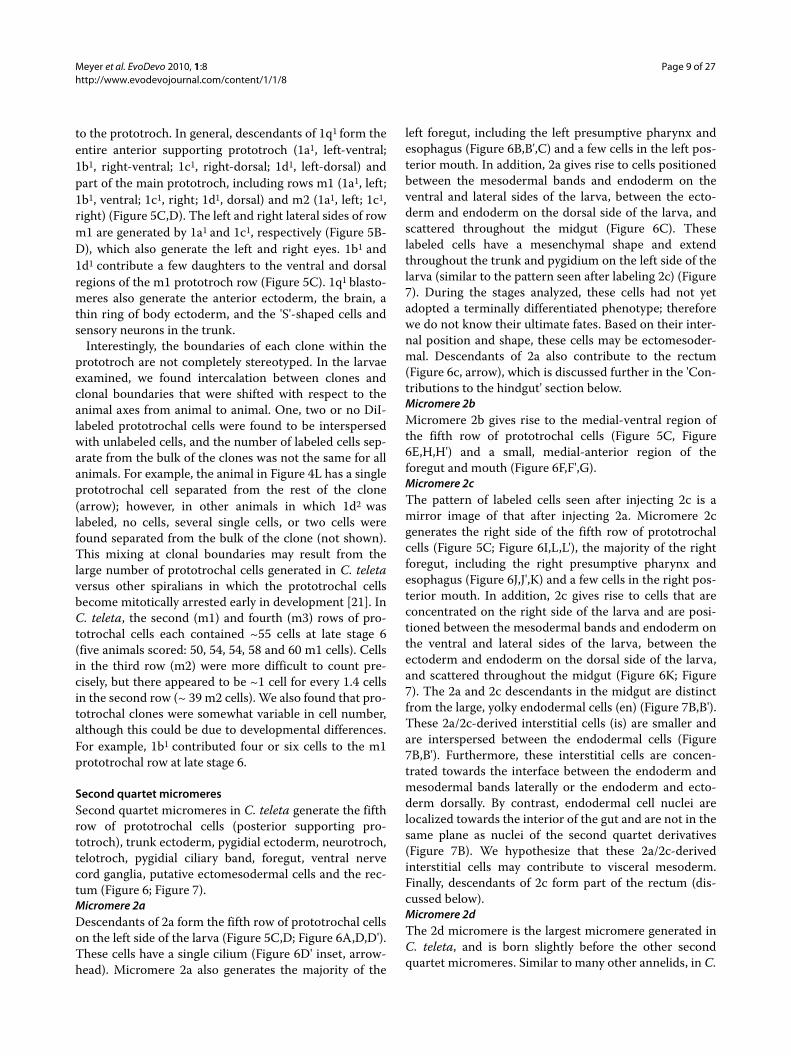
to the prototroch. In general, descendants of 1q1 form theentire anterior supporting prototroch (1a1, left-ventral;1b1, right-ventral; 1c1, right-dorsal; 1d1, left-dorsal) andpart of the main prototroch, including rows m1 (1a1, left;1b1, ventral; 1c1, right; 1d1, dorsal) and m2 (1a1, left; 1c1,right) (Figure 5C,D). The left and right lateral sides of rowm1 are generated by 1a1 and 1c1, respectively (Figure 5B-D), which also generate the left and right eyes. 1b1 and1d1 contribute a few daughters to the ventral and dorsalregions of the m1 prototroch row (Figure 5C). 1q1 blasto-meres also generate the anterior ectoderm, the brain, athin ring of body ectoderm, and the 'S'-shaped cells andsensory neurons in the trunk.
Interestingly, the boundaries of each clone within theprototroch are not completely stereotyped. In the larvaeexamined, we found intercalation between clones andclonal boundaries that were shifted with respect to theanimal axes from animal to animal. One, two or no DiI-labeled prototrochal cells were found to be interspersedwith unlabeled cells, and the number of labeled cells sep-arate from the bulk of the clones was not the same for allanimals. For example, the animal in Figure 4L has a singleprototrochal cell separated from the rest of the clone(arrow); however, in other animals in which 1d2 waslabeled, no cells, several single cells, or two cells werefound separated from the bulk of the clone (not shown).This mixing at clonal boundaries may result from thelarge number of prototrochal cells generated in C. teletaversus other spiralians in which the prototrochal cellsbecome mitotically arrested early in development [21]. InC. teleta, the second (m1) and fourth (m3) rows of pro-totrochal cells each contained ~55 cells at late stage 6(five animals scored: 50, 54, 54, 58 and 60 m1 cells). Cellsin the third row (m2) were more difficult to count pre-cisely, but there appeared to be ~1 cell for every 1.4 cellsin the second row (~ 39 m2 cells). We also found that pro-totrochal clones were somewhat variable in cell number,although this could be due to developmental differences.For example, 1b1 contributed four or six cells to the m1prototrochal row at late stage 6.
Second quartet micromeresSecond quartet micromeres in C. teleta generate the fifthrow of prototrochal cells (posterior supporting pro-totroch), trunk ectoderm, pygidial ectoderm, neurotroch,telotroch, pygidial ciliary band, foregut, ventral nervecord ganglia, putative ectomesodermal cells and the rec-tum (Figure 6; Figure 7).Micromere 2aDescendants of 2a form the fifth row of prototrochal cellson the left side of the larva (Figure 5C,D; Figure 6A,D,D').These cells have a single cilium (Figure 6D' inset, arrow-head). Micromere 2a also generates the majority of the
left foregut, including the left presumptive pharynx andesophagus (Figure 6B,B',C) and a few cells in the left pos-terior mouth. In addition, 2a gives rise to cells positionedbetween the mesodermal bands and endoderm on theventral and lateral sides of the larva, between the ecto-derm and endoderm on the dorsal side of the larva, andscattered throughout the midgut (Figure 6C). Theselabeled cells have a mesenchymal shape and extendthroughout the trunk and pygidium on the left side of thelarva (similar to the pattern seen after labeling 2c) (Figure7). During the stages analyzed, these cells had not yetadopted a terminally differentiated phenotype; thereforewe do not know their ultimate fates. Based on their inter-nal position and shape, these cells may be ectomesoder-mal. Descendants of 2a also contribute to the rectum(Figure 6c, arrow), which is discussed further in the 'Con-tributions to the hindgut' section below.Micromere 2bMicromere 2b gives rise to the medial-ventral region ofthe fifth row of prototrochal cells (Figure 5C, Figure6E,H,H') and a small, medial-anterior region of theforegut and mouth (Figure 6F,F',G).Micromere 2cThe pattern of labeled cells seen after injecting 2c is amirror image of that after injecting 2a. Micromere 2cgenerates the right side of the fifth row of prototrochalcells (Figure 5C; Figure 6I,L,L'), the majority of the rightforegut, including the right presumptive pharynx andesophagus (Figure 6J,J',K) and a few cells in the right pos-terior mouth. In addition, 2c gives rise to cells that areconcentrated on the right side of the larva and are posi-tioned between the mesodermal bands and endoderm onthe ventral and lateral sides of the larva, between theectoderm and endoderm on the dorsal side of the larva,and scattered throughout the midgut (Figure 6K; Figure7). The 2a and 2c descendants in the midgut are distinctfrom the large, yolky endodermal cells (en) (Figure 7B,B').These 2a/2c-derived interstitial cells (is) are smaller andare interspersed between the endodermal cells (Figure7B,B'). Furthermore, these interstitial cells are concen-trated towards the interface between the endoderm andmesodermal bands laterally or the endoderm and ecto-derm dorsally. By contrast, endodermal cell nuclei arelocalized towards the interior of the gut and are not in thesame plane as nuclei of the second quartet derivatives(Figure 7B). We hypothesize that these 2a/2c-derivedinterstitial cells may contribute to visceral mesoderm.Finally, descendants of 2c form part of the rectum (dis-cussed below).Micromere 2dThe 2d micromere is the largest micromere generated inC. teleta, and is born slightly before the other secondquartet micromeres. Similar to many other annelids, in C.

Meyer et al. EvoDevo 2010, 1:8http://www.evodevojournal.com/content/1/1/8
Page 10 of 27
teleta, descendants of 2d generate the majority of ecto-derm posterior to the prototroch (Figure 6M,O,Q,R,R',S).This includes both the segmented body ectoderm and thenon-segmented pygidial ectoderm, which are separatedby the telotroch (Figure 6M,O,S). One exception is a thinring of 1d-derived cells that wrap around the trunk justposterior to the prototroch (Figure 6T,T', arrow). 2d also
forms the neurotroch (Figure 6M), telotroch (tt) (Figure6M,S) and pygidial ciliary bands. In addition, descendantsof 2d form the ventral nerve cord (vn) (Figure 6Q), twosmall clusters of cells on the left-dorsal and right-dorsalsides of the brain (Figure 6P,P', arrow) and cells along thecircumesophageal connectives (Figure 6P,P',Q, arrow-heads). Although 2d does not contribute to the foregut
Figure 6 Second quartet micromeres generate foregut and trunk ectoderm. (A-T) Z-stacks of merged, confocal images of late stage 6 larvae 5 days after labeling second quartet micromeres with DiI. The channels are DiI (red), phalloidin (green) and TO-PRO-3 (blue) except for (D), (H), (L) and (T), which are DiI and phalloidin, and (Q) and (S), which are DiI and TO-PRO-3. Panels labeled with an apostrophe (for example, B',D') are single-chan-nel images of DiI that correspond to the multichannel images with the same letter (for example, B, D). A subset of panels are cropped, digitally mag-nified, z-stacks of confocal images through the foregut (B, F, J, N), prototroch (D, H, L), brain (P), forming ventral nerve cord (Q), body ectoderm (R) and dorsal body ectoderm just posterior to the prototroch (T). In (B, F, J, N) the foregut is outlined with a dashed line. The inset in (D') is an inverted, digitally magnified image of a single DiI-labeled prototroch cell with cilium (arrowhead). In (P) one brain lobe is outlined with a dashed line, and clus-ters of 2d descendants in the brain (arrow) are indicated. In (P, Q) 2d descendants in the circumesophageal connectives (arrowheads) are indicated. In (R') the boundary between mesoderm and endoderm is indicated with a dashed line. In (T) the arrow points to unlabeled ectodermal cells (arrow). The blastomere labeled with DiI is indicated to the left of each row and the view is indicated in the lower-left corner of each panel (dors = dorsal, vent = ventral, llat = left lateral, rlat = right lateral). Anterior is to the left in all ventral, dorsal and left lateral images, and to the right in all right lateral images. The position of the mouth is indicated with an asterisk. ec = ectoderm, en = endoderm, ms = mesoderm, pt = prototroch, tt = telotroch, vn = ventral nerve cord.

Meyer et al. EvoDevo 2010, 1:8http://www.evodevojournal.com/content/1/1/8
Page 11 of 27
(Figure 6N, N'; Figure 6O, dashed line), labeled surfaceectodermal cells extend around the mouth opening (Fig-ure 6M).
Third quartet micromeresThe third quartet micromeres generate cells in themouth, a small region of internal foregut, mesoderm inthe head, and cells surrounding the anus. In addition, thethird quartet micromeres form the left and right meso-dermal bands, which give rise to most of the circular andlongitudinal muscle fibers in the larva and the visceralmesoderm surrounding the foregut (Figure 8).Micromere 3aIn C. teleta, micromere 3a generates cells on the left sideof the mouth (Figure 8A,C) and a thin band of internallypositioned medial foregut tissue (Figure 8B,B',C). By stage8, some 3a descendants in the foregut contribute to a
putative valve between the esophagus and midgut, whichdoes not appear to be part of the foregut epithelium (notshown). Descendents of 3a also form a population ofmesodermal cells in the head (Figure 8C,D), including anumber of muscle cells with fibers extending posteriorly.These mesodermal cells are largely concentrated on theanterior side of the brain, with a few cells surrounding thelateral, ventral and posterior sides of the brain (Figure 8C,arrowhead; Figure 8D). Finally, 3a forms at least two neu-rons whose somas are positioned just anterior to themouth on the ventral face of the animal (Figure 8A,arrowheads). Axons from these neurons extend alongboth sides of the ventral nerve cord.Micromere 3bDescendants of 3b give rise to cells on the right side of themouth (Figure 8E) and a small, internally positioned,right-medial region of anterior foregut (Figure 8F-H). By
Figure 7 Ectomesodermal descendants from 2c. (A, B) Z-stacks of merged, confocal images from a dorsal (dors) view through a late stage 6 (A) or a stage 8 (B) larva after labeling micromere 2c. The channels are DiI (red), phalloidin (green) and TO-PRO-3 (blue). (A') Single-channel image of DiI staining shown in (A). (B) Cropped, digitally magnified image of the posterior trunk. (B') Single-channel phalloidin image corresponding to the mul-tichannel image in (B). A large endodermal (en) cell and two smaller interstitial (is) cells are indicated. Anterior is to the left in all images.

Meyer et al. EvoDevo 2010, 1:8http://www.evodevojournal.com/content/1/1/8
Page 12 of 27
stage 8, some 3b descendants in the foregut contribute toa putative valve between the esophagus and midgut (notshown). Because the 3b-derived and 3a-derived putativevalve cells are in very similar positions between theforegut and midgut, we think it likely that they contributeto the same valve.Micromere 3cThe 3c micromere forms the right mesodermal band(Figure 8I), which extends from just posterior of the telo-troch anteriorly to the foregut. In general, the right meso-dermal band forms both longitudinal and circular musclefibers, and wraps around the entire right side of theforegut (Figure 8J.J'). Micromere 3c also generates a smallnumber of cells in the right-posterior mouth (Figure 8I,K
L), cells that surround the right side of the anus (Figure8I,K, arrowhead), and a single, ciliated anterior neurotro-chal cell on the ventral midline (Figure 8I,K,L, closedarrowhead).Micromere 3dThe 3d micromere generates the left mesodermal band(Figure 8M), including longitudinal and circular bodywall muscle fibers and mesoderm wrapping around theleft foregut (Figure 8N,N'). 3d also gives rise to cells in theleft-posterior mouth (Figure 8M,O,P), cells surroundingthe left side of the anus (Figure 8M,O, arrowhead), and afew anterior ciliated neurotrochal cells (Figure 8M,O,P,closed arrowhead). In general, the 3d micromere forms amirror-image clone to 3c, although 3d also generates a
Figure 8 Third quartet micromeres form the mesodermal bands and ectodermal structures. (A-P) Z-stacks of merged, confocal images of late stage 6 larvae 5 days after labeling third quartet micromeres with DiI. The channels are DiI (red), phalloidin (green) and TO-PRO-3 (blue). Panels labeled with an apostrophe (for example, B', F') are single-channel images of DiI corresponding to panels with the same letter (for example, B, F). Panels (B, F, H, J, L, N, P) are cropped, digitally magnified, z-stacks of confocal images through the foregut (dashed line). Panel (D) is a cropped, digitally mag-nified, z-stack of confocal images through anterior mesoderm, with brain demarcated (dashed line). Arrowheads point to DiI-labeled neurons in (A), DiI-labeled anterior mesoderm in (C), and DiI-labeled cells surrounding the anus (I, K, M, O). Closed arrowheads point to DiI-labeled neurotroch cells in (I, K, M, O). The blastomere labeled with DiI is indicated to the left of each row, and the view is indicated in the lower-left corner of each panel (dors = dorsal, vent = ventral, llat = left lateral, rlat = right lateral). Anterior is to the left in all ventral and left lateral images, to the right in all right lateral images and down in all anterior images. The position of the mouth is indicated with an asterisk.

Meyer et al. EvoDevo 2010, 1:8http://www.evodevojournal.com/content/1/1/8
Page 13 of 27
small, internally positioned, medial-posterior region offoregut (Figure 8N,N',P).
Mesodermal band expansionAt stage 4, each mesodermal band is visible as a row ofsubsurface cells that extends from the telotroch anteri-orly towards the foregut. At stage 5, the right and leftmesodermal bands (Figure 9A, G, arrow), descendants
of 3c and 3d, respectively, begin to extend musclefibers (Figure 9A, D, G, J). At this stage, both longitudi-nal (Figure 9A,G, closed arrowhead) and circular (Fig-ure 9A, arrowhead) muscle fibers can be seen, as wellas muscle-cell soma that are positionally distinct fromthe mesodermal bands. These muscle cells originatefrom the mesodermal bands. Each mesodermal band isseveral cells wide along the dorsoventral and
Figure 9 Mesodermal band expansion. (A-L) Single-channel, z-stacks of confocal images of larvae (dashed line) after labeling micromeres 3c or 3d with DiI (white). In (A, G) mesodermal bands (arrows), forming circular muscle fibers (arrowheads), and forming longitudinal muscle fibers (closed ar-rowheads) are indicated. The blastomere labeled with DiI is indicated to the left of each row, the stage of each larva is indicated above each column and the view is indicated to the left of each row (vent = ventral, llat = left lateral, rlat = right lateral). Anterior is to the left in all ventral and left lateral images and to the right in all right lateral images. The position of the mouth is indicated with an asterisk. The position of the prototroch (pt) and tel-otroch (tt) are indicated.

Meyer et al. EvoDevo 2010, 1:8http://www.evodevojournal.com/content/1/1/8
Page 14 of 27
mediodistal axes, with the exception of the posteriorend, which contains a single large cell. By stage 6, themesodermal bands have begun expanding circumfer-entially in segmental rows. The first five segmentsextend dorsally (Figure 9B,H) but not ventrally (Figure9E,K). By stage 7, most of the mesodermal segmentshave expanded both dorsally (Figure 9C,I) and ven-trally (Figure 9F,L), and occupy the bulk of the larvalbody wall. The majority of the musculature in the headappears to arise from 3a, although thin muscle fibers,but not cell bodies, from 3c and 3d are visible in thehead (Figure 9A-C,F,H,I,L). Previously, precursors ofthe larval body segments were described by Eisig in1898 [14]. These structures were referred to as 'bauch-platten' or 'belly plates' and can be seen as a higherdensity of nuclei in the ventrolateral body of the larva,starting at stage 4. The belly plates expand dorsally andposteriorly as the larva develops [14,15,22]. Expansionof the mesodermal bands seen after labeling 3c and 3dcorresponds to recent descriptions of belly plateexpansion by Thamm and Seaver [22].
4dMicromere 4dWhen 4d is born, it is comparable in cell size to otherthird and fourth quartet micromeres. Concerning its fate,the 4d micromere generates a few longitudinal and circu-lar body wall muscle fibers (Figure 10A-B'), although 3cand 3d form the majority of larval muscle fibers. Descen-
dants of 4d are found at the posteriormost end of the gut,probably the presumptive anus (Figure 10A-B', D,D',closed arrowhead). These cells are positioned on the sur-face of the larva (Figure 10D, closed arrowhead) and con-tact the gut lumen (gl) (Figure 10D, arrow) at late stage 6.The 4d micromere also generates an internal pair of cellclusters that correspond to the presumptive primordialgerm cells (pgc) (Figure 10A-C', arrowheads). These cellsare in a similar position to nanos+, vasa+ and piwi+ cellsat similar stages ([23] and Seaver laboratory, unpublisheddata). After labeling 4d, we also see spots of DiI in theendoderm. We do not think that that these DiI spots arelabeled cells, because they do not appear to contain nucleiand they do not have a stereotypic position that remainsconstant from animal to animal. It is possible that somedescendants of 4d undergo programmed cell death andthat the remnants of these cells are visible in the endo-derm. At this time, we do not have evidence that 4d con-tributes to midgut endoderm.
Third and fourth quartet macromeresMacromeres 3A, 3B, 3C and 4D generate the endoderm(Figure 2N-P, R), which eventually forms the intestinalmidgut. In general, endoderm formed from 3A and 4D isrestricted to the left side of the embryo, whereas endo-derm from 3B and 3C is restricted to the right side of theembryo. Furthermore, endoderm descended from 3A and3B is more anteriorly positioned whereas endoderm from3C and 4D is more posteriorly positioned (Figure 2N-P,
Figure 10 Micromere 4d forms the primordial germ cells and anus. (A-D) Z-stacks of merged, confocal images of late stage 6 larvae 5 days after labeling micromere 4d with DiI. The channels are DiI (red), phalloidin (green) and TO-PRO-3 (blue). Panels labeled with an apostrophe (for example, A', B') are single-channel images of DiI that correspond to panels with the same letter (for example, A, B). Panels (C, D) are cropped, digitally magnified, z-stacks of confocal images showing the primordial germ cells (pgc) and anus, respectively. In (A-C) arrowheads point to primordial germ cells. In (A, B, D) a closed arrowhead points to DiI-labeled anal cells. In (B) the position of the mouth is marked with an asterisk. In (D) an arrow points to the gut lumen (gl). The view is indicated in the lower-right corner of each panel (vent = ventral, llat = left lateral). Anterior is to the left in all images.

Meyer et al. EvoDevo 2010, 1:8http://www.evodevojournal.com/content/1/1/8
Page 15 of 27
R), although the position of the clones within the gut var-ies from animal to animal.
Contributions to the hindgutFormation of the anusBecause several cells (2d, 3c, 3d and 4d) contribute to asmall region at the posterior end of the larva, includingthe anus, we examined the relative position of theseclones from a posterior view using confocal laser scan-ning microscopy (Figure 11). At late stage 6, there is arosette (diagrams) of surface cells in the center of thepygidium (Figure 11A-D). The number of cells in therosette increases from stage 5 to early stage 7. Immedi-ately beneath this rosette, concentric rings of musclefibers surround the rectum and possibly the posterior endof the midgut (Figure 11I-L). The forming gut lumen (gl)is also visible in the center of the rectum (Figure 10D, Fig-ure 11L, arrow). 2d descendants form the majority ofpygidial ectoderm. On the surface, 2d descendants form alarge number of the rosette cells and cells surroundingthe rosette (Figure 11, 2d diagram; Figure 11A, A', E, E').At a deeper focal plane, 2d descendants can be seen in thepygidial ectoderm that surrounds the rectum (Figure11I,I'). Descendants of micromere 3c form the right outerring of rosette cells (Figure 11, 3c diagram; Figure111B,B',F,F'). These cells extend below the surface and, ata deeper focal plane, abut the border between the muscle-fiber rings and the rectum (Figure 11J,J'). The posteriorclone arising from micromere 3d is a mirror image of thatgenerated by 3c (compare diagram 3d with 3c in Figure11; Figure 11C,C',G,G',K,K'). The cells generated by 3cand 3d form some of the concentric rings of musclefibers, although they appear to be only a subset of thefibers visible with phalloidin staining (not shown). Wecould not determine whether other micromeres also con-tribute to these muscle rings. Finally, 4d descendantsform a few cells in the center of the rosette (Figure 11, 4ddiagram; Figure 11D,D',H,H'). These 4d rosette cellsextend from the surface towards the interior, where theycontact the gut lumen (Figure 10D). We hypothesize thatthese 4d descendants form the future anus. 4d does notappear to generate any subsurface cells in the posteriorend of the larva (Figure 11L,L').Formation of the rectumBecause the gut continues to form throughout larvaldevelopment, we scored descendant clones at later stages(stage 8, 7 days after labeling) (Figure 12) to examinemore closely the micromere contributions to the poste-rior end of the alimentary canal. We individually injectedmicromeres 2a, 2c, 3Q and 4d with DiI. At stage 8, 1 daybefore metamorphosis, regions within the posterior endof the gut are more readily discernible than at stage 6. Atstage 8, a gut lumen can be seen extending from the pos-terior end of the esophagus to the anus, but not through
it (Figure 12D-F). At this stage, the rectum is distinguish-able from the midgut intestine by a few morphologicaltraits. First, the nuclear organization and cell size of therectal cells are distinct from the intestinal cells, which arelarger and more loosely organized (Figure 12F). Second,the gut lumen narrows along the dorsoventral axis as itpasses from the intestine into the rectum (Figure 12D).Third, the rectal portion of the gut lumen is more denselyciliated than the intestinal portion of the lumen whenanalyzed by anti-acetylated-tubulin staining (NPM,unpublished observations). We found that descendants of3C and 3D form the posterior end of the intestine, but notthe rectum (Figure 12A,A'). Descendants of 2a and 2cform the rectum (Figure 12B,B'), whereas descendants of4d form the presumptive anus (Figure 12C,C'). Interest-ingly, the anal cells derived from 4d do not appear tochange in shape or number (four cells) from stage 6 tostage 8 (compare Figure 10D,D' with Figure 12C,C').
DiscussionAxial relationships of the micromere quartetsIn spiralians for which a fate map exists, it has been notedthat descendants of the micromere quartets are arrangedsimilarly along the larval/adult body axes [6]. In the poly-clad platyhelminth Hoploplana inquilina, the nemerteanCerebratulus lacteus, the mollusk Patella vulgata, and theannelids C. teleta and P. dumerilii, first quartet microm-eres generate the left-ventral (1a), right-ventral (1b),right-dorsal (1c) and left-dorsal (1d) tissues. For the sec-ond and third quartet micromeres, the pattern seems lessconserved. In H. inquilina and C. lacteus, second quartetmicromeres are segregated into left (2a), right (2c), ven-tral (2b) and dorsal (2d) positions, whereas in P. vulgataand C. teleta, 2a and 2c clones are segregated into left andright positions, respectively. Descendants of third quartetmicromeres are distributed similarly to those of the firstquartet in C. lacteus and P. vulgata (Figure 13, Figure14A-C) [12,24-26]. In some spiralians, the symmetrybetween sets of clones is more conserved than the posi-tion of the clones relative to the animal axes. For example,in C. lacteus, P. vulgata, llyanassa obsoleta, Crepidula for-nicata, C. teleta and Helobdella robusta, 2a/2c and 3c/3dare mirror-image clones (Figure 14B, C) [10,25-28]. In themollusks P. vulgata, I. obseleta and C. fornicata, and thenemertean C. lacteus, 3a/3b descendants also generatemirror-image clones [10,25,26,28]. In two annelids and anemertean (C. teleta, H. robusta, P. dumerilii and C. lac-teus), 1a/b and 1c/d generate mirror-image clones (Figure14A) [12,26,27], whereas in mollusks (Chaetopleura api-culata, I. obseleta, C. fornicata), 1a/1c generate mirror-image clones [8,10,29]. Thus, although there are somesimilarities, the contributions relative to the plane ofbilateral symmetry vary between micromere quartetswithin and across species.

Meyer et al. EvoDevo 2010, 1:8http://www.evodevojournal.com/content/1/1/8
Page 16 of 27
Figure 11 Multiple micromeres contribute to the region surrounding the presumptive anus. (A-L) Z-stacks of merged, confocal images of late stage 6 larvae 5 days after labeling micromeres 2d (column 1), 3c (column 2), 3d (column 3) or 4d (column 4) with DiI. The channels are DiI (red), phal-loidin (green) and TO-PRO-3 (blue). Panels labeled with an apostrophe (for example, A', B') are single-channel images of DiI that correspond to panels with the same letter (for example, A, B). The depths of the confocal z-stacks, starting at the surface (A-D) and progressing into the larva, are indicated to the left of each row. The diagrams at the top of each column are of the posterior rosette cells and are drawn from the images immediately below them (that is, surface z-stacks). In (L) an arrow points to the forming gut lumen (gl). All images are from a posterior view with ventral down.

Meyer et al. EvoDevo 2010, 1:8http://www.evodevojournal.com/content/1/1/8
Page 17 of 27
Origins of nervous systemsIn annelids, the majority of anterior unsegmented ecto-derm is generated by the 1q micromeres, whereas themajority of segmented trunk ectoderm is generated bythe 2d micromere. Furthermore, the brain or supraesoph-
ageal ganglion comes from 1q (1q1 where studied), andthe ventral nerve cord comes from 2d in Amphitriteornata, Nereis, Scoloplos armiger, Helobdella and C.teleta (Figure 13; Figure 14A,B) [12,27,30-34]. Within thecontext of the largely conserved origin of the annelid cen-
Figure 12 Contributions to the developing gut at stage 8. (A-C) Cropped z-stacks of merged, confocal images through the posterior end of stage 8 larvae at the level of the gut lumen 7 days after labeling 3C, 2c or 4d with DiI. The channels are DiI (red), phalloidin (green) and TO-PRO-3 (blue). The cell labeled with DiI is indicated in the upper-right corner. Panels labeled with an apostrophe (for example, A', B') are single-channel images of DiI corresponding to panels with the same letter (for example, A, B). (D-F) Single-channel confocal z-stacks through the posterior end of stage 8 larvae labeled with phalloidin (D, E) or TO-PRO-3 (F) to show the intestinal lumen or cell nuclei, respectively. Panels (E, F) are corresponding single-channel images from (C). The view is indicated in the lower-right corner of each panel (vent = ventral, lat = lateral). Anterior is to the left in all images. mg = midgut, rc = rectum.

Meyer et al. EvoDevo 2010, 1:8http://www.evodevojournal.com/content/1/1/8
Page 18 of 27
tral nervous system from 1q and 2d, we find some uniquefeatures in C. teleta. Micromeres 1c1 and 1d1 generate themajority of the brain with only minor contributions from1a1 and 1b1 (Figure 13; Figure 14A,B). Furthermore, asmall population of 2d-derived cells are found in the
brain and positioned along the circumesophageal con-nectives (Figure 6P,Q; Figure 14B). Second quartet contri-butions to the brain have not been reported in annelids.However, in the leech H. robusta, daughters of 2d (dnopq'and dnopq") form putative glial cells in the supraesophageal
Figure 13 Lineage tree showing blastomere fates in Capitella teleta. D = dorsal, L = left, pT = posterior supporting prototroch, R = right, V = ven-tral.

Meyer et al. EvoDevo 2010, 1:8http://www.evodevojournal.com/content/1/1/8
Page 19 of 27
Figure 14 Summary fate map diagrams for Capitella teleta. (A-D) Patterns seen after injecting DiI into individual micromeres or third quartet mac-romeres in C. teleta. Each panel corresponds to one micromere tier, and each color corresponds to one quadrant. The views are ventral, left lateral through the mouth, anterior through the brain, anterior through the foregut, and posterior through the forming anus. D = dorsal, L = left, R = right, V = ventral.

Meyer et al. EvoDevo 2010, 1:8http://www.evodevojournal.com/content/1/1/8
Page 20 of 27
ganglion [27,35]. Based on their position, we think it isunlikely that the 2d-derived brain cells in C. teleta areglial.
There are several other cells that make minor contribu-tions to the nervous system in the trunk of annelids. In C.teleta, 1c1 and 1d1 give rise to putative trunk sensory neu-rons, and 3a generates two neurons near the mouth. In H.robusta, micromeres 2a, 2c, 3c and 3d (a", c", c''', d''',respectively) are all reported to give rise to putative neu-rons and/or mesenchyme in the proboscis and the ante-rior sucker. Furthermore, 3a (a''') forms a pair of neuron-like cells in the subesophageal ganglion that project to theproboscis and posteriorly into the trunk [27]. This pair ofneurons is similar in description to the pair of 3a-derivedneurons by the mouth in C. teleta (Figure 8A, arrow-heads). Unfortunately, we were unable to determinewhether micromeres other than 1q, 2d and 3a generateneurons in C. teleta. Based on clonal position, we think itlikely that the rows of peripheral neurons in the trunk areformed by 2d, and clusters of neurons in the foregut arederived from 2a and 2c. Taken together, these results sug-gest that the origin of the annelid nervous system from 1qand 2d is fairly conserved, although species-specific vari-ations may emerge once more animals are studied indetail.
Direct comparisons of nervous system fate mapsbetween annelids and other spiralians are more difficult.In gastropod mollusks, the ganglia of the central nervoussystem arise later in larval development [36-38], makingfate mapping technically challenging. The nemertean C.lacteus undergoes a radical metamorphosis, with the lar-val nervous system being entirely replaced by derivativesof imaginal discs. The polyclad flatworm H. inquilina alsohas a biphasic life cycle, and the relationship of the larvalnervous system to the adult nervous system is not clear.In the mollusks that have been examined, the anteriorectoderm is formed by descendants of the first quartetmicromeres [8,10,25,29]. This suggests that the apicalganglion and the cerebral ganglia are generated by 1qmicromeres, similar to annelids. There are some data thatsupport this hypothesis. In C. fornicata, 1a, 1c and 1dcontribute to the apical ganglion [10], and in C. apicu-lata, descendants of 1c1 and 1d1 form the apical organ [8].A similar situation may exist in C. lacteus, as micromeres1a and 1b were reported to give rise to the left and rightlarval cephalic discs [26]. In other nemerteans thatdevelop via a pilidium larva (heteronemerteans), thecephalic discs are thought to generate the head ectodermand cerebral ganglia of the juvenile [39-41].
Fate maps of ganglia in the body are more complicatedto compare. In mollusks, the nemertean C. lacteus, andthe polyclad flatworm H. inquilina, the 2d 'primary soma-toblast' does not form the majority of body ectoderm as itdoes in annelids [8-10,21,24,26,28]], and contributions to
ganglia in the body may also be more variable. In C. forni-cata, 2b contributes to the supra-intestinal and sub-intes-tinal ganglia, and 2a and 2c contribute to the left andright pedal ganglia. Interestingly, 2d gives rise to the pos-terior mantle cell [10], which is one of the first neurons todifferentiate, and its axons may serve as a scaffold forlater central nervous system development [36,38].Peripheral neurons in C. fornicata are generated by all 1qand 2q micromeres [10], whereas third quartet contribu-tions to the nervous system have not yet been deter-mined. In C. lacteus and H. inquilina, the first quartetforms the bulk of the larval ectoderm, and descendants ofmany first to third quartet micromeres contribute to thelarval nervous system [24,26]. However, in heteronemer-teans, the origin of the juvenile central nervous system inthe trunk is not clear and awaits results from fate-map-ping experiments. In contrast to annelids, in which 2dforms the majority of trunk ectoderm and all of the gan-glia in the ventral nerve cord, multiple blastomeres maygenerate the central and peripheral nervous systems inthe body of these other spiralians.
Origins of ciliated bandsPatterns of ciliation (for example, ciliated 'bands') inmarine larvae have played a central role in debates aboutthe homology of larval structures and life history evolu-tion across metazoan taxa [42-47]. The evolutionary sig-nificance of ciliary bands in metazoans remainsunresolved. For example, some authors think that ciliatedbands evolved as feeding structures associated withplanktotrophy [48-53], whereas others argue that they areprimarily locomotory in lecithotropic species [54-59].Spiralians are a rich group of animals in which to explorethese issues, as the pelagic phases of their life cycle bearmany distinct ciliated structures (for example, akrotroch,prototroch, metatroch, telotroch and neurotroch), andthe developmental origins of ciliated structures can befollowed in detail due to the stereotyped spiral cleavageprogram.
Modern intracellular cell-lineage studies such as thework presented here give insights into some of theseissues. The prototroch is found in a wide variety of spira-lian groups (including polychaetes, echiurans, sipuncu-lans, mollusks, nemerteans and entoprocts), andgenerally consists of multiple rows of cells bearing cilia.These larval cells are shed during metamorphosis and areoften larger in size than adjacent cells because theybecome mitotically arrested during early cleavage stages.In general, the 1q12 micromeres (accessory trochoblasts)contribute to the anteriormost prototrochal row (anteriorsupporting row), 1q2 micromeres (primary trochoblasts)largely form the two most heavily ciliated prototrochalrows (main prototroch) and the 2q11 micromeres (sec-ondary trochoblasts) contribute to the posteriormost

Meyer et al. EvoDevo 2010, 1:8http://www.evodevojournal.com/content/1/1/8
Page 21 of 27
prototrochal row (posterior supporting row). There arevariations in this pattern, and each tier of trochoblastshas been shown to contribute to multiple prototrochalrows. These generalizations are based on many early lin-eage studies (including [14,33,34,60-62] and on morerecent intracellular fate mapping experiments in mollusksand a paleonemertean [10,28,29,63-65]. The same generalpattern of contributions to each tier is found in C. teleta(Figure 5). To our knowledge, C. teleta is the only poly-chaete in which the precursors of all tiers of prototrochalprecursors have been individually labeled. The clonalcontribution to each row is less stereotyped than otherstudied spiralians, which is probably due to the relativelylarge number of cells in the prototroch. For example, inmany mollusks, the entire prototroch can contain as fewas 20 to 40 cleavage arrested cells [21], whereas the pro-totroch in C. teleta is composed of hundreds of smallercells that interdigitate at clonal boundaries.
In spiralians, the D quadrant appears to have reducedits contribution to the prototroch relative to the otherthree quadrants. In most species studied, including C.teleta, the 2a, 2b and 2c, but not 2d micromeres make acontribution to the prototroch. The prototroch of thepolychaetes A. ornata and Podarke obscura and the gas-tropod C. fornicata do not appear to have a contributionfrom 2d [10,34,62]. In P. obscura, descendents of 1a1, 1b1
and 1c1, but not 1d1 contribute to the prototroch [66]. Inthe chiton C. apiculata, the third quartet micromere, 3d,makes only a minor contribution to the prototroch [8]. Insummary, the prototroch is widely found among spira-lians, and the shared embryological origins of the pro-totroch between species provide evidence of it being ahomologous structure.
Detailed information on the developmental origin ofother trochal bands such as the metatroch, telotroch andneurotroch is much more limited. Intracellular fate-map-ping studies of these other ciliated bands have not beenperformed on many species and they are not found on allspiralian taxa. For example, aplacophoran mollusk larvaehave a telotroch [67], but gastropod mollusks and nemer-teans do not have telotrochs or neurotrochs, althoughsome mollusks have ciliated bands along the midline ofthe foot. Within annelids, not all polychaetes possess thesame complement of ciliary bands. For example, P.dumerilii does not possess a neurotroch or telotroch. Theneurotroch in the polychaete S. armiger is reported to bederived from the third quartet micromeres 3c and 3d[30]. In C. teleta, the first species in which intracellularlineage tracers were used to study the origin of a neu-rotroch, most of the neurotroch is generated by the 2dmicromere (Figure 14B), whereas only small contribu-tions near the mouth are provided by 3c and 3d (Figure 8,closed arrowheads). The telotroch and pygidial ciliarybands in C. teleta are generated entirely by derivatives of
the 2d micromere (Figure 13, Figure 14B). The meta-troch, the quintessential ciliary band required for'opposed band feeding' in spiralian larvae, is not presentin nemerteans nor in the majority of polychaete families,such as Capitellidae, which includes C. teleta [55,56]. Theorigin of the metatroch has only been studied in a singlespecies of polychaete, Polygordius, in which it wasreported to be derived from the third quartet micromeres3c and 3d [68]. If true, then the metatroch of Polygordiusis not developmentally homologous with the secondaryciliary band or the ciliated food grove of the molluscanvelum, which are derived from 2a, 2b and 2c [10]. Inanother example, the third quartet micromeres 3a and 3band derivatives of all first and second quartet micromeresgive rise to the ciliated bands in the pilidium larva of thenemertean C. lacteus [26]. Thus, it appears that distinctblastomeres can be co-opted to form various ciliatedbands in different spiralian lineages.
Nielsen suggests that prototrochs, metatrochs and telo-trochs are all derived from a circumblastoporal ciliatedband in a radially symmetrical, holopelagic ancestor, andthat their differentiation was established in connectionwith the evolution of a new body axis, the anterior-poste-rior axis, forming at an angle to the primary, apical-blas-toporal axis [46,52,69]. This is unlikely because mostciliated bands are not derived from the vicinity of theblastopore, which forms at the vegetal pole [70]. Further-more, the multiple embryonic origins of ciliary bandsimply that not all ciliary bands are homologous. The dis-tinct embryological origins of ciliary bands and the multi-tude of ciliary band types in spiralians indicate that ciliarybands are evolutionarily labile, thus caution must be usedin making statements about homology across distantlyrelated taxa.
Origins of mesodermThe developmental origins of mesodermal cell types in C.teleta represent significant modifications of the typicalspiralian fate map. One of the most important findingsfrom spiralian cell-lineage studies is the documentationof dual embryological origins of mesoderm. One source isfrom ectomesoderm (cells that give rise to both ectoder-mal and mesodermal derivatives) and the other fromendomesoderm ('embryonic precursors of both meso-derm and the endodermally derived intestine') [6,7].Endomesoderm arises from a single fourth quartetmicromere in the D quadrant, 4d, and its origin from 4dis a highly conserved feature within Spiralia (Table 2). InC. teleta, 4d generates mesodermal cell types typicallyfound in other spiralians, including primordial germ cellsand muscle cells, but it does not contribute to the endo-dermally derived intestine (Figure 13; Figure 14D).Although 4d makes a small contribution to the posteriorgut in C. teleta, this contribution is to the anus, an ecto-

Meyer et al. EvoDevo 2010, 1:8http://www.evodevojournal.com/content/1/1/8
Page 22 of 27
dermal structure (Figure 10D). Therefore, we argue thatin C. teleta, 4d is not a mesentoblast, but a mesectoblast.At first glance, it seems possible that 3D is the mesento-blast in C. teleta, because its descendants contribute toboth the endodermal midgut and mesodermal structures.However, 3D also generates ectoderm (4d-derived anus),eliminating it as a mesentoblast candidate (Figure 14C).The descendants of 4d (DM'') in Helobdella are similar tothose on C. teleta: 4d generates mesoderm and ectoderm,but not endoderm [32]. In the classic spiralian literature,there are other examples of 4d generating only mesodermin polychaete annelids, including in A. ornata and Areni-cola cristata [34,60]; however, these reports need to beconfirmed by direct intracellular labeling experiments. Itwill be important to determine how widespread is the 4drestriction to mesodermal fates within Annelida.
In Wilson's initial account of Nereis development, all 4dderivatives were described to be mesodermal [33]. How-ever, after Conklin's characterization of the Crepidula celllineage [5], Wilson re-examined the early cleavages of 4din Nereis and observed small daughter cells of 4d bornbefore the formation of the mesodermal bands. Thesedaughter cells contributed descendants to the posteriorendodermal archenteron [7]. Wilson proposed an evolu-tionary scenario to explain the observed variation in therelative contribution of 4d to endoderm and mesoderm in
spiralians [7]. He speculated that in the ancestral spiralianform, fourth quartet micromeres generated only endo-derm. In subsequent spiralian descendants, mesodermalderivatives eventually segregated entirely to 4d. Overtime, the capacity of 4d to form endoderm was reduced inannelids. From a current phylogenetic framework, it isdifficult to find support for the idea that the spiralianancestor lacked endomesoderm, because it is likely, basedon comparative molecular and lineage studies, that endo-mesoderm was present in the bilaterian ancestor [71-74].
In contrast to the relatively conserved embryologicalorigin of endomesoderm, the origin of ectomesoderm ismore variable across spiralians (Table 2). In C. teleta,micromeres 3a, 3c, 3d, 4d and possibly 2a, 2c and 3b gen-erate ectomesoderm (Figure 13, Figure 14B-D), and thecontribution to ectomesoderm by second and third quar-tet micromeres is shared with mollusks, annelids andnemerteans. A notable exception is the generation ofhead mesoderm by first quartet micromeres in H. robusta(Table 2) [27]. Newby also described a contribution tomesoderm from first quartet micromeres in the echiuranUrechis caupo [75], although this has not been directlyconfirmed by intracellular lineage tracers. It is notablethat the B quadrant does not generate mesoderm in C.teleta, with a minor possible exception of the 3b-derivedvalve between the esophagus and midgut, which may be
Table 2: Embryological sources of mesoderm in spiraliansa,b.
Species Ectomesoderm Endomesoderm Reference
Nemertean Cerebratulus 3a, 3b 4d [26]
Polyclad tubellarian Hoploplana 2b 4d [9,24]
Chaetopleura None 3D [8]
Mollusk Patella 3a, 3b 3D [25]
Ilyanassa 2b, 2c, 3d,3A, 3B, 3C
4d [28]
Crepidula 2c, 3a, 3b 4d [10]
Annelid Helobdella 1a (a'), 1b (b'), 1c (c'), 1d (d'), 3a (a'''), 3c (c'''), 3d (dm')4d (DM'')
None [27,32]
Platynereis 1A, 1B, 1C, 1D 4d [12]
Capitella 2a?, 2c?, 3a, 3b?, 3c, 3d, 4d none This report
aExamples included are taken from fate maps determined through use of intracellular lineage tracers injected into embryos at the 8-64 cell stage.bOnly blastomeres with confirmed mesoderm descendants are included.

Meyer et al. EvoDevo 2010, 1:8http://www.evodevojournal.com/content/1/1/8
Page 23 of 27
mesodermal. B quadrant contributions to ectomesodermare commonly found in spiralians, including in C. lacteus,P. vulgata, I. obsoleta, C. fornicata, Helobdella and P.dumerilii. In H. inquilina, 2b appears to be the only ecto-mesoblast (Table 2).
In addition to 4d not being a 'mesentoblast' in C. teleta,there is another modification of the typical 4d descendantfates. In most spiralians, 4d gives rise to the mesodermalbands, which extend along the anterioposterior axis andcontribute to the trunk mesoderm [76]. In C. teleta, themesodermal bands are generated by the third quartetmicromeres 3c and 3d rather than by 4d (Figure 13; Fig-ure 14C). In his classic study of Capitella development,Eisig also noted that 3c and 3d generate the coelomic lin-ings [14]. Mesodermal bands arising from the 3c and 3dmicromeres have not been previously reported for anyother spiralian. Although generation of the mesodermalbands by 3c and 3d is unique to Capitella among the ani-mals studied to date, other descendent cell types gener-ated by 3c and 3d, such as contributions to thestomodeum, are shared with P. vulgata, H. inquilina andC. lacteus [24-26]. In addition, in H. robusta and probablyin P. dumerilii, 3c and 3d generate mesoderm closelyassociated with the foregut [12,27], a pattern shared withC. teleta. Among the spiralians that have been examinedby intracellular fate mapping experiments, contributionsto visceral mesoderm by third quartet micromeres havenot been reported outside annelids. In conclusion, modi-fications of the spiralian program are complex, and donot represent simple heterochronic shifts of fate orwhole-cell fate transformations. In C. teleta, unlike inmost other spiralians, the size of the 4d blastomere is notobviously larger than other fourth quartet micromeres,and the 3c and 3d micromeres are similar in size to theother third quartet micromeres. It is interesting to con-sider whether 4d acts as the organizer in C. teleta, as itdoes in some other spiralians [76], or whether this func-tion is now assumed by cells that generate the mesoder-mal bands (3c and 3d).
In summary, the generation of the mesodermal bandsby 3c and 3d in C. teleta represents a radical departurefrom the highly conserved 4d-derived mesodermal bandsdescribed in other spiralians. Furthermore, our lineageresults indicate that although several distinct micromeresgenerate mesodermal derivatives in C. teleta, all of thesearise from ectomesoderm, a feature shared with anotherannelid, Helobdella.
Origins of the alimentary canalForegutIn spiralians, the anterior region of the gut is commonlyderived from second (2q) and third quartet (3q) microm-eres, although the contribution of particular blastomeresto mouth and foregut tissue varies among polychaete spe-
cies, and between polychaete annelids and other spiraliantaxa. Classic cell-lineage studies use the term 'stomato-blast' when referring to blastomeres that line the oral cav-ity and show that stomatoblasts are micromerederivatives of 2a to 2c in Nereis, 3a to 3d in A. cristata, 2ato 2c, 3a and 3b in S. armiger, and 2b, 3a and 3b in P.obscura [30,33,60,66]. These results are somewhat con-fusing because interpretations of particular cell fates arebased on the author's definition of 'stomodeum' (stomod-eum synonymous with pharynx [33], stomodeum synon-ymous with mouth [60]). Furthermore, some of theseearlier studies inferred the fates of anterior gut cells basedupon their positions during gastrulation. Some authorsassumed that the mouth forms from the blastopore (A.cristata [60], P. obscura [66], S. armiger [30]), whereasothers directly observed a distinct stomodeal opening atlater stages (Nereis [33], A. ornata [34], Capitella [14]).Therefore, details from earlier fate maps of polychaetesshould be interpreted and compared with caution.
In the fate map of C. teleta that we generated by intrac-ellular injections, descendants of 2a to 2c and 3a to 3dcontribute to cells in the mouth (Figure 13; Figure 14B,C).These results are generally consistent with results fromclassic lineage studies that examined micromere contri-butions to the stomodeum in other polychaete annelids.In C. teleta, the larval pharynx and esophagus showmajor contributions from 2a (left side) and 2c (right side)lineages, and a moderate contribution from 2b (central-anterior) (Figure 13; Figure 14B), which are similar in lin-eage and axial position to blastomeres traced by tradi-tional methods in Nereis, S. armiger and probably A.cristata [30,33,60]. Taken together with C. teleta, thesefour polychaete species generate four distinct foregutarchitectures in the adult, but during developmentalstages, the same micromeres form similar regions of theforegut. The larvae of these four species are predomi-nantly lecithotrophic, and fate maps may be less con-served between these species and planktotrophic larvaethat undergo substantial changes in larval tissues andorgan systems during metamorphosis.
Modern fate maps have been published only for oneother polychaete species, P. dumerilii [12]. In that study,'stomodeum' broadly refers to the developing foregut, andcontributions to the foregut fates were often inferred,because second or third quartet blastomeres were notinjected directly. By injecting blastomeres at the eight-cellstage, Ackerman et al. showed that descendents of 1Aand 1C, inferred to be 2a2 and 2c2, contribute to the leftand right stomodeum, respectively [12]. There was noapparent contribution to stomodeal tissue from 1B or 1D.This is in contrast to our findings in C. teleta, in whichdescendents of both B and D lineages could be traced tothe mouth and/or foregut tissue (Figure 13; Figure14B,C). Regarding oral ectoderm, Ackerman et al. [12]

Meyer et al. EvoDevo 2010, 1:8http://www.evodevojournal.com/content/1/1/8
Page 24 of 27
further corrected Wilson's [33] interpretations by statingthat 3a to 3d are unlikely to be involved in 'the generalectoblast of the circum-oral and circum-anal regions.'This observation is notable because we show by directinjection that 3a to 3d generate ciliated ectodermal cellsof the mouth in C. teleta. In the annelid H. robusta,Huang et al. found that all four first quartet micromerescontribute to the proboscis, along with 2a, 2c (a", c") and3a and 3c (a''', c'''; discussed below) [27]. Second quartetmicromeres having foregut-related fates are consistentwith our findings in C. teleta; however, first quartetmicromere contributions represent a significant depar-ture from both modern and traditional cell-lineage stud-ies in polychaetes.
Modern fate maps from molluscan and polyclad turbel-larian embryos also show important differences from ourmap in C. teleta. For example, the respective foregut fatesof second and third quartet micromeres in gastropodmollusks are essentially reversed in position relative tothe 2q and 3q micromeres in polychaete annelids. Inveliger larvae of both C. fornicata and I. obsoleta, 2qdescendants give rise to the cells lining the mouth, and 3qdescendants form the esophagus [10,28]. Within the tro-chophore larva of the limpet, P. vulgata, 2b, 3c and 3dall have internally positioned foregut fates [25]. In thepolyclad H. inquilina, micromeres 2a, 2b and 2c contrib-ute to oral ectoderm and associated cilia, whereas 3a, 3band 3c were found to label oral cilia [24]. Only one thirdquartet micromere, 3d, makes a significant contributionto the mouth in H. inquilina. Although the relative posi-tions of 2q and 3q descendants in the stomodeum of H.inquilina correlate with our results, one difference is thatin C. teleta, 2a, 2b and 2c micromeres generate internalforegut tissues, unlike the pattern from any 2q labelingobserved by Boyer et al. [24]. One generalization that canbe made here is that micromeres from A and C quadrantsin mollusk and polyclad embryos generate, respectively,the left and right structures of the larval mouth andforegut. For instance, Boyer et al. observed that in theblind gut of H. inquilina, 2a and 2c micromeres contrib-ute to left and right regions of the stomodeum [24], whichalso applies to C. teleta and other polychaetes. However,there are important foregut-specific contributions in C.teleta that do not conform to any general patterns, at leastuntil more spiralian taxa are examined by modern fate-mapping techniques.MidgutThe midgut in C. teleta and its intestinal counterpart inother spiralians are derived entirely from endoderm. Inmost spiralian taxa for which cell-lineage data are avail-able, including mollusks, annelids, nemerteans andsipunculans, 3A, 3B and 3C macromeres and all of theirdescendants generate midgut endoderm. Both descen-
dents of 3D (4D, 4d) also generate endoderm in mollusk,nemertean and sipunculan embryos [10,26,28,77]. Inpolychaetes, 4D contributes to endoderm. 4d also con-tributes to endoderm tissue in some polychaete species[7,66], but not in others [12,30,34,60] including C. teleta(Figure 13; Figure 14D).
Similar to the axial map of micromere derivatives inboth the foregut and nervous system, macromeres in C.teleta also contribute to the intestine in a quadrant-spe-cific pattern. Descendants of 3A and 4D are located pre-dominantly on the left side and those of 3B and 3C on theright side (Figure 14C). In P. dumerilii, 1A and 1C giverise to left and right midgut anlagen [12], which is consis-tent with a general pattern of quadrant-specific endo-derm contributions in polychaetes. Our intracellularlabeling also shows that the macromere contribution toanterior midgut in C. teleta makes a relatively abruptjunction with the esophagus, and therefore we concludethat there is no endodermal component of the foregut inthis species. Furthermore, the macromere contribution tothe intestine abuts the rectum at its posterior end, thus,endodermal cells do not generate hindgut tissue duringlarval development.HindgutCompared with other gut territories, the posterior end ofthe alimentary canal or 'hindgut', has received little atten-tion from previous cell-lineage studies on polychaetes.Traditionally the hindgut of polychaetes has been definedas a proctodeal invagination of ectoderm [18,19], imply-ing that cells lining the rectal canal are internalized deriv-atives of surface ectoderm. However, in some spiraliancell-lineage studies, the hindgut has been described asendodermal [10,28]. For example, in gastropod mollusks,the posterior end of the gut or intestine is endodermaland derives from descendants of 4d [10,28,76]. The 4d-derived portion of the larval gut in C. teleta consistssolely of a few surface cells restricted to the presumptiveanus, which we interpret as ectodermal. Late in larvaldevelopment, the gut canal passes through the posteriormidgut and rectum, but not the presumptive anus. Basedon this observation and our lineage data, we suggest thatan anal opening is the last developmental step in forminga through gut in C. teleta, and that the hindgut (rectumplus anus) does not form by a proctodeal invagination ofectoderm. During metamorphosis the hindgut regionprobably undergoes structural modifications, and withfurther development, it becomes considerably longerthan it was before the metamorphic transition from anon-feeding larva to a juvenile worm. We think it likelythat the 2a/2c-derived rectum undergoes such structuralmodification.
I6P. dumerilii, Poonamali cellular contributions to theproctodeum have not been characterized [12], whereas in

Meyer et al. EvoDevo 2010, 1:8http://www.evodevojournal.com/content/1/1/8
Page 25 of 27
Nereis, Wilson mentions that 3q micromeres contributeto 'portions' of the circum-anal region [33]. OnlyTreadwell reports that specific micromere descendents of2d2, 3c and 3d contribute to the proctodeal wall of P.obscura [66]. In C. teleta, we found that the rectum isderived from 2a and 2c, whereas the surrounding tissuesare generated from 2d, 3c and 3d micromeres (Figure14B, C), including a ring-like system of musculature thatmay function as a sphincter apparatus. The rectum abutsthe 3C/4D-derived midgut endoderm on its anterior endand the 4d-derived presumptive anus on its posterior end(Figure 14C,D). Our map of this region establishes themost complete modern reference of posterior gut territo-ries to date, and provides a framework that may be usedto assess whether any generalized trends exist in otherspiralians.
ConclusionsIntracellular injection of the lineage tracer DiI into indi-vidual blastomeres (1q, 1q1, 1q2, 2q, 3q, 4d, 2Q, 3Q and4D) in C. teleta has provided the most complete fate mapfor any polychaete annelid to date. Many features of theC. teleta fate map are similar to those found in otherannelids, including the generation of the anterior ecto-derm and prototroch by first quartet micromeres and themajority of post-trochal ectoderm by the primarysomatoblast 2d. Increased resolution through the use ofconfocal laser scanning microscopy enabled us to gener-ate a detailed fate map for regions such as the prototroch,mesoderm, nervous system and gut. Our analyses revealcomplex contributions to these structures from severalblastomere lineages. For example, more cells contributeto the posterior gut than previously documented for anyother spiralian and four to seven distinct blastomeresgenerate mesoderm in C. teleta, all from ectomesodermalsources. We also identified modifications of the typicalspiralian fate map in C. teleta; the most dramatic differ-ence is the formation of the mesodermal bands by 3c and3d rather than by 4d, which generates the mesodermalbands in all other spiralians examined. The fate map of C.teleta will provide a framework for future comparisonswith other spiralian fate maps and the foundation forfunctional experiments of cell-fate determination in thisspecies.
MethodsAnimal careA laboratory colony of C. teleta was maintained andembryos were collected as described previously [15], withthe exception that adult animals were kept in filtered sea-water (FSW) (passed through 20 μm filter) at 19°C. Afterinjection, embryos were raised at 19°C in FSW (through0.2 μm filter) with 60 μg/ml penicillin (Sigma-Aldrich
Co., St Louis, MO, USA) and 50 μg/ml streptomycin(Sigma-Aldrich Co.) added.
DiI labelingFor injections, the egg shell was permeabilized by a 30-second incubation in a 1:1 mixture of fresh 1 mol/Lsucrose and 0.25 mol/L sodium citrate followed by two tothree rinses with FSW. Individual blastomeres were pres-sure-injected with 1 part DiIC18(3) (1,1'-dioctadecyl-3,3,3',3'-tetramethylindocarbocyanine perchlorate; Invit-rogen, Carlsbad, CA, USA) saturated in ethanol to 19parts soybean oil (Wesson; ConAgra Foods Inc., Omaha,NE, USA) using aluminosilicate needles (Sutter Instru-ment Co., Novata, CA, USA). After labeling, uninjectedand injected animals from the same brood (same stage ofdevelopment) were raised for the same amount of time,and rates of development and morphological featureswere compared. An experiment was not scored unless90% of the injected and 90% of the uninjected animalswere healthy (morphologically normal and similar timingof development).
Animals at various developmental stages were imagedeither live or fixed (DiI fixation) [78]. For compound fluo-rescence microscopy, fixed animals were incubated over-night at 4°C in a fluorescently conjugated toxin thatrecognizes F-actin (BODIPY FL-phallacidin; Invitrogen)diluted 1:100 in phosphate-buffered saline (PBS), thenrinsed three to four times in PBS, incubated in 80%glycerol in PBS plus 0.125 μg/mL bisbenzimide Hoechst33342 (Sigma-Aldrich Co.) for 3 to 12 h at 4°C and thenanalyzed. For confocal laser scanning microscopy, ani-mals were incubated overnight at 4°C in a 1:1000 dilutionof a nucleic acid stain (TO-PRO-3 iodide; Invitrogen) and1:100 dilution of a phallotoxin (either BODIPY FL-phalla-cidin or Alexa Fluor 488-phalloidin; Invitrogen) in PBS,rinsed three to four times in PBS, then incubated inmounting media with an antifade reagent (SlowFadeGold; Invitrogen) for at least 3 h at 4°C, and analyzed.
MicroscopyFor compound fluorescence imaging, live and fixed DiI-labeled animals were imaged (Axioskop 2 Plus with anAxioCam HRm camera; both Carl Zeiss Inc., Munich,Germany) and analyzed (Openlab software, version 4.0.1;PerkinElmer Inc., Waltham, MA, USA). Confocal imag-ing was performed using a confocal microscope (LSM510; Carl Zeiss). Z-stack projections were then generated(ImageJ; National Institutes of Health, Bethesda, MD,USA). Figures were constructed using illustration soft-ware (Photoshop CS4 and Illustrator CS4; Adobe SystemsInc., San Jose, CA, USA).
Competing interestsThe authors declare that they have no competing interests.

Meyer et al. EvoDevo 2010, 1:8http://www.evodevojournal.com/content/1/1/8
Page 26 of 27
Authors' contributionsNPM carried out the injections, animal care, data collection and analyses, andillustration preparation. MJB and ECS helped with initial data analyses, and MJBcontributed to illustration preparation. MQM contributed to initial injections.All authors contributed to data interpretation and writing and editing of themanuscript. All authors read and approved this manuscript.
AcknowledgementsThis work was supported by the National Science Foundation (IOB05-44869 to ECS).
Author Details1Kewalo Marine Laboratory, Pacific Biosciences Research Center, University of Hawaii, 41 Ahui Street, Honolulu, Hawaii 96813, USA and 2Smithsonian Marine Station at Fort Pierce, 701 Seaway Drive, Fort Pierce, Florida 34949, USA
References1. Dunn CW, Hejnol A, Matus DQ, Pang K, Browne WE, Smith SA, Seaver E,
Rouse GW, Obst M, Edgecombe GD, Sørensen MV, Haddock SH, Schmidt-Rhaesa A, Okusu A, Kristensen RM, Wheeler WC, Martindale MQ, Giribet G: Broad phylogenomic sampling improves resolution of the animal tree of life. Nature 2008, 452:745-749.
2. Giribet G, Dunn CW, Edgecombe GD, Hejnol A, Martindale MQ, Rouse GW: Assembling the spiralian tree of life. In Animal Evolution: Genomes, Fossils, and Trees Edited by: Telford MJaDTJL. Oxford: Oxford University Press; 2009:52-64.
3. Hejnol A, Obst M, Stamatakis A, Ott M, Rouse GW, Edgecombe GD, Martinez P, Baguna J, Bailly X, Jondelius U, Wiens M, Müller WE, Seaver E, Wheeler WC, Martindale MQ, Giribet G, Dunn CW: Assessing the root of bilaterian animals with scalable phylogenomic methods. Proc Biol Sci 2009, 276:4261-4270.
4. Helmkampf M, Bruchhaus I, Hausdorf B: Multigene analysis of lophophorate and chaetognath phylogenetic relationships. Mol Phylogenet Evol 2008, 46:206-214.
5. Conklin EG: The embryology of Crepidula. J Morphol 1897, 13:1-226.6. Henry JJ, Martindale MQ: Conservation and innovation in spiralian
development. Hydrobiologia 1999, 402:255-265.7. Wilson EB: Considerations on cell-lineage and ancestral reminiscence.
Ann NY Acad Sci 1898, XI:1-27.8. Henry JQ, Okusu A, Martindale MQ: The cell lineage of the
polyplacophoran, Chaetopleura apiculata: variation in the spiralian program and implications for molluscan evolution. Dev Biol 2004, 272:145-160.
9. Boyer BC, Henry JQ, Martindale MQ: Dual origins of mesoderm in a basal spiralian: cell lineage analyses in the polyclad turbellarian Hoploplana inquilina. Dev Biol 1996, 179:329-338.
10. Hejnol A, Martindale MQ, Henry JQ: High-resolution fate map of the snail Crepidula fornicata: the origins of ciliary bands, nervous system, and muscular elements. Dev Biol 2007, 305:63-76.
11. Henry JJ, Martindale MQ: The origins of mesoderm in the equal-cleaving nemertean worm Cerebratulus lacteus. Biological Bulletin 1996, 191:286-288.
12. Ackermann C, Dorresteijn A, Fischer A: Clonal domains in postlarval Platynereis dumerilii (Annelida: Polychaeta). J Morphol 2005, 266:258-280.
13. Blake JA, Grassle JP, Eckelbarger KJ: Capitella teleta, a new species designation for the opportunistic and experimental Capitella sp. I, with a review of the literature for confirmed records. Zoosymposia 2009, 2:25-53.
14. Eisig H: Zur Entwicklungsgeschichte der Capitelliden. Mittheilungen Aus der Zoologischen Station Zu Neapel 1898, 13:1-292.
15. Seaver EC, Thamm K, Hill SD: Growth patterns during segmentation in the two polychaete annelids, Capitella sp. I and Hydroides elegans: comparisons at distinct life history stages. Evol Dev 2005, 7:312-326.
16. Eckelbarger KJ, Grassle JP: Interspecific variation in genital spine, sperm, and larval morphology in six sibling species of Capitella. Bull Biol Soc Wash 1987, 7:62-76.
17. Boyle MJ, Seaver EC: Developmental expression of foxA and gata genes during gut formation in the polychaete annelid, Capitella sp. I. Evol Dev 2008, 10:89-105.
18. Michel C: Intestine and digestive glands. The Ultrastructure of Polychaeta (Microfauna Marina) 1988, 4:5-175.
19. Tzetlin A, Purshke G: Pharynx and intestine. Hydrobiologia 2005, 535/536:199-225.
20. Rhode B: Larval and adult eyes in Capitella spec. I (Annelida, Polychaeta). J Morphol 1993, 217:327-335.
21. Damen P: Cell-lineage, and specification of developmental fate and dorsoventral organisation in the mollusc Patella vulgata. Thesis, Univ Utrecht 1994.
22. Thamm K, Seaver EC: Notch signaling during larval and juvenile development in the polychaete annelid Capitella sp. I. Dev Biol 2008, 320:304-318.
23. Dill KK, Seaver EC: Vasa and nanos are coexpressed in somatic and germ line tissue from early embryonic cleavage stages through adulthood in the polychaete Capitella sp. I. Dev Genes Evol 2008, 218:453-463.
24. Boyer BC, Henry JJ, Martindale MQ: The cell lineage of a polyclad turbellarian embryo reveals close similarity to coelomate spiralians. Dev Biol 1998, 204:111-123.
25. Dictus WJ, Damen P: Cell-lineage and clonal-contribution map of the trochophore larva of Patella vulgata (mollusca). Mech Dev 1997, 62:213-226.
26. Henry JJ, Martindale MQ: Conservation of the spiralian developmental program: cell lineage of the nemertean, Cerebratulus lacteus. Dev Biol 1998, 201:253-269.
27. Huang FZ, Kang D, Ramirez-Weber FA, Bissen ST, Weisblat DA: Micromere lineages in the glossiphoniid leech Helobdella. Development 2002, 129:719-732.
28. Render J: Cell fate maps in the Ilyanassa obsoleta embryo beyond the third division. Dev Biol 1997, 189:301-310.
29. Render J: Fate maps of the first quartet micromeres in the gastropod Ilyanassa obsoleta. Development 1991, 113:495-501.
30. Anderson DT: The embryology of the polychaete Scoloplos armiger. Q J Microsc Sci 1959, 100:89-166.
31. Nardelli-Haefliger D, Shankland M: Lox10, a member of the NK-2 homeobox gene class, is expressed in a segmental pattern in the endoderm and in the cephalic nervous system of the leech Helobdella. Development 1993, 118:877-892.
32. Weisblat DA, Kim SY, Stent GS: Embryonic origins of cells in the leech Helobdella triserialis. Dev Biol 1984, 104:65-85.
33. Wilson EB: The cell-lineage of Nereis. A contribution to the cytogeny of the annelid body. J Morphol 1892, 6:361-480.
34. Mead AD: The early development of marine annelids. J Morphol 1897, XIII:227-327.
35. Dohle W: The ancestral cleavage pattern of the clitellates and its phylogenetic deviations. Hydrobiologia 1999, 402:267-283.
36. Croll RP: Developing nervous systems in molluscs: navigating the twists and turns of a complex life cycle. Brain Behav Evol 2009, 74:164-176.
37. Dickinson AJ, Croll RP: Development of the larval nervous system of the gastropod Ilyanassa obsoleta. J Comp Neurol 2003, 466:197-218.
38. Dickinson AJ, Nason J, Croll RP: Histochemical localization of FMRFamide, serotonin and catecholamines in embryonic Crepidula fornicata (Gastropoda, Prosobranchia). Zoomorphology 1999, 119:49-62.
39. Bürger O: Studien zu einer Revision der Entwicklungsgeschichte der Nemertinen Ber Naturf Ges Freiburg; 1894.
40. Maslakova SA: The Invention of the Pilidium Larva in an Otherwise Perfectly Good Spiralian Phylum Nemertea. Integr Comp Biol 2010.
41. Salensky W: Morphogenetische Studien an Wurmern. II. Uber die Morphogenese der Nemertinen. Entwiklungsgeschichte der Nemertine im Inneren des Pilidiums. Mem Acad Sci St Petersb 1912, 30:1-74.
42. Arendt D, Technau U, Wittbrodt J: Evolution of the bilaterian larval foregut. Nature 2001, 409:81-85.
43. Garstang W: The morphology of the Tunicata and its bearing on the phylogeny of the Chordata. Q J Microsc Sci 1928, 72:51-54.
44. Hatschek B: Studien über die Entwicklungsgsgeschichte der Anneliden. Ein Beitrag zur Morphologie der Bilaterien. Arb Zool Inst Univ Wien 1878, 1:277-404.
Received: 21 May 2010 Accepted: 15 September 2010 Published: 15 September 2010This article is available from: http://www.evodevojournal.com/content/1/1/8© 2010 Meyer et al; licensee BioMed Central Ltd. This is an Open Access article distributed under the terms of the Creative Commons Attribution License (http://creativecommons.org/licenses/by/2.0), which permits unrestricted use, distribution, and reproduction in any medium, provided the original work is properly cited.EvoDevo 2010, 1:8

Meyer et al. EvoDevo 2010, 1:8http://www.evodevojournal.com/content/1/1/8
Page 27 of 27
45. Lacalli TC: Ciliary bands in echinoderm larvae: evidence for structural homologies and a common plan. Acta Zool 1993, 74:127-133.
46. Nielsen C, Nørrevang A: The Trochaea theory: an example of life cycle phylogeny. In The Origins and Relationships of Lower Invertebrates Edited by: Conway Morris S, George JD, Gibson R, Platt HM. Oxford: Clarendon Press; 1985:28-41.
47. Rieger RM: The biphasic life cycle--a central theme of metazoan evolution. Am Zool 1994, 34:484-491.
48. Jägersten G: Further remarks on the early phylogeny of Metazoa. Zool Bider Upps 1959, 33:79-108.
49. Jägersten G: Evolution of the Metazoan life cycle London: Academic Press; 1972.
50. Nielsen C: Larval ciliary bands and metazoan phylogeny. Fortschr Zool Syst Evolutionsforsch 1979, 1:178-184.
51. Nielsen C: Origin and evolution of animal life cycles. Biol Rev 1998, 73:125-155.
52. Nielsen C: How did indirect development with planktotrophic larvae evolve? Biol Bull 2009, 216:203-215.
53. Strathmann RR: The evolution and loss of feeding larval stages of marine invertebrates. Evolution 1978, 32:894-906.
54. Degnan SM, Degnan BM: The origin of the pelagobenthic metazoan life cycle: what's sex got to do with it? Integr Comp Biol 2006, 46:683-690.
55. Rouse GW: Trochophore concepts: ciliary bands and the evolution of larvae in spiralian Metazoa. Biol J Linn Soc 1999, 66:411-464.
56. Rouse GW: The epitome of hand waving? Larval feeding and hypotheses of metazoan phylogeny. Evol Dev 2000, 2:222-233.
57. Runnegar B: No evidence for planktotrophy in Cambrian molluscs. Evol Dev 2007, 9:311-312.
58. Salvini-Plawen LV: Was ist eine Trochophora? Eine Analyse der Larventypen mariner Protostomier. Zool Jb Anat 1980, 103:389-423.
59. Sly BJ, Snoke MS, Raff RA: Who came first--larvae or adults? origins of bilaterian metazoan larvae. Int J Dev Biol 2003, 47:623-632.
60. Child CM: The early development of Arenicola and Sternaspis. Arch EntwMech Org 1900, 9:587-723.
61. Heath H: The development of Ischnochiton. Zool Jahrb, Abt Anat Ontog Tiere 1899, 12:567-656.
62. Treadwell AL: The cell lineage of Podarke obscura. Zool Bull 1897, 1:195-203.
63. Damen P, Dictus WJ: Cell lineage of the prototroch of Patella vulgata (Gastropoda, Mollusca). Dev Biol 1994, 162:364-383.
64. Maslakova SA, Martindale MQ, Norenburg JL: Fundamental properties of the spiralian developmental program are displayed by the basal nemertean Carinoma tremaphoros (Palaeonemertea, Nemertea). Dev Biol 2004, 267:342-360.
65. Maslakova SA, Martindale MQ, Norenburg JL: Vestigial prototroch in a basal nemertean, Carinoma tremaphoros (Nemertea; Palaeonemertea). Evol Dev 2004, 6:219-226.
66. Treadwell AL: Cytogeny of Podarke obscura Verrill. J Morphol 1901, 17:399-486.
67. Okusu A: Embryogenesis and development of Epimenia babai (Mollusca Neomeniomorpha). Biol Bull 2002, 203:87-103.
68. Woltereck R: Beiträge zur praktischen Analyse der Polygordius-Entwicklung nach dem "Nordsee-" und dem "Mittelmeer-Typus". Arch EntwMech Org 1904, 18:377-403.
69. Nielsen C: Six major steps in animal evolution: are we derived sponge larvae? Evol Dev 2008, 10:241-257.
70. Martindale MQ, Hejnol A: A developmental perspective: changes in the position of the blastopore during bilaterian evolution. Dev Cell 2009, 17:162-174.
71. Henry JQ, Martindale MQ, Boyer BC: The unique developmental program of the acoel flatworm, Neochildia fusca. Dev Biol 2000, 220:285-295.
72. Martindale MQ, Henry JQ: Intracellular fate mapping in a basal metazoan, the ctenophore Mnemiopsis leidyi, reveals the origins of mesoderm and the existence of indeterminate cell lineages. Dev Biol 1999, 214:243-257.
73. Martindale MQ, Pang K, Finnerty JR: Investigating the origins of triploblasty: 'mesodermal' gene expression in a diploblastic animal, the sea anemone Nematostella vectensis (phylum, Cnidaria; class, Anthozoa). Development 2004, 131:2463-2474.
74. Technau U, Scholz CB: Origin and evolution of endoderm and mesoderm. Int J Dev Biol 2003, 47:531-539.
75. Newby WW: The Embryology of the Echiuroid Worm Urechis caupo Philadelphia: The American Philosophical Society; 1940.
76. Lambert JD: Mesoderm in spiralians: the organizer and the 4d cell. J Exp Zool B Mol Dev Evol 2008, 310:15-23.
77. Torrey JC: The early embryology of Thalassema mellita (Conn). Ann NY Acad Sci 1903, 14:165-246.
78. Meyer NP, Seaver EC: Neurogenesis in an annelid: characterization of brain neural precursors in the polychaete Capitella sp. I. Dev Biol 2009, 335:237-252.
doi: 10.1186/2041-9139-1-8Cite this article as: Meyer et al., A comprehensive fate map by intracellular injection of identified blastomeres in the marine polychaete Capitella teleta EvoDevo 2010, 1:8