a common exocytotic mechanism mediates axonal and ... · a common exocytotic mechanism mediates...
TRANSCRIPT

A Common Exocytotic Mechanism Mediates Axonal andDendritic Outgrowth
Sonia Martinez-Arca,1,4 Silvia Coco,2 Gaell Mainguy,3 Ursula Schenk,2 Philipp Alberts,1,4 Pascale Bouille,5Mauro Mezzina,5 Alain Prochiantz,3 Michela Matteoli,2 Daniel Louvard,4 and Thierry Galli1,4
1Membrane Traffic and Neuronal Plasticity, Institut National de la Sante et de la Recherche Medicale U536, Institut du Fer-a-Moulin, F-75005 Paris, France, 2Synaptic Development and Function, Cellular and Molecular Pharmacology and BrunoCeccarelli Centers, Consiglio Nazionale delle Ricerche, 20129 Milan, Italy, 3Developmental and Cellular Neurobiology,Centre National de la Recherche Scientifique (CNRS) Unite Mixte de Recherche (UMR) 8542, Ecole Normale Superieure,F-75005 Paris, France, 4Morphogenesis and Cell Signaling, CNRS UMR 144, Institut Curie, F-75005 Paris, France, and5Gene Therapy, Genethon III, Unite de Recherche Associee 1923, CNRS, BP 60, F-91002 Evry Cedex, France
Outgrowth of the dendrites and the axon is the basis of theestablishment of the neuronal shape, and it requires addition ofnew membrane to both growing processes. It is not yet clearwhether one or two exocytotic pathways are responsible for therespective outgrowth of axons and dendrites. We have previ-ously shown that tetanus neurotoxin-insensitive vesicle-associated membrane protein (TI-VAMP) defines a novel net-work of tubulovesicular structures present both at the leadingedge of elongating dendrites and axons of immature hippocam-pal neurons developing in primary culture and that TI-VAMP isan essential protein for neurite outgrowth in PC12 cells. Herewe show that the expression of the N-terminal domain ofTI-VAMP inhibits the outgrowth of both dendrites and axons in
neurons in primary culture. This effect is more prominent at theearliest stages of the development of neurons in vitro. Expres-sion of the N-terminal domain deleted form of TI-VAMP has theopposite effect. This constitutively active form of TI-VAMP lo-calizes as the endogenous protein, particularly concentrating atthe leading edge of growing axons. Our results suggest that acommon exocytotic mechanism that relies on TI-VAMP medi-ates both axonal and dendritic outgrowth in developingneurons.
Key words: axonal outgrowth; dendritic outgrowth; exocyto-sis; SNARE; TI-VAMP; adeno-associated virus; neurons in pri-mary culture
Growth of an axon and extension of a dendritic tree are the keymorphological features of neuronal development, and they defineneuronal shape (Prochiantz, 1995). Exocytosis plays a fundamen-tal role in axonal and dendritic outgrowths because both pro-cesses involve major increases in the surface area of the plasmamembrane (Futerman and Banker, 1996; Bradke and Dotti,1997). This new surface area is added at the tips of the elongatingprocesses (Craig et al., 1995; Zakharenko and Popov, 1998). Theaxon and the dendrites differ in their contents of protein early inneuronal development and are controlled by different factors(Prochiantz, 1995). Nevertheless, when the axon of a neuron issevered, a new axon forms from a pre-existing dendrite (Dotti andBanker, 1987; Bradke and Dotti, 2000a). The molecular machin-ery controlling the trafficking of membranes involved in axonal
and dendritic outgrowth is still poorly characterized, and it is notyet clear whether one or two independent exocytotic pathways areresponsible for the respective outgrowth of axons and dendrites(Bradke and Dotti, 2000b).
Soluble N-ethylmaleimide-sensitive fusion protein (NSF) at-tachment protein (SNAP) receptors (SNAREs) are importantproteins of membrane traffic because they are the core moleculesof membrane fusion (Sollner et al., 1993; Bock and Scheller, 1999;Jahn and Sudhof, 1999). In neurons, the plasma membrane targetSNAREs (t-SNAREs), syntaxin 1 and SNAP25, are found allalong the plasma membranes of axons and dendrites (Garcia etal., 1995; Galli et al., 1995). Both the outgrowth of axons and theoutgrowth of dendrites depend on SNAP25 because they areinhibited by the botulinum neurotoxins A and C1 but do notinvolve synaptobrevin 2 because they are not affected by tetanusneurotoxin (Osen-Sand et al., 1996; Igarashi et al., 1996; Grosse etal., 1999), which cleaves synaptobrevin 2. The recent discovery ofthe clostridial neurotoxin-resistant vesicular SNARE tetanusneurotoxin-insensitive vesicle-associated membrane protein (TI-VAMP) (Galli et al., 1998) (also called VAMP-7; Advani et al.,1998), the product of the Synaptobrevin-like gene 1 (D’Espositoet al., 1996) in neuronal cells (Coco et al., 1999; Martinez-Arca etal., 2000a,b) has provided an appealing hypothesis for the ob-served lack of effect of tetanus neurotoxin on axonal and den-dritic outgrowth (Johannes and Galli, 1998). An important struc-tural feature of TI-VAMP is its N-terminal extension of 100amino acids (Galli et al., 1998). We have shown that this domaininhibits the formation of SNARE complexes both in vivo and in
Received Dec. 22, 2000; revised March 9, 2001; accepted March 19, 2001.This work was supported in part by Action Concertee Incitative-Jeunes Cher-
cheurs (Grant 5254) from the Ministere de la Recherche et des Technologies to T.Gand by Telethon Italia (Grant 1042) and European Economic Community (GrantBIO4-CT98-0408) to M.M. S.M.A. is a recipient of a fellowship from Fondationpour la Recherche Medicale and P.A. from the Deutscher Akademischer Austausch-dienst. We thank the Production Service Unit of Genethon III for large-scale AAVvector production in the frame of the Gene Vector Production Network (GVPN;www.genethon.fr/gvpn) program. The GVPN is supported by the Association Fran-caise contre les Myopathies (Evry, France). We are grateful to Jean-Antoine Giraultfor his constant support and for critical reading of this manuscript. We thank JackKyte for helpful comments and corrections and R. Jahn and W. Hong for thegenerous gift of antibodies.
S.M., S.C., and G.M. contributed equally to this work.Correspondence should be addressed to Dr. Thierry Galli, “Membrane Traffic
and Neuronal Plasticity,” CNRS UMR144, Institut Curie, Section de Recherche, 26rue d’Ulm, 75231 Paris Cedex 05, France. E-mail: [email protected] © 2001 Society for Neuroscience 0270-6474/01/213830-09$15.00/0
The Journal of Neuroscience, June 1, 2001, 21(11):3830–3838

vitro. In agreement with this observation, we observed that neu-rite outgrowth is inhibited by expression in pheochromocytoma12 (PC12) cells of the N terminus of TI-VAMP and enhanced byexpression of a form of TI-VAMP deleted at its N terminus(Martinez-Arca et al., 2000a). Each process from a differentiatedPC12 cell contains both dendritic and axonal markers, and nonemake synaptic contacts so these cells cannot be considered to bea true model of neuronal differentiation and maturation. In thispaper, we expressed the N-terminal domain and the form ofTI-VAMP deleted at its N terminus in neurons to assay for therole of TI-VAMP in the outgrowth of axons and dendrites.
MATERIALS AND METHODSAntibodies and clones. A polyclonal antibody directed against greenfluorescent protein (GFP) was generated in rabbit and affinity-purifiedover recombinant glutathione S-transferase-coupled GFP. Mouse mono-clonal antibodies directed against GFP (clones 7.1 and 13.1; RocheDiagnostics, GmbH, Germany), the Golgi matrix protein of 130 kDa(GM130; Transduction Laboratories, Lexington, KY) and syntaxin 6(clone 30; Transduction Laboratories) have been described previously.Rabbit polyclonal antibody directed against calreticulin was from AffinityBioreagents. Mouse monoclonal antibody directed against synaptobrevin2 (clone 69.1) and rabbit polyclonal antibody directed against syntaxin 7were generous gifts of Drs. R. Jahn (Max Planck Institute, Goettingen,Germany) and W. Hong (Institute of Molecular and Cell Biology, Sin-gapore), respectively. Antibodies against excitatory amino acid carrier 1(EAAC1) and GluR1 were kind gifts of Drs. Rothstein (Johns HopkinsUniversity, Baltimore, MD) and Wenthold (Uniformed Services Univer-sity of the Health Sciences, Bethesda, MD), respectively, and were usedas previously described (Coco et al., 1997). The cDNA of human TI-VAMP and the GFP-fusion constructs have been previously described(Galli et al., 1998; Martinez-Arca et al., 2000a).
Electroporation of mouse embryonic brain. Brains were dissected outfrom mouse embryonic day 13 (E13) embryos in PBS and 0.6% glucose.Plasmids (2 mg/ml) were co-injected with 0.05% Fast-Green (Sigma, St.Louis, MO) in telencephalic vesicles using a glass capillary needle.Electroporation was performed essentially as described in Miyasaka et al.(1999), by five pulses (50 V, 50 msec) with a T-820 apparatus using a10-mm-diameter tweezertrode electrode (BTX, San Diego, CA). Afterelectroporation cells were dissociated in trypsin 0.05% (Life Technolo-gies, Cergy-Pontoise, France), plated on glass coverslips coated withmatrigel (Collaborative Biomedical Products, Bedford, MA), and cul-tured as in Mainguy et al. (2000). After the indicated times, cells werefixed with 4% PFA and either mounted with Vectashield-49,6-diamidino-2-phenylindole (DAPI) (Vector Laboratories, Burlingame, CA) for ob-servation of direct GFP-signal or permeabilized with 0.3% Triton X-100and processed for immunofluorescence as described (Coco et al., 1999).
Adeno-associated virus vectors construction, production, purification, andtitration. Recombinant adeno-associated virus (rAAV)-cytomegalovirus(CMV)-GFP-TI-VAMP, rAAV-CMV-GFP-Nter-TI-VAMP, andrAAV-CMV-GFP-DNter-TI-VAMP vectors were respectively obtainedfrom the pCR3.1 GFP-TI-VAMP, pCR3.1 GFP-Nter-TI-VAMP,pCR3.1 GFP-DNter-TI-VAMP, plasmids, harboring the correspondingtransgene and the pGG2 AAV plasmid. The latter plasmid is derivedfrom the pSUB201 plasmid, in which the expression is driven by hCMVpromoter and stabilized by the SV40 late polyA and a chimeric introncomposed of the 59 donor splice site of the first intron of the human bglobin gene (hBB) and the 39 acceptor splice site of the intron of an Iggene (IgG) heavy chain variable region. First, GFP-TI-VAMP, GFP-Nter-TI-VAMP, and GFP-DNter-TI-VAMP sequences were PCR-amplified by using specific primers and the high-fidelity pfu turbo poly-merase (Stratagene, La Jolla, CA) and further digested by NheIrestriction enzyme at the 39 end. These fragments were purified fromagarose gel by using the Geneclean kit (BIO101, Vista, CA) according tothe manufacturer’s procedure. Second, the pGG2 plasmid was cut byNheI and EcoRV enzymes to add the PCR-amplified cDNAs. Thecorrect orientation of the inserted sequences were checked by DNAsequencing analysis and agarose gel electrophoresis. Large-scale produc-tion and purification of vectors were performed by using the tripletransfection of 293 cells, followed by CsCl density gradients purification,as previously described (Xiao et al., 1998). The infectious particle con-centration is determined by a variation of the procedure previouslydescribed (Salvetti et al., 1998).
Adenoassociated viral infection of neurons. Cortical and striatal neuronswere prepared from rat E16 embryos as described previously (Rousseletet al., 1990). After dissociation, neurons were plated in collagen-coatedglass coverslips in chemically defined medium as above. Five hours afterplating cells were infected overnight with the described Aavs at a mul-tiplicity of infection of 100 in a final volume of 50 ml. The day after, theAavs were removed and cells were kept in regular medium for theindicated periods of time. The direct GFP signal from the Aav-encodedproteins could be detected 3 d after infection, however, because of thelow level of expression of the transgenes and to facilitate detection of theinfected neurons for subsequent quantitation, cells were fixed and per-meabilized as described above and stained with anti-GFP antibodies.
Transfection of hippocampal neurons with calcium phosphate. Calciumphosphate crystals were prepared as described in Maniatis et al. (1982).For transfection, neurons were placed in medium conditioned by corticalastrocytes for at least 15 hr. Calcium phosphate crystals were left for 4 hr,and the cells were then washed accurately with Krebs’–Ringer’s solutionand transferred in their previous medium.
Quantification of axonal and dendritic length in hippocampal neurons.Randomly chosen fields were taken with a Bio-Rad MRC-1024 ConfocalMicroscope equipped with a LaserSharp 3.2 software. Acquired imageswere processed and quantitatively analyzed with NIH Image 1.62 soft-ware from National Institute of Health, resulting in the analysis ofbetween 40 and 60 GFP-positive cells, for each condition and for eachindependent experiment. Multiple dendrites emerging from a same stalkwere counted as distinct entities. For immunocytochemistry, neuronswere permeabilized with 0.3% Triton X-100 and processed for immuno-fluorescence as described (Coco et al., 1999).
Quantification of axonal length in corticostriatal neurons. Randomlychosen fields were taken with a MicroMax CCD camera (PrincetonInstruments), resulting in the analysis of between 10 and 50 (in theelectroporation experiments) or between 25 and 200 (in the Aav exper-iments) GFP-positive cells, for each condition and for each independentexperiment. Quantification of axonal length was done using the Meta-morph software (Princeton Instruments). Double immunofluorescencewith neuronal markers was performed to verify exclusively quantificationof neuronal cells. The obtained data were analyzed for their statisticalsignificance with Sigma Stat (SPSS, Inc.).
Quantification of Nter-TI-VAMP-induced cell death. After electropora-tion of embryonic E13 brains, cells were dissociated and cultured in theabsence or presence of 200 mM zVAD (Calbiochem, La Jolla, CA) toinhibit caspases thus apoptosis. After 24 hr all the green fluorescentneuronal and non-neuronal cells remaining were scored.
RESULTSExpression of the N-terminal domain of TI-VAMPinhibits neuronal differentiationTo investigate the role of TI-VAMP in neuronal differentiation,we expressed GFP and GFP fused to the N-terminal domain ofTI-VAMP (GFP-Nter-TI-VAMP), using a calcium phosphate-based transfection method, in E18 rat hippocampal neurons thatwere cultured in the presence of feeding glial cells (Fig. 1).Neurons were transfected at 1 d in vitro (div) or 4 div andexamined 24 hr later. We observed that neurons expressing GFP-Nter-TI-VAMP grew shorter neurites, whereas those expressingGFP differentiated as nontransfected neurons (Fig. 1) (data notshown). We quantitated the growth of axons and dendrites, theseprocesses being defined by morphological criteria (Dotti et al.,1988). The inhibition affected both processes. Interestingly, den-drites of neurons expressing GFP-Nter-TI-VAMP were fewer innumber and shorter than those of neurons expressing GFP, after2 and 5 div; the strongest effect was after 2 div (Fig. 1B,C). Incomparison, the inhibitory effect of the expression of GFP-Nter-TI-VAMP on axonal outgrowth appeared stronger than the effecton dendritic outgrowth, when only the lengths of the processeswere compared (Fig. 1, compare B, D).
We confirmed that dendritic function was significantly alteredin hippocampal neurons expressing GFP-Nter-TI-VAMP be-cause a reduced localization of the glutamate transporter EAAC1
Martinez-Arca et al. • Exocytosis Mediating Axonal and Dendritic Outgrowth J. Neurosci., June 1, 2001, 21(11):3830–3838 3831

was seen in the dendrites of these cells compared with cellsexpressing only GFP (Fig. 2). EAAC1 is a transmembrane pro-tein expressed in dendrites of hippocampal neurons at very earlystages of their development, and thus is a good marker of vesic-ular transport to the growing dendritic plasma membrane (Cocoet al., 1997). Quantification of dendrites positive for EAAC1
showed that far fewer of them were from neurons expressingGFP-Nter-TI-VAMP than from neurons expressing only GFP(Fig. 2B). We did not observe any effect on the localization of theAMPA receptor subunit GluR1 (Fig. 2A). This lack of effectshows that only a subset of transport pathways are inhibited by theexpression of GFP-Nter-TI-VAMP.
Figure 1. Expression of the N-terminal domain of TI-VAMP inhibitsaxonal and dendritic growth. A, Hippocampal neurons from E18 ratstransfected with GFP or GFP-Nter-TI-VAMP 4 hr after plating and fixed24 hr later. Scale bar, 32 mm. B, Cells transfected as in A after 1 or 4 divwere recorded 24 hr later, and the dendritic length was measured. C, Cellstransfected as in A after 1 or 4 div were recorded 24 hr after transfection,and the number of dendrites on each cell was counted. D, Cells weretransfected as in A after 1 div with GFP or GFP-Nter-TI-VAMP, and theaxonal length was measured 24 hr later; shown are the mean values(6SEM) of between 40 and 60 analyzed cells. **p , 0.006; *p , 0.06.
Figure 2. Expression of the N-terminal domain of TI-VAMP affects thedistribution of EAAC1 but not GluR1 in hippocampal neurons. A, Four-day-old hippocampal neurons from E18 rats were transfected with GFP orGFP-Nter-TI-VAMP, and after 24 hr they were fixed and stained for theindicated proteins. Note the expected dendritic localization of EAAC1 incontrol neurons and in neurons transfected with GFP (right top panel andnontransfected cell in the right middle panel ) compared with its generallower expression and specifically its absence from the dendrites in cellsexpressing Nter-TI-VAMP (transfected cell in the right middle panel ). Bycontrast, the level of expression and the localization of GluR1 were notaffected by expression of GFP-Nter-TI-VAMP (compare the two cells inthe right bottom panel ). Scale bar, 21 mm. B, Cells transfected as in A werestained 24 hr later for EAAC1; shown are the mean values (6SEM) ofpercentage of GFP- or Nter-TI-VAMP-positive dendrites labeled also forEAAC1.
3832 J. Neurosci., June 1, 2001, 21(11):3830–3838 Martinez-Arca et al. • Exocytosis Mediating Axonal and Dendritic Outgrowth

Our observation that the inhibitory effect of GFP-Nter-TI-VAMP was smaller when the transfection was performed in 4 divhippocampal neurons compared with 1 div neurons prompted usto study axonal and dendritic outgrowth in embryonic neurons ofearlier stages. For doing so, we electroporated intact embryonicbrains from E13 mice, a stage of brain development in whichneuroblasts are very abundant. Then, we dissociated the corticesand striata and cultured neurons and astrocytes. The cells wereplated, cultured, and observed after 1–3 div. Using this approach,cells expressing GFP were abundant at both time points. Thedevelopment of neurons expressing GFP was indistinguishablefrom that of nontransfected neurons, and normal axonal anddendritic outgrowth was observed. However, after 1 div, we couldfind only few cells expressing GFP-Nter-TI-VAMP and after 3div, almost none were visible (Fig. 3A). Presumably, expression ofGFP-Nter-TI-VAMP at early times during development blockedneuronal differentiation and rapidly induced neuronal cell death.To test this presumption, we treated the cells with benzyloxy-carbonyl-Val-Ala-Asp-(OMe)-fluoromethylketone (zVAD), abroad-spectrum inhibitor of caspases (Polverino and Patterson,1997), shortly after plating. Cells treated with zVAD and express-ing GFP-Nter-TI-VAMP survived after 1 div but differentiation,as assessed by axonal and dendritic outgrowth, was severelyimpaired when compared with the corresponding cells expressingGFP alone (Fig. 3A). Quantification of survival showed thatzVAD reversed the pro-apoptotic effect induced by the expres-
sion of GFP-Nter-TI-VAMP (Fig. 3B). The neurons treated withzVAD and expressing GFP-Nter-TI-VAMP surviving after 1 divshowed a significant reduction in axonal length relative to cellsexpressing GFP alone (35 mm compared with 60 mm) (Fig. 3C),and only 16% of their axons were .50 mm compared with 62% inthe case of GFP. This result suggests that the deleterious effect ofthe expression of Nter-TI-VAMP was attributable, at least inpart, to inhibition of axonal and dendritic outgrowth. The elec-troporation of intact brain, however, led to a high level of expres-sion of the transgenes, and the apoptotic effect could be caused bynonspecific toxicity at high intracellular concentration, althoughthe control cells seemed normal. Therefore, we constructed re-combinant Aavs (Du et al., 1996; Slack and Miller, 1996) express-ing GFP or GFP-Nter-TI-VAMP and used them to infect corti-costriatal neurons. In this case also, the expression of GFP-Nter-TI-VAMP resulted in strong inhibition of axonal and dendriticoutgrowths, after 1, 2, and 3 div (Figs. 3D, 4). Staining with DAPIshowed that the nuclei of cells expressing GFP-Nter-TI-VAMP,but not the nuclei of cells expressing GFP, was condensed andfragmented. This effect was seen already in some GFP-Nter-TI-VAMP-expressing neurons after 1 div (data not shown) butaffected virtually all of the cells after 3 div (representative cellsare depicted in Fig. 3D). Thus, the expression of GFP-Nter-TI-VAMP, mediated by the corresponding recombinant Aav, re-sulted in neuronal cell death also, in spite of the fact that infectionwith Aav induced a much lower level of expression than electro-
Figure 3. Expression of the N-terminal do-main of TI-VAMP induces apoptosis. A,Corticostriatal neurons from intact embry-onic brains were electroporated with theindicated constructs and cultured for 24 hrin the absence (lef t panels) or presence(right panels) of the caspase inhibitor zVAD.Observe the increase in the number oftransfected cells in zVAD-treated Nter-TI-VAMP-electroporated cells compared withnontreated cells. In the case of GFP-electroporated cells, there is no differencebetween zVAD-treated or nontreated cells.Scale bar, 100 mm. B, Quantification of theapoptotic effect of the N-terminal domainof TI-VAMP in cells treated as in A; shownare the mean values (6SEM) of the numberof positive cells on each coverslip. C, Quan-tification of the effect in axonal length of theexpression of the N-terminal domain of TI-VAMP in cells treated as in A. Shown arethe mean values (6SEM) of a minimum of40 cells. D, Neurons infected with Aav car-rying GFP or Aav carrying GFP-Nter-TI-VAMP fixed 3 d after infection. A repre-sentative cell of each type is shown. Notethat the cell expressing GFP displays neu-rites and a normal nucleus compared with anoninfected cell, whereas the cell express-ing Nter-TI-VAMP is round, with no neu-rites and presents a typical apoptotic nu-cleus as seen with DAPI staining. Scale bar,20 mm.
Martinez-Arca et al. • Exocytosis Mediating Axonal and Dendritic Outgrowth J. Neurosci., June 1, 2001, 21(11):3830–3838 3833
poration. Indeed, the transfected peptides were detected by im-munofluorescence using an anti-GFP antibody in the case of viralexpression, whereas they could be seen by direct GFP-emittedfluorescence in the electroporation experiments. Consequently,
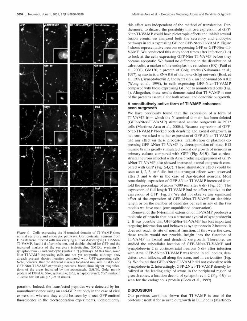
this effect was independent of the method of transfection. Fur-thermore, to discard the possibility that overexpression of GFP-Nter-TI-VAMP could have pleiotropic effects and inhibit severalfusion events, we analyzed both the secretory and endocyticpathways in cells expressing GFP or GFP-Nter-TI-VAMP. Figure4 shows representative neurons expressing GFP or GFP-Nter-TI-VAMP. We conducted this study short times after infection (1 d)to look at the cells expressing GFP-Nter-TI-VAMP before theybecame apoptotic. We found no difference in the distribution ofcalreticulin, a marker of the endoplasmic reticulum (ER) (Patil etal., 2000), GM130, a protein of Golgi stacks (Nakamura et al.,1997), syntaxin 6, a SNARE of the trans-Golgi network (Bock etal., 1997), synaptobrevin 2, and syntaxin 7, an endosomal SNARE(Wong et al., 1998), in cells expressing GFP-Nter-TI-VAMPcompared with those expressing GFP or to noninfected cells (Fig.4). Altogether, these results demonstrated that TI-VAMP is oneof the proteins essential for both axonal and dendritic outgrowth.
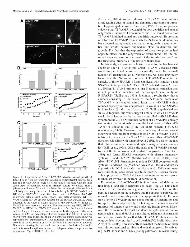
A constitutively active form of TI-VAMP enhancesaxon outgrowthWe have previously found that the expression of a form ofTI-VAMP from which the N-terminal domain has been deleted(GFP-DNter-TI-VAMP) stimulated neurite outgrowth in PC12cells (Martinez-Arca et al., 2000a). Because expression of GFP-Nter-TI-VAMP blocked both dendritic and axonal outgrowth inneurons, we asked whether expression of GFP-DNter-TI-VAMPhad any effect on these processes. Transfection of plasmids ex-pressing GFP-DNter-TI-VAMP by electroporation of intact E13murine brains greatly stimulated axonal outgrowth of neurons inprimary culture compared with GFP (Fig. 5A,B). Rat cortico-striatal neurons infected with Aavs producing expression of GFP-DNter-TI-VAMP also showed increased axonal outgrowth com-pared with GFP (Fig. 5A,C). These stimulatory effects could beseen at 1, 2, 3, or 6 div, but the strongest effects were observedafter 3 and 6 div in the case of Aav-treated neurons. Mostremarkably, expression of GFP-DNter-TI-VAMP increased four-fold the percentage of axons .300 mm after 6 div (Fig. 5C). Theexpression of full-length TI-VAMP had no effect relative to theexpression of GFP (Fig. 5). We did not observe any significanteffect of the expression of GFP-DNter-TI-VAMP on dendriticlength or on the number of dendrites per cell in any of the twomodels we have used (our unpublished observation).
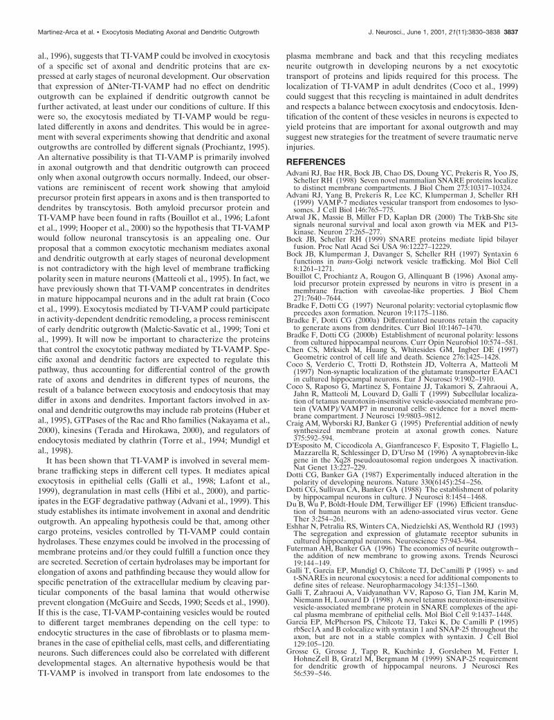
Removal of the N-terminal extension of TI-VAMP produces amolecule of protein that has a structure typical of synaptobrevin2. It was possible that GFP-DNter-TI-VAMP has lost importanttargeting information and behaves as synaptobrevin 2 because itdoes not reach its site of normal function. If this were the case,these results would not provide insight into the function ofTI-VAMP in axonal and dendritic outgrowth. Therefore, westudied the subcellular location of GFP-DNter-TI-VAMP andsynaptobrevin 2 in corticostriatal neurons 6 div after infectionwith Aavs. GFP-DNter-TI-VAMP was found in cell bodies, den-drites, axon hillocks, all along the axon, and in varicosities (Fig.6). We found that GFP-DNter-TI-VAMP did not colocalize withsynaptobrevin 2. Interestingly, GFP-DNter-TI-VAMP densely lo-calized at the leading edge of axons in the peripheral region ofgrowth cones, a location devoid of synaptobrevin 2 (Fig. 6E), asseen for the endogenous protein (Coco et al., 1999).
DISCUSSIONOur previous work has shown that TI-VAMP is one of theproteins essential for neurite outgrowth in PC12 cells (Martinez-
Figure 4. Cells expressing the N-terminal domain of TI-VAMP shownormal secretory and endocytic pathways. Corticostriatal neurons fromE16 rats were infected with Aav carrying GFP or Aav carrying GFP-Nter-TI-VAMP, fixed 1 d after infection, and double-labeled for GFP and theindicated markers of the secretory (calreticulin, GM130, syntaxin 6,synaptobrevin 2) and endocytic (syntaxin 7) pathways. At this time, someNter-TI-VAMP-expressing cells are not yet apoptotic, although theyalready present shorter neurites compared with GFP-expressing cells.Note, however, that the different markers localized similarly in GFP- andGFP-Nter-TI-VAMP-expressing neurons. Insets show higher magnifica-tions of the areas indicated by the arrowheads. GM130, Golgi matrixprotein of 130 kDa; Stx6, syntaxin 6; Syb2, synaptobrevin 2; Stx7, syntaxin7. Scale bar, 60 mm (15 mm in insets).
3834 J. Neurosci., June 1, 2001, 21(11):3830–3838 Martinez-Arca et al. • Exocytosis Mediating Axonal and Dendritic Outgrowth
Arca et al., 2000a). We have shown that TI-VAMP concentratesat the leading edge of axonal and dendritic outgrowths of imma-ture hippocampal neurons (Coco et al., 1999). Here, we provideevidence that TI-VAMP is essential for both dendritic and axonaloutgrowth in neurons. Expression of the N-terminal domain ofTI-VAMP inhibited axonal and dendritic outgrowth. Expressionof a form of TI-VAMP from which the N-terminal domain hasbeen deleted strongly enhanced axonal outgrowth in mouse cor-tical and striatal neurons but had no effect on dendritic out-growth. The fact that the expression of these two proteins hadopposite effects on the outgrowth of axons shows that the ob-served changes were not the result of the transfection itself butthe functional property of the proteins themselves.
In this study, we were not able to characterize the biochemicaleffects of Nter-TI-VAMP and DNter-TI-VAMP, because suchstudies in transfected neurons are technically limited by the smallnumber of transfected cells. Nevertheless, we have previouslyfound that the N-terminal domain of TI-VAMP inhibits thecapacity of this v-SNARE to form complexes with syntaxin 1 andSNAP25, its target Q-SNAREs in PC12 cells (Martinez-Arca etal., 2000a). TI-VAMP presents a long N-terminal extension thatis not present in members of the synaptobrevin family ofR-SNAREs (Galli et al., 1998). Preliminary results show that achimera consisting in the fusion of the N-terminal domain ofTI-VAMP with synaptobrevin 2 leads to a v-SNARE with areduced capacity to form complexes with syntaxin 4 and SNAP23in fibroblasts (S. Martinez-Arca and T. Galli, unpublished re-sults). Altogether, our results suggest a model in which TI-VAMPwould be a less active but a more controlled v-SNARE thansynaptobrevin 2. The N-terminal domain of TI-VAMP is unlikelyto contain targeting signals because the localization of DNter-TI-VAMP is similar to that of the full-length protein (Figs. 5, 6)(Coco et al., 1999). Moreover, the stimulatory effect on axonaloutgrowth resulting from expression of DNter-TI-VAMP (Fig. 5)is likely to be specific for TI-VAMP because DNter-TI-VAMPdoes not colocalize with synaptobrevin 2 (Fig. 6) despite the factthat it has a similar structure and high primary sequence similar-ity (Galli et al., 1998). Given the facts that TI-VAMP concen-trates at the tip of axonal and dendritic outgrowths (Coco et al.,1999) and forms SNARE complexes with plasma membranesyntaxin 1 and SNAP25 (Martinez-Arca et al., 2000a), thatDNter-TI-VAMP forms more abundant SNARE complexes withsyntaxin 1 and SNAP25 (Martinez-Arca et al., 2000a), and that itsexpression in PC12 cells (Martinez-Arca et al., 2000a) and neu-rons (this study) accelerates neurite outgrowth, it seems reason-able to propose that TI-VAMP mediates an important exocytoticmechanism involved in neuronal differentiation.
Expression of Nter-TI-VAMP inhibited neuronal differentia-tion (Fig. 1) and led to neuronal cell death (Fig. 3). This effectcannot be attributable to a general deleterious effect of thispeptide because both the secretory and the endocytic pathway arenormal in cells before they enter the apoptosis (Fig. 4). Expres-sion of Nter-TI-VAMP did not affect smooth ER generation andtransport, intra- and post-Golgi trafficking, and the formation andtransport of synaptic vesicle and endocytic fusion events. More-over, the expression of axonal and dendritic cytoskeletal compo-nents such as tau and MAP-2 is not altered (data not shown), andwe have previously shown that Nter-TI-VAMP inhibits neuriteoutgrowth but does not lead to cell death in PC12 cells (Martinez-Arca et al., 2000a). It was recently shown that the Shc site of TrkBcontrols both neuronal survival and axonal outgrowth by activat-ing the PI3-kinase and MEK signaling pathways, thus establishing
Figure 5. Expression of DNter-TI-VAMP activates axonal growth. A,Intact brains from E13 mice (top panels) or corticostriatal neurons fromE16 rats (bottom panels) were electroporated or infected with the indi-cated Aavs, respectively. Cells in primary culture were fixed after 2(electroporation) or 3 div (Aavs). Note the punctate distribution in thecell body and along the axon of both full-length GFP-TI-VAMP andGFP-DNter-TI-VAMP and the fact that GFP-DNter-TI-VAMP-expressing cells present longer axons than cells expressing GFP-TI-VAMP. Scale bar: 20 mm (top panels); 60 mm (bottom panels). B, Quan-tification of the effect in axonal growth of the expression of DNter-TI-VAMP in electroporated neurons. Neurons expressing GFP, GFP-TI-VAMP, or GFP-DNter-TI-VAMP were fixed after the indicated times,and the length of their axons was measured. In the top panels the meanvalues (6SEM) of percentage of axons longer than 50 or 100 mm areshown from three independent experiments; the bottom panels show tworepresentative experiments. C, Quantification of the effect on axonalgrowth of the expression of DNter-TI-VAMP in Aav-infected neurons.Neurons expressing the indicated constructs were fixed after 3 or 6 div,and their axonal length was measured; each panel shows a representativeexperiment. **p , 0.001; *p , 0.005.
Martinez-Arca et al. • Exocytosis Mediating Axonal and Dendritic Outgrowth J. Neurosci., June 1, 2001, 21(11):3830–3838 3835

a link between these two functions (Atwal et al., 2000). Therelationship between cell geometry and regulation of the balancebetween cell growth and apoptosis has also been reported (Chenet al., 1997). Our results suggesting that the apoptosis observedafter the expression of Nter-TI-VAMP is linked to the inhibitionof axonal and dendritic outgrowth point to the interdependencebetween neuronal survival and neurite outgrowth.
The fact that expression of Nter-TI-VAMP blocked both ax-onal and dendritic outgrowths (Figs. 1, 3) indicates that both
processes share common molecular mechanisms. As suggested byits localization at the leading edge of both axonal and dendriticgrowth cones, vesicles with fusion that is promoted by TI-VAMPcould mediate exposure at the cell surface of proteins that arerequired both for axonal and dendritic outgrowth (Coco et al.,1999). The effect of Nter-TI-VAMP on the dendritic expressionof EAAC1 (Fig. 2), a protein that may play a role in synaptogen-esis (Coco et al., 1997), but not on the expression of GluR1, aprotein of the mature dendrite (Eshhar et al., 1993; Richmond et
Figure 6. GFP-DNter-TI-VAMP does notcolocalize with synaptobrevin 2. Rat embry-onic neurons were infected with Aav carry-ing GFP-DNter-TI-VAMP. After 6 div, thecells were fixed and permeabilized, incu-bated with a polyclonal antibody anti-GFPand with a monoclonal antibody anti-synaptobrevin 2 (Syb2), and observed byconfocal microscopy. Low magnification im-ages are shown in A. In all the other panelshigh magnification images of a cell body (B),an axon (C), a varicosity (D), and a growthcone (E), respectively, are shown. GFP-DNter-TI-VAMP (small arrows) does not co-localize with endogenous synaptobrevin 2(B–E, large arrows) in any of the differentneuronal domains. A significant amount ofGFP-DNter-TI-VAMP was detected at theleading edge of the growth cone, in a regiondevoid of synaptobrevin 2. Scale bar: A, 90mm; B, C, E, 4.6 mm; D, 3 mm.
3836 J. Neurosci., June 1, 2001, 21(11):3830–3838 Martinez-Arca et al. • Exocytosis Mediating Axonal and Dendritic Outgrowth
al., 1996), suggests that TI-VAMP could be involved in exocytosisof a specific set of axonal and dendritic proteins that are ex-pressed at early stages of neuronal development. Our observationthat expression of DNter-TI-VAMP had no effect on dendriticoutgrowth can be explained if dendritic outgrowth cannot befurther activated, at least under our conditions of culture. If thiswere so, the exocytosis mediated by TI-VAMP would be regu-lated differently in axons and dendrites. This would be in agree-ment with several experiments showing that dendritic and axonaloutgrowths are controlled by different signals (Prochiantz, 1995).An alternative possibility is that TI-VAMP is primarily involvedin axonal outgrowth and that dendritic outgrowth can proceedonly when axonal outgrowth occurs normally. Indeed, our obser-vations are reminiscent of recent work showing that amyloidprecursor protein first appears in axons and is then transported todendrites by transcytosis. Both amyloid precursor protein andTI-VAMP have been found in rafts (Bouillot et al., 1996; Lafontet al., 1999; Hooper et al., 2000) so the hypothesis that TI-VAMPwould follow neuronal transcytosis is an appealing one. Ourproposal that a common exocytotic mechanism mediates axonaland dendritic outgrowth at early stages of neuronal developmentis not contradictory with the high level of membrane traffickingpolarity seen in mature neurons (Matteoli et al., 1995). In fact, wehave previously shown that TI-VAMP concentrates in dendritesin mature hippocampal neurons and in the adult rat brain (Cocoet al., 1999). Exocytosis mediated by TI-VAMP could participatein activity-dependent dendritic remodeling, a process reminiscentof early dendritic outgrowth (Maletic-Savatic et al., 1999; Toni etal., 1999). It will now be important to characterize the proteinsthat control the exocytotic pathway mediated by TI-VAMP. Spe-cific axonal and dendritic factors are expected to regulate thispathway, thus accounting for differential control of the growthrate of axons and dendrites in different types of neurons, theresult of a balance between exocytosis and endocytosis that maydiffer in axons and dendrites. Important factors involved in ax-onal and dendritic outgrowths may include rab proteins (Huber etal., 1995), GTPases of the Rac and Rho families (Nakayama et al.,2000), kinesins (Terada and Hirokawa, 2000), and regulators ofendocytosis mediated by clathrin (Torre et al., 1994; Mundigl etal., 1998).
It has been shown that TI-VAMP is involved in several mem-brane trafficking steps in different cell types. It mediates apicalexocytosis in epithelial cells (Galli et al., 1998; Lafont et al.,1999), degranulation in mast cells (Hibi et al., 2000), and partic-ipates in the EGF degradative pathway (Advani et al., 1999). Thisstudy establishes its intimate involvement in axonal and dendriticoutgrowth. An appealing hypothesis could be that, among othercargo proteins, vesicles controlled by TI-VAMP could containhydrolases. These enzymes could be involved in the processing ofmembrane proteins and/or they could fulfill a function once theyare secreted. Secretion of certain hydrolases may be important forelongation of axons and pathfinding because they would allow forspecific penetration of the extracellular medium by cleaving par-ticular components of the basal lamina that would otherwiseprevent elongation (McGuire and Seeds, 1990; Seeds et al., 1990).If this is the case, TI-VAMP-containing vesicles would be routedto different target membranes depending on the cell type: toendocytic structures in the case of fibroblasts or to plasma mem-branes in the case of epithelial cells, mast cells, and differentiatingneurons. Such differences could also be correlated with differentdevelopmental stages. An alternative hypothesis would be thatTI-VAMP is involved in transport from late endosomes to the
plasma membrane and back and that this recycling mediatesneurite outgrowth in developing neurons by a net exocytotictransport of proteins and lipids required for this process. Thelocalization of TI-VAMP in adult dendrites (Coco et al., 1999)could suggest that this recycling is maintained in adult dendritesand respects a balance between exocytosis and endocytosis. Iden-tification of the content of these vesicles in neurons is expected toyield proteins that are important for axonal outgrowth and maysuggest new strategies for the treatment of severe traumatic nerveinjuries.
REFERENCESAdvani RJ, Bae HR, Bock JB, Chao DS, Doung YC, Prekeris R, Yoo JS,
Scheller RH (1998) Seven novel mammalian SNARE proteins localizeto distinct membrane compartments. J Biol Chem 273:10317–10324.
Advani RJ, Yang B, Prekeris R, Lee KC, Klumperman J, Scheller RH(1999) VAMP-7 mediates vesicular transport from endosomes to lyso-somes. J Cell Biol 146:765–775.
Atwal JK, Massie B, Miller FD, Kaplan DR (2000) The TrkB-Shc sitesignals neuronal survival and local axon growth via MEK and P13-kinase. Neuron 27:265–277.
Bock JB, Scheller RH (1999) SNARE proteins mediate lipid bilayerfusion. Proc Natl Acad Sci USA 96:12227–12229.
Bock JB, Klumperman J, Davanger S, Scheller RH (1997) Syntaxin 6functions in trans-Golgi network vesicle trafficking. Mol Biol Cell8:1261–1271.
Bouillot C, Prochiantz A, Rougon G, Allinquant B (1996) Axonal amy-loid precursor protein expressed by neurons in vitro is present in amembrane fraction with caveolae-like properties. J Biol Chem271:7640–7644.
Bradke F, Dotti CG (1997) Neuronal polarity: vectorial cytoplasmic flowprecedes axon formation. Neuron 19:1175–1186.
Bradke F, Dotti CG (2000a) Differentiated neurons retain the capacityto generate axons from dendrites. Curr Biol 10:1467–1470.
Bradke F, Dotti CG (2000b) Establishment of neuronal polarity: lessonsfrom cultured hippocampal neurons. Curr Opin Neurobiol 10:574–581.
Chen CS, Mrksich M, Huang S, Whitesides GM, Ingber DE (1997)Geometric control of cell life and death. Science 276:1425–1428.
Coco S, Verderio C, Trotti D, Rothstein JD, Volterra A, Matteoli M(1997) Non-synaptic localization of the glutamate transporter EAAC1in cultured hippocampal neurons. Eur J Neurosci 9:1902–1910.
Coco S, Raposo G, Martinez S, Fontaine JJ, Takamori S, Zahraoui A,Jahn R, Matteoli M, Louvard D, Galli T (1999) Subcellular localiza-tion of tetanus neurotoxin-insensitive vesicle-associated membrane pro-tein (VAMP)/VAMP7 in neuronal cells: evidence for a novel mem-brane compartment. J Neurosci 19:9803–9812.
Craig AM, Wyborski RJ, Banker G (1995) Preferential addition of newlysynthesized membrane protein at axonal growth cones. Nature375:592–594.
D’Esposito M, Ciccodicola A, Gianfrancesco F, Esposito T, Flagiello L,Mazzarella R, Schlessinger D, D’Urso M (1996) A synaptobrevin-likegene in the Xq28 pseudoautosomal region undergoes X inactivation.Nat Genet 13:227–229.
Dotti CG, Banker GA (1987) Experimentally induced alteration in thepolarity of developing neurons. Nature 330(6145):254–256.
Dotti CG, Sullivan CA, Banker GA (1988) The establishment of polarityby hippocampal neurons in culture. J Neurosci 8:1454–1468.
Du B, Wu P, Boldt-Houle DM, Terwilliger EF (1996) Efficient transduc-tion of human neurons with an adeno-associated virus vector. GeneTher 3:254–261.
Eshhar N, Petralia RS, Winters CA, Niedzielski AS, Wenthold RJ (1993)The segregation and expression of glutamate receptor subunits incultured hippocampal neurons. Neuroscience 57:943–964.
Futerman AH, Banker GA (1996) The economics of neurite outgrowth–the addition of new membrane to growing axons. Trends Neurosci19:144–149.
Galli T, Garcia EP, Mundigl O, Chilcote TJ, DeCamilli P (1995) v- andt-SNAREs in neuronal exocytosis: a need for additional components todefine sites of release. Neuropharmacology 34:1351–1360.
Galli T, Zahraoui A, Vaidyanathan VV, Raposo G, Tian JM, Karin M,Niemann H, Louvard D (1998) A novel tetanus neurotoxin-insensitivevesicle-associated membrane protein in SNARE complexes of the api-cal plasma membrane of epithelial cells. Mol Biol Cell 9:1437–1448.
Garcia EP, McPherson PS, Chilcote TJ, Takei K, De Camilli P (1995)rbSec1A and B colocalize with syntaxin 1 and SNAP-25 throughout theaxon, but are not in a stable complex with syntaxin. J Cell Biol129:105–120.
Grosse G, Grosse J, Tapp R, Kuchinke J, Gorsleben M, Fetter I,HohneZell B, Gratzl M, Bergmann M (1999) SNAP-25 requirementfor dendritic growth of hippocampal neurons. J Neurosci Res56:539–546.
Martinez-Arca et al. • Exocytosis Mediating Axonal and Dendritic Outgrowth J. Neurosci., June 1, 2001, 21(11):3830–3838 3837

Hibi T, Hirashima N, Nakanishi M (2000) Rat basophilic leukemia cellsexpress syntaxin-3 and VAMP-7 in granule membranes. Biochem Bio-phys Res Commun 271:36–41.
Hooper NM, Trew AJ, Parkin ET, Turner AJ (2000) The role of pro-teolysis in Alzheimer’s disease. Adv Exp Med Biol 477:379–390.
Huber LA, Dupree P, Dotti CG (1995) A deficiency of the small GT-Pase rab8 inhibits membrane traffic in developing neurons. Mol CellBiol 15:918–924.
Igarashi M, Kozaki S, Terakawa S, Kawano S, Ide C, Komiya Y (1996)Growth cone collapse and inhibition of neurite growth by Botulinumneurotoxin C1: A t-SNARE is involved in axonal growth. J Cell Biol134:205–215.
Jahn R, Sudhof TC (1999) Membrane fusion and exocytosis. Annu RevBiochem 68:863–911.
Johannes L, Galli T (1998) Exocytosis: SNAREs drum up! Eur J Neu-rosci 10:415–422.
Lafont F, Verkade P, Galli T, Wimmer C, Louvard D, Simons K (1999)Raft association of SNAP receptors acting in apical trafficking inMadin-Darby canine kidney cells. Proc Nat Acad Sci USA96:3734–3738.
Mainguy G, Luz Montesinos M, Lesaffre B, Zevnik B, Karasawa M,Kothary R, Wurst W, Prochiantz A, Volovitch M (2000) An inductiongene trap for identifying a homeoprotein-regulated locus. Nat Biotech-nol 18:746–749.
Maletic-Savatic M, Malinow R, Svoboda K (1999) Rapid dendritic mor-phogenesis in CA1 hippocampal dendrites induced by synaptic activity.Science 283:1923–1927.
Maniatis T, Fritsch EF, Sambrook J (1982) Molecular cloning: a labora-tory manual, Ch 16, pp 33–36. Cold Spring Harbor, NY: Cold SpringHarbor Laboratory.
Martinez-Arca S, Alberts P, Zahraoui A, Louvard D, Galli T (2000a)Role of tetanus neurotoxin insensitive vesicle-associated membraneprotein (TI-VAMP) in vesicular transport mediating neurite out-growth. J Cell Biol 149:889–899.
Martinez-Arca S, Alberts P, Galli T (2000b) Clostridial neurotoxin-insensitive vesicular SNAREs in exocytosis and endocytosis. Biol Cell92:449–453.
Matteoli M, Verderio C, Krawzeski K, Mundigl O, Coco S, Fumagalli G,DeCamilli P (1995) Mechanisms of synaptogenesis in hippocampalneurons in primary culture. J Physiol (Paris) 89:51–55.
McGuire PG, Seeds NW (1990) Degradation of underlying extracellularmatrix by sensory neurons during neurite outgrowth. Neuron4:633–642.
Miyasaka N, Arimatsu Y, Takiguchihayashi K (1999) Foreign gene ex-pression in an organotypic culture of cortical anlage after in vivoelectroporation. NeuroReport 10:2319–2323.
Mundigl O, Ochoa GC, David C, Slepnev VI, Kabanov A, DeCamilli P(1998) Amphiphysin I antisense oligonucleotides inhibit neurite out-growth in cultured hippocampal neurons. J Neurosci 18:93–103.
Nakamura N, Lowe M, Levine TP, Rabouille C, Warren G (1997) Thevesicle docking protein p115 binds GM130, a cis-Golgi matrix protein,in a mitotically regulated manner. Cell 89:445–455.
Nakayama AY, Harms MB, Luo L (2000) Small GTPases Rac and Rhoin the maintenance of dendritic spines and branches in hippocampalpyramidal neurons. J Neurosci 20:5329–5338.
Osen-Sand A, Staple JK, Naldi E, Schiavo G, Rossetto O, Petitpierre S,Malgaroli A, Montecucco C, Catsicas S (1996) Common and distinctfusion proteins in axonal growth and transmitter release. J CompNeurol 367:222–234.
Patil AR, Thomas CJ, Surolia A (2000) Kinetics and the mechanism ofinteraction of the endoplasmic reticulum chaperone, calreticulin, withmonoglucosylated (Glc1Man9GlcNAc2) substrate. J Biol Chem275:24348–24356.
Polverino AJ, Patterson SD (1997) Selective activation of caspases dur-ing apoptotic induction in HL-60 cells. Effects of a tetrapeptide inhib-itor. J Biol Chem 272:7013–7021.
Prochiantz A (1995) Neuronal polarity: giving neurons heads and tails.Neuron 15:743–746.
Richmond SA, Irving AJ, Molnar E, McIlhinney RA, Michelangeli F,Henley JM, Collingridge GL (1996) Localization of the glutamatereceptor subunit GluR1 on the surface of living and within culturedhippocampal neurons. Neuroscience 75:69–82.
Rousselet A, Autillo-Touati A, Araud D, Prochiantz A (1990) In vitroregulation of neuronal morphogenesis and polarity by astrocyte-derived factors. Dev Biol 137:33–45.
Salvetti A, Oreve S, Chadeuf G, Favre D, Cherel Y, Champion-Arnaud P,David-Ameline J, Moullier P (1998) Factors influencing recombinantadeno-associated virus production. Hum Gene Ther 9:695–706.
Seeds NW, Haffke S, Christensen K, Schoonmaker J (1990) Cerebellargranule cell migration involves proteolysis. Adv Exp Med Biol265:169–178.
Slack RS, Miller FD (1996) Viral vectors for modulating gene expressionin neurons. Curr Opin Neurobiol 6:576–583.
Sollner T, Whiteheart SW, Brunner M, Erdjument-Bromage H, Geroma-nos S, Tempst P, Rothman JE (1993) SNAP receptors implicated invesicle targeting and fusion. Nature 362:318–324.
Terada S, Hirokawa N (2000) Moving on to the cargo problem ofmicrotubule-dependent motors in neurons. Curr Opin Neurobiol10:566–573.
Toni N, Buchs PA, Nikonenko I, Bron CR, Muller D (1999) LTP pro-motes formation of multiple spine synapses between a single axonterminal and a dendrite. Nature 402:421–425.
Torre E, McNiven MA, Urrutia R (1994) Dynamin 1 antisense oligo-nucleotide treatment prevents neurite formation in cultured hippocam-pal neurons. J Biol Chem 269:32411–32417.
Wong SH, Xu Y, Zhang T, Hong W (1998) Syntaxin 7, a novel syntaxinmember associated with the early endosomal compartment. J BiolChem 273:375–380.
Xiao X, Li J, Samulski RJ (1998) Production of high-titer recombinantadeno-associated virus vectors in the absence of helper adenovirus.J Virol 72:2224–2232.
Zakharenko S, Popov S (1998) Dynamics of axonal microtubules regu-late the topology of new membrane insertion into the growing neurites.J Cell Biol 143:1077–1086.
3838 J. Neurosci., June 1, 2001, 21(11):3830–3838 Martinez-Arca et al. • Exocytosis Mediating Axonal and Dendritic Outgrowth