a common antitussive drug, clobutinol, precipitates the...
TRANSCRIPT

1/36
A Common Antitussive Drug, Clobutinol,
Precipitates the Long QT2 Syndrome
Chloé Bellocq, Ronald Wilders, Jean-Jacques Schott, Bénédicte
Louérat-Oriou, Pierre Boisseau, Hervé Le Marec,
Denis Escande, Isabelle Baró
L’institut du thorax, INSERM U533, Nantes, France (C.B., J.J.S., B.L.O., D.E., I.B.);
Department of Physiology, Academic Medical Center, University of Amsterdam, the
Netherlands (R.W.); Laboratoire de Génétique Moléculaire, CHU de Nantes, France
(P.B.); L’Institut du Thorax, INSERM U533 and Clinique Cardiologique et des
Maladies Vasculaires, CIC INSERM de Nantes, France (H.L.M.)
Molecular Pharmacology Fast Forward. Published on July 27, 2004 as doi:10.1124/mol.104.001065
Copyright 2004 by the American Society for Pharmacology and Experimental Therapeutics.
This article has not been copyedited and formatted. The final version may differ from this version.Molecular Pharmacology Fast Forward. Published on July 27, 2004 as DOI: 10.1124/mol.104.001065
at ASPE
T Journals on July 26, 2018
molpharm
.aspetjournals.orgD
ownloaded from
MOLPHARM/2004/001065
2/36
Running title: Clobutinol and the LQT2 syndrome
Corresponding author: Isabelle Baró at INSERM U533, Faculté de Médecine, 1,
rue Gaston Veil, 44035 Nantes, France.
Phone: +33 2 40 41 28 48
Fax: +33 2 40 41 29 50
e-mail: [email protected]
text page: 35
tables: 1
figures: 8
references: 41
Abstract: 250 words
Introduction: 349 words
Discussion: 1500 words
Abbreviations
AP: action potential; HERG: human ether-a-go-go-related gene product; IKr: rapidly
activating component of the cardiac delayed rectifier K+ current; LQT: long-QT
syndrome; QTc: rate-corrected QT interval
This article has not been copyedited and formatted. The final version may differ from this version.Molecular Pharmacology Fast Forward. Published on July 27, 2004 as DOI: 10.1124/mol.104.001065
at ASPE
T Journals on July 26, 2018
molpharm
.aspetjournals.orgD
ownloaded from

MOLPHARM/2004/001065
3/36
Abstract
QT prolongation, a classical risk factor for arrhythmias, can result from a mutation in
one of the genes governing cardiac repolarization and also can result from the intake
of a medication acting as blocker of the cardiac K+ channel HERG. Here, we
identified the arrhythmogenic potential of a non-opioid antitussive drug, clobutinol.
The deleterious effects of clobutinol were suspected when a young boy, diagnosed
with congenital long QT syndrome, experienced arrhythmias while being treated with
this drug. Using the patch-clamp technique, we showed that clobutinol dose-
dependently inhibited the HERG K+ current with a half-maximum block concentration
of 2.9 µM. In the proband, we identified a novel A561P HERG mutation. Two others
LQT mutations (A561V and A561T) had previously been reported at the same
position. None of the three mutants led to sizeable current in heterologous
expression system. When co-expressed with wild-type (WT) HERG channels, the
three A561 mutants reduced the trafficking of WT and mutant heteromeric channels
resulting in decreased K+ current amplitude (dominant-negative effects). In addition,
A561P but not A561V and A561T mutants induced an ≈ -11 mV shift of the current
activation curve and accelerated deactivation, thereby partially counteracting the
dominant-negative effects. A561P mutation and clobutinol effects on the human
ventricular action potential characteristics were simulated using the Priebe-
Beuckelmann model. Our work shows that clobutinol has limited effects on WT action
potential but should be classified as a 'drug to be avoided by congenital long QT
patients' rather than as a 'drug with risk of torsades de pointes'.
This article has not been copyedited and formatted. The final version may differ from this version.Molecular Pharmacology Fast Forward. Published on July 27, 2004 as DOI: 10.1124/mol.104.001065
at ASPE
T Journals on July 26, 2018
molpharm
.aspetjournals.orgD
ownloaded from
MOLPHARM/2004/001065
4/36
QT prolongation is a risk factor in a number of cardiovascular and noncardiovascular
diseases. Among these, the congenital form of the long QT syndrome (LQT)
associates prolonged rate-corrected QT interval (QTc) with recurrent syncope and
sudden cardiac death resulting from torsades de pointes tachyarrhythmias. The long
QT syndrome may also result from the effects of numerous chemically unrelated
medications (acquired LQT) in patients with pre-existing normal QT or in patients
carrying a long QT gene mutation. At the cellular level, prolongation of the QT
interval reflects lengthening of the ventricular action potential (AP). In the human
heart, a key repolarizing potassium current is the rapidly activating component of the
delayed rectifier, IKr (Sanguinetti and Jurkiewicz, 1990). Its activation initiates
repolarization and terminates the plateau phase of the cardiac AP concomitantly to
the T wave on the ECG. IKr is related to pore-forming channel proteins encoded by
the human ether-a-go-go-related gene (HERG) (Curran et al., 1995; Sanguinetti et
al., 1995). Mutations in HERG account for chromosome 7-linked inherited long QT
syndrome 2 (LQT2; Keating, 1995; Sanguinetti et al., 1996). Finally, the vast majority
of drugs that produce the acquired LQT are blockers of HERG channels (Roden et
al., 1996).
In the present work, we have identified the arrhythmogenic potential of a
common drug, clobutinol, a centrally acting non-opioid antitussive drug widely used in
Europe as treatment for dry cough of infectious origin. The deleterious effects of the
molecule were suspected when a young boy, diagnosed with LQT2, experienced
syncopes and arrhythmias while being treated with clobutinol. We have evaluated the
inhibitory potency of clobutinol on wild-type HERG current. In the proband, we
identified a novel LQT2 mutation (A561P), which we functionally characterized in
This article has not been copyedited and formatted. The final version may differ from this version.Molecular Pharmacology Fast Forward. Published on July 27, 2004 as DOI: 10.1124/mol.104.001065
at ASPE
T Journals on July 26, 2018
molpharm
.aspetjournals.orgD
ownloaded from

MOLPHARM/2004/001065
5/36
recombinant expression system. Interestingly, two others mutations (A561V and
A561T) at the same position were previously reported (Curran et al., 1995; Dausse et
al., 1996) in congenital LQT2 patients. We thus examined and compared the
mechanism for HERG channel dysfunction in each of these three A561 mutations.
Our work shows that a common drug not previously identified as a QT prolonging
drug can precipitate the LQT2 syndrome.
This article has not been copyedited and formatted. The final version may differ from this version.Molecular Pharmacology Fast Forward. Published on July 27, 2004 as DOI: 10.1124/mol.104.001065
at ASPE
T Journals on July 26, 2018
molpharm
.aspetjournals.orgD
ownloaded from
MOLPHARM/2004/001065
6/36
Materials and methods
Clinical studies
The proband, an 11 year old boy, was diagnosed with a long QT duration in 1997. In
1999, he developed first symptoms in relation to torsades de pointes upon clobutinol
treatment. A familial investigation was initiated. All family members enrolled gave
written informed consent. Signature was applied by parents in case of children under
18. The protocol was approved by the local Committee for the Protection of Human
Subjects in Biomedical Research of Nantes University, France. Participants were
evaluated by history, review of medical records, and 12-lead electrocardiogram.
Correction of the duration of the QT interval as a function of cycle length was
performed using both Bazett's and Fridericia's formulae (QTcBazett=QT/2√RR and
QTcFridericia=QT/3√RR with RR expressed in seconds).
Mutation analysis
Genomic DNA was prepared from peripheral blood lymphocytes by standard
methods. Mutation analysis was conducted by direct sequencing of the gene using
an ABI 377 automated sequencer (Applied Biosystems, Foster City, CA). PCR
reactions for each of the amplicons representing the entire coding sequence and
splicing sites of KCNH2 but also KCNQ1, KNCE1 and SCN5A genes were performed
as previously described (Splawski et al., 1998; Wang et al., 1995).
Cell culture and transfection
African green monkey kidney cells (COS-7) were obtained from the American Type
Culture Collection. Cells were cultured at 37°C in a 5% CO2 humidified incubator in
Dulbecco’s modified Eagle medium supplemented with 10% fetal calf serum, 2 mM
L-glutamine and antibiotics (100 UI/mL penicillin, 100 µg/mL streptomycin, all from
GIBCO, Paisley, Scotland). Human wild-type HERG cDNA were subcloned into the
This article has not been copyedited and formatted. The final version may differ from this version.Molecular Pharmacology Fast Forward. Published on July 27, 2004 as DOI: 10.1124/mol.104.001065
at ASPE
T Journals on July 26, 2018
molpharm
.aspetjournals.orgD
ownloaded from

MOLPHARM/2004/001065
7/36
mammalian expression vector pBK under the control of the cytomegalovirus
promoter/enhancer (Stratagene, La Jolla, CA ). A561P, A561T, or A561V mutations
were introduced using standard molecular techniques. Cells were transfected using
the intranuclear microinjection technique or using polyethylenimine (PEI) as a
transfection reagent. A green fluorescence protein plasmid (pEGFP; Clonetech, Palo
Alto, CA) was used as an inert plasmid to ensure a constant plasmid concentration
and also to permit cell detection. The microinjection protocol using the Eppendorf
ECET microinjector 5246 system (Hamburg, Germany) has been previously
described by Mohammad-Panah et al. (1998). Plasmids (3 µg/mL pBK-CMV-WT or
mutated HERG plus 3 µg/mL pEGFP for homomeric channels, and 3 µg/mL pBK-
CMV-WT HERG plus 3 µg/mL pBK-CMV- mutated HERG for heteromeric channels)
were diluted in a buffer containing 40 mM NaCl, 50 mM HEPES, 50 mM NaOH, pH =
7.4 and supplemented with 0.5% fluorescein isothiocyanate-dextran (150 kDa).
Alternatively, cells were transfected when the culture reached 60-80% confluence
with plasmids complexed with PEI as previously reported (Pollard et al., 1998) with 2
µg plasmids per mL of culture medium. In patch-clamp experiments we used 20%
pBK-CMV-WT or mutated HERG plus 80% pEGFP for homomeric channels, and
20% pBK-CMV-WT HERG plus 20% pBK-CMV- mutated HERG and 60% pEGFP for
heteromeric channels. For immunolocalization experiments, we used 100% pBK-
CMV-WT or mutated HERG for homomeric channels and 50% pBK-CMV-WT HERG
plus 50% pBK-CMV- mutated HERG for heteromeric channels.
Patch-clamp recordings
Twelve to 24 hours after transfection, K+ currents from HERG-transfected COS-7
cells were recorded at 35°C using the whole cell configuration of the patch-clamp
technique. Cells were placed on the stage of an inverted microscope and
This article has not been copyedited and formatted. The final version may differ from this version.Molecular Pharmacology Fast Forward. Published on July 27, 2004 as DOI: 10.1124/mol.104.001065
at ASPE
T Journals on July 26, 2018
molpharm
.aspetjournals.orgD
ownloaded from

MOLPHARM/2004/001065
8/36
continuously superfused with Tyrode's solution. During the current measurements, a
microperfusion system allowed local application and rapid change of the different
extracellular solutions used to specifically record the K+ currents. Patch pipettes with
a tip resistance of 2.5-5 MΩ were electrically connected to a patch-clamp amplifier
(Axopatch 200A; Axon Instruments, Foster City, CA). Stimulation, data recording and
analysis were performed through an A/D converter (Tecmar TM100 Labmaster,
Scientific solutions, Mentor, Ohio) using Acquis1 software (Bio-logic, Claix, France).
WT and mutant HERG currents were measured using the following voltage protocol.
The membrane potential was clamped at a holding potential of –90 mV and every 3
s, a voltage prepulse to +10 mV was applied for 500 ms followed by the test pulse to
–70 mV for another 500 ms. The deactivating K+ current (or tail current) was
measured during polarization to -70 mV and normalized for each cell using the cell
capacitance. To evaluate the current activation, the prepulse voltage varied from
-100 to +60 mV for 500 ms. To evaluate the deactivation kinetics, the voltage test
pulse varied from -70 to -40 mV. Recorded human sub-endocardiac action potential
acquired at 1000 ms pacing length was also used to clamp the cells.
Immunolocalization experiments
Cells were plated 12 h after transfection on coverslips, left for 48 h in the incubator to
allow protein expression and then fixed in 4% PFA, permeabilized in 0.1% TRITON
X-100 and incubated overnight at 4 °C with a rabbit anti-erg antibody (1:1000;
Alomone, Jerusalem, Israel) in 1% BSA. Cells were rinsed in PBS and incubated for
1 hour at room temperature with a FITC-conjugated goat anti-rabbit antibody (1:200;
Sigma). Cells were mounted between slips and coverslips and placed on the stage of
an inverted Leica TCS NT confocal microscope (Wetzlar, Germany). Cells were
observed using a X63 oil objective.
This article has not been copyedited and formatted. The final version may differ from this version.Molecular Pharmacology Fast Forward. Published on July 27, 2004 as DOI: 10.1124/mol.104.001065
at ASPE
T Journals on July 26, 2018
molpharm
.aspetjournals.orgD
ownloaded from
MOLPHARM/2004/001065
9/36
Solutions and drugs
Tyrode's solution used for patch-clamp experiments contained (mM): 145 NaCl, 4
KCl, 1 MgCl2, 1 CaCl2, 5 HEPES, 5 glucose, pH = 7.4. The standard extracellular
solution to record K+ current contained (mM): 145 Na-gluconate, 4 K-gluconate, 7
hemi-Ca-gluconate (free-Ca2+: 1), 4 hemi-Mg-gluconate (free-Mg2+: 1), 5 HEPES, 5
glucose, 20 mannitol, pH = 7.4. Patch-clamp pipettes contained (mM): 145 K-
gluconate, 2 K2ATP, 2 hemi-Mg-gluconate (free-Mg2+: 0.1), 5 HEPES, 2 EGTA, pH =
7.2. Free activities were calculated using a software designed by GL Smith
(University of Glasgow, UK). Clobutinol (SILOMAT 20 mg/2 mL solution, Boehringer
Ingelheim) was diluted in extracellular solution as a 10-4 M stock solution. The test
solutions were prepared by further successive dilutions. E-4031 was prepared as 10-3
M stock solution with distilled water.
Computer modeling
Functional effects of the HERG mutations were tested by computer simulations using
the Priebe and Beuckelmann human ventricular cell model (1998). The
experimentally observed ≈70% reduction in HERG tail current density was
implemented by a 70% decrease in the fully activated conductance of IKr. The
experimentally observed −11 mV shift in voltage dependence of WT + A561P HERG
current activation was incorporated by a −11 mV shift in the IKr steady-state activation
curve, whereas the ≈30% decrease in the fast and slow time constants of HERG
channel deactivation were implemented by a 30% decrease in the corresponding IKr
time constant. Action potentials were elicited by repetitive stimulation with a 2 ms,
≈20% suprathreshold stimulus current.
The Priebe-Beuckelmann model produces an action potential that is typical for an
epicardial cell. To obtain endocardial and M cell models, we reduced the current
This article has not been copyedited and formatted. The final version may differ from this version.Molecular Pharmacology Fast Forward. Published on July 27, 2004 as DOI: 10.1124/mol.104.001065
at ASPE
T Journals on July 26, 2018
molpharm
.aspetjournals.orgD
ownloaded from

MOLPHARM/2004/001065
10/36
densities of the transient outward current (Ito), the slow component of the delayed
rectifier current (IKs) and the inward rectifier current (IK1) in the Priebe-Beuckelmann
model by 75%, 8%, and 11%, and by 13%, 54%, and 26%, respectively (see Conrath
et al., 2004), based on canine and human data (Liu et al., 1993; Liu et al., 1995;
Näbauer et al., 1996).
To measure restitution of action potential duration (APD restitution) in the single
human ventricular cell model, we used an S1-S2 stimulus protocol (Qu et al., 1999).
Following a period of pacing at a basic S1-S1 pacing interval of 1000 ms, S2 was
applied after a variable S1-S2 interval. The strengths of the 2 ms S1 and S2 stimuli
were fixed at 2.5 times threshold, and APD was defined using a threshold voltage of
−77.2 mV, which is near the voltage at which the action potential is 90% repolarized.
Models were coded using Compaq Visual Fortran 6.6 and run on a 3-GHz Intel
Pentium 4 processor workstation, applying an Euler-type integration scheme with a
5-µs step. All simulations were run for a sufficiently long time to reach steady-state
behavior.
Statistics
All data are presented as mean ± SEM. Statistical significance of the observed
effects was assessed by means of the Student t-test, Mann-Whitney rank sum test,
or two-way ANOVA when appropriate. A value of p < 0.05 was considered significant.
This article has not been copyedited and formatted. The final version may differ from this version.Molecular Pharmacology Fast Forward. Published on July 27, 2004 as DOI: 10.1124/mol.104.001065
at ASPE
T Journals on July 26, 2018
molpharm
.aspetjournals.orgD
ownloaded from
MOLPHARM/2004/001065
11/36
Results
Clinical characteristics
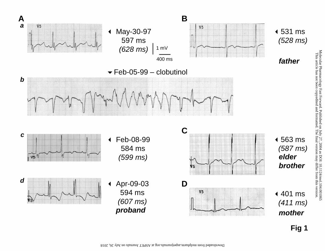
The proband, a boy born in 1992, was diagnosed for a tetralogy of Fallot and had
surgery several years before the drug-induced arrhythmia. His ECG follow-up is
illustrated in Figure 1Aa-d. Lengthening of the QT interval (QTcBazett = 628 ms and
QTcFredericia = 597 ms). was initially detected in 1997 in the absence of associated
symptoms (Figure 1Aa). In 1999, he experienced for the first time syncope and
torsades de pointes arrhythmias under clobutinol, a common antitussive drug (Figure
1Ab). The baseline serum K+ level indicated a moderate hypokalæmia to 3.5 mmol/L
before any treatment (Feb 05). However, the arrhythmic episodes did not cease
under potassium, magnesium or β-blocker therapy, even when the serum potassium
was normalized to 4.1 mmol/L. Torsades de pointes only ceased when the heart was
paced. After clobutinol treatment was discontinued, the QTc interval progressively
decreased to pre-treatment values with a time course comparable to clobutinol half-
life (23-32h; Zimmer et al., 1977) (Figure 1Ac). The kidney function was considered
as normal from urea and creatinine levels (Feb 05: 1.8 mmol/L (BUN: 5 mg/dL) and
44 mg/dL, respectively). Since 1999, the proband has no further arrhythmia or
symptoms, although he has retained a long QTc interval (Figure 1Ad). Altogether,
these data correlate the clobutinol intake to the torsades de pointes episodes.
Repolarization abnormalities were also detected in his asymptomatic father (Figure
1B) and in his elder brother (Figure 1C). Neither the brother nor the father has had a
history of cardiac surgery. The other relatives including his mother (Figure 1D)
presented normal ECG.
This article has not been copyedited and formatted. The final version may differ from this version.Molecular Pharmacology Fast Forward. Published on July 27, 2004 as DOI: 10.1124/mol.104.001065
at ASPE
T Journals on July 26, 2018
molpharm
.aspetjournals.orgD
ownloaded from

MOLPHARM/2004/001065
12/36
Mutation analysis
DNA sequencing of exon 7 in the proband (III-1; Figure 2) identified a heterozygous
G-to-C mutation at position 1681. This missense mutation was predicted to change
an Arginine for a Proline (A561P) within the S5 region of HERG. This mutation was
also identified in the proband's father and in his elder brother (II-1 ; III-2). DNA
sequencing of other genes related to LQT i.e. KCNQ1, KCNE1 and SCN5A,
encoding the α- and β-subunits of the IKs current and the α-subunit of the INa channel,
respectively, did not reveal other mutations.
Clobutinol blocks HERG current.
We investigated the sensitivity of homomeric wild-type (WT) channels to the non-
opioid antitussive clobutinol. Transfected COS-7 cells expressed a large K+ current
with biophysical properties reminiscent to those of the rapid component of the cardiac
delayed rectifier K+ current recorded in human cardiac cells (Figure 3A; Wang et al.,
1994). When repolarized to –70 mV, expressing COS-7 cells exhibited a large
deactivating current (I tail). The half-activation potential (V1/2) for this current was
calculated as –10.4 ± 5 mV (n = 10). Clobutinol at 10-5 M induced a slow decay of the
activating current recorded at +10 mV suggesting voltage-dependent block and a
lesser blocking effect at the initial phase of activation. This pattern of inhibition was
observed at the steady-state. After full activation at +10 mV, clobutinol inhibited the
HERG tail current with a half-maximum block concentration (IC50) of 2.9 · 10-6 ± 0.7 ·
10-6 M and a Hill coefficient of 0.9 (n = 9; Figure 3B).
As illustrated in Figure 3C, the HERG-related K+ current activated at potentials
positive to –50 mV and exhibited strong inward rectification. Deactivating K+ currents
reached a maximum after a prepulse to 10 mV and did not further increase with more
positive depolarizing prepulses (Figure 3D). As depicted in Figures 3 C and D,
This article has not been copyedited and formatted. The final version may differ from this version.Molecular Pharmacology Fast Forward. Published on July 27, 2004 as DOI: 10.1124/mol.104.001065
at ASPE
T Journals on July 26, 2018
molpharm
.aspetjournals.orgD
ownloaded from

MOLPHARM/2004/001065
13/36
clobutinol decreased activating and deactivating currents. The inset in Figure 3D
shows that clobutinol blocked the tail current more effectively at depolarized
potentials (p < 0.001). Recovery from block was also analyzed when the membrane
potential was repolarized to -70 mV. After a depolarization to +10 mV, the initial
deactivating current measured at the beginning of the repolarizing pulse (I tail init)
was significantly more inhibited than that measured 500 ms later (I tail late) (10-5 M
clobutinol, 71 ± 5% versus 45 ± 13%; inhibition percentage of I tail init and I tail late,
respectively; n = 7; p < 0.05). We conclude that the HERG block as produced by
clobutinol was voltage-dependent inasmuch as it was more pronounced at
depolarizing voltages, and as it developed with depolarization and relaxed with
repolarization.
Electrophysiological properties of the HERG A561P, HERG A561T and HERG
A561T channels
Since the non-opioid antitussive drug clobutinol had triggered arrhythmias in the
A561P HERG carrier, we evaluated the effects of this mutation on the channel
function. The functional effects of A561P were also compared to those caused by
A561T and A561V mutations. In cells injected into the nucleus with either A561P
HERG, A561T HERG or A561V HERG, no current was recorded unlike in cells
injected with WT HERG, suggesting that the three mutations led to a complete loss of
function (Figure 4A). Since LQT patients are heterozygous for these HERG
mutations, mutant and WT proteins were co-expressed with appropriate ratios to
mimic the genotype of the mutation carriers. As shown in Figure 4B, the tail K+
current density measured at –70 mV (depolarizing test pulse to +10 mV) was
markedly reduced to about 30% of the control current. We concluded that the three
mutations altered the WT HERG channel function in a dominant negative manner.
This article has not been copyedited and formatted. The final version may differ from this version.Molecular Pharmacology Fast Forward. Published on July 27, 2004 as DOI: 10.1124/mol.104.001065
at ASPE
T Journals on July 26, 2018
molpharm
.aspetjournals.orgD
ownloaded from
MOLPHARM/2004/001065
14/36
The absence of measurable K+ currents for mutated homomeric channels can be
related to either a defect in channel targeting to the plasma membrane or to altered
gating properties of channels. To test whether homomeric channels are inserted into
the cell membrane, immunolocalization experiments were conducted. As illustrated in
Figure 4A, WT HERG expressing cells showed an intracellular and plasma
membrane staining whereas membrane staining was undetectable with A561P,
A561T and A561V HERG channels suggesting that these channels do not insert into
the cell membrane. In cells co-transfected with WT and mutant HERG, confocal
microscopy revealed that membrane staining was still detectable though less
intensely (Figure 4B). These results suggest that the mutated channels are poorly
processed to the plasma membrane, and that heterotetramers coexpressed with the
WT protein are mostly retained in intracellular compartments.
In addition to the strong reduction in current amplitude, the A561P mutation also
modified the voltage-dependence of activation of the WT + A561P heteromeric
channels (Figure 5A and B) and produced a significant shift of V1/2 towards more
negative potentials (Table 1). The small 3–3.5 mV shift seen in cells co-expressing
WT and A561V or A561T HERG was not significant. On the other hand, the slope
factor of activation was slightly albeit significantly modified demonstrating the
alteration of the channel gating induced by the mutations.
In cells co-expressing WT and A561P HERG, we also observed a reduction in the
fast and slow deactivation time constants when the deactivating current traces were
fitted using a double exponential function (Figure 5C). The fast deactivating
component contributed to about 40% of the WT or WT + A561P current at any tested
potential. Altogether, the modifications as induced by the A561P mutation, i.e. current
reduction and faster deactivation kinetics counterbalanced by the shift in the
This article has not been copyedited and formatted. The final version may differ from this version.Molecular Pharmacology Fast Forward. Published on July 27, 2004 as DOI: 10.1124/mol.104.001065
at ASPE
T Journals on July 26, 2018
molpharm
.aspetjournals.orgD
ownloaded from

MOLPHARM/2004/001065
15/36
activation curve should lead to a milder loss of function of the WT + A561P channels
compared to the A561V or A561T mutations which only produced a decrease in
current amplitude.
In order to visualize more accurately the functional effects of the mutants, a human
ventricular action potential voltage clamp waveform was applied to cells expressing
WT or WT + mutated HERG channels. The normalized currents resulting from this
stimulation are depicted in Figure 6. The HERG current, shown as the E-4031-
sensitive current increased progressively as the action potential repolarized to reach
a peak. In accordance with the shift of the activation curve of WT + A561P channels,
an earlier WT + A561P HERG current peak was seen during the action potential time
course. Presumably, this behavior should prelude to a less pronounced QT interval
lengthening in comparison with WT + A561T or WT + A561V channels.
Clobutinol blocks WT + A561P HERG current
The effects of clobutinol were further tested in cells co-expressing WT and A561P
HERG channels. Figure 7 illustrates that the sensitivity of heteromeric channels to
the block produced by clobutinol was similar to that of homomeric channels (IC50 for
clobutinol on heteromeric WT + A561P HERG: 1.9 10-6 ± 0.7 10-6 M; Hill's coefficient:
1.1; n = 5).
Functional implications
Electrophysiological data demonstrate that each of the WT + A561P, WT + A561T,
and WT + A561V HERG currents is characterized by a decrease in current density to
≈30% of the control WT value. In addition, the WT + A561P HERG current shows
alterations in kinetics i.e. a -11 mV shift in voltage dependence of activation as well
as a ≈30% decrease in fast and slow time constants of deactivation. To assess the
functional consequences of these alterations, we carried out computer simulations
This article has not been copyedited and formatted. The final version may differ from this version.Molecular Pharmacology Fast Forward. Published on July 27, 2004 as DOI: 10.1124/mol.104.001065
at ASPE
T Journals on July 26, 2018
molpharm
.aspetjournals.orgD
ownloaded from

MOLPHARM/2004/001065
16/36
using the comprehensive human ventricular cell model by Priebe and Beuckelmann
(1998). Figure 8A (top) shows the model action potential with normal IKr (dotted line)
and with a 70% reduction in IKr (solid line), representing the experimentally observed
reduction in HERG current density. The reduction in repolarizing current induces a
prolongation of the action potential by 124 ms (arrow). The experimentally observed
alterations in kinetics result in an earlier activation of HERG channels and thus in a
larger IKr amplitude (Figure 8B). Despite the more rapid deactivation of the current,
repolarization is accelerated and the action potential shortened by 33 ms. When
combined, the two effects, i.e., the reduction in current density and alterations in
kinetics, are almost additive, with an AP prolongation of 97 ms (Figure 8C). We also
tested the effects of the experimentally observed changes in slope of the HERG
channel activation curve (Table 1), but the effects thereof were very small, with a
change in APD90 (∆APD90) typically less than 1–2 ms (data not shown). We also
determined ∆APD90 for pacing cycle lengths other than 1 s (Figure 8D). In all cases,
the effects are more pronounced at shorter cycle lengths, i.e., at higher heart rate,
which was also observed in a canine AP model for other mutations that affect IKr
(Mazhari et al., 2001). Figure 8D also shows that the A561P HERG mutation (open
triangles) exerts milder effects than the A561T or A561V mutations (open squares).
However, application of clobutinol (simulated by a 30% block of IKr, in accordance
with the estimated circulating drug concentration of 0.55–0.75 µM and the dose-
response curve in Figure 7; Zimmer et al. 1977) increases AP prolongation for the
A561P mutation to values larger than those for the more severe A561T or A561V
mutations (Figure 8D, closed triangles). With wild-type HERG channels, clobutinol
per se produces a mild prolongation of the action potential at all pacing cycle lengths
(Figure 8D, closed circles).
This article has not been copyedited and formatted. The final version may differ from this version.Molecular Pharmacology Fast Forward. Published on July 27, 2004 as DOI: 10.1124/mol.104.001065
at ASPE
T Journals on July 26, 2018
molpharm
.aspetjournals.orgD
ownloaded from
MOLPHARM/2004/001065
17/36
Increased dispersion of repolarization across the ventricular wall, either arising from
dynamic factors (steeper APD restitution) or from enhanced heterogeneity in intrinsic
electrophysiological properties among different cell types, is an important
determinant of the susceptibility to ventricular arrhythmias (see Qu et al., 2000).
Therefore, we first tested whether the HERG channel mutations would steepen the
APD restitution curve (Figure 8E). Again, the largest effects are observed for the
A561T or A561V mutations. However, application of clobutinol significantly steepens
the APD restitution curve for the A561P mutation, yielding a slope near 1 for the
steepest part of the curve. With wild-type HERG channels, clobutinol produces only
moderate alteration in the restitution curve. Second, we assessed the effects of the
HERG channel mutations on the intrinsic differences among the different cell types.
Figure 8F shows that the mutations preferentially prolong the M cell action potential,
thereby increasing the dispersion of repolarization. The largest dispersion is obtained
for the WT + A561P HERG current in the presence of clobutinol (189 ms vs. a control
value of 59 ms).
It has been hypothesized that the AP prolongation associated with LQT favors the
development of early afterdepolarizations (EADs) and triggered activity due to
reactivation of the L-type calcium current (ICa,L) during the AP plateau. In contrast
with simulation studies using the Luo-Rudy guinea pig-type AP model (Viswanathan
& Rudy, 1999), we did not observe EADs in any of the three cell types studied. As
discussed by Priebe and Beuckelmann (1998), the lower susceptibility of their human
ventricular cell model to the generation of EADs could reflect an inherent property of
human tissue. We did, however, observe considerably delayed repolarization for the
M cell model in case of the A561 HERG channel mutations, especially in response to
This article has not been copyedited and formatted. The final version may differ from this version.Molecular Pharmacology Fast Forward. Published on July 27, 2004 as DOI: 10.1124/mol.104.001065
at ASPE
T Journals on July 26, 2018
molpharm
.aspetjournals.orgD
ownloaded from

MOLPHARM/2004/001065
18/36
moderate increases in ICa,L, with, e.g., APD90 values exceeding 1 s upon a 25%
increase in ICa,L (data not shown).
In summary, Figure 8 shows that the effects of the A561P mutation on basic AP
properties are constantly less severe than those of the A561V or A561T mutation, but
become more severe if the WT + A561P HERG current is further reduced by the
application of clobutinol.
This article has not been copyedited and formatted. The final version may differ from this version.Molecular Pharmacology Fast Forward. Published on July 27, 2004 as DOI: 10.1124/mol.104.001065
at ASPE
T Journals on July 26, 2018
molpharm
.aspetjournals.orgD
ownloaded from
MOLPHARM/2004/001065
19/36
Discussion
This report is the first to detail the effects of clobutinol, a commonly used antitussive
drug, on the HERG cardiac K+ channel. The rhythmic incident correlated to clobutinol
intake in the proband revealed the potential effects of the drug and instigated the
present study, which also resulted in the identification of a novel HERG mutation
responsible for LQT2.
Drug-induced action potential prolongation
We found that clobutinol displays an IC50 value of ≈2 µM on HERG. As illustrated by
computer modeling, clobutinol would induce only mild modifications in the ventricular
action potential duration of unaffected individuals. The effects of clobutinol displayed
a positive voltage dependence, suggesting that the molecule interacts with an
activated state of the HERG channel. Drugs that block HERG current, are often
associated with QT prolongation and development of the ventricular arrhythmia
known as torsades de pointes. Among them, are terfenadine, astemizole and
cisapride. Published IC50 ranged between 56 nM and 431 nM for terfenadine (Rampe
et al., 1997, Chachin et al., 1999), and 69 nM and 480 nM for astemizole (Chachin et
al., 1999, Taglialatela et al., 1998). Cisapride IC50 values ranged between 4.3 nM in
HEK293 cells (Anson et al., 2004) and 124 nM in Xenopus oocytes (Fernandez et al.,
2004). We previously investigated the effects of cisapride and measured an IC50
value of 240 nM (Potet et al., 2001). We and others (Walker et al., 1999) showed that
the inhibition produced by cisapride increases during depolarization and is partially
removed during repolarization. In our previous and present works, the cells were
depolarized for 500 ms only, a duration close to the human action potential duration
but much shorter than the duration used by previous investigators (2 to 20 s-duration
prepulses). Since cisapride produces a time-dependent open-channel block, the
This article has not been copyedited and formatted. The final version may differ from this version.Molecular Pharmacology Fast Forward. Published on July 27, 2004 as DOI: 10.1124/mol.104.001065
at ASPE
T Journals on July 26, 2018
molpharm
.aspetjournals.orgD
ownloaded from

MOLPHARM/2004/001065
20/36
affinity of cisapride for HERG may be overestimated when long depolarization
duration protocols are used. Because clobutinol is also a voltage-dependent blocker,
it is more accurate to compare clobutinol and cisapride IC50 for relatively brief
depolarizations. Under these conditions, clobutinol has an IC50 value about 10 times
higher than that of terfenadine, astemizole or cisapride. This may explain why
clobutinol, despite widespread clinical use in Europe, has not yet been reported to
produce QT prolongation. On the other hand, the long QT syndrome may often be
missed in patients presenting syncope, seizures or drop attacks (Pacia et al., 1994).
This may hold for a previously reported case of grand mal seizure associated with
clobutinol overdose (Ramirez et al., 1993). In conclusion, the absence of other case
reports involving clobutinol and its relatively high IC50 value exclude this drug from
the list of drugs with risk of torsades de pointes, although the potential inhibitory
activity of clobutinol may represent an aggravating factor when the cardiac
repolarization reserve is already reduced as in congenital LQT patients like the
proband in the present study.
Trafficking of mutant HERG proteins
Among the mechanisms underlying LQT2, impaired trafficking of HERG protein is
increasingly recognized as a leading cause. In vitro, the A561P mutation caused
defects in intracellular protein transport to the plasma membrane. Furthermore, the
mutant subunit co-expression reduced the WT HERG function by a dominant
negative effect. Similar results were obtained with A561V or A561T HERG, two
previously identified LQT2 mutations (Curran et al., 1995; Dausse et al., 1996).
Studying the A561V dominant-negative mechanism, Ficker et al. (2000) observed
that co-assembly of WT with A561V HERG prevented the traffic of the heteromeric
channels to the plasma membrane. Our results are in favor of a similar mechanism
This article has not been copyedited and formatted. The final version may differ from this version.Molecular Pharmacology Fast Forward. Published on July 27, 2004 as DOI: 10.1124/mol.104.001065
at ASPE
T Journals on July 26, 2018
molpharm
.aspetjournals.orgD
ownloaded from
MOLPHARM/2004/001065
21/36
concerning A561T or A561P HERG mutants. Numerous HERG mutations leading to
trafficking deficiency have been described throughout the protein (see Paulussen et
al., 2002 for examples). Key regions of the protein for trafficking have been described
in the C-terminal part of the protein as well as in the N-terminal located PAS domain
(Kupershmidt et al., 2002; Akhavan et al., 2003; Paulussen et al., 2002). Retention of
mutant proteins has also been associated with increased affinity with the chaperones
Hsp70 and Hsp90 (Ficker et al., 2003). However the amino acids involved in this
association are localized in the S5-p-loop linker or in the C-terminal tail, unlike the
A561.
HERG channel mutations at position 561
Assuming random co-assembly of WT and mutant HERG subunits, and also that only
WT homotetramers are conductive, the K+ current resulting from heterozygous
expression should be reduced to 1/16th of the WT current value. Subsistence of a
current > 1/16th of WT K+ current indicates that the association of WT and mutated
HERG is less frequent than homomeric association or, alternatively, that some
heteromeric channels can reach the plasma membrane and effectively conduct K+
current. Concerning the A561P mutation, modifications of the heteromeric K+ current
characteristics, i.e. ≈-11 mV shift of the activation V1/2 and change in deactivation
kinetics, are in favor of the second hypothesis. Investigating the effects of A561V
HERG mutant, Kagan et al. (2000) suspected a small fraction of current being
passed by heteromeric channels. These authors also observed a shift of the
activation V1/2 in CHO cells from +0.7 mV in WT HERG expressing cells to -10.9 mV
in WT + A561V HERG expressing cells. We failed to observe such a ≈-10 mV shift in
COS-7 cells. It has to be mentioned however that the Kagan et al. study was
performed at room temperature unlike our (35°C). Studying WT HERG biophysical
This article has not been copyedited and formatted. The final version may differ from this version.Molecular Pharmacology Fast Forward. Published on July 27, 2004 as DOI: 10.1124/mol.104.001065
at ASPE
T Journals on July 26, 2018
molpharm
.aspetjournals.orgD
ownloaded from

MOLPHARM/2004/001065
22/36
properties in HEK293 cells at various temperatures, Zhou et al. (1998) calculated a
shift of V1/2 value from -14.2 mV at 23°C to -25.9 mV at 35°C. The latter value is
close to that of -21.5 mV obtained at the same temperature by Sanguinetti and
Jurkiewicz (1990) for the native IKr in guinea pig ventricular myocytes and to -20.7
mV as in the present study. Since no shift due to co-expression of WT and A561V
HERG proteins could be detected at +35°C, one can suspect that the mutation alters
the temperature sensitivity of the heteromeric channel activation. Alternatively, this
discrepancy may be explained by the cell model difference: CHO versus COS-7
cells.
Substitution of the alanine to a valine or a threonine at position 561 appears to have
no effect on heteromeric channels activity. Conversely, the substitution to a proline
that may induce a kink in the S5 helix altered its activation and deactivation. By
analogy to the Shaker channel structural model, A561 is the homolog of Shaker V414
and faces the S1-S4 voltage sensor (Torres et al., 2003; Neale et al., 2003).
Mutations of amino acids facing the voltage-sensor may impair the inter-subunit
interactions and the coupling of the voltage-sensing function to the channel opening.
Additionally, S5 structure modification may also alter the pore stability. Finally, since
the S4-S5 linker of HERG is involved in the voltage-dependence and kinetics of
channel activation and deactivation, it can be suggested that mutations in the S5
helix may be the origin of the heteromeric channels behavior changes by simple
allosteric changes with repercussions on S4-S5 function. However, the activation
modifications counterbalance the impaired trafficking consequences and overall lead
to milder mutation effects.
This article has not been copyedited and formatted. The final version may differ from this version.Molecular Pharmacology Fast Forward. Published on July 27, 2004 as DOI: 10.1124/mol.104.001065
at ASPE
T Journals on July 26, 2018
molpharm
.aspetjournals.orgD
ownloaded from
MOLPHARM/2004/001065
23/36
Novel LQT2 mutation
The penetrance of a mutation can be quite variable depending on the family but also
among carriers in the same family as reported for the A561V substitution (Priori et al.,
1999). In A561P gene carriers, the only symptom was the lengthening of the QT
interval until the proband experienced the drug-induced torsades de pointes. As
illustrated in the model simulation provided here, reduction of the resulting K+ current
induces the AP lengthening responsible for the QT interval prolongation. Unlike the
other A561 substitutions, the A561P mutation induced a more premature IKr current
that, despite its smaller amplitude, limits the AP duration prolongation. This difference
from A561V or T mutation could, at least in part, explain the milder phenotype of
A561P carriers.
Proarrhythmic effect of clobutinol
Since the torsades de pointes episode was independent of serum potassium, the
drug may be supposed to be the direct trigger of the arrhythmia. Clobutinol has a
renal elimination and no known metabolite (Zimmer et al., 1977). The normal kidney
function of the proband rules out a possible accumulation of the drug. As illustrated
by the computer simulation, clobutinol has only a minor effect on the WT AP duration.
Nevertheless, additive moderate reductions of IKr, (i) by the HERG A561P mutation
and (ii) by clobutinol, may enhance heterogeneity in intrinsic electrophysiological
properties among different ventricular cell types, an important determinant of the
susceptibility to ventricular arrhythmias. Finally, our work shows that less severe
mutation carriers are still at risk of developing arrhythmias when exposed to
additional pro-arrhythmic drug or pathophysiological triggers. It emphasizes the
necessity of information on risk factors such as the QT Drug List provided by the
University of Arizona Center for Education and Research on Therapeutics resuming
This article has not been copyedited and formatted. The final version may differ from this version.Molecular Pharmacology Fast Forward. Published on July 27, 2004 as DOI: 10.1124/mol.104.001065
at ASPE
T Journals on July 26, 2018
molpharm
.aspetjournals.orgD
ownloaded from
MOLPHARM/2004/001065
24/36
the pro-arrhythmic drugs (http://www.qtdrugs.org/). The reported accident does not
necessarily classify clobutinol in list 1 as a 'drug with risk of torsades de pointes' but
rather in list 3 listing 'drugs to be avoided by congenital long QT patients'.
This article has not been copyedited and formatted. The final version may differ from this version.Molecular Pharmacology Fast Forward. Published on July 27, 2004 as DOI: 10.1124/mol.104.001065
at ASPE
T Journals on July 26, 2018
molpharm
.aspetjournals.orgD
ownloaded from

MOLPHARM/2004/001065
25/36
Acknowledgements
We thank Estelle Roy, Béatrice Leray and Marie-Jo Louérat for expert technical
assistance.
This article has not been copyedited and formatted. The final version may differ from this version.Molecular Pharmacology Fast Forward. Published on July 27, 2004 as DOI: 10.1124/mol.104.001065
at ASPE
T Journals on July 26, 2018
molpharm
.aspetjournals.orgD
ownloaded from

MOLPHARM/2004/001065
26/36
References
Akhavan A, Atanasiu R and Shrier A (2003) Identification of a COOH-terminal
segment involved in maturation and stability of human ether-a-go-go-related gene
potassium channels. J Biol Chem 278:40105-40112.
Anson BD, Ackerman MJ, Tester DJ, Will ML, Delisle BP, Anderson CL and January
CT (2004) Molecular and functional characterization of common polymorphisms in
HERG (KCNH2) potassium channels. Am J Physiol 286:H2434-H2441.
Chachin M, Katayama Y, Yamada M, Horio Y, Ohmura T, Kitagawa H, Uchida S and
Kurachi Y (1999) Epinastine, a nonsedating histamine H1 receptor antagonist,
has a negligible effect on HERG channel. Eur J Pharmacol 374:457-460.
Conrath CE, Wilders R, Coronel R, de Bakker JMT, Taggart P, de Groot JR and
Opthof T (2004) Intercellular coupling through gap junctions masks M cells in the
human heart. Cardiovasc Res 62:407-414.
Curran ME, Splawski I, Timothy KW, Vincent GM, Green ED and Keating MT (1995)
A molecular basis for cardiac arrhythmia: HERG mutations cause long QT
syndrome. Cell 80:795-803.
Dausse E, Berthet M, Denjoy I, André-Fouet X, Cruaud C, Bennaceur M, Fauré S,
Coumel P, Schwartz K and Guicheney P (1996) A mutation in HERG associated
with notched T waves in long QT syndrome. J Mol Cell Cardiol 28:1609-1615.
Fernandez D, Ghanta A, Kauffman GW and Sanguinetti MC (2004) Physicochemical
features of the HERG channel drug binding site. J Biol Chem 279:10120-10127.
Ficker E, Dennis AT, Obejero-Paz CA, Castaldo P, Taglialatela M and Brown AM
(2000) Retention in the endoplasmic reticulum as a mechanism of dominant-
negative current suppression in human long QT syndrome. J Mol Cell Cardiol.
32:2327-2337.
This article has not been copyedited and formatted. The final version may differ from this version.Molecular Pharmacology Fast Forward. Published on July 27, 2004 as DOI: 10.1124/mol.104.001065
at ASPE
T Journals on July 26, 2018
molpharm
.aspetjournals.orgD
ownloaded from
MOLPHARM/2004/001065
27/36
Ficker E, Dennis AT, Wang L and Brown AM (2003) Role of the cytosolic chaperones
Hsp70 and Hsp90 in maturation of the cardiac potassium channel HERG. Circ
Res 92:e87-e100.
Kagan A, Yu Z, Fishman GI and McDonald TV (2000) The dominant negative LQT2
mutation A561V reduces wild-type HERG expression. J Biol Chem 275:11241-
11248.
Keating MT (1995) Genetic approaches to cardiovascular disease. Supravalvular
aortic stenosis, Williams syndrome, and long-QT syndrome. Circulation 94:142-
147.
Kupershmidt S, Yang T, Chanthaphaychith S, Wang Z, Towbin JA and Roden DM
(2002) Defective human Ether-à-go-go-related gene trafficking linked to an
endoplasmic reticulum retention signal in the C terminus. J Biol Chem 277:27442-
27448.
Liu DW and Antzelevitch C (1995) Characteristics of the delayed rectifier current (IKr
and IKs) in canine ventricular epicardial, midmyocardial, and endocardial
myocytes. A weaker IKs contributes to the longer action potential of the M cell.
Circ Res 76:351–365.
Liu DW, Gintant GA and Antzelevitch C (1993) Ionic bases for electrophysiological
distinctions among epicardial, midmyocardial, and endocardial myocytes from the
free wall of the canine left ventricle. Circ Res 72:671–687.
Mazhari R, Greenstein JL, Winslow RL, Marban E and Nuss HB (2001) Molecular
interactions between two long-QT syndrome gene products, HERG and KCNE2,
rationalized by in vitro and in silico analysis. Circ Res 89:33–38.
Mohammad-Panah R, Demolombe S, Riochet D, Leblais V, Loussouarn G, Pollard
H, Baró I and Escande D (1998) Hyperexpression of recombinant CFTR in
This article has not been copyedited and formatted. The final version may differ from this version.Molecular Pharmacology Fast Forward. Published on July 27, 2004 as DOI: 10.1124/mol.104.001065
at ASPE
T Journals on July 26, 2018
molpharm
.aspetjournals.orgD
ownloaded from

MOLPHARM/2004/001065
28/36
heterologous cells alters its physiological properties. Am J Physiol 274:C310-
C318.
Näbauer M, Beuckelmann DJ, Überfuhr P and Steinbeck G (1996) Regional
differences in current density and rate-dependent properties of the transient
outward current in subepicardial and subendocardial myocytes of human left
ventricle. Circulation 93:168–177.
Neale EJ, Elliott DJ, Hunter M and Sivaprasadarao A (2003) Evidence for
intersubunit interactions between S4 and S5 transmembrane segments of the
Shaker potassium channel. J Biol Chem 278:29079-29085.
Pacia SV, Devinsky O, Luciano DJ and Vazquez B (1994) The prolonged QT
syndrome presenting as epilepsy: a report of two cases and literature review.
Neurology 44:1408-1410.
Paulussen A, Raes A, Matthijs G, Snyders DJ, Cohen N and Aerssens J (2002) A
novel mutation (T65P) in the PAS domain of the human potassium channel
HERG results in the long QT syndrome by trafficking deficiency. J Biol Chem
277:48610-48616.
Pollard H, Remy JS, Loussouarn G, Demolombe S, Behr JP and Escande D (1998)
Polyethylenimine but not cationic lipids promotes transgene delivery to the
nucleus in mammalian cells. J Biol Chem 273:7507-7511.
Potet F, Bouyssou T, Escande D and Baró I (2001) Gastrointestinal prokinetic drugs
have different affinity for the human cardiac human ether-a-gogo K+ channel. J
Pharmacol Exp Ther 299:1007-1012.
Priebe L and Beuckelmann DJ (1998) Simulation study of cellular electric properties
in heart failure. Circ Res 82:1206–1223.
This article has not been copyedited and formatted. The final version may differ from this version.Molecular Pharmacology Fast Forward. Published on July 27, 2004 as DOI: 10.1124/mol.104.001065
at ASPE
T Journals on July 26, 2018
molpharm
.aspetjournals.orgD
ownloaded from
MOLPHARM/2004/001065
29/36
Priori SG, Napolitano C and Schwartz PJ (1999) Low penetrance in the long-QT
syndrome: clinical impact. Circulation 99:529-533.
Qu Z, Garfinkel A, Chen PS and Weiss JN (2000) Mechanisms of discordant
alternans and induction of reentry in simulated cardiac tissue. Circulation
102:1664-1670.
Qu Z, Weiss JN and Garfinkel A (1999) Cardiac electrical restitution properties and
stability of reentrant spiral waves: a simulation study. Am J Physiol 276:H269–
H283.
Ramirez MS, Rojas AM, Perez LA, Arias IA, Calles M and Aranguren A (1993) Grand
mal seizure and clobutinol overdose. Vet Hum Toxicol 35: 444.
Rampe D, Roy ML, Dennis A, Brown AM (1997) A mechanism for the proarrhythmic
effects of cisapride (Propulsid): high affinity blockade of the human cardiac
potassium channel HERG. FEBS Lett 417:28-32.
Roden DM, Lazzara R, Rosen M, Schwartz PJ, Towbin J and Vincent GM (1996)
Multiple mechanisms in the long-QT syndrome. Current knowledge, gaps, and
future directions. The SADS Foundation Task Force on LQTS. Circulation
94:1996-2012.
Sanguinetti MC, Curran ME, Spector PS and Keating MT (1996) Spectrum of HERG
channel dysfunction in an inherited cardiac arrhythmia. Proc Natl Acad Sci USA
93:2208-2212.
Sanguinetti MC, Jiang C, Curran ME and Keating MT (1995) A mechanistic link
between an inherited and an acquired cardiac arrhythmia: HERG encodes the Ikr
potassium channel. Cell 81:299-307.
Sanguinetti MC and Jurkiewicz NK (1990) Two components of delayed rectifier K+
current. J Gen Physiol 96:195-215.
This article has not been copyedited and formatted. The final version may differ from this version.Molecular Pharmacology Fast Forward. Published on July 27, 2004 as DOI: 10.1124/mol.104.001065
at ASPE
T Journals on July 26, 2018
molpharm
.aspetjournals.orgD
ownloaded from

MOLPHARM/2004/001065
30/36
Splawski I, Shen J, Timothy KW, Vincent GM, Lehmann MH and Keating MT (1998)
Genomic structure of three long QT syndrome genes: KVLQT1, HERG, and
KCNE1. Genomics 51:86-97.
Taglialatela M, Pannaccione A, Castaldo P, Giorgio G, Zhou Z, January CT,
Genovese A, Marone G and Annunziato L (1998) Molecular basis for the lack of
HERG K+ channel block-related cardiotoxicity by the H1 receptor blocker
cetirizine compared with other second-generation antihistamines. Mol Pharmacol
54:113-121.
Torres AM, Bansal PS, Sunde M, Clarke CE, Bursill JA, Smith DJ, Bauskin A, Breit
SN, Campbell TJ, Alewood PF, Kuchel PW and Vandenberg JI (2003) Structure
of the HERG K+ channel S5P extracellular linker: role of an amphipathic α-helix in
C-type inactivation. J Biol Chem 278:42136-42148.
Viswanathan PC and Rudy Y (1999) Pause induced early afterdepolarizations in the
long QT syndrome: a simulation study. Cardiovasc Res 42:530–542.
Walker BD, Singleton CB, Bursill JA, Wyse KR, Valenzuela SM, Qiu MR, Breit SN
and Campbell TJ (1999) Inhibition of the human ether-a-go-go-related gene
(HERG) potassium channel by cisapride: affinity for open and inactivated states.
Br J Pharmacol 128:444-450.
Wang Z, Fermini B and Nattel S (1994) Rapid and slow components of delayed
rectifier current in human atrial myocytes. Cardiovasc Res 28:1540-1546.
Wang Q, Shen J, Splawski I, Atkinson D, Li Z, Robinson JL, Moss AJ, Towbin JA and
Keating MT (1995) SCN5A mutations associated with an inherited cardiac
arrhythmia, long QT syndrome. Cell 80:805-811.
This article has not been copyedited and formatted. The final version may differ from this version.Molecular Pharmacology Fast Forward. Published on July 27, 2004 as DOI: 10.1124/mol.104.001065
at ASPE
T Journals on July 26, 2018
molpharm
.aspetjournals.orgD
ownloaded from

MOLPHARM/2004/001065
31/36
Zhou Z, Gong Q, Ye B, Fan Z, Makielski JC, Robertson GA and January CT (1998)
Properties of HERG channels stably expressed in HEK 293 cells studied at
physiological temperature. Biophys J 74:230-241.
Zimmer A, Buecheler A and Kaschke S (1977) Pharmacokinetic comparison of
clobutinol HCl (with and without orciprenaline SO4) as solution, liquid and syrup in
man. Arzneimittelforschung 27:2011-2017.
This article has not been copyedited and formatted. The final version may differ from this version.Molecular Pharmacology Fast Forward. Published on July 27, 2004 as DOI: 10.1124/mol.104.001065
at ASPE
T Journals on July 26, 2018
molpharm
.aspetjournals.orgD
ownloaded from

MOLPHARM/2004/001065
32/36
Footnotes
This work was supported by the Fondation de France and the Institut National de la
Santé et de la Recherché Médicale (INSERM). CB is financially supported by
INSERM/Pays-de-Loire. I.B. is recipient of a tenure position supported by the Centre
National de la Recherche Scientifique (CNRS).
This article has not been copyedited and formatted. The final version may differ from this version.Molecular Pharmacology Fast Forward. Published on July 27, 2004 as DOI: 10.1124/mol.104.001065
at ASPE
T Journals on July 26, 2018
molpharm
.aspetjournals.orgD
ownloaded from

33/36
Figure 1: ECG tracings in lead V5 of the proband (A), his father (B), his eldest
brother (C) and his mother (D). Only the mother was not A561P HERG carrier.
QTcBazett and QTcFredericia (italic between brackets) values are also indicated.
Figure 2: Pedigree of the family. Empty symbols (circles indicate females, and
squares males) depict unaffected members, gray symbols depict members
presenting a long QTc only whereas the black symbol represents the member who
experienced torsades de pointes. Carriers of A561P HERG mutations are indicated
with + sign and tested non-carriers with - sign.
Figure 3: A: Effects of clobutinol on HERG K+ currents expressed in COS-7 cells.
Representative current traces recorded in cells expressing WT HERG in control
condition, and in the presence of increasing clobutinol concentrations. Voltage
protocol (500-ms steps) as in the inset. B: Dose-response curve for clobutinol to
block the HERG tail current (n = 9). Voltage protocol as in A. Tail current was
normalized to the control value and expressed as a function of the drug
concentration. Solid line, fit of experimental data to the Hill equation:
y = a+d/[1+(x/c)b]
where x is the drug concentration, and b the Hill coefficient. IC50 value was calculated
as the drug concentration for which y = 50%. **: p < 0.01; ***: p < 0.001.C: Current-
voltage relation for activated current (prepulse current, Ipp) density in control (I
pp/Cm; ) and in the presence of 2.10-6 M clobutinol ( ; n = 10). Voltage protocol
consisted of 500 ms depolarizing and hyperpolarizing prepulses to various potentials
between –100 and +60 mV followed by a test pulse to –70 mV for 500 ms. Holding
potential: -90 mV; frequency: 0.33 Hz. D: Current-voltage relation for the deactivating
This article has not been copyedited and formatted. The final version may differ from this version.Molecular Pharmacology Fast Forward. Published on July 27, 2004 as DOI: 10.1124/mol.104.001065
at ASPE
T Journals on July 26, 2018
molpharm
.aspetjournals.orgD
ownloaded from

MOLPHARM/2004/001065
34/36
tail current (Itail) density under control conditions (I tail/Cm;) and with 2.10-6 M
clobutinol (; n = 10). ***: p < 0.001. Inset, Inhibition ratio (1 – [I tail in the presence
of 2.10-6 M clobutinol / I tail control]) versus prepulse potential (n = 9 to 10).
Figure 4: A: Current recorded from WT HERG, A561P HERG, A561T HERG or
A561V HERG injected COS-7 cells. Voltage protocol as in Fig 3B. B: Tail current
density recorded at –70mV after a depolarization to +10 mV in cells either injected
with cDNA coding for WT HERG, or co-injected with WT HERG cDNA plus A561P,
A561T or A561V HERG cDNA,. Statistical significance vs. WT: ***, p < 0.001. Insets:
confocal laser scanning images of COS-7 transfected with the different cDNAs. White
arrows: membrane staining.
Figure 5: A: Prepulse current density (I pp/Cm) versus potential in cells either
injected with WT HERG (n = 13) or co-injected with WT HERG plus A561P HERG
(n = 12), A561T HERG (n = 9) or A561V HERG (n = 9). Same voltage protocol as Fig
3C. B: Relative tail current vs. prepulse potential. Inset: representative current traces
recorded in cells polarized at -100, -40 (black arrows), 0, +20 and +40 mV (scales :
5 pA/pF and 200ms) in cells expressing WT HERG or WT + A561P HERG channels.
C: The tail current recorded at various potentials after a depolarization to +10 mV
was fitted using the following equation:
I(t) = a f exp(-t/τf) + a s exp(-t/τs)
where a f and a s represent the respective proportion of the fast (τf) and slow (τs )
deactivation processes. τf (bottom) and τs (top) are plotted as a function of test pulse
potential for HERG WT tail currents (n = 13), and for WT + A561P HERG tail currents
(n = 7). Statistical significance vs. WT: **: p < 0.01; *: p <0.05.
This article has not been copyedited and formatted. The final version may differ from this version.Molecular Pharmacology Fast Forward. Published on July 27, 2004 as DOI: 10.1124/mol.104.001065
at ASPE
T Journals on July 26, 2018
molpharm
.aspetjournals.orgD
ownloaded from
MOLPHARM/2004/001065
35/36
Figure 6: Respective contribution of HERG current during a cardiac action potential.
An action potential waveform recorded from a human ventricular myocyte was used
as the command voltage to clamp HERG transfected COS-7 cells. HERG currents
are expressed as the normalized E-4031-sensitive current (control current - residual
current in 300 nM E-4031).
Figure 7: Effects of clobutinol on WT + A561P HERG K+ currents expressed in COS-
7 cells. A: Representative current traces recorded in cells expressing WT + A561P
HERG in control condition, and in the presence of growing clobutinol concentrations.
Voltage protocol as in Fig 3A. B: Dose-response curve for WT + A561P HERG K+ tail
current (n = 4). Normalization and fitting as in Fig 3B.
Figure 8: The A561P HERG channel mutation exerts smaller effects on the cardiac
action potential than the A561V or A561T mutation, but not if the WT + A561P HERG
current is partially blocked by clobutinol. A–C: Action potentials elicited at 1 Hz (top)
and associated IKr (bottom) in case of (A) a 70% decrease in HERG current density,
as observed for all three mutations, (B) alterations in HERG current kinetics as
observed for the A561P mutation i.e. a –11 mV shift in voltage dependence of
activation as well as a ≈ 30% decrease in fast and slow time constants of
deactivation, and (C) both changes. Dotted lines indicate WT control. D: Change in
APD90 (∆APD90) at various pacing cycle lengths. E: APD restitution curves for each of
the HERG channel mutations. F: Effects of the HERG channel mutations on
differences in APD90 of endocardial, midmyocardial, and epicardial cells upon 1-Hz
stimulation.
This article has not been copyedited and formatted. The final version may differ from this version.Molecular Pharmacology Fast Forward. Published on July 27, 2004 as DOI: 10.1124/mol.104.001065
at ASPE
T Journals on July 26, 2018
molpharm
.aspetjournals.orgD
ownloaded from

36/36
Table 1: Activation parameters of WT, WT + A561P, WT + A561T, and WT + A561V
HERG currents
V1/2 (mV) k n
WT -20.7 ± 3.0 ns 8.0 ± 0.4 ns 13WT + A561P -31.8 ± 2.1 ** 10.0 ± 0.6 ** 12WT + A561T -24.2 ± 2.1 NS 6.0 ± 0.4 ** 9WT + A561V -23.5 ± 3.6 NS 6.2 ± 0.3 ** 9
Voltage protocol as described in Fig 3C. Tail currents were fitted using a Boltzmann
equation:
Itail / I tail max = 1 / 1 + exp[–(Vpp – V1/2)/k]
where I tail max is peak I tail, Vpp is the prepulse potential, V1/2 is the prepulse voltage for
which I tail is half of I tail max and k is the slope factor. WT was tested versus data
obtained when PEI was used as transfection vector (see clobutinol experiments) and
WT + A561's versus WT. Statistical significance: NS (or ns): not significant; **: p <
0.01.
This article has not been copyedited and formatted. The final version may differ from this version.Molecular Pharmacology Fast Forward. Published on July 27, 2004 as DOI: 10.1124/mol.104.001065
at ASPE
T Journals on July 26, 2018
molpharm
.aspetjournals.orgD
ownloaded from
A B
401 ms(411 ms)
D
1 mV
400 ms
proband
563 ms(587 ms)
C
elderbrother
mother
Fig 1
531 ms(528 ms)
father
May-30-97597 ms
(628 ms)
a
Feb-05-99 – clobutinolb
Feb-08-99584 ms
(599 ms)
c
Apr-09-03594 ms
(607 ms)
d
This article has not been copyedited and form
atted. The final version m
ay differ from this version.
Molecular Pharm
acology Fast Forward. Published on July 27, 2004 as D
OI: 10.1124/m
ol.104.001065 at ASPET Journals on July 26, 2018 molpharm.aspetjournals.org Downloaded from

I
3 -
II
III
1
4 -
1 + 2 + 3 -
2 -1 +
2 -
Fig 2
This article has not been copyedited and form
atted. The final version m
ay differ from this version.
Molecular Pharm
acology Fast Forward. Published on July 27, 2004 as D
OI: 10.1124/m
ol.104.001065 at ASPET Journals on July 26, 2018 molpharm.aspetjournals.org Downloaded from

B
prepulse potential (mV)-100 -50 0 50
I tai
l/Cm
(pA
/pF
)
0
5
10
clobutinol 2 . 10-6 M
***
prepulse potential (mV)0 50
inhi
bitio
n ra
tio
0.0
0.5
D
control
10-6 M
10-5 M
10 pA/pF
200 ms
A
-90 mV
+10 mV
-70 mV
prepulse
test pulse
clobutinol concentration (M)
I tai
l/ I t
ail c
ontr
ol
0.0
0.5
1.0
IC50 = 2.9 . 10-6 MHill coeff = 0.9
control 10-8 10-7 10-6 10-410-5
C
Fig 3prepulse potential (mV)-100 -50 0 50
I pp/
Cm
(pA
/pF
)
0
5
10
15 clobutinol 2 . 10-6 M
**
***
***
This article has not been copyedited and form
atted. The final version m
ay differ from this version.
Molecular Pharm
acology Fast Forward. Published on July 27, 2004 as D
OI: 10.1124/m
ol.104.001065 at ASPET Journals on July 26, 2018 molpharm.aspetjournals.org Downloaded from

300 pA
0
2
4
6
8
WT WT + A561P
WT + A561T
WT +A561V
I tai
l (pA
/pF
)
*** *** ***
WT + A561P WT + A561T WT + A561V
B
A
A561T HERG
A561P HERG
A561V HERG200 ms
WT HERG
10 µm
Fig 4
(34)
(26) (19) (15)
This article has not been copyedited and formatted. The final version may differ from this version.Molecular Pharmacology Fast Forward. Published on July 27, 2004 as DOI: 10.1124/mol.104.001065
at ASPE
T Journals on July 26, 2018
molpharm
.aspetjournals.orgD
ownloaded from

Fig 5
prepulse potential (mV)-100 -80 -60 -40 -20 0 20 40
I pp/
Cm
(pA
/pF
)
0
5
10
15
20A
τ slo
w(m
s)
100
150
200
250
**
test pulse potential (mV)-80 -70 -60 -50 -40 -30
τ fast
(ms)
20
30
40
* **
Cprepulse potential (mV)
-100 -80 -60 -40 -20 0 20 40
I tai
l/Cm
(pA
/pF
)
0
1B
0.5
WTWT + A561PWT + A561TWT + A561V
WTWT + A561PWT + A561TWT + A561V
WT + A561P
This article has not been copyedited and formatted. The final version may differ from this version.Molecular Pharmacology Fast Forward. Published on July 27, 2004 as DOI: 10.1124/mol.104.001065
at ASPE
T Journals on July 26, 2018
molpharm
.aspetjournals.orgD
ownloaded from

50 ms
50 mV
0
0
1
Fig 6
APWTWT + A561PWT + A561TWT + A561V
This article has not been copyedited and form
atted. The final version m
ay differ from this version.
Molecular Pharm
acology Fast Forward. Published on July 27, 2004 as D
OI: 10.1124/m
ol.104.001065 at ASPET Journals on July 26, 2018 molpharm.aspetjournals.org Downloaded from

I tai
l/ I t
ail c
ontr
ol
0.0
0.5
1.0
clobutinol concentration (M)
control 10-8 10-7 10-6 10-410-5
IC50 = 1.9 . 10-6 MHill coeff = 1.1
10 pA/pF
200 ms
A
B
control
10-6 M
10-5 M
-90 mV
+10 mV
-70 mV
Fig 7
This article has not been copyedited and formatted. The final version may differ from this version.Molecular Pharmacology Fast Forward. Published on July 27, 2004 as DOI: 10.1124/mol.104.001065
at ASPE
T Journals on July 26, 2018
molpharm
.aspetjournals.orgD
ownloaded from

This article has not been copyedited and formatted. The final version may differ from this version.Molecular Pharmacology Fast Forward. Published on July 27, 2004 as DOI: 10.1124/mol.104.001065
at ASPE
T Journals on July 26, 2018
molpharm
.aspetjournals.orgD
ownloaded from