a cis-regulatory mutation in troponin-i of drosophila ...acto-myosin interactions and a...
TRANSCRIPT
GENETICS | INVESTIGATION
A cis-Regulatory Mutation in Troponin-I ofDrosophila Reveals the Importance of ProperStoichiometry of Structural Proteins During
Muscle AssemblyHena Firdaus,*,†,1 Jayaram Mohan,*,‡,1 Sarwat Naz,* Prabhashankar Arathi,* Saraf R. Ramesh,‡
and Upendra Nongthomba*,2
*Department of Molecular Reproduction, Development and Genetics, Indian Institute of Science, Bangalore 560 012, India,‡Department of Studies in Zoology, University of Mysore, Manasagangotri, Mysore 570 006, India, and †Centre for Life Sciences,
Central University of Jharkhand, Ranchi 835 205, India
ORCID ID: 0000-0002-0616-5995 (U.N.)
ABSTRACT Rapid and high wing-beat frequencies achieved during insect flight are powered by the indirect flight muscles, the largest groupof muscles present in the thorax. Any anomaly during the assembly and/or structural impairment of the indirect flight muscles gives rise toa flightless phenotype. Multiple mutagenesis screens in Drosophila melanogaster for defective flight behavior have led to the isolation andcharacterization of mutations that have been instrumental in the identification of many proteins and residues that are important for muscleassembly, function, and disease. In this article, we present a molecular-genetic characterization of a flightless mutation, flightless-H (fliH),originally designated as heldup-a (hdp-a). We show that fliH is a cis-regulatory mutation of thewings up A (wupA) gene, which codes for thetroponin-I protein, one of the troponin complex proteins, involved in regulation of muscle contraction. The mutation leads to reduced levelsof troponin-I transcript and protein. In addition to this, there is also coordinated reduction in transcript and protein levels of other structuralprotein isoforms that are part of the troponin complex. The altered transcript and protein stoichiometry ultimately culminates in unregulatedacto-myosin interactions and a hypercontraction muscle phenotype. Our results shed new insights into the importance of maintaining thestoichiometry of structural proteins during muscle assembly for proper function with implications for the identification of mutations anddisease phenotypes in other species, including humans.
KEYWORDS Drosophila; flight muscles; troponin; muscle hypercontraction; protein stoichiometry
THE indirect flight muscles (IFMs) of Drosophila serve asa good genetic model system to study muscle develop-
ment, assembly of structural proteins, and regulation ofmuscle contraction (Vigoreaux 2006; Nongthomba et al.2007). Like the vertebrate skeletal muscles, IFM contractionis regulated by the influx of neurally stimulated intracellularCa2+. Stretch activation allows IFM to contract at a much
higher frequency and is similar to the asynchronous musclecontraction in the human heart (Peckham et al. 1990; Josephsonet al. 2000; Agianian et al. 2004; Moore et al. 2006). Inaddition, major structural proteins like myosin, actin, tropo-myosin (Tm), troponin (Tn), a-actinin, etc., that are involvedin the assembly of sarcomeres, are conserved in vertebratesand invertebrates and dispense similar functions. Mutationsin these sarcomeric proteins disrupt muscle structure andfunction (summarized in Vigoreaux 2006). Since IFMs arethe only fibrillar muscles present in a fly’s body, many ofthe myofibrillar proteins have IFM-specific isoforms (Cripps2006; Nongthomba et al. 2007) and are dispensable underlaboratory conditions, enabling the isolation of mutationswithout affecting other physiological activities. The majorityof mutations affecting the IFMs were isolated during muta-genesis screens for flightless behavior (Deak 1977; Homyk
Copyright © 2015 by the Genetics Society of Americadoi: 10.1534/genetics.115.175604Manuscript received February 28, 2014; accepted for publication March 1, 2015;published Early Online March 5, 2015.Supporting information is available online at http://www.genetics.org/lookup/suppl/doi:10.1534/genetics.115.175604/-/DC1.1These authors contributed equally to this work.2Corresponding author: Department of Molecular Reproduction, Development andGenetics, Indian Institute of Science, Bangalore 560 012, India.E-mail: [email protected]
Genetics, Vol. 200, 149–165 May 2015 149

and Sheppard 1977; Mogami and Hotta 1981; Deak et al.1982; Cripps et al. 1994; summarized in Cripps 2006). How-ever, molecular lesions for many of these mutations are yet tobe identified; as a result, plausible mechanisms that give riseto muscle phenotype of these mutations remain elusive.
Many of these flightless mutants are known to show amuscle phenotype that has been categorized as “hypercontrac-tion.” Hypercontraction is a phenomenon that leads to muscledefects like thinning and tearing, following uncontrolled acto-myosin interactions of otherwise normally assembled sarco-meric structures (Nongthomba et al. 2003). Mutations leadingto hypercontraction have been localized in structural genes suchas the upheld gene (up101) (Fyrberg et al. 1990; Nongthombaet al. 2003), flightin (fln0) (Reedy et al. 2000), Actin88F (Anand Mogami 1996; Nongthomba et al. 2003), Myosin heavychain (Mhc6, Mhc13, and Mhc19) (Kronert et al. 1995), wingsup A (wupAhdp-2) (Beall and Fyrberg 1991; Nongthombaet al. 2003), the protein phosphatase genes flapwing (flw1,flw6, and flw7) (Raghavan et al. 2000; Pronovost et al.2013), and calcineurin B2 (canB2EP(2)0774) (Gajewski et al.2006). Mutations producing hypercontraction fall in a largerepertoire of proteins, suggesting that there could be multiplegrounds for reaching the phenotype. Suppressor studies ofthe hypercontraction phenotype have led to the identificationof mutations in structural genes like Tm2 (Naimi et al. 2001),Mhc (Kronert et al. 1999; Nongthomba et al. 2003), and theintegrin adhesive complex protein PINCH (Pronovost et al.2013), providing very fruitful insights into the structure–functionrelationship of these proteins. Studies of these hypercontract-ing alleles have led to the conclusion that defects in Ca2+
regulation, structural defects, troponin–tropomyosin (Tn-Tm)regulation defects, and mechanical stress can lead to IFMhypercontraction (Nongthomba et al. 2003; Cammaratoet al. 2004; Pronovost et al. 2013). However, further workis required to identify genes/proteins and pathways involvedin the pathogenesis of hypercontraction to unravel new play-ers involved in the regulation of muscle contraction and pos-sible mechanisms leading to muscle dysfunction, as is the casewith most human myopathies.
Detailed characterization of the flightless mutations,which were isolated decades ago, has led to the identifica-tion of many proteins and residues that are important formuscle assembly, function and diseases (reviewed in Cripps2006; Vigoreaux 2006). Previously, we have shown detailedcharacterization of the mutation up1, which was isolated in1958 (Fahmy and Fahmy 1958) and which yielded newinsights into troponin-T (TnT) isoform switching, muscleassembly, and function (Nongthomba et al. 2007). In thepresent study, we have used a similar approach to charac-terize flightless-H (fliH), a flightless mutation generated byHomyk and Sheppard (1977) using a chemical mutagen ethylmethanesulfonate (EMS). fliH was considered to be an alleleof the heldup-a (hdp-a) mutation at the Beadex (Bx) locus.We found that fliH is a regulatory mutation of troponin-I(TnI), rather than Bx, with a hypercontraction muscle phe-notype. All the isoforms of TnI in Drosophila are encoded by
a single wupA gene. Many mutations already exist for thewupA gene, which showed developmental defects duringsarcomere assembly leading to muscle degeneration (Bealland Fyrberg 1991; Nongthomba et al. 2004). wupAhdp-2, asmentioned above, is a hypercontracting allele of TnI andinvolves a point mutation changing alanine to valine at po-sition 116 (Beall and Fyrberg 1991). Our study reveals thatfliH is a unique allele that confers a temperature-sensitivemuscle phenotype. This is the first mutation found in theregulatory region of any structural gene that leads to musclehypercontraction. This study also emphasizes the impor-tance of maintaining proper stoichiometry of structural pro-teins for proper functioning of the muscle.
Materials and Methods
Fly strains and crosses
All flies were maintained on corn flour–glucose–yeast–agar–medium. Canton-S served as the wild-type control, unlessotherwise mentioned. The fliH stock was obtained from theBloomington Drosophila Stock Center (BS #6028), and flieswere raised at 25� and 18� for various experiments. UH3-Gal4, line specific for adult IFM, was generated in our lab(Singh et al. 2014), and UAS-L9 was procured from AlbertoFerrus (Cajal Institute, Madrid). wee-P26 (Mhc-GFP) (Clyneet al. 2003), Tm2-GFP (Morin et al. 2001), and sls-GFP (Burkartet al. 2007) are fusion constructs wherein GFP-coding se-quence has been inserted at the C-terminal end of the nativeprotein. Act88F-GFP transgenic line leads to GFP expressionin IFM under the Act88F promoter (Barthmaier and Fyrberg1995). Mhc null allele and Y97 (transgenic fly line carryingHeadless myosin) have been described previously (Crippset al. 1999). up101 (Fyrberg et al. 1990), wupAhdp-3 (Barbaset al. 1993; Nongthomba et al. 2003), and wupAhdp-2
(Beall and Fyrberg 1991) are mutations of troponin-Tand troponin-I, respectively. All chromosomes and gene sym-bols are as in FlyBase (http://www.flybase.org). Pupae wereaged according to a method previously described (Fernandeset al. 1991).
Behavioral test
Flight test was performed as described previously (Drummondet al. 1991). A walking and jumping test was performed asdescribed previously (Naimi et al. 2001).
Polarized light microscopy
Fly hemithoraxes were prepared for polarized microscopy asdescribed (Nongthomba et al. 1999). Briefly, fly thoraxes werefrozen in liquid nitrogen, bisected longitudinally using a ra-zor blade, dehydrated in an alcohol series, cleared in methylsalicylate, and mounted using Di-n-butyl phthalate in Xy-lene mounting medium. The hemithoraxes were observedunder an Olympus SZX12 microscope and photographedusing an Olympus C-5060 camera under polarized lightoptics.
150 H. Firdaus et al.

Confocal microscopy
Flies were bisected as mentioned above, fixed in 4% para-formaldehyde, washed four times with PBT3 (0.3% Triton-X100 in phosphate buffer saline) for 15 min each, and stainedwith 1:200 diluted phalloidin–tetramethylrhodamine B iso-thiocyanate (phalloidin–TRITC) (50 mg/ml stock; Sigma)for 20 min. Anti-Mlp60A antibody raised in our lab was usedas a marker for Z-disc staining. Finally, the sections werewashed four times with PBT3 and mounted in Vectashieldmounting medium (Vector Labs). Imaging was done usinga Carl Zeiss LSM 510 META confocal microscope.
Genomic DNA isolation and PCR
Genomic DNA isolation was done by following Berkeley Dro-sophila Genome Project (http://www.flybase.org) protocol.DNA was diluted 1:100 times in milli-Q water and quantifiedusing a Bio-Rad spectrophotometer. Approximately 200 ng ofDNAwas used as template for amplifying the specified genomicregion. The following primers were used: TnI-URE1039F—59-GGGATTCCCCAATTTTATCT-39; TnI-URE1526R—59-CCGCTTGGAATTCAATGC-39; TnI-URE1818R—59-AACTGACATGGCAGAGCACA-39; TnI-URE57F—59-AACGCTCGGAACGAGAATGA-39; TnI-URE2078R—59-CTGAACGGGCCGACGATCCA-39;TnI-URE840F—59-GCGGCCAACATGCAAGATA-39; TnI-URE932R—59-TTCTTAGACCGTGCCACT-39; TnI-IRE1453F—59-ACTATACGGATAGGCTAGCA-39; TnI-IRE2021R—59-ATCGCACACGCCTACGATCT-39; TnI-IRE7F—59-CGATCCGTATCTGTATCCGT-39; TnI-IRE2495R—59-GGTTGCATGTTGCGTGGTTG-39;TnI-IRE1111F—59-CCGAAGGTCGTCATTGTCAGAA-39; and TnI-IRE1158R—59-CTTAGCGAAGGTAAGGCGTG-39.
Gel purification, cloning, and sequencing ofPCR products
PCR products were purified using a gel purification kit(Qiagen), ligated to pGEM-T Easy cloning vector (Promega),and transformed into Escherichia coli DH5a cells. Plasmidpreparations were done using a miniprep kit (Qiagen). DNAsequencing was done at Macrogen Inc. (Seoul, Korea) usingT7 sequencing primers or primers used to generate the frag-ments. Both the DNA strands, for a minimum of three clonesfrom each PCR, were sequenced; output was analyzed usingChromas Lite and ClustalW softwares.
Semiquantitative RT-PCR
Newly eclosed flies were kept in 70% ethanol and frozen at280� freezer. IFMs were dissected from these flies, and totalRNA was isolated using Trizol reagent (Sigma). The RNAamount was quantified by taking optical density (OD) at 260nm, and purity was assessed by calculating the OD ratio at260- and 280-nm wavelengths. Total RNA (2 mg) in a vol-ume of 20 ml was used for complementary DNA (cDNA)preparation using a first-strand cDNA synthesis kit (Fermen-tas). cDNA (1 ml) was used for carrying out the PCR. All thePCR reactions were performed at a predetermined nonsatu-rating cycle number for each gene. All the primers used havebeen described previously (Nongthomba et al. 2007). rp-49
(ribosomal protein-encoding RNA) was used as internalcontrol. The PCR products were resolved on 1.2% agarosegels, and images were captured using the JHBIO (JH BIOInnovations) gel documentation system, and gel quantifi-cation was done using the SpotDenso tool of AlphaEaseFCsoftware (Alpha Innotech). The data were processed usingMS Excel.
Quantitative real-time RT-PCR
Real-time quantitation was performed using cDNA equiva-lent to 20 ng of total RNA isolated from fly IFM. All the PCRreactions were carried out using Dynamo SYBRgreen mix(Finnzymes, Finland) in the ABI Prism 7900HT sequencedetection system (Applied Biosystems) and analyzed withSDS 2.1 software (Applied Biosystems). rp-49 primers wereused for normalization of RT-PCR data, and fold changeover control was calculated. Primers used in the presentstudy were tested beforehand for the presence of a singlepeak in the dissociation curve, which suggests that there isa single amplicon. The following primers were used: ribo-somal gene rp-49F—59-AAGCTGTCGCACAAATGG-39; rp-49R—59-ATCCGTAACCGATGTTGG-39; TnI-Ex4F—59-GGCTGATGATGAGGCTAAGA-39; TnI-Ex4R—59-TACGCAGCAGCAACCTGAGT-39; TnI-Ex6F—59-CCAGCGAAGGCGAATTG-39; andTnI-Ex6R—59-GATCGTTGATCTCCCAGTCT-39.
Protein extraction and Western blot
IFMs were removed from bisected flies preserved in 70%alcohol and homogenized in 13 buffer (0.1 M NaCl, 10 mMpotassium phosphate, pH 7.0, 2 mM EGTA, 2 mM MgCl2,1 mM DTT, 1 mM PMSF, and 0.5% Triton-X). The IFM lysatewas spun down to obtain protein pellet, which was furtherwashed with the same 13 buffer without Triton-X and thenboiled in SDS–sample buffer (0.0625 M Tris–Cl, pH 6.8, 2%SDS, 10% glycerol, 5% 2-mercaptoethanol, and 5 mg bro-mophenol blue) for 4 min at 95�. Samples were then re-solved in 12% PAGE gel in a mini electrophoresis unit(Amersham) at 100 V. The protein was then transferredfrom gel to PVDF membrane (Immobilon-P, Millipore) intransfer buffer (20% methanol, 25 mM Tris–base, and 150mM glycine). The membrane was blocked with 8% milksolution in Tris buffer saline (TBS, pH 7.4) for 1 hr and thenprobed with primary antibody at prescribed dilution over-night at 4�. The following antisera were used: anti-Drosophilatroponin-I (1:1000, raised in rabbit, gift from AlbertoFerrus); antiacetylated a-tubulin [1:1000, raised in mouse(Sigma)]. After washing three times with TBS, the mem-brane was incubated with HRP-conjugated secondary anti-body (1:1000, Bangalore Genei, Bangalore, India) for 3 hr atroom temperature. The membrane was then washed threetimes (15 min each) with TBST (TBS with 0.05% Tween 20)and 5 min with 0.5 M NaCl. Bands were detected by adding3,39-diaminobenzidine substrate along with 0.5% H2O2 onthe membrane or developed using the enhanced chemilumi-nescence method (Supersignal WestPico Chemiluminescentsubstrate, Pierce).
fliH Is a Troponin-I Mutation 151
Protein expression
Mef-2 forward primer (59-ATCTGTTCATATGGGCCGCA-39)and Mef-2 reverse primer (59-CTGCTGCTCGAGATGGTGTT-39)were used to amplify theMef-2 sequence from the pBluescriptvector containingMef-2 cDNA (provided by Richard Cripps,University of New Mexico). The cDNA was cloned inpET15b expression vector (Novagen) under T7 promoterusing NdeI and XhoI restriction enzymes. The protein wasexpressed in TNT Quick Coupled Transcription/Translationsystems (Promega) by adding 1 mg of purified Mef-2-pETplasmid vector, 1 mM methionine, and 40 ml of TNTQuick Master Mix and incubating the cocktail at 30� for30 min. The lysate from the reaction was used for furtherexperiments.
Electrophoretic mobility shift assays
Electrophoretic mobility shift assay (EMSA) was performedessentially as described (Sambrook et al. 1989). Probe DNAand unlabeled competitors were generated by annealingcomplementary oligonucleotides to generate a double-stranded DNA molecule with GG overhangs, thus creatingrecessed 39 ends. The following oligonuleotides were usedfor EMSA: Dmef2(+)—59-GGTGTCTATATTTAGCCC-39;Dmef2(2)—59-GGGGGCTAAATATAGACA-39 (Cripps et al.2004); CS1(+) (control sequence1)—59-GGCATTTATCCTCATAAACATAACTAATATTTCGATGA-39; CS1(2)—59-GGTCATCGAAATATTAGTTATGTTTATGAGGATAAATG-39; CS2(+)—59-GGAGTTGACTGAATACAAATTACTGTTTTCA-39;CS2(2)—59-GGTGAAAACAGTAATTTGTATTCAGTCAACT-39;FM1(+) (fliH mutant sequence1) —59-GGCATTTATTTATTCTCATAAACATAACTAATATTTCTATGA-39; FM1(2)—59-GGTCATAGAAATATTAGTTATGTTTATGAGGATAAATAAATG-39;FM2(+)—59-GGAGTTGACTGAATTACAAATACTGTTTCA-39;and FM2(2)—59-GGTGAAACAGTATTTGTAATTCAGTCAACT-39. Annealed sequences were radioactively labeled with32P-dCTP (BRIT, Hyderabad, India) using Klenow enzyme(Fermentas Life Sciences) and purified using G-25 columns.Mef-2 lysate was pre-incubated for 15 min on ice with 50mg/ml of poly (dI-dC) and binding buffer (20 mM Hepes–KOH, 60 mM KCl, 200 mM EDTA, 10% (w/v) glycerol).Competitors were then added and incubated for 15 minon ice. Finally, labeled probes were added and reactionwas incubated for 30 min. The entire reactions mixes wereloaded onto an 8% nondenaturing polyacrylamide gel thatwas run at 100 V at 4�. The gel was then dried for 1 hr andexposed overnight. The image was captured using a Typhoonimage scanner (Amersham Bioscience).
Luciferase assay
To determine the promoter activities of wild-type and fliHmutant, luciferase assay was performed. The 979-bp-longDNA fragment from the regulatory region of wupA wasPCR-amplified from wild-type and fliH mutant genomicDNA using the primer set TnI-URE840F—59-GCGGCCAACATGCAAGATA-39 and TnI-URE1818R—59-AACTGACATGGCAGAGCACA-39. The resultant fragments were inserted into
the pTZ57R/T vector (Fermentas) and sequenced. Thesefragments were further subcloned into the KpnI/XhoI sitesof the pGL3-luciferase vector (Promega). The C2C12 myo-blast cells were cultured in Dulbecco’s modified Eagle’s me-dium (DMEM, Gibco) supplemented with 10% FBS and 1%penicillin/streptomycin (Gibco). The C2C12 cells (6 3 104
cells/well) were grown in a 24-well plate (Nunc,ThermoScientific) for 24 hr until 70% confluence was reached.The cells were then transfected with pGL3-Basic (Control)wild-type 979 bp (Control-URE), and mutant 979 bp (fliH-URE) along with that of pRL-TK Renilla luciferase (Prom-ega) using lipofectamine 2000 (Invitrogen) as per themanufacturer’s instruction. Luciferase reporter activity wasestimated after 36 hr using the dual luciferase assay kit(Promega) and a TD-20/20 luminometer (Turner Design,Sunnyvale, CA) following the manufacturer’s protocol, andthe Renilla luciferase signal was normalized to the fireflyluciferase signal. Human foreskin fibroblast (HFF) cells wereused to reconfirm the promoter activation of wild type andthe fliH mutant. The HFF cells (devoid of the transcriptionfactor Mef2) were cultured in DMEM (Sigma Aldrich) sup-plemented with 10% FBS and 100 units/ml penicillin and100 mg/ml streptomycin (Invitrogen Life Sciences). The HFFcells (6 3 104 cells/well) were grown in a 12-well plate(Nunc, Thermo Scientific) for 24 hr until 70% confluencewas reached. The cells were then transfected with the pGL3-Basic, wild-type 979-bp (Control-URE) and mutant 979-bp(fliH-URE) plasmids along with the pRL-TK (Promega) plas-mid as transfection control. The cells were also transfectedwith pcDNA3.1(2) and pcDNA3.1-Mef2 along with pGL3-Basic, wild-type 979 bp (Control-URE), and mutant 979 bp(fliH-URE) along with that of the pRL-TK Renilla luciferase(Promega) using lipofectamine 2000 (Invitrogen) per man-ufacturer’s instructions. The luciferase reporter activity wasmeasured after 24 hr using the dual luciferase assay kit(Promega) and a TD-20/20 luminometer (Turner Design)following the manufacturer’s protocol. The Renilla luciferasesignal was normalized to the firefly luciferase signal. TheDLR assay system kit (Promega) was used to perform dualluciferase assays. Briefly, HFF cells were grown in 12-wellplates and transfected with 0.5 mg of pGL3-Basic, wild-type979 bp (Control-URE), and mutant 979 bp (fliH-URE) alongwith 0.5 mg of either the pCDNA3.1 empty vector orpCDNA3.1-Mef2 and 50 ng of pRL-TK (Renilla luciferaseunder the constitutively active thymidine kinase promoter)construct for 6 hr in plain DMEM using lipofectamine 2000(Invitrogen) reagent. After 6 hr cells were washed withDulbecco’s Phosphate-Buffered Saline, and fresh reconsti-tuted media was added for a period of 24 hr. Cells werelysed in 200 ml of 13 passive lysis buffer. The lysates weretransferred into microfuge tubes and centrifuged at 10,0003 gfor 10 min at 4� to pellet-down the debris. The superna-tant (10 ml) was taken in a fresh microfuge tube to which10 ml of the luciferase assay reagent II was added, and theactivity of the firefly luciferase was recorded using aluminometer (TD-20/20, Turner Designs). The activity of
152 H. Firdaus et al.
the Renilla luciferase was also measured after adding theRenilla substrate, Stop and Glo. The ratio of firefly andRenilla (relative luciferase units) was calculated andplotted.
The cDNA was prepared from HFF cells alone and HFFcells transfected with pCDNA3.1 + pGCL3-Basic emptyvector and pCDNA3.1-MEF2 + pGCL3-Basic empty vector.The following primers were used to see the expression ofDrosophila Mef2 in HFF cells: RPL-35A-F—59-GGGTACAGCATCACTCGGA; RPL-35A-R—59-ACGCCCGAGATGAAACAG; dMef2-F—59-ACGAGTCCCTCACCAACAAG; and dMef2-R—59-CGTGTAGCTGCTGTTTGGAA.
Results
fliH mutation leads to IFM hypercontraction phenotype
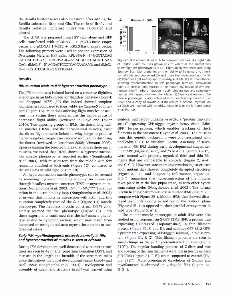
The fliH mutant was isolated based on a recessive flightlessphenotype in an EMS screen for flightless behavior (Homykand Sheppard 1977). fliH flies indeed showed completeflightlessness compared to their wild-type Canton-S counter-part (Figure 1A). Mutations affecting flight muscles or neu-rons innervating these muscles are the major cause ofdecreased flight ability (reviewed in Lloyd and Taylor2010). Two opposing groups of IFMs, the dorsal longitudi-nal muscles (DLMs) and the dorso-ventral muscles, assistthe direct flight muscles linked to wing hinge to producehigher wing-beat frequencies required for flight by distortingthe thorax (reviewed in Josephson 2006; Lehmann 2006).Upon examining the bisected thorax that houses these majormuscles, we noted that DLMs showed a hypercontraction-like muscle phenotype as reported earlier (Nongthombaet al. 2003), with muscles torn from the middle with fewremnants attached at both ends (Figure 1C), compared tothe six DLMs in wild type (Figure 1B).
All hypercontraction muscle phenotypes can be rescuedby removing myosin or reducing acto-myosin interactionthrough headless myosin construct (Y97) or myosin muta-tions (Nongthomba et al. 2003). Mhc2B (MhcP401S) is a mu-tation in the actin-binding loop (Nongthomba et al. 2003)of myosin that inhibits its interaction with actin, and thismutation completely rescued the fliH (Figure 1D) musclephenotype. The headless myosin construct (Y97) com-pletely rescued the fliH phenotype (Figure 1E). Boththese experiments confirmed that the fliH muscle pheno-type is due to hypercontraction, which may result fromincreased or unregulated acto-myosin interaction or me-chanical stress.
Early IFM myofibrillogenesis proceeds normally in fliH,and hypercontraction of muscles is seen at eclosion
During IFM development, well-demarcated sarcomere struc-tures are seen by 42 hr after puparium formation (APF), andincrease in the length and breadth of the sarcomere takesplace throughout the pupal development stages (Reedy andBeall 1993; Nongthomba et al. 2004). Development andassembly of sarcomeric structure in fliH was tracked using
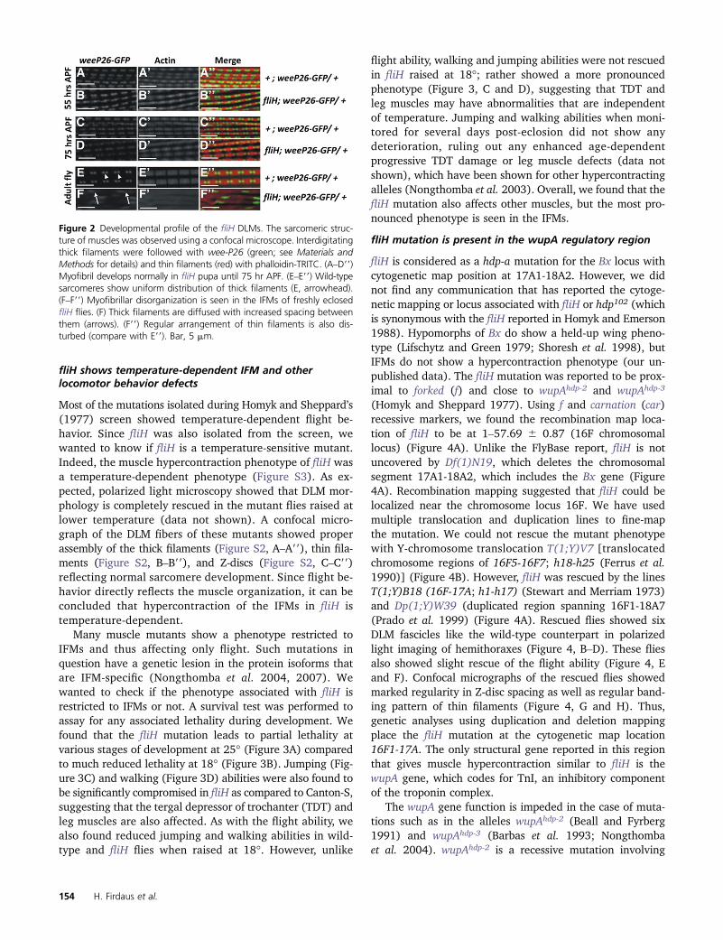
confocal microscopy utilizing wee-P26, a “protein trap con-struct” expressing GFP-tagged myosin heavy chain (Mhc-GFP) fusion protein, which enables tracking of thickfilaments in the sarcomere (Clyne et al. 2003). The musclesfrom this genetic background were counterstained withphalloidin-TRITC to visualize F-actin. Assembly of sarco-meres in fliH IFM during early developmental stages, i.e.,55 hr APF (Figure 2, B–B99) and 75 hr APF (Figure 2, D–D99),were normal with properly organized thick and thin fila-ments that are comparable to controls (Figure 2, A–A99and C–C99). However, sarcomere organization in just-eclosedadult mutant flies showed completely disrupted structures(Figure 2, F–F99 and Supporting Information, Figure S1,B–B99), suggesting that hypercontraction of the musclestakes place in at the late pupal stage, as with other hyper-contracting alleles (Nongthomba et al. 2003). The normalF-actin banding pattern was lost in mutant IFMs (Figure 2F9;compare with Figure 2E9). Mutant IFMs also showed disar-rayed myofibrils moving in and out of the confocal plane(Figure S1B99) as opposed to their parallel arrangement inwild type (Figure S1A99).
The mutant muscle phenotypes in adult IFM were alsostudied using tropomyosin-2-GFP (TM2-GFP, a protein trapexpressing GFP-tagged Tropomyosin-2), a thin filamentprotein (Figure S1, C and D), and sallimus-GFP (SLS-GFP,a protein trap expressing GFP-tagged sallimus), a Z-disc pro-tein (Figure S1, E–G). Thin filament proteins are seen insmall clumps in the fliH hypercontracted muscles (FigureS1D99). The regular banding patterns of Z-discs and sizeand spacing of the thin filaments were lost in freshly eclosedfliH IFMs (Figure S1, F–F9) when compared to control (Fig-ure S1E99). More pronounced dissolution of Z-discs andmyofilaments is observed in 2-day-old flies (Figure S1,G–G99).
Figure 1 IFM abnormalities in 3- to 5-day-old fliH flies. (A) Flight dataof Canton-S and fliH flies grown at 25�, where all the mutant fliesshow flightless phenotype (n = 50). Flight ability was measured usingSparrow Box, with gradation on their ability to fly upward (U), hori-zontally (H), and downward (D) and those that were could not fly (F).(B) Polarized light micrograph of wild-type DLMs. (C) fliH hemithoraxshowing hypercontraction muscle phenotype (arrows). Arrowheadpoints to normal jump muscles in the mutant. (D) Rescue of fliH phe-notype. Mhc2B harbors mutation in actin-binding loop and completelyrescues fliH hypercontraction phenotype. (E) Significant rescue of themuscle phenotype is also achieved with headless myosin construct(Y97) and a copy of myosin null (to reduce functional myosin). Allsix DLMs are marked with asterisks. Anterior is to the left and dorsalis on the top.
fliH Is a Troponin-I Mutation 153
fliH shows temperature-dependent IFM and otherlocomotor behavior defects
Most of the mutations isolated during Homyk and Sheppard’s(1977) screen showed temperature-dependent flight be-havior. Since fliH was also isolated from the screen, wewanted to know if fliH is a temperature-sensitive mutant.Indeed, the muscle hypercontraction phenotype of fliH wasa temperature-dependent phenotype (Figure S3). As ex-pected, polarized light microscopy showed that DLM mor-phology is completely rescued in the mutant flies raised atlower temperature (data not shown). A confocal micro-graph of the DLM fibers of these mutants showed properassembly of the thick filaments (Figure S2, A–A99), thin fila-ments (Figure S2, B–B99), and Z-discs (Figure S2, C–C99)reflecting normal sarcomere development. Since flight be-havior directly reflects the muscle organization, it can beconcluded that hypercontraction of the IFMs in fliH istemperature-dependent.
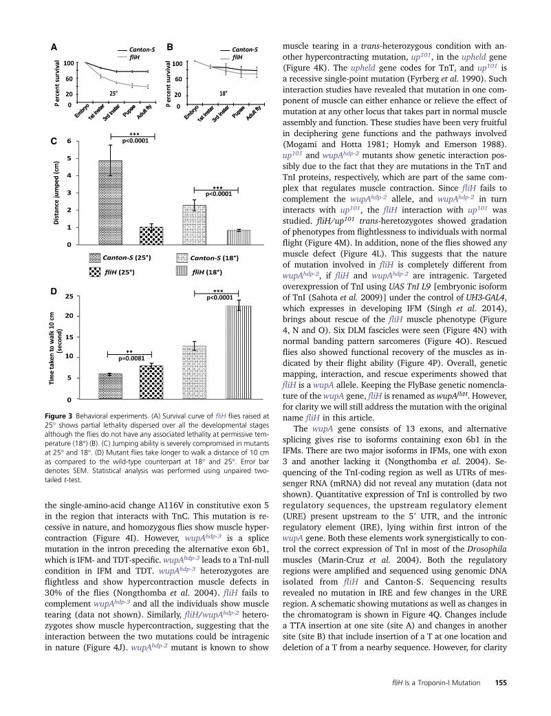
Many muscle mutants show a phenotype restricted toIFMs and thus affecting only flight. Such mutations inquestion have a genetic lesion in the protein isoforms thatare IFM-specific (Nongthomba et al. 2004, 2007). Wewanted to check if the phenotype associated with fliH isrestricted to IFMs or not. A survival test was performed toassay for any associated lethality during development. Wefound that the fliH mutation leads to partial lethality atvarious stages of development at 25� (Figure 3A) comparedto much reduced lethality at 18� (Figure 3B). Jumping (Fig-ure 3C) and walking (Figure 3D) abilities were also found tobe significantly compromised in fliH as compared to Canton-S,suggesting that the tergal depressor of trochanter (TDT) andleg muscles are also affected. As with the flight ability, wealso found reduced jumping and walking abilities in wild-type and fliH flies when raised at 18�. However, unlike
flight ability, walking and jumping abilities were not rescuedin fliH raised at 18�; rather showed a more pronouncedphenotype (Figure 3, C and D), suggesting that TDT andleg muscles may have abnormalities that are independentof temperature. Jumping and walking abilities when moni-tored for several days post-eclosion did not show anydeterioration, ruling out any enhanced age-dependentprogressive TDT damage or leg muscle defects (data notshown), which have been shown for other hypercontractingalleles (Nongthomba et al. 2003). Overall, we found that thefliH mutation also affects other muscles, but the most pro-nounced phenotype is seen in the IFMs.
fliH mutation is present in the wupA regulatory region
fliH is considered as a hdp-a mutation for the Bx locus withcytogenetic map position at 17A1-18A2. However, we didnot find any communication that has reported the cytoge-netic mapping or locus associated with fliH or hdp102 (whichis synonymous with the fliH reported in Homyk and Emerson1988). Hypomorphs of Bx do show a held-up wing pheno-type (Lifschytz and Green 1979; Shoresh et al. 1998), butIFMs do not show a hypercontraction phenotype (our un-published data). The fliH mutation was reported to be prox-imal to forked (f) and close to wupAhdp-2 and wupAhdp-3
(Homyk and Sheppard 1977). Using f and carnation (car)recessive markers, we found the recombination map loca-tion of fliH to be at 1–57.69 6 0.87 (16F chromosomallocus) (Figure 4A). Unlike the FlyBase report, fliH is notuncovered by Df(1)N19, which deletes the chromosomalsegment 17A1-18A2, which includes the Bx gene (Figure4A). Recombination mapping suggested that fliH could belocalized near the chromosome locus 16F. We have usedmultiple translocation and duplication lines to fine-mapthe mutation. We could not rescue the mutant phenotypewith Y-chromosome translocation T(1;Y)V7 [translocatedchromosome regions of 16F5-16F7; h18-h25 (Ferrus et al.1990)] (Figure 4B). However, fliH was rescued by the linesT(1;Y)B18 (16F-17A; h1-h17) (Stewart and Merriam 1973)and Dp(1;Y)W39 (duplicated region spanning 16F1-18A7(Prado et al. 1999) (Figure 4A). Rescued flies showed sixDLM fascicles like the wild-type counterpart in polarizedlight imaging of hemithoraxes (Figure 4, B–D). These fliesalso showed slight rescue of the flight ability (Figure 4, Eand F). Confocal micrographs of the rescued flies showedmarked regularity in Z-disc spacing as well as regular band-ing pattern of thin filaments (Figure 4, G and H). Thus,genetic analyses using duplication and deletion mappingplace the fliH mutation at the cytogenetic map location16F1-17A. The only structural gene reported in this regionthat gives muscle hypercontraction similar to fliH is thewupA gene, which codes for TnI, an inhibitory componentof the troponin complex.
The wupA gene function is impeded in the case of muta-tions such as in the alleles wupAhdp-2 (Beall and Fyrberg1991) and wupAhdp-3 (Barbas et al. 1993; Nongthombaet al. 2004). wupAhdp-2 is a recessive mutation involving
Figure 2 Developmental profile of the fliH DLMs. The sarcomeric struc-ture of muscles was observed using a confocal microscope. Interdigitatingthick filaments were followed with wee-P26 (green; see Materials andMethods for details) and thin filaments (red) with phalloidin-TRITC. (A–D99)Myofibril develops normally in fliH pupa until 75 hr APF. (E–E99) Wild-typesarcomeres show uniform distribution of thick filaments (E, arrowhead).(F–F99) Myofibrillar disorganization is seen in the IFMs of freshly eclosedfliH flies. (F) Thick filaments are diffused with increased spacing betweenthem (arrows). (F99) Regular arrangement of thin filaments is also dis-turbed (compare with E99). Bar, 5 mm.
154 H. Firdaus et al.
the single-amino-acid change A116V in constitutive exon 5in the region that interacts with TnC. This mutation is re-cessive in nature, and homozygous flies show muscle hyper-contraction (Figure 4I). However, wupAhdp-3 is a splicemutation in the intron preceding the alternative exon 6b1,which is IFM- and TDT-specific. wupAhdp-3 leads to a TnI-nullcondition in IFM and TDT. wupAhdp-3 heterozygotes areflightless and show hypercontraction muscle defects in30% of the flies (Nongthomba et al. 2004). fliH fails tocomplement wupAhdp-3 and all the individuals show muscletearing (data not shown). Similarly, fliH/wupAhdp-2 hetero-zygotes show muscle hypercontraction, suggesting that theinteraction between the two mutations could be intragenicin nature (Figure 4J). wupAhdp-2 mutant is known to show
muscle tearing in a trans-heterozygous condition with an-other hypercontracting mutation, up101, in the upheld gene(Figure 4K). The upheld gene codes for TnT, and up101 isa recessive single-point mutation (Fyrberg et al. 1990). Suchinteraction studies have revealed that mutation in one com-ponent of muscle can either enhance or relieve the effect ofmutation at any other locus that takes part in normal muscleassembly and function. These studies have been very fruitfulin deciphering gene functions and the pathways involved(Mogami and Hotta 1981; Homyk and Emerson 1988).up101 and wupAhdp-2 mutants show genetic interaction pos-sibly due to the fact that they are mutations in the TnT andTnI proteins, respectively, which are part of the same com-plex that regulates muscle contraction. Since fliH fails tocomplement the wupAhdp-2 allele, and wupAhdp-2 in turninteracts with up101, the fliH interaction with up101 wasstudied. fliH/up101 trans-heretozygotes showed gradationof phenotypes from flightlessness to individuals with normalflight (Figure 4M). In addition, none of the flies showed anymuscle defect (Figure 4L). This suggests that the natureof mutation involved in fliH is completely different fromwupAhdp-2, if fliH and wupAhdp-2 are intragenic. Targetedoverexpression of TnI using UAS TnI L9 [embryonic isoformof TnI (Sahota et al. 2009)] under the control of UH3-GAL4,which expresses in developing IFM (Singh et al. 2014),brings about rescue of the fliH muscle phenotype (Figure4, N and O). Six DLM fascicles were seen (Figure 4N) withnormal banding pattern sarcomeres (Figure 4O). Rescuedflies also showed functional recovery of the muscles as in-dicated by their flight ability (Figure 4P). Overall, geneticmapping, interaction, and rescue experiments showed thatfliH is a wupA allele. Keeping the FlyBase genetic nomencla-ture of the wupA gene, fliH is renamed as wupAfliH. However,for clarity we will still address the mutation with the originalname fliH in this article.
The wupA gene consists of 13 exons, and alternativesplicing gives rise to isoforms containing exon 6b1 in theIFMs. There are two major isoforms in IFMs, one with exon3 and another lacking it (Nongthomba et al. 2004). Se-quencing of the TnI-coding region as well as UTRs of mes-senger RNA (mRNA) did not reveal any mutation (data notshown). Quantitative expression of TnI is controlled by tworegulatory sequences, the upstream regulatory element(URE) present upstream to the 59 UTR, and the intronicregulatory element (IRE), lying within first intron of thewupA gene. Both these elements work synergistically to con-trol the correct expression of TnI in most of the Drosophilamuscles (Marin-Cruz et al. 2004). Both the regulatoryregions were amplified and sequenced using genomic DNAisolated from fliH and Canton-S. Sequencing resultsrevealed no mutation in IRE and few changes in the UREregion. A schematic showing mutations as well as changes inthe chromatogram is shown in Figure 4Q. Changes includea TTA insertion at one site (site A) and changes in anothersite (site B) that include insertion of a T at one location anddeletion of a T from a nearby sequence. However, for clarity
Figure 3 Behavioral experiments. (A) Survival curve of fliH flies raised at25� shows partial lethality dispersed over all the developmental stagesalthough the flies do not have any associated lethality at permissive tem-perature (18�) (B). (C) Jumping ability is severely compromised in mutantsat 25� and 18�. (D) Mutant flies take longer to walk a distance of 10 cmas compared to the wild-type counterpart at 18� and 25�. Error bardenotes SEM. Statistical analysis was performed using unpaired two-tailed t-test.
fliH Is a Troponin-I Mutation 155

these mutations are addressed as site A and site B muta-tions. MatInspector (Genomatix Software, Munich, Ger-many), a program to find transcription-factor-binding sites,
predicted Myocyte enhancer factor-2 (Mef-2)-binding sitespresent near site A and another falling at site B. The pre-dicted Mef2 sites are not similar to a previously published
Figure 4 Genetic mapping and rescue of fliH. (A) X-chromosome map showing cytological location 15F-19A. Green lines denote X-chromosome segmentsduplicated on Y chromosome for various transposition lines used in the study. Meiotic recombination mapping with forked (f) and carnation (car) places fliHmutation at 1–57.69 6 0.87 recombination map unit. (B) fliH was not rescued with T(1:Y)v7. Complete rescue of the DLM fiber morphology with T(1:Y)B18and T(1:Y)W39 transposition line (C and D, respectively). (E and F) Partial rescue of the flight ability of fliH mutants with above-mentioned transposition lines.(G and H) Confocal micrograph showing normal sarcomeric organization of DLMs in fly rescued with duplicated segments on Y chromosome. (I–M) fliHinteraction with other hypercontracting alleles. (I) wupAhdp-2 is a hypercontracting allele of TnI that shows muscle tearing. (H) fliH genetically interacts withwupAhdp-2 at 25�, and all the flies showmuscle hypercontraction. (K)wupAhdp-2 genetically interacts with TnT hypercontracting allele up101 in trans, leading tomuscle rupturing. (L) fliH and up101 trans-heterozygotes do not show muscle tearing. Arrows I–K point to muscle tearing due to hypercontraction. (M) Flightdata reveal that all the flies are flightless in fliH/wupAhdp-2 and wupAhdp-2/up101 trans-heterozygotes, whereas fliH/up101 females show gradation in flightability. Flies heterozygous for the fliHmutation serve as control for the flight test. (N–P) fliHmutants rescued by targeted overexpression of TnI transgenic linein IFM. (N) Polarized light image showing the rescue of DLM fibers with overexpression of a copy of TnI (w, UH3, fliH/Y; UAS-TnI-L9). (O) Myofiber showingcomplete rescue of the sarcomere organization as visualized by confocal imaging of w, UH3, fliH/Y; UAS-TnI-L9 flies. (P) Rescued fliH flies with TnI transgeneshow normal flight. (Q) Chromatogram showing mutations in fliH as analyzed by DNA sequencing. Mutations lie in upstream regulatory region of the wupAgene coding for TnI. Box indicates transcription factor Mef-2-binding site as predicted by MatInspector. Phallodin-TRITC for F-actin (red) and anti-Mlp60Aantibody localizing on Z-discs (green). Flight data and muscle analysis: n = 25, and all the flies were grown at 25�.
156 H. Firdaus et al.
consensus sequence (YTAWWWWTAR) Haberland et al.2007). However, Mef2 has also been known to bind to dif-ferent TA-rich sequences (Andrés et al. 1995).
Mutation abrogates Mef-2 binding to URE, leading toreduced TnI transcript and protein
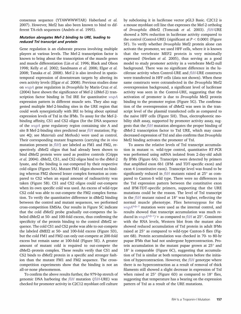
Gene regulation is an elaborate process involving multipleplayers at various levels. The Mef-2 transcription factor isknown to bring about the transcription of the muscle genesand muscle differentiation (Lin et al. 1996; Black and Olson1998; Kelly et al. 2002; Sandmann et al. 2006; Elgar et al.2008; Tanaka et al. 2008). Mef-2 is also involved in spatio-temporal expression of downstream targets by altering itsown activity levels (Elgar et al. 2008). Previous studies doneon wupA gene regulation in Drosophila by Marin-Cruz et al.(2004) have shown the significance of Mef-2 (dMef-2) tran-scription factor binding in TnI IRE for quantitative wupAexpression pattern in different muscle sets. They also sug-gested multiple Mef-2-binding sites in the URE region thatcould work synergistically with IRE to bring about optimalexpression levels of TnI in the IFMs. To assay for the Mef-2-binding affinity, CS1 and CS2 oligos (for the DNA sequenceof the wupA gene regulatory region containing site A andsite B Mef-2-binding sites predicted near fliH mutation; Fig-ure 4Q; see Materials and Methods) were used as control.Their corresponding mutant fragments (carrying the in vivomutation present in fliH) are labeled as FM1 and FM2, re-spectively. dMef2 oligos that had already been shown tobind dMef2 protein were used as positive controls (Crippset al. 2004). dMef2, CS1, and CS2 oligos bind to the dMef-2lysate, and the binding is out-competed by their respectivecold oligos (Figure 5A). Mutant FM1 oligos showed no bind-ing whereas FM2 showed lesser complex formation as com-pared to CS2 when an equal amount of radioactivity wastaken (Figure 5B). CS1 and CS2 oligos could out-competewhen its own specific cold was used. An excess of wild-typeCS2 cold was able to out-compete the FM2 complex forma-tion. To verify the quantitative difference in dMef2 bindingbetween the control and mutant sequences, we performedcross-competition EMSAs. Our results in Figure 5C indicatethat the cold dMef2 probe gradually out-competes the la-beled dMef2 at 50- and 100-fold excess, thus confirming thespecificity of the protein binding to the control dMef2 se-quence. The cold CS1 and CS2 probe was able to out-competethe labeled dMEf2 at 50- and 100-fold excess (Figure 5D),but the cold FM1 and FM2 can only out-compete at 200-foldexcess but remain same at 100-fold (Figure 5E). A greateramount of mutant cold is required to out-compete thedMef2–protein complex. These results verify that CS1 andCS2 binds to dMef2 protein in a specific and stronger fash-ion than the mutant FM1 and FM2 sequence. The cross-competition experiments show that the binding is not anall-or-none phenomenon.
To confirm the above results further, the 979-bp stretch ofgenomic DNA harboring the fliH mutation (fliH-URE) waschecked for promoter activity in C2C12 myoblast cell culture
by subcloning it in luciferase vector pGL3 Basic. C2C12 isa mouse myoblast cell line that expresses the Mef-2 orthologof Drosophila dMef2 (Tomczak et al. 2003). fliH-UREshowed a 50% reduction in luciferase activity compared tothe control (Control-URE) (significant at P, 0.0001 (Figure5F). To verify whether Drosophila Mef2 protein alone canactivate the promoter, we used HFF cells, where it is knownthat the vertebrate MEF2 protein is very minimallyexpressed (Neelam et al. 2005), thus serving as a goodmodel to study promoter activity in a vertebrate Mef2-nullbackground. There was no significant difference in the lu-ciferase activity when Control-URE and fliH-URE constructswere transfected in HFF cells (data not shown). When thesesame constructs were cotransfected in the Drosophila Mef2overexpression background, a significant level of luciferaseactivity was seen in the Control-URE, suggesting that theactivation of promoter is due to Drosophila Mef2 proteinbinding to the promoter region (Figure 5G). The confirma-tion of the overexpression of dMef2 was seen in the tran-script level of the plasmid transfected cells as compared tothe naive HFF cells (Figure 5H). Thus, electrophoretic mo-bility shift assay, supported by promoter activity assay, sug-gests that the fliH mutation abrogates the proper binding ofdMef-2 transcription factor to TnI URE, which may causedecreased expression of TnI and also confirms that DrosophilaMef2 binding activates the promoter of TnI.
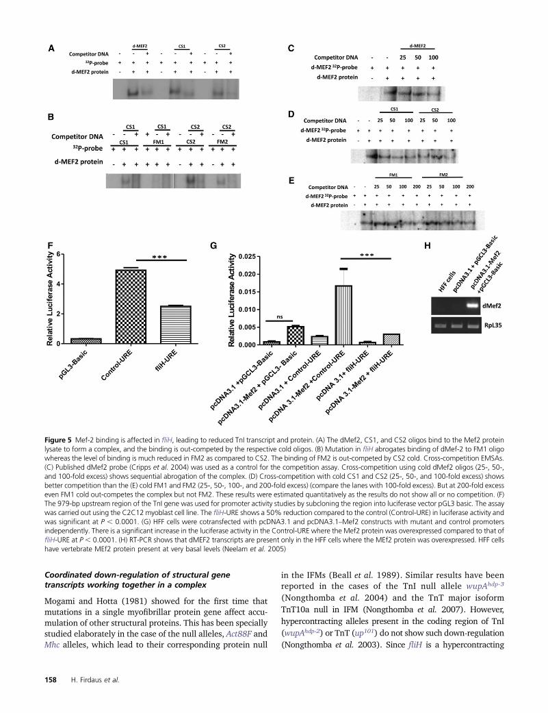
To assess the relative levels of TnI transcript accumula-tion in mutant vs. wild-type control, quantitative RT-PCRwas performed using mRNA isolated from 2-day-old adultfly IFMs (Figure 6A). Transcripts were detected by primersthat amplified exon 6b1 (IFM- and TDT-specific exon) andexon 4 (constitutive exon). Transcript level was found to besignificantly reduced in fliH mutants raised at 25� as com-pared to Canton-S wild type. There were no differences inthe TnI expression pattern between the constitutive exonand IFM-TDT-specific primers, supporting that the UREmutations could be the reason. The level of TnI transcriptin the fliH mutant raised at 18� was higher, reflecting thenormal muscle phenotype. Flies heterozygous for thewupAhdp-3 mutation were used as the internal control, andresults showed that transcript accumulation was much re-duced in wupAhdp-3/+ as compared to fliH at 25�. Consistentwith the RNA levels, Western blot from the mutant alsoshowed reduced accumulation of TnI protein in adult IFMsraised at 25� as compared to wild-type Canton-S flies (Fig-ure 6B). Protein accumulation was checked in 70- to 80-hrpupae IFMs that had not undergone hypercontraction. Pro-tein accumulation in the mutant pupae grown at 25� and18� is comparable (Figure 6C), suggesting that accumula-tion of TnI is similar at both temperatures before the initia-tion of hypercontraction. However, the fliH genotype wherethere is no hypercontraction as a result of removal of thickfilaments still showed a slight decrease in expression of TnIwhen raised at 25� (Figure 6D) as compared to 18� flies,suggesting that temperature has a bearing on the expressionpattern of TnI as a result of the URE mutations.
fliH Is a Troponin-I Mutation 157
Coordinated down-regulation of structural genetranscripts working together in a complex
Mogami and Hotta (1981) showed for the first time thatmutations in a single myofibrillar protein gene affect accu-mulation of other structural proteins. This has been speciallystudied elaborately in the case of the null alleles, Act88F andMhc alleles, which lead to their corresponding protein null
in the IFMs (Beall et al. 1989). Similar results have beenreported in the cases of the TnI null allele wupAhdp-3
(Nongthomba et al. 2004) and the TnT major isoformTnT10a null in IFM (Nongthomba et al. 2007). However,hypercontracting alleles present in the coding region of TnI(wupAhdp-2) or TnT (up101) do not show such down-regulation(Nongthomba et al. 2003). Since fliH is a hypercontracting
Figure 5 Mef-2 binding is affected in fliH, leading to reduced TnI transcript and protein. (A) The dMef2, CS1, and CS2 oligos bind to the Mef2 proteinlysate to form a complex, and the binding is out-competed by the respective cold oligos. (B) Mutation in fliH abrogates binding of dMef-2 to FM1 oligowhereas the level of binding is much reduced in FM2 as compared to CS2. The binding of FM2 is out-competed by CS2 cold. Cross-competition EMSAs.(C) Published dMef2 probe (Cripps et al. 2004) was used as a control for the competition assay. Cross-competition using cold dMef2 oligos (25-, 50-,and 100-fold excess) shows sequential abrogation of the complex. (D) Cross-competition with cold CS1 and CS2 (25-, 50-, and 100-fold excess) showsbetter competition than the (E) cold FM1 and FM2 (25-, 50-, 100-, and 200-fold excess) (compare the lanes with 100-fold excess). But at 200-fold excesseven FM1 cold out-competes the complex but not FM2. These results were estimated quantitatively as the results do not show all or no competition. (F)The 979-bp upstream region of the TnI gene was used for promoter activity studies by subcloning the region into luciferase vector pGL3 basic. The assaywas carried out using the C2C12 myoblast cell line. The fliH-URE shows a 50% reduction compared to the control (Control-URE) in luciferase activity andwas significant at P , 0.0001. (G) HFF cells were cotransfected with pcDNA3.1 and pcDNA3.1–Mef2 constructs with mutant and control promotersindependently. There is a significant increase in the luciferase activity in the Control-URE where the Mef2 protein was overexpressed compared to that offliH-URE at P , 0.0001. (H) RT-PCR shows that dMEF2 transcripts are present only in the HFF cells where the MEf2 protein was overexpressed. HFF cellshave vertebrate MEf2 protein present at very basal levels (Neelam et al. 2005)
158 H. Firdaus et al.
allele that does not fall in the above categories of mutations,transcript levels of other structural proteins were checkedthrough semiquantitative RT-PCR.
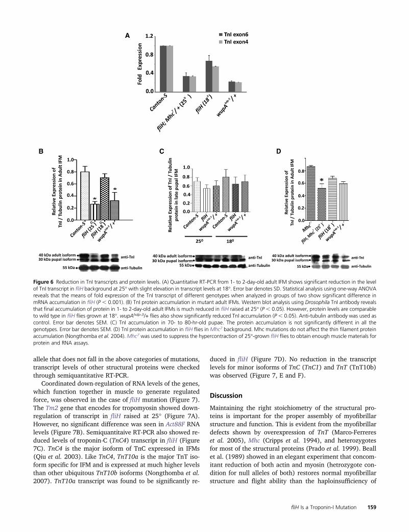
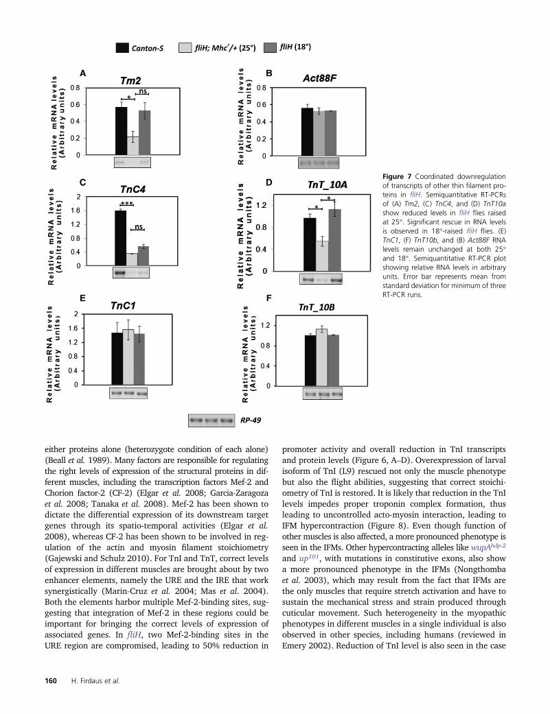
Coordinated down-regulation of RNA levels of the genes,which function together in muscle to generate regulatedforce, was observed in the case of fliH mutation (Figure 7).The Tm2 gene that encodes for tropomyosin showed down-regulation of transcript in fliH raised at 25� (Figure 7A).However, no significant difference was seen in Act88F RNAlevels (Figure 7B). Semiquantitaive RT-PCR also showed re-duced levels of troponin-C (TnC4) transcript in fliH (Figure7C). TnC4 is the major isoform of TnC expressed in IFMs(Qiu et al. 2003). Like TnC4, TnT10a is the major TnT iso-form specific for IFM and is expressed at much higher levelsthan other ubiquitous TnT10b isoforms (Nongthomba et al.2007). TnT10a transcript was found to be significantly re-
duced in fliH (Figure 7D). No reduction in the transcriptlevels for minor isoforms of TnC (TnC1) and TnT (TnT10b)was observed (Figure 7, E and F).
Discussion
Maintaining the right stoichiometry of the structural pro-teins is important for the proper assembly of myofibrillarstructure and function. This is evident from the myofibrillardefects shown by overexpression of TnT (Marco-Ferrereset al. 2005), Mhc (Cripps et al. 1994), and heterozygotesfor most of the structural proteins (Prado et al. 1999). Beallet al. (1989) showed in an elegant experiment that concom-itant reduction of both actin and myosin (hetrozygote con-dition for null alleles of both) restores normal myofibrillarstructure and flight ability than the haploinsufficiency of
Figure 6 Reduction in TnI transcripts and protein levels. (A) Quantitative RT-PCR from 1- to 2-day-old adult IFM shows significant reduction in the levelof TnI transcript in fliH background at 25� with slight elevation in transcript levels at 18�. Error bar denotes SD. Statistical analysis using one-way ANOVAreveals that the means of fold expression of the TnI transcript of different genotypes when analyzed in groups of two show significant difference inmRNA accumulation in fliH (P , 0.001). (B) TnI protein accumulation in mutant adult IFMs. Western blot analysis using Drosophila TnI antibody revealsthat final accumulation of protein in 1- to 2-day-old adult IFMs is much reduced in fliH raised at 25� (P , 0.05). However, protein levels are comparableto wild type in fliH flies grown at 18�. wupAhdp-3/+ flies also show significantly reduced TnI accumulation (P , 0.05). Anti-tubulin antibody was used ascontrol. Error bar denotes SEM. (C) TnI accumulation in 70- to 80-hr-old pupae. The protein accumulation is not significantly different in all thegenotypes. Error bar denotes SEM. (D) TnI protein accumulation in fliH flies in Mhc7 background. Mhc mutations do not affect the thin filament proteinaccumulation (Nongthomba et al. 2004).Mhc7 was used to suppress the hypercontraction of 25�-grown fliH flies to obtain enough muscle materials forprotein and RNA assays.
fliH Is a Troponin-I Mutation 159
either proteins alone (heterozygote condition of each alone)(Beall et al. 1989). Many factors are responsible for regulatingthe right levels of expression of the structural proteins in dif-ferent muscles, including the transcription factors Mef-2 andChorion factor-2 (CF-2) (Elgar et al. 2008; Garcia-Zaragozaet al. 2008; Tanaka et al. 2008). Mef-2 has been shown todictate the differential expression of its downstream targetgenes through its spatio-temporal activities (Elgar et al.2008), whereas CF-2 has been shown to be involved in reg-ulation of the actin and myosin filament stoichiometry(Gajewski and Schulz 2010). For TnI and TnT, correct levelsof expression in different muscles are brought about by twoenhancer elements, namely the URE and the IRE that worksynergistically (Marin-Cruz et al. 2004; Mas et al. 2004).Both the elements harbor multiple Mef-2-binding sites, sug-gesting that integration of Mef-2 in these regions could beimportant for bringing the correct levels of expression ofassociated genes. In fliH, two Mef-2-binding sites in theURE region are compromised, leading to 50% reduction in
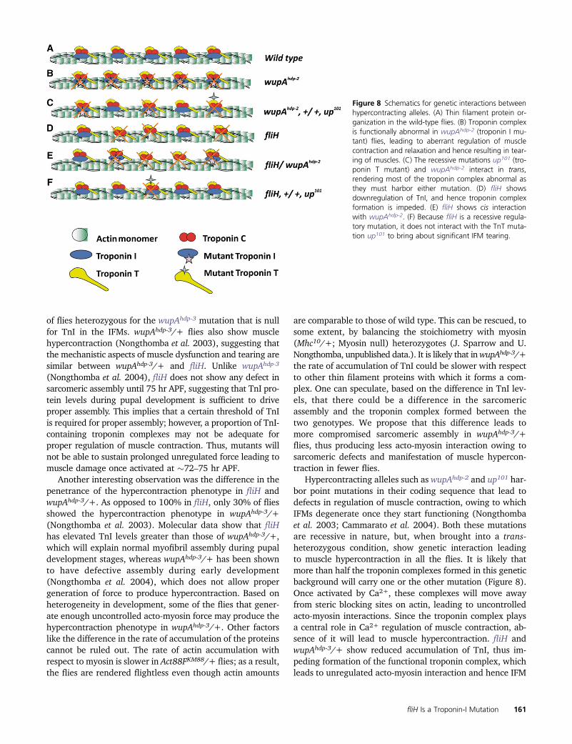
promoter activity and overall reduction in TnI transcriptsand protein levels (Figure 6, A–D). Overexpression of larvalisoform of TnI (L9) rescued not only the muscle phenotypebut also the flight abilities, suggesting that correct stoichi-ometry of TnI is restored. It is likely that reduction in the TnIlevels impedes proper troponin complex formation, thusleading to uncontrolled acto-myosin interaction, leading toIFM hypercontraction (Figure 8). Even though function ofother muscles is also affected, a more pronounced phenotype isseen in the IFMs. Other hypercontracting alleles like wupAhdp-2
and up101, with mutations in constitutive exons, also showa more pronounced phenotype in the IFMs (Nongthombaet al. 2003), which may result from the fact that IFMs arethe only muscles that require stretch activation and have tosustain the mechanical stress and strain produced throughcuticular movement. Such heterogeneity in the myopathicphenotypes in different muscles in a single individual is alsoobserved in other species, including humans (reviewed inEmery 2002). Reduction of TnI level is also seen in the case
Figure 7 Coordinated downregulationof transcripts of other thin filament pro-teins in fliH. Semiquantitative RT-PCRsof (A) Tm2, (C) TnC4, and (D) TnT10ashow reduced levels in fliH flies raisedat 25�. Significant rescue in RNA levelsis observed in 18�-raised fliH flies. (E)TnC1, (F) TnT10b, and (B) Act88F RNAlevels remain unchanged at both 25�and 18�. Semiquantitative RT-PCR plotshowing relative RNA levels in arbitraryunits. Error bar represents mean fromstandard deviation for minimum of threeRT-PCR runs.
160 H. Firdaus et al.
of flies heterozygous for the wupAhdp-3 mutation that is nullfor TnI in the IFMs. wupAhdp-3/+ flies also show musclehypercontraction (Nongthomba et al. 2003), suggesting thatthe mechanistic aspects of muscle dysfunction and tearing aresimilar between wupAhdp-3/+ and fliH. Unlike wupAhdp-3
(Nongthomba et al. 2004), fliH does not show any defect insarcomeric assembly until 75 hr APF, suggesting that TnI pro-tein levels during pupal development is sufficient to driveproper assembly. This implies that a certain threshold of TnIis required for proper assembly; however, a proportion of TnI-containing troponin complexes may not be adequate forproper regulation of muscle contraction. Thus, mutants willnot be able to sustain prolonged unregulated force leading tomuscle damage once activated at �72–75 hr APF.
Another interesting observation was the difference in thepenetrance of the hypercontraction phenotype in fliH andwupAhdp-3/+. As opposed to 100% in fliH, only 30% of fliesshowed the hypercontraction phenotype in wupAhdp-3/+(Nongthomba et al. 2003). Molecular data show that fliHhas elevated TnI levels greater than those of wupAhdp-3/+,which will explain normal myofibril assembly during pupaldevelopment stages, whereas wupAhdp-3/+ has been shownto have defective assembly during early development(Nongthomba et al. 2004), which does not allow propergeneration of force to produce hypercontraction. Based onheterogeneity in development, some of the flies that gener-ate enough uncontrolled acto-myosin force may produce thehypercontraction phenotype in wupAhdp-3/+. Other factorslike the difference in the rate of accumulation of the proteinscannot be ruled out. The rate of actin accumulation withrespect to myosin is slower in Act88FKM88/+ flies; as a result,the flies are rendered flightless even though actin amounts
are comparable to those of wild type. This can be rescued, tosome extent, by balancing the stoichiometry with myosin(Mhc10/+; Myosin null) heterozygotes (J. Sparrow and U.Nongthomba, unpublished data.). It is likely that inwupAhdp-3/+the rate of accumulation of TnI could be slower with respectto other thin filament proteins with which it forms a com-plex. One can speculate, based on the difference in TnI lev-els, that there could be a difference in the sarcomericassembly and the troponin complex formed between thetwo genotypes. We propose that this difference leads tomore compromised sarcomeric assembly in wupAhdp-3/+flies, thus producing less acto-myosin interaction owing tosarcomeric defects and manifestation of muscle hypercon-traction in fewer flies.
Hypercontracting alleles such as wupAhdp-2 and up101 har-bor point mutations in their coding sequence that lead todefects in regulation of muscle contraction, owing to whichIFMs degenerate once they start functioning (Nongthombaet al. 2003; Cammarato et al. 2004). Both these mutationsare recessive in nature, but, when brought into a trans-heterozygous condition, show genetic interaction leadingto muscle hypercontraction in all the flies. It is likely thatmore than half the troponin complexes formed in this geneticbackground will carry one or the other mutation (Figure 8).Once activated by Ca2+, these complexes will move awayfrom steric blocking sites on actin, leading to uncontrolledacto-myosin interactions. Since the troponin complex playsa central role in Ca2+ regulation of muscle contraction, ab-sence of it will lead to muscle hypercontraction. fliH andwupAhdp-3/+ show reduced accumulation of TnI, thus im-peding formation of the functional troponin complex, whichleads to unregulated acto-myosin interaction and hence IFM
Figure 8 Schematics for genetic interactions betweenhypercontracting alleles. (A) Thin filament protein or-ganization in the wild-type flies. (B) Troponin complexis functionally abnormal in wupAhdp-2 (troponin I mu-tant) flies, leading to aberrant regulation of musclecontraction and relaxation and hence resulting in tear-ing of muscles. (C) The recessive mutations up101 (tro-ponin T mutant) and wupAhdp-2 interact in trans,rendering most of the troponin complex abnormal asthey must harbor either mutation. (D) fliH showsdownregulation of TnI, and hence troponin complexformation is impeded. (E) fliH shows cis interactionwith wupAhdp-2. (F) Because fliH is a recessive regula-tory mutation, it does not interact with the TnT muta-tion up101 to bring about significant IFM tearing.
fliH Is a Troponin-I Mutation 161
hypercontraction. Similarly, one can explain the intragenicinteraction between fliH and wupAhdp-2 based on formationof the functional troponin complex. In this case, too, thetroponin complex formed either will lack TnI or will carrya mutated version leading to hypercontraction. fliH andup101 trans-heterozygotes show reduced flight ability withno obvious muscle tearing or hypercontraction. In this ge-netic background, up101 contributes a normal copy of theTnI locus, and one copy of the regulatory mutations isenough to express sufficient protein (fliH/+ flies are nor-mal); very few troponin complexes that carry mutant TnTwill be nonfunctional (such as up101/+), so flies show partialcomplementation.
Coordinated down-regulation of the expression of thinfilament genes in response to mutation in a single thinfilament protein has been very well documented in the caseof the null alleles of myosin (Mhc7), actin (Act88FKM88), TnI(wupAhdp-3), and TnT (up1). Such a phenomenon has notbeen reported for known hypercontracting alleles such aswupAhdp-2 and up101. Our result emphasizes that there iscoordinated down-regulation of major isoforms of TnC andTnT (TnC4 and TnT10a, respectively) as compared tothe minor isoforms TnC1 and TnT10b, which do not showany change in RNA levels in the IFMs. This could beexplained based on the formation of the functional troponincomplex and the interdependence of the expression level ofeach member of the complex by an unknown mechanism.Thus, in the absence of a single player of the complex, themessage for the other major interacting partners is also down-regulated. Studies involving coordinated down-regulation ofproteins that functions together in the same complex havealso been reported in other model organisms such as zebrafish(Sehnert et al. 2002).
Genetic variations lead to changes at the cellular andmolecular level affecting performance of the flight muscles.Apart from the intrinsic variables, an extrinsic factor such astemperature also plays an important role in flight ability.Certain species of moth cannot fly until they have pre-warmed their flight muscles (Esch 1988). Flight perfor-mance of the Canton-S flies, used as control in the presentstudy, showed reduced flight at 18� (Figure 3). They couldfly better at an elevated temperature, suggesting a physio-logical difference at the two extreme temperatures. The fliHmutation also shows a temperature-dependent effect in itsviability, walking, and jumping. However, a more profoundphenotype is seen in the IFMs that are raised at 25�, whichcorrelates with a reduced level of TnI transcript and proteinas compared to those at 18� and wild type. Other hyper-contracting alleles, such as flapwing, are known to showincreased viability and reduced hypercontraction when fliesare cultured in reduced temperature (Pronovost et al. 2013).Initiation of hypercontraction correlates with the movementof the thorax of the pupae within the pupal case, which ismore pronounced in 25�-raised flies than in those raised at18�. Moreover, one needs to keep the 18�-raised flies for atleast 2–3 hr at 25� to see flight, suggesting a limited move-
ment and lethargic nature of pupae and muscles at a lowertemperature. It is likely that the reduced activity of themuscles and less mechanical stress will allow the muscles toassemble completely, so that the muscle will show suppressionof the hypercontraction after eclosion. Our work has shownthat binding of Mef-2, a transcripton factor, which bringsabout correct expression of the structural genes, is affectedin the fliH mutant. However, defective binding of Mef-2 orany other transcription factors or changes in local structureof the DNA that brings about the temperature-sensitive effectneed to be worked out. Little is known about the mechanismsthat confer different temperature-dependent phenotypes. Re-search done on lower organisms like bacteria (Tamai et al.1998) and yeast (Nouraini et al. 1996) and in plants (Gilmouret al. 1998; Zhu et al. 2007) has shown that mutations in thecis-regulatory region show temperature-dependent pheno-types. Work done on the W3133 operon in bacteria showedthat an insertional mutation in the promoter region couldconfer a temperature-sensitive phenotype and affect transcrip-tion efficiency by stabilizing the DNA stem loop structure(Tamai et al. 1998). Such results have not been reported inhigher organisms, which may be due to higher-order organi-zation of the genome. However, temperature-sensitive muta-tions in protein-coding sequence have been isolated ineukaryotes wherein temperature difference might affectthe folding, stability, and function of the proteins (Reese andKatzenellenbogen 1991; Mondal et al. 2007; Hoeberichtset al. 2008).
In this study, we have shown that mutation in theregulatory region of TnI can lead to IFM hypercontraction.Mutations in the coding region of myofibrillar proteincausing myopathies in humans are very well docu-mented. However, there is no record of a mutation orsingle nucleotide polymorphism in the noncoding regionand regulatory region being associated with disease. Thismay be one reason for the large number of myopathic caseswhere the causative nature of mutation remains unknown.We propose from our study that regulatory mutations aswell as mutations leading to stoichiometric changes (splicesite mutants or nonsense mutations resulting in null protein)that may cause myopathic conditions can easily be identifiedby simple quantitation of transcripts and proteins byapplying the whole-genome approach. In humans, manymutations in sarcomeric proteins that lead to myopathicconditions have been identified. Cellular fiber disarray seenin hypercontracted IFM is also observed in the case ofhuman hypertrophic cardiomyopathies (Seidman and Seidman2001) and dystrophic muscles (Amato et al. 1998), suggest-ing that there may be parallel genetic pathways for hyper-contraction-induced cellular phenotypes. Conservation ofexpression of many remodeling proteins has been alreadyshown for hypercontracting Mhc alleles (Montana andLittleton 2006). Mutations have also been uncovered in hu-man TnI that lead to various cardiomyopathies and skeletalmyopathies (Kimura et al. 1997; Murphy et al. 2004; Gomeset al. 2005). The molecular mechanism by which these
162 H. Firdaus et al.

mutations lead to pathogenesis of myopathies remains un-clear. One needs to study the pathogenesis of these muta-tions in a model organism and follow the effects of otherfactors like environmental stress as well as different geneticbackgrounds. Overall, our results shed new insights into theimportance of the maintenance of structural protein stoichi-ometry during muscle assembly for proper function withimplications for identification of mutations and disease phe-notypes in other species, including humans.
Acknowledgments
We thank Sneha Raghuram and Meenakshi Sen at the IndianInstitute for Science-Confocal Facility for their technicalassistance; anonymous reviewers, John Sparrow (Universityof York), S. Mahadevan, and our lab members for theircritical comments and suggestions; Richard Cripps (Univer-sity of New Mexico) for the Mef-2 construct; Alberto Ferrus(Cajal Institute, Madrid) for flies and antibodies; Sathees C.Raghavan and M. Nishana (Indian Institute of Science) forhelp with EMSA experiments; Sunita Chopra for help in thecotransfection luciferase assays; and the Bloomington Dro-sophila Stock Center and the National Center for BiologicalSciences-Stock Centre (Bangalore, India) for providing flies.We thank the Indian Institute of Science and the Departmentof Science and Technology and Department of Biotechnology,Government of India, for financial assistance.
Literature Cited
Agianian, B., U. Krzic, F. Qiu, W. A. Linke, K. Leonard et al.,2004 A troponin switch that regulates muscle contraction bystretch instead of calcium. EMBO J. 23: 772–779.
Amato, A. A., K. Kagan-Hallet, C. E. Jackson, S. Lampkin, G. I.Wolfe et al., 1998 The wide spectrum of myofibrillar myopa-thy suggests a multifactorial etiology and pathogenesis. Neurol-ogy 51: 1646–1655.
An, H., and K. Mogami, 1996 Isolation of 88F actin mutants ofDrosophila melanogaster and possible alterations in the mutantactin structures. J. Mol. Biol. 260: 492–505.
Andrés, V., M. Cervera, and V. Mahdavi, 1995 Determination ofthe consensus binding site for MEF2 expressed in muscle andbrain reveals tissue-specific sequence constraints. J. Biol. Chem.270: 23246–23249.
Barbas, J. A., J. Galceran, L. Torroja, A. Prado, and A. Ferrús,1993 Abnormal muscle development in the heldup3 mutantof Drosophila melanogaster is caused by a splicing defect affect-ing selected troponin I isoforms. Mol. Cell. Biol. 13: 1433–1439.
Barthmaier, P., and E. Fyrberg, 1995 Monitoring developmentand pathology of Drosophila indirect flight muscles using greenfluorescent protein. Dev. Biol. 169: 770–774.
Beall, C. J., and E. Fyrberg, 1991 Muscle abnormalities in Dro-sophila melanogaster heldup mutants are caused by missing oraberrant troponin-I isoforms. J. Biol. Chem. 114: 941–951.
Beall, C. J., M. A. Sepanski, and E. A. Fyrberg, 1989 Geneticdissection of Drosophila myofibril formation: effects of actinand myosin heavy chain null alleles. Genes Dev. 3: 131–140.
Black, B. L., and E. N. Olson, 1998 Transcriptional control ofmuscle development by myocyte enhancer factor-2 (MEF2) pro-teins. Annu. Rev. Cell Dev. Biol. 14: 167–196.
Burkart, C., F. Qiu, S. Brendel, V. Benes, P. Haag et al.,2007 Modular proteins from the Drosophila sallimus (sls) geneand their expression in muscles with different extensibility. J.Mol. Biol. 367: 953–969.
Cammarato, A., V. Hatch, J. Saide, R. Craig, J. C. Sparrow et al.,2004 Drosophila muscle regulation characterized by electronmicroscopy and three-dimensional reconstruction of thin fila-ment mutants. Biophys. J. 86: 1618–1624.
Clyne, P. J., J. S. Brotman, S. T. Sweeney, and G. Davis,2003 Green fluorescent protein tagging Drosophila proteinsat their native genomic loci with small P-elements. Genetics165: 1433–1441.
Cripps, R. M., 2006 The contributions of genetics to the study ofinsect flight muscle function, pp. 2–15 in Nature’s Versatile En-gine: Insect Flight Muscle Inside and Out, edited by J. Vigoreaux.Springer/Landes Bioscience, New York.
Cripps, R. M., K. D. Becker, M. Mardahl, W. A. Kronert, D. Hodgeset al., 1994 Transformation of Drosophila melanogaster withthe wild-type myosin heavy-chain gene: rescue of mutant phe-notypes and analysis of defects caused by overexpression. J. CellBiol. 126: 689–699.
Cripps, R. M., J. A. Suggs, and S. I. Bernstein, 1999 Assembly ofthick filaments and myofibrils occurs in the absence of the my-osin head. EMBO J. 18: 1793–1804.
Cripps, R. M., T. L. Lovato, and E. N. Olson, 2004 Positive auto-regulation of the Myocyte enhancer factor-2 myogenic controlgene during somatic muscle development in Drosophila. Dev.Biol. 267: 536–547.
Deak, I. I., 1977 Mutations of Drosophila melanogaster that affectmuscles. J. Embryol. Exp. Morphol. 40: 35–63.
Deak, I. I., P. R. Bellamy, M. Bienz, Y. Dubuis, E. Fenner et al.,1982 Mutations affecting the indirect flight muscles of Dro-sophila melanogaster. J. Embryol. Exp. Morphol. 69: 61–81.
Drummond, D. R., E. S. Hennessey, and J. C. Sparrow,1991 Characterization of missense mutations in the Act88Fgene of Drosophila melanogaster. Mol. Gen. Genet. 226: 70–80.
Elgar, S. J., J. Han, and M. V. Taylor, 2008 Mef2 activity levelsdifferentially affect gene expression during Drosophila muscledevelopment. Proc. Natl. Acad. Sci. USA 105: 918–923.
Emery, A. E., 2002 The muscular dystrophies. Lancet 359: 687–695.
Esch, H., 1988 The effect of temperature on flight muscle poten-tials in honeybees and cuculiinid winter moths. J. Exp. Biol.135: 109–117.
Fahmy, O. G., and M. Fahmy, 1958 New mutants report. Drosoph.Inf. Serv. 32: 67–78.
Fernandes, J., and M. Bate, and K. VijayRaghavan,1991 Development of the indirect flight muscle of Drosophila.Development 113: 67–77.
Ferrús, A., S. Llamazares, J. L. de la Pompa, M. A. Tanouye, and O.Pongs, 1990 Genetic analysis of the Shaker gene complex ofDrosophila melanogaster. Genetics 125: 383–398.
Fyrberg, E. A., C. C. Fyrberg, C. Beall, and D. L. Saville,1990 Drosophila melanogaster troponin-T mutations engenderthree distinct syndromes of myofibrillar abnormalities. J. Mol.Biol. 216: 657–675.
Gajewski, K., and R. A. Schulz, 2010 CF2 represses actin 88F geneexpression and maintains filament balance during indirect flightmuscle development in Drosophila. PLoS ONE 5: e10713.
Gajewski, K. M., J. Wang, and R. A. Schulz, 2006 Calcineurinfunction is required for myofilament formation and troponin Iisoform transition in Drosophila indirect flight muscle. Dev. Biol.289: 17–29.
García-Zaragoza, E., J. A. Mas, J. Vivar, J. J. Arredondo, and M.Cervera, 2008 CF2 activity and enhancer integration are re-quired for proper muscle gene expression in Drosophila. Mech.Dev. 125: 617–630.
fliH Is a Troponin-I Mutation 163
Gilmour, S. J., D. G. Zarka, E. J. Stockinger, M. P. Salazar, and J. M.Houghton, 1998 Low temperature regulation of the Arabidop-sis CBF family of AP2 transcriptional activators as an early stepin cold induced COR gene expression. Plant J. 16: 433–442.
Gomes, A. V., J. Liang, and J. D. Potter, 2005 Mutations in humancardiac troponin I that are associated with restrictive cardiomy-opathy affect basal ATPase activity and the calcium sensitivity offorce development. J. Biol. Chem. 280: 30909–30915.
Haberland, M., M. A. Arnold, J. McAnally, D. Phan, Y. Kim et al.,2007 Regulation of HDAC9 gene expression by Mef2 estab-lishes a negative feedback loop in the transcriptional circuitryof muscle differentiation. Mol. Cell Biol. 27: 518–525.
Hoeberichts, F. A., E. Vaeck, G. Kiddle, E. Coppens, B. van de Cotteet al., 2008 A temperature-sensitive mutation in the Arabidop-sis thaliana phosphomannomutase gene disrupts protein glyco-sylation and triggers cell death. J. Biol. Chem. 283: 5708–5718.
Homyk, T., and C. P. Emerson Jr, 1988 Functional interactionsbetween unlinked muscle genes within haploinsufficient regionsof the Drosophila genome. Genetics 119: 105–121.
Homyk, T., and D. E. Sheppard, 1977 Behavioral mutants of Dro-sophila melaonogaster I. Isolation and mapping of mutationswhich decrease flight ability. Genetics 87: 95–104.
Josephson, R. K., 2006 Comparative physiology of insect flightmuscle, pp. 34–43 in Nature’s Versatile Engine: Insect Flight Mus-cle Inside and Out, edited by J. Vigoreaux. Springer/Landes Bio-science, New York.
Josephson, R. K., J. G. Malamud, and D. R. Stokes, 2000 Asynchronousmuscle: a primer. J. Exp. Biol. 203: 2713–2722.
Kelly, K. K., M. M. Stryder, and R. M. Cripps, 2002 DrosophilaMEF2 is a direct regulator of Actin57B transcription in cardiac,skeletal, and visceral muscle lineages. Mech. Dev. 110: 39–50.
Kimura, A., H. Harada, J. Park, H. Nishi, M. Satoh et al.,1997 Mutations in the cardiac troponin I gene associated withhypertrophic cardiomyopathy. Nat. Genet. 16: 379–382.
Kronert, W. A., P. T. O’Donnell, A. Fieck, A. Lawn, J. O. Vigoreauxet al., 1995 Defects in the Drosophila myosin rod permit sar-comere assembly but cause flight muscle degeneration. J. Mol.Biol. 249: 111–125.
Kronert, W. A., A. Acebes, A. Ferrus, and S. I. Bernstein,1999 Specific myosin heavy chain mutations suppress tropo-nin I defects in Drosophila muscles. J. Cell Biol. 144: 989–1000.
Lehmann, F. O., 2006 Muscle system design and integration, pp.230–241 in Nature’s Versatile Engine: Insect Flight Muscle Insideand Out, edited by J. Vigoreaux. Springer/Landes Bioscience,New York.
Lifschytz, E., and M. M. Green, 1979 Genetic identification ofdominant overproducing mutations: the Beadex gene. Mol.Gen. Genet. 171: 153–159.
Lin, M. H., H. T. Nguyen, C. Dybala, and R. V. Storti,1996 Myocyte-specific enhancer factor-2 acts cooperativelywith a muscle activator region to regulate Drosophila tropomy-osin gene muscle expression. Proc. Natl. Acad. Sci. USA 93:4623–4628.
Lloyd, T. E., and J. P. Taylor, 2010 Flightless flies: Drosophila mod-els of neuromuscular disease. Ann. N. Y. Acad. Sci. 1184: e1–e20.
Marco-Ferreres, R., J. J. Arredondo, B. Fraille, and M. Cervera,2005 Overexpression of troponin T in Drosophila musclescauses a decrease in the levels of thin-filament proteins. Bio-chem. J. 386: 145–152.
Marin-Cruz, M., R. Jose-Rodrigo, and A. Ferrus, 2004 Transcriptionof Drosophila troponin I gene is regulated by two conserved,functionally identical, synergistic elements. Mol. Biol. Cell 15:1185–1196.
Mas, J. A., E. Garcia-Zaragoza, and M. Cervera, 2004 Two func-tionally identical modular enhancers in Drosophila troponin Tgene establish the correct protein levels in different muscletypes. Mol. Biol. Cell 15: 1931–1945.
Mogami, K., and Y. Hotta, 1981 Isolation of Drosophila flightlessmutants which affect myofibrillar proteins of indirect flight mus-cle. Mol. Gen. Genet. 183: 409–417.
Mondal, K., K. VijayRaghavan, and R. Varadarajan, 2007 Designand utility of temperature-sensitive Gal4 mutants for condi-tional gene expression in Drosophila. Fly (Austin) 1: 282–286.
Montana, E. S., and J. T. Littleton, 2006 Expression profiling ofa hypercontraction-induced myopathy in Drosophila suggestsa compensatory cytoskeletal remodeling response. J. Biol.Chem. 281: 8100–8109.
Moore, S. A., C. J. Shilling, S. Westra, C. Wall, M. P. Wicklund et al.,2006 Limb-girdle muscular dystrophy in the United States. J.Neuropathol. Exp. Neurol. 65: 995–1003.
Morin, X., R. Daneman, M. Zavortink, and W. Chia, 2001 A pro-tein trap strategy to detect GFP-tagged proteins expressed fromtheir endogenous loci in Drosophila. Proc. Natl. Acad. Sci. USA98: 15050–15055.
Murphy, R. T., J. Mogensen, A. Shaw, T. Kubo, S. Hughes et al.,2004 Novel mutation in cardiac troponin I in recessive idio-pathic dilated cardiomyopathy. Lancet 363: 371–372.
Naimi, B., A. Harrison, M. Cummins, U. Nongthomba, S. Clarket al., 2001 A tropomyosin mutation suppresses troponin Imyopathy in Drosophila. Mol. Biol. Cell 12: 1529–1539.
Neelam, S., H. K. Harinivas, P. N. Pramod, Z. Ling, S. S. Marilynet al., 2005 ERK1/2 and MEK1/2 induced by Kaposi’s sarcoma-associated herpesvirus (human herpesvirus 8) early duringinfection of target cells are essential for expression of viralgenes and for establishment of infection. J. Virol. 79: 10308–10329.
Nongthomba, U., and N. B. Ramachandra, 1999 A direct screenidentifies new flight muscle mutants on the Drosophila secondchromosome. Genetics 153: 261–274.
Nongthomba, U., M. Cummins, J. Vigoreaux, and J. C. Sparrow,2003 Suppression of muscle hypercontraction by mutationsin the myosin heavy chain gene of Drosophila melanogaster.Genetics 164: 209–222.
Nongthomba, U., S. Clark, M. C. Cummins, M. Ansari, M. Starket al., 2004 Troponin I is required for myofibrillogenesis andsarcomere formation in Drosophila flight muscle. J. Cell Sci.117: 1795–1805.
Nongthomba, U., M. Ansari, D. Thimmaiya, M. Stark, and J. C.Sparrow, 2007 Aberrant splicing of an alternative exon inthe Drosophila troponin-T gene affects flight muscle develop-ment. Genetics 177: 295–306.
Nouraini, S., J. Hu, L. D. McBroom, and J. D. Friesen,1996 Mutations in an Abflp binding site in the promoter ofyeast rp026 shift the transcription start sites and reduce thelevel of RP026 mRNA. Yeast 12: 1339–1350.
Peckham, M., J. E. Molloy, J. C. Sparrow, and D. C. White,1990 Physiological properties of the dorsal longitudinal flightmuscle and the tergal depressor of the trochanter muscle ofDrosophila melanogaster. J. Muscle Res. Cell Motil. 11: 203–215.
Prado, A., I. Canal, and A. Ferrus, 1999 The haplolethal region atthe 16F gene cluster of Drosophila melanogaster: structure andfunction. Genetics 151: 163–175.
Pronovost, S. M., M. C. Beckerle, and J. L. Kadrmas,2013 Elevated expression of the integrin-associated proteinPINCH suppresses the defects of Drosophila melanogaster mus-cle hypercontraction mutants. PLoS Genet. 9: e1003406.
Qiu, F., A. Lakey, B. Agianian, A. Hutchings, G. W. Butcher et al.,2003 Troponin C in different insect muscle types: identifica-tion of two isoforms in Lethocerus, Drosophila and Anophelesthat are specific to asynchronous flight muscle in the adult in-sect. Biochem. J. 371: 811–821.
Raghavan, S., I. Williams, H. Aslam, D. Thomas, B. Szoor et al.,2000 Protein phosphatase 1 is required for the maintenance ofmuscle attachments. Curr. Biol. 10: 269–272.
164 H. Firdaus et al.

Reedy, M. C., and C. Beall, 1993 Ultrastructure of developingflight muscle in Drosophila. Dev. Biol. 160: 443–465.
Reedy, M. C., B. Bullard, and J. O. Vigoreaux, 2000 Flightin isessential for thick filament assembly and sarcomere stability inDrosophila flight muscles. J. Cell Biol. 151: 1483–1500.
Reese, J. C., and B. S. Katzenellenbogen, 1991 Mutagenesis ofcysteines in the hormone binding domain of the human estro-gen receptor. Alterations in binding and transcriptional activa-tion by covalently and reversibly attaching ligands. J. Biol.Chem. 266: 10880–10887.
Sahota, V. K., B. F. Grau, A. Mansilla, and A. Ferrus,2009 Troponin I and Tropomyosin regulate chromosomal sta-bility and cell polarity. J. Cell Sci. 122: 2623–2631.
Sambrook, J., E. F. Fritsch, and T. Maniatis, 1989 Molecular Clon-ing: A Laboratory Manual. Cold Spring Harbor Laboratory Press,Cold Spring Harbor, NY.
Sandmann, T., L. J. Jensen, J. S. Jakobsen, M. M. Karzynski, andM. P. Eichenlaub, 2006 A temporal map of transcription factoractivity: Mef2 directly regulates target genes at all stages ofmuscle development. Dev. Cell 7: 797–807.
Sehnert, A. J., A. Huq, B. M. Weinstein, C. Walker, and M. Fishman,2002 Cardiac troponin T is essential in sarcomere assemblyand cardiac contractility. Nat. Genet. 31: 106–110.
Seidman, J. G., and C. Seidman, 2001 The genetic basis for car-diomyopathy: from mutation identification to mechanistic para-digms. Cell 104: 557–567.
Shoresh, M., S. Orgad, O. Shmueli, R. Werczberger, D. Gelbaumet al., 1998 Overexpression Beadex mutations and loss-of
function heldup-a mutations in Drosophila affect the 39 regula-tory and coding components, respectively, of the Dlmo gene.Genetics 206: 283–299.
Singh, S. H., P. Kumar, N. B. Ramachandra, and U. Nongthomba,2014 Roles of the troponin isoforms during indirect flight mus-cle development in Drosophila. J. Genet. 93: 379–388.
Stewart, B., and J. R. Merriam, 1973 Segmental aneuploidy of theX-chromosome. Drosoph. Inf. Serv. 50: 167–170.
Tamai, E., T. Shimamoto, M. Tsuda, T. Mizushima, and T. Tsuchiya,1998 Conversion of temperature-sensitive to -resistant gene ex-pression due to mutations in the promoter region of the melibioseoperon in Escherichia coli. J. Biol. Chem. 273: 16860–16864.
Tanaka, K. K. K., A. L. Bryantsev, and R. M. Cripps, 2008 Myocyteenhancer factor 2 and Chorion factor 2 collaborate in activation ofthe myogenic program in Drosophila. Mol. Cell. Biol. 28: 1616–1629.
Tomczak, K. K., V. D. Marinescu, M. F. Ramoni, D. Sanoudou, F.Montanaro et al., 2003 Expression profiling and identificationof novel genes involved in myogenic differentiation. FASEB J.18: 403–405.
Vigoreaux, J., 2006 Molecular basis of muscle structure, pp. 143–156 in Muscle Development in Drosophila, edited by Helen Sink.Springer/Landes Bioscience, New York.
Zhu, J., C. Dong, and J. Zhu, 2007 Interplay between cold-responsive gene regulation, metabolism and RNA processing dur-ing plant cold acclimation. Curr. Opin. Plant Biol. 10: 290–295.
Communicating editor: L. Cooley
fliH Is a Troponin-I Mutation 165

GENETICSSupporting Information
http://www.genetics.org/lookup/suppl/doi:10.1534/genetics.115.175604/-/DC1
A cis-Regulatory Mutation in Troponin-I ofDrosophila Reveals the Importance of ProperStoichiometry of Structural Proteins During
Muscle AssemblyHena Firdaus, Jayaram Mohan, Sarwat Naz, Prabhashankar Arathi, Saraf R. Ramesh,
and Upendra Nongthomba
Copyright © 2015 by the Genetics Society of AmericaDOI: 10.1534/genetics.115.175604
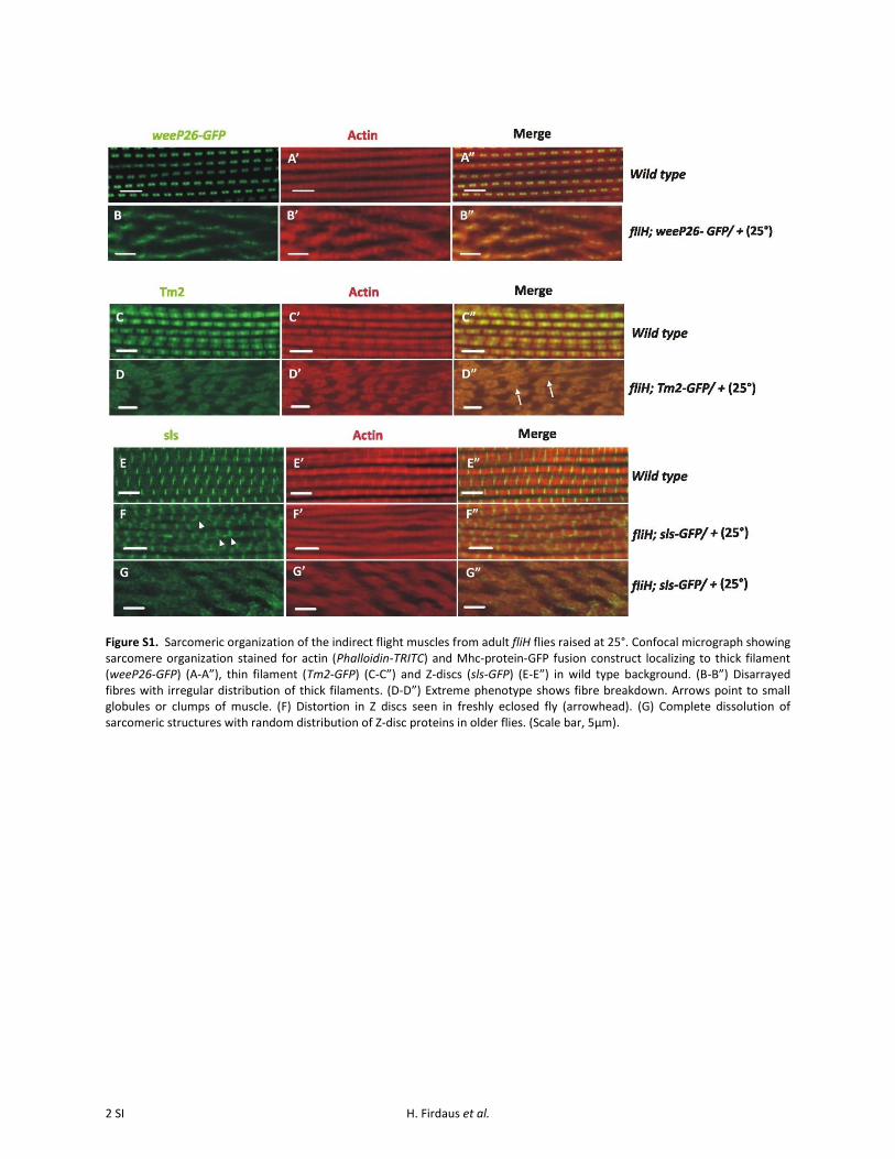
Figure S1. Sarcomeric organization of the indirect flight muscles from adult fliH flies raised at 25°. Confocal micrograph showing sarcomere organization stained for actin (Phalloidin-TRITC) and Mhc-protein-GFP fusion construct localizing to thick filament (weeP26-GFP) (A-A”), thin filament (Tm2-GFP) (C-C”) and Z-discs (sls-GFP) (E-E”) in wild type background. (B-B”) Disarrayed fibres with irregular distribution of thick filaments. (D-D”) Extreme phenotype shows fibre breakdown. Arrows point to small globules or clumps of muscle. (F) Distortion in Z discs seen in freshly eclosed fly (arrowhead). (G) Complete dissolution of sarcomeric structures with random distribution of Z-disc proteins in older flies. (Scale bar, 5µm).
2 SI H. Firdaus et al.

Figure S2. Assembly of the myofilaments in adult fliH flies raised at 18°. (A-C”) Confocal images which are comparable to the wild type (compare with representative pictures from wild type background in Figure S1). (Scale bar, 5µm).
Figure S3. Muscle hypercontraction in fliH starts at late puparium stages. (A-A”’) Act88F-GFP line was used to track the timing of IFM degeneration when genetically brought in the fliH background at 25°. Rupturing of the IFM is visible in adult flies post eclosion. Arrows point to DLMs, which are still intact at pupal stage, whereas arrowheads indicate site of muscle rupture in adult thorax. (B and C) Temperature shift assay to track exact pupal developmental stage in which IFM hypercontraction starts (details in supplementary materials and methods). (B) Temperature shift down experiment demonstrates that muscle degeneration in fliH mutant flies grown at 25°, starts after 75 hrs APF. (C) Temperature shift up assay supports the fact that degeneration of muscle fibre in fliH starts after 75 hrs APF. Asterisk denotes corrected development at 18° to match the development at 25° since pupal development is slow at 18°.
3 SI H. Firdaus et al.

Figure S2. Assembly of the myofilaments in adult fliH flies raised at 18°. (A-C”) Confocal images which are comparable to the wild type (compare with representative pictures from wild type background in Figure S1). (Scale bar, 5µm).
Figure S3. Muscle hypercontraction in fliH starts at late puparium stages. (A-A”’) Act88F-GFP line was used to track the timing of IFM degeneration when genetically brought in the fliH background at 25°. Rupturing of the IFM is visible in adult flies post eclosion. Arrows point to DLMs, which are still intact at pupal stage, whereas arrowheads indicate site of muscle rupture in adult thorax. (B and C) Temperature shift assay to track exact pupal developmental stage in which IFM hypercontraction starts (details in supplementary materials and methods). (B) Temperature shift down experiment demonstrates that muscle degeneration in fliH mutant flies grown at 25°, starts after 75 hrs APF. (C) Temperature shift up assay supports the fact that degeneration of muscle fibre in fliH starts after 75 hrs APF. Asterisk denotes corrected development at 18° to match the development at 25° since pupal development is slow at 18°.
4 SI H. Firdaus et al.