a case of enterocolic lymphocytic phlebitis mimicking ... · enterocolic lymphocytic phlebitis 103...
TRANSCRIPT

J o u r n a l o f R h e u m a t i c D i s e a s e sV o l . 2 1 , N o . 2 , A p r i l , 2 0 1 4http://dx.doi.org/10.4078/jrd.2014.21.2.101
□ Case Report □
101
<Received:April 23, 2013, Revised (1st: May 28, 2013, 2nd: June 19, 2013), Accepted:June 20, 2013>Corresponding to:Hyo Jin Choi, Division of Rheumatology, Department of Internal Medicine, Gachon University Gil Medical Center,
21 Namdongdae-ro 774-gil, Namdong-gu, Incheon 405-760, Korea. E-mail:[email protected]
pISSN: 2093-940X, eISSN: 2233-4718Copyright ⓒ 2014 by The Korean College of RheumatologyThis is a Free Access article, which permits unrestricted non-commerical use, distribution, and reproduction in any medium, provided the original work is properly cited.
A Case of Enterocolic Lymphocytic Phlebitis Mimicking Surgical Abdomen
Mi Ryoung Seo1, Tae Eun Kim2, Hee Jung Ryu1, Han Joo Baek1, Hyo Jin Choi1
Division of Rheumatology, Department of Internal Medicine, Gachon University School of Medicine, Gil Medical Center1, Incheon, Department of Pathology, Samsung Medical Centre,
Sungkyunkwan University School of Medicine2, Seoul, Korea
Vasculitis that involves the gastrointestinal (GI) tract often
occurs as part of a systemic inflammatory process. It is
a well-recognized manifestation of the small and medium
sized vessel vasculitides. Vasculitis of the GI tract may oc-
cur in isolation; although it can progress to a systemic
illness. It usually involves the arterioles, venules, and capil-
laries; however, it is very rare for only the venules to be
affected. Enterocolic lymphocytic phlebitis is a localized
vasculitis, typically affecting the small and medium-sized
intramural and mesenteric veins of the intestines. We re-
port a case of enterocolic lymphocytic phlebitis of the
colon. A 38-year-old woman was presented with hema-
tochezia and severe abdominal pain on the day of
admission. She had no history of intestinal disease or sys-
temic disease. Computed tomography showed an extremely
thickened wall of the colon, along with several air bubbles
in the colon with diffuse subcutaneous emphysema in the
abdominal wall. An emergency exploration laparotomy
and extended right hemicolectomy was performed. The pa-
tient recovered completely after surgery and remains well
without further therapy.
Key Words. Enterocolic lymphocytic phlebitis, Localized
gastrointestinal vasculitis, Single-organ vasculitis
Introduction
The term acute abdomen refers to signs and symptoms of
abdominal pain with tenderness and a clinical presentation that
often requires emergency surgical therapy (1). One cause of
acute abdomen is intestinal ischemia that can result from
vasculitis.
Various vasculitides affect the gastrointestinal (GI) tract and
are classified by type of affected vessel, etiology and under-
lying disease. Cancer, infection, drug, and systemic auto-
immune disease can present as GI vasculitis (2). Primary sys-
temic vasculitis also affect the GI tract, especially polyarteritis
nodosa, granulomatosis with polyangiitis (Wegener’s gran-
ulomatosis), eosinophilic granulomatosis with polyangiitis
(Churg-Strauss syndrome) and microscopic polyangiitis (3).
Localized vasculitis of the GI tract is very rare.
Enterocolic lymphocytic phlebitis (ELP) is a localized vascu-
litis typically affecting small and medium-sized intramural and
mesenteric veins of the intestines (4,5). We report a case of
ELP of the colon that was clinically and radiologically sus-
pected as intestinal ischemia accompanied by necrosis and
perforation of the colon.
Case Report
A 38-year-old woman was admitted to the hospital because
of hematochezia on the day of admission and severe abdomi-
nal pain with diarrhea of 36 hours’ duration. She had pre-
viously been healthy and had no history of intestinal or sys-
temic disease. She was a non-smoker and not taking any
medications. Blood pressure was 110/60 mmHg with a regular
heart rate of 96/min; body temperature at the time of admis-
sion was 37.0oC. On physical examination, the abdomen was
remarkable for tenderness in the right upper and lower with
rebound tenderness. A digital rectal examination revealed
fresh and clotted blood in the rectum.
102 Mi Ryoung Seo et al.
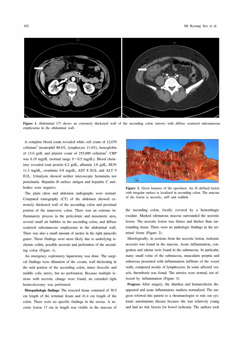
Figure 1. Abdominal CT shows an extremely thickened wall of the ascending colon (arrow) with diffuse scattered subcutaneous
emphysema in the abdominal wall.
Figure 2. Gross features of the specimen. An ill defined lesion
with irregular surface is localized in ascending colon. The mucosa
of the lesion is necrotic, stiff and reddish.
A complete blood count revealed white cell count of 12,670
cells/mm3 (neutrophil 80.6%, lymphocyte 11.4%), hemoglobin
of 13.0 g/dL and platelet count of 255,000 cells/mm3. CRP
was 6.19 mg/dL (normal range 0∼0.5 mg/dL). Blood chem-
istry revealed total protein 6.2 g/dL, albumin 3.8 g/dL, BUN
11.3 mg/dL, creatinine 0.8 mg/dL, AST 8 IU/L and ALT 9
IU/L. Urinalysis showed neither microscopic hematuria nor
proteinuria. Hepatitis B surface antigen and hepatitis C anti-
bodies were negative.
The plain chest and abdomen radiographs were normal.
Computed tomography (CT) of the abdomen showed ex-
tremely thickened wall of the ascending colon and proximal
portion of the transverse colon. There was an extreme in-
flammatory process in the pericolonic and mesenteric area,
several small air bubbles in the ascending colon, and diffuse
scattered subcutaneous emphysema in the abdominal wall.
There was also a small amount of ascites in the right paracolic
gutter. Those findings were most likely due to underlying is-
chemic colitis, possible necrosis and perforation of the ascend-
ing colon (Figure 1).
An emergency exploratory laparotomy was done. The surgi-
cal findings were dilatation of the cecum, wall thickening in
the mid portion of the ascending colon, intact ileocolic and
middle colic artery, but no perforation. Because multiple le-
sions with necrotic change were found, an extended right
hemicolectomy was performed.
Histopathologic findings: The resected tissue consisted of 30.5
cm length of the terminal ileum and 41.4 cm length of the
colon. There were no specific findings in the serosa. A ne-
crotic lesion 17 cm in length was visible in the mucosa of
the ascending colon, focally covered by a hemorrhagic
exudate. Marked edematous mucosa surrounded the necrotic
lesion. The necrotic lesion was firmer and thicker than sur-
rounding tissue. There were no pathologic findings in the ter-
minal ileum (Figure 2).
Histologically, in sections from the necrotic lesion, ischemic
necrosis was found in the mucosa. Acute inflammation, con-
gestion and edema were found in the submucosa. In particular,
many small veins of the submucosa, muscularis propria and
subserosa presented with inflammation infiltrate of the vessel
walls, composed mostly of lymphocytes. In some affected ves-
sels, thrombosis was found. The arteries were normal, not af-
fected by inflammation (Figure 3).
Progress: After surgery, the diarrhea and hematochezia dis-
appeared and acute inflammatory markers normalized. The sur-
geon referred this patient to a rheumatologist to rule out sys-
temic autoimmune disease because she was relatively young
and had no risk factors for bowel ischemia. The authors took

Enterocolic Lymphocytic Phlebitis 103
Figure 3. Microscopic findings of enterocolic phlebitis. The lesional mucosa and submucosa are shown in (A). The mucosa reveals
ischemic necrosis and the submucosa is hemorrhagic and edematous. The submucosal veins of the lesion (B) are filled with thrombi,
and show necrotizing vasculitis with fibrinoid degeneration. The vessels at the periphery (C and D) show lymphocytic phlebitis, relatively.
Special stain for elastin (D) demonstrated that arteries are spared. (A-C, Hematoxylin-eosin stain ×200; D, Elastic stain ×200).
more history and ran tests to evaluate systemic autoimmune
disease, especially systemic vasculitis, and hypercoagulable
states. She had no history of hypersensitivity to sunlight, malar
rash, arthritis, oral ulcerations, genital ulcerations and eye
disease. She was unmarried with no history of pregnancy.
Rheumatoid factor, antinuclear antibodies, antiphospholipid an-
tibodies (lupus anticoagulant, anticardiolipin IgG/IgA/IgM, and
anti-beta2-glycoprotein I IgG/IgM) and antineutrophil cytoplas-
mic antibodies were negative. Complement levels of C3 and
C4 were within a normal range. Hypercoagulable workup
showed no abnormalities (prothrombin time, activated partial
thromboplastin time, protein C, protein S, Factor V Leiden, an-
tithrombin, homocysteine, cryoglobulin). Stool bacteriological
and parasitological examinations were negative. In mesenteric
CT angiography and echocardiography, there were no abnor-
mal findings. There were no other manifestations of auto-
immune diseases such as systemic lupus erythematosus,
Behcet’s disease or systemic vasculitides.
Therefore, we diagnosed her as ELP by clinical manifes-
tations and histologic findings. The patient has recovered com-
pletely after the surgery without other therapy and was symp-
tom-free as of six months post-surgery.
Discussion
The vasculitides are a heterogeneous group of disorders char-
acterized by inflammation of blood vessel walls. The etiology
is usually unknown. Therefore, the vasculitides are mainly
classified by the type of vessels predominantly involved.
Another practical approach is to divide them into systemic or
localized vasculitides. The 2012 revised Chapel Hill
Consensus Conference defined the single-organ vasculitis as
vasculitis in arteries or veins of any size in a single organ,
with no features indicating that it is a limited expression of
a systemic vasculitis (2). Therefore, the term “single-organ

104 Mi Ryoung Seo et al.
vasculitis” is synonymous with localized vasculitis (6).
Vasculitis of the GI tract is a rare condition. The manifes-
tations of GI symptoms are wide, ranging from mild transient
abdominal pain to surgical abdomen. Systemic disorders such
as systemic lupus erythematosus, Behcet’s disease, mixed con-
nective tissue disease and rheumatoid arthritis can manifest as
vasculitis of GI tract (7). Indeed, primary systemic vasculi-
tides, especially medium and small vessel vasculitides, may
involve the GI tract. GI tract involvement of systemic vasculi-
tides has been reported at rates of 40∼60% for polyarteritis
nodosa, 30∼56% for microscopic polyangiitis, 20∼50% for
eosinophilic granulomatosis with polyangiitis, and 5∼11% for
granulomatosis with polyangiitis (3). GI involvement occurs
in IgA vasculitis (Henoch-Schönlein purpura), Kawasaki dis-
ease, Takayasu arteritis and giant cell arteritis (4,5,8). The GI
manifestations could be the first signs of those diseases, and
progress to systemic disorders. It is important to be aware that
isolated vasculitis of GI tract carries a risk for progression to
a systemic vasculitis. Therefore, isolated vasculitis should be
followed for at least six months before completing the diag-
nosis (9).
Localized vasculitis of the GI tract is an extremely rare
disease. Pagnoux and colleagues reviewed a series of small
and medium-sized vessel vasculitides with GI tract involve-
ment; only one of 62 patients presented with isolated vasculi-
tis of GI tract (3). Endoscopic biopsies have a low sensitivity
to diagnose GI vasculitis due to unable to obtain affected
vessels. In general, localized vasculitis of the GI tract is diag-
nosed after surgery (4,5,8,10). Burke and colleagues classified
isolated vasculitis of the GI tract into six groups histologically.
Those were polyarteritis, ELP, eosinophilic necrotizing in-
flammation of both arteries and veins, small-vessel vasculitis,
thromboangiitis obliterans, and giant cell arteritis (10).
The one cause of localized vasculitis of the GI tract is ELP.
This term is synonymous with mesenteric inflammatory ve-
no-occlusive disease, intramural mesenteric vasculitis, isolated
granulomatous phlebitis, lymphocytic venulitis, idiopathic my-
ointimal hyperplasia of mesenteric veins, and necrotizing and
giant cell granulomatous phlebitis (11). ELP is a rare type of
venulitis involving only the intramural and mesenteric veins
and venules of bowel wall. It spared the arteries and arterioles
as well as the systemic circulation. The inflammation can lead
to thrombotic obstruction and fibrointimal proliferation with
subsequent venous occlusion, causing edema and ischemia of
the involved intestinal segment. It can involve all sites of the
intestine, but the commonly affected sites are in the large
bowel, predominantly right colon (7,11). Clinically, the pre-
senting feature of ELP is subacute to acute intestinal ischemia
manifested as abdominal pain, hematochezia, and bloody
diarrhea. The endoscopic or imaging findings are not diag-
nostic, but computed tomography scanning often showed
thickened and edematous bowel walls. Our patient presented
with acute abdomen and remarkably thickened and edematous
right colon wall in CT. The etiology and pathogenesis of the
disease are undetermined, although the associations with some
medications, diversion colitis and lymphocytic colitis have
been proposed (12-14). But our patient was not taking any
medications and reported no history of surgery or previous GI
symptoms. The diagnosis of ELP depends on histopathology.
ELP was refractory to medical treatment and surgical re-
section of the affected bowel is usually curative without any
relapse. Of reported ELP, none progressed to systemic vascu-
litis to our knowledge (11-14).
Vasculitis of the GI tract can present as surgical abdomen.
If acute intestinal infarction develops in a young patient with-
out any risk factors of bowel ischemia, localized vasculitis
such as ELP should be considered. The authors report a case
of ELP that presented as an acute abdomen and recovered af-
ter surgery without other systemic treatment.
Summary
We report a 38-year-old woman presenting with acute ab-
dominal pain and bloody diarrhea. We first suspected in-
testinal ischemia leading to necrosis and bowel perforation.
The patient underwent emergency surgery. The pathologic di-
agnosis was ELP. She completely recovered after right hemi-
colectomy without further immunosuppressive treatment. GI
vasculitides can be a rare but noteworthy cause of acute
abdomen.
References
1. Ronald AS, Russell GP. Townsend: Sabiston Textbook of
Surgery. 19th ed. p. 1141, Philadelphia, Saunders, An im-
print of Elsevier, 2012.
2. Jennette JC, Falk RJ, Bacon PA, Basu N, Cid MC,
Ferrario F, et al. 2012 revised International Chapel Hill
Consensus Conference Nomenclature of Vasculitides.
Arthritis Rheum 2013;65:1-11.
3. Pagnoux C, Mahr A, Cohen P, Guillevin L. Presentation
and outcome of gastrointestinal involvement in systemic
necrotizing vasculitides: analysis of 62 patients with poly-
arteritis nodosa, microscopic polyangiitis, Wegener gran-
ulomatosis, Churg-Strauss syndrome, or rheumatoid ar-
thritis-associated vasculitis. Medicine (Baltimore) 2005;
84:115-28.
4. Gonzalez-Gay MA, Vazquez-Rodriguez TR, Miranda-Filloy
JA, Pazos-Ferro A, Garcia-Rodeja E. Localized vasculitis
of the gastrointestinal tract: a case report and literature
review. Clin Exp Rheumatol 2008;26(3 Suppl 49):S101-4.

Enterocolic Lymphocytic Phlebitis 105
5. Salvarani C, Calamia KT, Crowson CS, Miller DV,
Broadwell AW, Hunder GG, et al. Localized vasculitis of
the gastrointestinal tract: a case series. Rheumatology
(Oxford) 2010;49:1326-35.
6. Atisha-Fregoso Y, Hinojosa-Azaola A, Alcocer-Varela J.
Localized, single-organ vasculitis: clinical presentation
and management. Clin Rheumatol 2013;32:1-6.
7. Ahn E, Luk A, Chetty R, Butany J. Vasculitides of the
gastrointestinal tract. Semin Diagn Pathol 2009;26:77-88.
8. Garcia-Porrua C, Gutierrez-Duque O, Soto S, Garcia-
Rodeja E, Gonzalez-Gay MA. Localized vasculitis of the
gastrointestinal tract. Semin Arthritis Rheum 2006;35:
403-6.
9. Hernández-Rodríguez J, Hoffman GS. Updating single-
organ vasculitis. Curr Opin Rheumatol 2012;24:38-45.
10. Burke AP, Sobin LH, Virmani R. Localized vasculitis of
the gastrointestinal tract. Am J Surg Pathol 1995;19:
338-49.
11. Ngo N, Chang F. Enterocolic lymphocytic phlebitis: clin-
icopathologic features and review of the literature. Arch
Pathol Lab Med 2007;131:1130-4.
12. Jain R, Chetty R. Enterocolic lymphocytic phlebitis and
lymphocytic colitis: drug-related coexistent pathology. Int
J Colorectal Dis 2009;24:473-4.
13. Saraga E, Bouzourenne H. Enterocolic (lymphocytic)
phlebitis: a rare cause of intestinal ischemic necrosis: a
series of six patients and review of the literature. Am J
Surg Pathol 2000;24:824-9.
14. Chetty R, Hafezi S, Montgomery E. An incidental enter-
ocolic lymphocytic phlebitis pattern is seen commonly in
the rectal stump of patients with diversion colitis super-
imposed on inflammatory bowel disease. J Clin Pathol
2009;62:464-7.