a case of checkpoint inhibitor-induced celiac disease
TRANSCRIPT
CASE REPORT Open Access
A case of checkpoint inhibitor-inducedceliac diseaseDana Alsaadi1, Neil J. Shah2, Aline Charabaty3 and Michael B. Atkins2*
Abstract
Background: Immune checkpoint inhibitors (ICIs) have now become standard of care treatment for manymalignancies. ICIs are associated with unique immune mediated adverse events (irAEs) due to dysregulation ofimmune activation. As treatment with ICIs is becoming more common, rare irAEs are also being recognized. Herewe report a case of ICI-induced celiac disease.
Case: A 74-year-old Caucasian female with metastatic renal carcinoma received second line nivolumab (anti-PD1antibody) after initial disease progression on sunitinib. Ipilimumab was added after she failed to respond to sixcycles of nivolumab monotherapy. One week after her first cycle of combination treatment, she presented withnausea, vomiting, grade 1 diarrhea, and weight loss. She underwent endoscopy, which showed bile stasis in thestomach, normal appearing stomach mucosa, and nonbleeding erythematous mucosa in the duodenal bulb.Stomach biopsy showed moderate active chronic gastritis. Duodenal biopsy showed moderate chronic activeduodenitis with focal neutrophilic cryptitis, mucosal erosions, villous atrophy, mildly increased intraepitheliallymphocytes, and moderate chronic inflammation in the lamina propria pathognomonic of celiac disease.Symptoms improved with gluten-free diet, twice-daily omeprazole and anti-emetics and she was able to continueon treatment.
Conclusions: There has been only one published case reporting ICI-induced celiac disease. Our case reporthighlights a rare irAE (celiac disease) associated with ICI treatment. It is unclear whether the patient had previouslyundiagnosed celiac disease or whether ICIs triggered her enteritis. Our patient was able to continue treatment withICIs with dietary modifications, suggesting correct diagnosis is critical for optimal patient outcome.
Keywords: Nivolumab, Ipilimumab, Immune checkpoint inhibitor, Celiac disease, Immune-related adverse event
BackgroundImmune checkpoint inhibitors have become a mainstay inthe treatment of metastatic malignancies, such as melan-oma and lung cancer, as they increase the survival of pa-tients who failed conventional therapies. Nivolumab is ahuman monoclonal IgG4 antibody that inhibits the pro-grammed death-1 (PD-1) pathway, which is an importantregulator of the induction and maintenance of peripheraltolerance against malignant cells [1–3]. When a tumor cellligand binds the PD-1 receptor, a co-inhibitory moleculeexpressed on T-cells, it down-regulates the cellular immuneresponse. Nivolumab restores T-cell immunity by interfer-ing with co-inhibitory molecule induced T cell tolerance to
tumor cells. Ipilimumab is a human monoclonal IgG1kantibody that blocks cytotoxic T-lymphocyte–associatedantigen 4 (CTLA-4). CTLA-4 is a T-cell co-inhibitory mol-ecule that outcompetes the co-stimulatory molecule CD28for binding to B7 on antigen-presenting cells, therebydown-modulating cytotoxic T-cell function and allowingcellular proliferation. Ipilimumab binds to CTLA-4, whichis induced on activated T cells preventing down-regulationof cytotoxic T cell function. In addition, CTLA-4 is consti-tutively expressed on regulatory T-cells, where the bindingof ipilimumab leads to antibody dependent cellular cytotox-icity (ADCC), thereby eliminating a major immunosuppres-sive factor in the tumor microenvironment [4].While ICIs have revolutionized metastatic cancer
treatment, they produce unique immune-related adverseevents that include diarrhea and colitis. These side ef-fects vary in time of onset, but typically occur after the
© The Author(s). 2019 Open Access This article is distributed under the terms of the Creative Commons Attribution 4.0International License (http://creativecommons.org/licenses/by/4.0/), which permits unrestricted use, distribution, andreproduction in any medium, provided you give appropriate credit to the original author(s) and the source, provide a link tothe Creative Commons license, and indicate if changes were made. The Creative Commons Public Domain Dedication waiver(http://creativecommons.org/publicdomain/zero/1.0/) applies to the data made available in this article, unless otherwise stated.
* Correspondence: [email protected] Comprehensive Cancer Center, MedStar Georgetown UniversityHospital, 3800 Reservoir Road, NW, Washington, DC 20007, USAFull list of author information is available at the end of the article
Alsaadi et al. Journal for ImmunoTherapy of Cancer (2019) 7:203 https://doi.org/10.1186/s40425-019-0694-x
first few doses of ICI. ICI enterocolitis can be most ef-fectively managed when diagnosed early and immuno-suppressive therapy is initiated within the first five daysof symptoms [4]. Unrecognized or undertreated ICI-in-duced colitis can lead to bowel perforation and fatal out-come [5]. The choice of immunosuppressive therapydepends on the severity of irAE (grading is based on thecommon terminology criteria for adverse events(CTCAE) Version 5.0, 2017) [6]. For grade 1 diarrhea(which is an increase of less than 4 stools per day overthe patient’s baseline), symptomatic treatment with lo-peramide, rehydration, electrolytes substitution is ad-vised. For grade 2 diarrhea, steroid therapy with eitherbudesonide or 1 mg/kg prednisone is recommended. Incases of severe diarrhea (grade 3 and above), high doseIV corticosteroids such as methylprednisolone or dexa-methasone should be given. Grade 3 is defined as ≥7stools per day over baseline and necessitatinghospitalization for IV fluids. If no improvement is seenafter 3–5 days of high dose steroids, a dose of infliximab(IFX), a tumor necrosis factor-α (TNF-α) inhibitor, oroccasionally vedolizumab, an antibody to α4β7-integrinwhich facilitates T-cell trafficking into the gut mucosa,have been successfully used to achieve a clinical reso-lution of the ICI-induced colitis [7–10].As treatment with ICIs is becoming more common, rare
irAEs are also being recognized. While colitis is the maincause of diarrhea in ICI-treated patients, here we report acase of diarrhea due to ICI-induced celiac disease.
Case reportA 74-year-old Caucasian female with metastatic renal car-cinoma received second line nivolumab after initial diseaseprogression on sunitinib. She experienced grade 1 AST/ALT elevation and continued treatment. Ipilimumab wasadded after she failed to respond to six cycles of nivolu-mab monotherapy. One week after her first cycle of com-bination treatment, she presented with nausea, vomiting,and weight loss. She also had grade 1 diarrhea, which wastreated symptomatically with loperamide.She underwent upper endoscopy, which showed bile
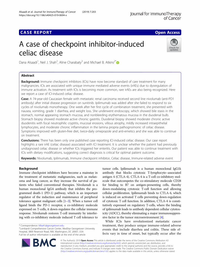
stasis in the stomach, normal appearing stomach mucosa,and nonbleeding erythematous mucosa in the duodenalbulb (Fig. 1). The second part of the duodenum showednormal mucosa without abnormalities. Stomach biopsyshowed moderate active chronic gastritis. Giemsa stain forHelicobacter Pylori was negative. Duodenal bulb biopsyshowed moderate chronic active duodenitis with focalneutrophilic cryptitis, mucosal erosions, villous atrophy,mildly increased intraepithelial lymphocytes, and moder-ate chronic inflammation in the lamina propria, suggestiveof celiac disease (Fig. 2). Immunohistochemistry was per-formed with antibodies against CD3, CD8, and CD56 torule out celiac disease because of villous atrophy. CD3
immunostains showed mildly increased intraepithelial Tcells, between twenty and thirty lymphocytes per hundredepithelial nuclei in the villi, but not to the usual degreeseen in celiac disease (which is defined as having greaterthan forty lymphocytes per hundred epithelial nuclei).Stains were negative for increased CD8-positive T cellsand CD56-positive Natural Killer cells.Serum tissue transglutaminase IgA antibody level was
elevated to 12 unit/mL (normal 0–3), which was diag-nostic for celiac disease. The patient was started on agluten-free diet for celiac disease, omeprazole 40 mg bymouth twice daily for gastritis, and the anti-emeticsondansetron and metoclopramide as needed. Symptomsimproved, and she was able to continue on treatment.The patient experienced a recurrence of symptoms, how-
ever, that was worse after each ICI infusion. Eight weeksafter her endoscopy, she was also started on budesonide 9mg by mouth daily and prochlorperazine three times a daywith meals. Symptoms improved with budesonide. The pa-tient also exhibited ICI-induced hypothyroidism and pan-creatitis, with an increase in lipase from baseline 77 to 400.She was treated with pancreatic enzymes and thyroidreplacement.Interval imaging was concerning for progression of
disease, and the patient discontinued nivolumab and ipi-limumab after receiving 4 cycles of combination therapy.She continued gluten-free diet and was able to gainweight. She was tapered off of budesonide over a periodof 6 months.
DiscussionImmune-checkpoints inhibitors have revolutionized thetreatment of metastatic malignancies; however, they cantrigger various organ-specific irAEs, such as nausea anddiarrhea, which can limit their use even with evidence ofregression of the underlying malignancy. One third of
Fig. 1 Endoscopic Picture of Duodenum. Inflammation in theduodenal bulb with non-bleeding erythematous mucosa
Alsaadi et al. Journal for ImmunoTherapy of Cancer (2019) 7:203 Page 2 of 5

patients treated with ipilimumab, an anti-CTLA-4 anti-body, develop diarrhea and 16% of patients will go on todevelop severe colitis, which can lead to perforations(0.5%) and/or colectomy [4, 11]. Nivolumab, an anti-PD-1 antibody, causes diarrhea in 8–19% of patients, ofwhom only 1% experience grade 3 or 4 diarrhea [5, 12,13]. Patients treated with a combination of ipilimumaband nivolumab have a 44% chance of developing diar-rhea, with grade 3 diarrhea accounting for 20% of allcases [12]. Typically, the onset of diarrhea occurs 6weeks after initiation of treatment, but can be delayedup to 6months after the last dose of ICI [13]. Patientscan also experience other irAEs separately or concomi-tantly, such as thyroiditis, myositis, and hepatitis, whichsuggests a systemic auto-immune like reaction to ICIs.While colitis is the most common cause of diarrhea in
the ICI-treated patient, alternative etiologies of diarrheamust also be considered. There has been only one pub-lished case reporting ICI-induced celiac disease due to
ipilimumab [14]. Our case report highlights a rare irAE, ce-liac disease, associated with ICI treatment. It is unclearwhether the patient had previously undiagnosed celiac dis-ease or whether ICIs triggered her enteritis, but the patientwas asymptomatic prior to initiation of ICI. Given that shealso exhibited other well-characterized concomitant irAEssuch as pancreatitis and hypothyroidism, we suspect thather celiac disease was triggered by ICIs. The initiation ofipilimumab in particular seemed to trigger her symptoms,which is in concordance with the literature that has shownthe strong immunogenic effects of ipilimumab comparedto other ICIs. The patient’s celiac diagnosis was coinciden-tal; given that her diarrhea was of a lower grade, she wastreated symptomatically with loperamide. Upper endoscopywas mainly performed for her nausea and vomiting.The pathogenesis of celiac disease is due to gluten-me-
diated activation of intestinal CD4+ T cells in the laminapropria. Gliadin peptides from gluten are converted bytissue transglutaminase (TTG) to a form that increases
a
b
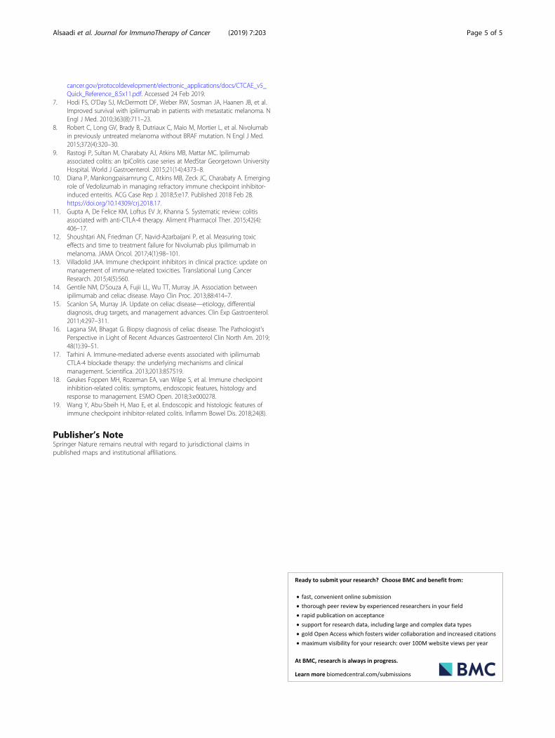
Fig. 2 Duodenal biopsy. a. Villous atrophy (black arrow) and chronic inflammation in the lamina propria with diffuse intraepithelial lymphocytosis(white arrow). b. Mucosal erosions (black arrows)
Alsaadi et al. Journal for ImmunoTherapy of Cancer (2019) 7:203 Page 3 of 5

their affinity for HLA-DQ2 and HLA-DQ8 moleculesand results in enhanced antigen presentation. Antigenpresenting cells activate CD4+ T-helper cells in the lam-ina propria, causing expansion of cells that produce anti-bodies to gliadin and TTG [15]. The histologichallmarks of celiac disease on small bowel biopsies areintraepithelial lymphocytosis, lymphoplasmacytic inflam-mation of the lamina propria, villous atrophy, and crypthyperplasia [16].While the pathogenesis of immune-mediated colitis
is not well understood, CTLA-4 blockade leads to T-cell activation that increases secretion of CD4 T-helper cell cytokines and cytolytic CD8 T-cell tissueinfiltration [17]. In contrast to celiac disease, ICI-in-duced colitis usually presents with an array of histo-logic findings. Usually biopsies demonstrate featuresof acute active colitis such as intraepithelial neutro-philic infiltrates or crypt abscesses, increased mono-nuclear cells in the lamina propria [18]. Both celiacdisease and ICI colitis show increased apoptotic cellsin crypts. Interestingly a subset of ICI colitis patientsmay demonstrate intraepithelial or basal lymphocytes,excess plasma cells in the lamina propria, andlymphocytic cryptitis on colonic biopsy, which ismore consistent with findings in chronic colitis [19].As histologic features may overlap, clinical features are
crucial for differentiation of distinct diseases. This pa-tient tested positive for tissue transglutaminase anti-bodies; the serum ELISA anti-TTG test has 93%sensitivity and 98% specificity for a celiac diagnosis [15].Typically celiac disease is diagnosed by biopsy whenthere are greater than forty lymphocytes per hundredepithelial nuclei in the villi. Thus, although the patient’shistology showed less than thirty lymphocytes per hun-dred epithelial nuclei, a diagnosis of early celiac diseasecan be made in conjunction with a positive serology.Gastroenterologists must consider histologic and endo-scopic features, clinical symptoms, and laboratory find-ings such as celiac serology and genetic testing to arriveat the correct diagnosis.Our patient was able to continue treatment with
ICIs with dietary modifications, suggesting correctdiagnosis is critical for optimal patient outcome. Ashighlighted by this case, active inflammation canaffect the small bowel and/or upper GI tract alone.For a patient with symptomatic diarrhea, evaluationshouldn’t be limited to colonoscopy alone, and biop-sies should be done to look for microscopic evidenceof inflammation even if the mucosa of the GI appearsnormal.Both early recognition and initiation of the appropriate
treatment of irAEs are crucial to relieve symptoms, avoidcomplications, and when indicated enable continued ICItherapy. While enterocolitis is by far the most common
cause of diarrhea, we here report a case of celiac diseaseinduced by ICI therapy. In a patient with symptomaticdiarrhea following initiation of ICIs, infectious pathologyshould be ruled out followed by initiation of systemiccorticosteroids. In patients with unusual features and/orfailure to respond to steroid treatment, considerationshould be given to a full endoscopic work up including acolonoscopy and exam of the terminal ileum and anupper endoscopy with biopsies in order to discern theunderlying etiology. This case of ICI-induced celiac dis-ease demonstrates that multidisciplinary collaborationamong oncologists, gastroenterologists, and pathologistsis crucial for correct diagnosis and treatment.
AbbreviationsCTCAE: Common Terminology Criteria for Adverse Events; ICI: Immunecheckpoint inhibitor; irAE: immune related adverse event; TTG: Tissuetransglutaminase
AcknowledgementsNot applicable.
Authors’ contributionsDA and NS wrote the case report. MA and AC treated the patient. Allauthors read and approved the final manuscript.
FundingThis work has no source of funding.
Availability of data and materialsNot applicable.
Ethics approval and consent to participateThis case report was exempt from the local ethical committee. Writteninformed consent was obtained from the patient for treatment.
Consent for publicationConsent for publication was obtained from the patient.
Competing interestsThe authors have no conflicts of interest to disclose.
Author details1Department of Internal Medicine, MedStar Georgetown University Hospital,Washington, DC, USA. 2Lombardi Comprehensive Cancer Center, MedStarGeorgetown University Hospital, 3800 Reservoir Road, NW, Washington, DC20007, USA. 3Sibley Memorial Hospital, Johns Hopkins University, 5255Loughboro Road NW, Washington, DC 20016, USA.
Received: 15 June 2019 Accepted: 24 July 2019
References1. Sundar R, Cho B, Brahmer JR, Soo RA. Nivolumab in NSCLC: latest evidence
and clinical potential. Ther Adv Med Oncol. 2015;7(2):85.2. Motzer RJ, Rini BI, McDermott DF, et al. Nivolumab for metastatic renal cell
carcinoma: results of a randomized phase II trial. JCO. 2015;33(13):1430–7.3. McDermott DF, Atkins MB. PD-1 as a potential target in cancer therapy.
Cancer Med. 2013;2(5):662–73.4. Graziani G, Tentori L, Navarra P. Ipilimumab: a novel immunostimulatory
monoclonal antibody for the treatment of cancer. Pharmacol Res. 2012;65(1):9–22.
5. Abdel-Rahman O, Helbling D, Schmidt J, et al. Treatment-related death incancer patients treated with immune checkpoint inhibitors: a systematicreview and meta-analysis. Clin Oncol. 2017;29(4):218.
6. US Department of Health and Human Services. National Cancer Institute.Common terminology criteria for adverse events (CTCAE). 2017. https://ctep.
Alsaadi et al. Journal for ImmunoTherapy of Cancer (2019) 7:203 Page 4 of 5
cancer.gov/protocoldevelopment/electronic_applications/docs/CTCAE_v5_Quick_Reference_8.5x11.pdf. Accessed 24 Feb 2019.
7. Hodi FS, O'Day SJ, McDermott DF, Weber RW, Sosman JA, Haanen JB, et al.Improved survival with ipilimumab in patients with metastatic melanoma. NEngl J Med. 2010;363(8):711–23.
8. Robert C, Long GV, Brady B, Dutriaux C, Maio M, Mortier L, et al. Nivolumabin previously untreated melanoma without BRAF mutation. N Engl J Med.2015;372(4):320–30.
9. Rastogi P, Sultan M, Charabaty AJ, Atkins MB, Mattar MC. Ipilimumabassociated colitis: an IpiColitis case series at MedStar Georgetown UniversityHospital. World J Gastroenterol. 2015;21(14):4373–8.
10. Diana P, Mankongpaisarnrung C, Atkins MB, Zeck JC, Charabaty A. Emergingrole of Vedolizumab in managing refractory immune checkpoint inhibitor-induced enteritis. ACG Case Rep J. 2018;5:e17. Published 2018 Feb 28.https://doi.org/10.14309/crj.2018.17.
11. Gupta A, De Felice KM, Loftus EV Jr, Khanna S. Systematic review: colitisassociated with anti-CTLA-4 therapy. Aliment Pharmacol Ther. 2015;42(4):406–17.
12. Shoushtari AN, Friedman CF, Navid-Azarbaijani P, et al. Measuring toxiceffects and time to treatment failure for Nivolumab plus Ipilimumab inmelanoma. JAMA Oncol. 2017;4(1):98–101.
13. Villadolid JAA. Immune checkpoint inhibitors in clinical practice: update onmanagement of immune-related toxicities. Translational Lung CancerResearch. 2015;4(5):560.
14. Gentile NM, D’Souza A, Fujii LL, Wu TT, Murray JA. Association betweenipilimumab and celiac disease. Mayo Clin Proc. 2013;88:414–7.
15. Scanlon SA, Murray JA. Update on celiac disease—etiology, differentialdiagnosis, drug targets, and management advances. Clin Exp Gastroenterol.2011;4:297–311.
16. Lagana SM, Bhagat G. Biopsy diagnosis of celiac disease. The Pathologist'sPerspective in Light of Recent Advances Gastroenterol Clin North Am. 2019;48(1):39–51.
17. Tarhini A. Immune-mediated adverse events associated with ipilimumabCTLA-4 blockade therapy: the underlying mechanisms and clinicalmanagement. Scientifica. 2013;2013:857519.
18. Geukes Foppen MH, Rozeman EA, van Wilpe S, et al. Immune checkpointinhibition-related colitis: symptoms, endoscopic features, histology andresponse to management. ESMO Open. 2018;3:e000278.
19. Wang Y, Abu-Sbeih H, Mao E, et al. Endoscopic and histologic features ofimmune checkpoint inhibitor-related colitis. Inflamm Bowel Dis. 2018;24(8).
Publisher’s NoteSpringer Nature remains neutral with regard to jurisdictional claims inpublished maps and institutional affiliations.
Alsaadi et al. Journal for ImmunoTherapy of Cancer (2019) 7:203 Page 5 of 5