a brain-computer interface for walking using eeg -...
TRANSCRIPT

The Netherlands, July 2012
A Brain-Computer Interface for walking using EEG
By Monica Perusquía HernándezIdentity number: 0756905
in partial ful�lment of the requirements for the degree of
Master of Science
in Human-Technology Interaction
Supervisors:
Dr. Raymond Cuijpers Faculty of Industrial Engeneering and Innovation Sciences, TU/e
Dr. Jason Farquhar Radboud University Nijmegen and Cognitive Arti�cial Intelligence,
Donders Institute for Brain, Cognition and Behaviour
Marianne Severens MSc. Sint Maartenskliniek Research Department and Cognitive Arti�cial In-
telligence, Donders Institute for Brain, Cognition and Behaviour

A BRAIN-COMPUTER INTERFACE FOR WALKING USING EEG 2
Preface
This thesis is the last step to complete the Master program in Human-TechnologyInteraction at the University of Technology Eindhoven. It is the result of several monthsof hard work, not only mine but also from a group of people who helped me to make thispossible. First of all, I would like to thank my wonderful supervisors Marianne Severens,Raymond Cuijpers and Jason Farquhar for their inspiring ideas, support, patience andenthusiasm. Also, I would like to thank Bart Nienhuis from the Sint Maartenskliniek forhis help during the setup of the experiments, for his advice regarding signal processing, andfor his feedback. I thank Antal Haans for his advice regarding the statistical analysis andthe design of the questionnaires. My sincere gratitude to all the people from the Researchand Education Department of the Sint Maartenskliniek for making pleasant my internship,for their advice during the presentations and for conducting the meetings in English just forme. Also, Victor Camelo and Monserrat Corona who kindly proofread this thesis. Thanksto all participants of the experiment and, �nally, thanks to my family, whose support hasbeen invaluable.
A BRAIN-COMPUTER INTERFACE FOR WALKING USING EEG 3
Summary
Brain-Computer Interfaces, or BCIs, allow the use of neurophysiological signals of thebrain to control external devices without using any other part of the body (Birbaumer &Cohen, 2007). Recently, it has been suggested that BCIs can be used in rehabilitation. In thecase of stroke patients, there is some evidence that imaginary movements might be helpfulby providing a means for practice when no actual movement can be performed (Mulder,2007), and BCIs can provide feedback for this imaginary-movement practice.
A common problem after a stroke is the loss of the ability to walk. Therefore, a BCIusing imaginary walking or attempted walking could help patients to relearn how to recoverthis ability. The present study proved the feasibility of decoding walking using EEG. Twodi�erent types of Event-Related Spectral Perturbations were used to distinguish walkingfrom no-walking in both actual walking and imaginary walking. Furthermore, an onlineBCI for walking was implemented successfully with classi�cation rates analogous to theo�ine classi�cation. Importantly, the results were robust for di�erent levels of automaticityin the walking task (simple walking, complex walking, and backwards walking).
In order to maximize the system performance, several tasks and features were chosenand compared, namely, task (simple and complex walking), spectral modulations (inter-stride modulations and intra-stride modulations) and frequency band (mu, beta and mu-beta). The mubeta band improved performance when compared with the mu and beta bandsseparately. However, neither di�erences in the automaticity of walking nor in the spectralmodulations were found. Only the e�ect of walking modality and spectral modulationswas signi�cant, showing that inter-stride modulations are better features during imaginarywalking, whereas the the intra-stride modulations yield better results during actual walking.
The interactions among task, performance and three subjective measures (sense ofagency, satisfaction and learnability) were also assessed. However, no signi�cant e�ects oftask and performance on the sense of agency, satisfaction and learnability were found.
Herein, recommendations are provided for the design of a BCI for rehabilitation. Inter-stride modulations could help during early stages in the rehabilitation process, whereas intra-stride could be more useful to tune practice according to the gait cycle phases. Furthermore,the mubeta band proved to be more suitable to achieve high performance. However, due tothe high variability between subjects, it is recommended to choose the best frequency bandper user.

A BRAIN-COMPUTER INTERFACE FOR WALKING USING EEG 4
Contents
Preface 2
Summary 3
1. Introduction 5
2. The Brain-Computer Interface cycle 7
3. The role of BCI in rehabilitation 10
4. EEG during Human Gait 12
5. The current study 15
6. Feasibility anlaysis: forward versus backward walking 18
Methods . . . . . . . . . . . . . . . . . . . . . . . . . . . . . . . . . . . . . . . . . . 18
Analysis . . . . . . . . . . . . . . . . . . . . . . . . . . . . . . . . . . . . . . . . . . 21
Results . . . . . . . . . . . . . . . . . . . . . . . . . . . . . . . . . . . . . . . . . . 24
Conclusions . . . . . . . . . . . . . . . . . . . . . . . . . . . . . . . . . . . . . . . . 28
7. Online BCI: simple versus complex walking 29
Methods . . . . . . . . . . . . . . . . . . . . . . . . . . . . . . . . . . . . . . . . . 29
Measurements . . . . . . . . . . . . . . . . . . . . . . . . . . . . . . . . . . . . . . . 31
Stimuli . . . . . . . . . . . . . . . . . . . . . . . . . . . . . . . . . . . . . . . . . . . 31
Analysis . . . . . . . . . . . . . . . . . . . . . . . . . . . . . . . . . . . . . . . . . 32
Results . . . . . . . . . . . . . . . . . . . . . . . . . . . . . . . . . . . . . . . . . . 34
Conclusions . . . . . . . . . . . . . . . . . . . . . . . . . . . . . . . . . . . . . . . . 38
8. General discussion 40
9. Conclusions 43
Appendixes 47
A. Forward versus Backward Walking . . . . . . . . . . . . . . . . . . . . . . . . . 47
B. Simple versus Complex Walking . . . . . . . . . . . . . . . . . . . . . . . . . . . 51
A BRAIN-COMPUTER INTERFACE FOR WALKING USING EEG 5
1. Introduction
Most children have dreamed about being a superhero granted with special abilitiesand powers: the power to �y, super-strength, telekinesis, perhaps even the ability to readminds. Such extra-sensorial and extra-motor powers seem to be con�ned to our imagination.However, they are not impossible anymore: advances in technology now make it possiblefor these dreams to come true. Technological extensions of the body can grant us abilitiesthat previously would have been impossible. In fact, evidence of these extended capabilitiesalready exists: airplanes have granted us the ability to �y, and vehicles have let us liftenormous weights. However, the control of such abilities still depends on physical interaction,using these technological extensions as elements external to our bodies.
Furthermore, the usage of such external tools is usually complicated. Interaction is socomplex, that it requires investing a huge amount of time on intensive training before beingable to optimally use such tools. Due to the inherent complexity of such technologies, thefeeling that humans are directly controlling the actions of the machine (i.e., sense of agency)and that the technology itself is part of their own body (i.e., sense of ownership) is almostimperceptible. To solve this problem, there are some trends in Human Computer Interactionwhich suggest several alternatives to diminish the complexity of the interfaces. Most of themcoincide in their enphasis on simple, one-purpose, location speci�c, interconnected devices(Buxton, 2001; Clark, 2003). Andrew Clark (2003) suggested that as tools become easierto use and more transparent, they become natural extensions of our body and of our mindand therefore, humans can be considered Natural-Born Cyborgs.
On the other hand, the possibility remains of fusing such interfaces and functionalitiesas extensions of our minds, literally. What would you feel if you could lift objects from acertain distance, with the sole will of your mind? What about controlling new stronger armsand legs as if they were your natural limbs? What would happen if you could see and hearmore frequencies of sound and light, besides the ones that our natural senses allow? Whatabout warping all your senses and your will to a faraway place? . . . Perhaps a virtual one?
The �rst step towards the implementation of a direct extension of our mind and bod-ies has already been taken through the intriduction of so-called Brain-computer Interfaces(BCIs). BCIs are a direct link between devices outside the body and mental activity (vanGerven, Farquhar, Schaefer, Vlek, Geuze, Nijholt, Ramsey et al., 2009). They allow the useof neurophysiological signals of the brain to control external devices (Birbaumer & Cohen,2007). BCIs have the potential of providing a good alternative for improving the sense ofagency and ownership provided by technology. However, their development is still rudimen-tary, and there is still much work left in order to reach a perfect coupling between humanminds and machines.
Nowadays, BCIs are typically used for communication and control (Wolpaw, Bir-baumer, McFarland, Pfurtscheller and Vaughan, 2002). One example of BCI for communi-cation is the so-called `visual speller'. This is a device that allows people to spell a wordby selecting letters from a grid. The interface consists of a matrix of letters which �ash inrows and columns. When the desired letter �ashes, a P300 evoked potential is generated300 ms after the presentation of the stimuli, making it possible to estimate which letter theuser is attending to (Wolpaw et al., 2002). On the other hand, BCIs for control have beenused with the explicit purpose of compensating for loss of motor ability in disabled patients.

A BRAIN-COMPUTER INTERFACE FOR WALKING USING EEG 6
A BCI could, for example, allow patients to drive external devices such as a wheelchairor prosthesis (Millán, Renkens, Mouriño and Gerstner, 2004; Enzinger, Ropele, Fazekas,Loitfelder, Gorani, Seifert, et al. 2008).
Since a BCI can provide feedback of imaginary and attempted movements when noactual movement can be executed, it has been suggested that it can be used in rehabilitationof stroke patients as well (Daly & Wolpaw, 2008; Vries & Mulder, 2007; Enzinger et al., 2008;Mulder, 2007; Prasad, Herman, Coyle, McDonough, & Crosbie, 2010; Cramer, Orr, Cohen,& Lacourse, 2007). By providing feedback of an attempted movement, BCIs can facilitatepractice of a lost brain functionality. There is some evidence that the use or disuse of a brainrepresentation can in�uence cortical representations (Mulder, 2007). Therefore, as a resultof constant training and facilitation of this training, reorganization of neural structures couldbe enhanced and lost motor abilities could be relearned.
A common problem after a stroke is the loss of the ability to walk. A BCI usingimaginary walking or attempted walking could help patients to recover this ability throughrelearning. The aims of this study are twofold. First, use EEG signals in order to determinewhether a (healthy) person is walking or not, for actual walking. Second, to build a BCI forimaginary walking, with similar brain signals. Di�erent features and tasks will be comparedin order to choose the combination that provides the best system performance. Furthermore,interactions between tasks, performance and the subjective assessment of the user about theusability and the sense of agency of the interface will be explored. Recommendations forfurther improvements of such BCI will be generated, focusing on necessary adaptations ofthe BCI for improved performance, while providing a good sense of agency and usability.
The structure of this report is as follows: in Section 2, a detailed description of themethodologies used to implement a BCI are provided. Section 3 describes current evidenceof the impact of BCIs in rehabilitation and Section 4 introduces relevant features of EEGsignals during human gait. Section 5 describes the research questions and hypothesis ofthe current study. Section 6 describes a feasibility study of the classi�cation of the EEGduring walking, and the technical details for such classi�cation. Section 7 describes the mainexperiment of this study, where the BCI was tested in an online setup and its subjectivequalities were assessed. Finally, Section 8 and 9 provide a general discussion of the resultsand conclusions, respectively.

A BRAIN-COMPUTER INTERFACE FOR WALKING USING EEG 7
2. The Brain-Computer Interface cycle
Despite the broad range of applications and paradigms that are used for implementingBCIs, most implementations follow a common set of steps. Gerven et al. (2009) proposed acycle for a BCI interface (Figure 1), from the measurement of the signal, to its transductioninto observable phenomena. In this cycle, the �rst stage corresponds to the sensing ofmental activity while the user engages in a cognitive task. Then, the signal is preprocessedin order to �lter noise and extract the characteristic features of the signal that will be usedfor classi�cation. Next, machine learning algorithms are used to make a prediction aboutthe intention of the user. Finally, this prediction is used to give feedback to the user, or toregulate the task at hand. The outcome signal can also be used to control a speci�c device.Since the user becomes aware of the device behavior, he is enabled to adjust his mentalactivity, towards the desired end.
In the following sections, each step of the BCI-cycle is described in detail.
Measurement. The required neurophysiological measurements of the brain arerecorded using two di�erent types of methods. Invasive methods require the implantation ofelectrodes on or in the brain. An example of an invasive method is the electro-cardiogram(ECoG), which provides a good signal-to-noise ratio and good detection of high frequen-cies. Other examples are the �Single microelectrode� (ME) and the �Micro-electrode array�(MEA), which consist of a single or multiple electrodes implanted in the brain, respectively.These methods are capable of detecting many forms of electrical potentials, single or multi-neuron spiking, as well as local �eld potentials. Both methods have fairly good temporaland spatial resolutions; however, they also have disadvantages such as the maintenance ofthe electrodes and di�culties with the communication of the internal device with externalprocessing units (Gerven et al., 2009).
Figure 1. : The BCI cycle as proposed by Gerven et al. (2009). While the user performs acognitive task his brain activity is measured. The signals acquired are then preprocessed toremove confounds. Then, the interesting characteristics for classi�cation (features) are ex-tracted. Next, a prediction is made about the nature of the features. Finally, this predictionis given as feedback or it is used to modify the user's task.

A BRAIN-COMPUTER INTERFACE FOR WALKING USING EEG 8
Non-invasive methods record the signals from outside the brain. Examples of non-invasive techniques are the Electroencephalogram (EEG) and the Magnetoencephalogram(MEG) technologies. The temporal resolution of both systems is very good when measuringmental activity in the brain. However, they do not provide a good spatial resolution and theyare susceptible to eye and muscle movements. Another example is the functional MagneticResonance (fMRI), which measures brain activity comparing the blood-oxygen level amongtasks. The fMRI achieves better spatial resolution than the EEG or MEG, at the cost oflosing temporal resolution (Juola, 2009).
Due to its temporal resolution and relative portability, EEG is particularly suitablefor BCI. Its working mechanism is based on using electrodes to measure electrical activitygenerated in the brain and transmitted on the scalp. These potentials are neurophysiologicalsignals that can be used to identify patterns useful to build a BCI.
Preprocessing. Before the signal measured by the EEG can be used, it has to be'cleaned.' In order words, the signal of interest has to be identi�ed and confounds haveto be removed. This is called preprocessing and implies the removal of faulty channels andoutliers. Also, the signal is usually 'detrended' to remove trends in the measurements causedby changes in the conductance between the electrode and the skin. Further preprocessingincludes the rejection of artifacts such as eye blinks, saccades and other EMG componentsdetected by the EEG electrodes.
Feature extraction. The next step is to identify the characteristics of the EEG signalthat are interesting for the problem at hand. Signatures are the association of a brain signalwith a mental state that uniquely caused it (Gerven et al., 2009). These signatures havebeen classi�ed into evoked and induced potentials. Both of them are responses locked toa stimulus; the di�erence between them is that the evoked potentials are responses phase-locked in the time domain and the induced potentials are locked in spectral power (i.e., inthe frequency domain) to the event (Gerven et al., 2009).
An example of induced response potentials are Event-Related Spectral Perturbations(ERSP). Within this category, Event-Related Spectral Desynchronizations (ERSD) andEvent-Related Spectral Synchronizations (ERSS) are of special interest because they arerelated to motor activity and movement preparation (Pfurtscheller, Stancák and Neuper,1996; Pfurtscheller, Brunner, Schlögl and Lopes da Silva, 2006). ERSDs are induced po-tentials that correspond to a decrease in spectral power during planning and execution of amovement. ERSDs are followed by an ERSS, which is the increase in spectral power afterthe movement (Pfurtscheller, Brunner, et al., 2006).
ERSD in the mu-rhythm frequencies (Described in proceeding paragraphs) in humanshas been linked to somatosensory stimulation or movement. Evidence for this has beenfound especially for hand movements (Pfurtscheller et al., 1996). Interestingly, ERSDsand ERSSs are present not only during actual movement, but also during imagery tasks(Pfurtscheller, Brunner, et al., 2006; Neuper et al., 2005). Motor imagery refers to thecognitive image of performing a movement, without executing the actual movement. If theimagery is performed in a �rst person perspective, it is called Kinesthetic Motor Imagery(KMI) and there is some evidence that it elicits stronger desynchronizations than VisualMotor Imagery (VMI), which is the imagery of seeing oneself walking (i.e., from an outsideperspective) (Neuper, Scherer, Reiner and Pfurtscheller, 2005).

A BRAIN-COMPUTER INTERFACE FOR WALKING USING EEG 9
Since motor imagery can elicit speci�c changes of potentials in the brain, it is a primecanditate for implementation of BCIs. Furthermore, it can be used at will, giving the userof the BCI the power of steering the interaction. The mu-rhythm and beta-band rhythmsare of special interest for identifying motor imagery. Mu-rhythm refers to an oscillation ofabout 8-14 Hz in the EEG signal, whereas beta-rhythm refers to an oscillation from 12-25Hz. Modulations in both frequency bands can be seen not only with actual movement, butalso if one imagines a movement (Pfurtscheller et al., 1996; Prasad et al., 2010). However,mu-band ERSDs for foot movements are more di�cult to detect because the foot area islocated within the mesial wall in the interhemispheric �sure. Beta-band oscillations arealso generated in the somatosensory cortex, but in contrast to mu-rhythms, they are easierto detect, and time courses of recovery after desynchronization are faster in beta than inmu-rhythms (Pfurtscheller et al., 1996).
Prediction. Once the features are selected, they are fed into a classi�er. A classi�er isan algorithm that assigns an external observation to one of several classess (Müller, Kraule-dat, Dornhege, Curio, and Blankertz, 2004). The rule to do this classi�cation is found byanalyzing the data and �tting a model that indicates a boundary between classes. A simpleapproach, is to use a linear regression to �t the data and use a threshold function to dif-ferentiate between di�erent classes. If there are only two classes, logistic regression can beused (Russell & Norving, 2010).
Such models are usually built by reducing the error between values predicted by themodel and the actual data. However, this might not always be bene�cial. Especially insmall samples, deviations might be large, and by reducing the error to explain the variancesin the sample (Müller et al., 2004), the classi�er might over�t the data. As a result, it willnot generalize to other samples of data. To solve the over�tting problem, a reglarizationparameter can be added to the regression problem (Müller, Krauledat, Dornhege, Curio,& Blankertz, 2004), which introduces restrictions (through penalizations) on how muchdata is required to switch the classi�cation threshold. The more data is available within aboundary, the more con�dent the classi�er is that it belongs to a certain class and the lowerthe penalization due to few data points.
Feedback. Finally, the result of the prediction can be used to control an externaldevice or can be shown to the user as feedback, so that he can adapt to the outcome of theclassi�cation. The range of modalities in which the feedback can be provided is wide andshould be designed according to the speci�c application of the BCI.
When such feedback is provided, the BCI is considered as �online.� In contrast to ano�ine setup where all data is gathered �rst and classi�cation is done afterward, an onlineBCI should do the classi�cation in real-time and should be robust to changes in the mentalactivity of the user and his adaptation to the feedback itself.
In summary, BCIs work in a closed loop which starts with measuring the brain activityof a person while he engages in a cognitive task. Then the signal is preprocessed and therelevant features extracted. Based on those features, a classi�er can learn about the user'sbrain signals and make a prediction about the intentions of the user and provide feedbackaccording to the results. Since the user is part of this loop, both machine learning and humanlearning are complementary; the proceeding section will explain how this interdependencecan be useful for rehabilitation of stroke patients.

A BRAIN-COMPUTER INTERFACE FOR WALKING USING EEG 10
3. The role of BCI in rehabilitation
A BCI using imaginary movements could help in rehabilitation, not only by providingthe means of bypassing spinal cord injuries, but also helping both Spinal Cord Injury (SCI)and stroke patients maintain and recover their brain functionalities by giving direct feedbackof imagined movements.
Motor impairment and functional disability are the major consequences of SpinalCord Injury (SCI) and stroke. The damaged spinal cord of SCI patients obstructs thecommunication between the brain and several parts of the body, depending on the level ofthe injury. On the other hand, a stroke is related to a distortion of the capacity of the brainto process neural information after a disturbance in the blood supply to the brain.
Even though both problems may result in motor impairments, the brain functionalityin patients with SCI is assumed to be intact and capable of driving limb movements (Crameret al., 2007; Enzinger et al., 2008), while in a stroke, the motor impairment is caused bybrain damage (Vries & Mulder, 2007). Consequently, rehabilitation strategies for theseimpairments follow di�erent paths. Rehabilitation after a stroke is mainly based on therapyand practice. In this case, functional recovery is attributed to the processes of reorganizationand substitution in the damaged brain. For SCI, rehabilitation could use BCIs, which allowthe use of neurophysiological signals of the brain to control external devices (Birbaumer &Cohen, 2007), and could therefore be used to bypass damaged areas in the spinal cord anddrive prosthesis or even the patients' muscles.
Even if the damage that caused the impairment is not localized in the brain, someabnormalities have been described in the brain motor functions of SCI patients, includingreduced activation, abnormal activation patterns and higher thresholds and latency for motorevoked potentials (Enzinger et al., 2008; Cramer et al., 2007). These abnormalities suggestthat indeed the disuse of a brain functionality can in�uence cortical representations. Inother words, reorganization of neural structures takes place as the result of depravation ofsensory input due to movements (Sadato et al., 1998; Halligan et al., 1993), and constanttraining is required in order to maintain the brain's motor system functionalities in goodshape.
Unfortunately, for an injured patient it is not always possible to practice the requiredmovements, either because he cannot move or because movements are painful. There is someevidence that imaginary movements might be helpful by providing a means of practice whenno actual movement can be performed Mulder (2007). The use of motor imagery would bebene�cial for rehabilitation because it is a direct representation of the movement that thepatient would like to recover.
Motor imagery training could be enhanced by providing the patient with feedbackof the movement. There are several types of feedback, including proprioceptive and visualfeedback. In stroke physiotherapy, a therapist helps the patient move the paralyzed limbs,enhancing proprioceptive feedback. Another example from therapy is the Lokomat System(Hocoma Inc.), which is a device designed to help injured patients simulate the movementof walking with slightly better results than traditional therapy (Mayr, Ko�er, Quirbach,Matzak, Fröhlich & Saltuari, 2007). The simulation of movement is achieved by using atreadmill with weight support and motorized boots. Visual feedback can also be provided bythe Lokomat Pro, which shows visual cues of walking on a screen with a virtual environment,
A BRAIN-COMPUTER INTERFACE FOR WALKING USING EEG 11
in which the patient walks and completes some tasks. This augmented feedback enhancesthe patient's active participation in the task.
Despite the potential of BCI and its possible bene�ts in rehabilitation, few studies haveexplored its feasibility. One example is the study by Prasad, Herman, Coyle, McDonough,& Crosbie (2010), where a game-based neurofeedback BCI was used to enhance movementrecovery in stroke patients. The lack of feasibility research is mainly due to the di�culty inidentifying signatures in the brain signal.
Some attempts to create a BCI for virtual walking using mental imagery of the handsand feet have been successful (Pfurtscheller, Leeb, et al., 2006); however, they requiredextensive training of three subjects, from which only one showed an steady increase in hiscontrol of the interface. Furthermore, they do not use imaginary walking to produce thewalking movement in the virtual environment.
Since imaginary movements can activate sensory-motor areas in the brain (Mulder,2007), they are considered embodied. A task that is more natural to the body -such asthe simple act of walking- could improve the sense of agency of the patient as well as thequality of the feedback he can gain from using a BCI. Accordingly, cortical representationsof walking during human gait have to be considered. To this aim, the next section willprovide a brief introduction to human gait and its cortical representation.

A BRAIN-COMPUTER INTERFACE FOR WALKING USING EEG 12
4. EEG during Human Gait
The Gait Cycle . Whittle (1996) de�ned walking as 'a method of locomotion involvingthe use of the two legs, alternately, to provide both support and propulsion,' and, whentalking about normal gait, the di�erence with running is that at least one foot is in contactwith the ground at all times. However, describing normal gait is di�cult because there arelarge di�erences in walk patterns depending on sex and age.
Furthermore, he de�ned the gait cycle as 'the time interval between two successiveoccurrences of one of the repetitive events of walking.' Thus, in order to de�ne a gait cycle,a start reference should be taken into account. Usually, an initial contact of a foot withthe ground is considered the starting point of the gait cycle, and it continues until thesame foot touches the ground again. Between these two contacts, several phases have beenidenti�ed. Namely, initial contact, opposite toe o�, heel rise, opposite initial contact, toeo�, feet adjacent and tibia vertical. For the purposes of this study, only toe o�s and heelstrikes will be considered for each foot: only left toe o� (LTO), left heel strike (LHS), righttoe o� (RTO) and right heel strike (RHS) are considered.
Figure 2 shows the di�erent phases of a gait cycle for forward and backward walking.The start of the gait cycle was set to left toe o� (LTO) in forward walking and left toestrike (LTS) in backward walking. As can be observed from this �gure, backward walkinghas analogous gait-cycle phases to forward walking. In fact, Grasso, Bianchi and Lacquaniti(1998) compared the kinematics and kinetics of both directions of locomotion. Accordingto their results, the kinematics (i.e., the spatial trajectory followed by the limbs duringwalking) of forward walking are very similar to those of backward walking when mirroredin time. However, the kinetics (i.e., the muscle activation during walk) of such movementsare di�erent.
Several measures have been used to characterize the timing of the walking process.Three of them are stride length, cadence, and walking speed (Whittle, 1996).
• The stride length corresponds to two consecutive steps (i.e., one gait cycle). On theother hand, the step length corresponds to the distance from one foot strike to the next,regardless of which foot touched the ground.
Figure 2. : Gait cycle phases. Four phases are considered within a step. In forward walking:left toe o� (LTO), left heel strike (LHS), right toe o� (RTO) and right heel strike (RHS). Inbackward walking: left toe strike (LTS), right heel o� (RHO), right toe strike (RTS) and leftheel o� (LHO). The �gure above shows the correspondence of phase between forward andbackward walking. Forward walking is quite similar to backward walking when mirrored intime. When a LTO occurs in forward walking, a LTS occurs in backward walking.
A BRAIN-COMPUTER INTERFACE FOR WALKING USING EEG 13
• Cadence is the number of steps taken in a given time (usually minutes), thus, itis a measure of time for half-gait cyles. An alternative is to measure cadence in steps perminute (heretofore referred to as stepping frequency).
• The speed of walking is the distance covered by the whole body in a given time andis usually measured in meters per second.
Given these de�nitions, the relationship among step length, stepping frequency and speedof walk can be simpli�ed to the de�nition of velocity: the speed of walking is given by theproduct of stepping frequency and step length (Whittle, 1996).
Measuring EEG during walking . The ultimate goal of this study is to use a BCI toprovide feedback about imaginary walking movements, which could be applied in rehabili-tation to improve the process of learning to walk again. To develop such a brain-computerinterface, it is crucial to identify the correlations between patterns of movements during thehuman gait cycle and EEG potentials.
The main challenge of using EEG during actual walking is to remove movement ar-tifacts inherent in the walking process and EMG artifacts generated from face and neckmuscles. Several attempts to remove these kinds of artifacts have been made for spokenlanguage production (De Vos, Riès, Vanderperren, Vanrumste, Alario, Van Hu�el, & Burle,2010) and walking (Gwin, Gramann, Makeig and Ferris, 2010; Gwin, Gramann, Makeig andFerris, 2011; Severens, Nienhuis, Desain and Duysens, 2012). In these studies, the aim wasto uncorrelate artifactual components from the EEG signal of interest.
Gwin et al. (2010) used Independent Component Analysis (ICA) to separate the mea-sured signal into maximally independent components and subsequently applied a component-based template regression to identify and spatially �lter gait-related movement artifacts. Ina second study, Gwin et al. (2011) con�rmed the potential of ICA to identify artifactualcomponents. Here, they used current dipole stimation for every component using an in-verse modeling approach. Components that exhibited signi�cant coupling with intra-stridechanges of spectral power were located in the anterior cingulate, posterior parietal, and sen-sorimotor cortex, suggesting that EEG recordings could identify cortical involvement duringhuman gait. In particular, peaks in the beta and alpha bands' spectral power were found inthe heel-strike and (approximately) in the middle of the double support phases, respectively.
Both studies demonstrated the potential of using ICA to remove gait-related artifacts.However, this requires a considerable amount of data, and the processing required to cal-culate all components is time-consuming. Furthermore, it has to be combined with otheralgorithms or visual selection to detect which components are useful. Unfortunatelly, inan online BCI context, time is not an abundant resource, and therefore, other alternativesshould be considered.
Aiming to remove muscle artifacts from EEG recordings of spoke language production,De Vos et al. (2010) proposed the use of a Blind Source Separation (BSS) technique calledCanonical Correlation Analysis (CCA) and compared it to other techniques such as ICA.CCA is a statistical method used to estimate the correlation between two variables. EEGand EMG components can be separated by combining CCA with the assumptions that (1)EEG and EMG sources are uncorrelated and that (2) EEG sources, contrary to EMG, areindividually uncorrelated. In their particular appication, De Vos et al. (2010) showed thatindeed BSS-CCA outperformed ICA in preserving the shapes of the ERP after removing

A BRAIN-COMPUTER INTERFACE FOR WALKING USING EEG 14
artifactual EMG components.In another study, Severens et al. (2012) showed that CCA can also be successfully
applied to disentangle EEG from EMG during walking. After removing EMG artifacts,the synchronizations between the gait cycle and the spectral power of the beta band, asshowed by Gwin et al. (2011), were con�rmed. During the study of Severens et al. (2012),participants had to walk at di�erent speeds, and the tasks also included stepping in place. Itwas shown that the intensity of activity is greater in heel-strike and toe-o� phases of the gaitcycle, and also that uncommon walking activity such as stepping in place and walking fastinvolves stronger modulations of cortical activity. This might suggest that cortical activitydepends on experience and familiarity with the movements (Peters, 2011; Severens et al.,2012).
In summary, the results of Gwin et al. (2011) and Severens et al. (2012) suggest thatit is feasible to use EEG as tool to measure cortical activity during walking, and that thereis cortical involvement during gait.
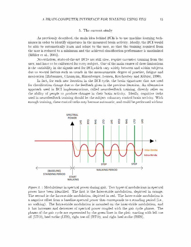
Severens et al. (2012) found two types of modulations in spectral power. The �rstis an overall power decrease in the mu and beta bands along the whole time span duringwhich steps were performed (i.e., inter-stride modulations); the second refers to spectralmodulations (SM) in the same frequency bands coupled to the gait-cycle (i.e., intra-stridemodulations). Figure 3 shows a schematic representation of such modulations, where theorange line shows the inter-stride spectral modulation and the red one the intra-stride spec-tral modulation. These two modulations show that there is cortical involvement during gait,which could be used as a feature to implement a BCI for walking.
A BRAIN-COMPUTER INTERFACE FOR WALKING USING EEG 15
5. The current study
As previously described, the main idea behind BCIs is to use machine learning tech-niques in order to identify signatures in the measured brain activity. Ideally, the BCI wouldbe able to automatically learn and adapt to the user, so that the training required fromthe user is reduced to a minimum and the achieved classi�cation performance is maximized(Müller et al., 2004).
Nevertheless, state-of-the-art BCIs are still slow, require extensive training from theuser, and have to be calibrated for every subject. One of the main causes of these limitationsis the variability in the signals used for BCI,which vary widely between and within subjectsdue to several factors such as trends in the measurements, degree of practice, fatigue andmotivation (Birbaumer, Ghanayim, Hinterberger, Iversen, Kotchoubey and Kübler, 1999).
In fact, for each new iteration in the BCI cycle, the brain signatures that are usedfor classi�cation change due to the feedback given in the previous iteration. An alternativeapproach used in BCI implementations, called neurofeedback training, directly relies onthe ability of people to produce changes in their brain activity. Ideally, cognitive tasksused in neurofeedback training should let the subject voluntary control brain activity. Withenough training, these control tasks may become automatic, and could be performed without
Figure 3. : Modulations in spectral power during gait. Two types of modulations in spectralpower have been identi�ed. The �rst is the Inter-stride modulation, depicted in orange.The second is the Intra-stride modulation, depicted in red. The Inter-stride modulation isa negative o�set from a baseline spectral power that corresponds to a standing period (i.e.,no walking). The Intra-stride modulation is mounted on the inter-stride modulation, andit has increases and decreases of spectral power coupled with the gait cycle phases. Thephases of the gait cycle are represented by the green lines in the plot: starting with left toeo� (LTO), heel strike (LHS), right toe o� (RTO), and right heel strike (RHS).

A BRAIN-COMPUTER INTERFACE FOR WALKING USING EEG 16
conscious e�ort or attention (Curran, 2003; Elbert, Rockstroh, Lutzenberger and Birbaumer,1980). User learning and user adaptation to the interface are key factors in the performanceof the BCI. It is the human's capability to adapt that allows for another application ofBCIs: they can be used as a tool for practice by providing feedback of the user's mentalstate (Nauper et al., 1999).
Other factors can also alter the performance of the BCI. There is some evidencethat the automaticity or the mental e�ort required to do a task also modi�es the qualityof the signature measured from the brain. For instance, Müller-Putz et al. (2007) foundsome evidence that SCI patients showed better imagery vividness than able-bodied subjects,maybe because they required more mental e�ort. Also, Severens et al. (2012) found someevidence that spectral power modulations during gait were stronger when the movement wasless automatic. Consequently, less automatic movements could improve the performance ofthe machine learning algorithm by making the boundary between classes more prominent.
For the purpose of rehabilitation, it is desired that the patient relearns how to controlhis brain activity in order to recover a lost ability. However, even when the goal is to learnhow to control one's brain activity, given the di�culty of making accurate predictions ofa mental state or instruction through a BCI, there is usually a chance that the predictionwill be wrong and that the instructions given by the user will be misinterpreted by theprediction algorithm. This implicit error might alter the experience of the user by givinghim the feeling that he is not the one controlling the interface. According to cognitiveneuroscience, this experience of controlling one's actions and -through this control- a�ectingthe external world is called 'sense of agency' (Coyle, Moore, Ola, Kristensson and Fletcher,2012;Gallagher, 2000). As a result of a decreased sense of agency, the patient's motivationto practice using the interface might decrease. Consequently, a balance between machinelearning, neurofeedback training and task design should be considered when designing a BCIfor rehabilitation.
Based on these considerations, the aim of the present study is to build a BCI todetermine if a person is actually walking or not, and to assess the tradeo� between theachieved system performance, and the sense of agency, learnability and satisfaction of theuser. Di�erent features and tasks will be explored in order to improve performance.
With this aim, two experiments were conducted. The �rst experiment used a compar-ison of forward and backward walking to prove the feasibility of using EEG to di�erentiatewalking and no-walking conditions using cortical activity (either in actual or imaginarywalking). Furthermore, di�erent features and tasks were tested to determine which methodprovides best system performance.
It was expected that the less automatic the movement, the more modulation in muand beta bands of activity over the motor cortex, resulting in increased classi�cation perfor-mance. Backwards walking and complex walking, being less automatic, could increase theperformance of the BCI when compared with forward walking and simple walking. However,it might improve performance at the expense of usability and the sense of agency.
A second experiment was designed to test the BCI in an online setup. During thisexperiment, simple walking and complex walking (i.e., walking at varying speeds) were com-pared while assessing sense of agency, learnability and satisfaction. Again, simple walkingwas less automatic than complex walking, and therefore better classi�cation rates were ex-pected. Furthermore, the sense of agency, satisfaction and learnability were assessed using

A BRAIN-COMPUTER INTERFACE FOR WALKING USING EEG 17
a questionnaire about the feedback provided. It was expected that:• The higher the system performance, the better the sense of agency.• The satisfaction and learnability will decrease with the complexity of the tasks.
However, there might be a mediating e�ect of system performance: the better the perfor-mance, the better the satisfaction.Recommendations for further improvements of the BCI will be presented with the aimof adapting the BCI to maximize performance, while providing a good sense of agency,learnability and satisfaction at the same time.

A BRAIN-COMPUTER INTERFACE FOR WALKING USING EEG 18
6. Feasibility anlaysis: forward versus backward walking
The goal of this experiment is to prove that walking (either actual or imaginary)can be distinguished from no-walking in an o�ine BCI setup. Di�erent tasks (forwardand backward walking) will be compared in order to determine the relationship between theclassi�cation performance and the automaticity of the movement. Di�erent frequency bandswill also be tested in order to determine which one provides best system performance.
Methods
Experiment Design. The experiment used a 2x2 within-subjects design. The twofactors were the type of task (forward vs. backward walking) and the walking modality(actual walking vs. imaginary walking). These four conditions were presented in eightblocks. In every block, each condition occurred once, in random order. In other words, allparticipants went through a total of eight repetitions of every condition (32 trials in total).
Participants. Twelve healthy volunteers with no history of major lower limb injury andno known neurological or locomotor de�cits participated in this study. The mean age of theparticipants was 29 (SD=5.94) years. Before the start of the experiment, the participantswere asked to read and sign a standard informed consent form, approved by the EthicalCommittee for Behavioral Scienti�c Research of the Faculty of Social Sciences, RadboudUniversity Nijmegen.
Experimental Setup. EEG was recorded using a TMSI-REFA 72-channel ampli�er(Twente Medical Systems International, the Netherlands) and a 64-channel electrode array ina 10-20 electrode placement system. The ground electrode was placed on the AFz-electrodelocation. The TMSI REFA ampli�er sampled the EEG signals at 500 Hz. Prior to themeasurement, electrode gel was used to ensure that the impedance was less than 50 kiloohms for each channel. The positions of 22 re�ective markers were recorded using a ten-camera motion capture system (Vicon, Vicon Motion Systems Inc.). Marker positions weresampled at 100 Hz. The markers were placed according to the Plugin-Gait Model. Sixteenmarkers were placed on the lower limbs as indicated in Figure 4, and six markers were placedon the cap to track the movements of the head during walking.
Subjects walked on a programmable treadmill at a velocity of 3 km/s, facing a monitorscreen placed 40 cm above the �oor and 150 cm away from the treadmill. In order towalk backwards, participants had to turn around (for both actual and imaginary walking).Identical screens were placed at both ends of the treadmill, to facilitate change of direction,as shown in Figure 5. In order to reduce movement artifacts, the EEG cap cable was clippedto a cable that hung on the ceiling with a device that could move along with the subject,and then connected to the ampli�er (see Figure 5).
The control of the �ow of the experiment was programmed using Brainstream (DCC,Radboud University Nijmegen, http://www.brainstream.nu), a Matlab application (TheMathworks, Natick, MA).
Procedure. Four walking tasks were executed by the participants: forward walking,imaginary forward walking, backward walking and imaginary backward walking. A trialoverview is shown in Figure 6. Every trial consisted of �ve seconds of standing (i.e., baseline

A BRAIN-COMPUTER INTERFACE FOR WALKING USING EEG 19
Figure 4. : Plugin-Gait Model. Sixteen re�ective markers were placed on the lower limbs;their locations were determined relative to the position of a bone as indicated in the picture.The markers were stuck directly on the skin with a double-sided tape.
period), followed by the presentation of the instructions on screen (one second). In theforward and backward walking conditions the treadmill was then turned on at a constantvelocity of 3 km/h. The walking task lasted 45 seconds plus nine seconds at the beginning,which is the time the treadmill takes to reach the desired speed. After the walking period,10 seconds were given to let the treadmill stop completely.
Experimental time course. After a brief introduction about the experiment was given,participants were asked to sign the informed consent form. Subsequently, the participant'sheight and weight were measured. Then, the EMG electrodes, EEG cap and the re�ectivemarkers were put in place. Measures of the leg length, knee width and ankle width weretaken, in order to model the position of the limbs with the Vicon System. The Vicon systemwas calibrated by taking static video samples of the subject. Position of the markers wasthen checked by the experimenter, and corrected if necessary.
Next, participants were asked to walk on the treadmill for a few minutes to reacha comfortable step length while the experimenter adjusted the metronome speed to thestepping rate. The metronome was adjusted for forward walking and it was kept the samefor backward walking. A metronome was adjusted to the stride frequency of the participant,and it was played both during the baseline period and the task period.
Participants were instructed not to chew and to blink as little as possible during

A BRAIN-COMPUTER INTERFACE FOR WALKING USING EEG 20
Figure 5. : Experiment setup. Green blocks represent Vicon cameras to track the re�ectivemarkers attached to the subject. Dim blocks are nearer to the observer perspective. Twoscreens showing the same stimuli were placed at both sides of the treadmill. The EEG cableconnecting the cap and the ampli�er was hung to the ceiling with a special device, as ameans of reducing movement artifacts.
Figure 6. : Trial overview. A trial started with a baseline (standing) period that lasted �veseconds, followed by on-screen instructions about the next condition. Then, nine secondswere required to start the treadmill. The subject walked or imagined himself walking for 45seconds and, �nally, 10 seconds were required to stop the treadmill during actual walkingconditions.

A BRAIN-COMPUTER INTERFACE FOR WALKING USING EEG 21
the trials. A set of written instructions for each condition was given to the participants.They were explicitly encouraged to imagine themselves (kinesthetic imagery) walking dur-ing the imaginary conditions. They were also instructed to synchronize their walking tothe metronome (which was already adjusted to their comfortable cadence), both duringimaginary and actual walking.
Visual stimuli consisted of a green �xation cross during the baseline period, an in-struction about the condition, and a black �xation cross during the task. Between trials,the researcher asked the participants to turn around when the next condition was in a dif-ferent direction than the previous one. Every condition was practiced once before the startof the experiment.
Analysis
The position of the markers was recorded, modeled and labeled using the Vicon Nexus1.7.1 Software. Each marker was labeled automatically by the software, according to a pre-de�ned model: the model for the legs was the plugin-gait model, and a custom-made modelwas used for the head consisting of one di�erent label for each marker. All trials were in-spected visually and any labeling errors were �xed manually. If a marker was missing duringone or more capture frames, the gaps had to be �lled by choosing one of the trajectoriessuggested by the Vicon Nexus Software. Two types of interpolation were suggested by Vi-con Nexus: either spline interpolation, or a Nexus-generated trajectory based on a selectedmarker located in the same bone as the missing marker. The second interpolation methodwas preferred because the spline �ll algorithm is susceptible to erratic motion in the lastframes before and after the marker gap. The spline method was used only when no markerswere available in the same segment. Missing markers in the limbs were caused by occlusionof the camera view with the treadmill's side handrails, with the ampli�er, and with its sup-port (Figure 5). After labeling and reconstructing the model, the information was exportedto a c3d �le with the Vicon Nexus software.
The EEG data was �rst imported to Matlab using the BioSig toolbox (Graz Techni-cal University) and was further processed using Matlab (Matworks Inc.). Additional pre-processing and classi�cation toolboxes were developed in-house at the Donders Institute,and can be provided upon request.
A program to classify walking and no-walking was implemented for Intra-stride spec-tral modulations and another was created for Inter-stride spectral modulations, as describedin the proceeding subsection. Representative data for no-walking was taken from the baselineperiod; for walking, representative data was used from the trial period after the instruction.The data was analyzed using a classi�er with di�erent frequency bands as features. Thesefrequency bands were the mu rythm (8-12Hz), beta rythm (12-25Hz) and both rythms to-gether (8-25Hz).
All classi�ers were applied separately to the data of each condition of the experimentdesign (i.e., walking vs. no-walking was assessed for forward walking, backward walking,imaginary forward walking and imaginary backward walking data independently). As aresult, the analysis yielded to 24 classi�cation rates per participant (i.e., 2 tasks x 2 walkingmodalities x 2 spectral modulation categories x 3 frequency bands).

A BRAIN-COMPUTER INTERFACE FOR WALKING USING EEG 22
Figure 7. : Classi�cation �ow diagram. Trials of di�erent conditions were analyzed separatelyusing two di�erent classi�ers. The �rst was designed for Inter-stride spectral modulations,and the second for Intra-stride modulations. The main di�erence between both implemen-tations was the preprocessing, as epochs were sliced di�erently.
Classi�cation. The data was analyzed o�ine according to the �ow diagram shownin Figure 7. The �rst step was to import the EEG data into Matlab. In order to reduceprocessing times, the data was downsampled from 500 Hz to 250Hz. Afterwards, the datawas sliced according to the 32 trials of the experiment, and was analyzed sepparately fordi�erent conditions. From this point, the preprocessing steps for the Inter-stride modulationswas di�erent from the Intra-stride modulations.
Inter-stride classi�cation. Each trial was sliced into epochs of 2.5 s, and each epochas then labeled as walking (1) for the period after the instruction, or no-walking (-1) forthe data taken from the baseline period. Next, a Common-Average Reference (CAR) wascalculated, the data was detrended and bad channels and outliers were excluded. Then, thedata was re-referenced using CAR and then EMG artifacts were removed using CanonicalCorrelation Analysis (CCA) with 0.7 as the minimum correlation threshold and 1.5 as thestardard deviation threshold. Next, Surface Laplacian was used to improve the EEG spatialresolution and to recalculate the values of the electrodes that were previously removed andthe Spectral Power Density per frequency bins of 2 Hz was calculated using overlapingHanning windows and the Welch method. Afterward, the frequency band of interest wasselected. Finally, a linear logistic regression classi�er was trained and tested using leave-one-out cross-validation.
Since the duration of the baseline was only �ve seconds and the trial itself lasted 45seconds, the amount of data for the no-walking condition was less than the data for thewalking condition. As a consequence, the classi�er weights were adjusted to account for the
A BRAIN-COMPUTER INTERFACE FOR WALKING USING EEG 23
smaller amount of information available for the no-walking class.
Intra-stride classi�cation. The intra-stride classi�cation is similar to the inter-stride clas-si�cation, except that the epochs were sliced di�erently. Here, the trials were sliced intoepochs of the same duration of a gait cycle, as detected from the markers for motion track-ing. A function was designed to detect online heel-strike and toe-o� occurrences based onthe kinematics of the feet and position of the markers. For actual walking conditions, thedetected heel-strike and toe-o�s were used to determine the phases of the gait cycle. How-ever, in imaginary conditions, movement is not performed and these labels are not available.Therefore, the metronome was used to generate time points of the step rhythm of the par-ticipant in the imaginary conditions.
Since stride length varies from one gait cycle to another, every epoch was rescaled usingspline interpolation so that every gait cycle could be comparable with the next. However,a disadvantage of rescaling is that the frequency content of the signal is altered. Because ofthis, the order of the preprocessing steps changed. First the signal was detrended, CAR wasapplied, bad channels and outliers were removed, the signal was re-referenced again, cleanedfrom EMG artifacts using CCA, the surface Laplacian was calculated and only then was thesignal sliced into epochs and a label of no-walking (-1) was assigned to the data correspondingto the baseline period, and walking (1) to the data measured after the condition's instruction.Following this process, the data was detrended once more and transformed into the frequencydomain using a Short-Time Fourier Transform algorithm with overlaping Hanning windows.Afterwards, the steps were rescaled and normalized so that time in every epoch representedthe percentage of completion of the gait cycle. Then, the frequency band of interest wasselected and �nally, a linear logistic regression classi�er was trained and tested using leave-one-out cross-validation. Analogous to the inter-classi�cation, the classi�er weigths weremodi�ed to account for the disbalanced amount of data available for the two classes.
Additionally, the grand average of the data after the preprocessing stage was calculatedand plotted. Plots of the EEG spectrum over all participants were analyzed for everyelectrode. Furthermore, the Areas Under the Curve of the Receiver-Operating Characteristiccurve were also calculated after the preprocessing stage and plotted per subject. By lookingat these plots it is possible to assess if the classi�er was trained on motor activity and if itwas in�uenced by motion artifacs and residual EMG artifacts.
Hypotheses tests. First, performance was de�ned as a balanced Classi�cation Rate(CR), in which the mean classi�cation rate of one class is averaged with the mean clas-si�cation rate of the second. CR rates were calculated per participant, across all blocks.Con�dence intervals for the classi�cation rates were calculated using a binomial proportioncon�dence interval as described by Müller-putz, Scherer, Brunner, Leeb and Pfurtscheller(2008).
SPSS 19 was used as a statistical package for data analysis. The e�ects of the task,walking modality and the features used on the classi�cation performance of the two types ofclassi�cation was assessed using a repeated measures ANOVA. Performance was the depen-dent variable and four within subjects factors were added to the model: task (forward vs.backward), walking modality (actual vs. imaginary walking), frequency band (mu vs. betavs. mubeta) and the type of spectral modulation used for classi�cation (intra vs. inter).
Contrasts within the repeated measures ANOVA factors were used to assess the hy-

A BRAIN-COMPUTER INTERFACE FOR WALKING USING EEG 24
(a) No walking (b) Walking
Figure 8. : Grand average spectral power in the mubeta band for the inter-stride modulationin Backwards Walking. A relative change of spectral power can be observed when comparingboth plots. Over all electrodes, the power during walking periods was less than in no-walkingperiods.
potheses proposed. It was expected that Actual walking would yieled better system per-formance, because ERSDs are stronger than in Imaginary walking. Backward walking wasexpected to a�ord better performance because the less automatic the movement, the moremodulations can be observed in the spectral power of the EEG.
Contrasts were also used to do pair-wise comparisons of the performance among dif-ferent frequency bands. Finally, di�erences in performance between the two classi�ers wasalso assessed.
Results
This section is divided in two parts. First, plots of the EEG spectrum and AUCs afterthe preprocessing stage will be described, to check that the signals used for classi�cationare EEG, and not EMG artifacts. Second, the CRs of the tested combinations of tasks andfeatures will be presented, and signi�cant di�erences in performance will be highlighted toanswer the question of what combination of tasks and features provides the best performance.
After the preprocessing stage of the classi�cation algorithm, the frequency content ofthe signals that were used for classi�cation were inspected. The grand average over all par-ticipants showed both the inter-stride (�gure 8) and the intra-stride spectral modulantions(�gures 9 and 10). These �gures show both the spectrograms per electrode, of the no-walkclass in the right and of the walk class in the left. The inter-stride change of spectral poweris shown for backward walking and it is visible when comparing �gure 8 (a) and (b). ThisERSD is visible as a widespread decrease in power in the walking class with respect to thenon-walking class. It is most visible near the motor cortex, lateralized around electrode C4(and in CP5 to a lesser extent) .
Intra-stride modulations are also visible when comparing walking and no-walking.
A BRAIN-COMPUTER INTERFACE FOR WALKING USING EEG 25
(a) No walking (b) Walking
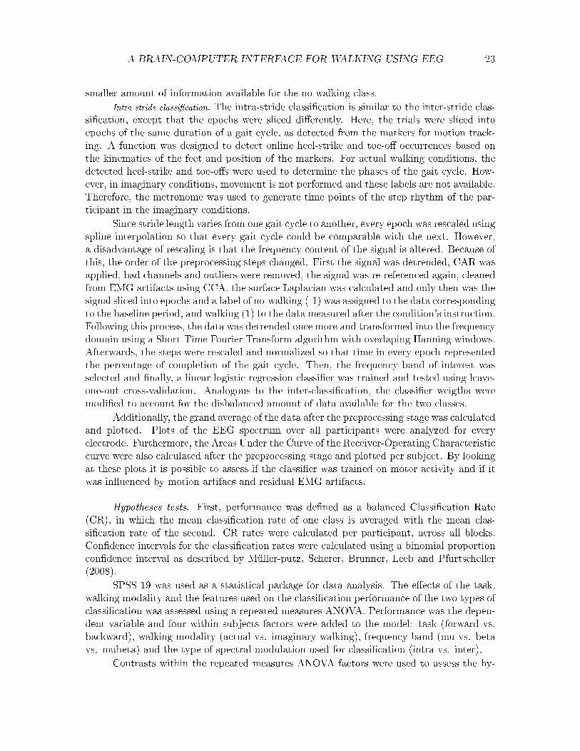
Figure 9. : Grand average spectral power in the mubeta band for the intra-stride modulationin Forward Walking. Intra-stride modulations can be observed during walking, especially inthe C2 and CP5 electrodes. Note that this is a plot relative to the baseline, and the colorscale represents relative changes.
Figure 9 shows the spectrum for forward walking plotted with a relative baseline. As canbe seen, during the walking class, there are regular modulations of power across the wholescalp, and they are specially visible at the C2 and CP5 electrodes. In contrast, these modu-lations are not present in the no-walking class. Interestingly, by eye inspection, intra-stridemodulations are more noticeable during forward walking than during backward walking.
On the other hand, the intra-stride modulations are not as clear for Imaginary Walk-ing. Figure 10 shows the case of Imaginary Forward Walking. Small modulations can be seennear the C4 and CP2 electrodes, approximatelly in phase with the modulations observed inactual walking conditions, but the magnitude of the power modulations across the scalp isalmost zero.
The AUCs of the spectral densities were also explored to assess in more detail thedi�erences in spectral power between the positive and the negative classess. These di�er-ences are the ones that the classi�er will probably consider, so they hint whether or notclassi�cation could use the ERSDs of interest. An AUC value of one is related to morepower in the walking class than in the no-walking one, and an AUC value of zero to morepower in the no-walking class than in the walking class.
Figure 11 shows the AUCs for Backward Walking. As can be observed from thepicture, the ERSD change was stronger around FCz in the beta band and C4 and CP5 inboth mu and beta bands.
Consistently with the inter-stride AUCs, the intra-stride version also showed that therelative change of power between walking and no-walking periods was bigger in the area ofthe CP4 electrode and its surroundings. Also, around CP5 the relative change was slighlyof higher magnitude than in other areas of the head.
Average classi�cation performance per subject per condition per spectral modulation

A BRAIN-COMPUTER INTERFACE FOR WALKING USING EEG 26
(a) No Walking (b) Walking
Figure 10. : Grand average spectral power in the mubeta band for the intra-stride modula-tion, Imaginary Forward Walking. Slight intra stride modulations can be observed speciallyin CP2 and CP4 in the walking period. Note that this is a plot relative to the baseline, andthe color scale represents relative change.
(a) Inter AUCs (b) Intra AUCs
Figure 11. : AUCs for backwards walking subject 5. Both Intra and inter analysis show thatthe relative change of power is more prominent around the C4, FCz and CP5 electrodes.

A BRAIN-COMPUTER INTERFACE FOR WALKING USING EEG 27
Figure 12. : O�ine classi�cation rates per subject for Imaginary Forward walking
and frequency band varied considerably between and within subjects (�gure 12). For theinter-stride version, averaged CRs ranged from 53% to 86%. Error bars in �gure 12 (left)also show that for two subjects classi�cation rates reached 90%. In these two conditons, forthe intra-stride classi�er, CRs also varied considerably, ranging from 52% to 82%, and themaximum CR trial-wise was around 89%, whereas the smallest CR was almost at chancelevel. In the inter-stride classi�cation, eight out of 12 participant had CRs signi�cantly abovechance level for all frequency bands, whereas for the intra-stride seven out of 12 performedabove chance level (95% con�dence intervals).
From these plots, it seems that CRs for intra-stride modulations are worse than thosefor the inter-stride. Figure 13 shows the average CRs per type of classi�er, grouped percondition. From here it can be observed that indeed the inter classi�cation seems to performbetter than the intra, but only in Imaginary conditions. For Actual conditions, intra-stridemodulations seem to give better performance. Furthermore, this pattern was consistent forall frequency bands. A repeated measures ANOVA revealed that this interaction of walkingmodality and spectral modulation had a signi�cant e�ect on classi�cation performance,F(1,11)=13.001, p<0.01, η2=0.0697.
As seen in �gure 13, regardless of the condition and type of classi�cation, classi�cationperformance for the three frequency bands is quite similar, although the mubeta band seemsto perform consistently better than the other two. Indeed, the main e�ect of the frequencyband used as a feature on performance was signi�cant, F(2,22)=6.039, p<0.5, η2=0.017.Pairwise comparisons revealed that the mu and the beta bands signi�cantly di�ered fromthe mubeta band, but not between each other.
In the inter-stride classi�cation, CR for Backward Walking is slightly higher comparedto Forward Walking, but not in the imaginary walking modality. For the intra-stride classi�-cation, this performance did not change much between task modalities. Di�erences betweenthe di�erent levels of task and spectral modality were not signi�cant (all p>0.05).
Results show that the main e�ect of the walking modality signi�cantly a�ected theperformance, F(1,11)=15.377, p<0.01, η2=0.288. This can also be con�rmed from the plotsin �gure 13, as actual walking was more easily detected in both classi�er types.

A BRAIN-COMPUTER INTERFACE FOR WALKING USING EEG 28
Figure 13. : O�ine classi�cation rates for Intra vs. Inter classi�cations in the three frequencybands
Conclusions
Classi�cation of walking versus no-walking was successfully implemented based onthe two types of spectral modulations proposed. Furthermore, classi�cation rates weresigni�cantly above chance for most subjects.
Because actual walking can elicit stronger modulations than imaginary walking, walk-ing modality was a factor that in�uenced performance signi�cantly, as expected. Di�erencesbetween di�erent frequency bands were also found. In most of the cases, the combined muand beta bands gave better results than did taking them separately. Contrary to the initialexpectations, no signi�cant di�erences between tasks were found, this is, backward walkingdid not di�er in classi�cation rate from forward walking.
As can be seen from the error bars in �gure 12, there was high variability within-subjects, and from �gure 13, variability was also large between-subjects. This is a wellknown problem in BCI, and might imply that even if a feature outperformed another, notall subjects can bene�t from this choice.
Despite these di�erences, a grand average of the EEG power spectra showed that bothtypes of modulations are located approximatelly on top of the motor cortex area and theposterior parietal area. Although both types of modulations are stronger on top of the motorcortex, the most prominent modulations were observed in areas related to hand movement.

A BRAIN-COMPUTER INTERFACE FOR WALKING USING EEG 29
7. Online BCI: simple versus complex walking
The previous experiment proved the feasibility of classi�ying EEG measurements intowalking or no walking for both actual and imaginary walking. The aim of this experimentwas to extend the �ndings of the previously implemented BCI to an online setup, and toassess the relationship between the performance of the BCI, the automaticity of the task(i.e., simple and complex walking), and three subjetive qualities, namely, sense of agency,learnability and satisfaction. Furthermore, an o�ine analysis of the data was performed, inorder to compare the classi�cation results with the previous experiment.
Methods
Experiment design. Two interdependent variables were considered for the online setupin a 2x2 within-subjects design: walking modality (i.e., actual vs. imaginary walking)and task (i.e., simple walking vs. walking at varying speeds). These four conditions werepresented in 16 blocks. In every block, each condition occurred once, in a counterbalancedorder. All participants went through a total of 16 repetitions of every condition (64 trialsin total). The �rst half of the blocks were used as input data to train the classi�cationfor every subject, and the second half were used to test the classi�cation performance ofthe BCI. During the second half, participants received feedback about the outcome of theclassi�cation.
Two blocks out of the eight blocks available for feedback were used as �challenge�blocks: during the imaginary conditions of that block, the participant was instructed toalternate between walking and no-walking at will. The purpose of this was twofold. First,it let the participant have a better feeling of the interface in a less controlled scenario.Second, it was used to prevent the participant from doing so without explicit instructiondue to skepticism that the interface is working. The order of the challenge blocks wascounterbalanced within the feedback blocks. Challenge trials were not included in the dataanalysis.
For o�ine performance analyses, an extra variable was included: frequency band ofthe selected features (i.e., mu and beta band). Dependent variables were classi�cation rate,as a technical measure of performance, and the sense of agency, satisfaction and learnabilityas part of the subjective assessment of the BCI.
Participants. Eleven healthy volunteers with no history of major lower limb injuryand no known neurological or locomotor de�cits participated and completed the �rst half ofthis study, and nine out of the 11 also completed the feedback blocks. The mean age of theparticipants was 25 (SD=2.19) years. Before the start of the experiment, the participantswere asked to read and sign a standard informed consent form, approved by the EthicalCommittee for Behavioral Scienti�c Research of the Faculty of Social Sciences, RadboudUniversity Nijmegen.
Experimental Setup. The experiment setup was similar to the setup of the previuousexperiment (as described in section 6 ), with some minor changes. The �rst di�erence wasthat instead of the whole Plugin-Gait Model, only seven markers were placed on the shoesof the participants: the RANK, RTOE, RHEE, LANK, LTOE and LHEE of the Plugin-Gait

A BRAIN-COMPUTER INTERFACE FOR WALKING USING EEG 30
Figure 14. : Trial overview. A trial started with a baseline (stand) period that lasted 10seconds, followed by instructions on screen about the next condition. Then, 9 secondswere required to start the treadmill. The subject walked or imagined himself walking for 30seconds and �nally 10 s were required to stop the treadmill during actual walking conditions.
Model were used. An additional marker was added in the left shoe, as assymetry betweenboth feet improved the online labeling of the Vicon Nexus software during the online setup.
Subjects walked on a programmable treadmill at a velocity programmed according tothe speed of the walking condition, facing a monitor screen placed 80 cm above the �oorand 150 cm away from de treadmill. In order to reduce movement artifacts, the same clipsetup was used as in the previous experiment (see �gure 5).
The control of the �ow of the experiment was programmed using Brainstream (DCC,Radboud University Nijmegen, http://www.brainstream.nu), and an application in Matlab(The Mathworks, Natick, MA).
Procedures. Four walking tasks were executed by the participants: simple walk-ing, imaginary simple walking, complex walking and imaginary complex walking. A trialoverview is shown in �gure 14. Every trial consisted of 10 seconds of standing (i.e., baselineperiod), followed by the presentation of the instruction on screen (1 second). In the sim-ple walking conditions the treadmill was then turned on at a constant velocity of 3 km/h.During complex walking the velocity of the treadmill changed four times. Both velocity andtime intervals for each velocity were randomized. Velocities varied from 2.5 km/h to 4.5km/h in steps of 0.5 km/h, time intervals varied from 6 s to 12 s. Together, the four velocityintervals lasted 35 s . The walking task lasted 30 seconds plus �ve seconds at the beginning,which is the time the treadmill takes to reach the desired speed. After the walking period,10 seconds were given to let the treadmill stop completely.
Experimental time course. The experiment consisted of three parts. During the �rstpart, the participant went through eight blocks of four conditions each. This part will bereferred as the 'o�ine training' of the experiment. The information obtained was used in thesecond part to build a classi�cation model to distinguish between walking and no-walking.This period (heretofore referred to as 'train classi�er'), lasted around 20 minutes in whichthe participant could take a rest. In the last part (called 'feedback'), the participant triedout the interface. Basically, the participant did the same task as in the o�ine training,but now on top of the visual stimuli, feedback was given about the classi�cation rate of theclassi�er. After each condition, participants completed a questionnaire of nine questionsabout sense of agency, satisfaction and learnability had to be completed. The feedback partof the experiment also consisted of eight blocks of four conditions each, and the particiapanthad an extra questionnaire after the 4th and 8th blocks. If requested by the participant, ashort break after the 4th feedback block was taken.

A BRAIN-COMPUTER INTERFACE FOR WALKING USING EEG 31
Analogous to the �rst experiment, participants received a description of the experi-ment, and signed a informed consent form. Then, the participant's height was measuredand the EMG electrodes, the EEG cap, and the re�ective markers were put in place. TheVicon system was calibrated by taking static video samples of the subject.
Next, participants were asked to walk on the treadmill for a few minutes to reach acomfortable step length while the experimenter adjusted a metronome speed to determinetheir cadence. Stride length was calculated using the speed of the treadmill (3 km/h) andtheir stepping frequency.
Finally, participants were instructed not to chew and to blink as little as possible dur-ing the trials. A set of written instructions for each condition was given to the participants.They were explicitly encouraged to imagine themselves (kinesthetic imagery) walking dur-ing the imaginary conditions. They were also instructed to synchronize their walking to thevisual stimuli, both in imaginary and actual walking. Before beginning the actual experi-ment, participants practiced the four conditions at least once, and as many times as theyrequested.
Measurements
A questionnaire of nine items was designed to measure sense of agency, satisfaction andlearnability. Each dimension was represented by three questions: a prototypical item andtwo synonyms. The prototypical question for sense of agency was based on the questionnairedesigned by Wegner, Sparrow and Winerman (2004). The satisfaction question was based ona usability questionnaire and was of the type �How satisfying was your experience with theinterface?�. The prototypical question for perceived learnability was also based on usabilityquestionnaires and was of the type �To what extent do you think you would need furtherpractice in order to master the interface or not?�
Participants answered the questionnaire in a continuous scale of 10 cm, once aftereach trial, and were explicitly asked to answer the questions thinking only about the trialthey just experienced.
Stimuli
• O�ine training
The visual stimuli consisted of a virtual treadmill, as depicted in �gure 15. In the baselineperiod (no-walking) it was shown as a static picture without colors (�gure 15-a). Duringthe walking period, the light stripes moved towards the participant in an animation loop.Stepping instructions were given as color cues. The central stripe of the treadmill �ashedin orange to indicate that a heel strike with the right foot should be made, and it �ashed inblue to indicate a left heel strike.
The distance between bars was adjusted to the step length of the participant and thefrequency of the orange-blue cues was set to the stepping frequency of the participant, asdetermined before the experiment (See previous section).
• Feedback
During the feedback part of the experiment, classi�cation rates were color coded on top of thevisual stimuli for stepping (�gure 16). The class predicted by the classi�er was determinedby the color of all stripes (orange was no-walking, blue was walking). The transparency ofthe bars was also modulated by the predicted probability that the class was indeed walking

A BRAIN-COMPUTER INTERFACE FOR WALKING USING EEG 32
(a) Static feedback. No walkingperiod
(b) Animation. Walking period
Figure 15. : Stimuli during the o�ine training part of the experiment. The leftmost �gureshows the virtual treadmill during the standing phase, without cues for stepping. The tworightmost �gures show two frames of the animation for walking. The stripes moved towardsthe user in an animation loop and the middle bars �ashed in orange when the participanthad to do a heel strike with his right foot, and blue when he had to do it with his left foot.
or not walking. The more transparent the color, the more close was the prediction to chancelevel. The more opaque the stripes became, the higher the probability the predicted classwas correct.
In the baseline period (no-walking) it was shown as a static picture with colors indi-cating the outcome of the classi�cation. During the walking period, the colored stripes wereanimated, and the same stepping cues were provided to the participant, in addition to theclassi�cation feedback (�gure 16).
Analysis
Since intra classi�cation was less e�ective for imaginary walking in the previous ex-periment, it was decided that for the online setup, only inter-stride classi�cation would beused. Also, after four pilots runs it became evident that intra-classi�cation was di�cult toimplement online using the Vicon Nexus 1.7.1 Software. Motion tracking of the feet usingthe Vicon system was not suitable for detecting heel strikes in real time. The labeling of themarkers was not done consistently and it had to be checked after the experiment. Further-more, the time required to train an intra-stride classi�er is approximately four times thatrequired for its inter-stride counterpart, which is lengthy for an online setup.
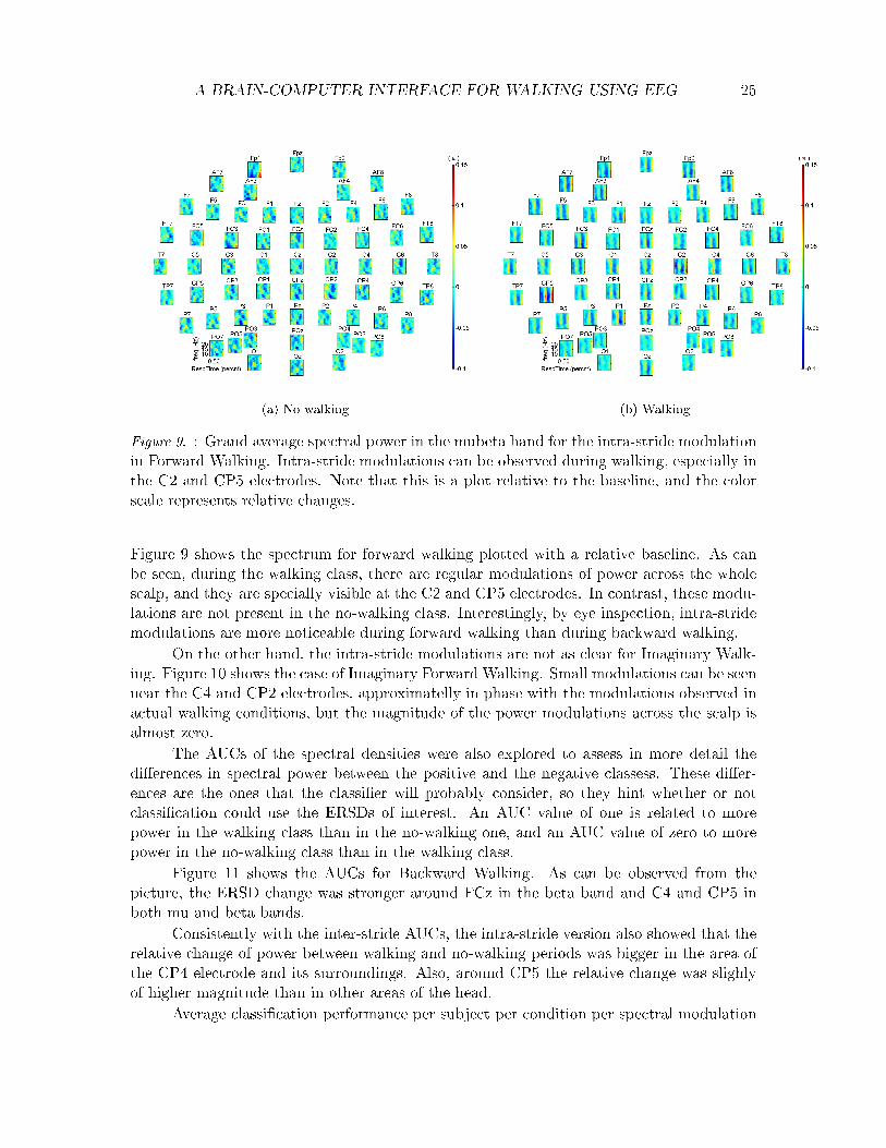
Online BCI implementation. The data analysis began in the second part of the ex-periment. The data gathered during the �rst eight blocks was used to train three classi�ers,one for each of the three proposed frequency bands: mu, beta and mubeta.
Afterwards, the three classi�ers were compared and the frequency band that gave thebest performance for that speci�c user was chosen and used during the online classi�cationshowed to the participant during the last eight blocks. In other words, the online classi�erused the classi�er recommended for that speci�c participant from his own data during theo�ine training part. This method was chosen because of the large di�erences in classi�cationrates between subjects during the previous experiment.
As in the previous experiment, the EEG data was �rst imported to Matlab usingthe BioSig toolbox (Graz Technical University) and was further processed using Matlab(Matworks Inc.). Additional pre-processing and classi�cation toolboxes were developed in

A BRAIN-COMPUTER INTERFACE FOR WALKING USING EEG 33
Figure 16. : Feedback Stimuli. During the feedback part of the experiment, the participantssaw the classi�cation rate color coded on top of the animation used to indicate the steppingrate. When all the stripes were orange, the classi�er guessed no-walking, and when blue,it guessed walking. Furthermore, the more transparent the stripes became, the less certainwas the classi�er of its classi�cation. In the top two �gures, no-walking was guessed, andin the top left, the predicted probability of no-walking was higher than in the top right.Analogously, in the bottom �gures, walking was guessed, and in the bottom left one theprobability of the person walking was higher than in the bottom right one.
house at the Donders Institute and can be provided upon request.The o�ine inter-stride classi�er developed during the previous analysis was used dur-
ing the train classi�er part of the experiment. The data used for o�ine classi�cation wasgathered during the �rst eight blocks. Next, the classi�er weights were extracted as a re-gression equation that could generate decision values for online classi�cation.
During the online experiment, data was recorded in 2.5 second segments. Each seg-ment was then analized following all preprocessing steps as described in �gure 7 for theinter-stride classi�cation. Finally, the decision value was calculated using the preprocesseddata and the classi�er weights extracted during the train classi�er analysis.
Each decision value was mapped to a p value ranging from 0 to 1 with the logisticfunction, where 0 represented the walking class, 1 the no-walking class and 0.5 chance level.In other words, p represents the probability that a piece of data belongs to one class oranother.
The p value was used to calculate the level of opacity of the stripes in the feedbackanimation, hence, the BCI cycle was complete and feedback was given to the user approxi-mately every 2.5 seconds.
Classi�cation rates were calculated by considering �rst the CR of the no-walking partof the trial (baseline), and then averaging it with the CR of the walking part (after theinstructions), which allowed for a balanced loss classi�cation rate to be obtained.
Subjective Assessment. The scores of each dimension of the questionnaire were ag-gregated into one measure. By averaging the three equivalent questions per questionnaire.

A BRAIN-COMPUTER INTERFACE FOR WALKING USING EEG 34
Afterwards, both the scores of the questionnaires and the classi�carion rates per trial weregrouped into conditions and averaged over blocks. Next, the e�ects of task and walkingmodality on the classi�cation performance, sense of agency, satisfaction and learnabilitywere assessed using a repeated measures ANOVA. Finally, the e�ects of performance oneach one of the dimensions of the questionnaire were assessed using pairs of correlations.
Classi�cation rates were also grouped per condition, averaged over participants, andplotted to inspect possible trends in the performance over blocks.
O�ine performance analysis. After the experiment, the training data was used tocheck the e�ects of task, walking modality and frequency band on the classi�cation rates.Also, the spectrograms and AUC's were inspected to check for correct artifact removal andthe localization of the ERDs used for classi�cation.
Results
First, results of the online performance analysis are given, as well as the results of theanalysis of the relationship between performance and the subjective measures. Next, resultsof the o�ine analysis of performance are given.
Online performance analysis and subjective assessment. Figure 17 shows the averageonline classi�cation rates per participant. Large error bars illustrate high within-subjectsvariations. Mean classi�cation rates per participant range from 52% to 96%. In average,classi�cation seems to be best for Simple walking, followed by Complex walking. The imagi-nary conditions seem to have similar classi�cation rates among them, although for Imaginarycomplex walking between-subjects variability is larger than for imaginary simple walking.Indeed, a trend was found in the e�ect of walking modality on performance: contrasts re-vealed that Actual walking has signi�cantly better performance than Imaginary walking,F(1,8)=3.766, p<0.05, η2=0.1128. However, the e�ects of the task on performance were notsigni�cant (p>0.05). From the three frequency bands available, the mu band was chosen asthe best feature option for 3 subjects (33%), the beta band for one (11%), and the mubetaband for �ve subjects (55%).
Sense of agency was rated, in average, as 75% for Actual walking conditions and 69%for imaginary conditions. Satisfaction was rated as 74% for actual walking conditions and69% for imaginary walking. Sense of agency was positively correlated with satisfaction inthe four conditions (p<0.01). Finally, learnability was rated as 52% for Actual walkingconditions and 50% for Imaginary conditions. A trend of the in�uence of tasks on the senseof agency was observed F(1,8)=4.163, p=0.076,η2= 0.018. However, e�ects of the task andwalking modality on the sense of agency were not signi�cant. For satisfaction, no e�ects oftask and walking modality were found. Finally, the e�ect of performance on the sense ofagency, satisfaction and learnability was assessed using correlation pairs - none of them wassigni�cant.
In order to explore possible learning trends re�ected in the classi�cation rates obtainedby the participants along blocks, CRs were grouped by condition and averaged over partici-pants. Figure 18 shows the resulting plot. Over the eight consecutive blocks a slight increasein performance for simple walking can be observed, whereas for complex walking classi�ca-tion rate seems to decrease. For the Imaginary simple walking condition, performance seemsto increase, but for complex walking the opposite e�ect can be seen.

A BRAIN-COMPUTER INTERFACE FOR WALKING USING EEG 35
Figure 17. : Online average classi�cation rates per participant and condition. The last grouprepresents the mean classi�cation rates overall participants.
Figure 18. : Learning trends. Online average classi�cation rates per block per condition.Error bars represent the standard deviation.
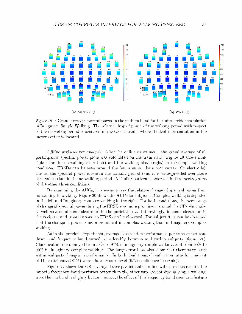
A BRAIN-COMPUTER INTERFACE FOR WALKING USING EEG 36
(a) No walking (b) Walking
Figure 19. : Grand average spectral power in the mubeta band for the inter-stride modulationin Imaginary Simple Walking. The relative drop of power of the walking period with respectto the no-walkig period is centered in the Cz electrode, where the feet representation in themotor cortex is located.
O�ine performance analysis. After the online experiment, the grand average of allparticipants' spectral power plots was calculated on the train data. Figure 19 shows mul-tiplots for the no-walking class (left) and the walking class (right) in the simple walkingcondition. ERSDs can be seen around the feet area on the motor cortex (Cz electrode),this is, the spectral power is less in the walking period (and it is widespreaded over moreelectrodes) than in the no-walking period. A similar pattern is observed in the spectrogramsof the other three conditions.
By examining the AUCs, it is easier to see the relative change of spectral power fromno walking to walking. Figure 20 shows the AUCs for subject 9, Complex walking is depictedin the left and Imaginary complex walking in the right. For both conditions, the percentageof change of spectral power during the ERSD was more prominent around the CPz electrode,as well as around some electrodes in the parietal area. Interestingly, in some electrodes inthe occipital and frontal areas, an ERSS can be observed. For subject 9, it can be observedthat the change in power is more prominent in complex walking than in Imaginary complexwalking.
As in the previous experiment, average classication performance per subject per con-dition and frequency band varied considerably between and within subjects (�gure 21).Classi�cation rates ranged from 58% to 97% in imaginary simple walking, and from 65% to99% in Imaginary complex walking. The large error bars also show that there were largewithin-subjects changes in performance. In both conditions, classi�cation rates for nine outof 11 participants (81%) were above chance level (95% con�dence intervals).
Figure 22 shows the CRs averaged over participants. In line with previous results, themubeta frequency band performs better than the other two, except during simple walking,were the mu band is slightly better. Indeed, the e�ect of the frequency band used as a feature
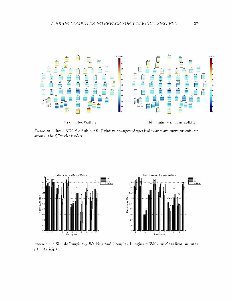
A BRAIN-COMPUTER INTERFACE FOR WALKING USING EEG 37
(a) Complex Walking (b) Imaginary complex walking
Figure 20. : Inter AUC for Subject 9. Relative changes of spectral power are more prominentaround the CPz electrodes.
Figure 21. : Simple Imaginary Walking and Complex Imaginary Walking classi�cation ratesper participant.
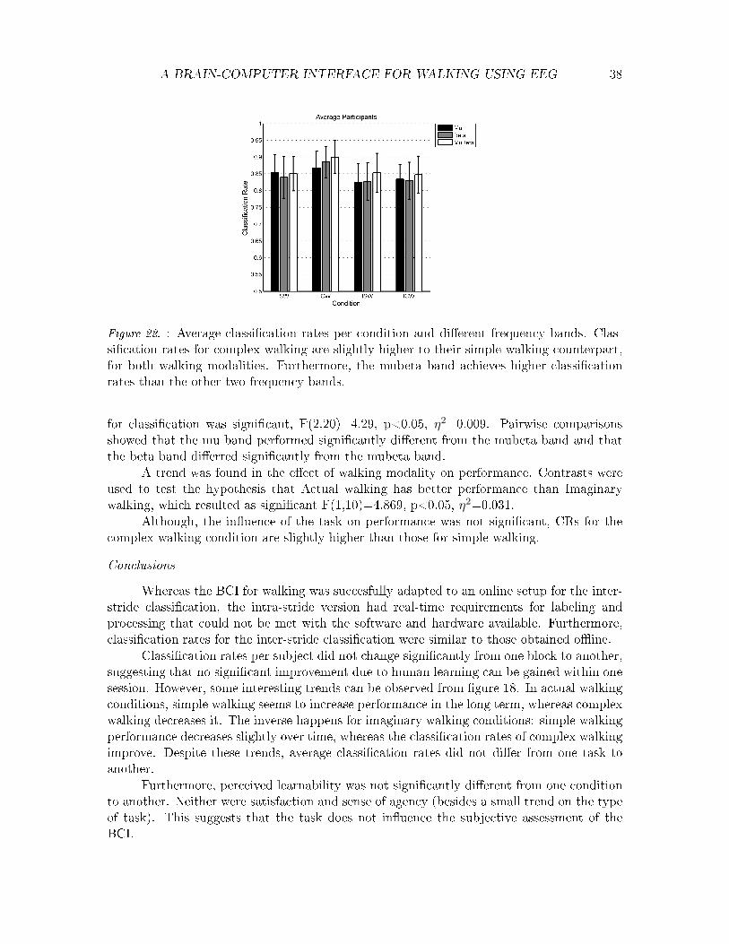
A BRAIN-COMPUTER INTERFACE FOR WALKING USING EEG 38
Figure 22. : Average classi�cation rates per condition and di�erent frequency bands. Clas-si�cation rates for complex walking are slightly higher to their simple walking counterpart,for both walking modalities. Furthermore, the mubeta band achieves higher classi�cationrates than the other two frequency bands.
for classi�cation was signi�cant, F(2,20)=4.29, p<0.05, η2=0.009. Pairwise comparisonsshowed that the mu band performed signi�cantly di�erent from the mubeta band and thatthe beta band di�erred signi�cantly from the mubeta band.
A trend was found in the e�ect of walking modality on performance. Contrasts wereused to test the hypothesis that Actual walking has better performance than Imaginarywalking, which resulted as signi�cant F(1,10)=4.869, p<0.05, η2=0.031.
Although, the in�uence of the task on performance was not signi�cant, CRs for thecomplex walking condition are slightly higher than those for simple walking.
Conclusions
Whereas the BCI for walking was succesfully adapted to an online setup for the inter-stride classi�cation, the intra-stride version had real-time requirements for labeling andprocessing that could not be met with the software and hardware available. Furthermore,classi�cation rates for the inter-stride classi�cation were similar to those obtained o�ine.
Classi�cation rates per subject did not change signi�cantly from one block to another,suggesting that no signi�cant improvement due to human learning can be gained within onesession. However, some interesting trends can be observed from �gure 18. In actual walkingconditions, simple walking seems to increase performance in the long term, whereas complexwalking decreases it. The inverse happens for imaginary walking conditions: simple walkingperformance decreases slightly over time, whereas the classi�cation rates of complex walkingimprove. Despite these trends, average classi�cation rates did not di�er from one task toanother.
Furthermore, perceived learnability was not signi�cantly di�erent from one conditionto another. Neither were satisfaction and sense of agency (besides a small trend on the typeof task). This suggests that the task does not in�uence the subjective assessment of theBCI.

A BRAIN-COMPUTER INTERFACE FOR WALKING USING EEG 39
Even though mean scores for sense of agency, satisfaction and learnability were, byeye, similar to mean classi�cation rates, they were not correlated to performance 1. In otherwords, changes in the sense of agency, satisfaction and learnability are not related to changesin performance.
Even though the sense of agency was not related to changes in performance, it isinteresting that the sense of agency did not change from actual walking to imaginary walking:this suggest that a link between one's intentions and the interface was created. Despitethe interesting results, it must be noted that the sample size was small, especially for thesubjective assessment.
Overall, the mubeta band achieved better performance than the mu and beta bandsseparately, con�rming results from the previous experiment. This result indicates thatthe di�erences among both bands are complementary and that the more information theclassi�er has, the better the achieved classi�cation rates.
As expected, Actual walking yielded better classi�cation results than imaginary walk-ing because modulations are stronger. However, this e�ect is small (about 3%). It is possiblethat the sample size (11 participants, o�ine analysis) was not large enough to �nd the e�ect.However, it is promising that the achieved performance in imaginary walking is similar tothat in actual walking.
The grand average of the spectrograms for this experiment showed an ERSD sur-rounding the Cz electrode where the feet area is located. This e�ect was con�rmed by theAUC plots, which showed that di�erences in spectral power between walking and no walkingperiods are larger around the CZ electrode. However, the AUC plots also showed increasesin spectral power in the walking periods with respect to the no-walking around frontal andoccipital electrodes and bigger in magnitude during actual walking conditons. This might berelated to residual movement artifacts causing di�erences between walking and no walkingespecially in the neck area. Since complex walking implies an actual movement, the di�er-ence between spectral power in walking and no-walking picked up in the back electrodes ismore prominent.
1The correlation could be overestimated because of the within-subjects design.

A BRAIN-COMPUTER INTERFACE FOR WALKING USING EEG 40
8. General discussion
The main goals of this study were to prove the feasibility of using walking imageryand EEG to implement a BCI for walking, and to determine which combination of tasks andfeatures could maximize the performance of the BCI while providing a good sense of agency,satisfaction and learnability. In order to achieve this, a BCI was successfully implementedusing di�erent combinations of features (espectral modulations and frequency bands), tasks(forward and backward walking) and walking modalities (actual and imaginary walking).The BCI was tested o�ine using the data from the �rst experiment. Afterwards, the sameprocessing pipeline was used to implement an online BCI for complex walking and simplewalking. During the online experiment, sense of agency, satisfaction and learnability wereassessed.
Despite the large variability among and within subjects, average classi�cation rateswere above chance level for most participants, demonstrating that we were successful in ourgoal of making a BCI for walking. Such variability should be considered when designing newBCIs, in order to choose the best combination of relevant features for every subject. Forinstance, the online version of the BCI was based on the best frequency band per subject.Among the aforementioned frequency bands, the mu and beta bands together gave betterresults than when tested separately. A possible explanation is that the more informationavailable, the better the classi�er can discriminate between the two classes.
An initial concern was the measurement of EEG during walking and the removal ofartifacts such as EMG and artifacts induced by movement. In order to partially solve thisissue, CCA was applied to the data at a preprocessing stage, with satisfactory results as canbe seen in �gure 9, this process was fast and worked well enough.2 Usually, EMG artifactsare high frequency components coupled to the gait cycle, similar to the EEG. However, inthis plot it can be seen that the signals detected by most occipital electrodes are �lteredand that the modulations picked up by the EEG are clearly represented above the motorand the parietal cortex.
Even though CCA was useful in removing EMG artifacts, some artifacts might remaindue to movement, as shown by the di�erences between the spectrograms and AUCs ofwalking and no-walking in the occipital electrodes in both experiments. As Severens et al.(2012) already suggested, an alternative would be to track head movements and uncorrelatethem from the EEG signal. This could be considered for further analysis of the current data.A second alternative would be to train the classi�er using less and more central electrodes toensure that only signals of interest are being considered for classi�cation. Previous studieshave already showed that, with enough training, a person is capable of adjusting his mentalactivity to control a BCI even with only one electrode (Wolpaw & McFarland, 2004).
In order to select the important electrodes for classi�cation, the localization of theERSDs of interest is also important. Our results suggest that cortical activity varies along
2Note: care must be taken in interperating the o�-line results as they may be over-estimated as the EMGremoval �lter was estimated on all the data before the classi�ers cross-validation loop was used to estimateperformance. This may lead to a double-dipping problem. According to Kriegeskorte et al. (2009), doubledipping is the use of the same data set for selection and selective analysis which often yields overestimatedclassi�cation rates. However, whilst a concern, as the on-line results (where this is not a problem) weresimilar to the o�-line results we do not believe this is a signi�cant issue. Follow-up studies should take thisinto account and include the CCA inside a nested cross-validation for o�ine analysis.

A BRAIN-COMPUTER INTERFACE FOR WALKING USING EEG 41
the gait cycle (as previously described by Gwin and colleagues (2011) and Severens et al.(2012)), and that these variations are located approximately in the vicinity of the primarymotor cortex and the posterior parietal areas. Posterior parietal cortex has been linked toselective and rhythmic attention, combination of sensorimotor and visuomotor information,and bimanual coordination (Gwin et al., 2011; Lange et al., 2005).
Another promising result is that the achieved performance in imaginary walking issimilar to that of actual walking, which con�rms theories that motor imagery is representedas an actual movement, to some extent (Lange et al., 2005). This is, participants were ableto achieve classi�cation rates of 70% in average for imaginary conditions within one session.Therefore, using motor imagery of walking, being a more intuitive movement, results inbetter classi�cation performance when compared to the use of other imaginary movementssuch as simple hand and feet movement as the movements used by Pfurtscheller, Leeb, etal. (2006).
Despite both types of spectral modulations were stronger in electrodes located on topof the motor cortex, the most prominent changes in the forward versus backward experi-ment were usually located in areas related to hand movement. This might be because therepresentation of one foot in the brain is opposite to the other, and the projection of thecurrent dipoles generated by them is attenuating each other and thus, the di�erences of po-tential detected by the EEG are those that are projected with di�erent angles (Pfurtscheller,Brunner, et al., 2006). Another possibility is that walking is a whole-body movement thatis not limited to the feet and, therefore, hand movement might be playing a role in theclassi�cation. Indeed, a potential disadvantage of the inter-stride spectral modulations isthat it is based on a motor ERSD, which any kind of movement can generate. Therefore, byusing inter-stride modulations there is no guarantee that the BCI is really working for footimagery. In contrast, intra-stride modulations are coupled to the gait cycle, but are muchweaker in magnitude than the inter-stride modulation, and resultingly are more di�cult tomeasure.
Although no signi�cant di�erences between the two types of classi�cation were found,the interaction of spectral modulation type and walking modality was signi�cant: intra-stride classi�cation seems to work better for Actual walking, and inter-stride classi�cationfor Imaginary walking. This might be because the subject did not perform the task correctly.Probably people are unable to imagine every phase of the gait cycle while keeping the stepfrequency of the metronome (or the visual stimuli, in the second experiment). Thus, intra-stride modulations are not clear during imaginary conditions. Furthermore, during actualwalking conditions the phases of the gait cycle were reliably identi�ed using the Viconre�ective markers and, in contrast, during imaginary walking conditions it was up to theability of the participant to synchronize every step with the metronome. If the stepping ratewas not locked to the stimuli, then the intra stride modulations would be dephased fromeach other as well, and would be cancelled out during the averaging. This may inidcatethat performance could be better with better trained subjects who can imagine detailed feetmovements according to a prede�ned rhythm.
Advantages and disadvantages of both types of spectral modulations during walkingcan be considered when designing a BCI for rehabilitation. Intra-stride modulations werebetter for classifying actual walking, whereas inter-stride modulations were better for imagi-nary walking. This �nding suggests that an inter-stride modulated BCI could be used at the

A BRAIN-COMPUTER INTERFACE FOR WALKING USING EEG 42
beginning of the rehabilitation process to detect any kind of imagery or attempted movementthat the patient can elicit. In this way, the timeless inter-stride modulations will help thepatient in the early stages of rehabilitation, and as his motor control improves, the practicecould be tuned with intra-stride modulations that could give time-sensitive feedback andconsider individual phases of the gait cycle. Further research should be conducted in orderto improve both types of classi�cation and to match their special qualities to develop a BCIthat could help patients relearn to walk. For example, a patient su�ering from a strokewould have limited -if any- motor abilities in his legs. In order to recover this function-ality, the rehabilitation process suggests constant practice. Right after the stroke, motoractivity in the brain would be limited, therefore, inter-stride modulations would be suitableto detect ERSDs related to attempted movements, even if the attempt was not accurate.As the training process goes on, the patient will gain mastery in imagining movements andhe will regain movement as well, little by little. Once at this stage, the patient could usethe intra-stride version of the BCI to practice every step accoring to the gait cycle. At thelatest stages of the rehabilitation, if the patient can already produce some movements, theproposed BCI could also be used to give feedback during actual walking.
For the rehabilitation process, it is important that a BCI provide senses of agency,enabling the patient to attribute the BCI feedback to his own e�orts. The results of this ex-periment also showed that the sense of agency did not di�er much between di�erent walkingmodalities: although a change in feedback is caused just by imagining a movement, to someextent, participants attributed the change to themselves. This phenomenon is evidence insupport of using motor imagery for rehabilitation. Imaginary and actual walking could beconsidered as interchangeable, and their consequences in the world are both attributed tothe self will power (Wegner et al., 2004; Gallagher, 2000).
Other e�ects of task and performance on the sense of agency, satisfaction and learn-ability were not signi�cant. Even within the few signi�cant results described above, thee�ect sizes were relatively small. The �rst possible explanation is the small sample size usedduring this experiment. Another limitation of this study was that the questionnaire usedwas not validated beforehand. It would have been bene�tial to test more subjects or to domore pilot runs to test the questionnaires. However, given the length of the experiments andthe time available for this project, a whole validation of the questionnaire and methodologywould not have been possible.
Given that setting up a BCI and testing it is time-consuming, and tiring for end-users,an alternative could be to simulate the BCI application, so that it can be tested quicklyand improved according to the user's needs during the design process. For instance, Boland,Quek, Tangermann and Williamson (2011) suggested performing fast prototyping of BCIsusing simulations of the expected BCI accuracy. By using these simulations, experiment setup times could be reduced and therefore, times between each iteration of the design cyclecould be optimized.
Future work could also include decoding of the direction of the movement or thewalking speed. Direction could be decoded by looking at the lateralization of the inter-stride modulations, whereas the speed could be inferred from the intra-stride modulations.
The high o�ine classi�cation rates achieved for forward and backward walking wereconsistent with the ones obtained in the o�ine and online analysis of the simple and complexwalking. This suggest that the BCI is robust for di�erent walking tasks and modalities.

A BRAIN-COMPUTER INTERFACE FOR WALKING USING EEG 43
9. Conclusions
The present study proved the feasibility of decoding walking and walking imageryusing EEG. Two di�erent types of ERSPs were used to distinguish walking from no-walkingin both actual walking and imaginary walking. Furthermore, an online BCI for walkingwas implemented successfully with classi�cation rates analogous to the o�ine classi�cation.Importantly, the results were robust for di�erent levels of automaticity in the walking task(simple walking, complex walking, backwards walking).
In order to maximize the system performance, several tasks and features were cho-sen and compared: task (simple and complex walking), spectral modulations (inter-stridemodulations and intra-stride modulations) and frequency band (mu, beta and mubeta). Themubeta band improved performance when compared with the mu and beta bands separately.However, no signi�cant di�erences were found between either the automaticity of walkingor the spectral modulations. Only the interaction between walking modality and spectralmodulations was signi�cant for the �rst experiment, showing that the inter-stride modula-tions are better at classi�ying during imaginary walking while the intra-stride modulationsare better at classifying during actual walking.
The interactions between task, performance and three subjective measures were alsoassessed. However, no signi�cant e�ects of task and performance on the sense of agency,satisfaction and learnability were found, probably due to the small sample size.
Recommendations for the design of a BCI for rehabilitation were also given. Findingsof the current research suggest that the intra-stride and inter-stride classi�cation methodscould constitute two phases of patient rehabilitation. Inter-stride modulations could helpduring early stages in the rehabilitation process, whereas intra-stride could be more usefulto �ne tune practice according to the gait cycle phases. Furthermore, the mubeta bandproved to be more suitable to achieve high classi�cation performance. However, due to thehigh variability between subjects, it is recommended to choose the best frequency band peruser.
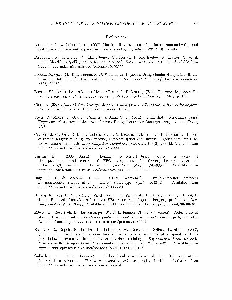
A BRAIN-COMPUTER INTERFACE FOR WALKING USING EEG 44
References
Birbaumer, N., & Cohen, L. G. (2007, March). Brain-computer interfaces: communication andrestoration of movement in paralysis. The Journal of physiology , 579 (Pt 3), 621�36.
Birbaumer, N., Ghanayim, N., Hinterberger, T., Iversen, I., Kotchoubey, B., Kübler, A., et al.(1999, March). A spelling device for the paralysed. Nature, 398 (6725), 297�298. Available fromhttp://www.ncbi.nlm.nih.gov/pubmed/10192330
Boland, D., Quek, M., Tangermann, M., & Williamson, J. (2011). Using Simulated Input into Brain-Computer Interfaces for User-Centred Design. International Journal of Bioelectromagnetism,13 (2), 86�87.
Buxton, W. (2001). Less is More ( More or Less ). In P. Denning (Ed.), The invisible future: Theseamless integration of technology in everyday life (pp. 145�179). New York: McGraw Hill.
Clark, A. (2003). Natural-Born Cyborgs: Minds, Technologies, and the Future of Human Intelligence(Vol. 29) (No. 3). New York: Oxford University Press.
Coyle, D., Moore, J., Ola, P., Paul, K., & Alan, C. F. (2012). I did that ! Measuring Users'Experience of Agency in their own Actions Trinity Centre for Bioengineering. Austin, Texas,USA..
Cramer, S. C., Orr, E. L. R., Cohen, M. J., & Lacourse, M. G. (2007, February). E�ectsof motor imagery training after chronic, complete spinal cord injury. Experimental brain re-search. Experimentelle Hirnforschung. Expérimentation cérébrale, 177 (2), 233�42. Available fromhttp://www.ncbi.nlm.nih.gov/pubmed/16944108
Curran, E. (2003, April). Learning to control brain activity: A review ofthe production and control of EEG components for driving brain-computer in-terface (BCI) systems. Brain and Cognition, 51 (3), 326�336. Available fromhttp://linkinghub.elsevier.com/retrieve/pii/S0278262603000368
Daly, J. J., & Wolpaw, J. R. (2008, November). Brain-computer interfacesin neurological rehabilitation. Lancet neurology , 7 (11), 1032�43. Available fromhttp://www.ncbi.nlm.nih.gov/pubmed/18835541
De Vos, M., Vos, D. M., Riès, S., Vanderperren, K., Vanrumste, B., Alario, F.-X., et al. (2010,June). Removal of muscle artifacts from EEG recordings of spoken language production. Neu-roinformatics, 8 (2), 135�50. Available from http://www.ncbi.nlm.nih.gov/pubmed/20480401
Elbert, T., Rockstroh, B., Lutzenberger, W., & Birbaumer, N. (1980, March). Biofeedback ofslow cortical potentials. I. Electroencephalography and clinical neurophysiology , 48 (3), 293�301.Available from http://www.ncbi.nlm.nih.gov/pubmed/6153349
Enzinger, C., Ropele, S., Fazekas, F., Loitfelder, M., Gorani, F., Seifert, T., et al. (2008,September). Brain motor system function in a patient with complete spinal cord in-jury following extensive brain-computer interface training. Experimental brain research.Experimentelle Hirnforschung. Expérimentation cérébrale, 190 (2), 215�23. Available fromhttp://www.springerlink.com/content/c8015l41hl558313/
Gallagher, I. (2000, January). Philosophical conceptions of the self: implicationsfor cognitive science. Trends in cognitive sciences, 4 (1), 14�21. Available fromhttp://www.ncbi.nlm.nih.gov/pubmed/10637618
A BRAIN-COMPUTER INTERFACE FOR WALKING USING EEG 45
Gerven, M. van, Farquhar, J., Schaefer, R., Vlek, R., Geuze, J., Nijholt, A., et al. (2009, August).The brain-computer interface cycle. Journal of neural engineering , 6 (4), 041001. Available fromhttp://www.ncbi.nlm.nih.gov/pubmed/19622847
Grasso, R., Bianchi, L., Lacquaniti, F., Owen, E., Cappellini, G., Ivanenko, Y. P., et al. (1998). MotorPatterns for Human Gait : Backward Versus Forward Locomotion. Journal of Neurophysiology ,1868�1885.
Gwin, J. T., Gramann, K., Makeig, S., & Ferris, D. P. (2010, June). Removal of movement artifactfrom high-density EEG recorded during walking and running. Journal of neurophysiology , 103 (6),3526�34. Available from http://www.ncbi.nlm.nih.gov/pubmed/20410364
Gwin, J. T., Gramann, K., Makeig, S., & Ferris, D. P. (2011, January). Electrocortical activityis coupled to gait cycle phase during treadmill walking. NeuroImage, 54 (2), 1289�96. Availablefrom http://www.ncbi.nlm.nih.gov/pubmed/20832484
Halligan, P. V., Marshall, J. C., Wade, D. T. W., Davery, J., & Morrison, D. (1993). Thumbin cheek? Sensory reorganization and perceptual plasticity after limb amputation. Neuroreport ,233�236.
Kriegeskorte, N., Simmons, W. K., Bellgowan, P. S. F., & Baker, C. I. (2009). Circular analysis insystems neuroscience - the dangers of double dipping. , 12 (5), 535�540.
Lange, F. P. de, Hagoort, P., & Toni, I. (2005, January). Neural topography and content ofmovement representations. Journal of cognitive neuroscience, 17 (1), 97�112. Available fromhttp://www.ncbi.nlm.nih.gov/pubmed/15701242
Mayr, A., Ko�er, M., Quirbach, E., Matzak, H., Fröhlich, K., & Saltuari, L. (2007). Prospec-tive, blinded, randomized crossover study of gait rehabilitation in stroke patients using theLokomat gait orthosis. Neurorehabilitation and neural repair , 21 (4), 307�14. Available fromhttp://www.ncbi.nlm.nih.gov/pubmed/17476001
Millán, J. R., Renkens, F., Mouriño, J., & Gerstner, W. (2004). Non-Invasive Brain-ActuatedControl of a Mobile Robot by Human EEG. IEEE Transactions on Biomedical Engineering , 51 ,1026�1033.
Mulder, T. (2007, January). Motor imagery and action observation: cognitive tools for rehabilitation.Journal of neural transmission (Vienna, Austria : 1996), 114 (10), 1265�78.
Müller, K., Krauledat, M., Dornhege, G., Curio, G., & Blankertz, B. (2004). Machine LearningTechniques For Brain-Computer Interfaces. Biomedical Technology , 49 , 11�22.
Müller-putz, G. R., Scherer, R., Brunner, C., Leeb, R., & Pfurtscheller, G. (2008). Better thanrandom ? A closer look on BCI results. International Journal of Bioelectromagnetism, 10 (1),52�55.
Müller-Putz, G. R., Zimmermann, D., Graimann, B., Nestinger, K., Korisek, G., & Pfurtscheller,G. (2007, March). Event-related beta EEG-changes during passive and attemptedfoot movements in paraplegic patients. Brain research, 1137 (1), 84�91. Available fromhttp://www.ncbi.nlm.nih.gov/pubmed/17229403
Neuper, C., Scherer, R., Reiner, M., & Pfurtscheller, G. (2005, December). Imageryof motor actions: di�erential e�ects of kinesthetic and visual-motor mode of imagery insingle-trial EEG. Brain research. Cognitive brain research, 25 (3), 668�77. Available fromhttp://www.ncbi.nlm.nih.gov/pubmed/16236487

A BRAIN-COMPUTER INTERFACE FOR WALKING USING EEG 46
Peters, E. (2011). Measurement of brain activity under di�erent walking circumstances on a treadmill(Tech. Rep.). Nijmegen: Sint Maatenskliniek.
Pfurtscheller, G., Brunner, C., Schlögl, a., & Lopes da Silva, F. H. (2006, May). Mu rhythm(de)synchronization and EEG single-trial classi�cation of di�erent motor imagery tasks. Neu-roImage, 31 (1), 153�9. Available from http://www.ncbi.nlm.nih.gov/pubmed/16443377
Pfurtscheller, G., Leeb, R., Keinrath, C., Friedman, D., Neuper, C., Guger, C., et al. (2006,February). Walking from thought. Brain research, 1071 (1), 145�52. Available fromhttp://www.ncbi.nlm.nih.gov/pubmed/16405926
Pfurtscheller, G., Stancák, A., & Neuper, C. (1996, November). Event-related synchronization (ERS)in the alpha band�an electrophysiological correlate of cortical idling: a review. International jour-nal of psychophysiology : o�cial journal of the International Organization of Psychophysiology ,24 (1-2), 39�46. Available from http://www.ncbi.nlm.nih.gov/pubmed/8978434
Prasad, G., Herman, P., Coyle, D., McDonough, S., & Crosbie, J. (2010, January). Applying abrain-computer interface to support motor imagery practice in people with stroke for upper limbrecovery: a feasibility study. Journal of neuroengineering and rehabilitation, 7 (1), 60.
Russell, S., & Norving, P. (2010). Arti�cial Intelligence (Third edit ed.). New Jersey: Pearson.
Sadato, N., Pascual-Leone, a., Grafman, J., Deiber, M. P., Ibañez, V., & Hallett, M. (1998, July).Neural networks for Braille reading by the blind. Brain : a journal of neurology , 121 ( Pt 7 ,1213�29. Available from http://www.ncbi.nlm.nih.gov/pubmed/9679774
Severens, M., Nienhuis, B., Desain, P., & Duysens, J. (2012). Feasibility of measuring Event RelatedDesynchronization with Electroencephalography during Walking. In Ieee engineering in medicine& biology society.
Vries, S. de, & Mulder, T. (2007, January). Motor imagery and stroke rehabilitation:a critical discussion. Journal of rehabilitation medicine : o�cial journal of the UEMSEuropean Board of Physical and Rehabilitation Medicine, 39 (1), 5�13. Available fromhttp://www.ncbi.nlm.nih.gov/pubmed/17225031
Wegner, D. M., Sparrow, B., & Winerman, L. (2004, June). Vicarious agency: experiencing con-trol over the movements of others. Journal of personality and social psychology , 86 (6), 838�48.Available from http://www.ncbi.nlm.nih.gov/pubmed/15149258
Whittle, M. W. (1996). Normal Gait. In Gait analysis, an introduction (pp. 53�107). Oxford:Butterworth Heinemann.
Wolpaw, J. R., Birbaumer, N., McFarland, D. J., Pfurtscheller, G., & Vaughan, T. M. (2002, June).Brain-computer interfaces for communication and control. Clinical neurophysiology : o�cialjournal of the International Federation of Clinical Neurophysiology , 113 (6), 767�91. Availablefrom http://www.ncbi.nlm.nih.gov/pubmed/12048038
Wolpaw, J. R., & McFarland, D. J. (2004, December). Control of a two-dimensional movementsignal by a noninvasive brain-computer interface in humans. Proceedings of the National Academyof Sciences of the United States of America, 101 (51), 17849�54.

A BRAIN-COMPUTER INTERFACE FOR WALKING USING EEG 47
Appendixes
A. Forward versus Backward Walking
Instruction forms . Welcome and thank you for participating. In this experimentwe are looking at EEG brain signal di�erences between forward and backward walking.Therefore, I will ask you to walk into this treadmill in both directions. This research will behelpful for rehabilitation as well as for Brain-Computer Interfaces, this is, people could movevirtual avatars, prosthesis, etc. with their minds. This is another reason why you will alsobe asked to imagine yourself walking backwards and forward while we take the appropriatemeasurements.
You will use an electrode cap, which has some electrodes on it. After you put it on,each electrode will be �lled with gel, in order to improve conductivity. You will also berequired to wears shorts, in order to place re�ective markers on several places from the hipsto your feet. In the schema below you can see the location of your body where the markerswill be placed. As you can see, the references are your bones, so the researchers will locatethe appropriate bones �rst, they will make some crosses where I'll put them later. Fourelectrodes will also be placed in your neck to measure electrical signals from your muscles,this will help us to reduce noise in the EEG signal.
In light of this explanation, please sign the informed consent form in next page. Withthis form you acknowledge that you know your basic rights as a participant: the datafrom this experiment, including the video recording, will be treated with con�dentiality andrespect and it will only be used for research purposes. If at any point during the experimentyou do not want to continue, you can quit without consequences. If you don't have anymore questions I would like to ask you to read the form and sign it.

MODEL 1
INFORMED CONSENT*
for participation in the scientific study:
Sensorimotor cortical activity during human gait: forward versus backward walking
- I am satisfied with the information about this experiment. I have read the written information (version code: 1)
carefully. I was given the opportunity to ask questions about the experiment, and my questions were answered
to my satisfaction. I have carefully considered my participation in the experiment. I understand that I have the
right to withdraw my participation in the experiment at any moment without having to give a justification.
- I consent with participation in this experiment.
- I consent that the data collected during this experiment and made anonymous can be used for
publication.
Name :
Date of birth :
Signature : Date:
-------------------------------------------------------------------------------------------------------------------------------------
- Signatory declares that the person named above has been informed both in writing and in person about the
experiment. Also, he/she declares that the participant may terminate participation in the experiment at any time
without consequence.
Name :
Function :
Signature : Date:
-------------------------------------------------------------------------------------------------------------------------------------
* This form is intended for research on people of 18 years and older able to give informed consent. In this type of research,
it is mandatory for the person concerned to give his or her consent personally.

A BRAIN-COMPUTER INTERFACE FOR WALKING USING EEG 49
General instructions
Please stand on the treadmill and look to the �xation point on the screen. Duringthe trial, please blink as less as possible, and please don't chew. Also, please try to keepyour head as still as possible, this is, try NOT to move much. This is important becausethe described movements (blinking, chewing and moving the head) would create noise in therecordings.
At the beginning you will see a green cross, please stand still and wait for the instruc-tion on screen. Four conditions are possible, and for each one of them you will have to doas described below.
Forward Walking
You will see an instruction on the screen. When the black �xation cross appears thetreadmill will start moving. Please start walking and look at the �xation cross also on thescreen. Try to keep your head still and your stride length constant. This is, the size of yoursteps should remain more-less the same. You will hear a metronome during the whole trial,which has been adapted to your walking speed. Try to synchronize your toe o� with the tickof the metronome, this is, every time you hear a tick, try to lift your foot from the ground.
Backward Walking
Please turn around. You will see an instruction on the screen. When the black �xationcross appears the treadmill will start moving. Please start walking and focus on the �xation-cross presented on screen. Try to keep your head still and your stride length constant. Thisis, the size of your steps should remain more-less the same. You will hear a metronomeduring the whole trial, which has been adapted to your walking speed. Try to synchronizeyour toe touching the ground with the tick of the metronome.
Imaginary Forward Walking
A black �xation cross will appear right after the instruction on screen. Then, startimagining that you are walking while looking at the �xation point on the screen. You willhear a metronome with two di�erent ticks. The loud tick will correspond to the left foot, andthe soft one to the right foot. You can use the metronome as a guide to imagine that yourfeet are touching the ground, one after the other. Imagine that you are moving your feetand legs as in the forward walking trial. One step after the other, imagine the rhythm youused, the constant stride length. Please imagine that you are walking in �rst person, this is,imagine the movement as if you were walking on the treadmill as in a real movement (notjust imagining looking at yourself walking). Please remember that this is just imaginarywalking, so don't contract or move any muscles at all.
Imaginary Backward Walking
Please turn around and stand on the treadmill. A black �xation cross will appear rightafter the instruction on screen. Then, start imagining that you are walking while looking atthe �xation point on the screen. You will hear a metronome with two di�erent ticks. Theloud tick will correspond to the left foot, and the soft one to the right foot. You can use themetronome as a guide to imagine that your feet are touching the ground, one after the other.Imagine that you are moving your feet and legs as in the backward walking trial. One step

A BRAIN-COMPUTER INTERFACE FOR WALKING USING EEG 50
after the other, imagine the rhythm you used, the constant stride length. Please imaginethat you are walking in �rst person, this is, imagine the movement as if you were walking onthe treadmill as in a real movement (not just imagining looking at yourself walking). Pleaseremember that this is just imaginary walking, so don't contract or move any muscles at all.
A BRAIN-COMPUTER INTERFACE FOR WALKING USING EEG 51
B. Simple versus Complex Walking
Instruction forms . Welcome and thank you for participating. In this experimentwe are looking at EEG brain signal di�erences between walking and no walking in fourdi�erent conditions. Therefore, I will ask you to walk into this treadmill. This research willbe helpful for rehabilitation as well as for Brain-Computer Interfaces, this is, people couldmove virtual avatars, prosthesis, etc. with their minds. This is another reason why you willalso be asked to imagine yourself walking while we take the appropriate measurements. Theexperiment will have three parts. During the �rst, we will gather some information of howyour EEG looks like during walking, so that in the second part, the computer can build amodel to distinguish between walking and no walking. In the second part, you do not haveto do anything, so it is break time. During the last part, you will have the opportunity totry out the interface. By doing the same task as in the �rst part, the computer will tryto guess if you are walking or not, or if you are imagining yourself walking or not. Aftereach condition, we will ask some questions about what do you think and how do you likethe interface. You will use an electrode cap, which has some electrodes on it. After youput it on, each electrode will be �lled with gel, in order to improve conductivity. You willalso be required to wear re�ective markers on your shoes or bare feet (this is your choice).Four electrodes will also be placed in your neck to measure electrical signals from yourmuscles, this will help us to reduce noise in the EEG signal. In light of this explanation,please sign the informed consent form in next page. With this form you acknowledge thatyou know your basic rights as a participant: the data from this experiment, including thevideo recording, will be treated with con�dentiality and respect and it will only be used forresearch purposes. If at any point during the experiment you do not want to continue, youcan quit without consequences. If you don't have any more questions I would like to askyou to read the form and sign it.

MODEL 1
INFORMED CONSENT*
for participation in the scientific study:
Brain-Computer Interface for Walking
- I am satisfied with the information about this experiment. I have read the written information (version code: 1)
carefully. I was given the opportunity to ask questions about the experiment, and my questions were answered
to my satisfaction. I have carefully considered my participation in the experiment. I understand that I have the
right to withdraw my participation in the experiment at any moment without having to give a justification.
- I consent with participation in this experiment.
- I consent that the data collected during this experiment and made anonymous can be used for
publication.
Name :
Date of birth :
Signature : Date:
-------------------------------------------------------------------------------------------------------------------------------------
- Signatory declares that the person named above has been informed both in writing and in person about the
experiment. Also, he/she declares that the participant may terminate participation in the experiment at any time
without consequence.
Name :
Function :
Signature : Date:
-------------------------------------------------------------------------------------------------------------------------------------
* This form is intended for research on people of 18 years and older able to give informed consent. In this type of research,
it is mandatory for the person concerned to give his or her consent personally.
A BRAIN-COMPUTER INTERFACE FOR WALKING USING EEG 53
General instructions
Please stand on the treadmill and look to the middle of the virtual treadmill on thescreen. During the trial, please blink as less as possible, and please don't chew. Also, pleasetry to keep your head as still as possible, this is, try NOT to move much. This is importantbecause the described movements (blinking, chewing and moving the head) would createnoise in the recordings.
At the beginning you will see a static version of the treadmill, please stand still andwait for the instruction on screen. This instruction will describe which of the four possibleconditions is next. For each one of them, you will have to do as described below.
Simple Walking
You will see an instruction on the screen. When the virtual treadmill appears, thetreadmill will start moving as well. In the treadmill you will see that two of the stripes arechanging colors. Please start walking and try to synchronize your steps with the colors onthe screen. When the orange stripe appears, please try to do a heel strike with your rightfoot, this is, every time you see the orange stripe appear, try to touch the ground with yourright heel. Similarly, when the blue stripe appears, please try to do a heel strike with yourleft foot. Try to keep your head still, and look to the same point during the whole trial.
Complex Walking
You will see an instruction on the screen. The mechanism will be de same as in simplewalking, with the sole di�erence that the velocity of the treadmill will be changing. Afterthe name of the condition, the virtual treadmill appears, the treadmill will start moving aswell. Try to synchronize your heel strikes of the right foot with the orange color, and theheel strikes of the left foot with the blue color. Again, try to keep your head still, and lookto the same point during the whole trial.
Imaginary Simple Walking
A virtual treadmill will appear right after the instruction on screen, however, thetreadmill will not move. Then, start imagining that you are walking while looking at thecolors of the stripes on the screen. You can use the colors as a guide to imagine that yourfeet are touching the ground, one after the other. Imagine that you are moving your feet andlegs as in the simple walking trial. One step after the other, imagine doing a heel strike withthe right foot when seeing the orange color, and a heel strike with the left foot when lookingat the blue stripe. Please imagine that you are walking in �rst person, this is, imagine themovement as if you were walking on the treadmill as in a real movement (not just imagininglooking at yourself walking). Please remember that this is just imaginary walking, so don'tcontract or move any muscles at all.
Imaginary Complex Walking
Analogously to the imaginary simple walking condition, a virtual treadmill will appearright after the instruction on screen and the treadmill will not move. Then, start imaginingthat you are walking while looking at the colors of the stripes on the screen. You can usethe colors as a guide to imagine that your feet are touching the ground, one after the other.Imagine that you are moving your feet and legs as in the complex walking trial. One step

A BRAIN-COMPUTER INTERFACE FOR WALKING USING EEG 54
after the other, imagine doing a heel strike with the right foot when seeing the orange color,and a heel strike with the left foot when looking at the blue stripe. The frequency of thechanges in the color of the stripes will vary, so that you have to imagine yourself walkingfaster or slower, according to what you see. Please imagine that you are walking in �rstperson, this is, imagine the movement as if you were walking on the treadmill as in a realmovement (not just imagining looking at yourself walking). Please remember that this isjust imaginary walking, so don't contract or move any muscles at all.
Feedback instructions
Now we are going to test the interface. The instructions are the same as for the trainblock, but now the guess of the classi�er will be color coded on the screen. Walking isrepresented with light blue and no walking with orange. You will see that all the stripes ofthe treadmill are changing between these two colors according to the guess of the classi�eron what you are doing. In other words, you should see the stripes in orange before theinstruction, because you won't be walking, and blue after the instruction, because you willbe walking. If the classi�er is pretty sure about its guess, then you will see solid colors,and you will see the colors become transparent as the classi�er becomes unsure about thecondition.
Even though the colors of all the stripes are changing, you will still see that two stripesare changing between orange and blue, as before. Please use them to guide your steps whilewalking or imagining yourself walking, just as you did in the training part.
Please remember to look to the middle of the virtual treadmill on the screen. Duringthe trial, please blink as less as possible, and please don't chew. Also, please try to keepyour head as still as possible, this is, try NOT to move much. This is important becausethe described movements (blinking, chewing and moving the head) would create noise in therecordings.
Questionnaire . (Next page)

Block:Condition:Trial:
How much control did you feel that you had over the movement?Not at all Very much
To what extent you think you would need further practice in order to master the interface or not?Not at all Very much
How satisfying was your experience with the interface?Not at all
satisfyingVery satisfying
To what degree did you feel you were causing the movement?not at all Very much
Was the level of control you perceived over the trial constant or not?
Not constant
at all Very constant
How content are you with the interaction?Not at all
contentVery content
Did you feel an increase or a decrease of the control you had over this condition?decrease Increase
To what extend you enjoyed or not the interaction?
Not enjoyed
it at all
Enjoyed it very
much
Did you feel as if your skill in generating signals decreased or increased over the trial?not at all Very much