a beginner’s guide to identifying the protostelids
TRANSCRIPT
A Beginner’s Guide to Identifying The Protostelids
Frederick W. SpiegelJohn D. ShadwickLora A. Lindley
Matthew W. Brownand
George Nderitu
University of Arkansas, FayettevilleJuly, 2007

IntroductionProtostelids are small slime molds in the taxon Amoebozoa with most species found in the subtaxon
Eumycetozoa, the group which also includes the myxomycetes and dictyostelid cellular slime molds. The protostelids are the least well known and most easily overlooked of the slime molds. This is because almost every species has delicate, microscopic fruiting bodies and because relatively few researchers have studied them since their recognition in the early 1960s.
The amoeboid trophic stages of protostelids occur as predators of bacteria and fungal cells on decaying vegetation that may be standing (aerial) or in the litter. The decaying plant tissuemay be primary (leaves, stems, inflorescences, etc) or secondary (wood and bark). Occasionally protostelids may occur on herbivore dung or the humus layer of soil. There is recent evidence that the trophic cells occur on submerged decaying vegetation in aquatic habitats.
Protostelids are recognized when the amoebae develop into sporocarps, or fruiting bodies. Sporocarps are found by the microscopic examination of bits of substrate that have been kept moist for several days on an agar plate. A typical protostelid sporocarp consists of a delicate stalk that supports a single spore, although there are a few species that regularly have 2 or 4 or even 8 spores. Almost all species of protostelids may be identified to species on the basis of sporocarp morphology.
We presently accept that there are 36 species of slime molds that have been described as protostelids. Examples of the sporocarps of each of these are presented in this guide, and information is presented to ease their identification. Users of earlier, less inclusive guides have found species identification to be reasonably straightforward and most could narrow down species to one or two choices in their first few uses of the guide when they were dealing with species that were covered. Now you can see all the described species, so if you are really stumped it is quite possible that you have encountered an undescribed species. Our studies have shown that there may be more than twice as many undescribed as described species. However, since most of these are relatively uncommon, this guide will allow you to identify more than 90% of the protostelids you encounter.
Essentially all the species covered in this guide are shown fruiting on primary isolation plates (see below). When you are looking for protostelids, this is the quickest way to find and record them presence. While culturing of protostelids is not too difficult, it is time consuming. Therefore, the ability to identify them on primary isolation plates allows you to survey them much more effectively.
A typical habitat in a temperate deciduous forest with most of the microhabitats in which protostelids occur: standing dead plants, forest floor litter, bark of living and dead trees, and rotting logs.

The guide is prefaced with a section on how to collect and prepare substrates for observation of protostelids and an illustrated guide to the terminology used for sporocarp morphology. It is followed by a visual guide to protostelids arranged on the basis of sporocarp features. As this is an identification guide, there is no discussion of taxonomy or classification, and closely related species may not be placed together.
The images of sporocarps taken with bright field microscopy as they grow on primaryisolation plates with either a 10x, 20x, 40x or 50x objective. In order to show whole sporocarps orgroups of sporocarps in focus, these images are montages compiled with the Auto-Montage system produced by Syncroscopy. For you to see all the features in these images, you should focus up and down on a specimen. Each taxon is illustrated with an image or images indicatiing its diagnostic features, and the images are accompanied by a brief description of how common the taxon is and the habitats and microhabitats in which it is most likely to be encountered. Because trophic cells are often difficult to observe in primary isolation plates, there will be few images pointing out features of amoebae, though the easily observed prespore cells, the transition from amoeba to developing sporocarp, will be shown in cases where it is particularly helpful for identification.
Single (left) and montaged (right) images of clusters of sporocarps of Nematostelium gracile. Note the muchgreater amount of detail that is in focus in the montaged image.
Collecting and Plating Protostelid Samples1. Go to a terrestrial habitat that is likely to contain protostelid substrates, and collect bits of
substrate and place them in a paper bag or envelope. Label the collection with a unique collectionnumber, and record locality data (preferably with GPS), habitat type, microhabitat, substrate, and collector. If the collection is to be plated and observed at a later date, air dry it before storage. If the collection is damp, it will rot and become overrun with filamentous fungi if not plated within a day or two. (If you are collecting submerged substrates from aquatic environments, use plastic bags and plate the substrates as soon as possible.)

Natasha Jones collecting protostelid substrates along a roadside in New Zealand. In the background,Steve Stephenson is taking a GPS reading.
2. Upon return to the lab, cut the substrate into small pieces using sterilized scissors, soak them completely in sterile, distilled water, and plate ca. 8 lines of one or more pieces in a circle about half way between the center and edge of a 15cm petri plate of wMY agar (0.02g malt extract, 0.02g yeast extract, 0.75g dibasic potassium phosphate, 15g agar/L distilled water). A line of pieces should be 2-2.5cm long and no more than 1cm wide. This is a primary isolation plate.
John Shadwick plating out substrates in the lab (left). A primary isolation plate with eight pieces of substrate(right).

3. Incubate the primary isolation plates at room temperature for at least 3 days, then examine them with a compound microscope using a 10x-20x objective. Scan the edge of each piece of substrate and the agar immediately surrounding it for sporocarps of protostelids. Record the species that occur on and around each piece. The greatest abundance of protostelids typically occurs between 5 and 14 days after the substrates are plated. It is usually a good idea to examine each plate once or twice during this period.
Fred Spiegel examining a primary isolation plate for protostelids with the compound microscope (left). A clusterof sporocarps of Nematostelium gracile fruiting from the side of a piece of substrate (right).

Morphological Features to Look For on aProtostelid Sporocarp
Sp
Sp
St
B
A
A
H
Sh
Spore (Sp): Shape, Number, Relative Size, Deciduous or Nondeciduous, if Deciduous then Actively or Passively Shed, Hilum (H) Present or Absent, Sheath (Sh) Features
Stalk (St): Long (> 2 spore diameters in length) or Short (< 1.5 spore diameter in length), Taper, Relatively Thick or Thin, Straight or Sinuous, Stiff or Flexuous, Apophysis Present or Absent, Basal Disc (B) appearance, Stalk Present or Absent after Spore Shed
Other Features: Shape of Prespore Cell, if seen, Gregarious or Scattered, Watch to see if air currents cause the spore to flag (wave about) on the top of the stalk, the whole sporocarp to wave or the spores to fall off.
Note: A single species can be very variable in size, but, unless otherwise noted, sporocarp proportions tend to be fairly constant over the size range. For instance, the spores of N. ovatum can range from less than 5um to over 15um and the stalks from <30um to >120um, respectively.
Nematostelium ovatum Schizoplasmodium cavostelioides

Protostelids – NOT!Here are some examples of organisms that you are likely to encounter in a PIP that are not protostelids. Two real protostelids are shown for comparison. All images are the same magnification.
A B C
D E F G
A, B. Protostelids: Protostelium mycophaga (A) and Soliformovum irregularis (B). C, D. Fungi. Note that “stalks” are associated with hyphae and spore masses contain several spores. Always focus up and down carefully to look for hyphae. Some Acremonium-like and Mortierella-like fungi can really fool you at first glance. E. Acrasis sp. These heterolobosean cellular slime molds often occur along with protostelids, but protostelids never have such broad stalks or multicellular chains of spores. F. Myxobacterium. These usually brightly colored bacteria that “run around in gangs” fruit in a manner that is reminiscent of mycetozoans, but their bright colors and robust stalks are unlike anything found in protostelids. G. The standing amoeba Sappinia pedata. This species is common on litter from moist habitats and herbivore dung. Scale Bar: 100µm

Identification GuideThe following section includes images of sporocarps of all the described protostelids. The plates
are arranged based upon the number of spores, whether the spore is deciduous, and the relative length of the stalk. In most instances, each species is illustrated in habit in a Primary Isolation Plate (PIP) along with some details in supplemental images. Image A always presented at the same size in all the plates with the scale bar equal to 100um. Supplemental images are presented at different sizes to emphasize diagnostic features, and the scale for each will be designated if it is different from 100um.
Each plate of images is followed by a description along with hints on how to recognize the species.
The section of species plates is followed by a quick reference guide showing all the species, except the macroscopic Ceratiomya spp., together in side by side comparison at the same magnification.
Keep the following general hints in mind when trying to observe protostelids on PIP:1. Use the 10x objective on a compound microscope to make observations. This allowsyou to scan the area around a piece of substrate with enough resolution to identify protostelids to species. High power dissecting scopes lack the necessary resolution for a beginner and objectives higher than 10x can easily bump into the pieces of substrate. Note: If you have access to a long working distance 20x or 50x objective, this can be very helpful for checking details since there is no risk of running the lens into pieces of substrate. These lenses are expensive and only practical for those who will use them a lot.2. Develop a search image. Protostelids are easy to see once you learn what to look for. Protostelidsporocarps look superficially similar to the sporangio/conidiophores of some fungi. The best way to distinguish protostelids from fungi is to remember that protostelid spores have a refractive index similar to a water drop. Fungal spores are more opaque. Also, protostelid stalks tend to be more delicate than conidiophores. If the stalks are on the agar, focus down into the agar, a fungus will be continuous with a hypha, a protostelid stalk will rest on a basal disk. Remember, sporocarps float. If in doubt, flood and area with sterile distilled water, and the sporocarps will float on the surface on their hydrophobic basal disks. Review: Protostelids – NOT!3. Learn to keep moving. On PIP, make observations while focusing up and down while moving around the piece of substrate. Protostelids tend to be easier to see this way. Focusing helps to give one a picture of the whole sporocarp, and it tends to “jump out” when one focuses through it. Remember, the images here are montages of several planes of focus.4. Remember, sporocarps are spore dispersal structures. They are evanscent. Many species drop spores or actively discharge them. Other microorganisms in PIP may disrupt them. It is good to go back and look at a patch of protostelids to see if some sporocarps are developing whether some have shed spores, etc. It is amazing how much the “landscape” can change over a day or so. Myxomycete plasmodia and nematodes, common members of the PIP “community” can have a huge effect on the ease with which protostelid sporocarps can be observed. One really has to look hard to see protostelids on a plate that is “myxo stomped.” or “worm stomped.”5. Don’t look for trophic cells. They are often mixed in with lots of other protists, buried in colonies of bacteria or yeasts, or just plain hard to see. However, if trophic cells are to be observed accurately, the protostelid of interest must be cultured.6. Protostelids mix. Don’t be surprised to see 2 to 5 different species fruiting intermixed with each other. If species are being recorded this means all sporocarps have to be looked at carefully.

Spores single, deciduous; stalks long
Protostelium mycophaga Olive & Stoianovitch Species Complex
B
B C
PSP
Sporocarp size variable but constant in proportions, degree of spore deciduousness variable. Several morphotypes which may represent several taxa known (see also next page).Stalk: tapered at maturity, flexuous, often allowing whole sporocarp to flag (wave) in air currents, often with a kink and small apophysis (arrows), with upper portion remaining straight of shriveling with slightly swollen tip when spore shed (circles in B & C). The straight-stalked and contorted-stalk-tip morphotypes may be different taxa. This species can be detected by the stalks alone.Spore: spherical smooth, may appear fainltly orange (C), may be slightly obpyriform in some patches of sporocarps.Prespore Cell (PSP): Elliptical when viewed from above, often present in primary isolationplates (PIP). Scale bar: 100µm, A, B; 50µm, C.Comments: The most common species of protostelid worldwide. Occurs on all types of substrates on which protostelids are found but is most common on dead primary tissues of standing plants. May be found singly or in dense fruitings on PIP.Similar species: The rare Planoprotostelium aurantium O. & S (which see)., which is nondeciduous. When cultured, P. aurantium amoebae can produce flagella. However, P. aurantium may prove to be part of the P. mycophaga Species Complex.
A
C

Spores single, deciduous; stalks long
Protostelium mycophaga Species Complex, continuedMorphotypes which may represent distinct species in the P. mycophaga Species Complex. A1, Typical P. mycophaga most similar to the Nomenclatural Type. D. Pm Repeater, a morphotype in which the spore may germinate in situ and refruit one or more times creating a long, flexuous series of stalks. Spores in this morphotype often are slightly obpyriform. E. Little Pm. This probable species has proportions and morphology simiilar to the morphotype with the contorted-stalk-tip shown on the previous page, but it rarely exceeds 30um in total height. It is extremely deciduous, perhaps with active spore release, but unlike the similar-sized P. nocturnum (which see), its stalks (St) remain after the spores are shed. In fact the field of small stalks is often more commonly seen than whole sporocarps. Unlike the less common P. okumukumu which has straight stalks, the empty stalks of Little PM are contorted. F. This form is, with its swollen base (B) is consistent with P. mycophaga var. crassipes Olive. It may represent a morphotype of P. aurantium which also sometimes shows a swollen base.Scale bar: 100µm for all.
A1 D
St
St
E F
B

Spores single, deciduous; stalks long
Protostelium nocturnum Spiegel
A
PSP
PSP
This species is ballistosporous, and the entire stalk disappears when the spore is discharged. All one sees is developing and mature sporocarps, but never any empty stalks.Stalk: similar in shape and proportion to P. mycophaga, but smaller. Appears turgid at first, then becomes more contorted and twisted. Disappears suddenly at spore dispersal.Spore: spherical and smooth, often found on lower surface of petri dish lid above a heavyfruiting. Usually discharged within an hour of maturing. Fig. B = T0. Fig. C=T90 min.Prespore Cell (PSP): similar to P. mycophaga, but smaller than all but Little Pm.Scale bar: 100µm in all.Comments: Relatively common; among the smallest of protostelids; often seen most abundantly in the hours before and after dawn. Should be checked about an hour after the first obervation to see that whole fruiting body has disappeared. Found on same types of substrates and microhabitats as P. mycophaga.Similar species: P. mycophaga at the small end of its size range (Little PM) and P. okumukumu, but in both, stalks are persistent after spores fall.

Spores single, deciduous; stalks long
A
B C
Protostelium okumukumu Spiegel, Shadwick, & Hemmes
St
St
This recently desrcibed, ballistosporous species is more easily seen by the fields of stiff, straight stalk bases it leaves after spore discharge (Fig. A). These patches of stalks look like beard stubble, and the specific epithet is derived from the Hawaiian for “beard stubble”.Stalk: Bipartite with a long, stiff, persistent basal segment that articulates with a somewhat reflexed, spherical to ovoid apophysis (Fig. B, C) that apparently bursts at spore discharge. When intact, the spore and apophysis flag at the articulation.Spore: Spherical. Usually discharged with in an hour of maturation.Scale bar: 100µm, A, 50µm, B,C.Prespore Cell (PSP): Similar to P. mycophaga and P. nocturnum, intermediate in size.Comments: This species is relatively uncommon, though it occurs in many sites on the Hawaiian islands of O’ahu, Lana’i and Moloka’i. It is also reasonably easy to find in North American grasslands. Look for patches of stalks with occasional intact sporocarps where the spores/apophyses flag easily in air currents.Similar species: Little Pm often leaves patches of stalks, but these are contorted at the tips similar to the morphotype shown in Fig. B, C. on the P. mycophaga page. The ballistosporous Soliformovum expulsum has a bipartite stalk, but the apical segment is narrow, and none of the stalk remains after spore release.

Spores single, deciduous; stalks long
Protostelium pyriformis Olive & Stoianovitch
A
C
D
B
C
PSP
A
Size may vary along same range as P. mycophaga. Flagging motion of spores at tip of stalk (Fig. B, multiple exposure of spores) very eye catching.Stalk: narrow, gently tapered, straight to gently curved, not flexuous, with small knob-likeapophysis (A in Fig. D) inserted into socket in base of spore. Develops in invagination in risingsporogen.Spore: obpyriform, campanulate, with socket in narrow basal part of spore. Usually swingsfreely back and forth in air currents prior to falling from stalk.Prespore Cell (PSP): elliptical, superficially similar to that of P. mycophaga Fig. C.Microcyst: irregular to stellate, often hard to see on PIP. C in Fig. DScale bar: 100µm, A, C, and D; approximately 40µm, B.Comments: Not uncommon, it is more abundant in the tropics than in temperate regions. Found most often on decaying primary tissues on both standing plants and in litter. Occasionally on bark or dead twigs.Similar species: About the same size as P. mycophaga, but spore shape different, stalk stiffer. Spore shape and movement similar to Endostelium zonatum but smaller and stalk smooth, not regularly beaded.

Spores single, deciduous; stalks long
Protostelium arachisporum OliveA very distinctive species that varies considerably in size (compare Figs. A, B and C). Stalk: narrow with little taper, widens noticably just above basal disk (B in Figs. A - C), withsmall knob-like apophysis (A in Fig. A, B) that extends into small socket in spore. Length may vary from 1.5 to 5 times the length of the spore.Spore: ovate to elongate (Fig. A, B) and constricted such that it looks like a peanut (Fig. C), occasionally almost spherical, often reflexed (Fig. B) May flag at top of stalk like P. pyriformis or E. zonatum in air currents. Small socket at base of spore fits over apophysis (only seen at high power if spore mounted on slide). Spore often slowly changes shape while on stalk.Prespore Cell (PSP): slightly ellipsoid to round in outline (Fig. D).Scale bar: 100µm in all.Comments: This fairly common species appears to be more abundant in the tropics than intemperate regions. It occurs on all kinds of substrates from both standing dead plants and litter.Though it varies a lot in size and spore shape, it usually fruits abundantly with many sporocarpshaving the peanut-shaped spores. It probably represents a species complex and is unlikely to be a member of the eumycetozoans.Similar species: When spores are nearly spherical, this species may, on cursory examination,be mistaken for P. mycophaga. When its spores are more ovate, the sporocarps superficially resemble a small Nematostelium ovatum , but that species has a more robust stalk with a pronounced bulbous apophysis. With familiarity, this species is easy to recognize despite its degree of variability.
B
B
A
A
A
B
D
B
B
C A

Spores single, deciduous; stalks long
Soliformovum irregularis (Olive & Stoian.) SpiegelThis is one of the largest, most common species in temperate. It often fruits intermixed with other species, especially P. mycophaga whose stalk tips are visible at the top of Fig. A (Pm). Stalk: very long with gentle taper and a hastate apophysis (arrows, Fig. A, B,C) at the tip, straightto gently curved, not flexuous. Easily recognized even after spores dropped. Spore: spherical, readily dropped from stalk. In open plates often drying into “football” shape attip of stalk (Fig. D), readily recovers shape when plate closes and relative humidity increases.Prespore Cell (PSP): early on with a raised, refractile center and more transluscent rim, like a “sunny-side-up” fried egg.; a readily seen key character for the genus in PIP. Scale bar: 100µm, A, B, C, E; 50µm, DComments: This species is one of the three or four most common species encountered in anysurvey worldwide. It greatly prefers attached dead primary tissue on standing plants. Originallydescribed as Protostelium irregularis O. & S.Similar species: None if examined carefully. It is usually larger than P. mycophaga and moredelicate than Nematostelium gracile. Once the apophysis is seen, there is no doubt. However, the apophysis may be difficult to discern in some orientations and in some strains.
Pm
A B C E
D
Spores single, deciduous; stalks long
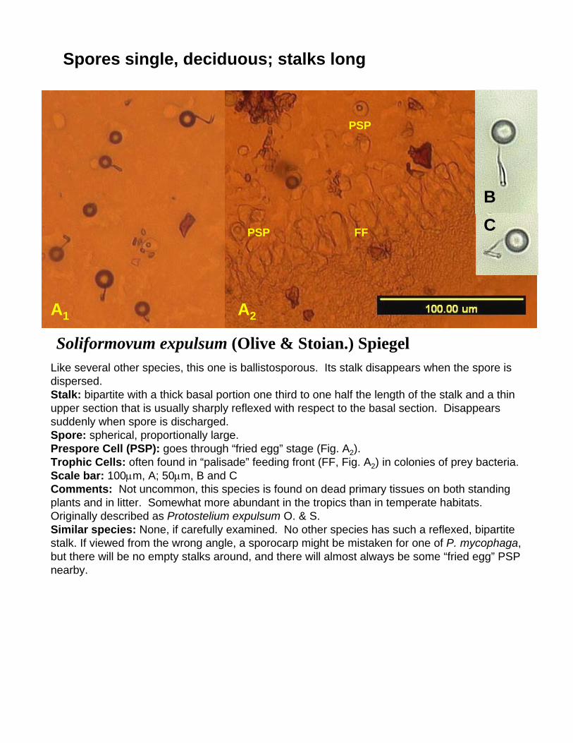
Soliformovum expulsum (Olive & Stoian.) Spiegel
A1 A2
BC
PSP
PSP FF
Like several other species, this one is ballistosporous. Its stalk disappears when the spore isdispersed. Stalk: bipartite with a thick basal portion one third to one half the length of the stalk and a thinupper section that is usually sharply reflexed with respect to the basal section. Disappearssuddenly when spore is discharged.Spore: spherical, proportionally large.Prespore Cell (PSP): goes through “fried egg” stage (Fig. A2).Trophic Cells: often found in “palisade” feeding front (FF, Fig. A2) in colonies of prey bacteria.Scale bar: 100µm, A; 50µm, B and CComments: Not uncommon, this species is found on dead primary tissues on both standingplants and in litter. Somewhat more abundant in the tropics than in temperate habitats. Originally described as Protostelium expulsum O. & S.Similar species: None, if carefully examined. No other species has such a reflexed, bipartitestalk. If viewed from the wrong angle, a sporocarp might be mistaken for one of P. mycophaga,but there will be no empty stalks around, and there will almost always be some “fried egg” PSPnearby.

Spores single, deciduous; stalks long
Endostelium amerosporum Olive
A B C
This is a relatively rare species. Three varieties are described for it based on size, but we have encountered it too few times to know whether those distinctions are merited.Stalk: long, broad and refractile along entire length. Untapered to slightly tapered. With a distinct, knob-like apophysis (Fig. A). As rising sporogen produces stalk, it is surrounded by an ensheathing extension of the cell (not shown but see E. zonatum.Spore: spherical (Fig. A-C) to ovate (Fig. C). Appears to have large warts which are bacteria attached to surface. Flags little or not at all in air currents.Prespore cell: Not shown; reported to be circular in outline.Scale bar: 100µm for all.Comments: Though this species appears to be rare it is distinctive and not easily missed.Similar species: None, though the rising stage is somewhat similar to E. zonatum. We have encountered a myxobacterium that looks similar to this but the sorus of cells slides down the stalk so that the apex sticks through the top.

Spores single, deciduous; stalks long
Endostelium zonatum (Olive & Stoian.) Olive, Bennett & Deasey
A
B
C D E
This reasonably common species is the easiest of all protostelids to identify by its beaded stalk.Stalk: long, quickly tapering from the base then tapering more gradually to the apex, beaded with alternating thick “vesicles” and thin “internodes”; with a slightly swollen apophysis (A in Fig.D). Developing in an invagination in the tapered base of the sporogen (Fig. E). Readilyrecognizable even after spore is shed (Fig. D)Spore: obpyriform to nearly spherical with extension with a socket that fits over apex of stalk,campanulate, freely swinging in air currents at apex of stalk (multiple exposure of swingingspore in Fig. C).Prespore Cell (PSP): not shown. Circular in outline.Scale bar: 100µm in all.Comments: This species is often found with lots of widely spaced fruiting bodies. It is mostoften found on substrates of primary tissues on standing plants. Frequently, when it is found, the substrate was collected from a relatively dry habitat that is exposed to direct sunlight.Originally described as Protostelium zonatum.Similar species: None. If the beads on the stalk are not really pronounced, it may be mistakenfor P. pyriformis, but that species is smaller. We have observed an undescribed species that is similar that has a smooth stalk, but a very rough surfaced spore. However, until that species can be isolated and carefully studied, it is difficult to say more. During the rising sporogen stage this is somewhat similar to E. amerosporum.
A

Spores single, deciduous; stalks long
Ceratiomyxella tahitiensis Olive & Stoianovitch/Nematosteliumgracile (Olive & Stoian.) Olive & Stoianovitch Complex
A B
CHH
Two described species identically share this easily recognized morphotype. Study is under way to determine whether they are truly distinct or variable with respect to the presence of an amoeboflagellate state in the life cycle of C. tahitiensis.Stalk: very long and robust, usually though not alway refractile along its entire length when viewed with a 10x objective (Compare Fig. A and Fig. D. next page), with a knob-shaped apophysis (arrows, Fig. A, B).Spore: spherical to slightly turbinate with distinct ring-shaped hilum with a raised edge thatarticulates with apophysis (H in Fig. C). A spore can be determined to be C. tahitiensis if it greminates to produce cysts in which amoeboflagellates develop. (Fig. E, F next page) or if the culture that develops from it has both small amoebae and reticulate plasmodia (Fig. G next page). A spore of N. gracile will produce only plasmodia in culture.Prespore Cell (PSP): initially a series of connected mounds, circular in outline, that develop from a plasmodial trophic state (Fig. H next page) .Scale bar: 100µm, A, C, D-H; 50µm, B.Comments: This species complex is often one of the three or four most common species in samples from the lowland tropics where it is found on decaying primary tissues on both standing plants and in the litter. It is fairly common in the litter in temperate regions and uncommon on standing plants. It is almost absent at high latitudes and above 2500m. Clusters of fruiting bodies are the rule. Originally, N. gracile was described as Schizoplasmodium gracile O. & S.

Spores single, deciduous; stalks long
Ceratiomyxella tahitinsis Olive & Stoianovitch/Nematosteliumgracile (Olive & Stoian.) Olive & Stoianovitch Complex, cont’dSimilar species: In an earlier version of the Guide, we stated that C. tahitiensis was very rare and N. gracile was much more common. Recent culturing has shown just the opposite to be true. Though we have isolated cultures consistent with N. gracile in the past, we are no longer confident that protostelids with this morphotype are necessarily two species and this is under study. Outside this complex, larger specimens of S. irregularis may superficially be similar, but that species has more slender stalks, a hastate apophysis, and spores that lack a raised hilum. From some orientations it is hard to distinguish N. ovatumfrom this complex because its ovate spore is oriented to appear circular in outline. Also, if only stalks are present one can conclude that either members of this complex or N. ovatumis present, but spores need to be seen to be sure.
D H
E
F G

Spores single, deciduous; stalks long
Nematostelium ovatum (Olive & Stoian.) Olive & Stoianovitch
A B
C
D H
Along with E. zonatum, this is one of the easiest species of protostelids to identify.Stalk: long (5-10 times the length of the spore), robust, appears refractive along entire length,even with 10x objective (most long stalked protostelids have a stalk that appears as a blackline at this magnification), with a distinct, knob-shaped apophysis (arrows in Figs. B, C).Spore: Ovate to nearly ellipsoid, with distinct, ring-shaped hilum with a raised edge thatarticulates with apophysis (H in Fig. D)
Prespore Cell (PSP): Not shown, identical to that of C. tahitiensis/N. gracile.Scale bar: 100µm, A; 50µm, B-DComments: This is a relatively common species in litter in temperate regions. It is less commonon standing dead plant parts. It occurs a bit more frequently in temperate latitudes than in thetropics. Usually it is encountered in clusters with the stalks angling away from each other asin Fig. A. These clusters arise following the fragmentation of the plasmodium into severalclosely spaced prespore cells. Originally described as Schizoplasmodium ovatum O. & S.Similar species: None. At the smaller end of its size range it may first appear to be a largeP. arachisporum, but its stalk is much more robust. If empty stalks are encountered, fallenspores must be found to distinguish it from C. tahitiensis/N. gracile.

Spores single, deciduous; stalks long
Schizoplasmodium seychellarum Olive & Stoianovitch
A B
This is among the rarest of protostelids, but with its large round spores with lateral droplets and robust, long stalk, it is easy to recognize.Stalk: about 4 spore diameters in length and very stout with almost no taper, ending with a distinct somewhat goblet-like apophysis (arrow Fig.A, B). Persistent and obvious after spore discharge.Spore: large and spherical, though often drying and becoming indented (Fig B) with a raised, ring-like apophysis that articulates with the apophysis of the stalk (H, Fig. A). Just prior to spore discharge, a lateral droplet forms (Fig. A). Sudden disappearance of the droplet results in the sudden discharge of the spore.Prespore cell (PSP): Not shown, identical to that of C. tahitiensis/N. gracile.Scale bar: 100µm for all.Comments: This species is very rare but may be found on aerial dead plant parts or in litter just about anywhere protostelids are found. Expect it in less than 1% of samples.Similar species: Essentially hard to misidentify. Its stalks are considerably longer than either S. cavostelioides or S. obovatum. Occasional, malformed individual sporocarps of C. tahitiensis/N. gracile will have stalks that look like this, but will be surrounded bynormally proportioned stalks. Since S. seychellarum produces several sporocarps per plasmodium, it will have several similar looking sporocarps in a group (Fig. A, B).
H

Spores single, deciduous; stalks short
Schizoplasmodium cavostelioides Olive & Stoianovitch
H
SH
SH
This common species is ballistosporous, and its large spore is rarely seen on the stalk. ThisIs one of only two short stalked species with deciduous spores.Stalk:l very short and thick, with a distinct cup-shaped apophysis (arrows, Figs. A and B) thatappears as a distinct ring when viewed from above (ST, arrows, Fig. A). Usually seen afterspore discharge, often grouped in clusters.Spore: Spherical, smooth, with distinct, ring-shaped hilum with raised edge (H in Fig. A) that articulates with apophysis. Spores usually seen discharged and often collapsed on agar surface in PIP. Spores still on stalk often bear a swollen, hydrated sheath (SH in Fig. A and B)that may appear as a lateral droplet or surround the spore completely. Prespore Cell (PSP): Identical to C. tahitiensis/N. gracile (Fig. C).Scale bar: 100µm, A,C; 50µm, BComments: This is a fairly common species that is most abundant on substrates from standingdead plants in temperate habitats. However, it is not uncommon in the tropics. The swellingof the sheath, originally interpreted as a gas bubble, appears just before spore discharge.Discharge occurs suddenly, and the spore is thrown many micrometers.Similar species: None. The short stalked species of Schizoplasmodiopsis have thin stalks, and Cavostelium apophysatum has nondeciduous, rough surfaced spores. The rare S. obovatumhas obovate to obconic spores.
B C
A

Spores single, deciduous; stalks short
Schizoplasmodium obovatum Olive & StoianovitchThis is another rare, but easy to recognize protostelid.Stalk: Short, less than the height of the spore topped with a goblet shaped apopysis. Looks like a small crater from above (circles).Spore: obovate to obconic with a distinct raised, ringlike hilum (H). When still on the stalk, its width is obviously narrow (arrows). Just before spore discharge a lateral droplet forms (not shown). Discharged spores often seen lying on agar some distance from stalks.Prespore cell (PSP): Not shown, identical to that of C. tahitiensis/N. gracile.Scale bar: 100µmComments: As with S. seychellarum, this species is very rare but may be found in any part of the world where protostelids occur.Similar species: Except for its obovate to obconic spores, this species is quite similar to the much more common S. cavostelioides.
H
H
A
B C D
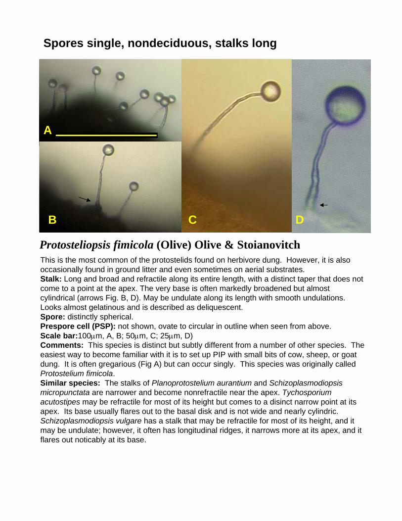
Protosteliopsis fimicola (Olive) Olive & Stoianovitch
Spores single, nondeciduous, stalks long
This is the most common of the protostelids found on herbivore dung. However, it is also occasionally found in ground litter and even sometimes on aerial substrates. Stalk: Long and broad and refractile along its entire length, with a distinct taper that does not come to a point at the apex. The very base is often markedly broadened but almost cylindrical (arrows Fig. B, D). May be undulate along its length with smooth undulations. Looks almost gelatinous and is described as deliquescent.Spore: distinctly spherical.Prespore cell (PSP): not shown, ovate to circular in outline when seen from above.Scale bar:100µm, A, B; 50µm, C; 25µm, D)Comments: This species is distinct but subtly different from a number of other species. The easiest way to become familiar with it is to set up PIP with small bits of cow, sheep, or goat dung. It is often gregarious (Fig A) but can occur singly. This species was originally called Protostelium fimicola.Similar species: The stalks of Planoprotostelium aurantium and Schizoplasmodiopsismicropunctata are narrower and become nonrefractile near the apex. Tychosporiumacutostipes may be refractile for most of its height but comes to a disinct narrow point at its apex. Its base usually flares out to the basal disk and is not wide and nearly cylindric. Schizoplasmodiopsis vulgare has a stalk that may be refractile for most of its height, and it may be undulate; however, it often has longitudinal ridges, it narrows more at its apex, and it flares out noticably at its base.

Planoprotostelium aurantium Olive & Stoianovitch
Spores single, nondeciduous; stalks long
A B C
This rare protostelid is a member of the Protostelium mycophaga complex that is usually nondeciduous.Stalk: long, thin, and narrow and nonrefractile along its length with a slight taper. It may have a slight kink near the very apex similar to what is often seen in P. mycophaga.Spore: Spherical, often slightly pink in color.Prespore cell: not shown, ovate in outline when seen from above as in P. mycophaga.Scale bar: 100µm for all.Comments: This species can only be identified with certainty if its trophic cells are flooded under which conditions they may become flagellated (Fig C). Even if the flagella cannot be seen, the cells can be seen to wiggle quickly back and forth. There can be one to several flagella per cell and pointed extensions form with a flagellum extending from each point (arrows, Fig. C indicate where flagella are extending from these points). The best way to be certain of the ability to produce flagella is to isolate and germinate spores. Less than 1% of the P. mycophaga-like protostelids that are thus treated prove to be P. aurantium.Similar species: Except for being nondeciduous, identical to P. mycophaga, and some strains of that species have spores that do not fall easily. Most strains of P.aurantiumhave proportionally longer stalks than strains of P. mycophaga. S. micropunctata and T. acutostipes both have stalks that taper to a much finer point.

Spores single, nondeciduous; stalks long
Tychosporium acutostipes Spiegel, Moore & Feldman
B A C
A relatively uncommon species that tends to fruit in very dense stands on pieces of substrateand the surrounding agar in PIP. Stalk: appearing stiff, relatively thick and only slightly tapered for over 90% of its length, oftensomewhat undulate (Fig. C), tapering sharply to a fine point at the very upper end (arrows,Figs B and C). Spore: spherical to broadly turbinate with slightly raised hilum that appears similar to a lunar crater at the junction with the stalk (Fig D)Prespore Cell (PSP): not shown, essentially identical to that of P. mycophaga.Scale bar: 100µm, A, 50µm, B, C, 20µm D.Comments: This species is widespread on decaying primary tissues, especially those on standing plants. It occurs worldwide. One microhabitat where it can be found with some regularity is on decaying emergent aquatic plants such as rushes and cat tails. Althoughsporocarps are sometimes seen fruiting singly or in small groups, this species tends to fruitin thick stands.
Similar species: This species has much stiffer stalks than P. mycophaga, and its spores arenondeciduous. It is much more likely to be mistaken for Schizoplasmodiopsis vulgare (whichsee) with which it frequently co-occurs; however, S. vulgare has a stalk that is relatively thickall the way to its junction with the spore, and its spores are relatively irregular in outline because of a reticulum of low ridges that cover the spore wall. This species may also beconfused with the very rare Schizoplasmodiopsis micropunctata (which see), but that specieshas a more delicate stalk with a very thin, hairlike tip.
D

Spores single, nondeciduous; stalks long
Schizoplasmodiopsis micropunctata Olive & Stoianovitch
B A
This is one of the rarest protostelids.Stalk: proportionally very long, narrowing suddenly to a hairlike tip that may be as long as25% the diameter of the spore (arrow, Fig. B, C). This tip may be so thin that the spore almostappears as if it is suspended above the tip of the stalk.Spore: spherical to broadly turbinate, with minute spines reported to be visible under oil immersion with phase contrast (not shown)Prespore Cell (PSP): not well described.Scale bar: 100µm, A; 50µm, B; 20µm, CComments: This species is very rare, but seems to be encountered worldwide in locales wherelarge numbers of samples have been collected. It occurs in usually <0.25% of samples.Because of its rarity, it is difficult to say much about likely microhabitat preferences.Similar species: It is most simliar to T. acutostipes, but it is more delicate, and has thedistinct hairlike stalk tip.
C

Spores single, nondeciduous; stalks long
Schizoplasmodiopsis vulgare Olive & Stoianovitch
B C A
This is a commonly encountered species that often tends to fruit very heavily. It is certainly themost common long stalked species with nondeciduous spores.Stalk: relatively thick along its entire length, tapering but never to a sharp point. Some undulations may be seen along the stalk along with some short, longitudinal ridges (Fig. B, C) Length may vary from slightly more than one spore diameter to several spore diameters though most sporocarps in PIP appear as those shown here.Spore: essentially spherical but course looking because of low ridges formed by a reticulum ofspore wall thickenings that appear as slight bumps (arrows, Figs. B and C). These may be moreor less obvious and are best detected with objectives of 20x or higher. Spore diameter is relatively constant even when stalk length varies.Prespore Cell (PSP): not shown, round in outline.Scale bar: 100µm, A; 50µm B, C.Comments: This is a species that is worldwide and distribution and found on decaying primarytissues about equally on standing plants and in the litter. In cool, moist habitats, it is often one of
the only species encountered. Though somewhat variable in stalk length, it is usually relatively easy to identify by its coarse appearance. It is another species that is fairly common on decaying emergent aquatic plants.Similar species: T. acutostipes is the most likely species to be confused with S. vulgare, butthe former has a proportionally thinner stalk which tapers to a fine point. However, one mustbe careful since the two species will often fruit together with the patches of sporocarpsintermixed. It can be distinguished from P. fimicola by its coarser looking stalks.

Spores single, nondeciduous; stalks long
Schizoplasmodiopsis reticulata Olive & Stoianovitch
A
C B
This is a relatively rare species that occurs in situations wherever S. vulgare is likely to be found. Like that species, it often fruits very heavily on a substrate. Its distinctive spores makeit easy to recognize.Stalk: relatively longer and usually more gracile and finely tapered than that of S. vulgare. The two sporocarps marked with arrows in Fig. B show stalks whose bases are not blockedby the substrate such that their whole length is obvious. Spore: essentially spherical with a distinct, raise reticulum of ridges that is easily visible evenat low magnifications (arrows, Fig. A-C).Prespore Cell (PSP): not shown, round in outline.Scale bar: 100µm, A,B: 50µm C.Comments: Though this species is widespread, it is rare. Nontheless, its reticulate spores areso distinctive that it is almost impossible to mistake for any other species. This particular example was observed on substrates from the Ozarks of Arkansas and has stalks that are onthe proportionally shorter end of the range stalk lengths. Some fruitings may have stalks that are up to two spore diameters longer.Similar species: Only an occasional fruiting of S. vulgare that has a coarse reticulum on the spores might be mistaken for S. reticulata. It may be the case that the two species are veryclosely related or conspecific and that spore reticulation is a variable trait.

Spores single, nondeciduous; stalks short
Schizoplasmodiopsis amoeboidea Olive & Whitney
A B C
This is a common species which may occur a scattered individual sporocarps in PIP or ingroups.
Stalk: short, rarely >1.5 times the diameter of the spore, narrows suddenly to a thinner tip(arrow, Fig. B), often hard to detect in PIP because of irregularities in surface of substrateor because sporocarps on agar are seen directly from above.
Spore: spherical, usually among the largest seen in any protostelids, but may vary in size.Prespore Cell (PSP): Fig. C, oval to round in outline.Scale bar: 100µm, A; 50µm, B, CComments: This species is found relatively regularly on any kind of substrate on which protostelids may occur. Similar species: Whenever a short stalked species occurs in a PIP, every effort should bemade to see a stalk, even if it means knocking sporocarps over. This species is very oftenquite difficult to distinguish from Schizoplasmodiopsis pseudoendospora because sporocarpsat the smaller end of the size range of S. amoeboidea overlap in size with those of S.pseudoendospora. Two distinguishing characters to look for are that the stalk of this specieshas the narrow section at its apex while the stalk of S. pseudoendospora tapers evenly to itstip, and this species tends to fruit singly or when grouped, the sporocarps are irregularlyspaced. However, it is probably the case that one species is occasionally misidentified as theother on PIP.
Spores single, nondeciduous; stalks short
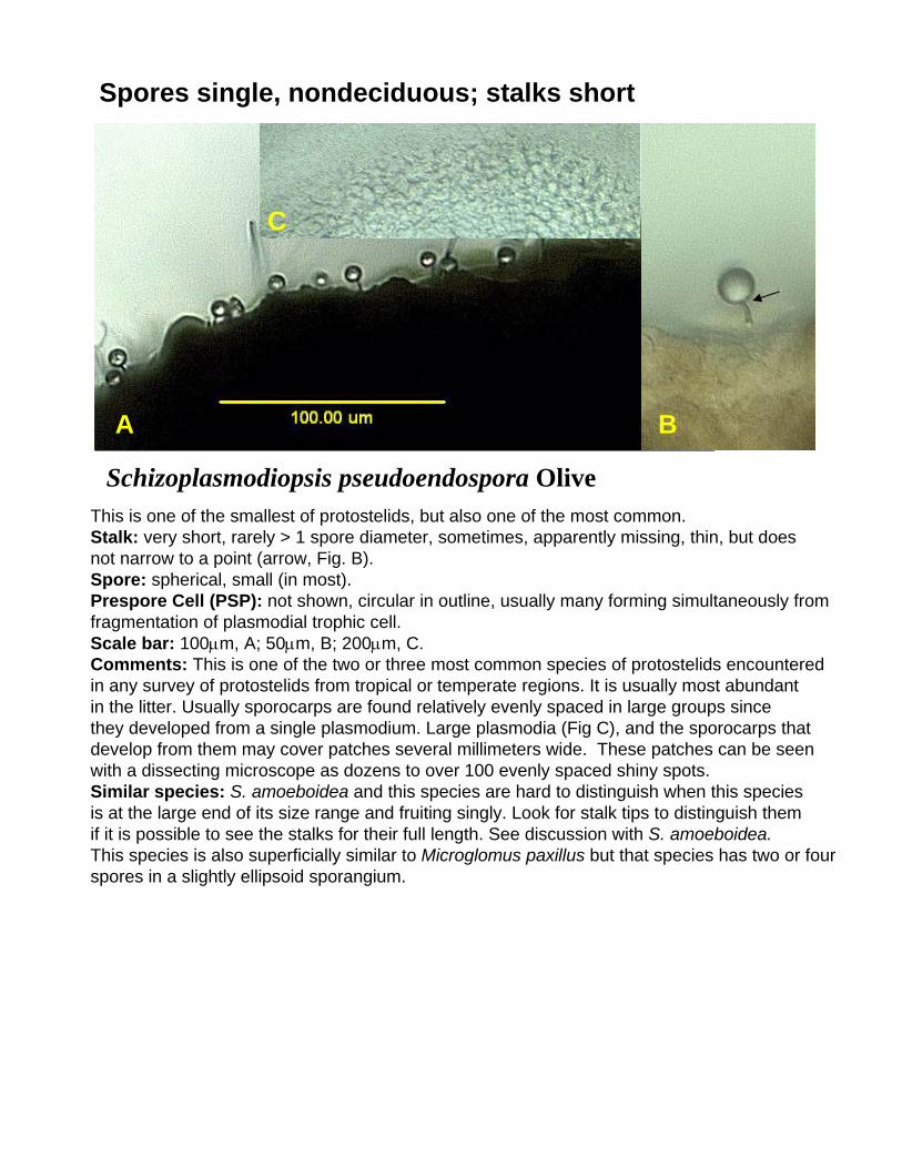
Schizoplasmodiopsis pseudoendospora Olive
A B
This is one of the smallest of protostelids, but also one of the most common.Stalk: very short, rarely > 1 spore diameter, sometimes, apparently missing, thin, but doesnot narrow to a point (arrow, Fig. B).Spore: spherical, small (in most).Prespore Cell (PSP): not shown, circular in outline, usually many forming simultaneously fromfragmentation of plasmodial trophic cell.Scale bar: 100µm, A; 50µm, B; 200µm, C.Comments: This is one of the two or three most common species of protostelids encounteredin any survey of protostelids from tropical or temperate regions. It is usually most abundantin the litter. Usually sporocarps are found relatively evenly spaced in large groups sincethey developed from a single plasmodium. Large plasmodia (Fig C), and the sporocarps thatdevelop from them may cover patches several millimeters wide. These patches can be seen with a dissecting microscope as dozens to over 100 evenly spaced shiny spots.Similar species: S. amoeboidea and this species are hard to distinguish when this speciesis at the large end of its size range and fruiting singly. Look for stalk tips to distinguish themif it is possible to see the stalks for their full length. See discussion with S. amoeboidea.This species is also superficially similar to Microglomus paxillus but that species has two or four spores in a slightly ellipsoid sporangium.
C

Spores single, nondeciduous; stalks short
Cavostelium apophysatum Olive
A
B CA
This is a fairly common specie in the tropics and relatively uncommon in temperate regions.Though it may vary quite a bit in overall size from isolate to isolate, the proportions of thesporocarp are constant (compare Figs. A and B).Stalk: very short, wide, with a cup-like apophysis that occupies one third to more than onehalf the total length of the stalk.Spore: spherical, roughened in appearance by the presence of numerous spines and warts onthe spore wall (Fig. C). This spore wall sculpturing often makes the spores appear to beless refractile than those of other protostelids. Some spores may contain two cells in some isolates. Such spores look ellipsoid from above and from the side.Prespore Cell (PSP): not shown, circular in outline.Scale bar: 100µm, A, B; 50µm, C.Comments: This species is usually found with dense patches of sporocarps. It may be foundin litter or on standing dead plants, though it is more found a bit more frequently in the latter.As stated above, it appears to be considerably more common in the tropics.Similar species: If sporocarps of this species can be viewed from the side, its thick stalkdistinguishes it from the short stalked species of Shizoplasmodiopsis. Since its spores arenondeciduous and roughened, it is easy to distinguish from Schizoplasmodium cavostelioides.
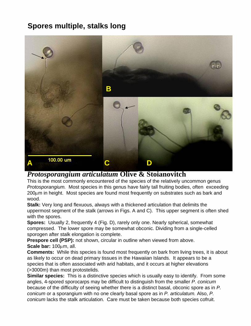
Spores multiple, stalks long
Protosporangium articulatum Olive & StoianovitchThis is the most commonly encountered of the species of the relatively uncommon genus Protosporangium. Most species in this genus have fairly tall fruiting bodies, often exceeding 200µm in height. Most species are found most frequently on substrates such as bark and wood.Stalk: Very long and flexuous, always with a thickened articulation that delimits the uppermost segment of the stalk (arrows in Figs. A and C). This upper segment is often shed with the spores. Spores: Usually 2, frequently 4 (Fig. D), rarely only one. Nearly spherical, somewhat compressed. The lower spore may be somewhat obconic. Dividing from a single-celled sporogen after stalk elongation is complete. Prespore cell (PSP): not shown, circular in outline when viewed from above.Scale bar: 100µm, all.Comments: While this species is found most frequently on bark from living trees, it is about as likely to occur on dead primary tissues in the Hawaiian Islands. It appears to be a species that is often associated with arid habitats, and it occurs at higher elevations (>3000m) than most protostelids.Similar species: This is a distinctive species which is usually easy to identify. From some angles, 4-spored sporocarps may be difficult to distinguish from the smaller P. conicumbecause of the difficulty of seeing whether there is a distinct basal, obconic spore as in P. conicum or a sporangium with no one clearly basal spore as in P. articulatum. Also, P. conicum lacks the stalk articulation. Care must be taken because both species cofruit.
A C D
B

Spores multiple, stalks long
A B
Protosporangium bisporum Olive & StoianovitchThis very uncommon protostelid is likely to be found on bark of living treesStalk: very long, thin, and flexuous though rarely sharply bent with pointed angles.Spore: hemispherical in a group of two to form a spherical to slightly ellipsoidal sporangium.Prespore cell (PSP): not shown, circular in outline when viewed from above.Scale bar: 100µ for both.Comments: This species is almost exclusively found on bark, often in dense stands of hundreds of sporocarps. The two spores are often hard to distinguish in standing sporocarpsbut if they touch the agar, they are easily seen. No other protostelid has such long, thin, and smoothly flexuous stalks.Similar species: Sporocarps of P. fragile with spherical sporangia are similar; however, their flexuous stalks are bent at sharp angles.

Spores multiple, stalks long
Protosporangium conicum BennettThis is the second most common species of the genus, though it is much more likely to be encountered in arid habitats than anywhere else.Stalk: long, thin, somewhat flexuous, waving readily in air currents.Spores: Always four per sporocarp, with the one that attaches to the stalk obconic; the other three spherical, arranged at distal end of obconic spore. Usually shed as a unit.Prespore Cell (PSP): unknown.Scale bar: 100µm for allComments: This species has been encountered exclusively on bark of living trees. It is the smallest of the four described species of Protosporangium. The other species have stalks that are usually twice the length of this species. All these are also found most often on bark or on wood and rarely on decaying primary tissues.Similar species: None, the obconic spore is absolutely distinctive though if it cannot be seen, it may be difficult to distinguish from P. articulatum with which it often co-occurs.
A B

Spores multiple, stalks long
Protosporangium fragile Olive & Stoianovitch
A C
B
This is a relativley uncommon species found primarily on bark of living trees or on rotting wood.Stalk: very long and flexuous, usually bending in distinct kinks. Flag readily in even slight air currents (Note blurred examples in Fig. A, B) Several sporocarps will often tangle together (Fig. C).Spores: Almost always in a group of four. The spores may each be perfect quarter spheres such that the sporangium appears nearly spherical, or the individual spores can be subspherical such that the sporangium appears slightly tetrahedral to morulate (Fig. C)Prespore cells (PSP): unknown.Scale bar: 100µm for all.Comments: This species has the longest and most delicate stalks of any protostelid and usually fruits in dense patches so that tangles of fruiting bodies (Fig C) are extremely common.Similar species: Most likely to be confused with P. bisporum when the sporangium is spherical.

Spores multiple, stalks short
A B
C D E
Clastostelium recurvatum Olive & StoianovitchThis relatively uncommon protostelid is easy to recognize with its recurved stalk and smallsporangium of two spores. It is best seen early in the morning because its spores are usually all dispersed by mid morning.Stalk: bipartite with a short, persistent, apiculate base (arrows in Fig. C) and an inflated, recurved upper portion that bursts to disperse the spores. Stalk bases seen as dark spotsfrom above after spores dispersed (circle in Fig. E)Spores: smooth and spherical in two spored sporangium, slightly pendulous when stalk is mature. Sporangium articulates to stalk between the two spores (arrows Fig. B,E). Shot off as a unit.Prespore Cell (PSP): not shown, circular in outline.Scale bar: 100µm A,B; 50µm C-D.Comments: Found most often aerial dead plant parts or bark of living trees. Appears to bemore common in the tropics than in mid to high latitudes.
Similar species: None of the described species is similar to c. recurvatum. However, thereis an uncommon undescribed species most often encountered on bark which is superficiallyresembles it. The undescribed species, for which no trophic state has yet been observed,is the same size and recurved, but it has a single, highly sculptured spore and the stalk islongitudinally ridged and tapers from base to tip. The undescribed species often appearsto be yellow to gold under a dissecting microscope.

Spores multiple, stalks short
Echinosteliopsis oligospora Reinhardt & Olive
A B C
SH
SHSH
This common species is treated as short stalked because it is usually first observed on PIPwith a hydrated sheath that makes the diameter of the sporangium greater than the lengthof the stalk.Stalk: straight to gently curved with a pronounced taper from base to tip.Spores: 1-8, usually 4, spherical to laterally compressed, usually of different sizes within asingle sporangium, spore walls smooth within a hygroscopic sheath that is usually hydrated in the saturated air of a culture plate. When hydrated, spores appear suspended in sheath (Fig. A and B). When dried, one spore at apex of stalk, others arranged around its distal half (Fig. C).Prespore Cell (PSP): not shown, circular in outline.Scale bar: 100µm, all.Comments: This species is common worldwide on decaying primary tissues on both standing plants and in the litter. It seems to show patches of local abundance, being verycommon at some collecting sites and absent at others. Similar species: This species is distinct from all other protostelids; however, it may atfirst be mistaken for a small example of the myxomycete genus Echinostelium. It lacks both a columella and a capillitium, and the arrangement of spores is unlike any myxomycetewhen the sheath dries down. In addition the spores usually vary in size within the sporocarp,and they lack any spore wall sculpturing. Spiegel has seen an aggregative ciliate that produces a sorocarp that looks similar to this species on one occasion. Ciliated cellsgerminate from the undescribed ciliate while amoebae germinate from E. oligospora.

Spores multiple, stalks short
Microglomus paxillus Olive & Stoianovitch
B A
C
This is a relatively uncommon species of protostelid which is hard to identify if not observedcarefully.Stalk: very short, tapered, narrows to a thin tip.Spores: two or four compressed against each other to form an ellipsoid (Fig. B) to almostspherical sporangium. Careful focusing, especially with objective of 20x or higher willreveal the individual spores (arrows in Fig. C point to the four spores).Prespore Cell (PSP): not shown, circular in outline.Scale bar: 100µm, A, B; 50µm, C.Comments: This species is small and easily misidentified if one does not look at it carefully.Any small, short-stalked protostelid whose “spore” looks ellipsoid should be carefully examined to see if it is really a sporangium. This species has been found on primary tissueson both standing plants and in litter and on bark of living trees.Similar species: Most likely to be misidentified as s. pseudoendospora.

Spores multiple, stalks short
Echinostelium bisporum (Olive & Stoian.) Whitney & Olive
A B C
SH
Actually the smallest described species of myxomycetes, this relatively common species ismost likely to be detected when examining substrates for protostelids.Stalk: short, stiff with small flange shaped apophysis (not shown).Spores: always two per sporangium, each spherical to subspherical arranged linearly, distalspore usually slightly smaller. Spore walls minutely spiny with a hilum of fused spines on each where they contact each other (arrow, Fig. C). Often, the hygroscopic sheath is swollenwhen hydrated when culture plate is first opened (SH in Fig. B).Prespore Cell (PSP): not shown, circular in outline.Scale bar: 100µm, A, B, D; 50µm, C.Comments: This little myxomycete is found on all kinds of substrates where protostelids arefound. It may be a bit more common in the tropics than in temperate areas. Origniallydescribed as Cavostelium bisporum Olive & Stoian.Similar species: None, this species is unique and nearly impossible to misidentify. However, if you encounter what appears to be a 4-spored example of E. bisporum, it is probably the much less common E. lunatum Olive & Stoianovitch which has an articuated half-cup shaped columella (Fig. D).
D

Sporocarps arranged on a slime column or pad depositedby a plasmodium prior to dividing into prespore cells.
Ceratiomyxa hemisphaerica Olive& StoianovitchThis rarely found species has been encountered fewer than a dozen times.Stalk: very long and flexuous, in groups of a few dozen to 100 resting on a small, hemispherical mound of slime deposited by the plasmodium prior to its cleavage into prespore cells. The slime mound in this image does not show well. It is better viewed from the side.
Spore: spherical to ellipsoid, deciduous.Prespore Cell (PSP): not shown, circular in outline.Scale bar: 100µmComments: When this species has been found, it has occurred on rotting wood or fairly substantial primary tissue substrates such as tree fern petioles. It is usually detected by seeing a cluster of sporocarps that radiate outward from a common base.
Similar species: At first glance, an individual sporocarp looks similar to one of Protosporangium bisporum (which is not illustrated in this Guide). While that species is often gregarious, it has no common slime mound, and it has two spores instead of one.

Sporocarps arranged on a slime column or pad depositedby a plasmodium prior to dividing into prespore cells.
Ceratiomyxa fruticulosa (Mull.) MacbrideThis is probably the most commonly encountered of all eumycetozoans, and it is one of thefew macroscopic protostelids (even though it is claimed for the myxomycetes in much of the literature). It has slime columns that may show a number of morphologies, and thoughit is usually white, a fructification may show a whole range of colors, especially early indevelopment. Fructifications may range in size from a few millimeters to over a meter.The species occurs most frequently on rotting wood in mesic to wet forests a day or twoafter a soaking rain. It is very rarely found in moist chamber cultures for myxomycetes (aswas the case with this specimen) and has never been reported in PIP for protostelids. Individual sporocarps have straight stalks about 30-50um long and deciduous, ellipsoidalspores.
Scale bar: 100µmSimilar species: The tropical species Ceratiomyxa morchella and C. sphaerosperma are much smaller. The former is shaped like a small morel and the latter has a distinct, stalk like base to the slime column with an irregular, spheroidal head of finger like extensions that support the sporocarps.

Sporocarps arranged on a slime column or pad depositedby a plasmodium prior to dividing into prespore cells.
Ceratiomyxa morchella WeldenThis macrophotograph of the tropical C. morchella was taken in the field on rotting wood in Costa Rica. Its spherical heads are pitted like a small morels. The individual sporocarpshave stalks and spores similar in morphology to C. fruticulosa.

Sporocarps arranged on a slime column or pad depositedby a plasmodium prior to dividing into prespore cells.
Ceratiomyxa sphaerosperma Boedijn
Courtesy of Adam Rollins
This second predominantly tropical Ceratiomyxa is often found fruiting on woody shells of nuts. It differs from the other two species primarily because its spores are spherical.

A. Protostelium mycophaga B. P.nocturum C. P. okumukumu D. P. pyriformis E. P. arachisporum F. Soliformovum irregularisG. S. expulsum H. Endostelium amerosporum I. E. zonatum J.Ceratiomyxella tahitiensis/Nematostelium gracile K.N. ovatumL. Schizoplasmodium seychellarum M. S. cavostelioides N. S. obovatum O. Protosteliopsis fimicola P. Planoprotostelium aurantiumQ. Tychosporium acutostipes R. Schizoplasmodiopsis micropunctata S. S. vulgare T. S. reticulata U. S. amoeboideaV. S. pseudoendospora W. Cavostelium apophysatum X. Protosporangium articulatum Y. P. bisporum Z. P. conicum AA. P. fragile BB. Clastostelium recurvatum CC. Echinosteliopsis fimicola DD. Microglomus paxillus EE. Echinostelium bisporumFF. Ceratiomyxa hemisphaerica. Scale bar = 100µm `
A C D E
B
F
G
H
I
J K L N O
M
P Q R T
SU V W
X Y Z AA FF
BB CC DD EE
Same Size Comparison of All Microscopic Protostelids
All the figures on the comparison plate are of sporocarps as they would appear with a 10x objective used for scanning a primary isolation plate. Remember to look for sporocarps on the agar surface surrounding a piece of substrate and fruiting out from the surface of a piece of substrate. With a little practice, one can learn to be fairly efficient at finding protostelids within a few hours and to identify them within a few days to a week.
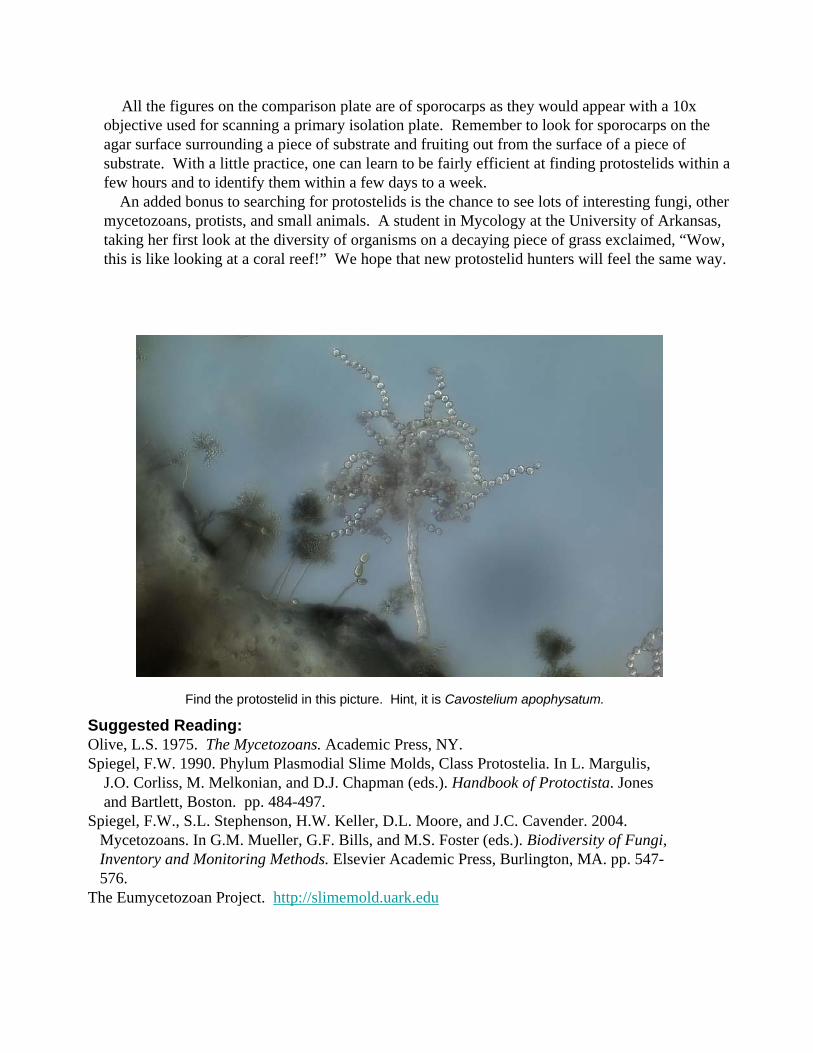
An added bonus to searching for protostelids is the chance to see lots of interesting fungi, other mycetozoans, protists, and small animals. A student in Mycology at the University of Arkansas, taking her first look at the diversity of organisms on a decaying piece of grass exclaimed, “Wow, this is like looking at a coral reef!” We hope that new protostelid hunters will feel the same way.
Find the protostelid in this picture. Hint, it is Cavostelium apophysatum.
Suggested Reading:Olive, L.S. 1975. The Mycetozoans. Academic Press, NY.Spiegel, F.W. 1990. Phylum Plasmodial Slime Molds, Class Protostelia. In L. Margulis,
J.O. Corliss, M. Melkonian, and D.J. Chapman (eds.). Handbook of Protoctista. Jonesand Bartlett, Boston. pp. 484-497.
Spiegel, F.W., S.L. Stephenson, H.W. Keller, D.L. Moore, and J.C. Cavender. 2004.Mycetozoans. In G.M. Mueller, G.F. Bills, and M.S. Foster (eds.). Biodiversity of Fungi,Inventory and Monitoring Methods. Elsevier Academic Press, Burlington, MA. pp. 547-576.
The Eumycetozoan Project. http://slimemold.uark.edu