a 52-year-old man with ecchymotic leg ulcers · a 52-year-old man with ecchymotic leg ulcers ... a...
TRANSCRIPT
232 J La State Med Soc VOL 165 May/June 2013
Journal of the Louisiana State Medical Society
CliniCal Case of the Month
A 52-Year-Old Man With Ecchymotic Leg Ulcers
Seema Walvekar, MD; Jessica L. Johnson, PharmD, BCPS; Emily Kauffman, DO; Rachna Jetly, MD; Bennett P. deBoisblanc, MD, FCP, FACCP, FCCM
CASE PRESENTATION
A 52-year-old man presented to the emergency depart-ment with a one-day history of pain and bluish discoloration of the tips of the great toes of both his feet that rapidly pro-gressed to worsening pain, swelling, and discoloration of both feet and legs. His past medical history was significant for many years of heavy alcohol use and an episode of un-provoked venous thromboembolism two months prior to presentation. At that time, he was found to be heterozygous for Factor V Leiden mutation. After initial anticoagulation with fondaparinux, he was started on warfarin 7.5 mg daily. A removable inferior vena cava filter was placed for unclear indications. In the emergency department, his review of systems was remarkable for a one-day history of melena with hematemesis. On subsequent questioning, he admit-ted to only sporadic compliance with his warfarin therapy and laboratory monitoring.
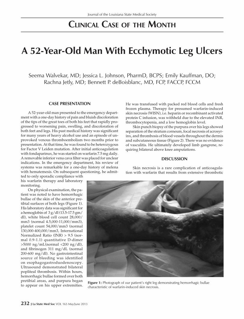
On physical examination, the pa-tient was noted to have hemorrhagic bullae of the skin of the anterior pre-tibial surfaces of both legs (Figure 1). His laboratory data was significant for a hemoglobin of 3 g/dl (13.5-17.5 gm/dl), white blood cell count 28,000/mm3 (normal 4.5,000-11,000/mm3), platelet count 54,000/mm3 (normal 130,000-400,000/mm3, International Normalized Ratio (INR) > 9.5 (nor-mal 0.9-1.1) quantitative D-dimer >5000 ng/mL(normal <200 ng/dl), and fibrinogen 311 mg/dL (normal 200-600 mg/dl). No gastrointestinal source of bleeding was identified on esophagogastroduodenoscopy. Ultrasound demonstrated bilateral popliteal thrombosis. Within hours, hemorrhagic bullae formed over both pretibial areas, and purpura began to appear on his upper extremities.
He was transfused with packed red blood cells and fresh frozen plasma. Therapy for presumed warfarin-induced skin necrosis (WISN), i.e. heparin or recombinant activated protein C infusion, was withheld due to the elevated INR, thrombocytopenia, and a low hemoglobin level.
Skin punch biopsy of the purpura over his legs showed separation of the stratum corneum, focal necrosis of acrosyr-inx, and thrombosis of blood vessels throughout the dermis and subcutaneous tissue (Figure 2). There was no evidence of vasculitis. He ultimately developed limb gangrene, re-quiring bilateral above knee amputations.
DISCUSSION
Skin necrosis is a rare complication of anticoagula-tion with warfarin that results from extensive thrombotic
Figure 1: Photograph of our patient’s right leg demonstrating hemorrhagic bullae characteristic of warfarin-induced skin necrosis.

J La State Med Soc VOL 165 May/June 2013 233
occlusion of small vessels, resulting in large areas of der-mal necrosis requiring debridement or limb amputation. Clinical mimics of WISN include: necrotizing fasciitis, pur-pura fulminans, calciphylaxis, cryoglobinemia, cholesterol embolization, heparin-induced skin necrosis, and lupus anticoagulant-associated skin necrosis. These conditions must be ruled out to consider a diagnosis of WISN.
The most plausible hypothesis explaining WISN is that initiation of warfarin treatment causes a transient imbalance of pro- and anti-coagulant factors that favors thrombosis. Within the first 24 hours of an initial dose, warfarin reduces the anticoagulant activity of protein C by almost 50%. How-ever, the activity of Vitamin K-dependent procoagulant factors is inhibited by warfarin at a much slower rate due to the longer half-lives of these factors (Table). The slower clearance of the procoagulant factors leads to transient hy-percoagulability and the potential for thrombosis during this early phase of warfarin therapy.1,2 This mechanism is more pronounced when higher doses of warfarin are used or when a patient has a genetic or acquired protein C de-ficiency. Heparin bridging is used to inhibit procoagulant
clotting factors and thus, to prevent WISN in patients with protein C deficiency until this procoagulant factors are cleared, typically by day five of therapy.3
WISN has an estimated incidence of 0.01-0.1% in the general population of patients receiving oral Vitamin K antagonists.1 However, as many as one-third of these cases occur in patients who are heterozygous for protein C or S deficiency.4 WISN has not only been associated with ac-quired and hereditary deficiencies of proteins C and S, but also with activated protein C (APC) resistance secondary to Factor V Leiden mutation.4-8
APC downregulates the production of thrombin by enzymatically deactivating clotting factors Va and VIIIa. APC resistance is a common risk factor for thrombosis, and between 20% and 60% of thrombophilic patients suffer from some form of APC resistance.9,10 The most common point mutation leading to APC resistance among Caucasians is at the R506Q site of Factor Va (FVa), known as the Factor V Leiden mutation.
Procoagulant FVa is deactivated by APC with an initial cleavage of the FVa peptide at the Arg506 position, followed by a second cleavage at Arg306. The Factor V Leiden R506Q point mutation, a substitution of arginine with glutamine, prevents cleavage at that position by APC. Although the mutant FVa can be inactivated by cleavage at Arg306, this cleavage is tenfold slower without prior cleavage at Arg506. Once cleaved at Arg506 by APC, deactivated FV functions as a cofactor in the APC-mediated degradation of Factor VIIIa. Individuals with Factor V Leiden mutation therefore have decreased deactivation of both FVa and FVIIIa and increased risk of thrombosis.9-11
Individuals who are heterozygous for the Factor V
Table: Half-life of Vitamin K-depedent clotting factors1,2
Protein C 8 hours
Factor IX 24 hours
Factor X 48 hours
Factor II 2-5 days
Factor IV 1-6 hours
Protein S 42 hours
Figure 2: Photomicrograph of a skin punch biopsy demonstrating thrombosis of blood vessels (arrows) throughout the dermis and subcutaneous tissue.
234 J La State Med Soc VOL 165 May/June 2013
Journal of the Louisiana State Medical Society
Leiden mutation carry a risk of venous thrombosis five to seven times higher than that of the general population; ho-mozygous subjects have a risk 80 times higher. The incidence of Factor V Leiden mutation among the general Caucasian population is 0-8%, but up to 60% of patients with venous thromboembolism may test positive for Factor V Leiden mutation.9-11
Approximately 95% of patients who are heterozygous for Factor V Leiden mutation at the chromosome R506Q demonstrate APC resistance.8 Additional sources of APC resistance include other point mutations of Factor V, spon-taneous generation of auto-antibodies targeting Factor V, and dysfunction or deficiency of APC cofactors, including protein S.
CASE SUMMARY
Our patient was predisposed to developing WISN. He was heterozygous for Factor V Leiden mutation at the R506Q position, increasing his risk of thrombosis. Patients with Factor V Leiden mutation are at increased risk of WISN, particularly when warfarin is initiated in high doses or without heparin bridging. Our patient admitted to inter-mittent compliance to prescribed therapy and monitoring. He stopped and started high-dose warfarin several times without monitoring or heparin bridging. He had no evidence of infection, no heparin use, no lupus anticoagulant, and no serum cryoglobins. His biopsy showed characteristic intra-vascular thrombosis of small cutaneous vessels. Based on his history, laboratory data, histopathology, and that he was heterozygous for Factor V Leiden mutation, we confirmed the diagnosis of WISN.
Alcoholism also contributed to the development of WISN in our patient. Acute alcohol ingestion, as in binge drinking, impairs hepatic metabolism of warfarin and in-creases the effective anticoagulant dose. Patients with Factor V Leiden mutation requiring anticoagulation with warfarin should receive extensive counseling regarding alcohol use and the risk of re-initiating therapy after self-discontinuation without heparin bridging. Consideration should be given to alternative oral anticoagulation (rivaroxaban, dabigatran) for patients with risk factors for WISN.
REFERENCES
1. Chan YC, Valenti D, Mansfield AO, Stansby G. Warfarin induced skin necrosis. Br J Surg 2000;87(3): 266–72.
2. McKnight JT, Maxwell AJ, Anderson RL. Warfarin necrosis. Arch Fam Med 1992 Sept;1(1):105-8.
3. Ansell J, Hirsch J, et al. The Pharmacology and Management of the Vitamin K Antagonists: The Seventh ACCP Conference on Antithrombotic and Thrombolytic Therapy. Chest 2004;126:204S-233S.
4. Broekmans AW, Teepe RG, van der Meer FJ, et al. Protein C and coumarin-induced skin necrosis. Thromb Res 1986;6:137.
5. Goldberg SL, Orthner CL, et al. Skin necrosis following a prolonged administration of coumarin in a patient with inherited protein C deficiency. Am J of Hematology 1991;38:64-6.
6. Teepe RG, Broekmans AW, Vermeer BJ, Nienhuis AM, Loeliger EA. Recurrent coumarin-induced skin necrosis in a patient with an acquired functional protein C deficiency. Arch Dermatol 1986;122(12):1408.
7. Sallah S, Abdallah JM, Gagnon GA. Recurrent warfarin-induced skin necrosis in kindreds with protein S deficiency. Haemostasis 1998;28(1):25.
8. Makris M, Bardhan G, Preston FE. Warfarin induced skin necrosis associated with activated protein C resistance. Thromb Haemost 1996;75(3):523.
9. Nicolaes GAF, Dahlbäck B. Congenital and Acquired Activated Protein C Resistance. Seminars in Vascular Medicine 2003;03(1): 033-046.
10. Dahlbäck B. The discovery of activated protein C resistance. J Thromb Haemost 2003 Jan;1(1):3-9.
11. Nazarian RM, Van Cott EM, Zembowicz A, Duncan LM. Warfarin-induced skin necrosis. J Am Acad Dermatol 2009;60(2):325-332.
Drs. Walvekar and Kauffman are with the Department of Internal Medicine at the Louisiana State University Health Sciences Center in New Orleans. Dr. Kauffman is a Resident there. Dr. Johnson is a Clinical Assistant Professor at Xavier University of Louisiana College of Pharmacy and is affiliated with Interim Louisiana Hospital, Medical Intensive Care Unit at LSUHSC-New Orleans. Dr. Jetly is with the Department of Pathology at LSUHSC-New Orleans. Dr. deBoisblanc is a Professor of Medicine and Pathology and is affiliated with the Pulmonary section of Critical Care at LSUHSC-New Orleans.