971–60 is the temperature of echocardiographic contrast media important?
TRANSCRIPT
JACC February 1995 ABSTRACTS 247A
ages were acquired at a sampling rate of 30 Hz. The mean transit rate ofAlbunex® through the myocardium was derived by fitting a y-variate functionto the background-subtracted time-intensity plots. The mean transit rate ofAlbunex® decreased as the hematocrit decreased. On multivariate analysis,the hematocrit and the p02 were the best correlates of mean microbubbletransit rate (y : 0.06 hematocrit - 0.002 p02 + 0.72, r: 0.72, p < 0.001).
We conclude that MCE, can be used to assess reversible microvascular injury. This technique, therefore, has great potential for understanding dynamicchanges in microvascular function in-vivo, particularly those modulated bythe endothelium.
occ. The slight increase noted in normalized epicardial flow during these 3stages in the controls (0.31 ± 0.11 to 0.33 ± 0.010 to 0.38 ± 13, P < 0.01)was significantly less than in PC dogs, and was not detected with MCE.
In conclusion, in this canine model, collateral flow to an occluded vascular bed is enhanced by prior brief ischemia/reperfusion in a distantly locatedcoronary bed. Furthermore, the resulting decrease in RA size appears ongoing during the sustained occ. "Remote" PC may facilitate the recruitmentof preformed collaterals situated between the 2 beds, and this phenomenonmay help explain the reductions in infarct size associated with this PC model.
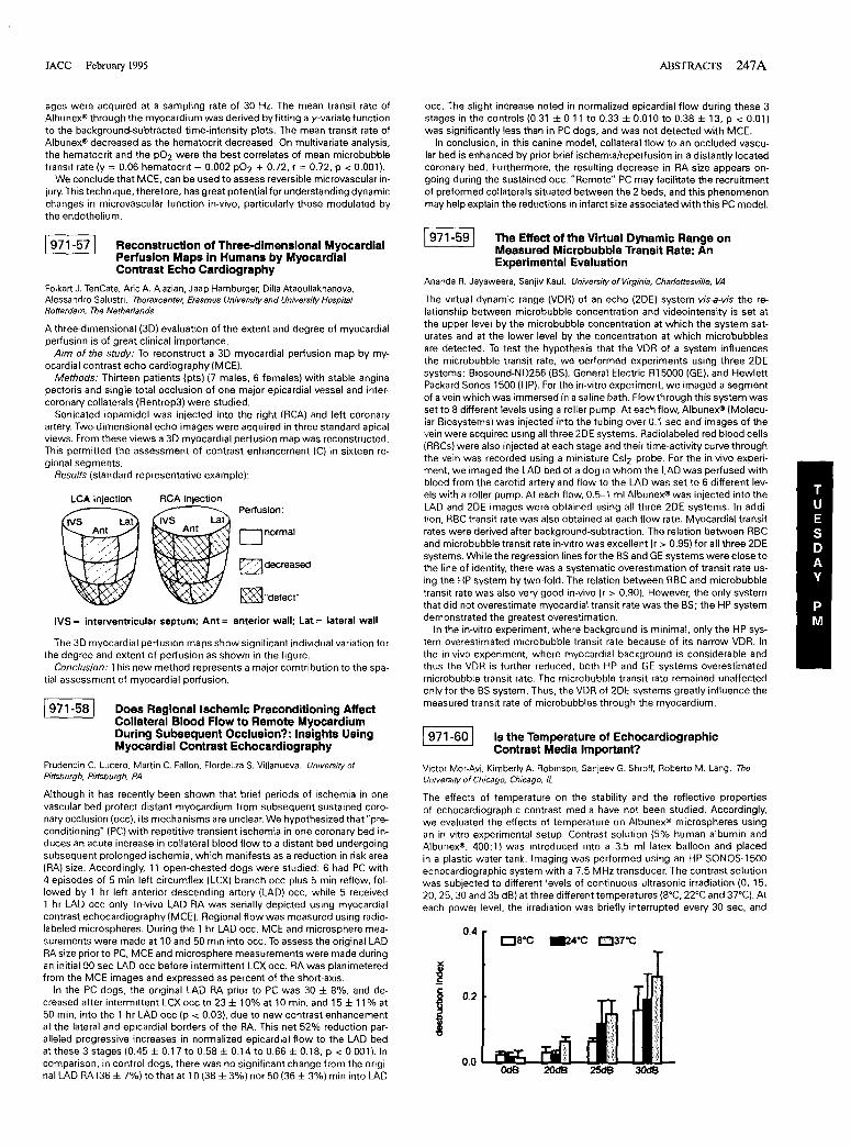
IVS: interventricular septum; Ant = anterior wall; Lat = lateral wall
The Effect of the Virtual Dynamic Range onMeasured Microbubble Transit Rate: AnExperimental Evaluation
Is the Temperature of EchocardiographicContrast Media Important?
Victor Mor-Avi, Kimberly A. Robinson, Sanjeev G. Shroff, Roberto M. Lang. TheUniversity of Chicago, Chicago, IL
Ananda R. Jayaweera, Sanjiv Kaul. University of Virginia, Charlottesville, VA
The virtual dynamic range (VDR) of an echo (2DE) system vis-a-vis the relationship between microbubble concentration and videointensity is set atthe upper level by the microbubble concentration at which the system saturates and at the lower level by the concentration at which microbubblesare detected. To test the hypothesis that the VDR of a system influencesthe microbubble transit rate, we performed experiments using three 2DEsystems: Biosound-ND256 (BS). General Electric RT5000 (GE), and HewlettPackard Sonos 1500 (HP). For the in-vitro experiment, we imaged a segmentof a vein which was immersed in a saline bath. Flowthrough this system wasset to 8 different levels using a roller pump. At each flow, Albunex® (Molecular Biosystems) was injected into the tubing over 0.1 sec and images of thevein were acquired using all three 2DE systems. Radiolabeled red blood cells(RBCs) were also injected at each stage and their time-activity curve throughthe vein was recorded using a miniature Csl2 probe. For the in-vivo experiment. we imaged the LAD bed of a dog in whom the LAD was perfused withblood from the carotid artery and flow to the LAD was set to 6 different levels with a roller pump. At each flow, 0.5-1 ml Albunex® was injected into theLAD and 2DE images were obtained using all three 2DE systems. In addition, RBC transit rate was also obtained at each flow rate. Myocardial transitrates were derived after background-subtraction. The relation between RBCand microbubble transit rate in-vitro was excellent (r > 0.95) for all three 2DEsystems. While the regression lines for the BS and GE systems were close tothe line of identity, there was a systematic overestimation of transit rate using the HP system by two-fold. The relation between RBC and microbubbletransit rate was also very good in-vivo (r > 0.90). However, the only systemthat did not overestimate myocardial transit rate was the BS; the HP systemdemonstrated the greatest overestimation.
In the in-vitro experiment, where background is minimal, only the HP system overestimated microbubble transit rate because of its narrow VDR. Inthe in-vivo experiment, where myocardial background is considerable andthus the VDR is further reduced, both HP and GE systems overestimatedmicrobubble transit rate. The microbubble transit rate remained unaffectedonly for the BS system. Thus, the VDR of 2DE systems greatly influence themeasured transit rate of microbubbles through the myocardium.
1971-591
1971-60 I
~"defect"
~decreased
Dnormal
Perfusion:RCA injection
Reconstruction of Three-dimensional MyocardialPerfusion Maps in Humans by MyocardialContrast Echo Cardiography
LCA injection
IVS Lat
1971-57\
Does Regional Ischemic Preconditioning AffectCollateral Blood Flow to Remote MyocardiumDuring Subsequent Occlusion?: Insights UsingMyocardial Contrast Echocardiography
Prudencio C. Lucero, Martin C. Fallon, Flordeliza S. Villanueva. University ofPittsburgh, Pittsburgh, PA
Folkert J. TenCate, Aric A. Aiazian, Jaap Hamburger, Dilla Ataoullakhanova,Alessandro Salustri. Thoraxcenter. Erasmus University and University HospitalRotterdam, The Netherlands
The 3D myocardial perfusion maps show significant individual variation forthe degree and extent of perfusion as shown in the figure.
Conclusion: This new method represents a major comtribution to the spatial assessment of myocardial perfusion.
A three-dimensional (3D) evaluation of the extent and degree of myocardialperfusion is of great clinical importance.
Aim of the study: To reconstruct a 3D myocardial perfusion map by myocardial contrast echo cardiography (MCE).
Methods: Thirteen patients (pts) (7 males, 6 females) with stable anginapectoris and single total occlusion of one major epicardial vessel and intercoronary collaterals (Rentrop3) were studied.
Sonicated iopamidol was injected into the right (RCA) and left coronaryartery. Two-dimensional echo images were acquired in three standard apicalviews. From these views a 3D myocardial perfusion map was reconstructed.This permitted the assessment of contrast enhancement (C) in sixteen regional segments.
Results (standard representative example):
1971-581
Although it has recently been shown that brief periods of ischemia in onevascular bed protect distant myocardium from subsequent sustained coronary occlusion (occ). its mechanisms are unclear. We hypothesized that "preconditioning" (PC) with repetitive transient ischemia in one coronary bed induces an acute increase in collateral blood flow to a distant bed undergoingsubsequent prolonged ischemia, which manifests as a reduction in risk area(RA) size. Accordingly, 11 open-chested dogs were studied: 6 had PC with4 episodes of 5 min left circumflex (LCX) branch occ plus 5 min reflow, followed by 1 hr left anterior descending artery (LAD) occ, while 5 received1 hr LAD occ only. In-vivo LAD RA was serially depicted using myocardialcontrast echocardiography (MCE). Regional flow was measured using radiolabeled microspheres. During the 1 hr LAD occ, MCE and microsphere measurements were made at 10 and 50 min into occ. To assess the original LADRA size prior to PC, MCE and microsphere measurements were made duringan initial 90 sec LAD occ before intermittent LCX occ. RA was planimeteredfrom the MCE images and expressed as percent of the short-axis.
In the PC dogs, the original LAD RA prior to PC was 30 ± 8%, and decreased after intermittent LCX occ to 23 ± 10% at 10 min, and 15 ± 11 % at50 min, into the 1 hr LAD occ (p < 0.03), due to new contrast enhancementat the lateral and epicardial borders of the RA. This net 52% reduction paralleled progressive increases in normalized epicardial flow to the LAD bedat these 3 stages (0.45 ± 0.17 to 0.58 ± 0.14 to 0.66 ± 0.18, P < 0.001). Incomparison, in control dogs, there was no significant change from the original LAD RA (36 ± 7%) to that at 10 (38 ± 3%) nOr 50 (36 ± 3%) min into LAD
The effects of temperature on the stability and the reflective propertiesof echocardiographic contrast media have not been studied. Accordingly,we evaluated the effects of temperature on Albunex® microspheres usingan in vitro experimental setup. Contrast solution (5% human albumin andAlbunex®, 400:1) was introduced into a 3.5 ml latex balloon and placedin a plastic water tank. Imaging was performed using an HP SONOS-1500echocardiographic system with a 7.5 MHz transducer. The contrast solutionwas subjected to different levels of continuous ultrasonic irradiation (0, 15,20,25,30 and 35 dB) at three different temperatures (8'C, 22'C and 37'C). Ateach power level, the irradiation was briefly interrupted every 30 sec, and
0.4 ewe ~4OC E'J37OC

248A ABSTRACfS JACC February 1995
1972-103/
imaging and digitization performed over a 2 sec period. Mean videointensity in the balloon cross-sectional area was measured for every consecutivedigitized image. Results: (1) Videointensity was found to be directly relatedto temperature induced changes in microbubble volume. (2) Under continuous ultrasonic irradiation, videointensity decreased over time. The slope ofthis decrease, defined as destruction index, correlated with both transmitted power and temperature, reflecting the destructive effects of irradiation,which were more pronounced at higher temperatures.
Conclusion: Temperature is a major factor affecting stability and reflectivity of Albunex® microspheres. To improve the reproducibility of contrastenhancement, the temperature of the contrast media should be carefullycontrolled.
Experimental Myocardial Ischemia/Infarction
farct zone myeloperoxidase was reduced from 0,28 ± 0.1 to 0,1 ± 0,1 U/l00mg (p = 0,03),
Conclusion: Bolus IV administration of TLC-C53 at reperfusion suppressesexpression of ICAM-l and P-Selectin on ischemic but viable endothelium andprevents secondary (presumably lethal) WBC myocardial infiltration, The ef·feet of TLC-C53 on WBC adhesion molecule expression may be less marked,We propose that inhibition of adhesion molecules in ischemic myocardiumis the principle mechanism of infarct salvage by TLC-C53.
A Monoclonal Antibody Directed Against ICAM·1Reduces Myocardial Stunning FollowingIschemia and Reperfusion
David J, Lefer, David M, Flynn, Scott Morehouse, Khoa D, Vo, Andrew J, Buda,Tulane University School of Medicine, New Orleans, LA
Tuesday, March 21, 1995, 3:00 p.m.-5:00 p.m.Ernest N. Morial Convention Center, Hall EPresentation Hour: 4:00 p.m.-5:00 p.m.
Previous studies have demonstrated that monoclonal antibodies (MAbs) thatneutralize ICAM-l reduce myocardial necrosis following ischemia and reper·fusion, We investigated the effects of a MAb directed against rat ICAM-l(1A29) in a rat model of myocardial reperfusion injury, Isolated, Krebs bufferperfused rat hearts (n = 7/group) were subjected to 20 min of global ischemia followed by 45 min of reperfusion, Forty million human neutrophils(PMNs) and 5 cc of rat plasma were infused into each heart during the first5 min of reperfusion, Immunohistochemical staining of ischemic-reperfusedmyocardium revealed prominent endothelial ICAM-1 expression and PMNaccumulation in coronary arterioles and venules in untreated hearts, In addition, left ventricular developed pressure ILVDP), pressure-rate product (PRP).and coronary flow (CF) were measured at baseline and at 5 minute intervals throughout the 45 min reperfusion period, When compared to controlhearts receiving human PMNs and rat plasma alone, treatment with lA291300 iLg/heart) significantly (p < 0.Q1) enhanced recovery of LVOP after 45min of reperfusion (90,8 ± 12,7% vs. 42,8 ± 5,5%), The recovery of PRPat 45 min of reperfusion was 89,7 ± 15,6% in lA29 treated hearts compared to 42,2 ± 5,3% in the control hearts (p < 0,02), Furthermore, treatment with 1A29 significantly preserved CF at 45 min of reperfusion (73,9 ±5.3% vs, 48,9 ± 6,9%, P < 0,05). Histological analysis of hearts receivinglA29 demonstrated a significant reduction in PMN accumulation, We conclude that inhibition of ICAM-l mediated PMN adhesion in the initial phaseof reperfusion significantly attenuates neutrophil induced postischemic myocardial contractile dysfunction,
Diagnostic Abillty of a Single Admission Value ofSerum Myoglobin, Troponin-T and CK·MB InAcute Myocardial Infarction Patients
Birgit Jurlander, Peter Clemmensen, Smen Galatius.Jensen, Galen S, Wagner,Peer Grande, Department ofMedicine B, Rigshospitalet and Hillemd Sygehus,University of Copenhagen, DK: Division of Cardiology, Duke University, N. C.
Admission serum myoglobin 1Mb) and troponin-T(TNT) levels were evaluatedvs admission CK-MB (mass assay) levels and the admission ECG ST elevation(STtl >2 m V. for their diagnostic values in patients with suspected acutemyocardial infarction (AMI) before thrombolytic treatment.
Consecutive patients In = 153), presenting with chest pain at rest >30min but <12 hrs, unresponsive to nitroglycerin, were included, The ultimatediagnosis of AMI was based on a rise and fall pattern of CK-MB (catalyticassay) and a peak value> 15 U/I, and/or the development of Q-waves on theECG.
Cytoklne Antagonists Increase In the CirculationDuring Myocardial Ischemia and Reperfuslon
1 972-101 1
Maddalena Lettino, Anna Catania, Lorena Airaghi, Maria Grazia Manfredi, JamesM. Lipton 1. Ospedale Maggiore di Milano, Milano, Italy: 1UTSouthwestern MedicalCenter. Dallas, TX
In the present research, we tested the idea that cytokine antagonists arereleased during acute myocardial ischemia and reperfusion to counteractpro-inflammatory effects of cytokines. We investigated changes in plasmaconcentrations of the anti-cytokine molecules a-melanocyte stimulating hormone (a-MSH), interleukin-1 receptor antagonist (IL-1 raj, and soluble tumornecrosis factor receptor (s TNF r) in patients with acute myocardial infarction(AMI) or unstable angina (UA) treated with thrombolytic agents compared topatients who did not receive thrombolysis, The study includes 32 pts presenting with prolonged chest pain and ST-segment changes on ECG; thefinal diagnosis was AM I in 24 and UA in 8 patients; 26 pts received thrombolytic therapy, There was no difference in the duration of chest pain atpresentation between the thrombolysis and no thrombolysis groups, Bloodsamples were collected at admission in the CU and at 3 h intervals for 24h, Comparisons of plasma a-MSH concentrations in thrombolysis and untreated groups showed highly significant differences: whereas concentrations of the peptide were elevated in early samples of patients treated witha thrombolytic agent, they were consistently low in untreated subjects, IL-lra levels were likewise greater 3 and 6 h post treatment in patients whounderwent thrombolysis, whereas there was no significant difference forplasma s TN Fr between the two groups. We suggest that during myocardialischemia and reperfusion anti-cytokine molecules released within the injuredmyocardium reduce inflammation caused by cytokines and other mediatorsof inflammation.
Suppression of ICAM-1 and P-Selectin AdhesionMolecule Expression by Bolus IV LlposomalPGE1 (TLC-C53) Immediately Prior toReperfuslon in a Two Hour CanineInfaret/Reperfuslon Model
The admission Mb, TNT and CK-MB concentrations were significantlyhigher in the AMI patients compared to the No-AMI patients, and there wasno overlap of the 95% CI of the medians between the two groups, For AMIdiagnosis, the predictive values of positive and negative tests were 0.84 and0,62 for Mb > 11°iL9/l; 0,70 and 0,62 for TNT >0,02 iLg/l; 1.00 and 0,53 forCK-MB >9,5 iLg/l, and 0.85 and 0,67 for the ECG STt,
Conclusion: A rise in serum Mb and TNT at admission, even below theusual diagnostic cut-off values for AMI diagnosis, has an acceptable diag·nostic accuracy, For more prompt and widespread use of fibrinolytic therapywe propose a lowering of the diagnostic cut-off values for serum Mb, TNTand CK-MB for admission diagnosis of MI.
Richard Smalling, Margaret Uthman, Nagendra Ramanna, Edward Yeh,James Amirian, Patty Felli, Steve Feld, Michel Accad, Andrew Concoff, C.Wayne Smith, Mark Entman, Andrew Janoff, U r Medical School and Baylor Collegeof Medicine, Houston. Texas
Bolus administration of 2 iLlkg Liposomal PGE l (TLC-C53) after 2 hrs of ischemia inhibits WBC accumulation into ischemic tissue and significantly reduces infarct size upon reperfusion. To test the hypothesis that inhibition ofadhesion molecule expression was the mechanism of this observation westudied 11 dogs subjected to 2 hrs of ischemia followed by 3 hrs of reperfusion, Animals were randomly assigned to receive either placebo or 2iL/kgTLC-C53 just prior to reperfusion, Samples obtained from the coronary sinusat baseline and at various intervals throughoutthe experiment were analyzedfor expression of L-Selectin (CL2) and C011 b (MY 904) on WBC using indirect immunofluorescence and flow cytometry. After sacrifice, tissue samplesobtained from the infarct, border, risk and control regions were analyzed forWBC infiltration (myeloperoxidase) and expression of ICAM-1 (CL18) and PSelectin (M03) on the endothelium using immuno-histochemistry, Intensityof staining was graded from °(no staini to 31intense stain),
Results: No differences in WBC mean channel fluorescence between thetreatment and control groups could be detected for either L-Selectin orC011 b. Risk region expression of ICAM-l was reduced from 2,6 ± 0,6 to0.9 ± 0.6 (p < 0,001) and P-Selectin from 1,6 ± 0,6 to 0,9 ± 0,8 (p < 0,01), In-
minutes to admissionadmission Mb (iL9A)admission TNT (itgA)admission CK-M B(iL9A)admission ECG (STtI
AMI (n = 81)(median (95% CII)
179,6 1100-210)123,0 179.~1540)0,08210.03-<l.15)54 (36-7,6)n = 50
No-AMI(n = 72)(median (95% ell)
179.7 (105-240)46.2(40.0-506)
0.003 (0.00-0,01)1.85114-26)n=9
p
NS<0.00001<0.00005<0.00001<0.00005