9/10/2012 chapter 21ems.jbpub.com/aehlert/paramedic/docs/ppt_lectures/chapter_021.pdf · 9/10/2012...
TRANSCRIPT

9/10/2012
1
Chapter 21
Pulmonary
2
Learning Objectives
Explain the importance of the respiratory tract and the prevalence of pulmonary disease
Explain the basic role of pulmonary diagnostic testing in medical care
Identify the anatomy of the upper airway
3
Learning Objectives (Cont’d)
Describe the etiology, epidemiology, history, physical findings; develop a treatment plan for upper respiratory tract infection, epiglottitis, croup, bacterial tracheitis, peritonsillar abscess
Describe the etiology, epidemiology, history, physical findings; develop a treatment plan for upper airway obstruction, trauma, tracheostomy
Copyright © 2013 by Jones & Bartlett Learning, LLC, an Ascend Learning Company

9/10/2012
2
4
Learning Objectives (Cont’d)
Describe the etiology, epidemiology, history, physical findings; develop a treatment plan for disorders of pleura, mediastinum, lung, chest wall: costochondritis, pleurisy, pneumomediastinum, pneumothorax, pleural effusion, noncardiogenic pulmonary edema, acute respiratory distress syndrome
Identify the anatomy of the lower airway
5
Learning Objectives (Cont’d)
Describe the etiology, epidemiology, history, and physical findings; develop a treatment plan for asthma, bronchiolitis, bronchopulmonary dysplasia, chronic obstructive pulmonary disease, cystic fibrosis, pneumonia, lung abscess, pulmonary thromboembolism, hyperventilation syndrome, atelectasis, tumors
6
Learning Objectives (Cont’d)
Describe the etiology, epidemiology, history, and physical findings; develop a treatment plan for pulmonary infection such as pneumonia, tuberculosis, aspiration
Describe the etiology, epidemiology, history, and physical findings; develop a treatment plan for environmental, occupational exposure to inhaled agents/irritants, gases, fumes, vapors
Copyright © 2013 by Jones & Bartlett Learning, LLC, an Ascend Learning Company

9/10/2012
3
7
Introduction
Respiratory tract Gaseous exchange for body Expels waste, balance blood, body chemistry
Patient airway Diseases of the respiratory tract
Upper airway Lower airway
8
Approach to the Patient
Primary assessment Respiratory distress indicators
• Mental status change• Dyspnea at rest• Severe cyanosis• Absent breath sounds• Audible stridor
• Difficulty speaking• Tachycardia• Pallor, diaphoresis• Retractions• Accessory muscle use
9
Approach to the Patient (Cont’d)
History Chief complaint Onset time, activities at onset Prior episodes, diagnoses Past treatments, medical history Medication and compliance Allergies
Copyright © 2013 by Jones & Bartlett Learning, LLC, an Ascend Learning Company

9/10/2012
4
10
Approach to the Patient (Cont’d)
History Assessment
• Infection signs• Cough, productive/nonproductive, color sputum
11
Approach to the Patient (Cont’d)
Physical examination Patient position can provide clues Mental status
12
Approach to the Patient (Cont’d)
Physical examination Respiratory compromise indicators
• Inability to complete sentences• Accessory muscle use• Pursed lips on exhalation• Exaggerated chest movement with minimal air
movement, tracheal tugging• Cyanosis, pallor, diaphoresis
Copyright © 2013 by Jones & Bartlett Learning, LLC, an Ascend Learning Company

9/10/2012
5
13
Approach to the Patient (Cont’d)
Physical examination Vital signs
• Tachycardia, respiratory distress• Bradycardia, severe hypoxia, imminent cardiac arrest• Blood pressure, unchanged in respiratory distress• Respiratory rate, quality, patterns• Extremities
14
Approach to the Patient (Cont’d)
Diagnostic testing Pulse oximetry Peak flow meters
15
Approach to the Patient (Cont’d)
Diagnostic testing Capnography
• Phase I Beginning of exhalation when air from anatomic dead
space is being exhaled Baseline
Copyright © 2013 by Jones & Bartlett Learning, LLC, an Ascend Learning Company

9/10/2012
6
16
Approach to the Patient (Cont’d)
Diagnostic testing Capnography
• Phase II CO2 from larger bronchi begins to pass sensor Expiratory upslope Sharp increase in CO2 concentration passing sensor,
rapid departure of waveform from baseline Rapidly departs from Phase I, vertical line
17
Approach to the Patient (Cont’d)
Diagnostic testing Capnography
• Phase III Alveolar plateau CO2-rich alveolar air passing sensor Flat, straight/slightly angled upward
18
Approach to the Patient (Cont’d)
Diagnostic testing Capnography
• Phase 0 End of exhalation, beginning of inhalation CO2 levels passing sensor quickly drop to 0 Quick return of waveform to baseline Straight line, rapidly returns to baseline
Copyright © 2013 by Jones & Bartlett Learning, LLC, an Ascend Learning Company

9/10/2012
7
19
Approach to the Patient (Cont’d)
Normal Capnogram
20
Approach to the Patient (Cont’d)
Diagnostic testing Capnography
• Changes in any phase when the respiratory system is impaired
• Vertical axis, amount of CO2 exhaled• Horizontal axis, time of exhalation• Junction of Phase II and III, 90° angle• Repetitive, consistent alterations investigated• Expiratory phase prolongation, phase III lengthened
21
Approach to the Patient (Cont’d)
Diagnostic testing Capnography
• Shark fin waveform• ETCO2 determines if bronchoconstriction is acute or
chronic• Phase 0 abnormalities, inhalation problems• Decreased CO2
Copyright © 2013 by Jones & Bartlett Learning, LLC, an Ascend Learning Company

9/10/2012
8
22
Upper Airway
Anatomy of the upper airway Nasopharynx Oropharynx Humidify, clean air entering the lower
respiratory tract Mucus secreted in sinuses, nasopharynx,
oropharynx
23
Upper Airway (Cont’d)
24
Upper Airway (Cont’d)
Assessment Ensure airway
patency Quality, frequency
of respirations Minute volume
Copyright © 2013 by Jones & Bartlett Learning, LLC, an Ascend Learning Company

9/10/2012
9
25
Upper Airway (Cont’d)
Assessment Oropharynyx
• Check for obstruction• Assess mucous membranes, tongue for hydration• Assess aspiration potential
26
Upper Airway (Cont’d)
Assessment of upper airway Retractions
• Examine neck, spaces above clavicles
• Accessory muscle use
• Palpate trachea for deviation
27
Upper Airway (Cont’d)
Retractions
Copyright © 2013 by Jones & Bartlett Learning, LLC, an Ascend Learning Company

9/10/2012
10
28
Upper Airway (Cont’d)
Acute upper airway disorders Upper respiratory tract infection
• Infection of any structures Sinusitis Pharyngitis Laryngitis Tonsilitis
29
Upper Airway (Cont’d)
Acute upper airway disorders Upper respiratory tract
infection• History and physical findings
Headache Nasal congestion Nasal drainage Nasal inflammation Sore throat Cough Mucus production with cough
Fever Chills Muscle aches Once infected,
lifelong immunity to virus
30
Upper Airway (Cont’d)
Acute upper airway disorders Upper respiratory tract infection
• Differential diagnosis May precede serious infection Meningitis Sinusitis Pneumonia
Copyright © 2013 by Jones & Bartlett Learning, LLC, an Ascend Learning Company

9/10/2012
11
31
Upper Airway (Cont’d)
Acute upper airway disorders Upper respiratory tract infection
• Therapeutic interventions Supportive Position of comfort O2
Pulse oximeter Oxygen saturation >95% Respiratory distress signs, IV, cardiac monitor,
capnography
32
Upper Airway (Cont’d)
Acute upper airway disorders Epiglottis
• Potentially life-threatening infection of airway supraglottic structures
• Inflammation on tongue base, aryepiglottic folds, arytenoids, tonsils, epiglottic
33
Upper Airway (Cont’d)
Hypopharynx Structures
Copyright © 2013 by Jones & Bartlett Learning, LLC, an Ascend Learning Company

9/10/2012
12
34
Upper Airway (Cont’d)
Acute upper airway disorders Epiglottis
• Edema, trachea, supraglottic areas Airway
narrowing, closure
35
Upper Airway (Cont’d)
Acute upper airway disorders Epiglottis
• History and physical findings, adults URI precedes Difficulty swallowing Painful swallowing Sore throat Muffled voice tachycardia Pain on palpation of anterior neck Sniffing position
36
Upper Airway (Cont’d)
Acute upper airway disorders Epiglottis
• History and physical findings, children Acute High fever Anxious Sniffing position Breathing difficulty Stridor Voice absence Drooling Difficulty swallowing
Copyright © 2013 by Jones & Bartlett Learning, LLC, an Ascend Learning Company

9/10/2012
13
37
Upper Airway (Cont’d)
Acute upper airway disorders Epiglottis
• Therapeutic interventions Supplemental O2
Rapid transport Must recognize the diagnosis, notify the hospital Airway equipment nearby for impending respiratory
failure
38
Upper Airway (Cont’d)
Acute upper airway disorders Croup
• Upper airway infection
• Just below the glottis
• Swollen, inflamed mucosa
39
Upper Airway (Cont’d)
Acute upper airway disorders Croup
• “Seal bark” cough, 3-4 days• Hoarse, inspiratory stridor• No difficulty swallowing, drooling• Low-grade fever
Copyright © 2013 by Jones & Bartlett Learning, LLC, an Ascend Learning Company

9/10/2012
14
40
Upper Airway (Cont’d)
Acute upper airway disorders Croup
• Minor croup Minimal distress Normal mental status Well hydrated Stridor when agitated Cough intermittent Mild tachycardia Mild tachycardia
41
Upper Airway (Cont’d)
Acute upper airway disorders Croup
• Moderate croup Alert, interactive Irritable Stridor Classic cough Tachypnea Tachycardia Retractions
42
Upper Airway (Cont’d)
Acute upper airway disorders Croup
• Severe croup Respiratory failure, complete obstruction risk Fatigue Altered mental status Hypoxia, hypercarbia Severe respiratory distress Stridor
Copyright © 2013 by Jones & Bartlett Learning, LLC, an Ascend Learning Company

9/10/2012
15
43
Upper Airway (Cont’d)
Acute upper airway disorders Croup
• Therapeutic interventions Patient calm, comfortable, position of comfort Humidified O2, pulse oximeter Oxygen saturation >95% Nebulized saline, epinephrine Transport Respiratory failure/arrest, bag-mask with 100% O2
44
Upper Airway (Cont’d)
Acute upper airway disorders Bacterial tracheitis
• Bacterial infection of the trachea
45
Upper Airway (Cont’d)
Acute upper airway disorders Bacterial tracheitis
• History and physical findings Fever Chills Inspiratory stridor Barking, brassy cough Hoarseness Degree of dyspnea Absent drooling
Copyright © 2013 by Jones & Bartlett Learning, LLC, an Ascend Learning Company

9/10/2012
16
46
Upper Airway (Cont’d)
Acute upper airway disorders Bacterial tracheitis
• Therapeutic interventions Antibiotics Supporting, maintaining airway IV access Antipyretics per protocol Transport, position of comfort Supplemental humidified O2
Respiratory failure/arrest, ET 0.5/1.0 mm smaller than normal
47
Upper Airway (Cont’d)
Acute upper airway disorders Peritonsillar
abscess (PTA)• Bacterial infection
on back of the oropharynx
• Rooted in adenoid tonsil tissue
48
Upper Airway (Cont’d)
Acute upper airway disorders Peritonsillar abscess (PTA)
• History and physical findings Febrile Notable difficulty swallowing Headache Malaise Neck pain “Hot potato voice” Unilateral swelling of posterior throat
Copyright © 2013 by Jones & Bartlett Learning, LLC, an Ascend Learning Company

9/10/2012
17
49
Upper Airway (Cont’d)
Acute upper airway disorders Peritonsillar abscess (PTA)
• Therapeutic interventions Patient calm, comfortable, position of comfort O2, pulse oximeter, saturation >95% If respiratory failure/arrest, bag-mask with 100% O2
IV fluids Antipyretics per protocol
50
Upper Airway (Cont’d)
Acute upper airway disorders Foreign body airway obstruction
• Upper airway blockage by foreign object
51
Upper Airway (Cont’d)
Acute upper airway disorders Foreign body airway obstruction
• Factors Seizures Intoxication by alcohol/drugs Decreased mental status Chronic medical conditions TIA, cerebrovascular accident Respiratory distress Feeding tubes Bowel structures
Copyright © 2013 by Jones & Bartlett Learning, LLC, an Ascend Learning Company

9/10/2012
18
52
Upper Airway (Cont’d)
Acute upper airway disorders Foreign body airway obstruction
• History and physical findings Sudden, severe coughing Wheezing with no asthmatic history Inability to speak Unexplained dyspnea Auscultation of lung, unilateral wheezing, rhonchi,
crackles Retractions Drooling Tripod position
53
Upper Airway (Cont’d)
Acute upper airway disorders Foreign body airway obstruction
• Therapeutic interventions Airway and oxygenation maintenance Supplemental O2
Cough to dislodge
54
Upper Airway (Cont’d)
Acute upper airway disorders Foreign body airway obstruction
• Complete obstruction Abdominal/chest thrusts Visual airway inspection, Magill forceps Suction Cricrothyrotomy per medical direction Immediate removal
Copyright © 2013 by Jones & Bartlett Learning, LLC, an Ascend Learning Company

9/10/2012
19
55
Upper Airway (Cont’d)
Acute upper airway disorders Trauma
• Penetrating, blunt neck trauma can be life-threatening
56
Upper Airway (Cont’d)
Acute upper airway disorders Trauma
• History and physical findings MVC, blunt force seen Cricoid cartilage positioned anteriorly, airway
obstruction Tracheal rings bruised, edema threatens airway
57
Upper Airway (Cont’d)
Acute upper airway disorders Trauma
• Therapeutic interventions Cervical spine precautions Changes in vital signs, dyspnea, changes in voice,
hoarseness, upper airway stridor, accessory muscle use
Bleeding, aggressive airway management In-line cervical spine stabilization Suction, oxygenation
Copyright © 2013 by Jones & Bartlett Learning, LLC, an Ascend Learning Company

9/10/2012
20
58
Upper Airway (Cont’d)
Acute upper airway disorders Tracheostomy
• Hole surgically placed in the trachea to support respiration
• Indications Obstructive sleep apnea Pickwickian syndrome Upper airway obstruction/cancer Laryngeal cancer Ventilator support for 2+ weeks
59
Upper Airway (Cont’d)
Tracheostomy
60
Upper Airway (Cont’d)
Acute upper airway disorders Tracheostomy
• Drawbacks Lung infections Pneumonia
Copyright © 2013 by Jones & Bartlett Learning, LLC, an Ascend Learning Company

9/10/2012
21
61
Upper Airway (Cont’d)
Acute upper airway disorders Tracheostomy
• Therapeutic interventions Dyspnea, additional O2
Assisted with bag-mask connected directly to tracheostomy hub
Displacement, transfer to ED Intubate through ostomy hole
62
Pleura, Mediastinum, Chest Wall Disorders
Anatomy and physiology Chest wall
• Multiple layers of skin, bone, muscle, connective tissue
63
Pleura, Mediastinum, Chest Wall Disorders (Cont’d)
Thorax Bones, Muscles
Copyright © 2013 by Jones & Bartlett Learning, LLC, an Ascend Learning Company

9/10/2012
22
64
Pleura, Mediastinum, Chest Wall Disorders (Cont’d)
Anatomy and physiology Respiration
• Inspiration, signal passed from brain
• Diaphragm contracts• Thoracic cavity
increases
65
Pleura, Mediastinum, Chest Wall Disorders (Cont’d)
Anatomy and physiology Mediastinum
• Area between lungs• Heart• Aorta• Inferior, superior venae cava• Trachea• Main bronchi• Does not directly contribute to respiration• Allows return of deoxygenated blood, circulation of
oxygenated blood
66
Pleura, Mediastinum, Chest Wall Disorders (Cont’d)
Mediastinum
Copyright © 2013 by Jones & Bartlett Learning, LLC, an Ascend Learning Company

9/10/2012
23
67
Pleura, Mediastinum, Chest Wall Disorders (Cont’d)
Anatomy and physiology Chemosensitive areas
• CO2 levels increase, respirations increase• CO2 levels decrease, respirations decrease
Hypoxic drive• Primary drive for respiration fails, drives respirations• Never withhold O2
68
Costochondritis Benign chest pain History and physical findings
• Inflammation of cartilage connecting the ribs to sternum
• Joints affected• Infection• Strain, sprain• Worse with deep breathing
Pleura, Mediastinum, Chest Wall Disorders (Cont’d)
69
Pleura, Mediastinum, Chest Wall Disorders (Cont’d)
Costochondritis Therapeutic interventions
• Antiinflammatory medications• O2
• IV access• Pulse oximetry, capnography, cardiac monitoring• Analgesics per medical direction
Copyright © 2013 by Jones & Bartlett Learning, LLC, an Ascend Learning Company

9/10/2012
24
70
Pleura, Mediastinum, Chest Wall Disorders (Cont’d)
Pleurisy Painful rubbing of pleural lining Etiology
• Visceral pleura• Parietal pleura
71
Pleura, Mediastinum, Chest Wall Disorders (Cont’d)
Pleurisy History and physical findings
• Pain worsened by breathing• Shallow breaths• Nausea, vomiting• Diaphoresis• Auscultation of lungs reveals “rub”• Hypoxic from decreased respirations to avoid pain
72
Pleura, Mediastinum, Chest Wall Disorders (Cont’d)
Pleurisy Therapeutic interventions
• Oxygenation saturation• Monitoring capnography• ECG monitoring• IV access
Copyright © 2013 by Jones & Bartlett Learning, LLC, an Ascend Learning Company

9/10/2012
25
73
Pleura, Mediastinum, Chest Wall Disorders (Cont’d)
Pneumomediastinum Air in mediastinum Etiology
• Spontaneously• Chest trauma• Mechanical ventilation• Asthma• Emphysema• Lung/chest tumors• Cocaine use
• Violent emesis/coughing
• Childbirth• Alveoli rupture, air
into surrounding structures
• Bacteria, infection
74
Pleura, Mediastinum, Chest Wall Disorders (Cont’d)
Pneumomediastinum Air in mediastinum
• History and physical findings Indistinct chest pain Mild dyspnea Subcutaneous emphysema Hamman’s sign
75
Pleura, Mediastinum, Chest Wall Disorders (Cont’d)
Pneumomediastinum Air in mediastinum
• Therapeutic interventions Oxygenation Ventilation IV access ECG monitoring Rapid transport
Copyright © 2013 by Jones & Bartlett Learning, LLC, an Ascend Learning Company

9/10/2012
26
76
Lower Airway
Anatomy of lower airway Trachea Bronchi Bronchioles Alveoli
77
Lower Airway (Cont’d)
Anatomy of lower airway Sterile Mucus production Air distributed to alveoli Metabolism byproducts
78
Lower Airway (Cont’d)
Lower Airway Structure
Copyright © 2013 by Jones & Bartlett Learning, LLC, an Ascend Learning Company

9/10/2012
27
79
Lower Airway (Cont’d)
Lung Tissue Gas Exchange
80
Lower Airway (Cont’d)
Assessment Continuous Discontinuous Both upper lobes Middle lung Lower lobes Posterior from the
bottom
81
Lower Airway (Cont’d)
Lung Lobes
Copyright © 2013 by Jones & Bartlett Learning, LLC, an Ascend Learning Company

9/10/2012
28
82
Lower Airway (Cont’d)
Assessment Abnormal breath sounds
• Stridor Harsh, high-pitched inspiratory sound over the neck Restricted movement through the upper airway Larynx, trachea obstruction
83
Lower Airway (Cont’d)
Assessment Abnormal breath sounds
• Crackles Rales, wet lung sounds Fluid in smaller airways Pneumonia, pulmonary edema Inhalation, discontinuous Fine Medium Coarse
84
Lower Airway (Cont’d)
Assessment Abnormal breath sounds
• Rhonchi Rattling, rumbling Continuous, louder during exhalation Fluid in larger airways Cleared by coughing Pneumonia Congestion from URIs, COPD
Copyright © 2013 by Jones & Bartlett Learning, LLC, an Ascend Learning Company

9/10/2012
29
85
Lower Airway (Cont’d)
Assessment Abnormal breath sounds
• Wheezing Musical whistling Turbulent air movement through the constricted
bronchioles Continuous, louder during exhalation Pitches singular, variable
86
Lower Airway (Cont’d)
Assessment Abnormal breath sounds
• Wheezing causes Asthma, COPD Toxic inhalation Bronchospasm Congestive heart failure Emphysema Croup
Pneumothorax Pneumonia Anaphylaxis Foreign body
obstruction, tumor
Pulmonary edema
87
Lower Airway (Cont’d)
Traumatic pleural and pulmonary injuries Pneumothorax
• Partial/full lung collapse
Copyright © 2013 by Jones & Bartlett Learning, LLC, an Ascend Learning Company

9/10/2012
30
88
Lower Airway (Cont’d)
Traumatic pleural and pulmonary injuries Pneumothorax
• Etiology Trauma Rib fractures Spontaneous without underlying lung disease Spontaneous with underlying lung disease Mechanical ventilation Barotrauma
89
Lower Airway (Cont’d)
Traumatic pleural and pulmonary injuries Pneumothorax
• Sudden onset Dyspnea Chest pain Tachycardia hypoxia Hyperpnea Cough
Diaphoresis Altered mentation Cyanosis Subcutaneous
emphysema
90
Lower Airway (Cont’d)
Traumatic pleural and pulmonary injuries Pneumothorax
• Therapeutic interventions Oxygenation, ventilation IV access Pulse oximetry Capnography ECG monitoring Position of comfort Tension pneumothorax possible
Copyright © 2013 by Jones & Bartlett Learning, LLC, an Ascend Learning Company

9/10/2012
31
91
Nontraumatic Lung Diseases
Pleural effusion Fluid collects in the
pleural cavity• Water, protein,
white blood cells, plasma components
92
Nontraumatic Lung Diseases (Cont’d)
Pleural effusion Causes
• Congestive heart failure• Bacterial pneumonia• Cancer• Pulmonary embolus• Advanced liver disease• Pancreatitis• Vascular disease• TB
93
Nontraumatic Lung Diseases (Cont’d)
Pleural effusion History and physical findings
• Underlying causes• No breath sounds in lower lung part• Pleural friction rub• Tactile fremitus• Egophony• Bronchophony• Accessory muscle use
Copyright © 2013 by Jones & Bartlett Learning, LLC, an Ascend Learning Company

9/10/2012
32
94
Nontraumatic Lung Diseases (Cont’d)
Pleural effusion Therapeutic interventions
• Oxygenation, ventilation• IV access• Pulse oximetry• Capnography• ECG monitoring
95
Nontraumatic Lung Diseases (Cont’d)
Noncardiogenic pulmonary edema Condition in which
fluid builds up in the alveoli in the absence of heart failure
96
Nontraumatic Lung Diseases (Cont’d)
Noncardiogenic pulmonary edema Etiology
• High permeability in capillary beds• Fluid leaks into the interstitial space and alveoli• Plasma proteins leave the capillary beds, increase
oncotic pressure, further fluid escape• Surfactant production decreases, alveolar collapse• Widened interstitial space interferes with diffusion,
CO2
Copyright © 2013 by Jones & Bartlett Learning, LLC, an Ascend Learning Company

9/10/2012
33
97
Nontraumatic Lung Diseases (Cont’d)
Noncardiogenic pulmonary edema Causes
• High altitude• Pulmonary embolus• Drowning• Acute glomerulonephritis• Fluid overload• Aspiration• Inhalation injury
• Neurogenic pulmonary edema
• Allergic reaction• Acute respiratory
distress syndrome• Cause not
understood
98
Nontraumatic Lung Diseases (Cont’d)
Noncardiogenic pulmonary edema History and physical findings
• Distinguish from heart failure Lack of jugular venous distention Lack of peripheral edema Adequate cardiac output, end-organ perfusion Initial insult
99
Nontraumatic Lung Diseases (Cont’d)
Noncardiogenic pulmonary edema Primary findings
• Dyspnea• Orthopnea• Crackles• Rales• Tachypnea• Tachycardia• Hypoxemia• Hypoxia• Anxiety
Copyright © 2013 by Jones & Bartlett Learning, LLC, an Ascend Learning Company

9/10/2012
34
100
Nontraumatic Lung Diseases (Cont’d)
Noncardiogenic pulmonary edema Therapeutic interventions
• Oxygenation, ventilation• IV access• Pulse oximetry, capnography, ECG monitoring• Continuous positive airway pressure• Elevate the upper body, dangle the feet• Inflatable pressure bag, descend altitude• Diuretics
101
Nontraumatic Lung Diseases (Cont’d)
Acute respiratory distress syndrome Alveoli damaged from illness/injury Impair O2, CO2 exchange Respiratory failure, dyspnea, hypoxia,
pulmonary edema
102
Nontraumatic Lung Diseases (Cont’d)
Acute respiratory distress syndrome Possible triggers
• Aspiration• Cardiopulmonary bypass surgery• Sepsis• Multiple blood transfusions• O2 toxicity• Trauma, burns• Pneumonia, TB
Copyright © 2013 by Jones & Bartlett Learning, LLC, an Ascend Learning Company

9/10/2012
35
103
Nontraumatic Lung Diseases (Cont’d)
Acute respiratory distress syndrome History and physical findings
• Preceded by massive insult• Signs
Shortness of breath Rapid breathing Inadequate oxygenation Decreased lung compliance
104
Nontraumatic Lung Diseases (Cont’d)
Acute respiratory distress syndrome Therapeutic interventions
• O2
• Pulse, oximeter, capnography, ECG monitor• Positive end-expiratory pressure/continuous positive
airway pressure
105
Nontraumatic Lung Diseases (Cont’d)
Obstructive and restrictive pulmonary diseases Asthma
• Reactive airway disease• Maintains normal acid-base balance• Prolonged attack, respiratory acidosis• Components
Bronchospasm Bronchial edema Excessive mucus production
Copyright © 2013 by Jones & Bartlett Learning, LLC, an Ascend Learning Company

9/10/2012
36
106
Nontraumatic Lung Diseases (Cont’d)
Obstructive and restrictive pulmonary diseases Asthma
• Etiology and demographics Allergies Middle, lower airways develop edema Bronchial smooth muscle contraction Bronchial wall edema, thickening Thick secretions Preceded by upper respiratory infection
107
Nontraumatic Lung Diseases (Cont’d)
Obstructive and restrictive pulmonary diseases Asthma
• History and physical findings Exposure to trigger causes acute onset of respiratory
distress Bronchial collapse more prevalent during exhalation,
air trapping, hyperinflation of lungs Chest tightness Inhalation inability
108
Nontraumatic Lung Diseases (Cont’d)
Obstructive and restrictive pulmonary diseases Asthma
• History and physical findings Current compared to past episodes Successful and unsuccessful therapies Prescribed inhaler use Medical condition, contraindication for corticosteroids
Copyright © 2013 by Jones & Bartlett Learning, LLC, an Ascend Learning Company

9/10/2012
37
109
Nontraumatic Lung Diseases (Cont’d)
Obstructive and restrictive pulmonary diseases Asthma
• History and physical findings Other medications may have caused attack, inhibit
treatment (aspirin, beta-blockers) Tripod position Dyspnea, wheezing, cough Distinguish between clear and absent lung sounds
110
Nontraumatic Lung Diseases (Cont’d)
Obstructive and restrictive pulmonary diseases Asthma
• Findings Anxiety Agitation Tachypnea Tachycardia Difficulty completing
sentences
Prolonged expiration Pallor, cyanosis Decreased muscle
tone Accessory muscle
use
111
Nontraumatic Lung Diseases (Cont’d)
Obstructive and restrictive pulmonary diseases Asthma
• Impending respiratory failure Patient fatigue Absent breath sounds Profound diaphoresis Altered mental status
Copyright © 2013 by Jones & Bartlett Learning, LLC, an Ascend Learning Company

9/10/2012
38
112
Nontraumatic Lung Diseases (Cont’d)
Obstructive and restrictive pulmonary diseases Asthma
• Peak flow meter
113
Nontraumatic Lung Diseases (Cont’d)
Obstructive and restrictive pulmonary diseases Asthma
• Capnography Bronchoconstriction, prolonged expiration before
shark fin waveform Elevated ETCO2 levels, prolonged episode
114
Nontraumatic Lung Diseases (Cont’d)
Obstructive and restrictive pulmonary diseases Asthma
• Therapeutic interventions Ventilation, oxygenation Position of comfort, humidified O2
Continuous positive airway pressure (CPAP) Pulse oximeter, capnography cardiac monitor
Copyright © 2013 by Jones & Bartlett Learning, LLC, an Ascend Learning Company

9/10/2012
39
115
Nontraumatic Lung Diseases (Cont’d)
Obstructive and restrictive pulmonary diseases Asthma
• Therapeutic interventions Nebulizer therapy IV corticosteroids, adjunct Theophylline, in addition to inhaled beta agonists,
adjunct Epinephrine, full respiratory failure, intramuscularly Status asthmaticus, respiratory failure
116
Nontraumatic Lung Diseases (Cont’d)
Obstructive and restrictive pulmonary diseases Bronchiolitis
• Acute, infectious inflammatory disease• Upper, lower airway tracts• Obstruction of small airways
117
Nontraumatic Lung Diseases (Cont’d)
Obstructive and restrictive pulmonary diseases Bronchiolitis
• Risk factors Low birth weight Crowded living conditions Daycare exposure Smoking exposure Chronic heart and lung conditions Respiratory syncytial virus
Copyright © 2013 by Jones & Bartlett Learning, LLC, an Ascend Learning Company

9/10/2012
40
118
Nontraumatic Lung Diseases (Cont’d)
Obstructive and restrictive pulmonary diseases Bronchiolitis
• History and physical findings Bronchiole obstruction from inflammation, edema Hyperinflation of the lungs Increased wheezing Course crackles
119
Nontraumatic Lung Diseases (Cont’d)
Obstructive and restrictive pulmonary diseases Bronchiolitis
• History and physical findings Nasal flaring Grunting Cyanosis Crackles Wheezing Apnea rarely
Atelectasis Dehydration differentiates
it from asthma Tachycardia Tachypnea Accessory muscle use
120
Nontraumatic Lung Diseases (Cont’d)
Obstructive and restrictive pulmonary diseases Bronchiolitis
• Therapeutic interventions Supportive Position of comfort Pulse oximeter, capnography O2, saturation >95% Nebulized beta agonists Hydration
Copyright © 2013 by Jones & Bartlett Learning, LLC, an Ascend Learning Company

9/10/2012
41
121
Nontraumatic Lung Diseases (Cont’d)
Obstructive and restrictive pulmonary diseases Bronchopulmonary dysplasia
• Child needing O2 supplementation at 28 days, maintain PO2 of 50 mm Hg
• Inflammation, scarring of smaller airways and alveoli
122
Nontraumatic Lung Diseases (Cont’d)
Obstructive and restrictive pulmonary diseases Bronchopulmonary dysplasia
• Etiology and demographics Prolonged positive-pressure ventilation, high O2
concentrations
123
Nontraumatic Lung Diseases (Cont’d)
Obstructive and restrictive pulmonary diseases Bronchopulmonary dysplasia
• History and physical findings Premature, low birth weight Tachypnea Tachycardia Retractions Nasal flaring
Copyright © 2013 by Jones & Bartlett Learning, LLC, an Ascend Learning Company

9/10/2012
42
124
Nontraumatic Lung Diseases (Cont’d)
Obstructive and restrictive pulmonary diseases Bronchopulmonary dysplasia
• Auscultation Decreased breath sounds Rhonchi Fine crackles Wheezing
125
Nontraumatic Lung Diseases (Cont’d)
Obstructive and restrictive pulmonary diseases Bronchopulmonary dysplasia
• Therapeutic interventions O2, be careful of amount O2 saturation, 90-95% Supportive care
126
Nontraumatic Lung Diseases (Cont’d)
Obstructive and restrictive pulmonary diseases COPD
• Description Bronchitis Emphysema Asthma Varying degrees/combination Long-term tobacco abuse, exposure to inhaled toxins
Copyright © 2013 by Jones & Bartlett Learning, LLC, an Ascend Learning Company

9/10/2012
43
127
Nontraumatic Lung Diseases (Cont’d)
Obstructive and restrictive pulmonary diseases COPD
• Bronchitis is predominant Mucus overproduction Cell enlargement in lungs, airways Productive cough 3+ months, 2+ years Hypoventilation of alveoli, drops O2 level in blood Acidosis Increased cardiac output, RBC production
128
Nontraumatic Lung Diseases (Cont’d)
Normal Lung Bronchitis
129
Nontraumatic Lung Diseases (Cont’d)
Obstructive and restrictive pulmonary diseases COPD
• Emphysema Involves alveoli Alveolar destruction Alveolar coalescence Destruction of elastin fibers surrounding the alveoli Chronic hypoxia, hypercarbia
Copyright © 2013 by Jones & Bartlett Learning, LLC, an Ascend Learning Company

9/10/2012
44
130
Nontraumatic Lung Diseases (Cont’d)
Obstructive and restrictive pulmonary diseases COPD
• Emphysema Blebs on lung surface, possible pneumothorax Polycythemia Muscle wasting, malnourished appearance Barrel chest
131
Nontraumatic Lung Diseases (Cont’d)
Obstructive and restrictive pulmonary diseases COPD
• Emphysema Chronic dyspnea Little or no cough, little mucus production Tripod position Mental status changes Heart problems, cor pulmonale, ventricular failure
132
Nontraumatic Lung Diseases (Cont’d)
Obstructive and restrictive pulmonary diseases COPD
• Asthma Bronchiole hyperstimulation, constriction Wheezing, dyspnea
Copyright © 2013 by Jones & Bartlett Learning, LLC, an Ascend Learning Company

9/10/2012
45
133
Nontraumatic Lung Diseases (Cont’d)
Obstructive and restrictive pulmonary diseases COPD
• Therapeutic interventions Oxygenation,
ventilation O2 saturation at
least 90%
134
Nontraumatic Lung Diseases (Cont’d)
Obstructive and restrictive pulmonary diseases COPD
• Therapeutic interventions Respiratory distress causes CO2 narcosis Reduce bronchoconstriction airway secretions IV access ECG monitoring, pulse oximetry, capnography, peak
expiratory flow testing Transport, reassess
135
Nontraumatic Lung Diseases (Cont’d)
Obstructive and restrictive pulmonary diseases Cystic fibrosis
• Genetic disease• Pulmonary distress complication
Copyright © 2013 by Jones & Bartlett Learning, LLC, an Ascend Learning Company

9/10/2012
46
136
Nontraumatic Lung Diseases (Cont’d)
Obstructive and restrictive pulmonary diseases Cystic fibrosis
• Etiology and demographics Thicker secretions Lung disease Liver, pancreas Whites Median life span, 36.8 years
137
Nontraumatic Lung Diseases (Cont’d)
Obstructive and restrictive pulmonary diseases Cystic fibrosis
• History and physical findings Similar presentation to COPD, pneumonia Cough Chest wall pain, tender Dyspnea Fever Crackles
138
Nontraumatic Lung Diseases (Cont’d)
Obstructive and restrictive pulmonary diseases Cystic fibrosis
• Therapeutic interventions Supplemental O2, ventilation Monitor oxygenation, ventilatory, cardiac functions
Copyright © 2013 by Jones & Bartlett Learning, LLC, an Ascend Learning Company

9/10/2012
47
139
Nontraumatic Lung Diseases (Cont’d)
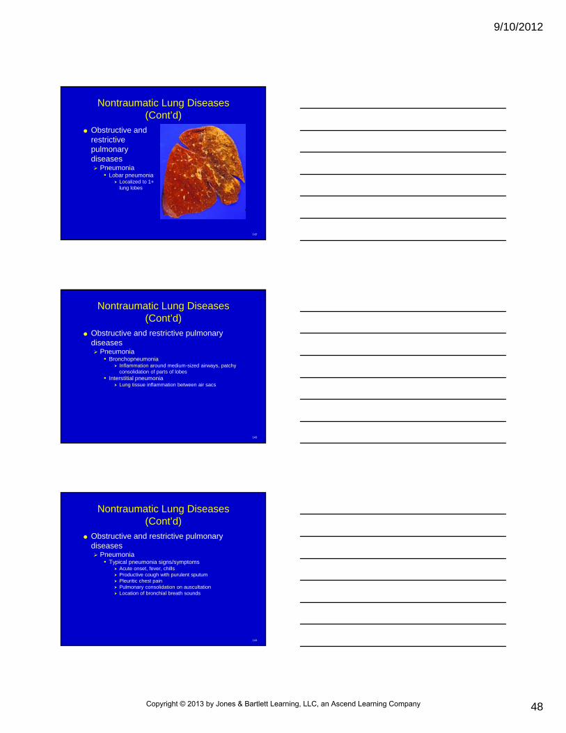
Obstructive and restrictive pulmonary diseases Pneumonia
• Infection in terminal breathing spaces, alveoli
140
Nontraumatic Lung Diseases (Cont’d)
Obstructive and restrictive pulmonary diseases Pneumonia
• Virus Respiratory syncytial virus Parainfluenza Influenza Adenovirus
141
Nontraumatic Lung Diseases (Cont’d)
Obstructive and restrictive pulmonary diseases Pneumonia
• Bacteria Haemophilus influenzae Streptococcus pneumoniae Mycoplasma spp. Klebsiella spp. Pseudomonas spp.
• Fungi
Copyright © 2013 by Jones & Bartlett Learning, LLC, an Ascend Learning Company
9/10/2012
48
142
Nontraumatic Lung Diseases (Cont’d)
Obstructive and restrictive pulmonary diseases Pneumonia
• Lobar pneumonia Localized to 1+
lung lobes
143
Nontraumatic Lung Diseases (Cont’d)
Obstructive and restrictive pulmonary diseases Pneumonia
• Bronchopneumonia Inflammation around medium-sized airways, patchy
consolidation of parts of lobes• Interstitial pneumonia
Lung tissue inflammation between air sacs
144
Nontraumatic Lung Diseases (Cont’d)
Obstructive and restrictive pulmonary diseases Pneumonia
• Typical pneumonia signs/symptoms Acute onset, fever, chills Productive cough with purulent sputum Pleuritic chest pain Pulmonary consolidation on auscultation Location of bronchial breath sounds
Copyright © 2013 by Jones & Bartlett Learning, LLC, an Ascend Learning Company

9/10/2012
49
145
Nontraumatic Lung Diseases (Cont’d)
Obstructive and restrictive pulmonary diseases Pneumonia
• Typical pneumonia signs/symptoms Crackles Lethargy Anorexia Tachypnea Tachycardia Chest, side, back pain
146
Nontraumatic Lung Diseases (Cont’d)
Obstructive and restrictive pulmonary diseases Pneumonia
• Atypical pneumonia Nonproductive cough Extrapulmonary symptoms Headache Myalgias Fatigue Sore throat Nausea, vomiting, diarrhea Fever, chills
147
Nontraumatic Lung Diseases (Cont’d)
Obstructive and restrictive pulmonary diseases Pneumonia
• Complicating factors, difficult treatment Age COPD Heart disease Alcoholism Diabetes AIDS
Copyright © 2013 by Jones & Bartlett Learning, LLC, an Ascend Learning Company

9/10/2012
50
148
Nontraumatic Lung Diseases (Cont’d)
Obstructive and restrictive pulmonary diseases Pneumonia
• Therapeutic interventions Optimize oxygenation Respiratory status monitoring ET intubation IV access Hypovolemic, septic shock treatment Antibiotics Personal protective equipment (PPE)
149
Nontraumatic Lung Diseases (Cont’d)
Obstructive and restrictive pulmonary diseases Lung abscess
• Pus collections in lung tissues
• Aspirated gastric contents
150
Nontraumatic Lung Diseases (Cont’d)
Obstructive and restrictive pulmonary diseases Lung abscess
• History and physical findings Productive cough of sputum with unusual odors Dyspnea Fever Night sweats Decreased appetite Weight loss Chest/chest wall pain
Copyright © 2013 by Jones & Bartlett Learning, LLC, an Ascend Learning Company

9/10/2012
51
151
Nontraumatic Lung Diseases (Cont’d)
Obstructive and restrictive pulmonary diseases Lung abscess
• History and physical findings Low grade fever Tachypnea Tachycardia Crackles Rhonchi Decreased breath sounds Percussion dullness Empyema, pus collection outside lung in pleural space
152
Nontraumatic Lung Diseases (Cont’d)
Obstructive and restrictive pulmonary diseases Lung abscess
• Therapeutic interventions Supplemental O2
Supportive care Intubate Antibiotics
153
Nontraumatic Lung Diseases (Cont’d)
Obstructive and restrictive pulmonary diseases Aspiration pneumonia and pneumonitis
• Gastric acid, food• Inflammatory response• Hypoxia, respiratory failure
Copyright © 2013 by Jones & Bartlett Learning, LLC, an Ascend Learning Company

9/10/2012
52
154
Nontraumatic Lung Diseases (Cont’d)
Obstructive and restrictive pulmonary diseases Aspiration pneumonia and pneumonitis
• At risk Altered mental status Intoxicated Chronic disability Feeding tubes Stroke history Head trauma Airway control problems
155
Nontraumatic Lung Diseases (Cont’d)
Obstructive and restrictive pulmonary diseases Aspiration pneumonia and pneumonitis
• Aspiration pneumonitis Lung, bronchoalveolar irritation, aspirated stomach
acid Swelling in alveoli
• Aspiration pneumonia Bacteria in lower lungs Pulmonary abscess possible
156
Nontraumatic Lung Diseases (Cont’d)
Obstructive and restrictive pulmonary diseases Aspiration pneumonia and pneumonitis
• History and physical findings Dyspnea, airway obstruction Altered mental status Fever Chills Dyspnea on exertion Orthopnea Pleuritic chest pain Productive cough Respiratory distress
Copyright © 2013 by Jones & Bartlett Learning, LLC, an Ascend Learning Company

9/10/2012
53
157
Nontraumatic Lung Diseases (Cont’d)
Obstructive and restrictive pulmonary diseases Aspiration pneumonia and pneumonitis
• Therapeutic interventions Aggressive airway control IV access Standard monitoring
158
Nontraumatic Lung Diseases (Cont’d)
Obstructive and restrictive pulmonary diseases Pulmonary
tuberculosis• Pulmonary disease• Mycobacterium
tuberculosis• Transmitted via
airborne droplets
159
Nontraumatic Lung Diseases (Cont’d)
Obstructive and restrictive pulmonary diseases Pulmonary TB
• Stage 1 Bacterium inhaled into lungs Standard inflammatory, immune responses triggered If defenses contain, eradicate bacteria, does not
progress
Copyright © 2013 by Jones & Bartlett Learning, LLC, an Ascend Learning Company

9/10/2012
54
160
Nontraumatic Lung Diseases (Cont’d)
Obstructive and restrictive pulmonary diseases Pulmonary TB
• Stage 2 Bacteria multiply rapidly Tubercle formed, infected lung area Infection spreads throughout the lymphatic system,
vascular system, body Possible extrapulmonary TB Pericarditis, peritonitis, GI disease
161
Nontraumatic Lung Diseases (Cont’d)
Obstructive and restrictive pulmonary diseases Pulmonary TB
• Stage 3 2-3 weeks after first stage Inflammatory, immune responses contain spread of
infectious organisms Granulomas formed, tissue destruction occurs Dormant survival, years
162
Nontraumatic Lung Diseases (Cont’d)
Obstructive and restrictive pulmonary diseases Pulmonary TB
• Stage 4 Dormant bacteria reactivates Tubercle erodes, releases contained bacteria back
into the lung
Copyright © 2013 by Jones & Bartlett Learning, LLC, an Ascend Learning Company

9/10/2012
55
163
Nontraumatic Lung Diseases (Cont’d)
Obstructive and restrictive pulmonary diseases Pulmonary TB
• Active TB Productive cough Fever Weight, appetite loss Hemoptysis Chest, chest wall pain Fatigue
Irritability Weakness Headache Chills Fever Night sweats
164
Nontraumatic Lung Diseases (Cont’d)
Obstructive and restrictive pulmonary diseases Pulmonary TB
• Elderly Atypical presentation Chronic cough Failure to thrive Fewer respiratory symptoms Fever Dyspnea Rhonchi, rales Upper lobe affinity
165
Nontraumatic Lung Diseases (Cont’d)
Obstructive and restrictive pulmonary diseases Pulmonary TB
• Therapeutic interventions Detailed history of TB exposure Initiate respiratory isolation with masks Notify the ED ASAP Emergent risk PPE
Copyright © 2013 by Jones & Bartlett Learning, LLC, an Ascend Learning Company

9/10/2012
56
166
Nontraumatic Lung Diseases (Cont’d)
Obstructive and restrictive pulmonary diseases Pulmonary thromboembolism
167
Nontraumatic Lung Diseases (Cont’d)
Obstructive and restrictive pulmonary diseases Pulmonary thromboembolism
• Pulmonary embolus• Thrombus becomes dislodged, travels through the
bloodstream• Lodges in the pulmonary artery, obstructs blood flow
to portion of the lung• Often start in leg, through inferior vena cava, to right
ventricle, pulmonary artery
168
Nontraumatic Lung Diseases (Cont’d)
Obstructive and restrictive pulmonary diseases Pulmonary thromboembolism
• Etiology and demographics Difficult to diagnose Wide range of clinical signs Sudden death Comorbid conditions
Copyright © 2013 by Jones & Bartlett Learning, LLC, an Ascend Learning Company

9/10/2012
57
169
Nontraumatic Lung Diseases (Cont’d)
Obstructive and restrictive pulmonary diseases Pulmonary thromboembolism
• History and physical findings Virchow’s triad Sedentary lifestyle Smoking Deep vein thrombosis history Long bone fractures Pregnancy Recent leg or hip surgery Contraceptive use Stasis of position
170
Nontraumatic Lung Diseases (Cont’d)
Obstructive and restrictive pulmonary diseases Pulmonary thromboembolism
• Right-to-left shunt• Atelectasis• Hypoxemia• Pulmonary hypertension• Right heart strain
171
Nontraumatic Lung Diseases (Cont’d)
Obstructive and restrictive pulmonary diseases Pulmonary thromboembolism
• Hypotension• Acute onset dyspnea (50%)• Pleuritic chest pain (two-thirds)• Anxiety• Tachycardia• Fever
Copyright © 2013 by Jones & Bartlett Learning, LLC, an Ascend Learning Company

9/10/2012
58
172
Nontraumatic Lung Diseases (Cont’d)
Obstructive and restrictive pulmonary diseases Pulmonary thromboembolism
• Cough• Chills• Mucus production• Tachypnea• Skin cool, pale, diaphoretic, cyanotic• If hypotensive, shock index >1
173
Nontraumatic Lung Diseases (Cont’d)
Obstructive and restrictive pulmonary diseases Pulmonary thromboembolism
• Therapeutic interventions Supplemental O2
IV access ECG monitoring
174
Nontraumatic Lung Diseases (Cont’d)
ECG
Copyright © 2013 by Jones & Bartlett Learning, LLC, an Ascend Learning Company

9/10/2012
59
175
Nontraumatic Lung Diseases (Cont’d)
Obstructive and restrictive pulmonary diseases Hyperventilation syndrome
• Excess ventilation• Tachypnea/rapid respirations• Hyperpnea• Respiratory alkalosis caused by increased CO2
elimination
176
Nontraumatic Lung Diseases (Cont’d)
Obstructive and restrictive pulmonary diseases Hyperventilation syndrome
• History and physical findings Preceding stressful or emotional event “Not getting enough air” Tingling in finger tips, toes, around the mouth Chest pain Dizziness, lightheadedness
177
Nontraumatic Lung Diseases (Cont’d)
Obstructive and restrictive pulmonary diseases Hyperventilation syndrome
• Physical findings Agitation Anxiety Tachypnea Tachycardia Generalized weakness Syncope
Hypocalcemia in muscles/carpopedal spasm
Trousseau’s sign Lung sounds clear O2 saturation, 100%
Copyright © 2013 by Jones & Bartlett Learning, LLC, an Ascend Learning Company

9/10/2012
60
178
Nontraumatic Lung Diseases (Cont’d)
Obstructive and restrictive pulmonary diseases Hyperventilation syndrome
• Therapeutic interventions Coaching to slow respiratory rate O2, ECG IV access
179
Nontraumatic Lung Diseases (Cont’d)
Obstructive and restrictive pulmonary diseases Atelectasis
• Partial/full alveoli collapse in parts of the lung• Lung fully expanded• Inhibits oxygenation
180
Nontraumatic Lung Diseases (Cont’d)
Obstructive and restrictive pulmonary diseases Atelectasis
• Etiology and demographics Failure to take deep breaths regularly After surgery Risk for pneumonia Prevented by regular coughing, deep breathing
Copyright © 2013 by Jones & Bartlett Learning, LLC, an Ascend Learning Company

9/10/2012
61
181
Nontraumatic Lung Diseases (Cont’d)
Obstructive and restrictive pulmonary diseases Atelectasis
• History and physical findings Chest radiograph Chest wall pain Dyspnea Coughing Fever Breath sounds, “snapping” on inhalation ETCO2 decreased
182
Nontraumatic Lung Diseases (Cont’d)
Obstructive and restrictive pulmonary diseases Atelectasis
• Therapeutic interventions Oxygenation IV access Standard respiratory monitoring
183
Nontraumatic Lung Diseases (Cont’d)
Obstructive and restrictive pulmonary diseases Tumors
• Benign, noncancerous• Malignant, cancerous• Primary• Secondary
Copyright © 2013 by Jones & Bartlett Learning, LLC, an Ascend Learning Company

9/10/2012
62
184
Nontraumatic Lung Diseases (Cont’d)Lung Cancer
185
Nontraumatic Lung Diseases (Cont’d)
Obstructive and restrictive pulmonary diseases Tumors
• Undiagnosed Malaise Dyspnea on exertion Hemoptysis Weight loss Long smoking history
186
Nontraumatic Lung Diseases (Cont’d)
Obstructive and restrictive pulmonary diseases Tumors
• Examination Thin Pallor Cough Hemoptysis Fever Stridor Wheezing
Copyright © 2013 by Jones & Bartlett Learning, LLC, an Ascend Learning Company

9/10/2012
63
187
Nontraumatic Lung Diseases (Cont’d)
Obstructive and restrictive pulmonary diseases Tumors
• Examination Rhonchi Crackles Dyspnea Chest pain Low O2 saturation Pulmonary effusion Absent breath sounds in lower lung
188
Nontraumatic Lung Diseases (Cont’d)
Obstructive and restrictive pulmonary diseases Tumors
• Therapeutic interventions O2, assist respirations Advance directive before intubation, CPR Bronchodilators Pain relief
189
Nontraumatic Lung Diseases (Cont’d)
Obstructive and restrictive pulmonary diseases Environmental and occupational inhalation
exposures• Mixing chemicals• Solvent-based chemical in enclosed spaces
Copyright © 2013 by Jones & Bartlett Learning, LLC, an Ascend Learning Company

9/10/2012
64
190
Nontraumatic Lung Diseases (Cont’d)
Obstructive and restrictive pulmonary diseases Environmental and occupational inhalation
exposures• History and physical findings
Similar to asthma Dyspnea Wheezing Tachypnea Anxiety Coughing Tearing Drooling
191
Nontraumatic Lung Diseases (Cont’d)
Obstructive and restrictive pulmonary diseases Environmental and occupational inhalation
exposures• Therapeutic interventions
Remove the offending substance Remove patient from the house Crew safety PPE House ventilation
192
Nontraumatic Lung Diseases (Cont’d)
Obstructive and restrictive pulmonary diseases Environmental and occupational inhalation
exposures• Therapeutic interventions
Supportive care Vital signs, O2 saturation Humidified O2 >90% Inhaled beta agonists Identify offending agent MSDS Decontamination per protocol
Copyright © 2013 by Jones & Bartlett Learning, LLC, an Ascend Learning Company

9/10/2012
65
193
Ventilator Management
O2-powered ventilators Determine inspiratory time, inspired volume Assist control (A/C)
194
Ventilator Management (Cont’d)
O2-powered ventilators Synchronous intermittent mandatory ventilation
(SIMV)• Inspire at will• Default rate• Partially/mostly awake
195
Ventilator Management (Cont’d)
Noninvasive mechanical ventilation Continuous positive airway pressure
• Slight positive pressure delivery• Neuromuscular weakness• Chronic pulmonary edema• Obstructive sleep apnea
Copyright © 2013 by Jones & Bartlett Learning, LLC, an Ascend Learning Company

9/10/2012
66
196
Ventilator Management (Cont’d)
CPAP Equipment
197
Ventilator Management (Cont’d)
Noninvasive mechanical ventilation Bilevel positive
airway pressure• Delivered through
tight-fitting mask• Two positive
pressure levels delivered
198
Ventilator Management (Cont’d)
Noninvasive mechanical ventilation Respiratory failure
• Signs Sleepy, intermittently combative, agitated Decreased muscle tone Decreased responsiveness level, response to pain Inadequate respiratory rate, effort, chest excursion Tachypnea with periods of bradycardia, slowing to
bradypnea/agonal breathing
Copyright © 2013 by Jones & Bartlett Learning, LLC, an Ascend Learning Company

9/10/2012
67
199
Ventilator Management (Cont’d)
Noninvasive mechanical ventilation Respiratory failure
• Causes Chest trauma Head trauma TIA COPD Status asthmaticus Severe pneumonia Intracranial hemorrhage
Drug, alcohol intoxication
Tension pneumothorax
Severe aspiration Congestive heart
failure
200
Ventilator Management (Cont’d)
Noninvasive mechanical ventilation Respiratory failure
• Management Aggressive airway control ET intubation Mechanical ventilation
201
Chapter Summary
Pulmonary dysfunction, result of interference with ventilation, interference with diffusion, interference with perfusion, combinations of factors
Copyright © 2013 by Jones & Bartlett Learning, LLC, an Ascend Learning Company

9/10/2012
68
202
Chapter Summary (Cont’d)
Indicators of life-threatening respiratory distress include Alterations in mental status Dyspnea at rest Severe cyanosis Absent breath sounds Audible stridor Difficulty in speaking Tachycardia, pallor/diaphoresis Retractions/accessory muscle use
203
Chapter Summary (Cont’d)
Hypoxia/hypercarbia can cause confusion, restlessness, irritability, lethargy, coma
Indicators of respiratory compromise include inability to complete sentences, accessory muscle use in respiratory effort, pursed lips on exhalation
204
Chapter Summary (Cont’d)
Patients in respiratory distress are often tachycardic Bradycardia associated with severe hypoxia,
may indicate imminent cardiac arrest Diagnostic tools available for evaluation of
respiratory distress; effectiveness of treatment includes pulse oximetry, peak flow meters, capnography
Copyright © 2013 by Jones & Bartlett Learning, LLC, an Ascend Learning Company

9/10/2012
69
205
Chapter Summary (Cont’d)
Respiratory diseases categorized as two major varieties: upper respiratory conditions and lower respiratory conditions
Upper respiratory diseases affect/limit inspired/expired air Upper respiratory infection can cause a wide
array of respiratory symptoms, including headache, nasal congestion, nasal drainage, nasal inflammation, sore throat, coughing, muscle aches, mucus production with cough, fever, chills
206
Chapter Summary (Cont’d)
Epiglottitis, potentially life-threatening infection of supraglottic structures of airway resulting in inflammation of the base of the tongue, aryepiglottic folds, arytenoids, tonsils, epiglottis itself Requires rapid transport, specialty care
207
Chapter Summary (Cont’d)
Croup (laryngotracheobronchitis) affects young children, manifested by infection of the upper airways Area below the glottis is most commonly
affected, resulting in swollen, inflamed mucosa Bacterial tracheitis, serious infection of
thetrachea often requiring hospitalization
Copyright © 2013 by Jones & Bartlett Learning, LLC, an Ascend Learning Company

9/10/2012
70
208
Chapter Summary (Cont’d)
Peritonsillar abscess, painful, frightening illness Begins when bacterial infection forms on the
back of the oropharynx, rooted in richly vascular tissues of adenoid tonsils
Rarely may compromise upper airway Foreign bodies in airway can range from
pins/needles to toys; the majority of patients are young children, some adults
209
Chapter Summary (Cont’d)
Penetrating, blunt trauma to neck has the potential to generate life-threatening injuries; most result from knife/gunshot wounds Blunt trauma more frequently seen in motor
vehicle crashes Direct-force blunt trauma to cricoid cartilage is
capable of crushing cartilage, creating airway obstruction
210
Chapter Summary (Cont’d)
In some chronic medical conditions, tracheostomy must be surgically performed to support respiration
Costochondritis, inflammation of cartilage in anterior chest that causes chest pain
Pleurisy, painful rubbing of pleural lining, cause of the inflammation is often unknown
Copyright © 2013 by Jones & Bartlett Learning, LLC, an Ascend Learning Company

9/10/2012
71
211
Chapter Summary (Cont’d)
Air in mediastinum is called pneumomediastinum, may occur spontaneously or as a result of trauma to the chest, mechanical ventilation, asthma, emphysema, lung/chest tumors, cocaine use, violent emesis/coughing, childbirth, among other causes
Lower respiratory diseases limit the ability of the body to oxygenate blood
212
Chapter Summary (Cont’d)
Pneumothorax, collection of air in pleural space Spontaneous pneumothorax occurs in the
absence of trauma Primary spontaneous pneumothorax occurs
without underlying lung disease Secondary spontaneous pneumothorax occurs
with underlying lung disease
213
Chapter Summary (Cont’d)
Fluid collected in pleural cavity, pleural effusion Pulmonary embolism, common cause of
pleural effusions in patients <40 years old, coexisting embolus must be considered
Noncardiogenic pulmonary edema (NCPE), fluid accumulates in the alveoli in the absence of heart failure, noted in overdoses secondary to opioids, salicylates, cyclic antidepressants, other medications
Copyright © 2013 by Jones & Bartlett Learning, LLC, an Ascend Learning Company

9/10/2012
72
214
Chapter Summary (Cont’d)
Acute respiratory distress syndrome, alveoli are damaged because of significant illness/injury Some alveoli collapse, others fill with fluid,
impairing exchange of oxygen and carbon dioxide
As the syndrome progresses, more alveoli are affected, gas exchange is further impaired, respiratory failure ensues resulting in dyspnea, hypoxia, pulmonary edema
215
Chapter Summary (Cont’d)
Asthma, type I allergic reaction associated with inflammatory response, expressed in lower airways Reactive airway disease, underestimated
216
Chapter Summary (Cont’d)
Bronchiolitis, acute, infectious, inflammatory disease of the upper and lower respiratory tracts that results in obstruction of small airways; larger airways of older children and adults tolerate respiratory syncytial virus infection better than the airways ofinfants
Copyright © 2013 by Jones & Bartlett Learning, LLC, an Ascend Learning Company

9/10/2012
73
217
Chapter Summary (Cont’d)
Bronchopulmonary dysplasia may occur in preterm infants as a result of prolonged treatment with positive-pressure ventilation , high oxygen concentrations
COPD may include varying degrees of bronchitis, emphysema, asthma; usually caused or worsened by tobacco abuse
218
Chapter Summary (Cont’d)
Cystic fibrosis (CF), genetic disease in which glands create thicker-than-normal secretions that cause chronic infections, resulting in the most common complication of the disease—pulmonary infections Most fatalities from the disease result from
progressive lung disease
219
Chapter Summary (Cont’d)
Pneumonia, infection in alveoli, particularly deadly in elderly, can be caused by a wide variety of pathogens, ranging from bacteria to fungi to viruses; can prevent oxygenation, requires aggressive medical treatment
Copyright © 2013 by Jones & Bartlett Learning, LLC, an Ascend Learning Company

9/10/2012
74
220
Chapter Summary (Cont’d)
Pulmonary abscesses, collections of pus in lung tissue itself, often the result of aspiration of gastric contents; can take weeks to develop clinical signs Empyema is a collection of pus outside the
lung in the pleural space
221
Chapter Summary (Cont’d)
Inspiration of fluids not intended for lungs is aspiration Aspiration of gastric acid, food from upper
airway/ stomach can cause inflammatory response, can lead to hypoxia, respiratory failure
Aspiration pneumonitis is lung, bronchoalveolar irritation caused by aspirated stomach acid
222
Chapter Summary (Cont’d)
Tuberculosis (TB), pulmonary disease caused by the bacterium Mycobacterium tuberculosis, transmitted by airborne droplets Active TB is characterized by productive
cough, fever, weight loss; symptoms include hemoptysis, chest/chest wall pain, weight/appetite loss, fatigue, irritability, weakness, headache, chills, fever, night sweats
Requires respiratory isolation
Copyright © 2013 by Jones & Bartlett Learning, LLC, an Ascend Learning Company

9/10/2012
75
223
Chapter Summary (Cont’d)
Pulmonary embolus (PE) occurs when a thrombus (clot) becomes dislodged, travels (clot is now embolus), lodging in pulmonary artery, obstructs blood flow to portion of the lung Clot may consist of blood, fat, air Difficult to diagnose, potentially fatal condition
224
Chapter Summary (Cont’d)
Hyperventilation, excess ventilation, results in respiratory alkalosis secondary to increased elimination of carbon dioxide Psychogenic hyperventilation is a diagnosis of
exclusion; can be considered only after all other causes of respiratory distress are ruled out
225
Chapter Summary (Cont’d)
Atelectasis, partial/full physical collapse of the alveoli in parts of the lungs; lung fully expanded, but collections of alveoli collapsed, inhibiting oxygenation
Copyright © 2013 by Jones & Bartlett Learning, LLC, an Ascend Learning Company

9/10/2012
76
226
Chapter Summary (Cont’d)
Pulmonary tumors have two varieties: benign (noncancerous) and malignant (cancerous) Malignant tumors may originate in the lung
(primary tumors) and may have spread from some other location, such as the liver, stomach, pancreas (called secondary tumors)
227
Chapter Summary (Cont’d)
Environmental chemical exposures are often the result of mixing chemicals, usually cleaning products, use of solvent-based chemicals in enclosed spaces Respiratory symptoms include dyspnea,
wheezing, tachypnea, anxiety, coughing, sometimes coughing mucus, tearing, or drooling
Exposure to inhaled toxins immediately managed by removal of the patient and medical team to a safe area
228
Chapter Summary (Cont’d)
Continuous positive airway pressure (CPAP), delivery of slight positive pressure to prevent airway collapse, improve oxygenation, ventilation in spontaneously breathing patients Patient wears mask that covers mouth, nose,
providing continuous increased airway pressure throughout the respiratory cycle as patient breathes
CPAP may be used to assist ventilation in patients with neuromuscular weakness, chronic pulmonary edema, obstructive sleep apnea
Copyright © 2013 by Jones & Bartlett Learning, LLC, an Ascend Learning Company

9/10/2012
77
229
Chapter Summary (Cont’d)
Bilevel positive airway pressure (BiPAP), delivered through tight-fitting mask Two levels of positive pressure are delivered:
one is delivered during inspiration to keep the airway open as patient inhales; the other (lower) pressure is delivered during expiration to reduce the work of exhalation
Care should be used when mechanically ventilating patients’ lungs to avoid pneumothorax
230
Questions?
Copyright © 2013 by Jones & Bartlett Learning, LLC, an Ascend Learning Company