77-88 immuno-proteomics analysis between omv of vaccine
TRANSCRIPT

77
*Corresponding author: Fereshteh Shahcheraghi, PhD, Department of Bacteriology, Pasteur Institute of Iran, Teh-ran, Iran. Telefax: +98-2166405535Email: [email protected]
*Corresponding author: Seyed Davar Siadat, PhD, De-partment of Mycobacteriology and Pulmonary Research, Pasteur Institute of Iran, Tehran, Iran. Telefax: +98-2166968854Email: [email protected]
Immuno-proteomics analysis between OMV of vaccine and dominant wild type strains of Bordetella pertussis in Iran
Ali Badamchi1, Fariborz Bahrami2, Alireza Hadizadeh Tasbiti3, Shamsi Yari3, Morvarid Shafiei1, Fereshteh Shahcheraghi1*, Seyed Davar Siadat3*
1Department of Bacteriology, Pasteur Institute of Iran, Tehran, Iran2Department of Immunology, Pasteur Institute of Iran, Tehran, Iran
3Department of Mycobacteriology and Pulmonary Research, Pasteur Institute of Iran, Tehran, Iran
Received: June 2019, Accepted: February 2020
ABSTRACT Background and Objectives: Despite widespread vaccination programs against pertussis, there has been a worldwide re-surgence of the disease in recent years. We aimed to investigate protein composition of outer membrane vesicles (OMV) of Bordetella pertussis (Bp) and to evaluate the immunogenicity of OMV antigens both in the vaccine and the dominant wild type strains in Iran. Materials and Methods: The OMV were purified from both vaccine and wild type strains. The immunoreactivity of the OMVs was investigated by exposing sera taken from the patients and the vaccinated infants. The protein profiles of OMVs were compared using two-dimensional electrophoresis. The LC-MS/MS was used to analyse and identify differentially ex-pressed protein spots. Results: The two type strains showed differences in their 2D gel protein profile. Further analysis of selected proteins from the dominant Iranian strains using LC-MS/MS demonstrated that the identified proteins fell into different functional catego-ries including (i) metabolism, (ii) membrane transport and secretion system, (iii) biosynthesis and degradation, (iv) adaption, adhesion, pathogenicity, conserved hypothetical and protection responses. Moreover, a number of immunogenic proteins were identified including Bp 2434 (serine protease) and Bp 1616 (putative DNA binding protein) from the vaccine and the wild type strains, respectively which could be considered as potential antigens for an OMV vaccine. Conclusion: OMV Bp could be considered as an alternative vaccine against pertussis, containing the bacterium’s protein antigens that can confer equal efficacy compared to a whole bacterial cell vaccine with advantages such as less side effects and lower costs than acellular pertussis vaccines.
Keywords: Bordetella pertussis; Outer membrane vesicles; Vaccine; Mass spectrometry analysis
Volume 12 Number 2 (April 2020) 77-88
OR
IGIN
AL
ART
ICLE
INTRODUCTION
Pertussis, also known as whooping cough, is one of the main vaccine-preventable infectious diseases, caused by a coccobacillus bacterium, named Borde-tella pertussis (Bp). As estimated by World Health Organization (WHO), 50 million cases and 300’000 deaths are caused by pertussis every year (1). After 1940s, the public vaccination programs which were based on a formalin-killed whole bacterial cell vac-cines (wP) reduced the incidence and the mortality

http://ijm.tums.ac.ir
AlI BADAMCHI ET Al.
78 IRAN. J. MICROBIOL. Volume 12 Number 2 (April 2020) 77-88 http://ijm.tums.ac.ir
rate in the developed countries (2). However, high fe-vers with or without febrile seizures, swelling, pain and redness at the site of injection have been reported as the most common side effects of wP vaccines (3). Later, acellular pertussis vaccines (aP) were devel-oped in recent decades (4). Although aP vaccines are less reactogenic than wP vaccines, the duration of protection offered by aP vaccines has been report-ed to be shorter (5). Despite widespread vaccination programs by aP and wP vaccines, there has been a worldwide resurgence of pertussis in recent years and it remains a major cause of vaccine-preventable deaths, particularly in the developing countries (6). Moreover, the causes of pertussis outbreaks and its rising incidences in some countries including Iran, after 60 years of history with pertussis vaccination program, have remained poorly understood.
Outer membrane vesicles (OMVs) are spherical blebs with an average diameter of 20-300 nm that are naturally released from Gram-negative bacteria into the environment (7). OMVs of Bp (omvPV) are considered as a potential vaccine candidate against pertussis (8). An immune response with mixed Th1/Th2/Th17 lymphocytes has been detected in om-vPV-mediated immunizations (8, 9). However, om-vPV induces higher serum IgG antibody and neutro-phil response with less pro-inflammatory cytokines compared to wP. On the other hand, omvPV stim-ulates both innate and adaptive immune responses (8) while it can improve efficacy in terms of vaccine reactogenicity, as observed in animal models stud-ies (8). The question is whether omvPV can induce efficient immune responses against pertussis in hu-mans. The aim of the present study was to elucidate the immunoproteomic differences between OMVs of a dominant Bp vaccine strain (named Bp 134), compared to a wild type Bp strain isolated from a patient in Iran (named Bp IP91), using qualitative proteomic analyses. Protein profiles were generated using a multi-combinatorial approach of SDS-PAGE, two-dimensional electrophoresis (2DE) and Liq-uid chromatography–mass spectrometry (LC-MS/MS) tryptic peptide detection and OMP identifica-tion via database search analysis. Western blot was used to identify Bp immunoreactive proteins. Our preliminary study revealed that six differential-ly-expressed proteins in OMV of the wild strains provide novel targets for further evaluation and de-velopment of an efficient regional vaccine against pertussis.
MATERIAlS AND METHODS
Bacterial strains. Bp vaccine strain 134 (Bp 134) as well as the wild-type strain (Bp IP91) (10) were cultured on Bordet-Gengou (BG) agar medium (BG; Difco, USA), containing 15% sheep blood. Bp IP91 was selected as a representative predominant circu-lating strain in Iran. This strain was included in the cluster of predominant PFGE pattern and isolated in 2012 from a 4 months old female baby. Bp 134 was obtained from Department of human vaccine and serum, Razi vaccine and Serum Research Institute, Karaj, Iran. The genotype of the strains were as fol-lows: Bp 134 (ptxS1B, Fim 3-1, prn1 and cyaA2) and Bp IP91 (ptxS1A, Fim 3-2, prn2 and cyaA2) (10).
Purification and protein quantification of OMVs. OMVs were prepared as previously de-scribed (9), with minor modifications. Briefly, Bp IP91 and Bp 134 strains were grown in Stainer Sholte (SS) broth at (36 ± 1 °C; 160 rpm) until they reached late log phase at 25 and 27 h, respectively. Heat-killed whole cells (56 °C for 30 min) of each strain were used in the experiments. Culture samples from the log phase were centrifuged at 10,000 × g for 20 min at 4 °C and the pellets were re-suspended in 20 mM Tris–HCl, 2 mM EDTA, pH 8.5 (TE buffer). The suspension was sonicated on ice for 10 min with a Dismembrator (Fisher). After two centrifugations (10,000 × g for 20 min at 4 °C), the supernatant was pelleted at 100,000 × g for 2 h at 4 °C. This pellet was re-suspended in 1.5% (w/v) deoxycholate (DOC) in TE buffer. Six ml of this suspension was added to 2 ml of an aquatic sucrose 60% (w/v) solution. Af-ter centrifugation at 100,000 × g for 2 h at 4 °C, the OMV bands were observed at TE/sucrose interphase and dialyzed overnight at 4 °C with 10 mM Tris HCl buffer (pH 8.0) with a 12-kDa cutoff dialysis tube (Sigma-Aldrich, Germany). To ensure that the su-pernatant was free of viable bacteria, 1 mL of the supernatant was streaked on BG agar and incubated at 37 °C for up to 72 h. The OMVs were stored in 1% glycerol and 0.001% sodium azide at 4 °C. OMVs of the strains were quantified by Bradford protein assay (Bio-Rad) using bovine serum albumin (BSA) as the standard (11).
Transmission electron microscopy (TEM). Electron microscopy was performed by suspending OMVs in 0.1 M ammonium acetate (pH 7.0). A drop-

http://ijm.tums.ac.ir
OMV OF VACCINE AND wIlD TYpE STRAINS OF BORDETEllA pERTUSSIS
IRAN. J. MICROBIOL. Volume 12 Number 2 (April 2020) 77-88 79 http://ijm.tums.ac.ir
let of this suspension was placed on a grid, coated with a 10 nm carbon-reinforced 10 nm Formvar film. After 30 s, the excess fluid was removed by absorb-ing with a filter paper and the grids were stained with 2% (w/v) phosphotungstic acid (pH 5.2 with KOH). Examination was done with a JEM 1200 EX Jeol mi-croscope (Japan).
Measurement of IgG-pT and IgA-pT in se-rum samples. Serum samples were collected from 100 infants in two age groups including 35 samples from infants younger than two months of age and 65 samples from infants 7-12 months old. The samples were stored at -20 °C until use. The levels of IgG-PT (Pertussis toxin) and IgA-PT -were measured us-ing a commercial ELISA kit (IBL-Hamburg GmbH, Germany), with a manufacturer’s stated sensitivity of > 95% (12). All IgG and IgA antibody concentra-tions were expressed as geometric mean concentra-tions (GMCs). Informed consent was obtained from parents of all participants in this study. The Ethical Committee of Pasteur Institute of Iran approved this study (letter no 96.0201.20877).
western blot analysis of OMV protein content. The OMV samples were subjected to Western blot-ting analysis. Briefly, 250 µl trichloroacetic acid (TCA) were added to 1.0 ml OMV content and incu-bated for 10 min at 4 °C. The tubes were spun down at 21,000 × g for 5 min. The pellet was washed three times with cold acetone and dried at room tempera-ture (RT). Protein expression was checked using SDS-PAGE followed by either Coomassie staining or immunoblotting analysis. Samples were prepared by adding 5 × SDS loading dye, denatured at 95 °C for 5 min and centrifuged at 10000 × g for 1 min. The samples were separated on an SDS-PAGE (12.5%) gel. The gel was run at 160V for 1 h in Tris-Glycine SDS running buffer. Thirty five µg of proteins from OMV of Bp were run using (12.5%) SDS-PAGE (9) and the separated proteins were subsequently trans-ferred to PVDF membranes within 15 h at 50 mA. The membranes were blocked with 1.5% BSA in TBST overnight at 4 °C (BioRad) and then washed twice with TBST. The membranes were incubated overnight at 4 °C with pooled sera from children who were vaccinated with Diphtheria-Tetanus-Pertussis (DTwP); and negative control sera as shown in the Table 1. Triplicate washing of the membranes with TBST was performed and then they were incubat-
ed with goat anti-mouse HRP-conjugated secondary antibody (1:1000-1:2000 dilutions; Thermo Scientific Pierce, USA) for 2 h at laboratory temperature. The membranes were subsequently double-washed with TBST and finally developed with DAB (3, 3′-Diami-nobenzidine) substrate (Millipore Sigma, USA).
Two dimensional gel electrophoresis (2DE). 2DE was performed using the Ettan IPGphor 3 isoelecter-ic focusing system (GE Healthcare, USA). The incu-bation of the samples for 2DE was performed after sample preparation and IR-labeling. About 350 μg OMV Bp proteins was incubated overnight at labo-ratory temperature in DeStreak Rehydration solution (final volume = 250 μL) which contained 7 M urea, 2 M thiourea, 100 mM, 4% CHAPS, 0.2% carrier am-pholyte, 0.0002% bromophenol blue, 4 mg of 50 mM dithiothreitol (DTT) and 1.5 µL of IPG buffer (pH 3-10 NL) (GE Healthcare). Immobiline Dry Strips pH 3-10 NL, 7 cm (GE Healthcare) were rehydrat-ed with protein sample in an IPGbox (GE Health-care). Isoelectric focusing (IEF) was performed in an Ettan IPGphor 3 IEF system (GE Healthcare) according to the following conditions: 300 V for 30 min, 1000 V for 30 min, 5000 V for 90 min, finally 5000 V for 8 KVh, at 20 °C and a current limit of 50 µA per strip. After IEF, strips were equilibrated in 3 mL of equilibration buffer (150 mM Tris-HCl, pH 8.8, 6 M urea, 50% glycerol, 20% SDS, and bro-mophenol blue) with DTT (2%) for 15 min at RT, followed by 3 mL of equilibration buffer with io-doacetamide (2.5%) for 15 min at RT. The proteins were separated in second dimension on 10% SDS - PAGE in a vertical electrophoretic dual gel system and were visualized by Coomassie (R -250) staining method (13).
Mass spectrometry procedure. Protein bands were sent to PhenoSwitch Bioscience Canada for
Table 1. Classification of sera samples into different groups
Control sera
Clinical symptomsVaccination statusIgG-PT (IU1 ⁄ml) IgA-PT (IU ⁄ml)
patient (n=4)YesNo(52.7 ± 18.8)2
(23.9 ± 1)
Vaccinated (n=16)NoYes(94.8 ± 12.8)(2.6 ± 1.5)
Negative (n=16)NoNo(2.2 ± 3.5)(1.1 ± 2)
1IU, international unit; 2(mean ± SD).

http://ijm.tums.ac.ir
AlI BADAMCHI ET Al.
80 IRAN. J. MICROBIOL. Volume 12 Number 2 (April 2020) 77-88 http://ijm.tums.ac.ir
mass spectroscopy, briefly, sample preparation gel bands were dehydrated in 50 mM tris pH 8.0 + 50% acetonitrile and rehydrated with 10mM DTT for 15' at 65 °C. Proteins were then alkylated with 15 mM iodoacetamide for 30 minutes in the dark, at RT. Gel bands were dehydrated again to remove excess re-agents and were rehydrated in 50 mM tris pH 8.0 + 1 µg of Trypsin/LysC. The digestion was carried over night at 37 °C with agitation. Peptides were extracted from the gel by dehydratation with 50% acetonitrile in 5% formic acid for 30 min, followed twice. Aceto-nitrile was evaporated by speedvac and the remain-ing peptides were purified by reversed phase solid phase extraction before LC-MS/MS analysis. Acqui-sition was performed with an ABSciex TripleTOF 5600 (ABSciex, Foster City, CA, USA) apparatus. The electrospray interface with a 25 μm i.d. capil-lary was used and coupled to an Eksigent μUHPLC (Eksigent, Redwood City, CA, USA). Analyst TF 1.7 software was used to control the instrument and for data processing and acquisition. Acquisition was performed in Information Dependant Acquisition (IDA) mode. A 5.2 kV source voltage was set and maintained at 225 °C, curtain gas was adjusted at 27 psi (the first gas; 12 psi and the second; 10 psi). A re-versed phase HALO C18-ES column 0.3 μm i.d., 2.7 μm particles, 50 mm long (Advance Materials Tech-nology, Wilmington, DE) which was maintained at 50 °C was used for separation. Samples were injected by loop overfilling into a 5 μl loop. For the 5 minutes (IDA) LC gradient, the mobile phase consisted of the following: solvent A (0.2% v/v formic acid and 3% DMSO v/v in water) and solvent B (0.2% v/v formic acid and 3% DMSO in EtOH) at a flow rate of 10 μl/min.
Data analysis. All runs were analyzed simultane-ously with Protein PilotTM v4.0 software (14). In ad-dition to spot identification by PMF, a detailed anal-ysis of these identified immunoreactive proteins was performed using a variety of bioinformatics tools. A modified version of a previously developed approach was used to predict proteins localization at the sub-cellular scale. The Pfam (protein families database; http://www.sanger.ac.uk/Software/Pfam/) was used for such identifications and the pI/MW tool which is available at http://www.expasy.org/tools/pi_tool.html/) was used for estimation of the theoretical molecular weights and the isoelectric points.
RESUlTS
Characterization of OMVS using TEM. OMVs with a mean size of 80 nm ranging from 20 to 250 nm were obtained from Bp IP91 and Bp 134 strains (Fig. 1) which were compatible with the size ranges of OMVs in previous studies (15).
Serum samples were classified into three control groups. The mean age of the study population was six months. The GMCs of anti-PT and anti-o anti-bodies were 18.8 and 17.9 EU/ml, respectively. The average titer for IgG-PT and IgA-PT (IU/ml) were 40.43 ± 33.02 and 6.23 ± 5.3 (Mean ± SD), respec-tively. The infants were divided into two groups, namely; less than 2 months of age and 7-12 months old. Among 100 cases enrolled in this study, 35 in-fants were younger than 2 months of age and 65 cases were between 7-12 months. Members of the former group were not vaccinated, although the participants in the second group were vaccinated by DTwP. The antibody titration results showed that 11.4 (4/35) and 11.4 (4/35) percent of the infants < 2 months were positive for IgA-PT and IgG-PT, respectively. On the other hand, the frequencies of the infants aging 7-12 months were 7.7 (5/65) and 76.9 (50/65) percent for IgA and IgG titers, respectively. In the next step, the sera were analyzed with re-spect to the positivity or negativity for IgA and IgG, clinical symptoms and the infant’s vaccination sta-tus, in order to select the control groups. As shown in Table 1, the samples were classified into three groups, including a patients group which were not vaccinat-ed, positive for IgA and IgG and with clinical symp-toms, a vaccinated group who were positive for IgG (and negative for IgA) and a negative control group who were negative for all the selection criteria. Ac-cording to Table 1, 36 classified serum samples were selected for Western blot analysis. The mean anti-PT titer was higher in the vaccinated group, compared with the patients group. Meanwhile, the mean IgA titer was elevated in the patients group, indicating the nature of the disease.
OMV’s of the wild type strain were more im-muno-reactive than the vaccine strain. According to SDS-PAGE results (Fig. 2A), a similar pattern was illustrated for OMV Bp 134 and OMV Bp IP91 strains. Further experiments, using Western immu-noblotting with the serum samples indicated that

http://ijm.tums.ac.ir
OMV OF VACCINE AND wIlD TYpE STRAINS OF BORDETEllA pERTUSSIS
IRAN. J. MICROBIOL. Volume 12 Number 2 (April 2020) 77-88 81 http://ijm.tums.ac.ir
Fig. 1. TEM and SEM images of negatively-stained OMVs, extracted from Bp. Bar: 300 nm.
Fig. 2. Protein profiling of OMVs of Bp. (A) 25 mg OMV Bp 134 and OMV Bp IP91 separated by SDS-PAGE. The gels were stained by Coomassie. (B) Immunoblotting was performed on the serum samples (1:200 dilution), obtained from the vaccinat-ed children. (C) Immunoblotting was performed using monoclonal Antibodies (FHA 1:2000, PRN 3:10000, Ptx 3:1000). The presence of pre-FHA and pre-PRN is observed as double bands for each antigen. (D) Immunoblotting was performed using serum samples from 4 patients (1:100 dilution).

http://ijm.tums.ac.ir
AlI BADAMCHI ET Al.
82 IRAN. J. MICROBIOL. Volume 12 Number 2 (April 2020) 77-88 http://ijm.tums.ac.ir
OMVs of the vaccine strain possess bands with 41 and 48 KDa molecular weights which were absent in OMV of the wild type strain (Fig. 2B). In order to investigate whether the observed difference in se-rum immune-blotting was due to some of the main antigens included in aP vaccines, immunoblotting was performed using monoclonal antibodies against FHA, Prn and Ptx antigens. As speculated for the Fig. 2C, the observed differences in serum-immune blot result could not be related to these antigens. This further emphasized the presence of those antigens in OMV’s of both strains. In order to discriminate the reactivity of the patients sera compared with the vaccinated infants against the vaccine and wild type strains, a separate set of experiments was conducted using OMV strains incubated with the patients pooled sera. As depicted in Fig. 2D, the patients pooled sera interacted with the wild type strain which revealed more immunogenic bands, compared with the vac-cine strain. These results may be indicative of the fact that OMVs of the wild type strain express more immuno-reactive antigens than the vaccine strain.
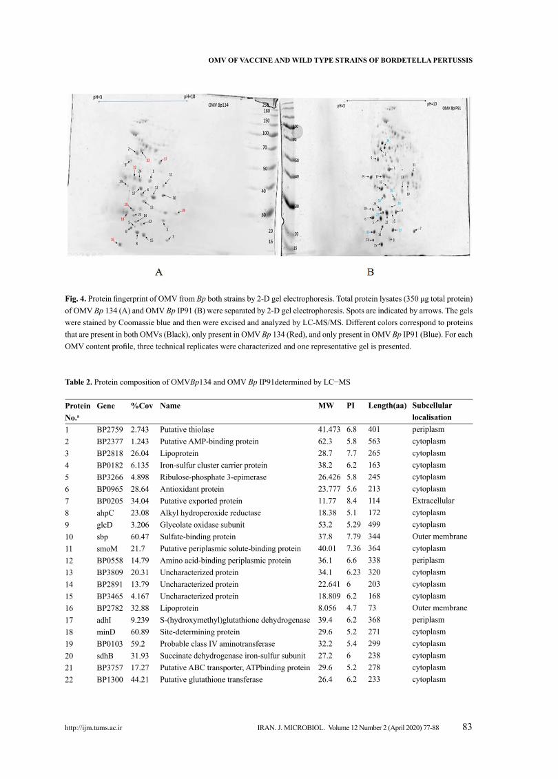
proteomics and differential expression of pro-teins. The differences between OMV Bp 134 and OMV Bp IP91 groups are shown in Fig. 3. The sig-nificant differences between 2-DE images of OMV
Fig. 3. Functional classes of proteins identified from OMV of Bp.
Bp 134 and OMV Bp IP91 were observed. All the spots were matched by a gel-to-gel comparison and the relative abundance difference (volume percent) of each spot was determined. The spots which had abundance at least significantly different by ± 2 fold in OMV Bp IP91 versus OMV Bp 134 were chosen for identification. The protein spots which were vig-orous to other spots to enable them to be picked up with confidence were chosen. Using these criteria, 40 spots statistically confirmed protein variations between OMV Bp 134, OMV Bp IP91 were selected. In total, 37 proteins were successfully identified and marked on the gel as shown in Fig. 4. The results ob-tained from recognized proteins and their functions are summarized in Table 2. As shown in Fig. 3, after annotation of the function-al classes of a total of 37 identified proteins, they fell into 6 categories as follows: (i) metabolism (29%), (ii) membrane transport and secretion system (7%), (iii) biosynthesis and degradation (10%), (iv) adaptaion, adhesion, pathogenicity, conserved hypothetical and protection responses (25%), (v) regulation, cell divi-sion, pseudogenes and phage-related (8%), and (vi) Miscellaneus (21%). It was found that the majority of identified proteins in OMV Bp 134 was down-reg-ulated. The proteins including Lipoprotein (spot no. 3) and Ribulose-phosphate 3-epimerase (spot no. 4)
http://ijm.tums.ac.ir
OMV OF VACCINE AND wIlD TYpE STRAINS OF BORDETEllA pERTUSSIS
IRAN. J. MICROBIOL. Volume 12 Number 2 (April 2020) 77-88 83 http://ijm.tums.ac.ir
Fig. 4. Protein fingerprint of OMV from Bp both strains by 2-D gel electrophoresis. Total protein lysates (350 μg total protein) of OMV Bp 134 (A) and OMV Bp IP91 (B) were separated by 2-D gel electrophoresis. Spots are indicated by arrows. The gels were stained by Coomassie blue and then were excised and analyzed by LC-MS/MS. Different colors correspond to proteins that are present in both OMVs (Black), only present in OMV Bp 134 (Red), and only present in OMV Bp IP91 (Blue). For each OMV content profile, three technical replicates were characterized and one representative gel is presented.
Table 2. Protein composition of OMVBp134 and OMV Bp IP91determined by LC−MS
protein No.a
12345678910111213141516171819202122
Gene
BP2759BP2377BP2818BP0182BP3266BP0965BP0205ahpCglcDsbpsmoMBP0558BP3809BP2891BP3465BP2782adhIminDBP0103sdhBBP3757BP1300
%Cov
2.7431.24326.046.1354.89828.6434.0423.083.20660.4721.714.7920.3113.794.16732.889.23960.8959.231.9317.2744.21
Name
Putative thiolasePutative AMP-binding proteinLipoproteinIron-sulfur cluster carrier proteinRibulose-phosphate 3-epimeraseAntioxidant proteinPutative exported proteinAlkyl hydroperoxide reductaseGlycolate oxidase subunitSulfate-binding proteinPutative periplasmic solute-binding proteinAmino acid-binding periplasmic proteinUncharacterized proteinUncharacterized proteinUncharacterized proteinLipoproteinS-(hydroxymethyl)glutathione dehydrogenaseSite-determining proteinProbable class IV aminotransferaseSuccinate dehydrogenase iron-sulfur subunitPutative ABC transporter, ATPbinding proteinPutative glutathione transferase
Mw
41.47362.328.738.226.42623.77711.7718.3853.237.840.0136.134.122.64118.8098.05639.429.632.227.229.626.4
pI
6.85.87.76.25.85.68.45.15.297.797.366.66.2366.24.76.25.25.465.26.2
length(aa)
40156326516324521311417249934436433832020316873368271299238278233
Subcellular localisationperiplasmcytoplasmcytoplasmcytoplasmcytoplasmcytoplasmExtracellularcytoplasmcytoplasmOuter membranecytoplasmperiplasmcytoplasmcytoplasmcytoplasmOuter membraneperiplasmcytoplasmcytoplasmcytoplasmcytoplasmcytoplasm

http://ijm.tums.ac.ir
AlI BADAMCHI ET Al.
84 IRAN. J. MICROBIOL. Volume 12 Number 2 (April 2020) 77-88 http://ijm.tums.ac.ir
Table 2. Continuing...
23
2425262728293031323334353637
BP0961
BP2963BP1702odhBBP1616BP3550bopNBP2533BP0361gltAsdhAlpdABP3277mmsBBP2434
9.639
5.2943.4091.9827.712.5451.91846.0725.7913.994.8995.87245.4519.192.828
Electron transfer flavoprotein beta subunit
Putative membrane proteinProbable enoyl-CoA hydratase/isomeraseoxoglutarate dehydrogenase complexPutative DNA-binding proteinAraC-family regulatory proteinPutative outer protein NInorganic pyrophosphataseUncharacterized proteinCitrate synthaseSuccinate dehydrogenase flavoprotein subunitDihydrolipoyl dehydrogenasePutative enoyl-CoA hydratase3-hydroxyisobutyrate dehydrogenasePeriplasmic serine endoprotease DegPlike
26.819
40.05528.55641.82518.5230.638.9920.04216.8848.4664.862.330.129.652.1
5.7
7.65.75.36.2955.35.26.036.16.16.25.77.6
249340
261404166275365178159436596596275297495
cytoplasm, periplasmOuter membranecytoplasmcytoplasmcytoplasmcytoplasmcytoplasmcytoplasmOuter membranecytoplasmcytoplasmcytoplasmcytoplasmcytoplasmperiplasm, Outer membrane
Identifications were confirmed by LC- MS/MS ion search and significant matches (p < 0.05) were retained (Swiss-Prot protein database).a: Protein numbering refers to spot numbers in Fig. 4.
were down-regulated proteins in OMV Bp 134. Al-kyl hydroperoxide reductase (AhpC; spot no. 8) was overexpressed in the OMV Bp 134. The role of AhpC in detoxification of reactive oxygen species has been shown in previous studies. While a number of protein spots were common among OMV of the vaccine strain and OMV of B. pertussis IP91. There were multiple spots unique to B. pertussis IP91, i.e., Dihydrolipoyl dehydrogenase (spot 34; BP0995), Putative DNA-binding protein (spot 27; BP1616), Inorganic pyrophosphatase (spot 30; BP2533), a Putative enoyl-CoA hydratase (spot 25; BP3277). Five of protein spots were common among OMV of the vaccine strain tested and OMV of Bp IP91 including (BP2759), (BP2377), (BP2818), (BP3266) (BP0965), (BP0205) and BP0558. (Figs. 4A and B).
DISCUSSION
Bordetella pertissis is an important pathogen during childhood that causes pertussis. The protec-tive effectiveness of present wP vaccines is not sat-isfactory. Meanwhile, the high costs of aP, and its major problem of inducing Th2 immune response
are its main drawbacks (16). Therefore, omvPV could be considered as an alternative vaccine types against pertussis (17). Considering the low toxicity of omvPV vaccines and their similarity to wP with respect to stimulation of the immune system, vac-cines incorporating OMV from a dominant Bp strain of each geographical region could be considered as an alternative solution for the current pertussis di-lemma (18). Therefore, the identification of antigenic proteins for a new candidate vaccine is in high de-mand. The OMV-based vaccines are attractive and useful tools for the complete coverage of vaccination against pertussis while there is still a high preva-lence of pertussis among populations of all ages in Iran (19). Here we designed a study to investigate the OMV’s of a vaccine strain and a dominant wild type strains of Bp isolates from Iran. Moreover, a quanti-tative proteomic approach was used to compare the whole-cell vaccine stain 134 with the other regional strains. In our study, 9 (9%) of infants with PT-IgA were positive for pertussis. Among them, 5 subjects had received 3 doses of wP. The vaccine effectiveness was only estimated to be 33% after receiving 3 doses of wP. Similar low effectiveness results of wP have also been reported in other studies (20). For instance,

http://ijm.tums.ac.ir
OMV OF VACCINE AND wIlD TYpE STRAINS OF BORDETEllA pERTUSSIS
IRAN. J. MICROBIOL. Volume 12 Number 2 (April 2020) 77-88 85 http://ijm.tums.ac.ir
two trials conducted in Europe, have measured the efficacy of wP, given in 3 primary doses at 2, 4 and 6 months interval. Moreover, Gustafsson and Gre-co (20, 21) have conducted similar trials in Sweden and Italy, respectively and all of these studies have indicated low effectiveness rates (36%-48.3%) of wP. These have used a particular Connaught vaccine (wP), and it has been suggested that the observed lower efficacies may have been due to the vaccine strain or the poor quality of the vaccine batches. Meanwhile, Blennow and colleagues who have con-ducted a trial in Sweden using Sauer strains, have found an effectiveness rate of ~71% (22). However in our study, poor immunogenicity was detected for wP even after administration of 3 doses of the vaccine. However, not all wP vaccines showed equal efficacy, reflecting substantial differences in quality between the manufacturers (23). The fact that selected DTwP were poorly effective has raised some questions regarding the utility of all DTwP vaccines. Hence, the resurgence of pertussis in Iran may have been contributed to the low effec-tiveness of wP used. De Greeff et al. results show that, although vaccination programs have reduced pertussis morbidity during the childhood, they have been ineffective to counter the increased infection rate in adolescent and adult pertussis. Indeed, the high circulation of Bp in the latter age categories may limit the effectiveness of pediatric vaccination (24). Based on our findings, it seems that a stronger immune response could be observed in children vac-cinated with wP, compared with infants infected with the wild-type strain. Waning immunity is regarded as the most significant cause of pertussis resurgence (10). Also, mutations in pertussis toxin as well as pertactin genes are considered as important causes of waning of the vaccine immunity (25). Moreover, prevalence of pertussis outbreaks in adolescents and adults have been revealed even in countries in which the wP or aP vaccination programs have been con-ducted (19, 26). Generally, wP induces a complex immune re-sponse, due to presence of many bacterial antigens. It induces the production of antibodies against Bp key virulence factors, namely, Ptx, FHA, PRN, ACT, LPS, DNT, FIM2/3 and BrkA (16). Raeven et al. (8) have reported that OMV of Bp 1917 has 16 protein with significant immunogenic properties. We char-acterized OMV Bp 134 and OMV Bp IP91 strains by proteomic analyses and compared their proteome
with other strains which had been previously report-ed (27). Immunoblotting was performed using mono-clonal antibodies (i.e. FHA 1:2000, PRN 3:10000 and Ptx 3:1000). According to our results, the candidate antigens for OMV vaccine could be Ptx, FHA and PRN. In addition, antibody responses against Bp should preferably be directed against antigens such as FHA, PT and PRN, since they have immune eva-sive properties (15) and are common in OMV pro-files of both the vaccine and the wild type strains. A comparative analysis of the respective proteomes revealed striking differences between the protein profiles of OMV vaccine and OMV wild type strains. We identified 37 reproducible proteins which had sig-nificant changes in their expression levels. A general up-regulation in the proteins levels was observed in OMV IP91 whereas the expression of these proteins levels (namely, those involved in carbohydrate me-tabolism, lipoprotein biosynthesis and adaptation) were generally down-regulated in OMV 134, due to significant energy cost for OMV production. Hydrogen-peroxide (H2O2) which has disruptive effects on the metabolism and blocks growth, is commonly generated in biological habitats under defined environmental conditions and even by cel-lular immune responses. However, some biological reactions lead to detoxification of H2O2. For example, RPE (ribulose-phosphate 3-epimerase) in E. coli de-toxifies H2O2 (28, 29). Interestingly in this study we observed that RPE was overexpressed in wild-type IP91 strain which may protect bacteria against the cellular immune responses. The low expression of AhpC in the wild type strain shows that the vaccine strain could be more immu-nogenic than the wild type strain. It has been shown that AhpC is a potent antigen and it induces a prom-ising T cell-mediated immune response in patients with acute Melioidosis (30). The down-regulation of AhpC has been reported in a persistent isolate of Borkholderia cenocepacia during a murine infection (31). The antioxidant protein AhpC has been consid-ered as a potential vaccine candidate against various bacterial infections by Helicobacter pylori, Bacillus anthracis, Streptococcus zooepidemicus and Burk-holderia pseudomallei (32-34). Our results pointed to some other identified out-er-membrane proteins such as iron-sulfur cluster car-rier protein (Bp0182), conserved hypothetical protein (Bp3465) and lipoproteins (Bp2782 and Bp2818). They serve as protective immunogens and are among

http://ijm.tums.ac.ir
AlI BADAMCHI ET Al.
86 IRAN. J. MICROBIOL. Volume 12 Number 2 (April 2020) 77-88 http://ijm.tums.ac.ir
the principal components of wP vaccines which have been developed against pertussis (27). The function of lipoproteins is unknown, although studies with pathogenic forms of E. coli have suggested that they play roles in vesicle formation (35). The lipoproteins from Bp2782 and Bp2818 which were identified in this study are abound in the outer-membranes of Gram-negative bacteria and for long have been con-sidered as potential target antigens for vaccine devel-opment. The immunogenicity of a lipoprotein from the putative outer-membrane of Bordetella bronchi-septica has been recently demonstrated (36). Liu et al. have revealed that the recombinant lipoprotein could induce humoral and cell-mediated immune re-sponses in mice (36). From the 37 identified proteins using LC-MS/MS in current study, 25 proteins including (Bp 0103, Bp 0205, Bp 0558, Bp 0627, Bp 0777, Bp 0961, Bp 0962, Bp 0965, Bp 0995, Bp 1126, Bp 1300, Bp 1487, Bp 1616, Bp 1857, Bp 2360, Bp 2361, Bp 2434, Bp 2533, Bp 2533, Bp 2770, Bp 2818, Bp 3228, Bp 3277, Bp 3552 and Bp 3757) were found in other regional study by Tefon (37). Bp 2434 (serine protease) and Bp 1616 (putative DNA binding protein) were found in all 4 strains studied, including Bp Sadat, Bp Tahoma І, Bp 134 and Bp IP91 (37). Serine endoprotease has various effects on the immune response like activa-tion, migration and leukocyte apoptosis according to Ruiz-Perez et al study (38). It has been found that serine protease protein SphB1 is involved in the mat-uration of FHA in Bp (37, 39). Bacterial DNA binding proteins, also known as histone-like proteins are basic, small proteins which protect DNA in harsh environmental conditions and their immunogenicity has been shown by Al-tındiş and colleagues (40). During infections, many Gram-negative pathogens secrete their virulence fac-tors via type III secretion system and in Bordetellae, this system is active in Bvg+ phase (41). Putative out-er protein N (BopN), has been reported as type III secreted protein in B. bronchiseptica. In our study, we found that BopN is expressed at a high level in OMV IP91 strain. This indicates that this protein is highly expressed during the natural infection and in-duces high immune responses.
CONClUSION
The shortcomings of wP and aP vaccines, such as
their severe side effects and high costs, respective-ly, have been recently highlighted. OMV VP-based vaccines could be considered as interesting alterna-tive against pertussis. Such complex vaccines which contain the bacterium protein antigens can confer a similar efficacy as wP with lesser side effects and lower costs.
ACKNOwlEDGEMENTS
This project was supported by Pasteur Institute of Iran. The results described in this paper were part of AB’s Ph.D. thesis.
REFERENCES
1. Folaranmi T, Pinell-McNamara V, Griffith M, Hao Y, Coronado F, Briere EC. Systematic review and me-ta-analysis of pertussis epidemiology in Latin America and the Caribbean: 1980–2015. Rev Panam Salud Pu-blica 2017; 41: e102.
2. World Health Organization. Pertussis vaccines: Who position paper. Wkly Epidemiol Rec 2015; 90: 433-460.
3. Klein NP. Licensed pertussis vaccines in the United States: history and current state. Hum Vaccin Immuno-ther 2014;10:2684-2690.
4. Ryan EJ, Nilsson L, Kjellman NI, Gothefors L, Mills KH. Booster immunization of children with an acellu-lar pertussis vaccine enhances Th2 cytokine produc-tion and serum IgE responses against pertussis toxin but not against common allergens. Clin Exp Immunol 2000; 121:193-200.
5. Gambhir M, Clark TA, Cauchemez S, Tartof SY, Swerd-low DL, Ferguson NM. A change in vaccine efficacy and duration of protection explains recent rises in per-tussis incidence in the United States. PLoS Comput Biol 2015; 11(4):e1004138.
6. Cherry JD. Epidemic pertussis in 2012—the resurgence of a vaccine-preventable disease. N Engl J Med 2012; 367:785-787.
7. Park SB, Jang HB, Nho SW, Cha IS, Hikima JI, Ohtani M, et al. Outer membrane vesicles as a candi-date vaccine against edwardsiellosis. PLoS One 2011; 6(3):e17629.
8. Raeven RH, van der Maas L, Tilstra W, Uittenbogaard JP, Bindels TH, Kuipers B, et al. Immunoproteomic profiling of Bordetella pertussis outer membrane vesi-cle vaccine reveals broad and balanced humoral immu-nogenicity. J Proteome Res 2015; 14:2929-2942.
9. Hozbor D, Rodriguez ME, Fernandez J, Lagares A,
http://ijm.tums.ac.ir
OMV OF VACCINE AND wIlD TYpE STRAINS OF BORDETEllA pERTUSSIS
IRAN. J. MICROBIOL. Volume 12 Number 2 (April 2020) 77-88 87 http://ijm.tums.ac.ir
Guiso N, Yantorno O. Release of outer membrane ves-icles from Bordetella pertussis. Curr Microbiol 1999; 38:273-278.
10. Heravi FS, Nikbin VS, Lotfi MN, Badiri P, Ahmadi NJ, Zahraei SM, et al. Strain variation and antigenic diver-gence among Bordetella pertussis circulating strains isolated from patients in Iran. Eur J Clin Microbiol In-fect Dis 2018;37:1893-1900.
11. Bradford MM. A rapid and sensitive method for the quantitation of microgram quantities of protein utiliz-ing the principle of protein-dye binding. Anal Biochem 1976; 72:248-254.
12. Hendrikx LH, Öztürk K, De Rond LG, De Greeff SC, Sanders EA, Berbers GA, et al. Serum IgA responses against pertussis proteins in infected and Dutch wP or aP vaccinated children: an additional role in pertussis diagnostics. PLoS One 2011; 6(11):e27681.
13. Yari S, Tasbiti AH, Ghanei M, Shokrgozar MA, Vazi-ri B, Mahdian R, et al. Proteomic analysis of sensitive and multi drug resistant Mycobacterium tuberculosis strains. Microbiology 2016; 85:350-358.
14. Pérez A, Merino M, Rumbo-Feal S, Álvarez-Fraga L, Vallejo JA, Beceiro A, et al . The FhaB/FhaC two-part-ner secretion system is involved in adhesion of Acineto-bacter baumannii AbH12O-A2 strain. Virulence 2017; 8:959-974.
15. Saha S, Raghava GP. Prediction of continuous B-cell epitopes in an antigen using recurrent neural network. Proteins 2006; 65:40-48.
16. Gardy JL, Laird MR, Chen F, Rey S, Walsh CJ, Ester M, et al. PSORTb v.2. 0: expanded prediction of bacte-rial protein subcellular localization and insights gained from comparative proteome analysis. Bioinformatics 2005; 21: 617-623.
17. Asensio CJ, Gaillard ME, Moreno G, Bottero D, Zurita E, Rumbo M, et al. Outer membrane vesicles obtained from Bordetella pertussis Tohama expressing the lipid A deacylase PagL as a novel acellular vaccine candi-date. Vaccine 2011; 29:1649-1656.
18. Kim YH, Kim KA, Kim YR, Choi MK, Kim HK, Choi KJ, et al. Immunoproteomically identified GBAA _0345, alkyl hydroperoxide reductase subunit C is a potential target for multivalent anthrax vaccine. Pro-teomics 2014;14:93-104.
19. Chen Z, He Q. Immune persistence after pertussis vac-cination. Hum Vaccin Immunother 2017; 13:744-756.
20. Robbins JB, Schneerson R, Kubler-Kielb J, Keith JM, Trollfors B, Vinogradov E, et al. Toward a new vaccine for pertussis. Proc Natl Acad Sci U S A 2014; 111:3213-3216.
21. Asl YM, Akhi MT, Soroush MH, Sefidan FY, Mou-sapour J, Hejazi ME, et al. Clinical Manifestations and Seasonality of Pertussis in Azerbaijan, Iran. Infect Dis Clin Pract 2018; 26:145-149.
22. Greco D, Salmaso S, Mastrantonio P, Giuliano M, Tozzi AE, Anemona A, et al. A controlled trial of two acellular vaccines and one whole-cell vaccine against pertussis. N Engl J Med 1996; 334:341-348.
23. Gustafsson L, Hallander HO, Olin P, Reizenstein E, Storsaeter J. A controlled trial of a two-component acellular, a five-component acellular, and a whole-cell pertussis vaccine. N Engl J Med 1996; 334:349-355.
24. Blennow M, Hedenskog S, Granström M. Protective effect of acellular pertussis vaccines. Eur J Clin Micro-biol Infect Dis 1988; 7:381-383.
25. De Greeff SC, De Melker HE, Van Gageldonk PG, Schellekens JF, van der Klis FR, Mollema L, et al. Se-roprevalence of pertussis in The Netherlands: evidence for increased circulation of Bordetella pertussis. PLoS One 2010; 5(12): e14183.
26. van der Maas NA, Mooi FR, de Greeff SC, Berbers GA, Conyn-van Spaendonck MA, de Melker HE. Pertussis in the Netherlands, is the current vaccination strategy sufficient to reduce disease burden in young infants. Vaccine 2013; 31:4541-4547.
27. Zhao Z, Xue Y, Tang X, Wu B, Cheng X, He Q, et al . Immunogenicity of recombinant protective antigen and efficacy against intranasal challenge with Bordetella bronchiseptica. Vaccine 2009; 27:2523-2528.
28. Sobota JM, Imlay JA. Iron enzyme ribulose-5-phos-phate 3-epimerase in Escherichia coli is rapidly dam-aged by hydrogen peroxide but can be protected by manganese. Proc Natl Acad Sci U S A 2011; 108:5402-5407.
29. Gu M , Imlay JA. Superoxide poisons mononuclear iron enzymes by causing mismetallation. Mol Microbiol 2013; 89:123-134.
30. Reynolds C, Goudet A, Jenjaroen K, Sumonwiriya M, Rinchai D, Musson J, et al. T Cell Immunity to the Al-kyl Hydroperoxide Reductase of Burkholderia pseudo-mallei: a correlate of disease outcome in acute meli-oidosis. J Immunol 2015; 194:4814-4824.
31. Shinoy M, Dennehy R, Coleman L, Carberry S, Schaf-fer K, Callaghan M, et al. Immunoproteomic analysis of proteins expressed by two related pathogens, Bur-kholderia multivorans and Burkholderia cenocepacia, during human infection. PLoS One 2013;8(11): e80796.
32. Velineni S, Timoney JF. Identification of novel immu-noreactive proteins of Streptococcus zooepidemicus with potential as vaccine components. Vaccine 2013; 31:4129-4135.
33. O’Riordan AA, Morales VA, Mulligan L, Faheem N, Windle HJ, Kelleher DP. Alkyl hydroperoxide reduc-tase: a candidate Helicobacter pylori vaccine. Vaccine 2012; 30:3876-3884.
34. Zhao Z, Xue Y, Tang X, Wu B, Cheng X, He Q. Im-munogenicity of recombinant protective antigen and efficacy against intranasal challenge with Bordetella

http://ijm.tums.ac.ir
AlI BADAMCHI ET Al.
88 IRAN. J. MICROBIOL. Volume 12 Number 2 (April 2020) 77-88 http://ijm.tums.ac.ir
bronchiseptica. Vaccine 2009; 27:2523-2528.35. McBroom AJ, Johnson AP, Vemulapalli S, Kuehn MJ.
Outer membrane vesicle production by Escherichia coli is independent of membrane instability. J Bacteriol 2006;188:5385-5392.
36. Liu Y, Chen H, Wei Q, Xiao C, Ji Q, Bao G. Immune efficacy of five novel recombinant Bordetella bronchi-septica proteins. BMC Vet Res 2015; 11:173.
37. Tefon BE, Maaß S, Özcengiz E, Becher D, Hecker M, Özcengiz G. A comprehensive analysis of Bordetella pertussis surface proteome and identification of new immunogenic proteins. Vaccine 2011; 29:3583-3595.
38. Ruiz-Perez F, Wahid R, Faherty CS, Kolappaswamy K, Rodriguez L, Santiago A, et al. Serine protease au-totransporters from Shigella flexneri and pathogenic Escherichia coli target a broad range of leukocyte gly-
coproteins. Proc Natl Acad Sci U S A 2011; 108:12881-12886.
39. Vijayakumar V, Santiago A, Smith R, Smith M, Rob-ins-Browne RM, Nataro JP, et al. Role of a class 1 serine protease autotransporter in the pathogenesis of Citrobacter rodentium colitis. Infect Immun 2014: 82:2626-2636.
40. Altındiş E, Tefon BE, Yıldırım V, Özcengiz E, Becher D, Hecker M, et al. Immunoproteomic analysis of Bor-detella pertussis and identification of new immunogen-ic proteins. Vaccine 2009; 27:542-548.
41. Yuk MH, Harvill ET, Cotter PA, Miller JF. Modula-tion of host immune responses, induction of apoptosis and inhibition of NF-κB activation by the Bordetella type III secretion system. Mol Microbiol 2000; 35:991-1004.