709 ' # '6& *#7 & 8 - intechcdn.intechopen.com/pdfs-wm/31026.pdf · oxidized low...
TRANSCRIPT

3,350+OPEN ACCESS BOOKS
108,000+INTERNATIONAL
AUTHORS AND EDITORS115+ MILLION
DOWNLOADS
BOOKSDELIVERED TO
151 COUNTRIES
AUTHORS AMONG
TOP 1%MOST CITED SCIENTIST
12.2%AUTHORS AND EDITORS
FROM TOP 500 UNIVERSITIES
Selection of our books indexed in theBook Citation Index in Web of Science™
Core Collection (BKCI)
Chapter from the book Coronary Artery DiseasesDownloaded from: http://www.intechopen.com/books/coronary-artery-diseases
PUBLISHED BY
World's largest Science,Technology & Medicine
Open Access book publisher
Interested in publishing with IntechOpen?Contact us at [email protected]

8
Oxidized Low Density Lipoprotein, Statin Therapy and Carotid Stenosis
Elias Skopelitis, Dimitrios Levisianou, Theodore Gialernios and Sofoklis Kougialis
General Hospital of Nikaia and Piraeus Greece
1. Introduction
1.1 Carotid stenosis and atheromatous process
Carotid artery stenosis due to atherosclerosis is a major complication of hyperlipidemia, diabetes mellitus and hypertension. Moreover, the extent of carotid intima media thickness is a measure of atheromatosis and therefore of cardiovascular disease (CVD).
The effect of cholesterol in the process of atheromatosis is now well established. High levels of total cholesterol (TC), as well as of low-density lipoprotein (LDL), very low-density lipoprotein (VLDL), intermediate-density lipoprotein (IDL), lipoprotein a (Lp-α), and triglycerides (TG), coupled with decreased levels of high-density lipoprotein (HDL) are responsible for the creation of atheromatous plaques (Assmann & Schulte, 1992; Hokanson & Autsin, 1996; Katsouras et al., 2001). Of the above factors, LDL cholesterol, and especially the oxidized LDL (oxLDL) is considered as the most important contributor of atheromatosis (Anderson et al., 1996).
The atheromatous process is completed in the following three stages:
1. In the first stage, LDL cholesterol enters the vessel wall, binds to glucosaminoglucanes, which are part of the extracellular matrix of the intima. This binding is facilitated by apolipoprotein B-100 (ApoΒ–100). The accumulation of LDL in the vessel wall contributes to the formation of fatty strikes. Following adhesion to the vessel wall, LDL undergoes oxidation by free radicals produced locally, the molecule is altered and chemokines are produced by adjacent vessel wall cells, such as monocyte chemoattractant protein-1 (MCP–1), together with growth factors, which are responsible for the accumulation of monocytes and macrophages. The latter cause further oxidation of LDL, resulting in negative charge, recognition by scavenger receptors located on macrophage membrane and increased uptake of LDL inside the macrophages, as these receptors are not inhibited by increased intracellular concentration of cholesterol. The final result is an enormous accumulation of LDL in the macrophages, which are transformed to foam cells. These cells represent the first step in the atheromatous process (Durrington & Sinderman, 2002) (figure 1).
2. During the second stage, the atheromatous plaque is formed. Foam cells produce growth factors and together with oxLDL result to the attraction of smooth muscle cells.
www.intechopen.com

Coronary Artery Diseases
126
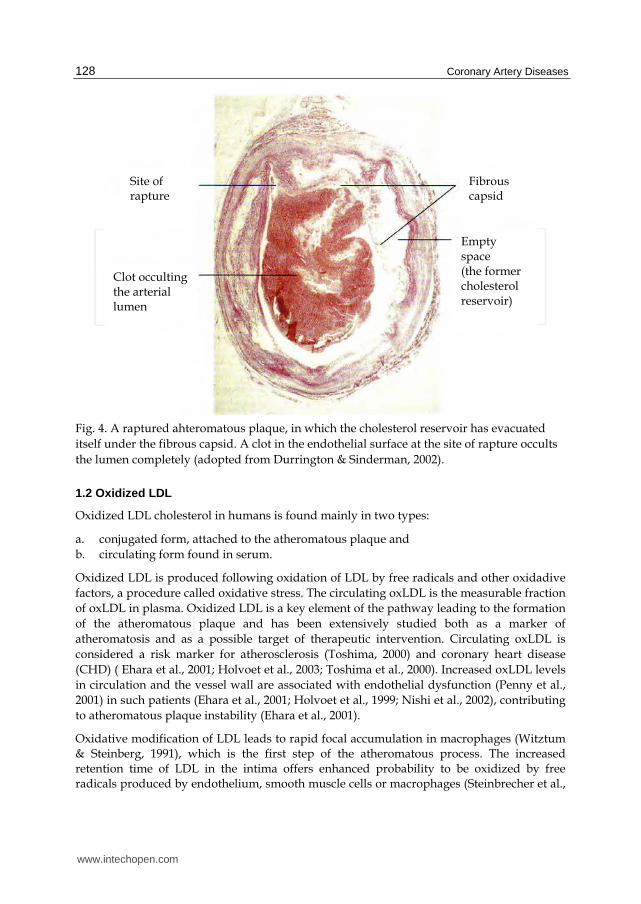
The latter are then differentiated to fibroblasts and start producing collagen. This collagen covers foam cells, which either are destroyed or are forced to apoptosis. The final result is the formation of a pool of extracellular cholesterol trapped under a fibrous capsid (figure 2). The part which is close to the yet intact vessel wall is the active site of the plaque, where the foam cells are produced. As the plaque extents to the inner layers of the vessel wall, the point of foam cell formation becomes instable and may cause rapture of the plaque (Durrington & Sinderman, 2002) (figure 3).
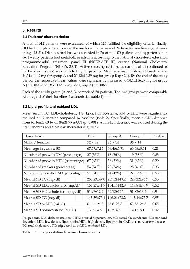
3. In the third stage, that of the complicated lesion, the rapture of the fibrous capsid of the atheromatous plaque leads to massive evacuation of the cholesterol reservoir. The artery may occult due to the accumulation of platelets and subsequent clotting, leading to acute ischemia or infarction (figure 4). If not so, then the plaque will be further enlarged (Durrington & Sinderman, 2002).
Fig. 1. Atherogenesis. Fatty strikes are characterized by macrophages containing an excess of
lipids (foam cells). Foam cells are derived by blood monocytes which are attracted to vessel
intima and start phagocytosing lipoproteins, such as oxLDL. The conversion of fatty strike
to atheroma depends on proliferation and differentiation of smooth muscle cells to
fibroblasts. The latter produce collagen resulting in intima thickening. As the lesion extents
further, foam cells are destroyed releasing large amounts of cholesterol trapped in a fibrous
capsid. The active site of atheroma is the point which is adjacent to normal endothelium,
where foam cells are formed (adopted with persmission from Durrington & Sinderman,
2002).
Collagen
Smooth muscle cell proliferation
growth factors
Oxidized LDL
scavenger receptors
Fc receptors autoantibodies
Decreased macrophage mobility
chemotactic agents
cytotoxicity oxidation
Circulating LDL
www.intechopen.com
Oxidized Low Density Lipoprotein, Statin Therapy and Carotid Stenosis
127
Avoiding the formation and the instability of the atheromatous plaque is top priority for
patients at risk for cardiovascular events. Statins may contribute towards this direction
(Corti et al., 2002; Nissen et al., 2004)
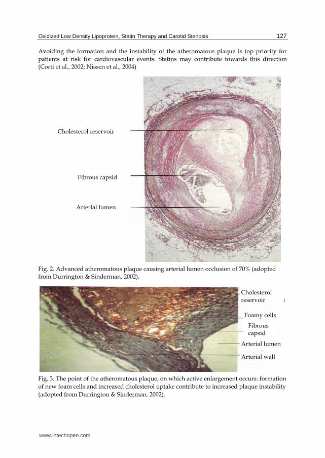
Fig. 2. Advanced atheromatous plaque causing arterial lumen occlusion of 70% (adopted
from Durrington & Sinderman, 2002).
Fig. 3. The point of the atheromatous plaque, on which active enlargement occurs: formation
of new foam cells and increased cholesterol uptake contribute to increased plaque instability
(adopted from Durrington & Sinderman, 2002).
Arterial lumen
Fibrous capsid
Cholesterol reservoir
Cholesterol reservoir
Foamy cells
Fibrous capsid
Arterial lumen
Arterial wall
www.intechopen.com
Coronary Artery Diseases
128
Fig. 4. A raptured ahteromatous plaque, in which the cholesterol reservoir has evacuated
itself under the fibrous capsid. A clot in the endothelial surface at the site of rapture occults
the lumen completely (adopted from Durrington & Sinderman, 2002).
1.2 Oxidized LDL
Oxidized LDL cholesterol in humans is found mainly in two types:
a. conjugated form, attached to the atheromatous plaque and
b. circulating form found in serum.
Oxidized LDL is produced following oxidation of LDL by free radicals and other oxidadive
factors, a procedure called oxidative stress. The circulating oxLDL is the measurable fraction
of oxLDL in plasma. Oxidized LDL is a key element of the pathway leading to the formation
of the atheromatous plaque and has been extensively studied both as a marker of
atheromatosis and as a possible target of therapeutic intervention. Circulating oxLDL is
considered a risk marker for atherosclerosis (Toshima, 2000) and coronary heart disease
(CHD) ( Ehara et al., 2001; Holvoet et al., 2003; Toshima et al., 2000). Increased oxLDL levels
in circulation and the vessel wall are associated with endothelial dysfunction (Penny et al.,
2001) in such patients (Ehara et al., 2001; Holvoet et al., 1999; Nishi et al., 2002), contributing
to atheromatous plaque instability (Ehara et al., 2001).
Oxidative modification of LDL leads to rapid focal accumulation in macrophages (Witztum & Steinberg, 1991), which is the first step of the atheromatous process. The increased retention time of LDL in the intima offers enhanced probability to be oxidized by free radicals produced by endothelium, smooth muscle cells or macrophages (Steinbrecher et al.,
Fibrous capsid
Site of rapture
Clot occulting the arterial lumen
Empty space (the former cholesterol reservoir)
www.intechopen.com

Oxidized Low Density Lipoprotein, Statin Therapy and Carotid Stenosis
129
1984). Oxidized LDL then acts chemotactic for monocytes and smooth muscle cells through binding to scavenger receptors (D. Li et al., 2002), leading to the formation of foamy cells. Oxidized LDL is also capable to elicit endothelial dysfunction by altering the secretory activity of endothelial cells (D. Li et al., 2002), inhibiting the nitric oxide-mediated vasodilatation through reduction of the expression of endothelial nitric-oxide synthase (eNOS), inducing the expression of adhesion molecules on the endothelium thus mediating the adhesion of monocytes to intima (D. Li et al., 2002), and inducing the expression of inflammatory cytokines (Steinberg, 1997). Indeed, oxLDL is a potent inducer of inflammation (Steinberg et al., 1989), contributing to the chronic inflammatory process which results to atherosclerosis (Ross, 1999).
1.3 Statins
The 3-hydroxy-3-methylglutaryl-coenzyme A reductase inhibitors, or statins, reduce serum TC, LDL cholesterol, apolipoprotein B (apoB), and, to a lesser degree, TG and Lp-a levels. Statins also have pleiotropic effects (Bellosta et al., 2000), such as the modulation of inflammatory molecules and monocyte maturation and differentiation (Bellosta et al., 2000), the suppression of smooth muscle-cells migration and proliferation (Bellosta et al., 2000), the reduction of the monocyte adhesion to the endothelium (Weber et al., 1997), the restoration of the impaired endothelium-dependent vessel wall relaxation (Jarvisalo et al., 1999), and the modification of cell-mediated LDL oxidation (Giroux et al., 1993; Aviram et al., 1998). All of the above mechanisms contribute to the reversion of atheromatosis. Undeniably, statins reduce the incidence of coronary events and are a cornerstone in the primary and secondary prevention of CHD (D.Y. Li et al., 2001). Previous studies have detected some efficacy in reducing the circulating oxLDL levels, but whether this effect is due to the reduction of LDL or is an independent, pleiotropic phenomenon remains a matter of controversy (Kwak & Mach, 2001; Robinson et al., 2005). Furthermore, little is known about the definite clinical benefit of such oxidative marker reduction.
The aim of the present study was to evaluate the efficacy of atorvastatin in reducing stenosis, to investigate the effect on oxLDL and to search for possible associations of oxLDL modification with changes of stenosis in patients managed conservatively and in pre-treated with percutaneous catheter interventional procedures patients with carotid atheromatosis. We hypothesise that atorvastatin therapy will confer remission of oxLDL levels in vivo and this will be associated with significant reduction of carotid artery stenosis.
2. Patients and methods
Between January 2005 and February 2008 a total of 100 patients were randomly selected from the lipid clinic and the carotid angioplasty clinic of a large tertiary hospital in Athens for inclusion in the study. Informed consent was obtained from each patient at recruitment according to our institutional policies. Eligible were patients with carotid artery atheromatosis from various causes (not only dyslipidemia) and with a range of predisposing factors. Exclusion criteria included: acute cardiovascular disease, severe or unstable angina pectoris, clinically evident cardiac failure, severe arrhythmias, recent surgical procedures, inflammatory diseases, active liver disease or liver impairment, excessive alcohol consumption (>4 drinks /day) or history of alcohol abuse, known allergic reaction to statins, poorly controlled diabetes mellitus as defined by a haemoglobin A1c (HbA1c) level of >7 %,
www.intechopen.com

Coronary Artery Diseases
130
uncontrolled hypertension indicated by systolic blood pressure (SBP) >140 mmHg and/or diastolic pressure >85 mmHg, history of deep vein thrombosis, bleeding tendency, serum triglycerides >350 mg/dl, evidence of thyroid dysfunction, use of systemic steroids or other anabolics, pernicious anaemia, impaired vitamin B12 or folate acid levels, abnormal serum urate at baseline, serum creatinine phosphokinase elevation of >1.5 fold at baseline, pregnancy or lactation, and end-stage renal disease or dialysis.
Patients were allocated into two groups according to the degree of carotid artery stenosis: those with arterial lumen occlusion of ≥70% in at least one common or internal carotid vessel consisted group A; those with stenosis <70% comprised group B. Patients in both groups were naive to statin therapy or if otherwise, a 6-month washout period was allowed before enrolment in the study. Group A underwent percutaneous transluminal carotid angioplasty with stenting by the same interventional cardiologist, prior to the initiation of statin therapy. Those patients were additionally administered clopidrogrel and salicylate. Both groups had to follow an American Heart Association step II diet and were encouraged to exercise.
According to the study protocol, all patients were placed on atorvastatin once daily at bedtime in individualised doses, tittered to achieve and maintain serum LDL cholesterol levels of <100 mg/dl (and ideally <70 mg/dl, if hypertension, renal impairment, smoking, hyperlipidemia, symptomatic peripheral arterial obstructive disease, or diabetes mellitus were present). Patients were prescribed statins even in the absence of hyperlipidemia, as the aim of the study was to investigate the effect of statin on oxLDL and carotid stenosis in a common atherogenic patient population. The most common doses used to achieve the above levels of LDL ranged between 10 to 40 mg, while seldom it was required to administer higher doses such as 60 mg (median atorvastatin dose for the total population = 20 mg, range 10 – 60 mg). The use of other drugs known to act synergistically with statins causing rhabdomyolysis was prohibited during the study. Adverse events were assessed in every visit in a non-specific manner: every newly reported symptom was documented as possible adverse reaction due to statin therapy and subsequently evaluated by an expert in clinical biochemistry. Adherence to the medication regimen was assessed indirectly by the low LDL levels compared with baseline.
Medical anamnesis, anthropometrics, smoking habits, blood pressure, and laboratory investigations comprising of complete blood count, fasting glucose, HbA1c, liver and kidney biochemistry, detailed lipid profile (TC, LDL cholesterol, HDL cholesterol, serum TG, apoB, and apolipoprotein A), urate, B12 and folate, thyroid function tests, homocysteine, Lp-a, and oxLDL were obtained at baseline and during follow-up visits, which were arranged at baseline, one, three, and six months; the final assessment was carried out in 12 months. Blood samples were collected after an at least 12-hour fast and a light, low-fat meal the night before sample collection was advised. Venous blood samples were collected in standard biochemistry vacutainer tubes. For the analysis of homocysteine and whole blood count, ethylenediaminetetraacetic acid (EDTA) vacutainer was used. Serum for biochemistry analysis was obtained by centrifugation (4000 g) at 4°C for 7 min and was immediately tested.
Lipid profiles (TC, HDL, TG) were determined using commercially available enzymatic colourimetric methods (Dade Behring, Newark, USA) with a Dade Behring analyser. LDL was calculated with the use of Friedewald's formula as all had TG <350 mg/dl (Puccetti et
www.intechopen.com

Oxidized Low Density Lipoprotein, Statin Therapy and Carotid Stenosis
131
al., 2002). For the measurement of circulating oxLDL, a commercially available kit (Mercodia, Uppsala, Sweden), based on a double antibody (4E6 and mouse monoclonal antiapoB) (Holvoet et al., 1996) capture ELISA test, was used. This method primarily detects malondialdehyde LDL (MDA-LDL). The normative range (reference range) in our lab was 31-61 mU/l. Apolipoprotein A, B and Lp-a were measured using immuno-nephelometry with rabbit antisera (Dade Behring, Newark, USA) in a Dade Behring analyser.
The evaluation of stenosis was conducted by Triplex ultrasonography using an Apogee 800 plus scanner with a 7.5 MHz transducer (ATL Inc., Bothell WA, USA) at baseline and 12 months. The stenosis was calculated in three sections in each common and internal carotid artery, and the final measure was the mean value of the three. The value of stenosis in the most occluded vessel was used in the statistical analysis. Specifically, the internal carotid artery (ICA) and common carotid artery (CCA) bilaterally were evaluated for each patient using coloured and grey Doppler ultrasonography. An effort was made to completely visualize the vessels. Additionally, the pulse wave was estimated with Doppler phasmatometry as well as the blood flow velocity of the two vessels. Results were recorded in a validated form. Stenosis was defined as the presence of visual plaque in coloured or grey Doppler. The degree of stenosis was calculated by measuring the decrease of the lumen diameter and the maximum systolic blood flow velocity. In difficult cases, other parameters were taken into account, such as ICA/CCA max blood flow velocity ratio and the ICA end-diastolic velocity. A degree of stenosis >70% was considered as sever and angioplasty was advised. A degree of stenosis between 60 – 70% was defined as high, between 50 – 60% as moderate and <50% as mild. High, moderate and mild stenoses were treated conservatively. The intima media thickness (IMT) and plaque morphology were not studied due to specific lab requirements, not readily available in our institution.
2.1 Statistical analysis
Continuous variables were presented as mean values ± standard deviation, while qualitative variables were presented as absolute and relative frequencies. Normality tests were applied using the Kolmogorov-Smirnov criterion as well as Shapiro-Wilk test. Univariate analysis was initially applied to test the associations of oxLDL with carotid stenosis for each patient group as well as to identify first order correlations with various clinical parameters. Correlations between skewed continuous or discrete variables were evaluated using Spearman's p-coefficient, whereas correlations of normally distributed variables were evaluated by calculating the Pearson's r-coefficient. Comparisons between normally distributed, continuous variables and categorical variables were made using the Student t-test. Analysis of categorical data was carried out with the [chi]2 test or Fischer's exact test when appropriate.
The association of oxLDL with carotid stenosis was also tested through multiple Cox proportional hazard model. The results obtained were presented as Hazard Ratios (HR) and the 95% Confidence Intervals (CI). A backward elimination procedure was applied to all multivariate models (using P<5% as the threshold for removing a variable from the models). All models were adjusted for age, gender, SBP and TC. Kaplan-Meier curves concerning stenosis over the study period were plotted and Log rank test was performed. All reported P-values were based on two-sided tests and compared to a significance level of 5%. STATA 8.0 software (Stata Corporation, 2003, Texas, USA) was used for the analysis.
www.intechopen.com
Coronary Artery Diseases
132
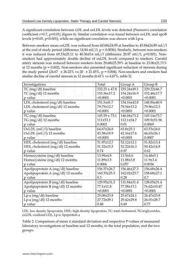
3. Results
3.1 Patients’ characteristics
A total of 612 patients were evaluated, of which 123 fulfilled the eligibility criteria; finally,
100 had complete data to enter the analysis, 76 males and 24 females, median age 68 years
(range 45-81). Diabetes mellitus was recorded in 26 of the 100 patients and hypertension in
66. Twenty patients had metabolic syndrome according to the national cholesterol education
programme-adult treatment panel III (NCEP-ATP III) criteria (National Cholesterol
Education Program (NCEP), 2001). Active smoking (defined as current of discontinued as
far back as 5 years) was reported by 58 patients. Mean atorvastatin dose at baseline was
24.31±11.49 mg for group A and 20.62±10.39 mg for group B (p=0.1). By the end of the study
period, the respective mean values were significantly increased to 30.45±16.27 mg for group
A (p=0.044) and 28.75±17.57 mg for group B (p=0.007).
Each of the study group (A and B) comprised 50 patients. The two groups were comparable
with regard of their baseline characteristics (table 1).
3.2 Lipid profile and oxidzed LDL
Mean serum TC, LDL-cholesterol, TG, Lp-a, homocysteine, and oxLDL were significantly
reduced at 12 months compared to baseline (table 2). Specifically, mean oxLDL dropped
from 62.26±22.03 to 44.49±21.75 mU/l (p<0.001). A marked decrease was noticed during the
first 6 months and a plateau thereafter (figure 5).
Characteristic Total Group A Group B P value
Males / females 72 / 28 36 / 14 36 / 14
Mean age in years ± SD 67.57±7.15 68.46±5.71 66.68±8.31 0.21
Number of pts with DM (percentage) 37 (37%) 18 (36%) 19 (38%) 0.83
Number of pts with HTN (percentage) 67 (67%) 36 (72%) 31 (62%) 0.29
Number of smokers (percentage) 54 (54%) 29 (54%) 25 (46%) 0.33
Number of pts with CAD (percentage) 51 (51%) 24 (47%) 27 (53%) 0.55
Mean ± SD TC (mg/dl) 232.23±47.8 235.24±49.2 229.22±46.7 0.53
Mean ± SD LDL cholesterol (mg/dl) 151.27±41.7 154.16±42.8 148.84±40.9 0.52
Mean ± SD HDL cholesterol (mg/dl) 51.97±12.7 52.12±12.1 51.82±13.4 0.9
Mean ± SD TG (mg/dl) 145.59±73.1 146.04±73.2 145.14±73.7 0.95
Mean ± SD oxLDL (mU/l) 64.66±24.8 65.8±25.3 63.53±24.5 0.65
Mean ± SD homocysteine (mU/l) 13.99±4.8 13.5±4.6 14.47±5.1 0.32
Pts: patients, DM: diabetes mellitus, HTN: arterial hypertension, MS: metabolic syndrome, SD: standard deviation, LDL: low density lipoprotein, HDL: high density lipoprotein, CAD: coronary artery disease, TC: total cholesterol, TG: triglycerides, oxLDL: oxidized LDL
Table 1. Study population baseline characteristics.
www.intechopen.com
Oxidized Low Density Lipoprotein, Statin Therapy and Carotid Stenosis
133
A significant correlation between LDL and oxLDL levels was detected (Pearson's correlation coefficient r=0.7, p<0.01) (figure 6). Similar correlation was found between oxLDL and apoB levels (r=0.65, p<0.001), while no significant correlation was shown with Lp-a.
Between smokers mean oxLDL was reduced from 60.68±24.09 at baseline to 45.84±24.89 mU/l at the end of study period (difference 14.84 mU/l, p = 0.0036). Similarly, between non-smokers it was reduced from 69.33±25.11 to 40.36±5.6 mU/l (difference 28.97 mU/l, p<0.001). Non-smokers had approximately double decline of oxLDL levels compared to smokers. Carotid artery stenosis was reduced between smokers from 29.68±25.59% at baseline to 23.06±21.71% at 12 months (p = 0.002). Non-smokers also presented significant reduction of stenosis during the study period (24.67 ± 26.22% vs 20 ± 21.45%, p = 0.004). Non-smokers and smokers had similar decline of carotid stenosis in 12 months (6.61% vs 4.67%, table 3).
Investigations Total Group A Group B
TC (mg/dl) baseline TC (mg/dl) 12 months p value
232.23 ± 47.8 153.36±17.2 <0.0001
235.24±49.1 154.24±16.9 <0.0001
229.22±46.7 152.48±17.7 <0.0001
LDL cholesterol (mg/dl) baseline LDL cholesterol (mg/dl) 12 months p value
151.5±41.7 79.75±12.7 <0.0001
154.16±42.8 79.54±13.2 <0.0001
148.84±40.9 79.96±12.3 <0.0001
TG (mg/dl) baseline TG (mg/dl) 12 months p value
145.59 ± 73.1 111±53.1 0.0002
146.04±73.2 112.1±54.7 0.01
145.14±73.7 109.9±51.96 0.0069
OxLDL (mU/l) baseline OxLDL (mU/l) 12 months p value
64.67±24.8 43.38±18.9 <0.0001
65.8±25.3 42.16±17.6 <0.0001
63.53±24.6 44.65±26.1 0.0007
HDL cholesterol (mg/dl) baseline HDL cholesterol (mg/dl) 12 months p value
51.97±12.7 51.32±15.5 0.74
52.12±12.1 52.22±16.3 0.97
51.82±13.4 50.42±14.8 0.62
Homocysteine (mg/dl) baseline Homocysteine (mg/dl) 12 months p value
13.99±4.8 11.89±3.5 0.0006
13.5±4.6 11.88±3.8 0.057
14.48±5.1 11.9±3.4 0.0036
Apolipoprotein A (mg/dl) baseline Apolipoprotein A (mg/dl) 12 months p value
156.57±26.7 160.35±25.3 0.3
156.46±27.3 162.02±23.7 0.28
156.68±26.4 158.68±27.1 0.7
Apolipoprotein B (mg/dl) baseline Apolipoprotein B (mg/dl) 12 months p value
129.95±31.3 77.1±11.8 <0.0001
131.84±31.4 77.58±13.1 <0.0001
128.05±31.4 76.62±10.47 <0.0001
Lp-a (mg/dl) baseline Lp-a (mg/dl) 12 months p value
25.08±23.8 27.72±29.1 0.48
25.67±24.1 29.42±29.8 0.49
24.47±23.8 26.01±28.7 0.77
LDL: low density lipoprotein, HDL: high density lipoprotein, TC: total cholesterol, TG:triglycerides, oxLDL: oxidized LDL, Lp-a: lipoprotein a
Table 2. Comparison of mean ± standard deviation and respective P-values of measured laboratory investigations at baseline and 12 months, in the total population, and the two groups.
www.intechopen.com

Coronary Artery Diseases
134
5010
015
020
025
0m
gr/d
l
0 1 2 3 4Time periods of observation
TC LDLoxLDL TG
Lipids alteration during observation period
TC: total cholesterol, LDL: low density lipoprotein cholesterol, oxLDL: oxidized LDL, TG: triglycerides, 0=baseline, 1=one month, 2=three months, 3=six months, 4=twelve months.
Fig. 5. Time curve of change of total cholesterol, LDL cholesterol, triglycerides and oxidized LDL levels during the observation period.
LDL: low density lipoprotein cholesterol, oxLDL: oxidized LDL, CI: confidence intervals.
Fig. 6. Correlation of low density lipoprotein with oxidized LDL levels at baseline (Pearson’s correlation coefficient r=0.7, p<0.001).
0
50
100
150
Ox LDL %
50 100 150 200 250LDL
Ox LDL 95% CI
Fitted values
p<0.001
Correlation LDL with Ox LDL
www.intechopen.com

Oxidized Low Density Lipoprotein, Statin Therapy and Carotid Stenosis
135
In further analysis, the group of smokers was subdivided to mild (≤5 cigarettes/day),
moderate (5 – 15 cigarettes/day) and heavy (≥15 cigarettes/day) smokers. The statistical
significant reduction of oxLDL levels and degree of carotid stenosis was apparent in the
subgroup of mild smokers (oxLDL at baseline 48.24±8.74 mU/l vs 41.54±9 mU/l at 12
months, p = 0.027 and stenosis at baseline 27.63±25.68% vs 23.42±21.74% at 12 months, p =
0.009), while it was not apparent in the subgroups of moderate and heavy smokers (oxLDL
at baseline 86.82±37.7 mU/l vs 42.92±10.77 mU/l at 12 months, p = 0.077 and stenosis at
baseline 34±31.9% vs 22±24.9% at 12 months, p = 0.186, for moderate smokers; respective
values for oxLDL were 66.29±15.88 mU/l vs 34.81±5.48 mU/l, p = 0.06 and for stenosis
32.14±24.13% vs 22.86±22.8%, p = 0.174, for heavy smokers). The above described effect of
smoking was taken into consideration during Cox-regression analysis.
Smokers P value Non Smokers P value
OxLDL (mU/l)
baseline
12 months
difference
60.68±24.09
45.48±24.89
14.84
0.0036
69.33±25.11
40.36±5.6
28.97
0.001
Stenosis (%)
baseline
12 months
difference
29.68±25.59
23.06±21.71
6.61
0.002
24.67±26.22
20±21.45
4.67
0.004
Correlation of oxLDL
change with stenosis
change in 12 months
Pearson’s
r = 0.412
0.021
Pearson’s
r = 0.198
0.03
oxLDL: oxidized LDL.
Table 3. Comparison of mean oxidized LDL values and degree of carotid stenosis change during the 1 year follow-up period, between smokers and non-smokers.
Within group B, the subgroup of patients with high degree of stenosis (>60%) had oxLDL
63.47±19.18 mU/l at baseline, while those with moderate and mild degree of stenosis (<60%)
had 40.32±20.72 mU/l (p<0.001). Corresponding values at 12-months were 33.18±17.78
mU/l and 38.81±29.02 mU/l, representing a marked decline for patients with >60% initial
stenosis and a far less decline for patients with <60% initial stenosis; yet those differences
were not statistically significant (table 4).
Stenosis >60<70% Stenosis <60% P value
Baseline
Mean oxLDL
63.47±19.18 mU/l
40.32±20.72 mU/l
< 0.001
12 months
Mean oxLDL
33.18±17.78 mU/l
38.81±29.02 mU/l
NS
LDL: low density lipoprotein cholesterol, oxLDL: oxidized LDL, NS: non significant.
Table 4. Comparison of mean oxidized LDL levels at baseline and 12 months within patients of group Β (n = 50), according to degree of stenosis at enrollment.
www.intechopen.com

Coronary Artery Diseases
136
3.3 Anthropometrics
Body mass index (BMI), weight, waist circumference and waist:hip ratio did not change
significantly during the study period.
3.4 Carotid stenosis
Patients in group A had null stenosis at recruitment due to prior angioplasty with stenting.
At the end of the 12-month statin therapy, no case of clinically important restenosis (>70%)
was reported in this group (as restenosis was defined any increase of the carotid lumen
diameter >5%). Patients in group B had mean percentage of stenosis at baseline 47.6±13.2%,
which was significantly reduced following 12-month statin therapy (37.7±15.7%, p<0.001)
(table 5).
Baseline 12 months P value
Mean (%) carotid stenosis ± standard deviation
47.6 ± 13.2 37.7 ± 15.7 0.001
Table 5. Change of the percentage of carotid artery stenosis between baseline and 12months
for patients in group B.
3.5 Association of stenosis with oxidized LDL
Group B patients in the highest quartile of oxLDL values had a 12-month risk ratio for
restenosis of 1.025, 95%CI=1.006-1.044, p=0.0083 (figure 7). After adjusting for gender,
age, smoking, SBP, TC, and LDL levels, these patients demonstrated a HR for restenosis of
4.319 compared with those in the lowest quartile (p<0.001, figure 7). This means that an
increase of oxLDL by one unit increases the degree of carotid stenosis by 2.5%, for
patients in group B. A weak but significant correlation was detected between oxLDL
levels and the degree of carotid artery stenosis (r=0.17, p=0.018). Similar correlation was
found between LDL cholesterol levels and carotid stenosis (r=0.18, p=0.0085). The
strength of Pearson’s correlation of mean oxLDL change with degree of carotid stenosis
change during the 12-month period was greater for smokers compared to non-smokers
(table 3).
3.6 The effect of LDL levels
Patients in group B who achieved LDL levels <70 mg/dl during the observation period had
a greater (28.08±28% vs 22.31±22.7%, difference 5.77%, p = 0.06) reduction of carotid stenosis
compared to those with LDL levels between 70 and 100 mg/dl (26.98±25.3% vs 21.35±21.3%,
difference 5.63%, p < 0.001), but this difference was not statistically significant. Thus, in
conservatively treated group B, further reduction of LDL than the limit of 100 mg/dl was
not associated with additional improvement of stenosis.
www.intechopen.com

Oxidized Low Density Lipoprotein, Statin Therapy and Carotid Stenosis
137
4. Discussion
This study demonstrates that atorvastatin administered in indivudualised doses, tittered to maintain serum LDL cholesterol levels <100 mg/dl, significantly decreased lipid profile and oxLDL, reduced carotid artery stenosis in patients managed conservatively and prevented restenosis in patients with prior angioplasty. Oxidized LDL in this study correlated positively with the degree of carotid artery stenosis; it was also shown by multivariate analysis that oxLDL represented an independent risk factor for restenosis. To our knowledge this is the first prospective study with a long observation period of 12 months to report such a clear, significant reduction of oxLDL levels following atorvastatin therapy for carotid atheromatosis of various causes and to report an association of the degree of oxLDL reduction with remission of carotid stenosis. It is also of major importance that this robust, long-standing decline of oxLDL was achieved with doses of atorvastatin used in everyday clinical practice. Interestingly, this beneficial effect was completed in the first six months, while practically no further reduction was noticed past this time point.
oxLDL=0: low quartile of oxLDL levels, oxLDL=1: high quartile of oxLDL levels, 0=baseline, 1=one month, 2=three months, 3=six months, 4=twelve months.
Fig. 7. Kaplan Meier survival analysis for the estimation of the risk ratio for restenosis according to the levels of oxidized LDL (oxLDL). With red line those with oxLDL levels in the highest quartile of the values. With blue line those with oxLDL levels in the lowest quartile of the measurements (risk ratio 1.025, logrank test p<0.001).
The mechanism by which statins modulate oxLDL levels has been controversial in the literature. Moreover, the association of oxLDL level modification with improvement of carotid atheromatosis and clinical outcome is not unequivocally established by large, double-blinded, randomised trials. Under this perspective, the present observational study provides reasonable evidence that reducing oxLDL may independently improve carotid stenosis.
0.00
0.25
0.50
0.75
1.00C a R O T I D S T E N O S I S % 0 1 2 3 4 5
Time of observation (in five periods)
oxLDL = 0 oxLDL = 1
Logrank test p<0.001 Kaplan Meier Curves for Carotid Stenosis
www.intechopen.com
Coronary Artery Diseases
138
Carotid IMT is a validated measure of carotid atherosclerosis. It is well established that
carotid atherosclerosis, serves as an independent surrogate marker for CHD (Vasankari et
al., 2001) and CVD (van Tits et al., 2006). Nevertheless, in the present study it was preferred
to estimate the degree of carotid stenosis with a more direct approach, because this is more
readily available in most hospital settings and because there is an obvious relation with
clinical symptoms and signs. Besides, it represents a reliable method with sufficient
reproducibility and it is practically the method of choice when evaluated patients candidate
for endarterectomy or angioplasty. Evaluating carotid stenosis in turn, is an established
method for estimating coronary risk (Vasankari et al., 2001) and cardiovascular risk (van
Tits et al., 2006). Other parameters of vessel wall function, such as IMT and plaque
morphology, even if clearly associated with cardiovascular risk in the literature, require well
equipped laboratory and are not readily available in our hospital. Future research on the
field should, ideally, comprise such measurements.
Oxidized LDL has long been recognized as a risk factor for carotid atherosclerosis in asymptomatic men (Liu et al., 2004) and has also been linked with CVD (Robbesyn et al., 2004). Oxidized LDL levels (Papathanasiou et al., 2008), autoantibodies against epitopes of oxLDL (Papathanasiou et al., 2008) and oxLDL:LDL ratio (Vasankari et al., 2001) are independently associated with increased risk for coronary atheromatosis and ischemic heart disease. Increased levels of oxLDL (Ehara et al., 2001) and MDA-LDL (Holvoet et al., 1999) in such cases are related to plaque instability. On the other hand, it has been reported that oxLDL is weakly associated with carotid IMT, but not with carotid plaque occurrence (Hulthe & Fagerberg, 2002). Oxidized LDL impairs endothelium relaxation (Harrison et al., 1987) by inhibition of the expression of eNOS and of the transport pathways of nitric oxide (NO) from the endothelial cell, reduces the responsiveness of smooth muscle cell to NO (Keaney et al., 1996), inhibits the NO-mediated vasodilation (Harrison et al., 1987; Simon et al., 1990; Steinberg, 1997), induces the expression of adhesion molecules (Frostegard et al., 1990), acts directly chemotactic to circulating monocytes (Steinberg, 1997), stimulates endothelial cells to produce MCP-1 (Cushing et al., 1990), facilitates monocyte adhesion to intima (Mehta et al., 1995), exhibits cytotoxic properties against endothelial cells (Steinberg, 1997), and induces the expression of inflammatory molecules (Steinberg, 1997). All of the above contribute directly to dysfunction of the endothelium (Witztum & Steinberg, 1991) and foam cell formation, which is the first step in the development of fatty streaks (Ross, 1999), the first visible step of atherosclerosis. These effects are mediated by preferential binding of oxLDL with type A scavenger receptors (SRA, SRA-II and CD36) on subendothelial resident macrophages and smooth muscle cells (Li et al., 1995) and lectin-like oxLDL receptor-1 (LOX-1) on endothelial cells (Sawamura et al., 1997) rather than the typical LDL receptor, resulting in an unrestricted uptake of cholesterol.
Statins reduce the incidence of cardiovascular events, an effect attributable to their
hypocholesterolemic properties (Archbold & Timmis, 1999). However, the extent of clinical
benefit and accumulating laboratory evidence suggest additional mechanisms of action, the
so-called pleiotropic effects (Bellosta et al., 2000). The most important among such effects are
the suppression of smooth muscle cell migration and proliferation (Bellosta et al., 1998), the
reduction of monocyte adhesion to the vascular endothelium (Weber et al., 1997), the
improvement of endothelial function (Jarvisalo et al., 1999), the inhibition of cell-mediated
LDL oxidation (Aviram et al., 1998; Giroux et al., 1993), the immuno-modulation of
www.intechopen.com

Oxidized Low Density Lipoprotein, Statin Therapy and Carotid Stenosis
139
monocyte maturation and differentiation, and the modification of production of
inflammatory cytokines (Rothe et al., 1999).
Atorvastatin suppresses cellular uptake of oxLDL from differentiating monocytes by reducing the expression of LOX-1 and scavenger receptors (Fuhrman et al., 2002) and accelerates the LDL-receptor-mediated removal of the non oxidized LDL particles (Vasankari et al., 2005). Hydroxymetabolites of atorvastatin protect the LDL against oxidation (van Tits et al., 2006). The antioxidant potency of atorvastatin metabolites has been confirmed by the reduction of IgG antibodies against LDL, a marker well-associated with CHD (Aviram et al., 1998). It has even been reported that these active atorvastatin metabolites may have greater anti-atherosclerotic effects than other statin molecules (Mason et al., 2004).
In acute coronary syndromes, atorvastatin therapy was linked to modulation of short- and long-term immune response towards LDL due to inhibition of lipoprotein-associated phospholipase A2 (Lp-LPA2) enzyme (Papathanasiou et al., 2008). The apparent benefit from statin therapy after acute coronary events may also be attributed to the stabilization of the plaque and removal of oxLDL from the vessel wall (Tsimikas et al., 2004). Increased mobilization of oxidized phospholipids from the vessel wall, transient binding with apoB-100 particles and clearance from the circulation may be the possible underlying mechanism. Under this perspective the increase in oxLDL:apoB ratio detected with atorvastatin therapy might represent a marker of oxLDL efflux from the vessel wall. Removal of oxLDL contributes to improved endothelial function as oxLDL is highly immunogenic and vasoconstrictive. In our study there was no significant change in oxLDL:apoB ratio. Atorvastatin also inhibits the oxLDL-mediated LOX-1 expression by endothelial cells, the uptake of oxLDL in endothelium and the oxLDL-mediated reduction of protein kinase B (PKB) phosphorylation (Li et al., 2001). The activation of PKB is critical for the expression of eNOS, which promotes vessel relaxation. However, a meta-analysis provided no clear evidence that statin therapy have a favourable effect on oxLDL (Balk et al., 2003).
In STAT trial (Mulder et al., 2007) the antibodies against oxLDL were equally decreased with both aggressive and conventional lipid-lowering therapy. This indicates that the statin-related reduction of oxLDL is not a dose-dependent phenomenon, a finding which is in agreement with our results. It might therefore represent a pleiotropic effect, independent -at least partially- from the hypo-cholesterolemic action. A study by Orem et al. (2002) detected a significant decrease of autoantibodies against oxLDL with low doses of atorvastatin (10 mg), similar to doses used in our study. In statin exposed patients, intensification of the regimen offers no additional benefit and only those with LDL >125 mg/dl benefited from a more aggressive statin therapy (Mulder et al., 2007). Statins have a dose-related response with regard to clinical outcome, but this dose-related response has not been confirmed with regard to oxidative stress (Ky et al., 2008). This might alternatively be explained by the hypothesis that statins achieve their uttermost benefit on oxLDL within a certain time point (Mulder et al., 2007), after which further continuation of treatment serves only the purpose of maintenance. Atorvastatin has been shown to reduce small dense LDL subfractions, remnant-like particles cholesterol and oxLDL, and improve endothelial function, after just few weeks of therapy (Miyagishima et al., 2007; Sakabe et al., 2003). Such time-related effect has not been fully elucidated, but may possibly account for our finding that in the first six months there was an accelerated decline of oxLDL levels followed by a milder reduction rate thereafter.
www.intechopen.com

Coronary Artery Diseases
140
Additional pleiotropic effects of statins have been reported in the literature and might
account for the observed beneficial effects in the current study. Lysophosphatidylcholine is
elevated during LDL oxidation and is responsible for some of the biological effects of
oxLDL. Atorvastatin alters the ability of oxLDL to impair the endothelium relaxation, by
modulating the hydrolysis of phosphatidylcholine to lysophosphatidylcholine when LDL is
being oxidized (Zhu et al., 2000). Statins remove predominately "aged LDL" from plasma,
which is more prone to oxidation (Orem et al., 2002), through stimulation of hepatic LDL
receptor activity and inhibition of very-low density lipoprotein (VLDL) and LDL production
by the liver cells (Orem et al., 2002). Statins also reduce oxygen species generation (Ky et al.,
2008). Atorvastatin promotes adipocyte uptake of oxLDL in rabbits by increasing the
expression of CD36 and peroxisome proliferators-activated receptor γ (PPARγ) in
adipocytes (Zhao & Zhang, 2004). The increased expression of such receptors by adipocytes
results to internalization of oxLDL and clearance from plasma, converting adipocytes to an
oxLDL-buffering pool (Zhao & Zhang, 2004). Reduction of oxLDL in patients with CHD
with atorvastatin 10 mg parallel with an increase of adiponectin, which has anti-atherogenic,
anti-inflammatory and anti-diabetic properties through reduction of insulin resistance
(Miyagishima et al., 2007). The CARDS study reported a significant degree of preventive
activity of atorvastatin against myocardial infarction in eucholesterolemic diabetic patients,
conceivably attributed to such improvement of insulin sensitivity (Miyagishima et al., 2007).
Statins also diminish the expression of CD40 and CD40 ligand in vascular cells, smooth
muscle cells and macrophages, which are promoted by oxLDL and are considered
proatherogenic (Schonbeck et al., 2002). Other anti-inflammatory pathways include
reduction of C-reactive protein (Hogue et al., 2008), chemokines, major histocompatibility
complex II molecules, matrix-degrading enzymes, and procoagulant tissue factor
(Schonbeck et al., 2002). Atorvastatin reverses the oxLDL-mediated inhibition of vascular
endothelial growth factor-induced endothelial progenitor cell differentiation via the
phosphatidylinositol 3 kinase/Akt pathway (Imanishi et al., 2003), which restores the
oxLDL-related inhibition of mature endothelial cells migration (Imanishi et al., 2003). This
could improve neovascularization and collateral vessel formation in response to tissue
ischemia. Atorvastatin also suppresses platelet activity (Puccetti et al., 2005) by reducing the
expression of CD36 and LOX-1, which are present in platelets (Puccetti et al., 2005;
Sawamura et al., 1997), thus inhibiting the oxLDL-mediated platelet hyperactivity (Puccetti
et al., 2005). Statins reduce the oxLDL-derived expression of adhesion molecules (E- and P-
selectins, vascular cell adhesion molecule 1 [VCAM-1] and intercellular adhesion molecule 1
[ICAM-1]) in human coronary artery endothelial cells, through up-regulation of eNOS
expression, which regulates the expression of adhesion molecules in endothelial cells (Li et
al., 2002). Statins also diminish the oxLDL-mediated activation of nuclear factor-κΒ (NF-κB)
(D. Li et al., 2002), which regulates the transcription of adhesion molecule genes (Robbesyn
et al., 2004). In diabetic patients with dyslipidemia atorvastatin reduced CVD and markers
of inflammation, adhesion and oxidation, such as C-reactive protein (CRP), soluble ICAM-1,
soluble VCAM-1, E-selectin, matrix metalloproteinase 9, secretory phospholipase A2
(sPLA2), and oxLDL, the latter by 38,4% (Hogue et al., 2008). Moreover, the change of
oxLDL levels correlated with the change of sICAM-1 and E-selectin levels, suggesting that
statins could possibly counteract the oxLDL-associated increase of NF-κΒ, and therefore, the
production of such cell adhesion molecules (Hogue et al., 2008). Statins also enhance
www.intechopen.com
Oxidized Low Density Lipoprotein, Statin Therapy and Carotid Stenosis
141
scavenger receptor expression in macrophages, and increase plaque stability via reduction
of metalloproteinases (Hogue et al., 2008).
The reduction of oxLDL and of carotid stenosis in our study was relevant for both smokers and non-smokers. However, subsequent subgroup analysis showed that the beneficial effect of statin use concerns mostly the subgroup of mild smokers, while no such effect was noticed for moderate and heavy smokers. How smoking may diminish the beneficial effect of statins on oxLDL and carotid stenosis is not yet clarified in the literature. A reasonable assumption might be that, since smoking increases the oxidative stress, it contributes to enhanced LDL oxidation (Van Himbergen et al., 2004). Moreover, studies in animal models, have demonstrated that smoking alters the immunologic response to oxLDL by reducing the production of antibodies against these molecules, i.e. causing a kind of immune suppression regarding the response to oxLDL. Thus, it has been shown to increase carotid IMT (Tani et al., 2004).
The Mercodia oxLDL detects the MDA-modified apoB (Holvoet et al., 1996). It has been proposed that oxLDL looses its predictive value for CVD when adjustment for apoB level is performed (Ky et al., 2008). In several studies though, a significant reduction of Mercodia oxLDL with atorvastatin 10 mg was still detected even after adjustment for apoB, (Holvoet et al., 2003; Ky et al., 2008; van Tits et al., 2006), while in other studies no adjustment for LDL or apoB levels was made (Ky et al., 2008; Sasaki et al., 2002). In our study the oxLDL:apoB ratio remained unchanged, but in the multivariate analysis the reduction of oxLDL was still significant after adjustment for apoB and LDL levels.
In patients with familial hypercholesterolemia a lack of association between oxLDL and IMT was reported at baseline, however two years therapy with atorvastatin 80 mg was associated with regression of carotid IMT (van Tits et al., 2004). The LDL subfraction profile and autoantibodies against oxLDL remained unchanged. Nevertheless, the rate of oxidation and the amount of dienes formed decreased and this was linked to lessening of atherosclerosis. In our study the reduction of carotid stenosis was associated with decreased oxLDL levels. Besides, the unchanged oxLDL autoantibodies levels do not preclude the reduction of oxLDL, as was indicated in another study involving dialysis patients, where atorvastatin therapy reduced plasma oxLDL, whereas oxLDL autoantibodies did not changed significantly (van den Akker et al., 2003).
Disadvantages of the study were the relatively small size, the lack of a control group comprising of patients with carotid stenosis not on statin therapy, which would be unethical, the fact that researchers were not blinded to the patients’ status, the lack of randomization of the dose-schedules and the use of only one method to detect oxLDL.
5. Conclusion
This prospective, cross-sectional study with such a long observation period provided enough evidence to postulate a favourable effect of low-dose atorvastatin therapy on oxLDL, which was additionally associated with improvement of stenosis in patients with carotid atheromatosis. We thus, assume that oxLDL may represent a far more sensitive risk factor for carotid stenosis, than LDL itself or apoB. Further studying is required to confirm such findings and to establish a clear clinical and pathophsiologic link between oxLDL and carotid stenosis.
www.intechopen.com

Coronary Artery Diseases
142
6. Acknowledgment
The authors wish to acknowledge Dr. Antonios Polydorou for performing the
catheterizations and stenting of the carotid arteries in the group of patients that underwent
intervention prior entering the study. We also thank him for allowing us access to the
records of the angioplasty laboratory. Finally we are grateful for valuable advice and
reviewing this manuscript before publishing.
We also wish to acknowledge Dr. Ioannis Dermitzakis for the critical contribution in
evaluating the degree of stenosis of our patient population, as director of the
ultrasonography laboratory in our institution. Without his help and valuable assistance this
whole project would not have been completed.
The authors finally acknowledge Mrs. Anna Zervou for carrying out the biochemical
laboratory measurements with diligence and accuracy, overlooking tiredness, physical
and emotional strain. We thank her for her personal commitment in the success of this
research.
7. References
Anderson TJ, Meredith IT, Charbonneau F, Yeung AC, Frei B, Selwyn AP, Ganz P. (1996) Endothelium – dependant coronary vasomotion relates to the susceptibility of LDL to oxidation in humans. Circulation, vol. 93, No. 9, (May 1996), pp. 1647-1650, ISSN 0009-7322
Archbold RA., Timmis AD. (1999) Modification of coronary artery disease progression by cholesterol-lowering therapy: the angiographic studies. Current Opinion in Lipidology, vol. 10, No. 6, (Dec 1999), pp 527-534, ISSN 0957-9672
Assmann G, Schulte H. (1992) Relation of high density lipoprotein cholesterol and triglycerides to incidence of atherosclerotic coronary artery disease (the PROCAM experience). Prospective cardiovascular Münster study. The American Journal of Cardiology, vol. 70, No. 7, (Sep 1992), pp. 733–737, ISSN 0002-9149
Aviram M., Rosenblat M., Bisgaier CL., Newton RS. (1998) Atorvastatin and gemfibrozil metabolites, but not the parent drugs, are potent antioxidants against lipoprotein oxidation. Atherosclerosis, vol. 138, No. 2, (Jun 1998), pp 271-280, ISSN 0021-9150
Balk EM., Lau J., Goudas LC., Jordan HS., Kupelnick B., Kim LU., Karas RH. (2003). Effects of statins on nonlipid serum markers associated with cardiovascular disease: a systematic review. Annals of Internal Medicine, vol. 139, No. 8, (Oct 2003), pp. 670-682, ISSN 0003-4819
Bellosta S., Bernini F., Ferri N., Quarato P., Canavesi M., Arnaboldi L., Fumagalli R., Paolleti R., Corsini A. (1998) Direct vascular effects of HMG-CoA reductase inhibitors. Atherosclerosis, Suppl. 137, (Apr 1998), pp. 101-119, ISSN 0021-9150
Bellosta S., Ferri N., Bernini F., Paoletti R., Corsini A. (2000) Non-lipid-related effects of statins. Annals of Medicine, Vol. 32, No. 3, (Apr 2000), pp. 164-176, ISSN 0785-3890
Corti R., Fuster V., Fayad ZA., Worthley SG., Helft G., Smith D., Weinberger J., Wentzel J., Mizsei G., Mercuri M., Badimon JJ. (2002) Lipid lowering by simvastatin induces regression of human atherosclerotic lesions: two years’ follow–up by high–resolution noninvasive magnetic resonance imaging. Circulation, Vol. 106, No. 23, (Dec 2002), pp. 2884–2887, ISSN 0009-7322
www.intechopen.com
Oxidized Low Density Lipoprotein, Statin Therapy and Carotid Stenosis
143
Cushing SD, Berliner JA, Valente AJ, Territo MC, Navab M, Parhami F, Gerrity R, Schwartz CJ, Fogelman AM. (1990) Minimally modified low density lipoprotein induces monocyte chemotactic protein 1 in human endothelial cells and smooth muscle cells. Proceedings of the National Academy of Sciences of the United States of America, Vol. 87, No. 13, (Jul 1990), pp. 5134-5138, ISSN 1091-6490
Durrington P, Sniderman A. (2002) Epidemiology and pathophysiology. In: Hyperlipidemia. Durrington P, Sniderman A. pp. 29–31 Health Press, ISBN 1‐903734‐23‐1, Oxford, UK
Ehara S, Ueda M, Naruko T, Haze K, Itoh A, Otsuka M, Komatsu R, Matsuo T, Itabe H, Takano T, Tsukamoto Y, Yoshiyama M, Takeuchi K, Yoshikawa J, Becker AE.(2001) Elevated levels of oxidized low density lipoprotein show a positive relationship with the severity of acute coronary syndromes. Circulation, vol. 103, No. 15, (Apr 2001), pp. 1955-1960, ISSN 0009-7322
Expert Panel on Detection, Evaluation, and Treatment of High Blood Cholesterol in Adults. (2001) Executive Summary of The Third Report of The National Cholesterol Education Program (NCEP) Expert Panel on Detection, Evaluation, And Treatment of High Blood Cholesterol In Adults (Adult Treatment Panel III). JAMA, vol. 285, No. 19, (May 2001), pp. 2486-2497, ISSN 0098-7484
Frostegard J, Nilsson J, Haegerstrand A, Hamsten A, Wigzell H, Gidlund M. (1990) Oxidized low density lipoprotein induces differentiation and adhesion of human monocytes and the monocytic cell line U937. Proceedings of the National Academy of Sciences of the United States of America, vol. 87, No 3, (Feb 1990), pp 904-908, ISSN 1091-6490
Fuhrman B, Koren L, Volkova N, Keidar S, Hayek T, Aviram M. (2002) Atorvastatin therapy in hypercholesterolemic patients suppresses cellular uptake of oxidized-LDL by differentiating monocytes. Atherosclerosis, vol. 164, No. 1, (Sep 2002), pp 179-185, ISSN 0021-9150
Giroux LM, Davignon J, Naruszewicz M. (1993) Simvastatin inhibits the oxidation of low-density lipoproteins by activated human monocyte-derived macrophages. Biochimica et Biophysica Acta, vol. 1165, No. 3, (Jan 1993), pp 335-338, ISSN 0006-3002
Harrison DG, Freiman PC, Armstrong ML, Marcus ML, Heistad DD. (1987) Alterations of vascular reactivity in atherosclerosis. Circulation Research, vol. 61, No. 5 Pt 2, (Nov 1987), pp. II74-1180, ISSN 0009-7300
Hogue JC, Lamarche B, Tremblay AJ, Bergeron J, Gagne C, Couture P. (2008) Differential effect of atorvastatin and fenofibrate on plasma oxidized low-density lipoprotein, inflammation markers, and cell adhesion molecules in patients with type 2 diabetes mellitus. Metabolism, vol. 57, No. 3, (Mar 2008), pp 380-386, ISSN 0026-0495
Hokanson JE, Autsin MA. (1996) Plasma triglyceride level is a risk factor for cardiovascular disease independent of high–density lipoprotein cholesterol level: a meta–analysis of population–based prospective studies. Journal of Cardiovascular Risk, vol. 3, No. 2, (Apr 1996), pp. 213–219, ISSN 1350-6277
Holvoet P, Donck J, Laneloos M, Brouwers EK, Arnout J, Lesaffre E, Vanrenterghem Y, Collen D. (1996) Correlation between oxidized low density lipoproteins and von Willebrand factor in chronic renal failure. Journal of Thrombosis and Haemostasis, vol. 76, No. 5, (Nov 1996), pp. 663-669, ISSN 1538-7933
www.intechopen.com
Coronary Artery Diseases
144
Holvoet P, Collen D, Van de Werf F. (1999) Malondialdehyde-modified LDL as a marker of acute coronary syndromes. JAMA, vol. 281, No. 18, (May 1999), pp. 1718-1721, ISSN 0098-7484
Hulthe J, Fagerberg B. (2002) Circulating oxidized LDL is associated with subclinical atherosclerosis development and inflammatory cytokines (AIR Study). Arteriosclerosis, Thrombosis, and Vascular Biology, vol. 22, No. 7 (Jul 2002), pp 1162-1167, ISSN 1079-5642
Imanishi T, Hano T, Matsuo Y, Nishio I. (2003) Oxidized low-density lipoprotein inhibits vascular endothelial growth factor-induced endothelial progenitor cell differentiation. Clinical and Experimental Pharmacology and Physiology, vol. 30, No. 9, (Sep. 2003), pp. 665-670, ISSN 0305-1870
Järvisalo MJ, Toikka JO, Vasankari T, Mikkola J, Viikari JS, Hartiala JJ, Raitakari OT. (1999) HMG CoA reductase inhibitors are related to improved systemic endothelial function in coronary artery disease. Atherosclerosis, vol.147, No. 2, (Dec 1999), pp. 237-242, ISSN 0021-9150
Katsouras CS, Karabina SA, Tambaki AP, Goudevenos JA, Michalis LK, Tsironis LD, Stroumbis CS, Elisaf MS, Sideris DA, Tselepis AD. (2001) Serum lipoprotein (a) concentrations and apoprotein (a) isoforms: association with the severity of clinical presentation in patients with coronary heart disease. Journal of Cardiovascular Risk, vol. 8, No. 5, (Oct 2001), pp. 311–317, ISSN 1350-6277
Keaney JF, Jr., Guo Y, Cunningham D, Shwaery GT, Xu A, Vita JA. (1996) Vascular incorporation of alpha-tocopherol prevents endothelial dysfunction due to oxidized LDL by inhibiting protein kinase C stimulation. The Journal of Clinical Investigation, vol. 98, No. 2, (Jul 1996), pp. 386-394, ISSN 0021-9738
Kwak BR, Mach F. (2001) Statins inhibit leukocyte recruitment: new evidence for their anti-inflammatory properties. Arteriosclerosis, Thrombosis, and Vascular Biology, vol. 21, No. 8, (Aug 2001), pp. 1256-1258, ISSN 0276-5047
Ky B, Burke A, Tsimikas S, Wolfe ML, Tadesse MG, Szapary PO, Witztum JL, FitzGerald GA, Rader DJ. (2008) The influence of pravastatin and atorvastatin on markers of oxidative stress in hypercholesterolemic humans. Journal of the American College of Cardiology, vol. 51, No. 17, (Apr 2008), pp. 1653-1662, ISSN 0735-1097
Li D, Chen H, Romeo F, Sawamura T, Saldeen T, Mehta JL. (2008) Statins modulate oxidized low-density lipoprotein-mediated adhesion molecule expression in human coronary artery endothelial cells: role of LOX-1. Journal of Pharmacology and Experimental Therapeutics, vol. 302, No. 2, (Apr 2002), pp. 601-605, ISSN 0022-3565
Li DY, Chen HJ, Mehta JL. (2001) Statins inhibit oxidized-LDL-mediated LOX-1 expression, uptake of oxidized-LDL and reduction in PKB phosphorylation. Cardiovascular Research, vol. 52, No. 1, (Oct 2001), pp. 130-135, ISSN 0008-6363
Li H, Freeman MW, Libby P. (1995) Regulation of smooth muscle cell scavenger receptor expression in vivo by atherogenic diets and in vitro by cytokines. The Journal of Clinical Investigation, vol. 95, No. 1, (Jan 1995), pp. 122-133, ISSN 0021-9738
Liu ML, Ylitalo K, Salonen R, Salonen JT, Taskinen MR. (2004) Circulating oxidized low-density lipoprotein and its association with carotid intima-media thickness in asymptomatic members of familial combined hyperlipidemia families. Arteriosclerosis, Thrombosis, and Vascular Biology, vol. 24, No. 8, (Aug 2004), pp. 1492-1497. ISSN 0276-5047
www.intechopen.com
Oxidized Low Density Lipoprotein, Statin Therapy and Carotid Stenosis
145
Mason RP, Walter MF, Jacob RF, (2004) Effects of HMG-CoA reductase inhibitors on
endothelial function: role of microdomains and oxidative stress. Circulation, vol.
109, 21 Suppl. 1, (Jun 2004), pp. II34-41, ISSN 0009-7322
Mehta A, Yang B, Khan S, Hendricks JB, Stephen C, Mehta JL, (1995) Oxidized low-density
lipoproteins facilitate leukocyte adhesion to aortic intima without affecting
endothelium-dependent relaxation. Role of P-selectin. Arteriosclerosis, Thrombosis,
and Vascular Biology, vol. 15, No 11, (Nov 1995), pp. 2076-2083, ISSN 0276-5047
Miyagishima K, Hiramitsu S, Kato S, Kato Y, Kitagawa F, Teradaira R, Shinohara R, Mori K,
Kimura H, Ueda T, Ohtsuki M, Morimoto S, Hishida H, (2007) Efficacy of
atorvastatin therapy in ischaemic heart disease - effects on oxidized low-density
lipoprotein and adiponectin. Journal of International Medical Research, vol. 35, No. 4,
(Jul-Aug 2007), pp. 534-539, ISSN 0300-0605
Mulder DJ, van Haelst PL, Wobbes MH, Gans RO, Zijlstra F, May JF, Smit AJ, Tervaert JW,
van Doormaal JJ, (2007) The effect of aggressive versus conventional lipid-lowering
therapy on markers of inflammatory and oxidative stress. Cardiovascular Drugs and
Therapy, vol. 21, No. 2, (Apr 2007), pp. 91-97, ISSN 0920-3206
Nishi K, Itabe H, Uno M, Kitazato KT, Horiguchi H, Shinno K, Nagahiro S, (2002) Oxidized
LDL in carotid plaques and plasma associates with plaque instability.
Arteriosclerosis, Thrombosis, and Vascular Biology, vol. 22, No 10, (Oct 2002), pp. 1649-
1654, ISSN 0276-5047
Nissen SE, Tuzcu ME, Schoenhagen P, Brown GB, Ganz P, Vogel RA, Crowe T, Howard G,
Cooper CJ, Brodie B, Grines CL, DeMaria AN, REVERSAL investigators. (2004)
Effect of intensive compared with moderate lipid lowering therapy on progression
of coronary atherosclerosis. A randomized controlled trial. JAMA, vol. 291, No. 9,
(Mar 2004), pp. 1071-1080, ISSN 0098-7484
Orem C, Orem A, Uydu HA, Celik S, Erdol C, Kural BV, (2002) The effects of lipid-lowering
therapy on low-density lipoprotein auto-antibodies: relationship with low-density
lipoprotein oxidation and plasma total antioxidant status. Coronary Artery Disease,
vol. 13, No. 1, (Feb 2002), pp. 65-71, ISSN 0954-6928
Papathanasiou AI, Lourida ES, Tsironis LD, Goudevenos JA, Tselepis AD, (2008) Short- and
long-term elevation of autoantibody titers against oxidized LDL in patients with
acute coronary syndromes. Role of the lipoprotein-associated phospholipase A2
and the effect of atorvastatin treatment. Atherosclerosis, vol. 196, No. 1, (Jan 2008),
pp. 289-297, ISSN 0021-9150
Penny WF, Ben-Yehuda O, Kuroe K, Long J, Bond A, Bhargava V, Peterson JF, McDaniel M,
Juliano J, Witztum JL, Ross J Jr, Peterson KL, (2001) Improvement of coronary
artery endothelial dysfunction with lipid-lowering therapy: heterogeneity of
segmental response and correlation with plasma-oxidized low density lipoprotein.
Journal of the American College of Cardiology, vol. 37, No. 3, (Mar 2001), pp. 766-774,
ISSN 0735-1097
Puccetti L, Pasqui AL, Pastorelli M, Bova G, Cercignani M, Palazzuoli A, Angori P, Auteri A,
Bruni F, (2002) Time-dependent effect of statins on platelet function in
hypercholesterolaemia. European Journal of Clinical Investigation, vol. 32, No. 12, (Dec
2002), pp. 901-908, ISSN 0014-2972
www.intechopen.com

Coronary Artery Diseases
146
Puccetti L, Sawamura T, Pasqui AL, Pastorelli M, Auteri A, Bruni F, (2005) Atorvastatin reduces platelet-oxidized-LDL receptor expression in hypercholesterolaemic patients. European Journal of Clinical Investigation, vol. 35, No. 1, (Jan 2005), pp. 47-51, ISSN 0014-2972
Robbesyn F, Salvayre R, Negre-Salvayre A, (2004) Dual role of oxidized LDL on the NF-kappaB signaling pathway. Free Radical Research, vol. 38, No. 6, (Jun 2004), pp. 541-551, ISSN 1071-5762
Robinson JG, Smith B, Maheshwari N, Schrott H. (2005) Pleiotropic effects of statins: benefit beyond cholesterol reduction? A meta-regression analysis. Journal of the American College of Cardiology, vol. 46, No. 10, (Nov 2005), pp. 1855-1862, ISSN 0735-1097
Ross R. (1999) Atherosclerosis--an inflammatory disease. New England Journal of Medicine, vol. 340, No. 2, (Jan 1999), pp. 115-126, ISSN 0028-4793
Rothe G, Herr AS, Stohr J, Abletshauser C, Weidinger G, Schmitz G, (1999) A more mature phenotype of blood mononuclear phagocytes is induced by fluvastatin treatment in hypercholesterolemic patients with coronary heart disease. Atherosclerosis, vol. 144, No. 1, (May 1999), pp. 251-261, ISSN 0021-9150
Sakabe K, Fukuda N, Wakayama K, Nada T, Shinohara H, Tamura Y, (2003) Effects of atorvastatin therapy on the low-density lipoprotein subfraction, remnant-like particles cholesterol, and oxidized low-density lipoprotein within 2 weeks in hypercholesterolemic patients. Circulation Journal, vol. 67, No. 10, (Oct 2003), pp 866-870, ISSN 1738-5520
Sasaki S, Kuwahara N, Kunitomo K, Harada S, Yamada T, Azuma A, Takeda K, Nakagawa M. (2002) Effects of atorvastatin on oxidized low-density lipoprotein, low-density lipoprotein subfraction distribution, and remnant lipoprotein in patients with mixed hyperlipoproteinemia. The American Journal of Cardiology, vol. 89, No. 4, (Feb 2002), pp. 386-389, ISSN 0002-9149
Sawamura T, Kume N, Aoyama T, Moriwaki H, Hoshikawa H, Aiba Y, Tanaka T, Miwa S, Katsura Y, Kita T, Masaki T, (1997) An endothelial receptor for oxidized low-density lipoprotein. Nature, vol. 386, No. 6620, (Mar 1997), pp. 73-77, ISSN 0028-0836
Schönbeck U, Gerdes N, Varo N, Reynolds RS, Horton DB, Bavendiek U, Robbie L, Ganz P, Kinlay S, Libby P, (2002) Oxidized low-density lipoprotein augments and 3-hydroxy-3-methylglutaryl coenzyme A reductase inhibitors limit CD40 and CD40L expression in human vascular cells. Circulation, vol. 106, No. 23, (Dec 2002), pp. 2888-2893, ISSN 0009-7322
Simon BC, Cunningham LD, Cohen RA. (1990) Oxidized low density lipoproteins cause contraction and inhibit endothelium-dependent relaxation in the pig coronary artery. Journal of Clinical Investigation, vol. 86, No. 1, (Jul 1990), pp. 75-79, ISSN 0021-9738
Steinberg D, Parthasarathy S, Carew TE, Khoo JC, Witztum JL. (1989) Beyond cholesterol. Modifications of low-density lipoprotein that increase its atherogenicity. New England Journal of Medicine, vol. 320, No. 14, (Apr 1989), pp. 915-924, ISSN 0028-4793
Steinberg D. (1997) Low density lipoprotein oxidation and its pathobiological significance. The Journal of Biological Chemistry, vol. 272, No. 34, (Aug 1997), pp. 20963-20966, ISSN 0021-9258
www.intechopen.com
Oxidized Low Density Lipoprotein, Statin Therapy and Carotid Stenosis
147
Tani S, Dimayuga PC, Anazawa T, Chyu KY, Li H, Shah PK, Cercek B, (2004) Aberrant
antibody responses to oxidized LDL and increased intimal thickening in apoE-/-
mice exposed to cigarette smoke. Atherosclerosis, vol. 175, No. 1, (Jul 2004), pp. 7-14,
ISSN 0021-9150
Steinbrecher UP, Parthasarathy S, Leake DS, Witztum JL, Steinberg D. (1984) Modification of
low density lipoprotein by endothelial cells involves lipid peroxidation and
degradation of low density lipoprotein phospholipids. Proceedings of the National
Academy of Sciences of the United States of America, vol. 81, No. 12, (Jun 1984), pp.
3883-3887, ISSN 1091-6490
Toshima S, Hasegawa A, Kurabayashi M, Itabe H, Takano T, Sugano J, Shimamura K,
Kimura J, Michishita I, Suzuki T, Nagai R. (2000) Circulating oxidized low density
lipoprotein levels. A biochemical risk marker for coronary heart disease.
Arteriosclerosis, Thrombosis, and Vascular Biology, vol. 20, No. 10, (Oct 2000), pp. 2243-
2247, ISSN 0276-5047
Holvoet P, Harris TB, Tracy RP, Verhamme P, Newman AB, Rubin SM, Simonsick EM,
Colbert LH, Kritchevsky SB. (2003) Association of high coronary heart disease
risk status with circulating oxidized LDL in the well-functioning elderly:
findings from the Health, Aging, and Body Composition study. Arteriosclerosis,
Thrombosis, and Vascular Biology, vol. 23, No. 8, (Aug 2003), pp. 1444-1448, ISSN
0276-5047
Tsimikas S, Witztum JL, Miller ER, Sasiela WJ, Szarek M, Olsson AG, Schwartz GG;
Myocardial Ischemia Reduction with Aggressive Cholesterol Lowering (MIRACL)
Study Investigators, (2004) High-dose atorvastatin reduces total plasma levels of
oxidized phospholipids and immune complexes present on apolipoprotein B-100 in
patients with acute coronary syndromes in the MIRACL trial. Circulation, vol. 110,
No. 11, (Sep 2004), pp. 1406-1412, ISSN 0009-7322
van den Akker JM, Bredie SJ, Diepenveen SH, van Tits LJ, Stalenhoef AF, van Leusen R,
(2003) Atorvastatin and simvastatin in patients on hemodialysis: effects on
lipoproteins, C-reactive protein and in vivo oxidized LDL. Journal of Nephrology,
vol. 16, No. 2, (Mar-Apr 2003), pp. 238-244, ISSN 1121-8428
Van Himbergen T, Roest M, De Waart F, De Graaf J, Voorbij H, Van Tits L, Stalenhoef A.
(2004) Paraoxonase genotype, LDL-oxidation and carotid atherosclerosis in male
life-long smokers. Free Radical Research, vol. 38, No. 6, (Jun 2004), pp. 553-560, ISSN
1071-5762
van Tits LJ, Smilde TJ, van Wissen S, de Graaf J, Kastelein JJ, Stalenhoef AF. (2004) Effects of
atorvastatin and simvastatin on low-density lipoprotein subfraction profile, low-
density lipoprotein oxidizability, and antibodies to oxidized low-density
lipoprotein in relation to carotid intima media thickness in familial
hypercholesterolemia. Journal of Investigative Medicine, vol 52, No. 3, (Apr 2004), pp.
177-184, ISSN 1080-5589
van Tits LJ, van Himbergen TM, Lemmers HL, de Graaf J, Stalenhoef AF. (2006) Proportion
of oxidized LDL relative to plasma apolipoprotein B does not change during statin
therapy in patients with heterozygous familial hypercholesterolemia.
Atherosclerosis, vol. 185, No. 2, (Apr 2006), pp. 307-312, ISSN 0021-9150
www.intechopen.com
Coronary Artery Diseases
148
Vasankari T, Ahotupa M, Toikka J, Mikkola J, Irjala K, Pasanen P, Neuvonen K, Raitakari O, Viikari J. (2001) Oxidized LDL and thickness of carotid intima-media are associated with coronary atherosclerosis in middle-aged men: lower levels of oxidized LDL with statin therapy. Atherosclerosis, vol. 155, No. 2, (Apr 2001), pp. 403-412, ISSN 0021-9150
Vasankari T, Ahotupa M, Viikari J, Nuotio I, Vuorenmaa T, Strandberg T, Vanhanen H, Tikkanen MJ. (2005) Effects of statin therapy on circulating conjugated dienes, a measure of LDL oxidation. Atherosclerosis, vol. 179, No. 1, (Mar 2005), pp. 207-209, ISSN 0021-9150
Weber C, Erl W, Weber KS, Weber PC, (1997) HMG-CoA reductase inhibitors decrease CD11b expression and CD11b-dependent adhesion of monocytes to endothelium and reduce increased adhesiveness of monocytes isolated from patients with hypercholesterolemia. Journal of American College of Cardiology, vol. 30, No. 5, (Nov 1997), pp. 1212-1217, ISSN 0735-1097
Witztum JL, Steinberg D. (1991) Role of oxidized low density lipoprotein in atherogenesis. Journal of Clinical Investigation, vol. 88, No. 6, (Dec 1991), pp. 1785-1792, ISSN 0021-9738
Zhao SP, Zhang DQ, (2004) Atorvastatin enhances cellular uptake of oxidized LDL in adipocytes from hypercholesterolemic rabbits. Clinica Chimica Acta, vol. 339, No. 1-2, (Jan 2004), pp. 189-194, ISSN 0009-8981
Zhu Q, McMaster J, Mymin D, Dembinski T, Hatch G, Choy PC, Kroeger EA, (2000) Effects of atorvastatin treatment on the oxidatively modified low density lipoprotein in hyperlipidemic patients. Molecular and Cellular Biochemistry, vol. 207, No. 1-2, (Apr 2000), pp. 9-17, ISSN 0300-8177
www.intechopen.com

Coronary Artery DiseasesEdited by Dr. Illya Chaikovsky
ISBN 978-953-51-0238-0Hard cover, 332 pagesPublisher InTechPublished online 07, March, 2012Published in print edition March, 2012
InTech EuropeUniversity Campus STeP Ri Slavka Krautzeka 83/A 51000 Rijeka, Croatia Phone: +385 (51) 770 447 Fax: +385 (51) 686 166www.intechopen.com
InTech ChinaUnit 405, Office Block, Hotel Equatorial Shanghai No.65, Yan An Road (West), Shanghai, 200040, China
Phone: +86-21-62489820 Fax: +86-21-62489821
This book has "wide geography" both literally and figuratively. First of all, this book brings togethercontributions from around the world, both from post-industrial countries and developing world. This is natural,because coronary artery disease is becoming pandemic worldwide. CAD is the single most frequent cause ofdeath in developed countries, causes about 1 in every 5 deaths. Mortality from cardiovascular disease ispredicted to reach 23.4 million in 2030. Moreover, in the developing world, cardiovascular disease tends toaffect people at a younger age and thus could negatively affect the workforce and economic productivity. Themorbidity, mortality, and socioeconomic importance of CAD make its diagnosis and management fundamentalfor all practicing physicians. On another hand, the book widely represents "geography" of CAD itself, i.e. manyvarious aspects of its pathophysiology, epidemiology, diagnosis, treatment are touched in this book. This bookdoes not pretend on complete and integral description of the Coronary artery disease. Rather, it containsselected issues on this complex multifactorial disease. Nevertheless, we hope that readers will find CoronaryArtery Disease useful for clinical practice and further research.
How to referenceIn order to correctly reference this scholarly work, feel free to copy and paste the following:
Elias Skopelitis, Dimitrios Levisianou, Theodore Gialernios and Sofoklis Kougialis (2012). Oxidized Low DensityLipoprotein, Statin Therapy and Carotid Stenosis, Coronary Artery Diseases, Dr. Illya Chaikovsky (Ed.), ISBN:978-953-51-0238-0, InTech, Available from: http://www.intechopen.com/books/coronary-artery-diseases/oxidized-low-density-lipoprotein-statin-therapy-and-carotid-stenosis