7 sec etching time, in primary teeth- 2012
DESCRIPTION
REDUCE ETCHING TIME FOR PRIMARY TEETHTRANSCRIPT

/ JofIMAB; Issue: 2012, vol. 18, book 2 / 191
HYBRID LAYER THICKNESS IN PRIMARY ANDPERMANENT TEETH – A COMPARISON BETWEENTOTAL ETCH ADHESIVES
Natalia Gateva, Rossitza KabaktchievaDepartment of Pediatric Dentistry, Faculty of Dental Medicine,Medical University, Sofia, Bulgaria
Journal of IMAB - Annual Proceeding (Scientific Papers) 2012, vol. 18, book 2
DOI: 10.5272/jimab.2012182.191
SUMMARYPurpose: The aim this study is to compare the hybrid
layer thickness and its micromorphological characteristicsin samples from primary and permanent teeth followingapplication of total etch adhesives.
Materials and methods: On intact specimens of 20primary and 10 permanent teeth was created flat dentinsurfaces. The patterns were divided in 6 groups. Twodifferent total etch adhesive systems were used – one treesteps (OptiBond, Kerr) and one two steps (Exite, VivaDent).In groups 3, 4, 5 and 6 recommended etching time was used- 15 s, in groups 1 and 2 the etching time was reduced to 7s. After applying the adhesive, resin composite build-upswere constructed. Thus restored samples are stored in salinesolution for 24 hours at temperature 37°C. Then they aresubjected to thermal stress in temperature between 5°C to55°C for 1,500 cycles and to masticatory stress – 150,000cycles with force 100 N in intervals of 0.4 s. After that theteeth are cut through the middle in medio-distal directionwith a diamond disc. SEM observation was done toinvestigate the thickness of the hybrid layer and the presenceof microgaps. Statistical analysis was performed withANOVA and Tukey's tests.
Results: SEM observation showed significantdifferences of the hybrid layer thickness between primaryand permanent teeth under equal conditions and afterdifferent etching time. Group 6 presented the highest averagethickness 8.85 ì and group 1 the lowest average in hybridlayer 3.74 ì.
Conclusion: In primary teeth the hybrid layerthickness increases with the increased etching time. Thehybrid layer thickness in primary teeth is greater than thatof the hybrid layer in permanent teeth under equalconditions. For primary teeth it is more appropriate to reducethe etching time to 7s to obtain a hybrid layer with betterquality.
Key words: primary teeth, permanent teeth, hybridlayer, total etch adhesive, etching time
INTRODUCTIONAdhesion to dental structures is based on a process,
where inorganic dental structure is replaced by syntheticresin (34, 37). This process includes two phases. The firstone is represented by elimination of calcium phosphates andwith creation of micropores on the surfaces of the enameland the dentine. The second one, the so called hybridizationphase, is represented by infiltration and subsequentpolymerization of the resin into created superficialmicropores (1, 4, 37). Both phases facilitate the bonding ofthe adhesive onto the dental structure. Adhesive penetratesinto the dentine following, or parallel with its chemicalprocessing (conditioning). This adhesive forms anintermediate layer, called hybrid layer (1, 24). Thushybridization is a process of micromechanical interlocking,which provides demineralization, infiltration andpolymerization, and was first described by Nakabayashi etal (17, 34). The hybrid layer is covered by a thin layer ofadhesive, to which the composite is bonded (1, 24).
The application protocol of total etch adhesivesincludes as a separate step the application of acid to removethe smear layer and the smear plugs. At the same time thedentine is demineralized 0.5-0.75 ìm of depth. Peritubulardentine is removed, which leads to enlargement of thedentine tubuli, and their orifices become funnel-like (4, 20,33, 35, 36).
It is followed by demineralization of the inperitubular dentine and a network of collagen fibres isrevealed, so that such hybrid layer can be formed (12, 15,18, 19, 35, 38).
The acids used for etching of the dentine surfaceremove the smear layer much faster in primary than inpermanent teeth (10, 18, 19, 20, 26, 27, 39), by causingdeeper demineralization in primary teeth (20). The reasonfor that is the difference in the chemical composition andreactivity of the primary teeth dentine as compared to thedentine of permanent ones (14, 18, 19, 20, 23), so thebuffering capacity of the mineral phase is smaller. Thedentine of primary teeth is less mineralized, therefore it hasa weaker neutralizing effect of the mineral phase, which inturn determines reduced buffer capacity to action of acids
192 / JofIMAB; Issue: 2012, vol. 18, book 2 /
as compared to permanent teeth. It is established that longeretching results in formation of a thicker hybrid layer (7, 8)and facilitates the creation of unstable demineralized zonein the hybrid layer, which leads to reduction of bondingstrength (6, 18, 19, 26). It is the quality, not the thicknessof the formed hybrid layer, that is important for the strengthof the bonding with the dentine. Therefore a thicker hybridlayer does not mean greater adhesive bond strength (2, 7,8, 18, 19, 22, 28, 30, 31).
The aim of this study is to compare the hybrid layerthickness and its micromorphological characteristics insamples from primary and permanent teeth followingapplication of total etch adhesives.
The following tasks were therefore laid down:
- To measure the hybrid layer thickness in samplesfrom both dentitions after 15 s etching.
- To measure the hybrid layer thickness in primary
teeth after 7 s etching- To compare the hybrid layer thickness following
different etching time, and between samples from bothdentitions.
- To evaluated the resin dentin interfacialmorphology in samples from both dentitions.
MATERIALS AND METHODSThe teeth were collected from healthy adults and
children, after signed informed consent (from the parents forchildren) for the use of these teeth in the experiment. Theprimary teeth are extracted due to physiological changes ordue to orthodontic testimony, and the permanent - due toperiodontal problems.
Grouping of experimental samples. Thirty intact teeth(20 primary molars and 10 primary premolars and molars)were selected. The teeth were divided in 6 groups as shownon Table 1.
Preparation of the dental surface. Using round turbineburr (ISO 806 314 001534 012 for primary teeth and806 314 001534 014 for permanent teeth) and water cooling,a medio-distal cut is made through the central occlusalfissure. Depth of the cut is compatible with the size of theburr. The depth of the enamel and dentine to be removedfrom the occlusal surface is marked in advance, whichallows for removal of relatively compatible layer of enameland dentine for each of the experimental samples. A cutparallel to the occlusal surface is done with high-speed burr(ISO 806204108524835010) and under water cooling. Thecut is made up to the controlled depth determined by theinitial cut with the round burr. The surface is smoothed witha polishing disc. This leads to the formation of a smoothdentine surface, which is at compatible distance from thecentral fissure. Samples are observed with optic microscopeOLYMPUS VANOX-T under zoom 25x to 100x to establishwhether the enamel has been completely removed from theocclusal surface.
The exposed dental surface (enamel and dentine) isetched with 37% phosphoric acid for 7 s in groups 1 and 2and for 15 s for groups 3, 4, 5, 6. After the etching thesurface of the samples is washed with aerosol spray, and then
dried with air for 5s from a distance of 20-25cm.All adhesive systems are applied according to
manufacturer’s instructions, except for groups 1 and 2,where the etching time is reduced from 15s to 7s. Samplesare restored with lightcuring composite (Tetric EvoCeram,Ivoclar Vivadent, shade A3), applied in two layers, each of1.0 mm. Each of the layers is lightcured for 40 s withlightcuring light (Coltolux75, Curing Light, Whaledent).Finally the restorations are polished with polishing discs(ISO 9687.900 140) and cups (ISO 658 204 030503 090).Thus filled samples are stored in saline solution for 24 hoursat temperature 37°C. Then they are subjected to thermalstress in temperature between 5°C to 55°C for 1,500 cycles(Temperaturwechsel ZAHN-PRUGERAT BT1 unit) and tomasticatory stress – 150,000 cycles with force 100 N inintervals of 0.4 s. Stress tests were carried out in theDepartament of Conservative Dental Treatment in theMedical University, Graz, Austria.
After removal of existing roots at the level of cement-enamel junction, the teeth are cut through the middle inmesio-distal direction with a diamond disc (ISO350 514 220) and water cooling. The samples are decalcifiedwith 36% silicone-free phosphoric acid for 10 s and
Table 1. Grouping of experimental samples.
Group Teeth N Etching time Adhesive systemGroup 1 Primary teeth 5 7 s OptiBond FL (Kerr) – 3 steps-total etchGroup 2 Primary teeth 5 7 s Exite (Ivoclar,Vivadent) - 2 steps-total etchGroup 3 Permanent teeth 5 15 s OptiBond FL (Kerr) – 3 steps-total etchGroup 4 Permanent teeth 5 15 s Exite (Ivoclar,Vivadent) - 2 steps-total etchGroup 5 Primary teeth 5 15 s OptiBond FL (Kerr) – 3 steps-total etchGroup 6 Primary teeth 5 15 s Exite (Ivoclar,Vivadent) - 2 steps-total etch
/ JofIMAB; Issue: 2012, vol. 18, book 2 / 193
deproteinized with 5% hypochoride for 120 s in order toremove the observed surface from smears.
Preparation for SEM examination. Prepared samplesare placed on aluminium discs. They are then covered invacuum with golden powder in a media of argon-cathodeatomization with JEOL JFC - 1200 Fine coater. Researchwas carried out with scanning electron microscope typeJEOL JSM - 5510 SEM with 750x zoom. In order to observethe morphology of the adhesive and of the hybrid layer,photographs were done on the borderline surface betweenthe adhesive and the dental structures.
Below are the criteria to establish the quality of theadhesive bond in teeth from both dentitions:
· Formation of hybrid and of adhesive layer· The thickness of the hybrid layer is measured on
the photos with graph paper and compasses in three points
– in both ends and in the middle of the photos. Resultsobtained in millimeters were converted to microns.
· Characteristics of the formed resin tags – form inthe basis, at the hybrid layer
· Micromorphology of the hybrid layer – formationof micropores in the resin-dentin interface.
RESULTSGroups 1 and 2In those two groups were researches samples from
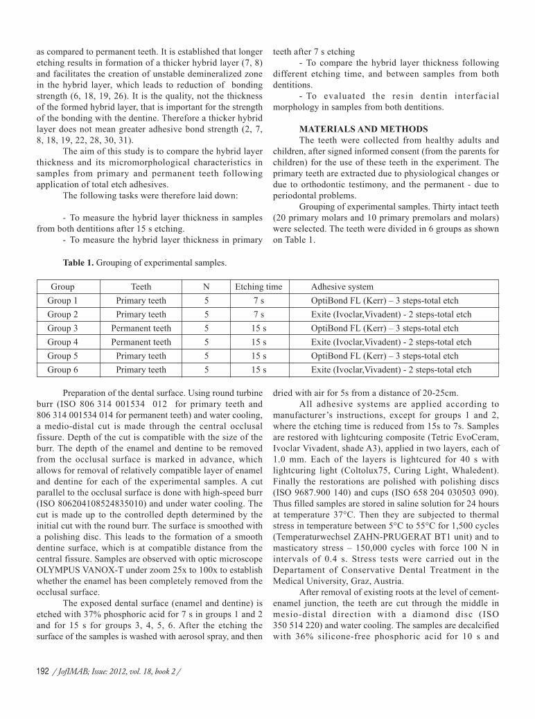
deciduous molars. On teeth from group 1 was applied 3-stepadhesive system OptiBond FL (Kerr). Samples weredemineralized with 37% phosphoric acid for 7s. Adhesivesystem was applied according to manufacturer’s instructions,only the etching time was shortened to 7s (fig. 1, 2, 3).
Fig. 1, 2 and 3. SEM of hybrid (between the arrows) and adhesive (Ad) layer in the borderline area in deciduousmolar etched with 37% phosphoric acid for 7s and adhesive system OptiBond FL (Kerr) (3-step, total etching). Multipleresin tags (R) are observed in dentine (D), C = composite.
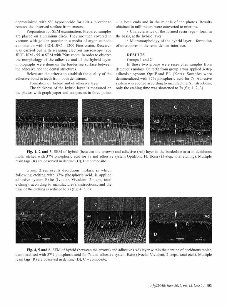
Group 2 represents deciduous molars, in whichfollowing etching with 37% phosphoric acid, is appliedadhesive system Exite (Ivoclar, Vivadent, 2-steps, totaletching), according to manufacturer’s instructions, and thetime of the etching is reduced to 7s (fig. 4, 5, 6).
Fig. 4, 5 and 6. SEM of hybrid (between the arrows) and adhesive (Ad) layer within the dentine of deciduous molar,demineralised with 37% phosphoric acid for 7s and adhesive system Exite (Ivoclar Vivadent, 2-steps, total etch). Multipleresin tags (R) are observed in dentine (D); C = composite.
194 / JofIMAB; Issue: 2012, vol. 18, book 2 /
From the photographs analyzed it could be concludedthat the thickness of the formed hybrid layer is greater in thesamples from group 2. The hybrid layer in the samples fromgroup 2 is more irregular, with varying thickness alongside.In the samples from both groups a significant number of resintags with funnel shape are established, and they penetrate indepth into the dentine tubuli (fig. 1, 2, 3, 4, 5 and 6).
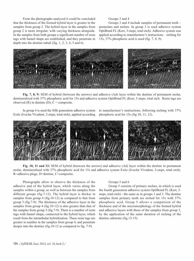
Fig. 7, 8, 9: SEM of hybrid (between the arrows) and adhesive (Ad) layer within the dentine of permanent molar,demineralized with 37% phosphoric acid for 15s and adhesive system OptiBond FL (Kerr, 3-steps, total etch. Resin tags areobserved (R) in dentine (D); C = composite.
Fig. 10, 11 and 12: SEM of hybrid (between the arrows) and adhesive (Ad) layer within the dentine in permanentmolar, demineralised with 37% phosphoric acid for 15s and adhesive system Exite (Ivoclar Vivadent, 2-steps, total etch).R=adhesive plugs, D=dentine, C=composite.
Photographs allow to observe the thickness of theadhesive and of the hybrid layer, which varies along thesamples within a group, as well as between the samples fromdifferent groups (fig.7-12). The hybrid layer is thicker insamples from group 4 (fig.10-12) as compared to that fromgroup 3 (fig.7-9). The thickness of the adhesive layer in thesamples from group 4 (fig.10-12) is also greater than that ofthe samples from group 3 (fig.7-9). There is a number of resintags with funnel shape, connected to the hybrid layer, whichresult from the intratubular hybridization. These resin tags aregreater in number in the samples from group 4, and penetratedeeper into the dentine (fig.10-12 as compared to fig. 7-9).
Groups 5 and 6Group 5 consists of primary molars, in which is used
the fourth generation adhesive system OptiBond FL (Kerr, 3-steps, total etch) - the same as in groups 1 and 3. The dentinesamples from primary teeth are etched for 15s with 37%phosphoric acid. Group 5 allows a comparison of thethickness and of the micromorphology of the formed hybridand adhesive layers with those of the samples from group 3,by the application of the same duration of etching of thedentine substrate (fig.13-15).
In group 4 is used the fifth generation adhesive systemExite (Ivoclar Vivadent, 2-steps, total etch), applied according
Groups 3 and 4Groups 3 and 4 include samples of permanent teeth –
premolars and molars. In group 3 is used adhesive systemOptiBond FL (Kerr, 3-steps, total etch). Adhesive system wasapplied according to manufacturer’s instructions – etching for15s, 37% phosphoric acid is used (fig. 7, 8, 9).
to manufacturer’s instructions, following etching with 37%phosphoric acid for 15s (fig.10, 11, 12).

/ JofIMAB; Issue: 2012, vol. 18, book 2 / 195
Fig. 13, 14 and 15: SEM of hybrid (between the arrows) and adhesive (Ad) layers in primary molar etched with 37%phosphoric acid for 15s and adhesive system OpyiBond FL (Kerr, 3-steps, total etch). C=composite, R=resin tags, D= dentine.
Group 6 consists of primary molars, in which is usedthe fifth generation adhesive system – Exite (Ivoclar Vivadent,2-steps, total etch) – the same as in groups 2 and 4. Thedentine samples from primary teeth are etched for 15s with37% phosphoric acid. Group 6 allows for comparison of the
Fig. 16, 17 and 18: SEM of hybrid (between the arrows) and adhesive (Ad) layers in primary molar etched with 37%phosphoric acid for 15s and adhesive system Exite (Ivoclar, Vivadent, 2-steps, total etch). C=composite, R=resin tags,D=dentine.
The figures allow to establish that the thickness of thehybrid layer in both used generations of adhesive systems isdifferent, as it is different between the samples frompermanent teeth in groups 3 and 4 (fig.7-12), and is differentbetween the samples from primary teeth in groups 5 and 6(fig.13-18). When comparing the thickness of the hybrid layerwithin the dentine, it is established that it is thicker in thesamples from groups 3 and 4 as compared to groups 5 and 6(fig.7-18). Resin tags with funnel shape are observed in thesamples from all groups. There are also microgaps betweenthe hybrid and the adhesive layer in samples from group 5
Table 2. Average values of the thickness of the hybrid layer (in microns) from adhesive systems with total etching inprimary and permanent teeth.
Group Etching Time Adhesive system HL Thickness – average value (ì) mean±SEGroup 1 – primary teeth 7 s OptiBond FL 3,74±0,32Group 2 – primary teeth 7 s Exite 4,70±0,18Group 3 – permanent teeth 15 s OptiBond FL 4,46±0,13
thickness and of the micromorhology and the formed hybridand adhesive layer with those from the samples of group 4,by the application of the same duration of etching of thedentine substrate (fig.16-18).
(fig.14-15), and group 6 (fig.16 and 18).By comparing the photographs of the samples from
group 1 with group 5 (fig.1-3 and 13-15), as well as betweenthose from group 2 (fig.4-6) with group 6 (fig.16-18) it isestablished that the hybrid layer is thicker, with microcracksin samples etched for 15s (fig.14-16 and fig. 18).
Statystical analysis of the resultsTable 2 represents the average values of the measured
thickness of the hybrid layer for different groups of samplesand applied adhesive systems with total etching.

196 / JofIMAB; Issue: 2012, vol. 18, book 2 /
The analysis of the results shows statisticallysignificant difference in the thickness of the hybrid layerbetween the samples of different experimental groups(Table 3).
In comparing the results for the thickness of theformed hybrid layer, it is established that there is a differencebetween samples from group 1 and group 2, between samplesfrom group 3 and group 4, and also between samples fromgroup 5 and group 6 (Table 3). A thicker hybrid layer isformed following application of 2-steps total etch adhesivesystem (Exite - groups 2, 4, and 6), as compared to applicationof 3-steps total etch adhesive (OptiBond FL - groups 1, 3 and5). The difference in the measured mean thickness of thehybrid layer is statistically relevant between the pairs of
groups (Table 3)(p<0.0001).A comparison was made between the average values
of the thickness of the hybrid layer for the combinations inpairs between experimental groups 1, 3, and 5 where we usedcomparison between the two dentitions with different etchingtime and the same adhesive system OptiBond FL (3-steps,total etching), by applying one-way ANOVA analysis (Table4). All pairs are with confirmed statistical relevance of thedifferences in the values of the thickness of the hybrid layer(Table 4).
Table 3. Hybrid layer thickness in samples from experimental groups.
Group N photographs HL Thickness(ì) mean±SD t ÐGroup 1 Group 2 5 5 3.74±0.32 4.70±0.18 5.84 <0.001*Group 3 Group 4 5 5 4.46±0.13 7.36±0.56 11.28 <0.0001*Group 5 Group 6 5 5 5.66±0.11 8.85±0.41 16.80 <0.0001*
*The difference is statistically significant.
Table 4. Thickness of the hybrid layer in pairs of experimental groups 1, 3 and 5 and adhesive system OptiBondFL (Kerr).
Group In comparison Average Difference 95% confidential interval for meanP*with in HL Thickness Lower Bound Upper Bound
Group 1 Group 3 -0.72 -1.26 -0.19 <0.0001Group 5 -1.92 -2.46 -1.38 <0.0001
Group 2 Group 5 -1.19 -1.42 -0.96 <0.0001*Empirical level of statistical relevance (ANOVA).
It is established that there is statistically significantdifference in the thickness of the hybrid layer in the group ofprimary teeth (groups 1 and 5) with different time for etching– 7 and 15 s, the greater thickness is measured in 15s etching(group 5) (p<0.0001); there is statistically significantdifference in the hybrid layer thickness with etching for 15sbetween the samples from two dentitions (groups 3 and 5),with greater mean thickness of the hybrid layer measured inthe samples from primary teeth (group 5) (p<0.0001); andstatistically significant difference in the thickness of the hybrid
layer between group 1 and group 3 with greater thickness ofthis layer established in permanent teeth (group 3)(p<0.0001)and using the same adhesive system OptiBond FL (Table 4).
The same comparison is also done between groups 2,4 and 6 in which was applied the 2-steps total etc adhesivesystem (Exite), by using one-way ANOVA analysis (Table 5).All pairs are with confirmed statistical significant of thedifferences in the values of the thickness of the hybrid layer(Table 5).
Group 4 – permanent teeth 15 s Exite 7,36±0,56Group 5 – primary teeth 15 s OptiBond FL 5,66±0,11Group 6 – primary teeth 15 s Exite 8,85±0,41

/ JofIMAB; Issue: 2012, vol. 18, book 2 / 197
It is established that there is statistically significantdifference in the thickness of the hybrid layer in the group ofprimary teeth (groups 2 and 6) with different time for etching– 7 and 15 s, the greater thickness is measured in 15s etching(group 6)(p<0.0001)(Table 5); there is statisticallysignificant difference in the hybrid layer thickness withetching for 15s between the samples from two dentitions(groups 4 and 6), with greater thickness established in thesamples from primary teeth (group 6)(p<0.0001)(Table 5);and statistically significant difference in the thickness of thehybrid layer in group 2 (primary teeth) and group 4(p<0.0001) with greater thickness established in the samplesfrom permanent teeth (group 4) and using the same adhesivesystem Exite (Table 5).
DISCUSSIONThe samples from all experimental groups were
restored with the mediation of adhesive systems belonging toEtch&Rinse strategy for adhesion with dental structures. Inthem adhesion is achieved by the formation of a hybrid layer,or so called zone of internal diffusion of the adhesive in thedemineralized dentine (15, 16, 17, 19).
Descriptive analysis of the figures we observed showspresence of formed adhesive and hybrid layers, resin tags withfunnel-like shape, resulting from intratubular hybridization,and connected with the hybrid layer, and penetrating invarious depth into the dentine (fig. 1-18). There areestablished differences in the thickness of the formed adhesiveand hybrid layers, they are thicker in the samples where 2-steps adhesive system was applied – groups 2, 4, and 6respectively, and the formed resin tags are also greater numberand penetrate deeper in the same groups (fig.4-6, 10-12, 16-18). It is considered that the formed resin tags also enforcethe strength of the bonding with the dentine. In adhesives withEtch&Rinse the formed resin tags increase the total strengthagainst shear bond strength.
There are established significant differences in thethickness and in the quality of the formed hybrid layer. In thesamples from primary teeth etched for 15s it is observed thepresence of microgaps between the adhesive and the hybridlayer and the dentine, as well as microgars inside the hybridlayer (fig.14-16 and 18). This means that under equalconditions the same adhesive system leads to formation ofhybrid layer with different characteristics in samples from
primary and permanent teeth (fig.7-9 as compared with fig.13-15, and fig.10-12 as compared with fig.16-18). At the sametime in etching for 7s the formed adhesive and hybrid layersare with similar characteristics (fig.1-6) with those from thesamples of permanent teeth etched for 15s (fig.7-12) and itis very important that there are no microgaps in those layers.
For the dentine bond strength of paramount importanceis the quality of the formed hybrid layer, not its thickness, soa thicker hybrid layer does not mean greater adhesive bondstrength (1, 7, 8, 18, 19, 22).
The results from our research show that acid etchingof the dentine of primary teeth with 37% phosphoric acid for7s (group 1 and 2) results in formation of a hybrid layer inall of the samples observed (fig.1-6).
Acid etching of primary teeth for 15s (groups 5 and6) results in formation of a hybrid layer with significantlygreater thickness than that in the samples from groups 1 and2 (7s etching)(p<0.05), as well as in comparison with that inthe groups with samples from permanent teeth – groups 3 and4, etched for 15s (Table 3-5, fig.1-18). Similar to the resultsof a previous research of ours for the nature of the changesin the dentine following acid etching (5), this research alsoconfirm the fact that the acid used is more aggressive on theprimary dentine than on the dentine of permanent teeth, otherconditions being equal. That leads to a deeperdemineralization of the intertubular dentine and results in theformation of a significantly thicker hybrid layer in primaryteeth (Table 4 and 5). There is a chance that such deeplydemineralized dentine may not be completely impregnated bythe adhesive systems and this creates risks of the formationof an uninfiltrated by the adhesive zone between the hybridlayer and the intact dentine structure. In that case the removedmineral matrix is not completely replaced by the primer ofthe adhesive system, which facilitates the penetration of theadhesive, there remains a more unstable zone in the basis ofthe hybrid layer, which is a possible route for micro- andnanopermeability, enzymic and hydrolytic decomposition andfollowing all that – an area for failure of the bonding (7, 18,19, 25, 26). The reason for that are also likely to be themicrocracks we observed in the samples from groups 5 and6 (fig.14-16 and 18).
Our research shows that the application of the sameadhesive system with the same clinical protocol in primaryand in permanent teeth leads to formation of adhesive and
Table 5. Hybrid layer thickness in pairs of experimental groups 2, 4, and 6 and adhesive system Exite.
Group In comparison Average Difference 95% confidential interval for average P*with in HL thickness Lower Bound Upper Bound
Group 2 Group 4 -2.65 -3.59 -1.71 <0.0001Group 6 -4.14 -4.83 -3.46 <0.0001
Group 4 Group 6 -1.49 -2.44 -0.53 <0.0001*Empirical level of statistical relevance (ANOVA).

198 / JofIMAB; Issue: 2012, vol. 18, book 2 /
REFERENCES:1. Bolanos-Carmona V, Gonzalez-Lopez
S, Briones-Lujan T, De Haro-Munoz C, dela Macorra JC. Effects of Etching Time ofPrimary Dentin on Interface Morphologyand Microtensile Bond Strength. DentMater. 2006 Dec;22(12):1121-1129.[PubMed] [CrossRef]
2. Bolanos-Carmona V, Gonzalez-LopezS, De Haro-Munoz C, Briones-Lujan MT.Interfacial Morphology and Bond Strengthof Self-Etching Adhesives to Primary DentinWith or Without Acid Etching. J BiomedMater Res B Appl Biomater. 2008Nov;87(2):499-507. [PubMed] [CrossRef]
3. Eick JD, Gwinnett AJ, Pashley DH,Robinson SJ. Current Concepts on Adhesionto Dentin. Crit Rev Oral Bio Med. 1997;8(3): 306-335. [PubMed] [CrossRef]
4. Eliades G, Watts DS, Eliades T.Dental hard tissues and bonding. Interfacialphenomena and related properties. Springer2005, 89-122
5. Gateva N. Comparative SEMexaminations of alterations on dentinesurface of primary and permanent teeth afteretching. Problemi na dentalnata medizina2008; 34(1): 71-84. (in Bulgarian)
6. Hals E. Observation on Giant Tubulesin Human Coronal Dentin by LightMicroscopy and Microradiography. Scand JDent Res. 1983 Feb;91(1):1-7 [PubMed]
7. Hashimoto M, Ohno H, Endo K,Kaga M, Sano H, Oguchi H. The Effect ofHybrid Layer Thickness on Bond Strength:Demineralized Zone of Hybrid Layer. DentMater. 2000 Nov;16(6):406-411. [PubMed]
8. Hashimoto M, Ohno H, Kaga M,Sano H, Tay FR, Oguchi H, et al: Over-etching Effects on Micro-tensile BondStrength and Failure Patterns for TwoDentin Bonding Systems. J Dent. 2002 Feb-Mar;30(2-3):99-105. [PubMed]
9. Hosoya Y, Marshall SJ, Watanabe LG,
Marshall GW. Microhardness of CariousDeciduous Dentine. Oper Dent 2000 Mar-Apr;25(2):81-89. [PubMed]
10. Koutsi V, Noonan RG, Horner JA,Simpson MD, Matthews WG, Pashley DH.The effect of Dentin Depth on thePermeability and Ultrastructure of PrimaryMolars. Pediatr Dent. 1994 Jan-Feb;16(1):29-35. [PubMed]
11. Lohbauer U, Nikolaenko SA,Petschelt A, Frenkenberger R. Resin Tags donot Contribute to Dentin Adhesion in Self-etching Adhesives. J Adhes Dent. 2008Feb;10(2):97-103. [PubMed]
12. Lopez GC, Baratieri VLN, Caldeirade Andrada MA, Vieira LCC. Dentaladhesion: Present state of the art and futureperspectives. Quintessence Int. 2002Mar;33(3):213-224. [PubMed]
13. Mahoney E., Holt A., Swain M.,Kilpatric N.: The Hardess and Modulus ofElasticity of Primary Molar Teeth: an Ultra-micro-indentation Study. J Dent. 2000Nov;28(8):589-594. [PubMed] [CrossRef]
14. Marquezan M, da Silveira BL,Burnett LH Jr, Rodrigues CR, Kramer PF.Microtensile Bond Strength of Contem-porary Adhesives to Primary Enamel andDentin. J Clin Pediatr Dent. 2007 Winter;32(2):127-132. [PubMed]
15. Marshall GW Jr, Marshall SJ,Kinney JH, Balooch M. The DentinSubstrate: Structure and Properties Relatedto Bonding. J Dent. 1997 Nov;25(6):441-458. [PubMed] [CrossRef]
16. Monksgaard EC. Wet or Dry,Normal or Deproteinized Dentin Surface asSubstrate for Dentin Adhesives. ActaOdontol Scand. 2002 Jan;60(1):60-64.[PubMed]
17. Nakabayashi N., Kojima K.,Masuhara E.: The promotion of adhesion bythe infiltration of monomers into tooth
substrates. J Biomed Mater Res. 1982May;16(3):265-273. [PubMed]
18. Nör JE, Feigal RG, Dennison JB,Edwards CA. Dentin Bonding: SEMComparison of the Resin-Dentin Interface inPrimary and Permanent Teeth. J Dent Res.1996 Jun;75(6):1396- 1403. [PubMed]
19. Nor JE, Feigal RG, Dennison JB,Edwards CA. Dentin Bonding: SEMComparison of the Dentin Surface inPrimary and Permanent Teeth. Pediatr Dent.1997 May-Jun;19(4);246-252. [PubMed]
20. Osorio R, Aguilera FS, Otero PR,Romero M, Osorio E, Garcya-Godoy F,Toledano M. Primary dentin etching time,bond strength and ultra-structurecharacterization of dentin surfaces. J Dent.2010 Mar;38(3):222-231. [PubMed][CrossRef]
21. Pashley DH, Ciucchi B, Sano H,Carvalho RM, Russell CM. Bond strengthversus dentine structure: A modellingapproach. Arch Oral Biol. 1995 Dec; 40(12):1109-1118. [PubMed] [CrossRef]
22. Perdigao J., Lopes M.: The Effect ofEtching Time on Dentin Demineralization.Quintessence Int. 2001; 32: 19-26
23. Pioch T, Garsia-Godoy F, DuschnerH, Koch MJ, Staehle HJ, Dorfer CE. Effectof cavity preparation instruments (oscillatingor rotating) on the composite-dentin inter-face in primary teeth. Dental Materials 2003Jun;19(4): 259-263. [PubMed] [CrossRef]
24. Rauch RU, Losche GM, RosanskyJ, Roulet JF. Comparison of quantitativemargin analysis and dye penetration. J DentRes. 1996; 75: abstr.187; 41
25. Sano H, Shono T, Takatsu T, HosodaH. Microporous Dentin Zone BeneathResin-Impregnated Layer. Oper Dent. 1994Mar-Apr;19(2):59-64. [PubMed]
26. Sardella TN, de Castro FL, SanabeME, Hebling J. Shortening of Primary
hybrid layer with different characteristics. They are withgreater thickness in the samples from primary teeth. Thevalues for the thickness of the hybrid layer are withstatistically significant difference between the two dentitions,and with other conditions equal, the measured hybrid layerin primary teeth is thicker (p<0.05, Table 2, 4 and 5) (2, 18,19).
Conclusions:Based on the results we obtained as well as on the
results from the specialized literature, it could be accepted
that:1. In primary teeth the hybrid layer thickness
increases with the increased etching time.2. The greater thickness of the hybrid layer does not
guarantee reliable and lasting adhesive bonding.3. The hybrid layer thickness in primary teeth with
the same concentration and duration of the etching isgreater than that of the hybrid layer in permanent teeth.
4. For primary teeth it is more appropriate to reducethe etching time to 7s to obtain a hybrid layer with betterquality.

/ JofIMAB; Issue: 2012, vol. 18, book 2 / 199
Corresponding author:Natalia GatevaDepartment of Pediatric Dentistry, Faculty of Dentistry, Medical University, Sofia1, St. Georgi Sofiiski str., 1431 Sofia, Bulgariae-mail: [email protected]
Dentin Etching Time and its Implication onBond Strength. J Dent. 2005 May;33(5):355-362. [PubMed] [CrossRef]
27. Shashikiran ND, Gunda S, SubbaReddy VV. Comparison of Resin-DentinInterface in Primary and Permanent Teethfor Three Different Durations of DentinEtching. J Indian Soc Pedod Prev Dent.2002 Dec;20(4):124-131. [PubMed]
28. Spencer P, Wang Y, Walker MP,Swafford JR. Molecular Structure of Acid-Etched Dentin Smear Layers - in situ Study.J Dent Res. 2001 Sep;80(9):1802-1807.[PubMed] [CrossRef]
29. Spreafico D, Semeraro S,Mezzanzanica D, Re D, Gagliani M, TanakaT, et al. The effect of air-blowing step on thetechnique sensitivity of four differentadhesive systems. J Dent. 2006 Mar;34(3):237-244. [PubMed] [CrossRef]
30. Stadtler P: Dentinhaftmittel.Stomatologie 2007; 104(4): 101–109.[CrossRef]
31. Tulunoglu O, Tulunoglu I. Resin-dentin interfacial morphology and shearbond strengths to primary dentin after long-
term water storage: An in vitro study.Quintessence Int. 2008 May;39(5): 427-437. [PubMed]
32. Uekusa S, Yamaguchi K, MiyazakiM, Tsubota K, Kurokawa H, Hoyosa Y.Bonding efficacy of single-step self-etchsystems to sound primary and permanenttooth dentin. Oper Dent. 2006 Sep-Oct;31(5):569-576. [PubMed] [CrossRef]
33. Van Meerbeek B, Conn LJ Jr, DukeE, Eick J, Robinson S, Guerrero D. Corre-lative transmission electron microscopyexamination of nondemineralized anddemineralized rezin-dentin interfacesformed by two dentin adhesive systems. JDent Res. 1996 Mar;75(3):879-888.[Pubmed] [CrossRef]
34. Van Meerbeek B, De Munck J,Yoshida Y, Inoue S, Vargas M, Vijay P, etal. Buonocore memorial lecture. Adhesionto enamel and dentin: current status andfuture challenges. Oper Dent. 2003 May-Jun;28(3):215-235. [PubMed]
35. Van Meerbeek B, Inokoshi S, BraemM, Lambrechts P, Vanherle G. Morpholo-gical aspects of the resin-dentin interdiffu-
sion zone with different dentin adhesivesystems. J Dent Res. 1992 Aug;71(8):1530-1540. [PubMed] [CrossRef]
36. Van Meerbeek B, Van Landuyt K,De Munck JD, Hashimoto M, Peumans M,et al. Technique-Sensitivity of Contempo-rary Adhesives. Dent Mater J. 2005 Mar;24(1):1-13. [PubMed]
37. Van Meerbeek B.,Vargas M., InoueS., Yoshida Y., Peumans M Lambrechts P.et all.: Adhesives and cements to promotepreservation dentistry. Operative Dentistry2001; 6: 119-144
38. Wang Y, Spencer P. Analysis ofAcid-Treated Dentin Smear Debris andSmear Layers Using Confocal RamanMicrospectroscopy. J Biomed Mater Res.2002 May;60(2):300-308. [PubMed]
39. Yaseen SM, Subba Reddy VV.Comparative evaluation of shear bondstrength of two self-etching adhesives (sixthand seventh generation) on dentin ofprimary and permanent teeth: An in vitrostudy. J Indian Soc Pedod Prev Dent. 2009Jan-Mar;27(1):33-38. [PubMed] [CrossRef]