62253747 drug allergy book
DESCRIPTION
allergi obatTRANSCRIPT
Di
lVl
Daniel VervloetMichel Pradal
Michel Castelain
DRUGALLERGY


- 1 -
Daniel Vervloet, Michel Pradal, Michel Castelain
Department of Pneumology and AllergyDepartment of DermatologyHopital Sainte Marguerite
Université de la MéditerrannéeMarseille FRANCE
DRUG ALLERGY

- 2 -
ISBN 91-973440-0-1
Daniel Vervloet/Michel Pradal/Michel Castelain
- 3 -
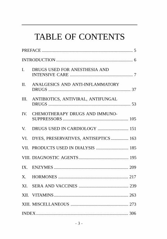
TABLE OF CONTENTSPREFACE .................................................................................. 5
INTRODUCTION ..................................................................... 6
I. DRUGS USED FOR ANESTHESIA ANDINTENSIVE CARE ......................................................... 7
II. ANALGESICS AND ANTI-INFLAMMATORYDRUGS .......................................................................... 37
III. ANTIBIOTICS, ANTIVIRAL, ANTIFUNGALDRUGS .......................................................................... 53
IV. CHEMOTHERAPY DRUGS AND IMMUNO-SUPPRESSORS ........................................................... 105
V. DRUGS USED IN CARDIOLOGY ............................ 151
VI. DYES, PRESERVATIVES, ANTISEPTICS ................ 163
VII. PRODUCTS USED IN DIALYSIS............................. 185
VIII. DIAGNOSTIC AGENTS............................................. 195
IX. ENZYMES ................................................................... 209
X. HORMONES ............................................................... 217
XI. SERA AND VACCINES ............................................. 239
XII. VITAMINS................................................................... 263
XIII. MISCELLANEOUS .................................................... 273
INDEX................................................................................... 306

- 4 -

- 5 -
PREFACEThe first version of this work was originally the thesis that Dr
Michel Pradal submitted for his Doctorate of Medicine in 1987.Thanks to Pharmacia & Upjohn Diagnostics Division, this firstversion was published in French in 1990, and received positivereviews among the French allergist community. Two years later,in 1992, we published a second edition, this time in English, in thehope that this work might be a practical help to allergists facingdifficulties in making etiologic diagnoses of drug allergies. Thesedifficulties reside in the fact that where drug allergy is concerned,there is but one general rule: “there are no general rules”. Eachdrug has its own metabolites and mechanisms, and the diagnostictools must be adapted to the type of reaction caused by the drug inquestion and to the mechanisms involved.
Seven years after this second edition, we thought it would beuseful to publish an updated version enriched with up-to-dateknowledge and recent references. To achieve this, I askeddermatologist and allergist Doctor Michel Castelain to join DoctorPradal and myself and enrich this work particularly in regard todelayed allergic reactions which often show skin reactions.
I hope that all the information found here will be useful in theday-to-day practice of allergology, and I thank Pharmacia & Upjohnfor its material support.
Daniel Vervloet

- 6 -
INTRODUCTIONEstimations of incidence figures for drug allergy show a varia-
tion. In a French study of 2067 adults aged from 20 to 67 visitinga health-care center for check-up examination, 14.7% of the studygroup reported reliable histories of systemic drug reactions.
Others have stated that allergic drug reactions account for 5-10% of adverse drug reactions. In a Swiss drug monitoring pro-gram, about 17% of hospitalized patients showed adverse drugreactions, 5.4 -5.9% appeared to be allergic.
Numerous mechanisms have been implicated, but severalremain obscure and this lack of knowledge accounts for thedifficulties in differentiating allergy from other side effects,assessing the incidence, risk factors, and management strategy fordiagnosis and prevention.
This book was written as a practical guide to allergy diagnosismanagement and the main drugs responsible. We have tried tospecify the main clinical manifestations and risk factors for eachof them, as well as the diagnostic methods (in vivo and, wherepossible, in vitro), known mechanisms and management.
- 7 -
I
DRUGS USED FORANESTHESIA ANDINTENSIVE CARE

- 8 -
LOCAL ANESTHETICS
These drugs anesthetize the area to which they are applied byblocking nerve conduction and preventing depolarization of cellmembranes.
There are two main families of local anesthetics:Benzoic acid ester group (group I):benzocaine, chlorprocaine, cocaine, piperocaine, procaine,propoxycaine, tetracaine.
Amides (group II):aromatic: articaine, bupivacaine, dibucaine, etidocaine,lidocaine, mepivacaine, prilocaine.thiophenic: alphacaine
Amides are by far the most used today.
INCIDENCE2 to 3% of local anesthesias (faints). True allergic reactions areexceedingly rare (less than than 1% of all reactions).
RISK FACTORSLong term topical application of anesthetic drugs (contactdermatitis).
CLINICAL MANIFESTATIONSReactions unrelated to drugs:
— psychomotor: hyperventilation, vaso-vagal, adrenergic— sympathetic stimulation— operative trauma
Toxic responses in normal subjects:— central nervous system effects— cardiovascular effects
DRUGS USED FOR ANESTHESIA AND INTENSIVE CARE

- 9 -
Reactions due to either adjuvants or injection, but not to the localanesthetic itself:
— associated drugs: epinephrine, sulfites, parabens, antibio-tics, analgesics;
— traumatic subcutaneous emphysema.
Type I allergic reactions: anaphylactic shock, urticaria, angioedemaare exceptional.Fixed drug eruptions: extremely rare (lidocaine, mepivacaine).Delayed-type hypersensitivity (contact allergy) is more frequentdue to the increasing use of topical preparations containing localanaesthetics of the amide group. Several of these cases have beenreported in chronically treated patients with topical applicationsof Emla cream (lidocaine + prilocaine). Cases of contact allergy tolidocaïne have been also reported (antihemorroidal cream,phlebectasias treatment).Historically, delayed-type hypersensitivity was reported mainlywith esters which are much less commonly used nowadays.Cross-reactivity among amides is not always encountered.
DIAGNOSTIC METHODSCutaneous testingPrick-tests are mostly negative.Intradermal skin-tests: false positive at 1/10 and pure concentration.Few case reports with type I clinical manifestations and positiveintradermal skin-tests with 1/10 000 to 1/100 concentrations.Patch-tests are useful in the diagnosis of type IV manifestations(contact dermatitis) to esters and amides.
No specific IgE found
MECHANISMSNo absolute demonstration of IgE-mediated reactions.The role of preservatives (parabens, sulfites) remains controversial.Type IV reactions: contact dermatitis (esters and amides).
DRUGS USED FOR ANESTHESIA AND INTENSIVE CARE

- 10 -
MANAGEMENTChallenge test remains the ”gold standard” in local anestheticreactions. Many suggested dosing protocols exist: for example (at15 minute intervals).
1° prick undiluted2° S.C 0.1 ml undiluted3° S.C 0.5 ml undiluted4° S.C 1 ml undiluted5° S.C 2 ml undiluted
Alternative therapies:— preservative-free preparations— be aware of cross-reactivity between esters (frequent) and
amides (less frequent)— antihistamines (diphenhydramine) may be used as anesthetics— general analgesia (N2O)— hypnosis
REFERENCES• Fisher M.M, Bowey C.J, ”Alleged allergy to local anaesthetics”, Anaesth.
Intensive. Care., 1997 ; 25 (6): 611-4• Cuesta-Herranz J, de las Heras M, Fernandez M, Lluch M, Figueredo E,
Umpierrez A, Lahoz C, “Allergic reaction caused by local anesthetic agentsbelonging to the amide group”, J.Allergy. Clin. Immunol., 1997 ; 99 (3):427-8
• Eggleston S.T, Lush L.W, “Understanding allergic reactions to localanesthetics”, Ann. Pharmacother, 1996 ; 30 (7.8): 851-7
• Gall H, Kaufmann R, Kalveram C.M, “Adverse reactions to local anesthetics:analysis of 197 cases”, J.Allergy. Clin. Immunol, 1996 ; 97 (4): 933-7
• Wasserfallen J.B, Frey P.C, ”Long-term evaluation of usefulness of skin andincremental challenge tests in patients with history of adverse reaction tolocal anesthetics”, Allergy, 1995 ; 50: 162-5
• Sindel L.J, de Shazo R.D, “Accidents resulting from local anesthetics. Trueor false allergy ?”, Clin. Rev. Allergy, 1991 ; 9 (3.4): 379-95.
DRUGS USED FOR ANESTHESIA AND INTENSIVE CARE

- 11 -
HYPNOTICS
BARBITURATES
METHOHEXITAL
Methohexital is a barbiturate derivative used for brief surgicalprocedures.
INCIDENCEReported incidence ranges from 1/7000 to 0/23000.0.2% in dentistry.No deaths have been reported.
CLINICAL MANIFESTATIONSEssentially cutaneous, mucosal (pruritus, urticaria, rash, edema)and cardiovascular.
Respiratory (bronchospasm) and digestive symptoms are unusual.
Symptoms are usually delayed (a few minutes after injection).
DIAGNOSTIC METHODSNone.
MECHANISMSNon specific histamine-release.
MANAGEMENTAvoidance.
DRUGS USED FOR ANESTHESIA AND INTENSIVE CARE

- 12 -
REFERENCES• Liu L.M, Liu P.L, Moss J, “Severe histamine-mediated reaction to rectally
administered methohexital”, Anesthesiology, 1984 ; 61 (1): 95-7• McDonald D, “Methohexitone in dentistry. Scientific results of 4379
administrations. 5: complications”, Dent. Anaesth. Sedat., 1982 ; 11 (2): 51-7• Harrison G.R, Thompson I.D, “Adverse reaction to methohexitone and galla-
mine”, Anaesthesia, 1981 ; 36 (1): 40-4• Driggs R.L, O’Day R.A, “Acute allergic reaction associated with methohexital
anesthesia: report of six cases” J. Oral. Surg., 1972 ; 30: 906-9.
THIOPENTAL
Frequently used hypnotic, barbiturate anesthetic.
INCIDENCE1/23000 to 1/29000 administrations.Deaths rarely reported.
RISK FACTORSPrevious exposure (94%).Female gender (gender ratio 3/1)
CLINICAL MANIFESTATIONSGeneral: anaphylactic shockRespiratory: bronchospasm.Cutaneous: flush, generalized erythema, angioedema, fixed drugeruption.Possible delayed reactions.Haematological: haemolytic anemia.
DRUGS USED FOR ANESTHESIA AND INTENSIVE CARE

- 13 -
DIAGNOSTIC METHODSCutaneous testingPrick-tests or intradermal tests. Dilution 1/1000 to 1/10 of 2.5%thiopental.Detection of thiopentone-reactive IgE antibodies by the RASTmethod ; which specificity is confirmed by hapten inhibitionstudies.
MECHANISMSImmediate hypersensitivity: Usually IgE-mediated.Allergenic determinants: pentyl and ethyl groups attached to posi-tion 5 on the pyrimidine ring nucleus and the secondary region ofthe ring, encompassing and including the attached hetero atom.Possible involvement of non-specific histamine release.
MANAGEMENTAvoidance.
REFERENCES• Bremang J.A, Halasi S, ”Fixed drug eruption associated with anaesthesia”,
Can.J.Anaesth., 1995 ; 42 (7): 628-30• Baldo B.A, Fisher M.M, “Diagnostic de l’allergie IgE dépendante aux
curares, au thiopental et aux opiacés”, Ann.Fr.Anesth.Reanim., 1993 ; 12(2): 173-81
• Baldo B.A, Fisher M.M, Harle D.G, “Allergy to thiopentone” Clin. Rev.Allergy, 1991 ; 9 (3-4): 295-308.
• Baldo B.A, Harle D.G, “Drug allergenic determinants (pp 11-51)”, In:Molecular approaches to the study of allergens. Monographs in allergy, Vol.28.BA Baldo ed. Karger, Basel, 1990.
• Harle D.G, Baldo B.A, Smal M.A, Wajon P, Fisher M.M, ”Detection ofthiopentone reactive IgE antibodies following anaphylactoid reactions duringanaesthesia”, Clin. Allergy, 1986 ; 16: 493-8.
DRUGS USED FOR ANESTHESIA AND INTENSIVE CARE

- 14 -
NON-BARBITURATES
ETOMIDATE
Short-acting general anesthetic.
INCIDENCEExceedingly rare.
CLINICAL MANIFESTATIONSEssentially cutaneous and gastrointestinal.Few cardiovascular and respiratory effects.
DIAGNOSTIC METHODSNo in vivo or in vitro diagnostic method currently available.
MECHANISMSEtomidate is a poor histamine releaser.
MANAGEMENTAvoidance.
REFERENCES• Fazackerley E.J, Martin A.J, Tolhurst-Cleaver C.L, Watkins J, “Anaphylactoid
reaction following the use of etomidate”, Anaesthesia, 1988 ; 43 (1): 953-4.• Bricker S.R, “Angioneurotic edema following etomidate/lignocaine”, Anaes-
thesia, 1987 ; 42 (3): 323-4.• Watkins J, “Etomidate, an “immunologically safe” anaesthetic agent”,
Anaesthesia., 1983 ; 38 (S): 34-8
DRUGS USED FOR ANESTHESIA AND INTENSIVE CARE

- 15 -
KETAMINE
INCIDENCEExceedingly rare.
CLINICAL MANIFESTATIONSGeneral: anaphylactic shock.Cutaneous: rash, urticaria.Respiratory: laryngospasm.
DIAGNOSTIC METHODS
Cutaneous testingIntradermal skin-tests: 0.05 ml of Ketalar 5% or ketamine base 1/100 and 1/10 positive in one patient.
MECHANISMSProbable IgE-mediated hypersensitivity.
MANAGEMENTAvoidance.
REFERENCES• Karayan J, Lacoste L, Breuil K, ”Allergie à la kétamine”,
Ann.Fr.Anesth.Reanim., 1990 ; 9 (4): 396-7• Mathieu A, Goudsouzian N, Swider M.T, “Reaction to ketamine:
anaphylactoid or anaphylactic ? “ Br. J. Anaesth., 1975 ; 47: 624-7
PROPANIDID
Short-acting general anesthetic used especially for toothextractions.
INCIDENCEVery high: 1 out of 500 to 1 out of 700 anesthesias.Severe reactions: 0.007% to 0.13%.
DRUGS USED FOR ANESTHESIA AND INTENSIVE CARE

- 16 -
CLINICAL MANIFESTATIONSGeneral: anaphylactic shock.Respiratory: bronchospasm.Dermatological: various delayed reactions.Digestive: nausea, vomiting, diarrhea.
DIAGNOSTIC METHODSComplement assay at the time of the accident (difficult).Blood histamine measurement at the time of the accident.
MECHANISMSResponsibility of solvent: cremophor E.L., which activates theclassical or alternative complement pathway, releasing C3aanaphylatoxin and leukocyte migration.Non-specific histamine release.
MANAGEMENTProhibited in many countries since 1983.Avoidance.
REFERENCES• Guerrero J, Trevilla J.M, de las Mulas M, Herrera J.C, Malagon F, ”Reaccion
anafilactica a la propanidida”, Rev. Esp. Anesthesiol Reanim., 1985 ; 32(5): 252-3.
• Christmas J.P, “Immune reaction to propanidid”, Anaesthesia, 1984 ; 39:470-3.
DRUGS USED FOR ANESTHESIA AND INTENSIVE CARE
PROPOFOL
Propofol (2-6 diisopropylphenol) is an alkyl phenol in a lipidvehicle (soybean oil, egg lecithin, glycerol) used in anesthesia.
INCIDENCE1/60 0001.2% of peroperative anaphylactic shocks in France.

- 17 -
DRUGS USED FOR ANESTHESIA AND INTENSIVE CARE
RISK FACTORSPrevious drug allergy.Allergy to muscle relaxants.Association with atracurium.
CLINICAL MANIFESTATIONSGeneral: anaphylactic shock.Cutaneous: urticaria, angioedema, facial edema, erythema.Respiratory: bronchospasm.Ocular: conjunctival chemosis.
DIAGNOSTIC METHODSCutaneous testing
Skin prick-tests.Intradermal skin-tests from 1 µg/ml to 100 µg/ml (1/1000 to 1/10). Positive in most patients with anaphylactic reactions.Specific IgE (RIA): positive in 10/14 propofol allergic patients.Leukocyte-specific histamine release: positive in 3/5 propofolallergic patients.
MECHANISMSIgE-mediated hypersensitivity in most cases.Propofol contains 2 isopropyl group that may act as the epitopes.
MANAGEMENTDo not use propofol in muscle relaxant-allergic patients.Do not use atracurium with propofol (high frequency ofanaphylactoid reactions).Avoidance.
REFERENCES• Laxenaire M.C, ”Utilisation du Diprivan* chez le patient allergique”,
Ann.Fr.Anesth.Reanim., 1994 ; 13 (4): 498-502• Mc Hale S.P, Konieczko K, ”Anaphylactoid reaction to propofol”, Anaes-
thesia., 1992 ; 47 (10): 864-5• de Leon-Casasola O.A, Weiss A, Lema M.J, “Anaphylaxis due to propofol”,
Anesthesiology, 1992 ; 77 (2): 384-6

- 18 -
• Laxenaire M.C, Mata-Bermejo E, Moneret-Vautrin D.A, Gueant J.L, “Life-threatening anaphylactoid reactions to propofol (Diprivan*)”,Anesthesiology, 1992 ; 77 (2): 275-80
• Mc Leskey C.H, “Anaphylactoid reactions following propofol-atracuriumsequence (letter ; comment)”, Can.J.Anaesth., 1990 ; 37 (8): 946-7
MORPHINOMIMETICS
CODEINE
Codeine (methylmorphine, morphine monomethyl ether) belongsto the opiod group. The main indication of codeine is as a coughsuppressant.
INCIDENCEVery low.
RISK FACTORSIntravenous use in children.
CLINICAL MANIFESTATIONSGeneral: arterial hypotension (intravenous route), pseudoscarletfever.Cutaneous: pruritus, urticaria, macular and maculopapulareruptions, angioedema, erythema multiforme, erythema nodosum,scarlatiniform rashes, fixed drug eruption (occupational dermatitisfrom codeine has been reported).Respiratory: bronchospasm.
DRUGS USED FOR ANESTHESIA AND INTENSIVE CARE

- 19 -
DIAGNOSTIC METHODSCutaneous testingSkin patch-tests: codeine phosphate 0.1% aq. (fixed drug erup
tion).codeine phosphate 0.001% to 0.033% aq.(urticarial rash).
Oral challenge tests (fixed drug eruption).
MECHANISMSNon immunological histamine release (pruritus, urticaria).Delayed-type hypersensitivity (urticaria, rash).Vasomotor depression, ganglionar blockade and histamine releasecould explain hypotension.
MANAGEMENTAvoid intravenous use in children.The risk of cross-sensitivity is higher with morphine congenersthan with phenylpiperidine or methadone-type agents.
REFERENCES• Gonzalo-Garijo M.A, Revenga-Arranz F, ”Fixed drug eruption due to codeine
(letter)” , Br.J.Dermatol., 1996 ; 135 (3): 498-9• Rodriguez F, Fernandez L, Garcia-Abujeta J.L, Maquiera E, Llaca H.F, Jerez
J, “Generalized dermatitis due to codeine”, Contact. Dermatitis. , 1995 ; 32(2): 120
• de Groot A.C, Conemans J, “Allergic urticarial rash from oral codeine”,Contact. Dermatitis., 1986 ; 14 (4): 209-14.
• Shanahan E.C, Marshall A.G, Garrett C.P.O, “Adverse reactions tointravenous codeine phosphate in children. A report of 3 cases”, Anaesthe-sia, 1983 ; 38 (1): 40-3.
DRUGS USED FOR ANESTHESIA AND INTENSIVE CARE

- 20 -
FENTANYL
Major morphinomimetic analgesic, used exclusively in anesthesia.
INCIDENCEExtremely rare.
RISK FACTORSPrevious sensitization to another phenylpiperidine drug.
CLINICAL MANIFESTATIONSGeneral: anaphylactic shock.Cutaneous: urticaria, angioedema, diffuse rash (transdermalfentanyl).Respiratory: bronchospasm.
DIAGNOSTIC METHODSCutaneous testingIntradermal skin-tests positive from 0.5 ng/ml to 5 ng/ml (2 casesreported).
MECHANISMSFentanyl does not induce non-specific histamine-release.IgE-mediated hypersensitivity (positive skin-tests).
MANAGEMENTAvoidance.
REFERENCES• Stoukides C.A, Stegman M, “Diffuse rash associated with transdermal
fentanyl”, Clin.Pharm., 1992 ; 11 (3): 222• Bennett M.J, Anderson L.K, Mc Millan J.C, Ebertz J.M, Hanifin J.M,
Hirshman C.A, “Anaphylactic reaction during anaesthesia associated withpositive intradermal skin test to fentanyl”, Can. Anaesth. Soc. J., 1986 ; 33(1): 75-8.
DRUGS USED FOR ANESTHESIA AND INTENSIVE CARE

- 21 -
• Pevny I, Danhauser I, ”Anaphylaktisher schock wahrend der narkose mitpositivem hauttest auf fentanyl und alloferin”, Anaesthesist., 1981 ; 30:400-4
DRUGS USED FOR ANESTHESIA AND INTENSIVE CARE
MEPERIDINE - PETHIDINE
Narcotic analgesic frequently used for analgesia and generalanesthesia (induction).
INCIDENCEVery low.
CLINICAL MANIFESTATIONSGeneral: anaphylactic shock.Cutaneous: flush, urticaria.Respiratory: cough, wheezing.
DIAGNOSTIC METHODSSpecific IgE (RAST): positive in a few cases of anaphylaxis.Immediate skin-tests may be positive (do not exceed a concent-ration of 1/100000).
MECHANISMSIgE-mediated hypersensitivity.Non-specific histamine release (meperidine is one of the strongesthistamine-releasers of all anaesthetic agents).
MANAGEMENTAvoidance.
REFERENCES• Flacke J.W, Flacke W.E, Bloom B.C, Van Etton A.P, Kripke B.J, “Histamine
release by 4 narcotics: a double blind study in humans”, Anesth. Analg.,1987 ; 66: 723-30.
• Levy J.H, Rockoff M.A, “Anaphylaxis to meperidine”, Anesth. Analg., 1982; 61 (3): 301-3
• Waisbren B.A, “Hypersensitivity to meperidine”, JAMA, 1978 ; 239 (14):1395

- 22 -
DIAGNOSTIC METHODSCutaneous testingIntradermal skin-tests: positive 1/100 000 (solution of 10 mg/ ml)Specific IgE (RIA morphine-sepharose) positive in a patient.
MECHANISMSNonspecific histamine release .IgE-mediated hypersensitivity: the morphine allergenic determi-nant comprises the cyclohexenyl ring, with a hydroxyl group atC6 and a methyl substituent attached to the N-atom.
MANAGEMENTAvoidance.Cross-reactivity with codeine and other opioids may occur.
REFERENCES• Harle D.G, Baldo B.A, Coroneos N.J, Fisher M.M, ”Anaphylaxis following
administration of papaveretum. Case report: implication of IgE antibodiesthat react with morphine and codeine and identification of an allergic deter-minant”. Anesthesiology., 1989 ; 71 (4): 489-94
• Rossi R, Dick W, “Anaphylactoide reaktion nach intravenöser injektion vonmorphinum hydrochloricum”, Anesthesist. , 1982 ; 31: 463-5
MORPHINE
Morphine, historically chief among the opioids , is widely used asan analgesic in various clinical situations (postoperative period,cancer).
INCIDENCEExtremely rare.
CLINICAL MANIFESTATIONSGeneral: anaphylactic shock.Cutaneous: urticaria.Respiratory: bronchospasm.
DRUGS USED FOR ANESTHESIA AND INTENSIVE CARE

- 23 -
DRUGS USED FOR ANESTHESIA AND INTENSIVE CARE
• Fahmy N.R, “Hemodynamics, plasma histamine and catecholamineconcentrations during an anaphylactoid reaction to morphine”,Anesthesiology, 1981 ; 55: 329-31
MUSCLE RELAXANTS
Family of agents widely used in general anesthesia to achievemuscle relaxation.Suxamethonium, Vecuronium, Pancuronium, Atracurium,Mivacurium, Rocuronium, Cisatracurium
INCIDENCEOne anaphylactic shock out of every 10000 general anesthesias.10% of these reactions are fatal.Muscle relaxants account for 60 to 70% of all allergic reactionsoccurring during general anesthesia.
RISK FACTORSUse of muscle relaxants with a “flexible chain”.Female gender (80% of cases).No involvement of atopy.Previous allergic reactions to muscle relaxants.
CLINICAL MANIFESTATIONSGeneral: collapse, tachycardia, arrhythmia, cardiac arrest.Respiratory: bronchospasm.Dermatological and mucosal: flush, diffuse erythema, diffuseurticaria, angioedema.Digestive: diarrhea.Haematological: disseminated intravascular coagulation.
DIAGNOSTIC METHODSIntradermal skin tests or prick tests (using undiluted musclerelaxants). High diagnostic performance if conducted with properconcentrations, i.e. for intradermal skin testing:

- 24 -
≤100 µg/ml for suxamethonium.≤200 µg/ml for pancuronium.≤100 µg/ml for rocuronium≤2 µg/ml for mivacurium≤400 µg/ml for vecuronium.≤10 µg/ml for atracurium.≤200 µg/ml for cisatracuriumSubjects remain positive for several years (up to 30 years).
Detection of anticurare and antisuxamethonium IgE antibodies byRAST methods.
Specific histamine release.
MECHANISMSImmediate IgE-mediated hypersensitivity: detection of IgE specificto muscle relaxants (quaternary ammonium ion determinants)accounting for cross reactions between different curarizinganesthetics and reactions upon first contact with a muscle relaxant(antihypertensors, antiseptics, antibiotics, cosmetics, and soaps witha quaternary ammonium ion structure).Mediator release from target cells. Leukocytes from subjects witha history of anaphylactic accidentsbrought into contact withincreasing concentrations of muscle relaxants in vitro, releasehistamine which can be measured (dose-dependent response).Histamine release from basophils involves IgE antibodies.Role of quaternary ammonium ion determinants. The rigidity ofthe chain linking quaternary ammoniums plays a triggering role inallergic reactions. ”Flexible” molecules with simple carbon chains(Suxamethonium) can stimulate sensitized cells more strongly thanrigid molecules (Pancuronium).Some reactions are not based on an IgE mechanism, but on a non-specific histamine release effect.
MANAGEMENTUse safer muscle relaxants such as Pancuronium rather thanSuxamethonium.
DRUGS USED FOR ANESTHESIA AND INTENSIVE CARE

- 25 -
DRUGS USED FOR ANESTHESIA AND INTENSIVE CARE
.
REFERENCES• Porri F, Lemiere C, Birnbaum J, Guilloux L, Lanteaume A, Didelot R, Char-
pin D, Vervloet D, ”Prevalence of muscle relaxants sensitivity in a generalpopulation: implications for a preoperative screening”, Clin. Exp. Allergy,1998 ; in press
• Nicklas R.A, “Anaphylaxis during general anesthesia, the intraoperative pe-riod and postoperative period”, J. Allergy. Clin. Immunol., 1998 ; 101:S512-6
• Laxenaire M.C et le groupe d’étude des réactions anaphylactoïdesperanesthesiques, “ Substances responsables des chocs anaphylactiquesperanesthesiques”, Ann. Fr. Anesth., 1996 ; 15: 1211-8
• Porri F, Pradal M, Rud C, Charpin D, Alazia M, Gouin F, Vervloet D, “Issystematic preoperative screening for muscle relaxants and latex allergyadvisable ?”, Allergy, 1995 ; 50: 374-7
• Birnbaum J, Vervloet D, “Allergy to muscle relaxants”, Clin. Rev. Allergy.,1991 ; 9 (3-4): 281-93
• Marone G, Stellato C, Mastronardi P, Mazzarella B, “Nonspecific histamine-releasing properties of general anesthetic drugs”, Clin. Rev. Allergy, 1991 ;9 (3-4): 269-80
• Leynadier F, Sansarricq M, Didier J.M, Dry J, “Prick-tests in the diagnosisof anaphylaxis to general anaesthetics”, Br. J. Anaesth., 1987 ; 59: 683-9
Systematic screening-tests in a general population are not advisabledue to the poor positive predictive value of the tests.Patients with a history of anaphylactic reactions:Conduct preoperative skin-tests with all muscle relaxants.(Frequent cross-reactivity between 2 or more muscle relaxants)
— If skin-tests are positive, do not use the offending muscle relaxants.
— If skin-tests are negative, the agents may be administered(good negative precitive value).
No preventive treatment can avoid anaphylactic reactions

- 26 -
DRUGS USED FOR ANESTHESIA AND INTENSIVE CARE
BENZODIAZEPINES
DIAZEPAM
Leading benzodiazepine, widely used against seizures and as amuscle relaxant.
INCIDENCELow in France but one injection out of 1000 when cremophor E.L.was used as the solvent (not in France).The most common offending molecules are diazepam andflunitrazepam.
CLINICAL MANIFESTATIONSGeneral: anaphylactic shock, collapse.Cutaneous: urticaria, angioedema, hives, flush, polymorphouserythema, purpura.Respiratory: dyspnea, bronchospasm.Renal: allergic interstitial nephropathy.
DIAGNOSTIC METHODSCutaneous testing
Usually negative.In one case prick-test positive with solvent.In one case patch-test positive.
Positive reintroduction test (with caution) if necessary.
MECHANISMSUnknown.Major role of solvents (chromophor E.L., propylene glycol, sodiumbenzoate).

- 27 -
Other hypothesis:Immediate IgE-mediated hypersensitivity: Prausnitz-Kustner testpositive in one case. The active metabolite common to allbenzodiazepines is desmethyldiazepam, which appears to be anantigenic molecule and accounts for cross reactivity among diffe-rent benzodiazepines.Complement activation.Non-specific histamine release.
MANAGEMENTManifestations are usually observed when cremophor E.L. is usedas a solvent (not used in France).
REFERENCES• Daumal M, Gillon J.C, Guilbert J.M, Line B, Manoury B, Pila Y, ”Syndrome
de detresse respiratoire aigue après choc anaphylactique, (rôle du diazépamet de la succinylcholine”), Cahiers d’Anesthésiologie, 1984 ; 32 (4): 313-5.
• Huttel M.S, Schou Olesen A, Stoffersen E, “Complement-mediated reactionsto diazepam with cremophor as solvent (Stesolid M.R.)”. Br. J. Anaesth.,1980 ; 52: 77-9
.
MIDAZOLAM
Midazolam hydrochloride is a short-acting imidazobenzodiazepineused in anesthesiology.
INCIDENCEExtremely rare.
CLINICAL MANIFESTATIONSGeneral: anaphylactic shock.Cutaneous: angioedema, rash.Respiratory: bronchospasm.
DIAGNOSTIC METHODSNone.
DRUGS USED FOR ANESTHESIA AND INTENSIVE CARE

- 28 -
MECHANISMSUnknown.
MANAGEMENTAvoidance.
REFERENCES• Fujita Y, Ishikawa H, Yokota K, ”Anaphylactoid reaction to midazolam”,
Anesth. Analgesia., 1994 ; 79 (4): 811-2• Yakel D.L Jr, Whittaker S.E, Elstad M.R, “Midazolam-induced angioedema
and bronchoconstriction”, Crit. Care. Res., 1992 ; 20 (2): 307-8
NEUROLEPTICS
DROPERIDOL
Droperidol is a neuroleptic drug (butyrophenone class) frequentlyused postoperatively as an antiemetic and a sedative.
INCIDENCEExceptional.
RISK FACTORSPhenothiazine hypersensitivity.
CLINICAL MANIFESTATIONSGeneral: anaphylactic shock.Cutaneous: angioedema (swollen tongue), erythema of face andtorso.Respiratory: bronchospasm.
Diagnostic methodsCutaneous testing
Intradermal skin-tests (1/1000) positive in 2 patients.
DRUGS USED FOR ANESTHESIA AND INTENSIVE CARE
- 29 -
MECHANISMSIgE-mediated hypersensitivity (positive cutaneous tests, one casewith positive Prausnitz-Kustner test).Increase in serum histamine due to inhibition of histamine-N-methyltransferase.
MANAGEMENTAvoidance.
REFERENCES• Palombaro J.F, Klingelberger C.E, “Angioedema associated with droperidol
administration”, Ann. Emerg. Med., 1996 ; 27 (3): 379-81• Corke P.J, Murray G, ”Angio-oedema with droperidol”, Anaesth. Intensive.
Care., 1993 ; 21 (3): 375• Clark R.J, “Tongue-swelling with droperidol”, Anaesth. Intesive. Care., 1993
; 21 (6): 898• Moss J, Verburg K.M, Henry D.P, “Droperidol inhibits histamine N-methyl
transferase”, Anesthesiology, 1985 ; 63: A303.• Occelli G, Saban Y, Pruneta R.M, Pourcher N, Michel A.M, Maestracci P,
“Deux cas d’anaphylaxie au droperidol”, Ann. Fr. Anesth. Reanim, 1984 ; 3(6): 440-2
DRUGS USED FOR ANESTHESIA AND INTENSIVE CARE
.
PLASMA SUBSTITUTES
DEXTRANS
Dextrans, glucose polymers, are used as plasma expanders,thromboembolic prevention and as distention and irrigating fluidsfor various endoscopic procedures. They are produced byLeuconostoc mesenteroides.
INCIDENCE1/2000 grade III /IV without hapten inhibition ; 1/70000 grade III/IV with hapten inhibition.Deaths reported.

- 30 -
DRUGS USED FOR ANESTHESIA AND INTENSIVE CARE
RISK FACTORSAtopy (grade I and II reactions).High levels of dextran-reactive antibodies (grade III and IVreactions).Clinical manifestations
GENERAL: ANAPHYLACTIC SHOCK, FEVER.Cutaneous: general itching, macular rash, urticaria, flushing,angioedema.Respiratory: dyspnea, tightness of chest, wheezing, coughing,pulmonary edema.Other: nausea, vomiting, joint pains, oliguria, convulsions,increased clotting time.Classification of dextran-induced adverse reactions.Grade I: skin manifestations (urticaria, flush, erythema), back pain,mild fever.Grade II: mild to moderate hypotension, gastrointestinaldisturbances.Grade III: severe hypotension, shock, bronchospasm.Grade IV: cardiac and/or respiratory arrest.Grade V: death.
DIAGNOSTIC METHODSCutaneous testing (usually negative).
Dextran reactive antibodies (passive hemagglutination).Dextran specific IgG and IgM (ELISA): the presence of a highlevel of specific IgG antibodies is a major risk factor.
MECHANISMSThe majority of adverse reactions are mild anaphylactoid, non-immunogenic reactions.The severe reactions are immunogenic (immune-complexmediated) anaphylactic reactions, caused by naturally occurringIgG class dextran-reactive antibodies . When dextran is infusedinto a patient with high titer of dextran-reactive antibodies, immune-complexes are generated and lead to the release of mediators.

- 31 -
MANAGEMENTHapten inhibition.20 ml of Promit (dextran 1 (15%)) must be injected prior to infu-sion of clinical dextrans. If more than 15 minutes have passedfollowing the injection before starting the infusion, another doseof dextran 1 should be given. This leads to a 35-fold reduction ofsevere (grade III/IV/V) reactions to clinical dextran.Concerning preloading in pregnant women prior to epiduralanalgesia, dextrans should be avoided and replaced by gelatins orcrystalloid solutions due to the risk to the fetus of anaphylacticshock in the mother (antidextran IgG cross the placentalmembrane). 18 neonatal deaths and 7 cases of neurologicalimpairment have been reported in France.
REFERENCES• Hedin H, Ljungstrom K.G, ”Prevention of dextran anaphylaxis. Ten years
experience with hapten dextran”, Int. Arch. Allergy. Immunol., 1997 ; 113(1-3): 358-9
• Bircher A.J, Hedin H, Berglund A, “Probable grade IV dextran-inducedanaphylactic reaction despite hapten inhibition”, J. Allergy. Clin. Immunol.,1995 ; 95 (2): 633-4
• Huhn A.M, “Anaphylactic reactions to high molecular weight dextran duringhysteroscopic surgery”, C.R.N.A, 1995 ; 6 (4): 167-71
• Ljungstrom K.G, “Safety of dextran in relation to other colloids-ten years’experience with hapten inhibition”, Infusionsther. Transfusionsmed., 1993 ;20: 206-10
• Barbier P, Jonville A.P, Autret E, Coureau C, “Fetal risks with dextrans duringdelivery”, Drug. Saf., 1992 ; 7 (1): 71-3
• Ring J, “Anaphylactoid reactions to intravenous solutions used for volumesubstitution”, Clin. Rev. Allergy, 1991 ; 9 (3-4): 397-414
GELATINS
Gelatins are used as plasma expanders in emergency situations.Fluid gelatins are produced by hydrolysis of collagen and chemicalmodification:
DRUGS USED FOR ANESTHESIA AND INTENSIVE CARE

- 32 -
— succinylation (modified fluid gelatin);— cross-linking with glyoxal and subsequent degradation by
oxidation and heating (oxypolygelatin);— cross-linking with hexamethylene diisocyanate (urea
linked gelatin).
INCIDENCEGelatins linked to urea: 0.146% to 0.852% administrations (0.048%of serious manifestations).Oxypolygelatins: 0.617% of administrations.Modified fluid gelatins: 0.066% to 0.338 % of administrations(0.016% of serious manifestations).
RISK FACTORSGelatin allergy (gummy bears, sweets/foods such as liquorice,marshmallows, corned beef, brawn, fruit yogurt, instant whippedcream, instant pudding, juices).Drug allergy.Male gender.
CLINICAL MANIFESTATIONSGeneral: anaphylactic shockCutaneous: urticaria +++.Respiratory: bronchospasm.ENT: sneezing.Classification of allergic reactions:I urticaria, flush.II same + nausea +dyspnea + tachycardia, arterial hypotension.III same + vomiting, diarrhea + bronchospasm + cyanosis +
shockIV same + respiratory arrest + cardiac arrest.V death.
DRUGS USED FOR ANESTHESIA AND INTENSIVE CARE

- 33 -
DIAGNOSTIC METHODSCutaneous testingIntradermal skin-tests: positive from 1/1000 to 1/10 in a few patientswith anaphylactic shock
Specific IgE (UniCAP/Pharmacia CAP System).
Histamine-release by blood basophils.
MECHANISMSDirect histamine-release (urea linked gelatins by excess ofdiisocyanate)IgE-mediated hypersensitivity.
MANAGEMENTAntihistamines H1 and H2 are useful in prevention of minorcutaneous reactions.Avoidance of gelatins.
REFERENCES• Laxenaire M.C, Charpentier C, Feldman L, ”Réactions anaphylactoïdes aux
substituts colloidaux du plasma: incidence, facteur de risque, mécanismes.Enquête prospective multicentrique française”, Ann.Fr.Anesth.Reanim., 1994; 13 (3): 301-10
• Duffy B.L, Harding J.N, Fuller W.R, Peake S.L, ”Cardiac arrest followingHaemaccel”, Anaesth. Intensive. Care., 1994 ; 22 (1): 90-2
• Ring J, “Anaphylactoid reactions to intravenous solutions used for volumesubstitution”, Clin. Rev. Allergy, 1991 ; 9(3-4): 397-414.
• Wahl R, Kleinhans D, “IgE-mediated allergic reactions to fruit gums andinvestigation of cross-reactivity between gelatin and modified gelatin-containing products”, Clin. Exp. Allergy., 1989 ; 19 (1): 77-80
• Vervloet D, Senft M, Dugue P, Arnaud A, Charpin J, “Anaphylactic reactionsto modified fluid gelatins”, J. Allergy. Clin. Immunol, 1983 ; 71: 535-40
DRUGS USED FOR ANESTHESIA AND INTENSIVE CARE

- 34 -
HYDROXYETHYLSTARCH (HES)
Hydroxyethylstarch or hetastarch is a synthetic colloid derived fromamylopectin used as plasma substitute.
pentastarch (250 000 D) hetastarch (480 000 D)
INCIDENCE0.058% to 0.085% (grade I to IV)0.006% (grade III/IV)Pruritus: 10 to 40%
CLINICAL MANIFESTATIONSGeneral: anaphylactic shock.Cutaneous: rash, urticaria, pruritus: beginning 1 to 3 weeks afterHES infusion ; lasted 6 weeks to 6 months ; related to the type(concentration and molecular weight of HES ) and total infusiondose .
DIAGNOSTIC METHODSCutaneous testing
Skin prick-tests.Intradermal skin-tests 0.05 ml Hetastarch 1/10 and pure: seldompositive.Anti-HES IgM, IgG, IgA (ELISA) IgM are found 1/1004 in patientsfollowing administration of starch with no clinical significance.One patient with high titers of IgG, IgM, IgA, 6 months afteranaphylactic shock.
MECHANISMSPoor histamine-releaser.Complement activation.Non IgE-HES specific antibodies.Pruritus: direct stimulation of cutaneous nerves by HES depositsin macrophages and endothelial cells.
DRUGS USED FOR ANESTHESIA AND INTENSIVE CARE

- 35 -
MANAGEMENTAvoidance.
REFERENCES• Dieterich H.J, Kraft D, Sirtl C, Laubenthal H, Schimetta W, Polz W, Gerlach
E, Peter K, ”Hydroxyethylstarch antibodies in humans: incidence and clinicalrelevance”, Anesth. Analg., 1998 ; 86 (5): 1123-6
• Gall H, Schultz K.D, Boehncke W.H, Kaufmann R, “Clinical andpathophysiological aspects of hydroxyethylstarch-induced pruritus: evaluationof 96 cases”, Dermatology., 1996 ; 192 (3): 222-6
• Cox N.H, Popple A.W, “Persistent erythema and pruritus, with a confluenthistiocytic skin infiltrate, following the use of a hydroxyethylstarch plasmaexpander”, Br.J.Dermatol., 1996 ; 134 (2): 353-7
• Kreimeier U, Christ F, Kraft B, Lauterjung L, Niklas M, Peter K, MessmerK, “Anaphylaxis due to hydroxyethylstarch reactive antibodies”, Lancet,1995 ; 346 (8966): 49-50
• Laxenaire M.C, Charpentier C, Feldman L, “Reactions anaphylactoïdes auxsubstituts colloidaux du plasma: incidence, facteurs de risque, mecanismes.Enquête prospective multicentrique française”, Ann.Fr.Anesth.Reanim., 1994; 13 (3): 301-310
• Cullen M.J, Singer M, “Severe anaphylactoid reaction to hydroxyethylstarch”,Anaesthesia, 1990 ; 45 (12): 1041-42
MANNITOL
Mannitol is a hexahydric alcohol closely related to the hexosesugars ; which has been used in a variety of clinical situations forits osmotic diuretic qualities (chemotherapy, cerebral edema).
INCIDENCEVery low. Fewer than 10 cases reported.
RISK FACTORSAtopy.
CLINICAL MANIFESTATIONSGeneral: anaphylactic shock.
DRUGS USED FOR ANESTHESIA AND INTENSIVE CARE

- 36 -
Cutaneous: urticaria, rash, pruritus, periorbital edema.
Respiratory: sneezing, bronchospasm, chest tight-ness.
DIAGNOSTIC METHODSCutaneous testing.
Intradermal skin-tests positive at 1/100.No specific IgE.Specific histamine-release (RIA method) positive in a patient withanaphylactic shock.
MECHANISMSHyperosmolar solutes are capable of inducing non-cytotoxicbasophil histamine release.IgE-mediated hypersensitivity: positive cutaneous tests, specifichistamine-release.
MANAGEMENTAvoidance.
REFERENCES• Schmid P, Wuthrich B, ”Peranaesthetic anaphylactoid shock due to
mannitol”, Allergy, 1992 ; 47 (1): 61-2• Ackland S.P, Hillcoat B.L, “Immediate hypersensitivity to mannitol: a po-
tential cause of apparent hypersensitivity to cisplatin”, Cancer. Treat. Rep.,1985 ; 69 (5): 562-3
• Mc Neill I.Y, “Hypersensitivity reaction to mannitol”, Drug. Intell. Clin.Pharm., 1985 ; 19 (7-8): 552-3
• Findlay S.R, Kagey-Sobotka A, Lichtenstein L.M, “In vitro basophilhistamine-release induced by mannitol in a patient with mannitol-inducedanaphylactoid reaction”, J. Allergy. Clin. Immunol., 1984 ; 73 (5.1): 578-83
• Lamb J.D, Keogh J.A, “Anaphylactoid reaction to mannitol”, Can. Anaesth.Soc. J., 1979 ; 26 (5): 435-6
DRUGS USED FOR ANESTHESIA AND INTENSIVE CARE

- 37 -
II
ANALGESICS ANDANTI-
INFLAMMATORYDRUGS
ANALGESICS AND ANTI-INFLAMMATORY DRUGS

- 38 -
ACETAMINOPHEN
Acetaminophen is a widely used analgesic and antipyretic.
INCIDENCEUncommon in non-aspirin sensitive patients.Specific acetaminophen allergy is probably exceptional.
CLINICAL MANIFESTATIONSUrticaria (61% of reactions), angioedema, maculopapular exan-themas, fixed drug eruption, pigmentary purpura, exfoliativedermatitis, pruritus, acute generalized exanthematous pustulosis.Anaphylactic shockBronchospasm.Rhinoconjunctivitis.
DIAGNOSTIC METHODSCutaneous testing.Skin prick-tests: one positive case reported following generalizedurticaria.Patch-tests: paracetamol syrup in pet. (30%) ; paracetamolsuppository in pet. (30%), pure paracetamol powder in pet. (30%):positive in 3 patients with delayed manifestations.
Specific IgE: one case reported following generalized urticaria.Oral challenge (50 mg, 100 mg, 250 mg, 500 mg, 750 mg) at 30minute intervals.
MECHANISMSAcetaminophen possesses a weak cycloxygenase inhibitor action.Aspirin-sensitive patients may react to high doses (> 650 mg) ofacetaminophen, which suggests cycloxygenase inhibition.Type I hypersensitivity to paracetamol is rare, but does exist.
ANALGESICS AND ANTI-INFLAMMATORY DRUGS

- 39 -
MANAGEMENTCross reactivity between acetaminophen and other NSAIDs isconflictual (0 to 34%).Low frequency has been reported when acetaminophen challengedoses of 650 mg or less are administered (0 to 6 %).Avoid high doses of acetaminophen (>1000 mg) in aspirin-sensitiveasthmatic patients.Use oral provocation challenge to ascertain that analgesicsubstitutes are safe .One case of aseptic pustular eruption due to paracetamol withworsening of lesions, (appearance of toxic epidermal necrolysis)and severe hemodynamic disturbances following intravenous useof propacetamol hydrochloride.
REFERENCES• Mendizabal S.L, Diez Gomez M.L, “Paracetamol sensitivity without aspirin
intolerance”, Allergy, 1998 ; 53 (4): 457-8• de Almeida M.A, Gaspar A.P, Carvalho F.S, Nogueira J.M, Pinto J.E, “Adverse
reactions to acetaminophen, ASA and NSAIDs in children: whatalternatives?”; Allergy. Asthma. Proc., 1997 ; 18 (5): 313-8
• Ownby D.R, “Acetaminophen-induced urticaria and tolerance of ibuprofenin an eight year-old child”, J. Allergy. Clin. Immunol., 1997 ; 99: 151-2
• de Coninck A.L, van Strubarq A.S, Pipeleers-Marichal M.A, Huyghens L.P,Suys E.T, Roseeuw D.I, “Acute generalized exanthematous pustulosis inducedby paracetamol. A case with severe hemodynamic disturbances”,Dermatology., 1996 ; 193 (4): 338-41
• Ibanez M.D, Alonso E, Munoz M.C, Martinez E, Laso M.T, “Delayedhypersensitivity reaction to paracetamol (acetaminophen)”, Allergy, 1996 ;51 (2): 121-3
• Schwarz N, Ham Pong A, “Acetaminophen anaphylaxis with aspirin andsodium salicylate sensitivity: a case report”, Ann. Allergy. Asthma. Immunol.,1996 ; 77: 473-4
• Settipane R.A, Schrank P.J, Simon R.A, Mathison D.A, Christiansen S.C,Stevenson D.D, “Prevalence of cross sensitivity with acetaminophen inaspirin-sensitive asthmatic subjects”, J. Allergy. Clin. Immunol., 1995 ; 96:480-5
• Leung R, Plomley R, Czarny D,”Paracetamol anaphylaxis”, Clin. Exp.Allergy., 1992 ; 22 (9): 831-3
ANALGESICS AND ANTI-INFLAMMATORY DRUGS

- 40 -
ANTHRALINIC ACIDDERIVATIVES
Group of NSAIDs including glafenine, antrafenine, floctafenine,and the fenamates (mefenamic acid, flufenamic, etofenamic andmeclofenamic acids).
INCIDENCE (CONFLICTUAL)Glafenine: 1/2000 to 3,4/100 000 courses. 14 cases of allergyrequiring hospital admission in the Netherlands in 1981.Antrafenine: 1.3/100 000 treatments.Floctafenine: 0.6/100 000 treatments.
RISK FACTORSIntolerance to other NSAIDs.
CLINICAL MANIFESTATIONSAnaphylactic shock.Urticaria, angioedema.Bronchospasm.Renal failure, hepatitis, blood dyscrasias.
DIAGNOSTIC METHODSCutaneous testing.One case of positive intradermal skin test with glafenine 0.1 to 1mg/ ml and meclofenamate 0.1 to 1 mg/ ml.
Leukocyte migration test: positive in 50% of patients with glafenineintolerance.
Basophil histamine release: positive in 16% of patients withglafenine intolerance.
Double blind placebo-controlled challenge.
ANALGESICS AND ANTI-INFLAMMATORY DRUGS

- 41 -
MECHANISMS (UNCERTAIN)IgE-mediated hypersensitivity: one case reported with positive skintests in a patient specifically allergic to anthralinic derivatives buttolerating aspirin and other NSAIDs.Intolerance: similar mechanisms to other NSAIDs.
MANAGEMENTAvoidance of glafenine and related molecules (withdrawn innumerous countries).Ascertain other NSAID tolerance.
REFERENCES• Fernandez-Rivas M, de la Hoz B, Cuevas M, Davila I, Quirce S, Losada E,
“Hypersensitivity reactions to anthralinic acid derivatives”, Ann. Allergy.,1993 ; 71 (6): 515-8
• Stricker B.H, de Groot R.R, Wilson J.H, “Glafenine-associated anaphylaxias a cause of hospital admission in the Netherlands”, Eur. J. Clin. Pharmacol.,1991 ; 40 (4): 367-71
• Stricker B.H, de Groot R.R, Wilson J.H, “Anaphylaxis to glafenine (letter)”,Lancet. 1990 ; 336 (8720): 943-4
• Cheymol G, Biour M, Bruneel M, Albengres E, Hamel J.D, “Bilan d’uneenquête nationale prospective sur les effets indésirables de la glafenine, del’antrafenine, et de la floctafenine”, Therapie. 1985 ; 40: 45-50
ASPIRIN AND NON STEROIDANTI-INFLAMMATORY DRUGS
(NSAIDs)
Aspirin and NSAIDs are widely used drugs in the field of pain andinflammatory disorders.
INCIDENCEAspirin induced asthma
— asthmatic adults: 10%
ANALGESICS AND ANTI-INFLAMMATORY DRUGS

- 42 -
Aspirin induced urticaria— chronic urticaria: 21 to 30% (mean 23%)— rhinitis and asthma: 1.5%— normal individuals: 0.3%.
RISK FACTORSFemale gender (urticaria).HLA DQW2 (controversial), decrease of incidence of DPB1 0401in both aspirin-tolerant and aspirin-sensitive asthmatics.Atopy (localized periorbital edema).
CLINICAL MANIFESTATIONSGeneral: anaphylactoid reaction, (zomepirac, tolmetine,diclofenac).Rhinoconjunctivitis/asthma: chronic eosinophilic rhinosinusitiswith or without nasal polyps and secondary purulent infection ofthe paranasal sinuses ; then asthma, usually severe andcorticodependant. Classic triad: rhinitis with nasal polyps, asthmaand ASA sensitivity.Cutaneous: chronic urticaria (in free patients and in patients withchronic urticaria), angioedema, isolated periorbital edema (youngersubjects; atopy, intolerance to multiple NSAIDs not structurallyrelated), Lyell’s syndrome (fenbrufen, indomethacin, piroxicam),purpura (phenylbutazone, salicylate, derivatives), photodermatitis(naproxen, piroxicam, tiaprofenic acid, benoxaprofen).Haematological: eosinophilia, cytopenia.Respiratory: hypersensitivity, pneumonitis (fever, cough,pulmonary infiltrates): most reactions occur in patients withinflammatory arthritis. Drugs involved: naproxen, sulindac,ibuprofen, azapropazone, indomethacin, piroxicam,phenylbutazone, oxyphenylbutazone, diclofenac.New triad: atopy, NSAIDs sensitivity and oral anaphylaxis fromaeroallergens (mites).
ANALGESICS AND ANTI-INFLAMMATORY DRUGS

- 43 -
DIAGNOSTIC METHODSCutaneous testing.Usually ineffective and negative ; one case of positive cutaneoustest with aspirin polylysine 2 mg/ ml in a patient with urticaria.
Specific IgE (controversial).IgE antibodies against platelet antigens.Specific antibodies to salicyloyl and O-methylsalicyloyl (RAST-RAST inhibition).
Controlled oral challenges: gold standard in the diagnostic ofNSAID hypersensitivity.Urticaria (aspirin).Day 1: placebo. Day 2: 100 mg , 200 mg. Day 3: 325 mg , 650 mgUrticarial responses are measured by skin scores recorded every 2hours.Rhinosinusitis/asthma.Oral challenge (single blind or double blind)
Day 1 Day 2 Day 3
8 H00 AM placebo ASA 3 or 30 mg 150 mg11H00 AM placebo ASA 60 mg 325 mg2H00 PM placebo ASA 100 mg 650 mg
Classical response (86%): FEV1 decrease > 20% + naso-ocularreaction.Asthma only: FEV1 decrease > or = 20%Rhinitis: naso-ocular reaction only.Mild asthma: FEV1 decrease 15 to 20% combined with naso-ocularreaction.No reaction.
Bronchial inhalation challenge with Lysine-ASA.Lysine-acetylsalicylate conjugate, available as a powder, solublein water (ASA-Lysine: 11.25 mg, 22.5 mg, 45 mg , 90 mg, 180mg, 360 mg).No severe bronchoconstriction.Easier to perform .
ANALGESICS AND ANTI-INFLAMMATORY DRUGS

- 44 -
Other drugs (single-blind, placebo-controlled drug challenge).Paracetamol 100 mg, 250 mg, 500 mg, at 60 minute intervals.Isonixin 100 mg, 400 mg, at 60 minute intervals.Salsalate 500 mg, 1000 m at 60 minute intervals.Diflunisal 100 mg, 500 mg, at 60 minute intervals.Mefenamic acid 50 mg, 125 mg, 250 mg at 60 minute intervals.Clonixin 50 mg, 125 mg at 60 minute intervals.Diclofenac 25 mg, 50 mg at 120 minute intervals.Piroxicam 10 mg, 20 mg at 120 minute intervals.Ketoprofen 10 mg, 25 mg, 50 mg at 120 minute intervals.
MECHANISMSLeukotriene C4, histamine and tryptase are released from cells inASA-sensitive asthmatics following ASA challenge.Leukotriene over-production is related to marked eosinophilic in-filtration of the mucosa.Administration of aspirin shifts the metabolism of arachidonic acidtowards the 5-lipooxygenase pathway with synthesis of leukotrienesulfidopeptides (LTC4, LTD4, LTE4) which are potentbronchoconstrictors.Metabolites of arachidonic acid (LTC4, LTD4, LTE4) may bedetected in urine and bronchial and nasal fluid following aspirinchallenge.Platelets have been implicated in the pathogenesis of asthmaintolerance, since they release free radicals of O2 and cytocidalmediators in response to NSAIDs.Platelets from ASA-sensitive patients become cytotoxic in thepresence of ASA.Studies performed in patients suffering from asthma due to aspirinintolerance have demonstrated higher levels of IL5, an elevatedcount of eosinophils, and higher levels of ECP compared to aspirin-tolerant asthmatic patients.
Sensitivity to aspirin-like drugs in ASA-sensitive patients.— prostaglandin synthetase inhibitors: indomethacin 100%,
fenoprofen 100%, naproxen 100%, zomepirac 80 to 100%,ibuprofen 97%, mefenamic acid 60%, phenylbutazone 42%
ANALGESICS AND ANTI-INFLAMMATORY DRUGS

- 45 -
— no prostaglandin synthetase inhibitors: sodium salicylate <1%, choline salicylate < 1%, salicylate < 1%, propoxyphene< 1%
MANAGEMENTURTICARIA.Avoidance of ASA/NSAIDs.Long-term desensitization does not appear feasible for patientswith ASA/NSAIDs-induced urticaria.
Hypersensitivity pneumonitis.Avoidance.Use of systemic corticosteroids.
RHINOSINUSITIS/ASTHMA.Avoidance of all NSAIDs.Asthmatics with normal sinus X-rays or CT scans of the sinuses.and asthmatics with clear evidence of IgE-mediated allergy are atlow risk of ASA sensitivity.Desensitization.
ASA desensitization may be considered in patients with:— uncontrolled respiratory inflammation despite appropriate
treatment (local and systemic corticosteroids).— patients requiring frequent sinus surgery.— patients with arthritis or recurrent arterial thromboembolic
diseases.Aspirin desensitization is accompanied by a reduced aspirin-induced production of sulfido peptide leukotrienes (LTE4).
Do not administer topical ophtalmic ketorolac, flurbiprofen,suprofen, and diclofenac in asthmatic patients with ASA sensitiv-ity due to the risk of bronchospasm.Nimesulide (4 nitro-2 phenoxymethane-sulfon-anilide) is anNSAID-inhibiting cox2 and is well-tolerated in 90 to 100% ofasthmatics and 92.8 to 98% of NSAID-intolerant patients.Imidazole salicylate, a new NSAID, which inhibits Tx A2 synthe-sis without interfering with cyclooxygenase pathway, is safe inASA-intolerant patients.
ANALGESICS AND ANTI-INFLAMMATORY DRUGS

- 46 -
REFERENCES• Szczeklik A, Dworski R, Mastalerz L, Prokop A, Sheller J.R, Nizankowska
E, Cmiel A, Oates J.A, “Salmeterol prevents aspirin-induced attacks of asthmaand interferes with eicosanoïd metabolism”, Am. J. Respir. Crit. Care. Med,1998 ; 158: 1168-72
• Zhu D.X, Zhao J.L, Mo L, Li H.T, “Drug allergy: identification andcharacterization of IgE-reactivities to aspirin and related compounds”, J.Invest. Allergol. Clin. Immunol., 1997 ; 7 (3): 160-8
• Quiralte J, Blanco C, Castillo R, Delgado J, Carrillo T, “Intolerance to nonsteroidal inflammatory drugs: results of controlled drug challenges in 98patients”, J. Allergy. Clin. Immunol., 1996 ; 98 (3): 678-85
• Nasser S.M, Pfister R, Christie P.E, Sousa A.R, Barker J, Schmitz-Schu-mann M, Lee P.H, “Inflammatory cell populations in bronchial biopsies fromaspirin-sensitive asthmatic subjects”, Am. J. Respir. Crit. Care. Med, 1996 ;153 (1): 90-6
• Bochenek G, Nizankowska E, Szczeklik A, “The atopy trait in hypersensitivityto non steroidal anti-inflammatory drugs”, Allergy, 1996 ; 51: 16-23
• Nasser S.M, Patel M, Bell G.S, Lee T.H, “The effect of aspirin desensitizationon urinary leukotriene E4 concentrations in aspirin-sensitive asthma”, Am.J. Respir. Crit. Care. Med, 1995 ; 151 (5): 1326-30
• Lee T.H, “Mechanism of aspirin sensitivity”, Am. Rev. Respir. Dis., 1992 ;145 (2.2): S34-6
• Stevenson D.D, Simon R.A, “Sensitivity to aspirin and non steroidal anti-inflammatory drugs”, In. Allergy. (Middleton E.R, Reed C.E, eds) page 1747-65. St Louis CV Mosby 1992
PROPACETAMOL
Propacetamol (N,N-diethyl glycidyl ester of acetaminophen) is asoluble bioprecursor of acetaminophen. It is composed of anacetaminophen molecule coupled to a vector: diethylglycine. Avery effective and well tolerated analgesic, it is a prodrug widelyused for its analgesic and antipyretic properties. Followingintravenous injection, propacetamol is readily hydrolized intoacetaminophen 2 and N,N-diethylglycine 3).
ANALGESICS AND ANTI-INFLAMMATORY DRUGS

- 47 -
INCIDENCESeveral cases of occupational contact dermatitis have been reportedin nurses handling propacetamol .
RISK FACTORSNurses frequently handling propacetamol (surgery, intensive careunits, oncology).
CLINICAL MANIFESTATIONSChronic eczema with vesiculous flares on the back of the handssometimes extending to wrists, forearms, and occasionally on theface.
DIAGNOSTIC METHODSCutaneous testing.Patch-tests with propacetamol are positive ; but negative with sol-vent, acetaminophen and diethylglycine.
MECHANISMSPatch-tests with N,N-diethylglycine phenyl ester (activated formof N,N- diethylglycine) in 10% pet. are positive in patients withcontact dermatitis to propacetamol. This strongly suggests thatpropacetamol acts as an activated form of N,N-diethylglycine,transferring this part of the molecule to nucleophilic residues ofproteins. So acetaminophen allergy has never been observed inpropacetamol-allergic patients.
MANAGEMENTThe wearing of gloves for preparing propacetamol is mandatory,especially in cases where allergic history enhances the risk ofoccupational sensitization.
REFERENCES• Berl V, Barbaud A, Lepoitevin J.P, “Mechanism of allergic contact dermatitis
from propacetamol: sensitization to activated N,N-diethylglycine”, ContactDermatitis, 1998 ; 38: 185-8
• Barbaud A, Trechot P, Bertrand O, Schmutz J.L, “Occupational allergy topropacetamol”, Lancet; 1995 ; 346: 902
ANALGESICS AND ANTI-INFLAMMATORY DRUGS

- 48 -
PYRAZOLINE DRUGS ORPYRAZOLONES
Clinical use of this family of analgesics was sharply curtailed dueto its potentially fatal bone marrow toxicity (agranulocytosis).Metamizol. Propylphenazone. Amidophenazone. Phenazone.
INCIDENCEMetamizol: from 0.13% to 2.4% of cutaneous reactions.
CLINICAL MANIFESTATIONSAnaphylactic shock.Urticaria, angioedema (allergy).Allergic vasculitis .Fixed drug eruption.Erythema multiforme.Toxic epidermal necrolysis .Maculopapular eruption.Allergy contact dermatitis.Bronchospasm (intolerance).
DIAGNOSTIC METHODSCutaneous testing.Intradermal skin-tests: positive at 0.001 to 0.01% in most patientswith allergic symptoms (anaphylactic shock, urticaria).
Specific IgE (RAST) for 1 - phenyl - 2.3 - dimethyl - 3 - pyrazoline- 5 - one found in 17/19 pyrazolone sensitive patients.
Oral challenge.Contraindicated if anaphylactic shock.Sometimes useful in patients with asthma or urticaria.
MECHANISMSIgE-mediated hypersensitivity: positive skin tests + specific IgE ;especially in patients with anaphylactic shock or urticaria.Intolerance: similar mechanisms to other NSAIDs.
ANALGESICS AND ANTI-INFLAMMATORY DRUGS

- 49 -
MANAGEMENTIn patients with specific pyrazolone allergy, ascertain safe otherNSAIDs.Avoid all NSAIDs in cases of intolerance.
REFERENCES• Zhu D, Becker W.M, Schulz K.H, Schubeler K, Schlaak M, “Detection of
IgE antibodies specific for 1 - phenyl - 2,3 - dimethyl - 3 - pyrazoline - 5 - oneby RAST*: a serological diagnostic method for sensitivity to pyrazolinedrugs”, Asian. Pac. J. Allergy. Immunol., 1992 ; 10 (2): 95-101
• Voigtlander V, “Adverse dermatological reaction to pyrazolones”, Agents.Actions. Suppl., 1986 ; 19: 303-11
· Szczeklik A, “Analgesics, allergy and asthma”, Drugs. 1986 ; 32 (s4): 148-63
TOPICAL NON STEROIDAL ANTI-INFLAMMA TORY DRUGS
(NSAIDs)
Widely used in the treatment of various eczemas.
INCIDENCEDifficult to evaluate.French contact dermatitis group (GERDA) recently reported 62observations.The first national pharmacovigilance inquiry (1993-95) in Francereported 260 cases of local intolerance to topical NSAIDs among11 million units sold.
RISK FACTORSSun exposure.Summertime.Arylpropionic derivatives (ketoprofen).
ANALGESICS AND ANTI-INFLAMMATORY DRUGS

- 50 -
CLINICAL MANIFESTATIONSContact dermatitis, photocontact dermatitis, contact erythemamultiforme.The mean duration of application is 13 days ; eruption may beprolonged (mean duration 25 days) with frequent regional orgeneralized extension.Oral administration of NSAIDs following cutaneous sensitizationmay lead to severe generalized eruption.
DIAGNOSTIC METHODSCutaneous testing.Patch-tests and photopatch-tests (UVA, UVB and total light).Oral challenge may be dangerous and should be avoided.
MECHANISMSDelayed contact hypersensitivity and virtual photocontact sensitiv-ity (preponderance of UVA).Cross-sensitivity exists between NSAIDs of the same family.Photo allergy is due to the benzophenone moiety of ketoprofen, orthiophene-phenylketone moiety of tiaprofenic acid but not to thearylpropionic function.
MANAGEMENTAvoidance of causative familyUse of NSAIDs of other families if mandatory.If there is photosensitivity to ketoprofen or tiaprofenic acid, avoidfenofibrate and benzophenones. Alminoprofen, fenoprofen,flurbiprofen, ibuprofen or naproxen may be tolerated in such cases.
REFERENCES• le Coz C.J, Bottlaender A, Scrivener J.N, Santinelli F, Cribier B.J, Heid E,
Grosshans E, “Photocontact dermatitis from ketoprofen and tiaprofenic acid:cross-reactivity study in 12 consecutive patients”, Contact Dermatitis, 1998; 38: 245-52
ANALGESICS AND ANTI-INFLAMMATORY DRUGS

- 51 -
• Bastien M, Milpied-Homsi B, Baudot S, Dutartre H, Litoux P,“Photosensibilisation de contact au ketoprofene. 5 observations.”, Ann.Derm. Venereol., 1997 ; 124: 523-6
• Milpied B, “Réseau Revidal et AINS topiques: un an d’expérience”, La lettredu GERDA, 1997 ; 14: 50-4
ANALGESICS AND ANTI-INFLAMMATORY DRUGS

- 52 -

- 53 -
III
ANTIBIOTICS,ANTIVIRAL,
ANTIFUNGALDRUGS
ANTIBIOTICS, ANTIVIRAL, ANTIFUNGAL DRUGS

- 54 -
ACYCLOVIR
Acyclovir is a structural analogue of guanosine which inhibits viralDNA-polymerase and is widely used as an antiviral drug in herpesand herpes-zoster infections.
INCIDENCERare.
RISK FACTORSAIDS.Repeated topical use .
CLINICAL MANIFESTATIONSTopical use: contact dermatitis.Systemic administration: generalized eczema, vesicular eruption,phlebitis, urticaria.
DIAGNOSTIC METHODSCutaneous testing.Patch-tests with acyclovir 5% in water or pet. must be read at 30min, 48 and 72 or 96 hours.Some cases of sensitization are due to vehicle constituents(propylene glycol).
MECHANISMSDelayed contact dermatitis.
MANAGEMENTAvoidance of acyclovir (topical and systemic) and valacyclovir.
REFERENCES• Montoro J, Basomba A, “Fixed drug eruption due to acyclovir”, Contact
Dermatitis, 1997 ; 36: 225-31• Goday J, Aguirre A, Gil-Ibarra N, Ezaguirre X, “Allergic contact dermatitis
from acyclovir”, Contact Dermatitis, 1991 ; 24: 380-1
ANTIBIOTICS, ANTIVIRAL, ANTIFUNGAL DRUGS

- 55 -
ALLYLAMINES
New class of antifungal agents (terbinafine).
INCIDENCESevere cutaneous side-effects: 1/100000.
CLINICAL MANIFESTATIONSRashes, urticaria, erythema multiforme (2 to 5 weeks after startingtreatment), Stevens-Johnson’s syndrome and toxic epidermalnecrolysis (5 to 8 days after the first intake).All cases were regressive within 4 to 18 days after stopping ter-binafine.
DIAGNOSTIC METHODSPatch-tests with 1% terbinafine are negative in Lyell’s syndrome.
MECHANISMSUnknown.
MANAGEMENTAvoidance.
REFERENCES• Mc Gregor J.M, Rustin M.H.A, “Terbinafine and erythema multiforme”,
Br.J.Dermatol., 1994 ; 131: 587-8• Carstens J, Wendelboe P, Sogaard H, Thestrup-Pedersen K, “Toxic epidermal
necrolysis and erythema multiforme following therapy with terbinafine”, Acta.Derm. Venereol., 1994 ; 74: 391-2
AMINOGLYCOSIDES
Aminoglycosides are broad-based heterosides with a broad-spectrum antibiotic action.
ANTIBIOTICS, ANTIVIRAL, ANTIFUNGAL DRUGS

- 56 -
Streptidine group.Streptomycin, dihydrostreptomycin, hydroxystreptomycin, mano-sidostreptomycin.
Desoxystreptamine group.1,3 substitution (trisaccharide): kanamycin, amikacin, gentamicin,tobramycin, sisomicin, netilmicin.1,2 substitution (tetrasaccharide): paramomycin, neomycin.
INCIDENCEHigh for neomycin and streptomycin: >2% of treatments.Occasional for gentamicin and amikacin: 0.1 to 2% of treatments.Uncommon for kanamycin: 0.1 to 0.5% of treatments.
CLINICAL MANIFESTATIONSGeneral: anaphylactic shock uncommon, fever (11% with long-term streptomycin), serum sickness.Cutaneous: urticaria, rash (11% with streptomycin; 0.8% withgentamicin; 0.6% with tobramycin), contact dermatitis (3 to 10%with streptomycin), exfoliative dermatitis.Haematological: eosinophilia, immunological blood dyscrasia.
DIAGNOSTIC METHODSCutaneous testing.Skin prick-tests: one case positive with systemic reaction in a pa-tient with anaphylactic shock following topical use of framycetin(framycetin sulfate 10 mg/ ml).
Patch-tests:* positive in 12% of patients treated with streptomycin.* positive in patients presenting contact dermatitis, rash, fever.* positive with neomycin in 10 to 35% of patients with leg ulcers.
The presence of antibodies against specific aminosides has rarelybeen demonstrated.
— antistreptomycin IgG antibodies in association withhemolytic anemia (direct and indirect Coombs);
— antierythrocyte antibodies (neomycin, gentamicin,kanamycin).
ANTIBIOTICS, ANTIVIRAL, ANTIFUNGAL DRUGS

- 57 -
MECHANISMSIgE-mediated hypersensitivity (rare).Cell-mediated delayed hypersensitivity for contact dermatitis(neomycin) ; neomycin is the antibiotic with the highest contactsensitizing power. Sensitization tends to occur on altered skin (legulcers) and with long-term application.
MANAGEMENTAvoid use of offending antibiotics for leg ulcers. The use ofneomycin should be avoided in dermatology.Cross sensitivity between aminosides has been documented(neomycin-framycetin: 93% ; neomycin-ribostamycin 73% ;neomycin-kanamycin 60% ; neomycin-gentamycin 46% ;neomycin-tobramycin 40%).
REFERENCES• Assier-Bonnet H, Revuz J, “La néomycine topique, risques et bénéfices. Plai-
doyer pour un retrait”, Ann. Dermatol. Venereol., 1997 ; 124: 721-5• Proebstle T.M, Jugert F.K, Merk H.F, Gall N, “Severe anaphylactic reaction
to topical administration of framycetin”, J. Allergy. Clin. Immunol., 1995 ;96 (3): 429-30
• Samsoen M, Metz R, Melchior E, Foussereau J, “Allergie croisée entre lesantibiotiques aminosides”, Ann. Dermatol. Venereol., 1979 ; 106 (8-9):683-9
• Chung C.W, Carson T.R, “Cross sensitivity of common aminoglycosideantibiotics”, Arch. Dermatol., 1976 ; 112: 1101-7.
• Rippen R.P, “Anaphylactic reaction after Chymacort® ointment”. Br. Med.J., 1966 ; 1: 1172.
STREPTOMYCIN
Streptomycin is a chemical complex substance, being composedof a central hexose (streptidine) linked to an amine-substituteddisaccharide. Widely used in the past (tuberculosis), its use hasdeclined drastically but is now routinely added to cell culture me-dia (PHA-LAK) and to Ham’s F-10 medium, which is used for invitro fertilization.
ANTIBIOTICS, ANTIVIRAL, ANTIFUNGAL DRUGS

- 58 -
INCIDENCEHigh in the past (> 2% of treatments).
CLINICAL MANIFESTATIONSGeneral: anaphylactic shock, fever, serum sickness.Cutaneous: urticaria, rash, contact dermatitis, exfoliative dermatitis.Haematological: eosinophilia, haemolytic anemia.
DIAGNOSTIC METHODSCutaneous testing.Skin prick-tests (1 to 10 mg/ ml).Intradermal skin-tests (1 mg/ ml).Patch-tests with streptomycin in 2% pet. positive in patients withcontact dermatitis.
Specific IgE (RAST, ELISA) few cases with positive results
Histamine-release test: one case with dose-dependent releasefollowing stimulation with streptomycin (1 ; 0.1 ; 0.01 µg/ml).
Drug re-challenge.
MECHANISMSHigh molecular mass impurities (streptomycin polymers) relatedto some reactions aminogroups of streptomycin are the allergensinvolved in immediate-type allergies.
MANAGEMENTDesensitization (3 hours) beginning with 1 mg intravenously.
REFERENCES• Perez R, Garces M, Marcos M, Alonso L, Carretero P, Juste S, Blanco J,
Navarro J, “Anaphylaxis induced by streptomycin in cell media”, Allergy,1996 ; 51 (2): 135-6
• Gauchia R, Rodriguez-Serna M, Silvestre J.F, Linana J.J, Ahaga A, “Alle rgiccontact dermatitis from streptomycin in a cattle breed”, Contact. Dermatitis,1996 ; 35 (6): 374-5
ANTIBIOTICS, ANTIVIRAL, ANTIFUNGAL DRUGS

- 59 -
• Abeck D, Kuwert C, Segnini-Torres M, Przybilla B, Ring J, “Streptomycin-induced anaphylactic reaction during in vitro fertilization (IVF)”, Allergy.,1994 ; 49 (5): 388-9
• Tinkelman D.G, Bock S.A, “Anaphylaxis presumed to be caused by beefcontaining streptomycin”, Ann. Allergy., 1984 ; 53 (3): 243-4
AMPHOTERICIN B
Liposomal preparations of amphotericin B have the advantage oflower toxicity compared with conventional preparations.Ambisome has proved effective in the treatment of fungal infectionsincluding candida, aspergillus and cryptococcus.
INCIDENCE3/187 in transplant recipients.One case of fatal cardiac arrest.
RISK FACTORSAIDS.
CLINICAL MANIFESTATIONSAnaphylactic shock, fever.Bronchospasm.Rash, erythema, edema.
DIAGNOSTIC METHODSNone.
MECHANISMSUnknown.The lipid content of the drug is suspected to be the culprit.
MANAGEMENTAnaphylaxis to ambisome does not preclude subsequent use ofamphotericin B, which is well tolerated.
ANTIBIOTICS, ANTIVIRAL, ANTIFUNGAL DRUGS

- 60 -
REFERENCES• Torre I, Lopez-Herce J, Vazquez P, “Anaphylactic reaction to liposomal
amphotericin B in children (letter)”, Ann. Pharmacother., 1996 ; 30 (9): 1036-7
• Ringden O, Andström E, Remberger M, Svahn B.M, Tollemar J, “Alle rgicreactions and other rare side effects of liposomal amphotericin”, Lancet,1994 ; 344 (8930): 1156-7
• Laing R.D.S, Milne L.J.R, Leen C.L.S, Malcolm G.P, Steers A.J.W,“Anaphylactic reactions to liposomal amphotericin”, Lancet. , 1994 ; 344(8923): 682
• Tollemar J, Ringden O, Andersson S, Sundberg B, Ljungman P, Tyden G, “Arandomized double-blind study of AmBisome (liposomal amphotericin B) inprophylaxis of invasive fungal infections in bone marrow transplantrecipients”, Bone Marrow Transplant., 1993 ; 12: 577-81
AZOLE DERIVATIVES
FLUCONAZOLE
Fluconazole is a triazole antifungal agent used in the treatment ofinfections of the nails, skin, oral and vaginal mucosa ; systemiccandidiasis, cryptococcal meningitis and as prophylaxis forcryptococcal infections in HIV patients.
INCIDENCEProbably very low.
CLINICAL MANIFESTATIONSAnaphylactic shock.Rashes, maculopapular eruptions, fixed drug eruptions,angioedema, toxic epidermal necrolysis, Stevens-Johnson’ssyndrome.
ANTIBIOTICS, ANTIVIRAL, ANTIFUNGAL DRUGS

- 61 -
DIAGNOSTIC METHODS
Cutaneous testing .One case with positive skin-prick tests 1/10.
MECHANISMSUnknown.
MANAGEMENTCross-reactivity with other azole derivatives is likely.Desensitization (orally): 0.2 mg to 400 mg in 15 days.
REFERENCES• Craig T.J, Peralta F, Boggavarapu J, “Desensitization for fluconazole
hypersensitivity”, J. Allergy. Clin. Immunol, 1996 ; 98 (4): 845-6• Neuhaus G, Pavic N, Pletscher M, “Anaphylactic reaction after oral
fluconazole”, B.M.J, 1991 ; 302 (6788): 1341
ITRACONAZOLE
Triazole antifungal agent used for treatment of histoplasmosis,coccidiodomycosis, paracoccidiodomycosis, sporotrichosis,candidosis and aspergillosis.
INCIDENCEProbably low.
CLINICAL MANIFESTATIONSErythematous rash.Maculopapular rash.
DIAGNOSTIC METHODSSkin-prick tests: negative.Drug re-challenge.
MECHANISMSUnknown.
ANTIBIOTICS, ANTIVIRAL, ANTIFUNGAL DRUGS

- 62 -
MANAGEMENTDesensitization.1 mg to 200 mg over 4 hours.
REFERENCES• Douglas R, Spelman D, Czarny D, O’Hehir R, “Desensitization to
itraconazole (letter, comment)”, J. Allergy. Clin. Immunol., 1997 ; 99 (2):269
• Bittleman D.B, Stapleton J, Casale T.B, “Report of successful desensitizationto itraconazole (see comments)”, J. Allergy. Clin. Immunol, 1994 ; 94 (2.1):270-1
KETOCONAZOLE
Ketoconazole is a synthetic imidazole-derivative antifungal agentused in the treatment of systemic and subcutaneous mycosis; alsoeffective in the treatment of dermatophytosis and superficialmycosis.
INCIDENCEVery low (4 cases published).
CLINICAL MANIFESTATIONSAnaphylactic shock.Pruritus, rash, urticaria, facial angioedema.
DIAGNOSTIC METHODSCutaneous testing.Skin-prick tests with 1, 10, 20, 40 mg/ ml positive with a concent-ration of 10 mg/ ml in a patient with allergic reaction toketoconazole.
No specific IgE found.
Histamine release test was positive in the same patient.
Oral challenge: 25 mg, 50 mg, 100 mg, 200 mg positive in thesame patient following a cumulative dose of 75 mg.
ANTIBIOTICS, ANTIVIRAL, ANTIFUNGAL DRUGS
- 63 -
MECHANISMSIgE-mediated hypersensitivity is likely.Cross-reactivity with other azole derivatives is possible, but notdemonstrated .
MANAGEMENTAvoidance.
REFERENCES• Beal M, Bernard C, Zenut M, Simmonnet F, Lavarenne J, Boch C, “Reaction
anaphylactique au ketoconazole”, Therapie., 1996 ; 51 (5): 604-5• Gonzalez-Delgado P, Florido-Lopez F, Saenz de San Pedro B, Cuevas-Agusti
M, Marin-Pozo J.F,”Hypersensitivity to ketoconazole”, Ann. Allergy., 1994; 73 (4): 326-8
METRONIDAZOLE
Nitroimidazole derivative used for the treatment of anaerobicinfections and trichomonas infections.
INCIDENCEUncommon.
CLINICAL MANIFESTATIONSFever, serum sickness.Bronchospasm.Urticaria, maculopapular rash, fixed drug eruption, pustulosis.
DIAGNOSTIC METHODS1.5 mg of 0.75% metronidazole vaginal gel applied to the vaginalmucosa induced wheals of 6 x 6 mm in one patient and 2.5 x 5 mmin another with severe allergic reactions to metronidazole.
MECHANISMSIgE-mediated hypersensitivity is likely in some cases.
ANTIBIOTICS, ANTIVIRAL, ANTIFUNGAL DRUGS

- 64 -
MANAGEMENTCross-reactivity between metronidazole and tinidazole (fixed drugeruption).Desensitization.Two different protocols have been published:
— 0.0025 mg to 1 000 mg in 8 steps (orally)— 5 µg to 125 mg intravenously (3h40), then 250 mg, 500 mg,
2 g orally (3 h).
REFERENCES• Pearlman M.D, Yashar C, Ernst S, Solomon W, “An incremental dosing
protocol for women with severe vaginal trichomoniasis and adverse reactionsto metronidazole”, Am. J. Obstet. Gynecol., 1996 ; 174 (3): 934-6
• Knowles S, Chhoudhury T, Shear N.H, “Medronidazole hypersensitivity”,Ann. Pharmacother., 1994 ; 28 (3): 325-6
• Kurohara M.L, Kwong F.K, Lebherz T.B, Klaustermeyer W.B,“Metronidazole hypersensitivity and oral desensitization”, J. Allergy Clin.Immunol., 1991 ; 88 (2): 279-80
BACITRACIN
Antibiotic polypeptide complex produced by Bacillus subtiliswidely used as topical antibacterial medication.
INCIDENCEImmediate allergy is infrequent (10 cases reported)Contact dermatitis is common.
RISK FACTORSCompromised skin barrier (leg ulcers, excoriated dermatitis, burns).Clinical manifestationsUsually severe: anaphylactic shock with hypotension
(80%) pruritus, flush,swelling of the tongue and faceurticaria
Contact dermatitis.
ANTIBIOTICS, ANTIVIRAL, ANTIFUNGAL DRUGS

- 65 -
DIAGNOSTIC METHODS
Cutaneous testing.Skin prick-tests positive in a few cases of anaphylaxis (intradermalskin-tests may be dangerous).Positive patch-tests in contact dermatitis.
MECHANISMSStrong evidence of IgE-mediated hypersensitivity includingpositive immediate skin tests, positive Prausnitz-Küstner tests,although IgE antibodies have not been disclosed by other methods.
MANAGEMENTAvoidance.
REFERENCES• Lin F.L, Woodmansee D, Patterson R, “Near-fatal anaphylaxis to topical
bacitracin ointment”, J. Allergy. Clin. Immunol, 1998 ; 101 (1.1): 136-7• Dyck E.D, Vadas P, “Anaphylaxis to topical bacitracin”, Allergy., 1997 ; 52:
870-1• Knowles S.R, Shear N.H, “Anaphylaxis from bacitracin and polymyxin B
(polysporin) ointment”, Int. J. Dermatol., 1995 ; 34 (8): 572-3• Eedy D.J, Mc Millan J.C, Bingham E.A, “Anaphylactic reactions to topical
antibiotic combinations”, Postgrad. Med. J., 1990 ; 66 (780): 858-9• Sprung J, Schedewie H.K, Campine J.P, “Intraoperative anaphylactic shock
after bacitracin irrigation”, Anesth. Analg., 1990 ; 71 (4): 430-3
CHLORAMPHENICOL
Antibiotic produced by Streptomyces venezuelae. It comprises anitrobenzene ring linked to propanol, with an amide group bin-ding to a derivative of dichloroacetamide acid.
INCIDENCEUncommon.
ANTIBIOTICS, ANTIVIRAL, ANTIFUNGAL DRUGS
- 66 -
RISK FACTORSAllergy to penicillin.Severe infection.Previous exposure to phenicols.
CLINICAL MANIFESTATIONSTopical use of chloramphenicol may lead to: contact dermatitis,anaphylactic shock, aplastic anemia.General: anaphylactic shock, fever.Respiratory: bronchospasm.Cutaneous: urticaria, maculopapular rash, angioedema, contactdermatitis.Haematological: aplastic anemia.
DIAGNOSTIC METHODSCutaneous testing.Positive scratch and patch test reported in some cases involvingcutaneous manifestations (patch-tests with chloramphenicol 1%in pet).
Antibodies against chloramphenicol have been found, with noobvious clinical manifestation.
MECHANISMSUnknown.The dichloroacetamide ring is probably the major antigenic deter-minant.
MANAGEMENTAvoidance.Cross-sensitivity between chloramphenicol and syntheticderivatives is likely.
REFERENCES• Le Coz C.J, Santinelli F, “Facial contact dermatitis from chloramphenicol
with cross-sensitivity to thiamphenicol”, Contact. Dermatitis., 1998 ; 38:108-9
ANTIBIOTICS, ANTIVIRAL, ANTIFUNGAL DRUGS

- 67 -
• Moyano J.C, Alvarez M, Fonseca J.L, Bellido J, Munoz-Bellido F.J, “Alle rgiccontact dermatitis to chloramphenicol”, Allergy., 1996 ; 51 (1): 67-9
• Mc Ghee C.N.J, Anastas C.M, “Widespread ocular use of topicalchloramphenicol: is there justifiable concern regarding idiosyncratic aplasticanaemia?”, Br. J. Ophtalmol., 1996 ; 80 (2): 182-4
• Liphshitz I, Loewenstein A, “Anaphylactic reaction following application ofchloramphenicol eye ointment”, Br. J. Ophtalmol., 1991 ; 75 (1): 64
• Schewach-Millet M, Shapiro D, “Urticaria and angioedema due to topicallyapplied chloramphenicol ointment”, Arch. Dermatol., 1985 ; 121: 587.
• Palchick B.A, Funk E.A, Mc Entire J.E, Hamory B.H, “Anaphylaxis due tochloramphenicol”, Am. J. Med. Sci., 1984 ; 288 (1): 43–5.
CLINDAMYCIN
Clindamycin is a semi-synthetic derivative of lincomycin activeagainst most gram-positive and anaerobic bacteria. Clindamycintogether with pyrimethamine has been used as alternative treatmentfor toxoplasmic encephalitis in AIDS patients.
INCIDENCEUp to 10% of patients treated develop rashes at the end of the firstweek, or during the second week of therapy.58% of patients treated with pyrimethamine/clindamycin havecutaneous reactions after an averageof 13 days.
CLINICAL MANIFESTATIONSGeneral: anaphylactic shock.Respiratory: bronchospasm.Cutaneous: contact dermatitis (very rare), pruritus, maculopapulareruptions, lip or palpebral edema, Stevens-Johnson syndrome,leukocytoclastic angeitis.
DIAGNOSTIC METHODS
Cutaneous testing.Skin prick-tests (150 mg/ ml): negative.
ANTIBIOTICS, ANTIVIRAL, ANTIFUNGAL DRUGS

- 68 -
Intradermal skin-tests: negative.Patch-tests (clindamycin phosphate 1% pet. or aq.): positive inpatients with contact dermatitis.
No specific IgE found.
Detection of hemagglutinating antibodies which specificity hasbeen confirmed by inhibition of hemagglutination.
MECHANISMSResidual impurities from the manufacturing process.
MANAGEMENTDesensitization (orally, in AIDS patients with toxoplasmicencephalitis).Day 1: 20 mg, 20 mg, 20 mgDay 2: 40 mg, 40 mg, 40 mgDay 3: 80 mg, 80 mg; 80 mgDay 4: 150 mg, 150 mg, 150 mgDay 5: 300 mg, 300 mg, 300 mgDay 6: 600 mg, 600 mg, 600 mgDay 7: 600 mg, 600 mg, 600 mg, 600 mgDiscontinuation of the treatment 9% to 14% due to adverse effects.
REFERENCES• Garcia R, Galindo P.A, Feo F, Gomez E, Fernandez F, “Delayed allergic
reactions to amoxycillin and clindamycin”, Contact. Dermatitis, 1996 ; 35(2): 116-7
• Caumes E, Bocquet H, Guermonprez G, Rogeaux O, Bricaire F, Katlama C,Gentilini M, “Adverse cutaneous reactions to pyrimethamine/sulfadiazineand pyrimethamine/clindamycin in patients with AIDS and toxoplasmicencephalitis”, Clin. Infect. Dis., 1995 ; 21 (3): 656-8
• Marcos C, Sopena B, Luna I, Gonzalez R, de la Fuente J, Martinez-VazquezC, “Clindamycin desensitization in an AIDS patient”, AIDS, 1995 ; 9 (10):1201-2
• Vidal C, Iglesias A, Saez A, Rodriguez M, “Hypersensitivity to clindamycin”,D. I. C. P, 1991 ; 25 (3): 317
ANTIBIOTICS, ANTIVIRAL, ANTIFUNGAL DRUGS

- 69 -
DAPSONE
Diaminodiphenylsulfone is traditionally used in the treatment ofleprosy and dermatitis herpetiformis; it is also part of the treatmentfor Pneumocystis carinii pneumonitis in HIV patients.
INCIDENCEUnknown, probably low (0.3%).Mortality: 11%.1949: 2% of patients treated with dapsone for leprosy.21 cases of sulfone syndrome due to dapsone reported up to 1994.
RISK FACTORSHIV patients.High doses.
CLINICAL MANIFESTATIONSDapsone hypersensitivity syndrome (occurring 2 - 8 weeks of star-ting therapy): fever, arthralgias, rash (erythema, maculopapulareruption, exfoliative dermatitis), haemolytic anemia,lymphocytosis, hepatitis, lymphadenopathy, hepatosplenomegaly,eosinophilia.Usual dose 50 to 300 mg/day: 85% rash, 40% haemolytic anemia.
DIAGNOSTIC METHODSLow complement levels.
MECHANISMSHypersensitivity to dapsone may be caused by metabolites ofdapsone-forming haptens, with formation of antidapsone antibo-dies.Dapsone is metabolized primarily via two pathways: N-acetylationand N-hydroxylation (oxidation). N-acetylation is mediated by N-acetyltranferase type 2 showing a bimodal pattern of activity; slowand fast acetylation. Dapsone N-hydroxylation is mediated byhuman lever microsomal enzymes P4503A4, 2C6 and 2C11. This
ANTIBIOTICS, ANTIVIRAL, ANTIFUNGAL DRUGS

- 70 -
pathway is thought to be the initial step in the formation of toxicintermediate metabolites (nitrosamines) that can induce haemolyticanemia.
MANAGEMENTUse 50 mg/day in adults, 25 mg/day in children.Corticosteroids (no controlled study).On withdrawing dapsone, patients usually recover within 2-8weeks.For the treatment of leprosy, replace with clofazimine (50 mg/day).For the treatment of dermatitis herpetiformis, replace with anothersulfonamide (sulfapyridine).
REFERENCES• Mc Kenna K.E, Robinson J, “The dapsone hypersensitivity syndrome
occurring in a patient with dermatitis herpetiformis”, Br.J.Dermatol., 1997 ;137 (4): 657-8
• Prussick R, Shear N.H, “Dapsone hypersensitivity syndrome”, J. Am. Acad.Dermatol., 1996 ; 35 (2.2): 346-9
• Bocquet H, Bourgault-Villada I, Delfau-Larue M.H, Wechsler J, Revuz J,Roujeau J.C, “Syndrome d’hypersensibilité à la dapsone. Clone T circulanttransitoire”, Ann. Dermatol. Venereol., 1995 ; 122 (8): 514-6
• Chalasani P, Baffoe-Bonnie H, Jurado R.L, “Dapsone therapy causing sulfonesyndrome and lethal hepatic failure in an HIV-infected patient”, South. Med.J., 1994 ; 87 (11): 1145-6
• de Soldenhoff R, “Are dapsone hypersensitivity reactions dose-related?”,Lepr. Rev., 1990 ; 61 (4): 391-2
ETHAMBUTOL
Ethambutol was widely used in the treatment of tuberculosis. Sideeffects other than ocular toxicity are rare.
INCIDENCECutaneous reactions: 0.5%.Drug fever: 0.3%.
ANTIBIOTICS, ANTIVIRAL, ANTIFUNGAL DRUGS

- 71 -
CLINICAL MANIFESTATIONSGeneral: fever.Cutaneous: pruritus, maculoerythematous rash, toxic epidermalnecrolysis, purpura.Respiratory: dyspnea, pulmonary infiltrates.Haematological: eosinophilia, neutropenia, thrombocytopenia.
DIAGNOSTIC METHODSDrug re-challenge is usually positive in the cases of fever ormaculopapular rash.
MECHANISMSUnknown.
MANAGEMENTDesensitization if absolutely necessary: 0.1 mg ; 0.5 mg; 1 mg; 2mg; 4 mg; 8 mg ; 16 mg; 32 mg; 50 mg; 100 mg; 200 mg at 45minutes intervals; then 400 mg 3H30 later; then 400 mg x 3 thenext day.
REFERENCES• Wong P.C, Yew W.W, Wong C.F, Choi H.Y, “Ethambutol-induced pulmonary
infiltrates with eosinophilia and skin involvement”, Eur. Respir. J., 1995 ; 8(5): 866-8
• Matz J, Borish L.C, Routes J.M, Rosenwasser L.J, “Oral desensitization torifampin and ethambutol in mycobacterial disease”, Am. J. Respir. Crit. CareMed., 1994 ; 149 (3.1): 815-7
• Wong C.F, Yew W.W, “Ethambutol-induced neutropenia and eosinophilia”,Chest, 1994 ; 106: 1638-9
• Pegram P.S Jr, Mountz J.D, O’Bar P.R, “Ethambutol-induced toxic epidermalnecrolysis”, Arch. Intern. Med., 1981 ; 141 (12): 1677-8
• Kerremans A, Majoor C.L, Gribnau F.W, “Hypersensitivity to ethambutol”,Tubercle., 1981 ; 62 (3): 215-7
ANTIBIOTICS, ANTIVIRAL, ANTIFUNGAL DRUGS

- 72 -
ISONIAZID
Isoniazid is a major antituberculosis drug.
INCIDENCE5% of patients.
CLINICAL MANIFESTATIONSGeneral: fever, flu-like syndrome.Cutaneous: morbilliform, maculopapular or urticarial rash, contactdermatitis (occupational in nurses and manufactories).Respiratory: lung infiltrates, interstitial pneumonia.
DIAGNOSTIC METHODSCutaneous testing.Intradermal skin-tests: 0.06 to 6 mg/ ml.Patch-tests: isoniazid 2% in dis. water (contact dermatitis).Specific IgE .
MECHANISMSIgE-mediated hypersensitivity in a few cases.Delayed hypersensitivity (contact dermatitis).
MANAGEMENTDesensitization is possible and effective. Start with 0.1 mg and goto 150 mg in 17 hours.
REFERENCES• Meseguer J, Sastre A, Malek T, Salvador M.D, “Systemic contact dermatitis
from isoniazid”, Contact Dermatitis, 1993 ; 28 (2): 110-1• Holland C.L, Malasky C, Ogunkoya A, Bielory L, “Rapid oral desensitization
to isoniazid and rifampin”, Chest, 1990 ; 98: 1518-9• Asai S, Shimoda T, Hara K, Fujiwara K, “Occupational asthma caused by
isonicotinic acid hydrazide (INH) inhalation”, J. Allergy Clin. Immunol.,1987 ; 80 (4): 578-82.
ANTIBIOTICS, ANTIVIRAL, ANTIFUNGAL DRUGS

- 73 -
• Fujiwara K, Saita T, Shimoda T, Asai S, Hara K, “Isonicotinic acid hydrazideas an antigen”, J. Allergy. Clin. Immunol., 1987 ; 80 (4): 582-5
• Gabrail N, “Severe febrile reaction to isoniazid”, Chest, 1987 ; 91: 620-1.
MACROLIDES
Widely used class of antibodies which act by inhibiting bacterialprotein synthesis.
INCIDENCEUncommon (0.5 to 2.3% of treatments) for skin reactions.Exceptional for anaphylaxis and acute respiratory failure.
RISK FACTORSPenicillin allergy.Lupus erythematosus.
CLINICAL MANIFESTATIONSAnaphylaxis.Asthma (spiramycin ++).Fixed drug eruption (erythromycin), urticaria, maculopapular rash,vasculitis (rare), contact dermatitis.Urticaria, vasculitis, fixed drug eruption and thrombocytopenicpurpura recently described with clarithromycin.
DIAGNOSTIC METHODSCutaneous testing: usually negative with erythromycin. One casewith positive skin prick-tests (erythromycin lactobionate 10 mg/ml)Positive patch-tests in fixed drug eruption (skin lesions).
Specific IgE: usually negative. One case with erythromycin specificIgE (sepharose radioimmunoassay).
ANTIBIOTICS, ANTIVIRAL, ANTIFUNGAL DRUGS

- 74 -
Leukocyte histamine release.
Drug re-challenge.
MECHANISMSIgE-mediated hypersensitivity in few patients.
MANAGEMENTAvoidance.Cross-reactivity among macrolides has not been demonstrated.
REFERENCES• Rosina P, Chieregato C, Schena D, “Fixed drug eruption from
clarithromycin”, Contact. Dermatitis., 1998 ; 38: 105-22• Jorro G, Morales C, Braso J.V, Pelaez A, “Anaphylaxis to erythromycin”,
Ann. Allergy. Asthma. Immunol., 1996 ; 77: 456-8• Pascual C, Crespo J.F, Quiralte J, Lopez C, Wheeler G, Martin-Esteban M,
“In vitro detection of specific IgE antibodies to erythromycin”, J. Allergy.Clin. Immunol., 1995 ; 95 (3): 668-71
• Igea J.M, Quirce S, De la Hoz B, Fraj J, Pola J, Diez-Gomez M.L, “Adversecutaneous reactions due to macrolides”, Ann. Allergy., 1991 ; 66: 216–8.
PENICILLIN AND OTHER BETA-LACTAMS
β-lactams: bactericidal antibiotics that act on bacteria during theirgrowth phase by inhibiting the formation of specific peptide bondson the bacterial wall.Penicillins: natural or semi-synthetic antibiotics with a corestructure consisting of a β-lactam fused to a thiazolidine (6 aminopenicillanic acid) differing simply in regard to their lateral fixedchain on the 6 amino function.Cephalosporins: antibiotics with a core structure consisting of aβ-lactam fused to a 1.3-thiazine ring system.
ANTIBIOTICS, ANTIVIRAL, ANTIFUNGAL DRUGS

- 75 -
Some anaphylactic reactions to cephalosporins are due to antibo-dies directed against specific side chains in these molecules ratherthan the β-lactam ring.
INCIDENCEPenicillins: 1/1 000 administrations, i.e. 0.7 to 10% of treatments.Responsible for 75% of anaphylactic deaths in the USA.Urticaria: 4.5% of treatments. Systemic reactions: 2% of treatments.Anaphylactic shock: 0.2% of treatments. Mortality: 0.02% oftreatments.Cephalosporins: 1.1 to 3% cutaneous reactions.
RISK FACTORSAtopy not implicated.Sensitivity to moulds not implicated.Intravenous administration.Mean age, 20 to 49 years.Prior history of penicillin reaction (risk x 6).
CLINICAL MANIFESTATIONSImmediate (< 1 h): anaphylactic shock, urticaria, angioedema,laryngospasm, bronchospasm (especially with benzylpenicillin).Accelerated (1 to 72 h): mainly urticaria.
— cutaneous: maculopapular rash, pruritis, erythemamultiforme, bullous erythema, erythrodermia (especiallywith amino penicillin)
— serum sickness— hematological: hemolytic anemia, neutropenia,
thrombocytopenia— renal: acute interstitial nephropathy.
Late (> 72 h): maculopapular rash, contact dermatitis
DIAGNOSTIC METHODSCutaneous testing.
ANTIBIOTICS, ANTIVIRAL, ANTIFUNGAL DRUGS

- 76 -
Scratch, prick or intradermal skin tests with:— Polylysine penicilloyl (main component): P. P. L. 6 10-5 M.— Variable amounts of minor components (M. D. M.):
benzylpenicillin, benzylpenicilloic acid, benzylpenilloate,benzylpenicilloylamine or benzylpenicilloate salt (nowstandardized on lyophilized form).
Begin with the polylysine penicilloyl prick-test. If negative do theintradermal test with polylysine penicilloyl. If negative do the pricktest with minor component mixture, and if this is negative do theintradermal test with minor component mixture.
Results: wheal:— 0—3 mm: negative test— 3—5 mm: undetermined test— 5—10 mm: positive test— > 10 mm: strongly positive test
In case of anaphylactic shock, begin the test with M. D. M. atdiluted concentrations (1/100 and 1/10).The reported incidence of positive skin-tests for detection of IgEsensitization in patients with suspected previous history of allergyto penicillin is less than 20%.Risk of anaphylactic reactions to penicillin is < 3% if skin tests formajor and minor determinants are negative.For drugs other than penicillin G, conduct empirical skin tests using0.25 mg/ ml; 2.5 mg/ ml; 25 mg/ ml for prick tests, and if negative2.5 mg/ ml and 25 mg/ ml for intradermal injection.N.B.: A patch test may be positive in patients with contact dermatitisto aminopenicillin.
Specific IgE: RAST and ELISA for the major determinant of themost common penicillins are available. However, there are no testsfor IgE antibodies to minor determinants.
MECHANISMSThe metabolites of benzyl penicillin are haptens and can lead tothe formation of antibodies by conjugating with a carrier to form acomplete antigen (serum or tissue protein).
ANTIBIOTICS, ANTIVIRAL, ANTIFUNGAL DRUGS

- 77 -
Reactions to penicillin may be caused by consuming milk or meatfrom cows treated with penicillin. Pregnant or nursing motherstreated with penicillin may induce reactions in fetuses or babies.Also occupational exposure, skin tests (theoretical).
IMMEDIATE IGE HYPERSENSITIVITY
Detection of IgE antibodies against major components (especiallyin accelerate reactions).Detection of IgE antibodies against minor components (especiallyin immediate reactions).Detection of IgE antibodies against side chains in some patientsallergic to cephalosporins.IgE-mediated reactions are responsible for anaphylactic shock,angioedema as well as some forms of urticaria.
CYTOTOXIC MECHANISMSDetection of IgG antibodies against erythrocytes and neutrophils.This mechanism is responsible for hemolytic anemia andleukoneutropenia.
HYPERSENSITIVITY DUE TO IMMUNE COMPLEXES
Implicated in serum sickness as well as in some forms of urticariaand maculopapular rash.
DELAYED CELL-MEDIATED HYPERSENSITIVITYResponsible for contact dermatitis and some form of maculopapularrash.Two particular problems:
— Ampicillin rash (5 to 8% of patients treated). Non-immunological mechanism: the amine function ofaminopenicillins causes changes in leukocyte function(especially lymphocytes), particularly if there is a concurrentdisease (viral infection, chronic lymphoid leukemia).
— Cross sensitivity between penicillin and cephalosporins: 8to 10% risk of allergy to cephalosporins in patients allergicto penicillin.
ANTIBIOTICS, ANTIVIRAL, ANTIFUNGAL DRUGS

- 78 -
MANAGEMENTPatients with a positive history of penicillin allergy and negativeskin-tests for major and minor determinants have a risk of < 3%.Patients with a positive history and a positive skin-test response topenicillin have a 50% or greater risk of repeat reaction after admi-nistration of penicillin.Patients with a positive history of penicillin allergy but withnegative skin-tests to major and minor determinants may receive acephalosporin at no greater risk than the general population.Patients with a positive history of penicillin allergy and positiveskin-tests to penicillin determinants have a 5-10% risk of clinicalreactions to cephalosporins. An alternative antibiotic class shouldbe proposed or provocation-desensitization with the cephalosporinif needed.The negative predictive value of skin-tests with cephalosporins isunknown.
Example of penicillin desensitization:0 min: 100 U orally (penicillin V)15 min: 200 U orally30 min: 400 U orally45 min: 800 U orally1 h: 1 600 U orally1 h 15: 3 200 U orally1 h 30: 6400 U orally1 h 45: 12800 U orally2 h: 25000 U orally2 h 15: 50000 U orally2 h 30: 100000 U orally2 h 45: 200000 U orally3 h: 400000 U orally3 h 15: 200000 U subcutaneous (penicillin G)3 h 30: 400000 U subcutaneous3 h 45: 800000 U subcutaneous4 h: 1 000000 U intramuscular
ANTIBIOTICS, ANTIVIRAL, ANTIFUNGAL DRUGS

- 79 -
REFERENCES• Nicklas R.A, “Bêta-lactam antibiotics”, J. Allergy. Clin. Immunol., 1998 ;
101: S498-501• Birnbaum J, Vervloet D, “Allergie aux penicillines”, Rev. fr. Allergol., 1997
; 37: 29-35• Bernstein I.L, Storms W.W, “Practice parameters for allergy diagnostic tes-
ting” , Ann. Allergy. Asthma. Immunol, 1995 ; 75 (6.2): 543-625• Adkinson N.F Jr, “Tests for immunological drug reactions”, In: Rose NR, de
Marco EC ed. Manual of clinical laboratory immunology. 4th ed. Washing-ton: American society of microbiology, 1992: 717-22
• Blanca M, Vega J.M, Garcia J, Carmona M.J, Terado S, Avila M.J, MirandaA, Juarez C, “Allergy to penicillin with good tolerance to other penicillins.Study of the incidence in patients allergic to betalactams”, Clin. Exp. Allergy,1990 ; 20: 475-81
• Redelmeier D.A, Sox H.C Jr, “The role of skin testing for penicillin allergy”,Arch. Intern. Med., 1990 ; 150: 1939-45.
• Saxon J, “Immediate hypersensitivity reactions to b-lactam antibiotics”, Ann.Int. Med., 1987 ;107: 204-15.
• Stark B.J, Earl H.S, Gross G.N, Lumry W.R, Goodman E.L, Sullivan T.J,“Acute and chronic desensitization of penicillin allergic patients using oralpenicillin” , J. Allergy Clin. Immunol., 1987 ; 79 (3): 523-32.
PENTAMIDINE
Aerosolized pentamidine is widely used for pneumocystis cariniipneumonitis in AIDS patients. Conversely, theintravenous formof the drug is seldom used, for reasons of efficacy and toxicity.
INCIDENCEBronchospasm: 10 to 29%.Cutaneous reactions: morbilliform eruptions 15%.
CLINICAL MANIFESTATIONSCutaneous: morbilliform eruptions, erythroderma; generalizedurticaria, contact urticaria ( nurses), facial rash.
ANTIBIOTICS, ANTIVIRAL, ANTIFUNGAL DRUGS

- 80 -
Respiratory: bronchospasm (aerosolized pentamidine), laryngealedema, tongue swelling, hypersensitivity pneumonitis.Other: conjunctivitis.
DIAGNOSTIC METHODSCutaneous testing.Skin-prick tests positive with 3 mg/ ml pentamidine isethionate incontact urticaria and some cases of bronchospasm.Intradermal skin-tests: false positive with 0.015 and 0.15 mg/ mlpentamidine.
Intravenous test dose is dangerous (2 deaths reported).
MECHANISMSNon specific histamine release (documented with intravenouspentamidine).IgE-mediated hypersensitivity (contact urticaria, some cases ofbronchospasm).Irritative effect (bronchospasm).
MANAGEMENTUse premedication with nebulized albuterol and/ or cromolynsodium prior to aerosolized pentamidine.Slow intravenous administration (decreases non specific histaminerelease).In patients with hypersensitivity to systemic pentamidine; cautiousadministration of aerosolized pentamidine is safe when thefollowing protocol is applied:Dilution of 300 mg of pentamidine isethionate in 20 ml of sterilewater (15 mg/ ml).
Dilution Volume inhaled Pentamidine (mg)1/10 000 4 0.0061/1 000 4 0.061/100 4 0.61/10 4 6Full strength 20 300
ANTIBIOTICS, ANTIVIRAL, ANTIFUNGAL DRUGS

- 81 -
REFERENCES• Carr A, Cooper D.A, “Pathogenesis and management of HIV-associated drug
hypersensitivity”, AIDS. Clin. Rev., 1995 - 96: 65-97• Belsito D.V, “Contact urticaria from pentamidine isethionate”, Contact.
Dermatitis., 1993 ; 29 (3): 158-9• Pradalier A, Vincent D, Bouée S, Girard P.M, “Bronchospasme de mécanisme
allergique a l’isethionate de pentamidine en aérosol”, Presse. Med., 1993 ;22 (11): 554
• Baum C.G, Sonnabend J.A, O’Sullivan M, “Prophylaxis of AIDS-relatedPneumocystis carinii pneumonia with aerosolized pentamidine in a patientwith hypersensitivity to systemic pentamidine”, J. Allergy. Clin. Immunol,1992 ; 90 (2): 268-9
• Gordin F.M, Simon G.L, Wofsy C.B, Mills J, “Adverse reactions totrimethoprim-sulphamethoxazole in patients with the acquiredimmunodefiency syndrome”, Ann. Intern. Med., 1984 ; 100: 495-9
PRAZIQUANTEL
Praziquantel is an isoquinolin drug widely used as trematodicide.
INCIDENCEExceptional (one case published).
RISK FACTORSUnknown.
CLINICAL MANIFESTATIONSUrticaria.Difficulty swallowing.Tightness of the chest.
DIAGNOSTIC METHODSNone.
MECHANISMSUnknown.
ANTIBIOTICS, ANTIVIRAL, ANTIFUNGAL DRUGS

- 82 -
MANAGEMENTDesensitization.Premedication with hydroxyzine, dexamethasone and prednisone6 hours prior to administration of praziquantel: 18 mg x 6 then 180mg x 3, then 360 mg x 3 (at 15 minute intervals).
REFERENCES• Huang S.W, “A clinical approach to a patient with praziquantel
hypersensitivity”, J. Allergy. Clin. Immunol., 1992 ; 90: 5-867
PYRAZINAMIDE
Synthetic pyrazine analogue of nicotinamide used in the treatmentof tuberculosis.
INCIDENCEUncommon.
CLINICAL MANIFESTATIONSMainly cutaneous: flushing, rash, urticaria.
DIAGNOSTIC METHODSCutaneous testing
Patch-tests: pyrazinamide 1% eth. and 10% eth. positive in onepatient with a pruriginous rash.
MECHANISMS.Undetermined.
MANAGEMENTDesensitization if absolutely necessary: starting dose 5 mg.Increasing by 50 to 100% every 30 minutes up to the total dose.
ANTIBIOTICS, ANTIVIRAL, ANTIFUNGAL DRUGS

- 83 -
REFERENCES• Shorr A.F, Trotta R.F, “Pyrazinamide hypersensitivity”, Chest , 1996 ; 109
(3): 855-6• Goday J, Aguirre A, Diaz-Perez J.L, “ A positive patch test in a pyrazinamide
drug eruption”, Contact Dermatitis, 1990 ; 22 (3): 181-2• Soyez F, Surpas P, Vestri R, Philip-Joet F, Arnaud A, “Réaction inhabituelle
au pirilène”, Rev. fr. Allergol., 1988 ; 28: 1.
QUININE
Quinine is the main alkaloid derived from cinchona bark. It is usedin medicine mainly as an antimalarial drug but also as an antipyreticand analgesic easily available in many over-the-counterpreparations (treatment of leg cramps).
INCIDENCEAnaphylaxis is uncommon.Thrombocytopenia is more frequent (1/1000 to 1/3500).
CLINICAL MANIFESTATIONSAnaphylactic shock, fever.Eczematous eruption (photoallergic), lichenoid photosensitivity,contact dermatitis (hair lotions), fixed drug eruption, allergicvasculitis, rash.Agranulocytosis, thrombocytopenia, hemolytic uremic syndrome,disseminated intravascular coagulation.
DIAGNOSTIC METHODSCutaneous testing.Intradermal skin-tests: positive 1/1000 in one case of anaphylaxis.Patch-tests (quinine 1% pet).Photo-patch tests.
ANTIBIOTICS, ANTIVIRAL, ANTIFUNGAL DRUGS

- 84 -
Serologic methodsQuinine dependent neutrophil antibodies IgG, IgM (immuno-fluorescence, agglutination).Quinine dependent platelet antibodies IgG, IgM (immuno-fluorescence).Quinine dependent erythrocyte antibodies (antiglobulin test).
Specific IgEPositive RAST and RAST-inhibition in one case of anaphylaxis.Oral provocation test: positive in one case of anaphylaxis.
MECHANISMSIgE-mediated hypersensitivity: few cases with positive cutaneoustests and specific IgE.Cytotoxicity: thrombocytopenia, anemia, leukopenia.Delayed cell-mediated hypersensitivity: contact dermatitis.
MANAGEMENTCross-sensitivity between quinine and quinidine (photoallergy)Quinine should be available one prescription only.Warning of quinine potential harmful effects should be printed onall over the counter preparations and on bottles of tonic-water.
REFERENCES• Maguire R.B, Stronsek D.F, Campbell A.C, “Recurrent pancytopenia,
coagulopathy and renal failure associated with multiple quinine-dependentantibodies”, Ann. Intern. Med., 1993 ; 119: 215-7
• Aster R.H, “Quinine sensitivity: a new cause of the hemolytic uremicsyndrome”, Ann. Intern. Med, 1993 ; 119: 243-4
• Ljunggren B, Hindsen M, Isaksson M, “Systemic quinine photosensitivitywith photoepicutaneous cross-reactivity to quinidine”, Contact. Dermatitis.,1992 ; 26: 1-4
• Pin I, Dor P.H, Vervloet D, Senft M, Charpin J, “Hypersensibilité immédiateà la quinine”, Presse Med., 1985 ; 14 (17): 967-9.
ANTIBIOTICS, ANTIVIRAL, ANTIFUNGAL DRUGS

- 85 -
QUINOLONES
Synthetic antibiotics first generation quinolones were used fortreatment of urinary tract infections because of their rapid excretionby kidney.New quinolones are used in various systemic infections.First generation: Nalidixic acid, Oxolinic acid, Pipemidic acid,FlumequinSecond generation: Pefloxacin, Norfloxacin, Ofloxacin,Ciprofloxacin, Enoxacin, LomefloxacinThird generation: Sparfloxacin.
INCIDENCE18 to 23/10 million days of treatment.In France 43 cases of anaphylaxis to quinolones were transmittedto pharmacovigilance in 1992 (first generation quinolones werealmost always involved).
RISK FACTORSAIDS.Female gender.Sun exposure (photosensitivity).Past exposure to quinolones or related compounds (Chloroquine,Glafenine, Tiliquinol, Nitroxolin).
CLINICAL MANIFESTATIONSGeneral: anaphylactic shock, fever, arthralgias.Cutaneous: maculopapular or bullous exanthema, pruritus,angioedema, fixed drug eruption and rarely Stevens-Johnson orLyell’s disease (enoxacin, sparfoxacin and lomefloxacin are well-known to cause photosensitivity).Differentiate from other side effects: gastrointestinal disturbance,neuropsychiatric manifestations.
ANTIBIOTICS, ANTIVIRAL, ANTIFUNGAL DRUGS
- 86 -
DIAGNOSTIC METHODSCutaneous testing.Skin- prick tests and intradermal tests give false positive results.Photo-patch tests with lomefloxacin are usually negative and cross-sensitivity with other quinolones is rarely reported.
Specific IgE: none.
Challenge test: 11 cases published.Cross-reactivity between first and second generation quinolones+++.
MECHANISMSUnknown.
MANAGEMENTEviction (all quinolones).If absolutely necessary: desensitization.Ciprofloxacin 0,05 mg to 150 mg (3H).
REFERENCES• Kimura M, Kawada A, “Photosensitivity induced by lomefloxacin with cross-
sensitivity to ciprofloxacin and fleroxacin”, Contact. Dermatitis., 1998 ; 38:180
• Bircher A.J, Rutishauser M, “Oral “desensitization” of maculopapularexanthema from ciprofloxacin”, Allergy. , 1997 ; 52: 1246-8
• Arboit F, Bessot J.C, Deblay F, Dietemann A, Charpentier C, Pauli G,“L’allergie aux quinolones. A propos de huit observations”, Rev. Fr. Allergol.,1997 ; 37 (1): 15-9
• Lantner RR, “Ciprofloxacin desensitization in a patient with cystic fibrosis”,J. Allergy.Clin. Immunol., 1995 ; 96: 1001-2
• Davila I, Diez M.L, Quirce S, Fraj J, de La Hoz B, Lazaro M, “Cross reactivitybetween quinolones. Report of three cases”, Allergy 1993 ; 48: 388-90
RIFAMPICIN
Rifampicin is a semi-synthetic broad-spectrum antibiotic veryeffective against mycobacteria, Brucella, and Staphylococci.
ANTIBIOTICS, ANTIVIRAL, ANTIFUNGAL DRUGS

- 87 -
INCIDENCEAnaphylactic shock is rare (6/30 000 reports of possible allergicreactions to rifampicin).Flu-like syndrome: rare when administered in daily regimens (0.1to 4%) ; frequent in intermittent or discontinuous regimens (20%).
RISK FACTORSAIDS.Intermittent treatment (flu-like syndrome, acute haemolytic anemia,renal failure, thrombocytopenic purpura).
CLINICAL MANIFESTATIONSGeneral: anaphylactic shock, serum sickness.Cutaneous: pruritus, erythema, facial swelling, maculopapular rash,urticaria, vasculitis, Stevens-Johnson’s syndrome, red mansyndrome.Respiratory: shortness of breath, bronchospasm.Haematological: thrombocytopenia, haemolytic anemia.Renal: renal failure.
DIAGNOSTIC METHODSCutaneous testing.
Intradermal skin-tests: 1/1000 to 1/10 (intravenous rifampicin 60mg/ ml): positive in a few cases of anaphylactic shock.
Specific IgE (RAST)Circulating rifampicin dependent antibodies, especially when in-termittent therapy is used (Coombs test, complement binding test,antiglobulin test)
Circulating immune complexes.
Haemolytic complement titers
MECHANISMSPossible IgE-mediated hypersensitivity: anaphylactic shock withimmediate positive skin-tests.
ANTIBIOTICS, ANTIVIRAL, ANTIFUNGAL DRUGS

- 88 -
Type II hypersensitivity: blood dyscrasias.Type III hypersensitivity: serum sickness.
MANAGEMENTDesensitization (contra-indicated if severe manifestations: renalfailure, thrombocytopenia).2 protocols:
— 0.1 mg ; 0.5 mg ; 1 mg ; 2 mg ; 4 mg ; 8 mg ; 16 mg ; 32 mg; 50 mg: 100 mg ; 150 mg at 45 minutes intervals, then 300mg 3H30 later, then 300 mg x 2 next day.
— 0.1 mg to 300 mg within 17 hours.
REFERENCES• Matz J, Borish .C, Routes J.M, Rosenwasser L.J, “Oral desensitization to
rifampin and ethambutol in mycobacterial disease”, Am. J. Respir. Crit. Care.Med, 1994 ; 149 (3.1): 815-7
• Parra F.M, Perez-Elias M.J, Cuevas M, Ferreira A, “Serum sickness-like illnessassociated with rifampicin”, Ann. Allergy., 1994 ; 73 (2): 123-5
• Cnudde F, Leynadier F, “The diagnosis of allergy to rifampicin confirmed byskin-test: letter ; comment”, Am. J. Med., 1994 ; 97 (4): 403-4
• Holland C.L, Malasky C, Ogunkoya A, Bielory L, “Rapid oral desensitizationto isoniazid and rifampin”, Chest, 1990 ; 98: 1518-9.
• Nessi R, Domenichini E, Fowst G, “Allergic reactions during rifampicintreatment: a review of published cases”, Scand. J. Respir. Dis. Suppl., 1973; 84: 15-19
RIFAMYCIN SV
Semi-synthetic antibiotic derived from Rifamycin B.Widely used by surgeons for local application.
INCIDENCEUncommon.
ANTIBIOTICS, ANTIVIRAL, ANTIFUNGAL DRUGS

- 89 -
CLINICAL MANIFESTATIONSContact dermatitis.Contact urticaria.Anaphylactic reactions.
DIAGNOSTIC METHODSCutaneous testing.Prick-tests 50µg/ml.Intradermal tests 50 µg/ml to 5000 µg/ml (positive in 4/4 patientswith anaphylaxis).Specific IgE: detected in at least one case.Histamine release (positive in 2/3 patients with anaphylaxis).Antiparasite cytotoxicity by platelets (positive in 4/4 patients withanaphylaxis).
MECHANISMSIgE-mediated allergy.
MANAGEMENTAvoidance.Possible cross-reactivity with Rifampicine and Rifabutine.
REFERENCES• Magnan A, Venemalm L, Porri F, Vervloet D, “Anaphylactic reaction to
rifamycin SV: presence of specific IgE antibodies”, J. Allergy. Clin. Immunol.,1999 in press
• Cardot E, Tille-Leblond I, Jeannin P, Facon A, Breuil K, Patte F, Tonnel A.B,“Anaphylactic reaction to local administration of Rifamycin SV”, J. Allergy.Clin. Immunol. 1995 ; 95: 1-7
• Piazza I, “Anaphylactic reaction to local administration of Rifamycin”(abstract), Allergologie. 1989 ; 12 (s): 96
• Grob J.J, Pommier G, Robaglia A, Collet-Vilette A.M, Bonerandi J.J, “Contacturticaria from Rifamycin”, Contact. Dermatitis. 1987 ; 16: 284-5
ANTIBIOTICS, ANTIVIRAL, ANTIFUNGAL DRUGS

- 90 -
SODIUM FUSIDATE
Sodium fusidate is a sodium salt of fusidic acid widely used forthe treatment of cutaneous infections with Staphylococcus aureus.
INCIDENCE25 cases of contact dermatitis have been reported.
RISK FACTORSAtopic dermatitis.Venous stasisLeg ulcers.
CLINICAL MANIFESTATIONSContact eczema.
DIAGNOSTIC METHODSPatch-tests with sodium fusidate in 1 to 2% in pet. or in 0.1%propylene glycol.
MECHANISMSType IV delayed hypersensitivity.
MANAGEMENTAvoidance.
REFERENCES• Giordano-Labadie F, Pelletier N, Bazex J, “Contact dermatitis from sodium
fusidate”, Contact Dermatitis, 1996 ; 34: 159
ANTIBIOTICS, ANTIVIRAL, ANTIFUNGAL DRUGS

- 91 -
SULFAMETHOXAZOLE-TRIMETHOPRIM
(SMX-TMP)
Both components of cotrimoxazole act as antifolate drugs byinhibiting the biosynthesis of tetrahydrofolic acid.Cotrimoxazole is widely used in AIDS patients with Pneumocystiscarinii pneumonia.
INCIDENCEGeneral population: 3 to 5%.AIDS patients treated with high-dose cotrimoxazole: 44 to 83%.Severe, life threatening idiosyncratic toxicity: 1/10000.
RISK FACTORS (uncertain)Degree of immunodeficiency (CD4+<200/mm3).Duration and dose of therapy.Coexisting viral infection.Slow acetylator phenotype.Atopic diathesis.
CLINICAL MANIFESTATIONSDifferentiate:Pharmacologic toxicity: blood dyscrasias associated with folatedeficiency, renal tubular acidosis, nausea and vomiting, headacheand neurological disturbances, hypoglycemia, goitrogenic effects.Intrinsic toxicity: renal toxicity, methaemoglobinaemia,keratoconjunctivitis sicca.Idiosyncratic or hypersensitivity toxicity:1° Sulfonamide allergy (rare).Anaphylactic shock.Bronchospasm.Urticaria, rash.2° Sulfonamide hypersensitivity reactions (AIDS).Occurring 7-12 days after starting treatment.
ANTIBIOTICS, ANTIVIRAL, ANTIFUNGAL DRUGS

- 92 -
Cutaneous rash: erythematous, maculopapular, pruritis, most pro-minent on the body and upper limbs.Fever.Anicteric hepatitis.Acute interstitial pneumonitis.Aseptic meningitis, myocarditis, serum sickness, uveitis,eosinophilia, leukocytosis.Other cutaneous manifestations: erythema nodosum, erythemamultiforme, lupus erythematosus, toxic epidermal necrolysis.
DIAGNOSTIC METHODS (in immediate type I reactions)
Cutaneous testing.Skin-prick tests with SMX-poly-L-tyrosine up to 1 mg/ ml.Intradermal skin-tests with SMX-poly-L-tyrosine 0.03 mg/ ml.27% positive in 44 patients with histories of allergic reaction tosulfamethoxazole. Inhibition of skin-test reactivity with a mono-valent inhibitor.
Specific IgE anti -SMX (RAST and RAST inhibition).
MECHANISMS1° IgE-mediated hypersensitivity (rare).positive skin-tests, specific IgE.The major determinant is N4-sulfonamidyl group2° Sulfonamide hypersensitivity reactions in AIDS patients.SMX is either acetylated or hydroxylated at the N4 position toform: N4 acetyl SMX (45-70%) and N4 hydroxyl SMX (2-5%):SMX-HA.The SMX-HA metabolic is believed to be critical in thepathogenesis of many of the SMX adverse effects.SMX is oxidized to SMX-HA by the cytochrome P450 and bymyeloperoxidase-dependent oxidation in neutrophils andmacrophages/ monocytes.SMX-HA auto-oxidizes to the more reactive nitroso metaboliteswith production of superoxide anion radicals.
ANTIBIOTICS, ANTIVIRAL, ANTIFUNGAL DRUGS

- 93 -
Nitroso-SMX reacts with glutathione to form an unstable semi-mercaptal which is reduced back to SMX-HA in the presence ofexcess glutathione.Slow acetylator phenotype is associated with a susceptibility tosulfonamide hypersensitivity reactions.Glutathione protects against the toxicity of SMX-HA to isolatedperipheral blood mononuclear cells by preventing its oxidation tonitroso SMX.The nitroso SMX which covalently binds to proteins is the ultimatetoxic metabolite.This binding is inhibited by acetylation, reaction with glutathioneor reduction back to SMX-HA and SMX.In summary, high incidence of hypersensitivity reactions tosulfonamides in AIDS patients is probably multifactorial: high-dose regimens, altered immunological responses, altered pathwaysof drug metabolism.There is no correlation between presence of anti-SMX-antibodies(IgG) and occurrence of adverse reaction, exceptthrombocytopenia.
MANAGEMENTCross-sensitivity between SMX-TMP and sulfadiazine is frequent.There is no evident cross-reactivity between sulfonamideantimicrobials and sulfamide diuretics (furosemide, chlorothiazide,acetazolamide) ; hypoglycemics (tolbutanol, chlorpropanol) orantihypertensives (diazoxide).Faced with hypersensitivity reaction to cotrimoxazole in AIDSpatients, there are 3 possibilities:1° Treatment throughout the duration of hypersensitivity.The rash (general exanthema, pruritic or non-pruritic, fever) maybe treatedsymptomatically with antihistamines and may resolve.2° Re-challengeA history of cutaneous rash is not a contra-indication to retreat-ment, since only 20 to 66% cutaneous reactions occur on re-chal-lenge.3° Desensitization.
ANTIBIOTICS, ANTIVIRAL, ANTIFUNGAL DRUGS
- 94 -
Numerous studies, involving no more than 45 patients differedgreatly with regard to the inclusion criteria, duration of the protocol,dose progression and success rate (33 to 96%). For example:
Hours Dose of SMX-TMP (mg)
D1 9 AM 4-0.811 AM 8-1.61 PM 20-45 PM 40-8
D2 9 AM 80-163 PM 160-329 PM 200-40
D3 9 AM 400-80
Risk factor of desensitization failure: female sex.Life-threatening reactions may occur during desensitization.
REFERENCES• Caumes E, Guermonprez G, Lecomte C, Katlama C, Bricaire F, “Efficacy
and safety of desensitization with sulfamethoxazole and trimethoprim in 48previously hypersensitive patients infected with human immunodeficiencyvirus” , Arch. Dermatol., 1997 ; 133 (4): 465-9
• Cribb A.E, Lee B.L, Trepanier L.A, Spielberg S.P, “Adverse reactions tosulfonamide and sulfonamide-trimethoprim antimicrobials: clinicalsyndromes and pathogenesis”, Adverse Drug. React. Toxicol. Rev., 1996 ;15 (1): 9-50
• Carr A, Cooper D.A, “Pathogenesis and management of HIV-associated drughypersensitivity”, AIDS. Clin. Rev., 1995 - 96: 65-97
·• Meekins C.V, Sullivan T.J, Gruchalla R.S, “Immunochemical analysis ofsulfonamide drug allergy: identification of sulfamethoxazole-substituted hu-man serum proteins”, J. Allergy. Clin. Immunol, 1994 ; 94 (6.1): 1017-24
· Gruchalla R.S, Sullivan T.J, “Detection of human IgE to sulfamethoxazoleby skin-testing with sulfamethoxazoyl-poly-L-tyrosine”, J. Allergy. Clin.Immunol., 1991 ; 88 (5): 784-92
ANTIBIOTICS, ANTIVIRAL, ANTIFUNGAL DRUGS

- 95 -
SULPHADIAZINE
Sulphadiazine combined with pyrimethamine is the most effectivefirst-line treatment of cerebral toxoplasmosis in AIDS patients(sulphadiazine 1-1.5 g q 6 hours p.o. x 6 weeks + pyrimethamine100-200 mg loading dose, then 50-75 mg / day p.o. x 6 weeks +leucovorin 10-20 mg / day p.o.).
INCIDENCEHigh, but no accurate findings.
RISK FACTORSAIDS.
CLINICAL MANIFESTATIONSGeneral: fever (10%), conjunctivitis.Cutaneous: rash (19%): maculoerythematous or morbilliform ;itching, facial angioedema, Stevens-Johnson’s syndrome.Haematological: leukopenia (40%), thrombocytopenia (12%).
DIAGNOSTIC METHODSCutaneous testing.Skin-prick tests (1 mg/ ml, 2 mg/ ml, 5 mg/ ml) are negative.
No specific histamine release.
MECHANISMSSee sulfamethoxazole-trimethoprim.
MANAGEMENTAlternative therapyReplace sulphadiazine with clindamycin, azithromycin,clarithromycin or atovaquoneDesensitization: 2 different protocols have been published:1°- Oral route in 5 days +/- corticosteroids
ANTIBIOTICS, ANTIVIRAL, ANTIFUNGAL DRUGS

- 96 -
Day 1: 10 µg to 200 µgDay 2: 300 µg to 8 mgDay 3: 15 mg to 500 mgDay 4: 500 mg: 1 x 4 / dayDay 5: 500 mg: 2 x 4 / day Success rate: 62%2°- Oral route in 10 days without corticosteroidsDay 1: 5 mg and 10 mg Day 6: 1 g and 1 gDay 2: 20 mg and 40 mg Day 7: 1 g/8 hoursDay 3: 80 mg and 160 mg Day 8: 1g/6 hoursDay 4: 250 mg and 500 mg Day 9: 1.5 g/6 hoursDay 5: 750 mg and 750 mg Day 10: 2 g/6 hours
Success rate: 100%
REFERENCES• Behbahani R, Moshfeghi M, Baxter J.D, “Therapeutic approaches for AIDS-
related toxoplasmosis”, Ann. Pharmacother., 1995 ; 29 (7-8): 760-8• Peters B.S, Carlin E, Weston R.J, Loveless S.J, Sweeney J, Weber J, Main J,
“Adverse effects of drugs used in the management of opportunistic infectionsassociated with HIV infection”, Drug. Saf., 1994 ; 10 (6): 439-54
• Tenant-Flowers M, Boyle M.J, Carey D, Marriott D.J, Harkness J.L, PennyR, Cooper D.A, “Sulphadiazine desensitization in patients with AIDS andcerebral toxoplasmosis”, AIDS., 1991 ; 5 (3): 311-5
• de la Hoz Caballer B, Fernandez-Rivas M, Fraj Lazaro J, Quirce Gancedo S,Davila Ruiz I, Puyana Ruiz J, Cuesta Herranz J, Alvarez Cuesta E, CuevasM, Perez Elias M, “Management of sulfadiazine allergy in patients withacquired immunodeficiency syndrome”, J. Allergy. Clin. Immunol., 1991 ;88 (1): 137-8
TETRACYCLINE GROUP
The tetracycline antibiotics are a group of broad spectrum proteinsynthesis inhibiting compounds used in the treatment of Gram+and Gram - infections. In dermatological practice, they arecommonly used in the treatment of acne and rosacea.Natural tetracyclines: basic tetracycline, chlortetracycline,oxytetracycline
ANTIBIOTICS, ANTIVIRAL, ANTIFUNGAL DRUGS

- 97 -
Semi-synthetic tetracyclines: doxycycline, minocycline
INCIDENCEUncommon.Deaths reported.
CLINICAL MANIFESTATIONSGeneral: anaphylactic shock, serum sickness, fever, arthralgia,arthritis.Respiratory: pneumonitis, bronchospasm.Cutaneous: pruritus, urticaria, erythema multiforme, Lyell’ssyndrome, Stevens-Johnson’s syndrome, fixed drug eruption,angioedema, contact dermatitis, photosensitivity.Hepatic: hepatitis.Renal: acute interstitial nephritis.Haematological: autoimmune hemolytic anemia, thrombo-cytopenia, leukopenia.3 distinct syndromes have been reported:
— hypersensitivity syndrome reaction (HSR): fever, rash,internal organ involvement, occurring 2 to 4 weeks after thestart of therapy
— serum sickness like reaction (SSLR): fever, rash, arthralgiaand/or lymphadenopathy, urticaria, exanthema
— drug induced lupus (DIL) .
DIAGNOSTIC METHODSCutaneous testing.
Intradermal skin-tests: positive in one patient with anaphylaxis.Patch-tests (chlortetracycline hydrochloride 0.5% in pet).
Hemagglutinating antibodies positive in patients with Lyell’ssyndrome .
Migration inhibitory factor (MIF) + mast cell degranulation testpositive in 4/15 patients.
Re-challenge with minocycline or tetracycline is not recommendedin patients with severe reactions.
ANTIBIOTICS, ANTIVIRAL, ANTIFUNGAL DRUGS

- 98 -
MECHANISMSThe potential reactive metabolites generated by minocycline maybind to tissue macromolecules, thereby causing direct cell damage,or they may act as haptens.
MANAGEMENTCross-sensitivity between tetracycline/doxycycline andminocycline concerning fixed drug eruptions is not constant.Patients who experienced a serious adverse event while receivingone of the tetracycline antibiotics must avoid all tetracyclines untilmore information is available.Patients receiving long-term minocycline therapy should have anantinuclear antibody test and assessment of hepatic transaminaselevels only if symptoms develop during their course of treatment.
REFERENCES• Shapiro L.E, Knowles S.R, Shear N.H, “Comparative safety of tetracycline,
minocycline and doxycycline”, Arch. Dermatol., 1997 ; 133 (10): 1224-30• Knowles S.R, Shapiro L.E, Shear N.H, “Serious adverse reactions induced
by minocycline and review of the literature”, Arch. Dermatol., 1996 ; 132(8): 934-9
• Harel L, Amir J, Livni E, Straussberg R, Varsano I, “Serum-sickness-likereaction associated with minocycline therapy in adolescents”, Ann.Pharmacother., 1996 ; 30: 481-3
• Bargman H, “Lack of cross-sensitivity between tetracycline, doxycycline andminocycline with regard to fixed drug sensitivity to tetracycline”, J. Am.Acad. Dermatol., 1984 ; 11 (5.1): 900-2
• Menon M.P.S, Das A.K, “Tetracycline asthma: a case report”, Clin. Allergy.,1977 ; 7: 285-90
TRIMETHOPRIM
Trimethoprim (2.4-diamino-5-(3',4',5'-trimethoxybenzyl)pyrimidine) is a synthetic folate antagonist anti-infective agent usedfor the treatment of urinary tract infections and for Pneumocystispneumonia in AIDS patients.
ANTIBIOTICS, ANTIVIRAL, ANTIFUNGAL DRUGS

- 99 -
INCIDENCEHigh.
CLINICAL MANIFESTATIONSAnaphylactic shock.Bronchospasm, hypersensitivity pneumonitis.Rash, pruritus, general urticaria, facial angioedema, fixed drugeruption.
DIAGNOSTIC METHODSCutaneous testing.Skin-prick tests with trimethoprim 10 mg/ ml in glycerol.
Specific IgERAST TMP-HSA + RAST inhibition (TMP-HSA coupled to bis-oxirane-activated sepharose or nitrocellulose paper disks).
MECHANISMSIgE-mediated hypersensitivity (positive cutaneous tests, specificIgE)3 different IgE antibody binding determinants:
— 3-4 dimethoxybenzyl group.— 2-4 diamino-5-(3',4'-dimethoxy-benzyl) pyrimidine group.— entire TMP molecule.
MANAGEMENTAvoidance.
REFERENCES• Pham N.H, Baldo B.A, Manfredi M, Zerboni R, “Fine structural specificity
differences of trimethoprim allergenic determinants”, Clin. Exp. Allergy, 1996; 26: 1155-60
• Cabanas R, Caballero M.T, Vega A, Martin-Esteban M, Pascual C,“Anaphylaxis to trimethoprim”, J. Allergy. Clin. Immunol, 1996 ; 97 (1.1):137-8
• Harle D.G, Baldo B.A, Smal M.A, Van Nunen S.A, “An immunoassay forthe detection of IgE antibodies to trimethoprim in the sera of allergic patients”,Clin. Allergy., 1987 ; 17: 209-16
ANTIBIOTICS, ANTIVIRAL, ANTIFUNGAL DRUGS

- 100 -
VANCOMYCIN
Preferred antimicrobial agent for the treatment of methicilline-resistant Staphylococcus aureus . Vancomycin is a complex tricyclicglycopeptide obtained from the nocardia species Amycolatopsisorientalis.
INCIDENCEAdults: 5 to 14%.Children: 1.6 to 35%.
RISK FACTORSAssociation with narcotics.Age < 40 years: risk factor for infusion-related and delayedreactions.Duration > 7 days: risk factor for delayed reactions.
CLINICAL MANIFESTATIONS“Red man syndrome”: flushing, pruritus, hypotension (occurs in50% to 90% of normal volunteers infused with 1 g of vancomycinover one hour).Delayed cutaneous eruptions: maculopapular rashes, exfoliativedermatitis, erythema multiforme, Stevens-Johnson’s syndrome,toxic epidermal necrolysis. Occurring 8 to 55 days after the startof treatment. Often associated with eosinophilia, fever, interstitialnephritis.Linear IgA bullous dermatosis.One case with dyspnea, fever, hypoxia and eosinophilia (inhaledvancomycin used in decontamination of the respiratory tract forallogenic bone marrow transplantation).
DIAGNOSTIC METHODSCutaneous testing.Skin-prick tests are usually negative.Intradermal skin-tests: few cases published positive with 0.02 mlat 0.1 µg/ml.
ANTIBIOTICS, ANTIVIRAL, ANTIFUNGAL DRUGS

- 101 -
Basophil histamine release test.One case published of specific histamine release and cross-reactivity between vancomycin and teicoplanin.
MECHANISMS“Red man syndrome” is due to histamine release into the blood byvancomycin with no antibody or complement involvement.Hypotension is linked to peripheral vasodilatation followinghistamine release. Myocardial dysfunction is secondary toendogenous myocardial histamine release, or direct inotropicmyocardial depression.IgE-mediated hypersensitivity reactions do exist in a few cases.
MANAGEMENTPREVENTION OF THE “RED MAN SYNDROME”:
Decreasing vancomycin doses.Slowing infusion rate (no faster than 10 mg/ min).Pretreatment with an antihistamine (hydroxyzine 50 mg 2 hoursbefore a vancomycin dose).
DESENSITIZATIONRush: 0.5 mg/500 ml// 4 hours to 1 000 mg/250 ml// 4 hours in 13days.Fast: 0.0001 mg/ ml infusion to 10 mg/ ml infusion in 100 minuteswith pretreatment (antihistamines).Cross-reactivity between vancomycin and teicoplanin remainscontroversial.
REFERENCES• Marik P.E, Ferris N, “Delayed hypersensitivity reaction to vancomycin”,
Pharmacotherapy., 1997 ; 17 (6): 1341-44• Korman P.M, Turnidge J.D, Grayson M.L, “Risk factors for adverse cutaneous
reactions associated with intravenous vancomycin”, J. Antimicrob.Chemother., 1997 ; 39 (3): 371-81
• Anne S, Middleton E Jr, Reisman R.E, “Vancomycin anaphylaxis andsuccessful desensitization”, Ann. Allergy., 1994 ; 73 (5): 402-4
ANTIBIOTICS, ANTIVIRAL, ANTIFUNGAL DRUGS

- 102 -
• Wong J.T, Ripple R.E, Mc Lean J.A, Marks D.R, Bloch K.J, “vancomycinhypersensitivity: synergism with narcotics and “desensitization” by a rapidcontinuous intravenous protocol”, J. Allergy. Clin. Immunol., 1994 ; 94 (2.1):189-94
ZIDOVUDINE
Dideoxynucleoside analog of thymidine, acting as a virostatic drugagainst HIV by interfering with viral reverse transcriptase.
INCIDENCEFew cases of zidovudine allergy have been published.
RISK FACTORSUnknown.
CLINICAL MANIFESTATIONSErythroderma.Maculopapular rash.Urticaria.Fever.Leukocytoclastic vasculitis.Bronchospasm.
DIAGNOSTIC METHODSNo in vitro or in vivo tests are available.
MECHANISMSUnknown.Possible production of toxic metabolites (like sulfonamides).
MANAGEMENTDesensitization.2 protocols have been published.
— 0.008 mg to 1 200 mg (37 days)— 10 mg to 500 mg (10 days)
The protective effect of corticosteroids is controversial.
ANTIBIOTICS, ANTIVIRAL, ANTIFUNGAL DRUGS

- 103 -
REFERENCES• Duque D, de la Puente J, Rodriguez F, Pellon L.F, Maquiera E, Jerez J,
“Zidovudine-related erythroderma and successful desensitization: a case rep-ort” , J. Allergy. Clin. Immunol., 1996 ; 98: 234-5
• Carr A, Penny R, Cooper D.A, “Allergy and desensitization to zidovudine inpatients with acquired immunodeficiency syndrome (AIDS)”, J. Allergy. Clin.Immunol., 1993 ; 91 (2): 683-85
• Mc Kinley G.F, Mazza D.S, Grieco M.H, “Urticarial reaction to zidovudine”,Lancet., 1990 ; 336: 384
ANTIBIOTICS, ANTIVIRAL, ANTIFUNGAL DRUGS

- 104 -
ANTIBIOTICS, ANTIVIRAL, ANTIFUNGAL DRUGS

- 105 -
IV
CHEMOTHERAPYDRUGS AND
IMMUNO-SUPPRESSORS
CHEMOTHERAPY DRUGS AND IMMUNOSUPPRESSORS

- 106 -
ALKYLATING AGENTS
CHLORAMBUCIL
Chlorambucil is an alkylating agent widely used in the treatmentof lymphoproliferative diseases.
INCIDENCEUncommon.
CLINICAL MANIFESTATIONSGeneral: fever, stomatitis, pharyngitis, conjunctivitis.Cutaneous: toxic epidermal necrolysis, maculopapular erythema,urticaria.Pulmonary: interstitial pneumonitis (14 cases up to 1994).Hematological: immune hemolytic anemia (antibody able to bindcomplement to erythrocytes only in the presence of chlorambucil).
DIAGNOSTIC METHODSCutaneous testing.Patch-tests: chlorambucil mixed in vaseline 5% and 10% positivein 2 cases of toxic epidermal necrolysis.Cutaneous biopsy (toxic epidermal necrolysis).Re-challenge test is often positive but harmful.
MECHANISMSUnproven, but type III allergic reaction is likely (immune complexdeposition).
MANAGEMENTAvoidance.Cross-reactivity between alkylating agents is exceptional.
CHEMOTHERAPY DRUGS AND IMMUNOSUPPRESSORS

- 107 -
REFERENCES• Aydogdu I, Ozcan C, Harputluoglu M, Karincaoglu Y, Turhan O, Ozcanu A,
”Severe adverse skin reaction to chlorambucil in a patient with chroniclymphocytic leukemia”, Anticancer. Drugs., 1997; 8 (5): 468-9
• Crestani B, Jaccard A, Israel-Biet D, Couderc L.J, Frija J, Clauvel J.P,“Chlorambucil - associated pneumonitis”, Chest., 1994; 105 (2): 634-6
• Pietrantonio F, Moriconi L, Torino F, Romano A, Gargovich A, “Unusualreaction to chlorambucil: a case report”, Cancer. Lett., 1990; 54 (3): 109-11
• Thompson-Moya L, Martin T, Heuft H.G, Neubauer A, Herrmann R, “Allergicreaction with immune hemolytic anemia resulting from chlorambucil”, Am.J. Hematol., 1989; 32 (3): 230-1
• Hitchins R.N, Hocker G.A, Thomson D.B, “Chlorambucil allergy- a seriesof three cases”, Aust. N.Z. J. Med., 1987; 17 (6): 600-2
CYCLOPHOSPHAMIDE
Nitrogen mustard derivative widely used in the treatment of variousmalignancies and auto-immune disorders.
INCIDENCELow. Fewer than 25 cases reported.
CLINICAL MANIFESTATIONS(higher with intravenous than oral route)Urticaria (+++) immediate or delayed.Anaphylactic shock +/- bronchospasm (+++).Angioedema.Vasculitis.
DIAGNOSTIC METHODSCutaneous testing with cyclophosphamide, ifosfamide, andcyclophosphamide metabolites: 4 hydroperoxycyclophosphamideand phosphoramide mustard.Prick-tests and intradermal tests: cyclophosphamide andifosfamide 1 mg/ ml and 10 mg/ ml; cyclophosphamide metaboli-tes 1 µg/ml to 10 mg/ ml.
CHEMOTHERAPY DRUGS AND IMMUNOSUPPRESSORS

- 108 -
A few cases with positive skin-tests to cyclophosphamide,ifosfamide, or metabolites.
No specific IgE.
MECHANISMSPossible IgE-mediated allergy. Cyclophosphamide is a lowmolecular weight compound able to form an immunogenic complexwith a carrier protein.Phosphoramide mustard contains the bischlorethylamine groupcommon to the nitrogen mustards leading to potential cross-reactivity with other nitrogen mustards (melphalan, chlorambucil,ifosfamide).
MANAGEMENTAvoidance.Use of an other nitrogen mustard (ifosfamide) is sometimes possibleunder strict medical supervision.
REFERENCES• Popescu N.A, Sheehan M.G, Kouides P.A, Loughner J.E, Condemi J.J, Looney
R.J, Leddy J.P, “Aller gic reactions to cyclophosphamide: delayed clinicalexpression associated with positive immediate skin-tests to drug metabolitesin five patients”, J. Allergy. Clin. Immunol., 1996; 97: 26-33
• Knysak D.J, Mc Lean J.A, Solomon W.R, Fox D.A, Mc Cune W.J, “Immediatehypersensitivity reaction to cyclophosphamide”, Arthritis. Rheum., 1994; 37(7): 1101-4
• Cromar B.W, Colvin M, Casale T.B, “Validity of skin tests tocyclophosphamide and metabolites”, J. Allergy Clin. Immunol., 1991; 88:965-7
• Kim H.C, Kesarwala H.H, Colvin M, Saidi P, “Hypersensitivity reaction to ametabolite of cyclophosphamide”, J. Allergy Clin. Immunol., 1985; 76 (4):591–4.
CHEMOTHERAPY DRUGS AND IMMUNOSUPPRESSORS

- 109 -
MECHLORETHAMINE
Antimitotic alkylating agent known as nitrogen mustardadministered intravenously in the treatment of hematological dis-orders and applied topically in the treatment of mycosis fungoidsand severe psoriasis.
INCIDENCEFrequent when applied topically.Uncommon when administered intravenously.
CLINICAL MANIFESTATIONSGeneral: anaphylactic shock.Respiratory: dyspnea.Cutaneous: pruritus, urticaria, angioedema, bullous reactions,contact dermatitis (topical use), erythema multiforme.
DIAGNOSTIC METHODSCutaneous testing: prick-tests and patch-tests positive in patientswith reactions to topical mechlorethamine.
MECHANISMSUnknown for immediate reactions.Cell-mediated hypersensitivity for contact dermatitis.
MANAGEMENTDesensitization in patients with mycosis fungoids.
— topical desensitization: from 0.01 mg/100 ml to 20 mg/100 ml
— intravenous desensitization.Not always successful.
REFERENCES• Pariser D.M, Childers R.C, Kechijian P, Halprin K.M, Taylor J.R,
“Intravenous desensitization to mechlorethamine in patients with psoriasis”,Arch. Dermatol., 1976; 112 (8): 1113-4
CHEMOTHERAPY DRUGS AND IMMUNOSUPPRESSORS

- 110 -
• Constantine V.S, Fuks Z.Y, Farber E.M, “Mechlorethamine desensitizationin therapy for mycosis fungoids. Topical desensitization to mechlorethamine(nitrogen mustard) contact hypersensitivity”, Arch. Dermatol., 1975; 111 (4):484-8
MELPHALAN
Nitrogen mustard class cytostatic alkylating agent, used as a firstline drug in the treatment of multiple myeloma.
INCIDENCE2.4% (intravenous route).Uncommon when administered orally (0.3%).No deaths reported.
RISK FACTORSIgA multiple myeloma (55% of cases).Intravenous route.
CLINICAL MANIFESTATIONS (at least 2 prior doses, up to28 previous doses)General: anaphylactic shock.Cutaneous: urticaria, angioedema, rash, pruritus.Respiratory: interstitial pneumonitis.
DIAGNOSTIC METHODSLeukocyte migration inhibition test: one positive test in a case ofinterstitial pneumonitis.
MECHANISMSAlkylation reaction may occur in vivo, and altered proteins mayserve as new antigens capable of stimulating antibodies to thehapten-protein complex.
CHEMOTHERAPY DRUGS AND IMMUNOSUPPRESSORS

- 111 -
MANAGEMENTAvoidance of the intravenous route. Some reactive patients maybe switched to oral melphalan with no further reaction.Cross reactivity with other alkylating agents is exceptional(cyclophosphamide).
REFERENCES• Weiss R.B, “Hypersensitivity reactions”, Semin.Oncol., 1992; 19 (5):
458-77• Liote H, Gauthier J.F, Prier A, Gauthier-Rahman S, Kaplan G, Akoun G,
“Pneumopathie interstitielle, aigüe, réversible, induite par le melphalan”,Rev. Mal. Respir., 1989; 6 (5): 461-4
• Lawrence B.V, “Anaphylaxis due to oral melphalan”, Cancer, Treat. Rep.,1980; 64 (4-5): 731-2
• Cornwell G.G. III, Pajak T.F, Mc Intyre O.R, “Hypersensitivity reactions toIV melphalan during treatment of multiple myeloma: Cancer and LeukemiaGroup B experience”, Cancer. Treat. Rep., 1979; 63 (3): 399-403
AMINOGLUTETHIMIDE
Estrogen biosynthesis inhibitor, producing a “medicaladrenalectomy” in patients with breast cancer.
INCIDENCECommon (>20%).
RISK FACTORSAssociated radiotherapy.
CLINICAL MANIFESTATIONSGeneral: anaphylactic shock, fever.Cutaneous: maculopapular rash, oral ulcerations, capillaritis.
CHEMOTHERAPY DRUGS AND IMMUNOSUPPRESSORS

- 112 -
DIAGNOSTIC METHODSNo in vivo or in vitro method is currently available for diagnosis,other than challenge by reintroduction.
MECHANISMSUndetermined.
MANAGEMENTAvoidance.Corticosteroid therapy may be useful.
REFERENCES• Vanek N, Hortobagyi G.N, Buzdar A.U, “Radiotherapy enhances the toxicity
of aminoglutethimide”, Med. Pediatr. Oncol., 1990; 18 (2): 162-4• Zambetti M, Brambilla C, Tancini G, Bonadonna G, “Aminoglutethimide in
postmenopausal breast cancer refractory to multiple hormonal and cytostatictreatments”, Tumori., 1987; 73 (4): 369-73
• Leloire O, Forzy G, Derreumaux L.L, Cordonnier D, Vincent G, “Reactionanaphylactique sévère à l’aminoglutéthimide (lettre)”, Presse. Med., 1986;15 (1): 34
CHEMOTHERAPY DRUGS AND IMMUNOSUPPRESSORS

- 113 -
ANTHRACYCLINE ANTIBIOTICS
DAUNORUBICIN
Antimitotic antibiotic used to treat hematological disorders.
INCIDENCEUncommon (1 to 2%).No deaths reported.
CLINICAL MANIFESTATIONSGeneral: fever; anaphylactic shock.Cutaneous: rash; urticaria, angioedema.
DIAGNOSTIC METHODSNo in vivo or in vitro method is currently available for diagnosis.
MECHANISMSUnknown.
MANAGEMENTAvoidance.Possible cross reactivity with doxorubicin.
REFERENCES• Ma D, Isbister J.P, “Cytotoxic-induced fulminant hyperpyrexia”, Cancer, 1980;
45: 2249–51.• Crowther D, Powles R.L, Bateman C.J.T, Beard M.E.J, Gauchi C.L, Wrigley
P.F.M, Malpas J.S, Hamilton-Fairley G, Bodley-Scott R., “Management ofadult acute myelogenous leukaemia”, Br. Med. J., 1973; 1: 131–7.
• Freeman A.I, “Clinical note: allergic reaction to daunomycin (NSC-82151)”,Cancer. Chemother. Rep., 1970; 54: 475-6
CHEMOTHERAPY DRUGS AND IMMUNOSUPPRESSORS

- 114 -
DOXORUBICIN
Doxorubicin is an anthracycline antibiotic isolated from culturesof Streptomyces peucetius It is used in the treatment ofhematological malignancies and solid and soft tissue tumors.Pegylated liposomal doxorubicin may be less cardiotoxic than freedoxorubicin but mucositis is increased.
INCIDENCEUncommon: urticaria 1/160 to 3%.One death reported.
RISK FACTORSClindamycin allergy?Intravenous route.
CLINICAL MANIFESTATIONSGeneral: anaphylactic shock.Cutaneous: pruritus, urticaria, rash, flush, flare reaction: erythema,pruritus, urticaria localized or adjacent to the site of infusion, hand-foot syndrome (palmar-plantar dysesthesia).E.N.T.: nasal congestion.Respiratory: bronchospasm.
DIAGNOSTIC METHODSDrug re-challenge.
MECHANISMSDirect degranulation of mast cells or circulating basophils withoutantibody mediation.Activation of alternate complement-activity pathway.
MANAGEMENTThe use of pegylated liposomal doxorubicin (PLD) increases thefrequency (7 to 9%) of hypersensitivity reactions in the first cyclesof treatment (flushing, shortness of breath, facial swelling,
CHEMOTHERAPY DRUGS AND IMMUNOSUPPRESSORS

- 115 -
headache, chills, back pain, tightness in the chest and throat,hypotension).Concerning use of intravesical doxorubicin:
— if the reaction is severe; give an other effective intravesicalagent
— if the reaction is mild and self-limiting; prophylactic admi-nistration of antihistamines may be useful.
REFERENCES• Alberts D.S, Garcia D.J, “Safety aspects of pegylated liposomal doxorubicin
in patients with cancer”, Drugs, 1997; 54 (S4): 30-5• Arena F.P, Sherlock S, “Doxorubicin hypersensitivity and clindamycin
(letter)” , Ann. Intern. Med., 1990; 112 (2): 150• Lee M, Sharifi R, “Generalized hypersensitivity reaction to intravesical
thiotepa and doxorubicin”, J. Urol., 1987; 138 (1):143-4
• Solimando D.A Jr, Wilson J.P, “Doxorubicin-induced hypersensitivityreactions”, Drug. Intell. Clin. Pharm., 1984; 18 (10): 808-11
• Collins J.A, “Hypersensitivity reaction to doxorubicin”, Drug. Intell. Clin.Pharm., 1984; 18 (5): 402-3
L-ASPARAGINASE
Polypeptide of bacterial origin (E. coli) widely used in the treatmentof acute lymphoblastic leukemia in children and adults.
INCIDENCEHighest of all antimitotic agents.Intravenous route: 15 to 33%.Intramuscular route: 6 to 18%.Deaths reported.
RISK FACTORSIntravenous use.Hiatus of 1 month or more between two courses.Non-association with prednisone and vincristine.Prior exposure months or years previously.
CHEMOTHERAPY DRUGS AND IMMUNOSUPPRESSORS

- 116 -
CLINICAL MANIFESTATIONSGeneral: anaphylactic shock.Cutaneous: pruritus, rash, urticaria, angioedema.Respiratory: laryngospasm, bronchospasm.
DIAGNOSTIC METHODSCutaneous testing.Ineffective (false positive and negative).
Specific IgE.Increased specific IgE antibodies found in patients in whom L-asparaginase infusions are followed by allergic reactions.
Specific IgM and IgG (microtiter solid-phase radioimmunoassay).High titers of IgG3 or IgG4 anti L-asparaginase may predict L-asparaginase allergy.Complement activation (C3d).L-asparaginase-specific IgG antibodies bind and activate thecomplement system.
MECHANISMSIgE-mediated hypersensitivity: a few cases.Complement activation induced by formation of immunecomplexes of L-asparaginase and specific IgM and IgG classantibodies.Leukotriene production by bone marrow-derived mast cells.
MANAGEMENTAvoidance, but hypersensitivity reactions to L-asparaginase do notimpact on the remission duration in adults with acute lymphoblasticleukemia.Use of alternative formulations:
— L-asparaginase derived from Erwinia chrysantemia: feweranaphylactic reactions and no cross-reactivity withEscherichia coli L-asparaginase
— Polyethyleneglycol-L-asparaginase: lower immunogenicity.Premedication (epinephrine).
CHEMOTHERAPY DRUGS AND IMMUNOSUPPRESSORS

- 117 -
Desensitization (in a 2 - year -old child with myelogenousleukemia).From 1.2 U/hour over 4.2 hours to 1200 U/hour over 3.8 hours.
REFERENCES• Larson R.A, Fretzin M.H, Dodge R.K, Schiffer C.A, “Hypersensitivity
reactions to L-asparaginase do not impact on the remission duration of adultswith acute lymphoblastic leukemia”, Leukemia. , 1998; 12 (5): 660-5
• Bonno M, Kawasaki H, Hori H, Umemoto M, Komada Y, Sakurai M, “Ra-pid desensitization for L-asparaginase hypersensitivity”, J. Allergy. Clin.Immunol., 1998; 101 (4.1): 571-12
• Stone H.D Jr, Dipiro C, Davis P.C, Meyer C.F, Wray B.B, “Hypersensitivityreactions to Escherichia Coli derived polyethylene glycolated-asparaginaseassociated with subsequent immediate skin-test reactivity to E. Coli - derivedgranulocyte colony stimulating factor”, J. Allergy. Clin. Immunol., 1998;101 (3): 429-31
• Ettinger L.J, Kurtzberg J, Voute P.A, Jurgens H, Halpern S.L, “An open-label multicenter study of polyethyleneglycol-L-asparaginase for the treatmentof acute lymphoblastic leukemia”, Cancer, 1995; 75 (5): 1176-81
• Fabry U, Korholz D, Jurgens H, Gobel U, Wahn V, “Anaphylaxis to L-asparaginase during treatment for acute lymphoblastic leukemia in children.Evidence of a complement activated mechanism”, Pediatr. Res., 1985; 19(4): 400-8
AZATHIOPRINE
Azathioprine is an imidazole analogue of 6-mercaptopurine. Thisimmunosuppressive agent has become the mainstay of therapy forrheumatological and dermatological conditions, and inflammatorybowel diseases.
INCIDENCEHypersensitivity reactions to azathioprine: more than 50 patientsreported (up to 1998) in English medical literature.
CHEMOTHERAPY DRUGS AND IMMUNOSUPPRESSORS

- 118 -
RISK FACTORSPresence of fever, gastrointestinal symptoms, or exacerbation ofthe underlying disease upon initiation of the drug are risk factorsfor a hypersensitivity reaction.Concomitant use of corticosteroids (hypotension).
CLINICAL MANIFESTATIONS(within 4 weeks of initiation of the treatment)General: fever (38/49), hypotension (4/49), tachycardia (2/49).Cutaneous: urticaria (8/49), maculopapular eruption (3/49),erythema multiforme, erythema nodosum, purpura, petechiae.Respiratory: dyspnea (5/49), pneumonitis (3/49), cough (1/49).Gastrointestinal: nausea (21/49), vomiting (21/49), diarrhea (10/49), jaundice (3/49), pancreatitis (3/49), hepatitis (3/49).Musculoskeletal: arthralgia (7/49), myalgia (6/49), rhabdomyolysis(1/49).Neurological: headache (7/49), meningismus (3/49), peripheralneuropathy (2/49), seizure (1/49).Renal: oliguria (3/49), acute interstitial nephritis (1/49).
DIAGNOSTIC METHODSNo antibodies to azathioprine or its metabolites have been found.Recurrence of symptoms with drug re-challenge (to be performedwith extreme caution).
MECHANISMSUnknown.Role of the imidazole side-chain?
MANAGEMENTAvoidance, especially if the initial dose of the medication elicits afebrile or systemic response.
CHEMOTHERAPY DRUGS AND IMMUNOSUPPRESSORS

- 119 -
REFERENCES• Fields C.L, Robinson J.W, Roy T.M, Ossorio M.A, Byrd R.P Jr,
“Hypersensitivity reaction to azathioprine”, South. Med. J., 1998; 91 (5):471-4
• Jones J.J, Ashworth J, “ Azathioprine-induced shock in dermatology patients”,J. Am. Acad. Dermatol., 1993; 29 (5.1): 795-6
• Rosenthal E, “Azathioprine shock”, Postgrad. Med. J., 1986; 62 (729):677-8
BLEOMYCIN
Cytostatic agent produced by a strain of Streptomyces verticillus.Bleomycin inhibits cell cleavage by blocking the uptake ofthymidine by DNA and leading to weakening and break-up of DNAchains.
INCIDENCEFever: 20 to 25% of patients.Fulminant reactions: 1 to 8% of patients.Deaths reported.
RISK FACTORSLymphoma (for fulminant reactions).Intravenous administration.
CLINICAL MANIFESTATIONSGeneral: fever, hypotension.Cutaneous: pruritus, flagellate dermatitis, angioedema, toxicepidermal necrolysis, erythematous rash.Respiratory: eosinophilic pneumonia, fibrosis.
DIAGNOSTIC METHODSNo in vivo or in vitro method is currently available for diagnosis.
CHEMOTHERAPY DRUGS AND IMMUNOSUPPRESSORS

- 120 -
MECHANISMSNon immunological mechanisms are likely.
MANAGEMENTAntihistamines and corticosteroids are sometimes effective.
REFERENCES• Haerslev T, Avnstorp C, Joergensen M, “Sudden onset of adverse effects due
to low-dosage bleomycin indicates an idiosyncratic reaction”, Cutis., 1993;52 (1): 45-6
• Yousem S.A, Lifson J.D, Colby T.V, “Chemotherapy-induced eosinophilicpneumonia. Relation to bleomycin”, Chest., 1985; 88 (1): 103-6
•· Khansur T, Little B, Tavassoli M, “Fulminant and fatal angioedema causedby bleomycin treatment”, Arch. Intern. Med., 1984; 144 (11): 2267
BUSULFAN
Alkylating agent able to act selectively against the myeloid cellline used in the treatment of chronic leukemia, polycythemia vera,and thrombocythemia.
INCIDENCEUncommon.
CLINICAL MANIFESTATIONSCutaneous (main manifestations): bullous eruptions, urticaria.Respiratory: interstitial pneumonia (often severe), pulmonaryfibrosis.
DIAGNOSTIC METHODSNo in vivo or in vitro method is currently available for diagnosis.
MECHANISMSUndetermined.
CHEMOTHERAPY DRUGS AND IMMUNOSUPPRESSORS
- 121 -
MANAGEMENTAvoidance.
REFERENCES• Akoun G, Milleron B, Mayaud C, “Pulmonary changes caused by cytostatic
drugs”, Ann. Med. Interne.(Paris), 1985; 136 (8): 671-6• Leyden M.J, Manoharan A, “Allopurinol-type” rash due to busulphan”,
Lancet., 1978; 2 (8093): 797
CYCLOSPORINE
Cyclosporine is a potent immunosuppressive agent used to pre-vent rejection of transplanted organs.
INCIDENCE23 cases of hypersensitivity have been reported (17 in patients, 5in normal volunteers).
CLINICAL MANIFESTATIONSGeneral: anaphylactic shock.Respiratory: dyspnea, bronchospasm.E.N.T.: laryngospasm.Cutaneous: erythematous skin rash (generalized or mainly on faceand trunk), urticaria, pruritus, periorbital edema. In the 17 patientsreported: 5/17 no previous exposure, 12/17 previous exposure tocyclosporine or cremophor EL (15 intravenous route, 2 oral route).
DIAGNOSTIC METHODSCutaneous testing is seldom performed: 2/17 patients had positiveskin-tests.Provocation challenge: oral or intravenous.
MECHANISMSThe organic solvent (cremophor E.L.) contained in the intravenoussolution (not in oral form) has been implicated.
CHEMOTHERAPY DRUGS AND IMMUNOSUPPRESSORS

- 122 -
IgE-mediated hypersensitivity (positive skin-tests, nospecific IgE found).Complement activation.Direct histamine release.
MANAGEMENTUse alternative formulations of cyclosporine:
— oral solution (cyclosporine 100 mg/ ml): Sandimmune(diluent: polyoxyethylated oleic glycerides).
— oral soft gelatin capsule: Sandimmune (diluent:polyoxyethylated glycolysed glycerides)
— oral solution microemulsion: Neoral (diluent: polyoxyl 40hydrogenated castor oil)
— oral soft gelatin capsule microemulsion: Neoral (diluent:polyoxyl 40 hydrogenated castor oil).
The soft, corn oil-based gelatin capsule appears to be the safestformulation of cyclosporine.
If high-dose intravenous cyclosporine is used:— cyclosporine solutions are incompatible with polyvinyl
chloride (PVC) plastics and must be prepared in non-PVCplastic bags, glass bottles or polypropylene syringes. Tubingused for the infusion must not contain PVC.
— cyclosporine should be diluted to 0.5 to 2.5 mg/ ml with 5%dextrose or 0.9% sodium chloride. After adding cyclosporineto the carrier fluid, the infusion must be mixed thoroughlyby shaking or swirling the bottle. The infusion fluid mustappear homogenous.
— the tubing system must be primed with 0.9% saline or 5%dextrose.
— high-dose cyclosporine should not be administered to thepatient unless the patient has received appropriatecorticosteroid and antihistamine premedication .
— During the first ten minutes of the first and secondcyclosporine infusions, supervision by medical personnelwith proper resuscitation skills is advisable.
CHEMOTHERAPY DRUGS AND IMMUNOSUPPRESSORS

- 123 -
REFERENCES• Volcheck G.W, Van Delenn R.G, “Anaphylaxis to intravenous cyclosporine
and tolerance to oral cyclosporine: case report and review”, Ann. Allergy.Asthma. Immunol., 1998; 80 (2): 159-63
• Liau-Chu M, Theis J.G.W, Koren G, “Mechanism of anaphylactoid reactions:improper preparation of high-dose intravenous cyclosporine leads to bolusinfusion of cremophor EL and cyclosporine”, Ann. Pharmacother., 1997; 31:1287-91
• Boehnke-Michaud L, “Methods for preventing reactions secondary tocremophor EL”, Ann. Pharmacother., 1997; 31: 1402-4
• Theis J.G.W, Liau-Chu M, Chan H.S, Doyle J, Greenberg M.L, Koren G,“Anaphylactoid reactions in children receiving high-dose intravenouscyclosporine for reversal of tumor resistance: the causative role of improperdissolution of cremophor EL”, J. Clin. Oncol., 1995; 13 (10): 2508-16
CYTARABINE
Antimitotic antimetabolite agent. This hydrosoluble pyrimidicnucleoside-resembling cytidin inhibits desoxycytidin synthesis bya competitive mechanism. It is used in the treatment of acuteleukemia and some solid tumors.
INCIDENCEType I reactions: uncommon.Cytarabine syndrome: up to 21% of treated patients in some reports.Toxic conjunctivitis: uncommon.Neutrophilic eccrine hidradenitis: uncommon.Palmar-plantar syndrome: rare.
CLINICAL MANIFESTATIONSType I reactions: dyspnea, chest pain, fever, angioedema, urticaria,hypotension.Cytarabine syndrome: fever, rigors, diaphoresis, myalgia,arthralgia, maculopapular rash, hypotension, conjunctivitis.Palmar-Plantar erythema: +/- generalized erythematousmaculopapular rashes.
CHEMOTHERAPY DRUGS AND IMMUNOSUPPRESSORS

- 124 -
Neutrophilic eccrine hidradenitis: tender, erythematous, induratedlesions (trunk + upper extremities).
DIAGNOSTIC METHODSType I reactions.
Cutaneous testing.Prick-tests are negative.A few cases of positive intradermal skin-tests at a concentrationof 4 µg/ml.Presence of specific IgE antibodies: in some patients withanaphylactic shock.Specific histamine release.
MECHANISMSType I reactions: IgE-mediated hypersensitivity is suggested byimmediately positive intradermal skin tests, detection of IgE antibo-dies and passive cutaneous anaphylaxis.Cytarabine syndrome, toxic conjunctivitis, neutrophilic eccrinehidradenitis: direct toxicity is likely.
MANAGEMENTType I reactions.Desensitization.Adults (1 case): starting with 10 ml of 0,002% cytarabine up to200 mg of cytarabine in 500 cc of saline.Children (1 case): 200 µg to 45 mg in 13 hours.Other reactions.Pretreatment with corticosteroids is sometimes helpful.
REFERENCES• Blanca M. Torres M.T, Giron M, Corzo J.L, Martinez-Valverde A,”Successful
administration of cytarabine after a previous anaphylactic reaction”,Allergy.1997; 52: 1009-1011
• Weiss R.B, “Hypersensitivity reactions” Sem. Oncology.1992; 19 (5):458-77
CHEMOTHERAPY DRUGS AND IMMUNOSUPPRESSORS

- 125 -
• Berkowitz F.E, Wehde S, Ngwenya E.T, “Anaphylactic shock due tocytarabine in a leukemic child”, Am. J. Dis. Child., 1987; 141: 1000-1
• Markman M, Howell S.B, King M, Pfeifle C, Nasserman S.I.,”Anaphylacticreaction to cytarabine: in vitro evidence that the immune response is IgE-mediated”. Med. Ped. Oncol. 1984; 12: 201–3
• Rassiga A.L, Schwartz H.J, Forman W.B, Crum E.D, “Cytarabine inducedanaphylaxis demonstration of antibody and successful desensitization”, Arch.Intern. Med. 1980; 140: 425-8
DACARBAZINE
Dacarbazine (DTIC) is an imidazole carboxamide used in thetreatment of malignant melanoma, sarcomas, Hodgkin’s diseaseand neuroblastoma.
INCIDENCEAnaphylactic shock: one case.More common for photosensitivity and hepatitis.
CLINICAL MANIFESTATIONSGeneral: anaphylactic shock (one case).Cutaneous: photosensitivity, localized skin reaction, pruritus,erythema, edema.Hepatic: allergic hepatitis (allergic hepatic veno-occlusive disorder:Budd-Chiari syndrome).
DIAGNOSTIC METHODSNone.
MECHANISMSUnknown.
MANAGEMENTAvoidance.
CHEMOTHERAPY DRUGS AND IMMUNOSUPPRESSORS

- 126 -
REFERENCES• Serrano G, Aliaga A, Febrer I, “Dacarbazine induced photosensitivity”,
Photodermatology, 1989; 6: 140-1• Abhyankar S, Rao S.P, Pollio L, Miller S.T, “Anaphylactic shock due to
dacarbazine”, Am. J. Dis. Child., 1988; 142 (9): 918• Mc Clay E, Lusch C.J, Mastrangelo M.J, “Allergy-induced hepatic toxicity
associated with dacarbazine”, Cancer Treat. Rep, 1987; 71 (2): 219-20• Koehn G.G, Balizet L.B, “Unusual local cutaneous reaction to dacarbazine”,
Arch. Dermatol., 1982; 118 (12): 1018-9
DIAZIQUONE
Diaziquone is an aziridinylbenzoquinone alkylating agent used invarious carcinomas.
INCIDENCE1 to 2%.
CLINICAL MANIFESTATIONS(occurring after several courses of diaziquone)
General: anaphylactic shock.Cutaneous: pruritus, urticaria.Respiratory: bronchospasm.
DIAGNOSTIC METHODSRecurrence of hypersensitivity reactions with drug re-challenge(3/3).
MECHANISMSThe solvent: N,N-dimethylacetamide could be involved inhypersensitivity reactions.
MANAGEMENTAvoidance.No protocols for prophylactic premedication have been yet pub-lished.
CHEMOTHERAPY DRUGS AND IMMUNOSUPPRESSORS

- 127 -
REFERENCES• Weiss R.B, “Hypersensitivity reactions”, Semin.Oncol., 1992; 19 (5): 458-
77• Posada J.G, O’Dwyer P.J, Hoth D.F, “Anaphylactic reactions to diaziquone”,
Cancer. Treat. Rep., 1984; 68 (10): 1215-7• Budman D.R, Schulman P, Vinciguerra V, Weiselberg L, Degnan T.J,
“Anaphylactoid reactions to AZQ”, Cancer Chemother. Pharmacol., 1982; 8(3): 317
EPIDOPHYLLOTOXINS
ETOPOSIDE
Semi-synthetic derivative of podophyllotoxin, active against anumber of tumors: germ cell neoplasms, small cell lung carcinomaand malignant lymphoma.
INCIDENCE93 cases reported up to 1996.3 deaths.High incidence in children with Hodgkin’s disease.
RISK FACTORSIntravenous route.
CLINICAL MANIFESTATIONSHypotension, bronchospasm, facial flushing, exanthema, dyspnea,fever, chills, tachycardia, chest tightness, cyanosis, hypertension(rare).Urticaria is uncommon.
DIAGNOSTIC METHODSNo in vivo or in vitro method is currently available for diagnosis.
CHEMOTHERAPY DRUGS AND IMMUNOSUPPRESSORS

- 128 -
MECHANISMSNon specific histamine release.The role of polysorbate 80 (Tween 80) used as an excipient in theparenteral formulation is doubtful.
MANAGEMENTLowering of the infusion rate.Continuing administration without modification (65% successful)Premedication with antihistamines and/ or corticosteroids.
REFERENCES• Hoetelmans R.M., Schornagel J.H, ten Bokkel Huinink W.W, Beijnen J.H,
“Hypersensitivity reactions to etoposide”, Ann. Pharmacother, 1996; 30 (4):367-71
• Hudson M.M, Weinstein H.J,Donaldson S.S, Greenwald C, Kun L, TarbellN.J, Humphrey W.A, Rupp C, Marina N.M, Wilimas J, Link M.P, “Acutehypersensitivity reactions to etoposide in a VEPA regimen for Hodgkin’sdisease”, J. Clin. Oncol., 1993; 11 (6): 1080-4
• Kellie S.J, Crist W.M, Pui C.H, Crone M.E, Fairclough D.L, Rodman J.H,“Hypersensitivity reactions to epidophyllotoxins in children with acutelymphoblastic leukemia”, Cancer. 1991; 67: 1070-5
• Eschalier A, Lavarenne J, Burtin C, Renoux M, Chapuy E, Rodriguez M,“Study of histamine release induced by acute administration of anti tumoragents in dogs”, Cancer Chemother. Pharmacol., 1988; 21: 246-50
TENIPOSIDE
Semisynthetic derivative of podophyllotoxin which interacts withtype II topoisomerase to induce DNA cross-links and double-strandbreaks.
INCIDENCE2 to 11%.41% of children with acute lymphoblastic leukemia treated withintensive multiagent chemotherapy.
CHEMOTHERAPY DRUGS AND IMMUNOSUPPRESSORS

- 129 -
RISK FACTORSChildren with neuroblastoma or brain tumors.High doses (children): 1500 to 2000 mg/ m2.
CLINICAL MANIFESTATIONS(often on the first dose)General: hypotension, oliguria, intravascular hemolysis, sweating,palor, fever.Respiratory: chest pain, wheezing.Cutaneous: flushing, urticaria, angioedema.
DIAGNOSTIC METHODSIn vitro histamine release from basophil leukocytes: non-specifichistamine release.One case with IgG 1 antibody to teniposide.
MECHANISMSCremophor EL is thought to be the culprit (see cremophor EL).
MANAGEMENTPremedication with diphenydramine +/- corticosteroids is useful.Etoposide does not usually cross-react.
REFERENCES• Kellie S.J, Crist W.M, Pui C.H, Crone M.E, Fairclough D.L, Rodman J.H,
Rivera G.K, “Hypersensitivity reactions to epipodophyllotoxins in childrenwith acute lymphoblastic leukemia”, Cancer. , 1991; 67 (4): 1070-5
• Siddall S.J, Martin J, Nunn A.J, “Anaphylactic reactions to teniposide”,Lancet., 1989; 1 (8634): 394
• Nolte H, Carstensen H, Hertz H, “VM-26 (teniposide)-induced hypersensitivityand degranulation of basophils in children”, Am. J. Pediatr. Hematol. Oncol.,1988; 10 (4): 308-12
• O’Dwyer P.J, King S.A, Fortner C.L, Leyland-Jones B, “Hypersensitivityreactions to teniposide (VM-26): an analysis”, J. Clin. Oncol., 1986; 4 (8):1262-9
CHEMOTHERAPY DRUGS AND IMMUNOSUPPRESSORS

- 130 -
5-FLUOROURACIL
Pyrimidine analogue used for the treatment of several types ofmalignancies. Topical 5-FU is widely used for the treatment ofactinic keratosis and warts in some countries.
INCIDENCEUnknown.
RISK FACTORSSeborrheic dermatitis (palmar-plantar dermatitis).Iterative long-term topical applications.
CLINICAL MANIFESTATIONSAnaphylactic shock (4 cases reported).Angioedema.Palmar-plantar dermatitis.Contact eczema by topical application, pigmentation abnormalities,phototoxicity and photosensitization.
DIAGNOSTIC METHODSCutaneous testing: one case with intradermal test positive(anaphylactic shock).Patch-tests and photopatch-tests with 0.5-1% 5-FU in pet.No specific IgE found.
MECHANISMSIgE-mediated hypersensitivity (one case of anaphylactic shock).Probable direct cytotoxic effect of 5 FU (palmar-plantar dermatitis).
MANAGEMENTAvoidance.
REFERENCES• Nadal C, Pujol R.M, Randazzo L, Marchello E, Alomar A, “Systemic contact
dermatitis from 5-Fluorouracil”, Contact. Dermatitis., 1996; 35: 124-5
CHEMOTHERAPY DRUGS AND IMMUNOSUPPRESSORS

- 131 -
• Milla Santos A, Sanchiz Medina F, “Anaphylactic reaction following i.v ad-ministration of 5-fluorouracil (letter)”, Cancer. Treat. Rep., 1986; 70 (11):1346
• Sridhar K.S, “Allergic reactions to 5-fluorouracil infusion”, Cancer., 1986;58: 862-4
• de Beer R, Kabakow B, “Anaphylactoid reactions associated with intravenousadministration of 5-fluorouracil”, N.Y. State. J. Med., 1979; 79: 1750-1
HYDROXYUREA
Antimetabolite acting primarily on cells in S phase. It is used inpatients with myeloproliferative disorders.
INCIDENCECutaneous manifestations: 10 to 35% of patients.Fever: 15 cases published (up to 1997).
CLINICAL MANIFESTATIONSGeneral: fever, appearing within the first few weeks after firstexposure; disappearing rapidly after discontinuation.Cutaneous: generalized dryness and scaling (xerosis),dermatomyositis-like eruption (dorsal hands), cutaneous atrophy,hyperpigmentation of skin and nails, ulcerative lichen planus-likedermatitis, fixed drug eruption (rare), oral or leg ulceration.Respiratory: alveolitis (rare).
DIAGNOSTIC METHODSSkin biopsy: epidermal thickening, flattening of thedermoepidermal junction, basal layer degeneration, colloid bodyformation.
MECHANISMSCutaneous reactions: Lichenoid hypersensitivity reaction?
Hydroxyurea toxicity on the basal layerof the epidermis?
CHEMOTHERAPY DRUGS AND IMMUNOSUPPRESSORS

- 132 -
MANAGEMENTHydroxyurea can be continued if necessary with no worsening ofcutaneous lesions.
REFERENCES• van der Klooster J.M, Sucec P.M, Stiegelis W.F, Hagenbeek A, “Fever caused
by hydroxyurea: a report of three cases and review of the literature”, Neth. J.Med., 1997; 51 (3): 114-8
• Senet P, Aractingi S, Porneuf M, Perrin P, Duterque M, “Hydroxyurea -induced dermatomyositis-like eruption”, Br. J. Dermatol., 1995; 133 (3):455-9
• Lossos I.S, Matzner Y, “Hydroxyurea-induced fever: case report and reviewof the literature”, Ann. Pharmacother., 1995; 29 (2): 132-3
• Kelly R.I, Bull R.H, Marsden A, “Cutaneous manifestations of long-termhydroxyurea therapy”, Australas. J. Dermatol., 1994; 35 (2): 61-4
METHOTREXATE
Folic acid antagonist used in the treatment of several neoplasmsand inflammatory disorders.
INCIDENCEAnaphylactic shock: fewer than 20 cases reported.
CLINICAL MANIFESTATIONSGeneral: anaphylactic shock.Cutaneous: pruritus, urticaria, angioedema, cutaneous vasculitis,severe epidermal toxicity.Respiratory: acute pneumonitis, bronchospasm.Hematological: pancytopenia, agranulocytosis, hemolytic anemia.
DIAGNOSTIC METHODSCutaneous testing.Skin prick-test: with methotrexate 10 mg/ ml. One positive case(anaphylactic shock).
CHEMOTHERAPY DRUGS AND IMMUNOSUPPRESSORS
- 133 -
Intradermal test: 0,1 ml of 25 mg/ ml solution of methotrexate.One case positive complicated by syndromic reaction (anaphylacticshock).No specific IgE found.No specific histamine release shown.Drug induced lymphocyte stimulation test (DLST): positive in2 cases of pancytopenia, and one case of agranulocytosis.IgG3 antibody in a case of hemolytic anemia.
MECHANISMSType I reactions: anaphylaxis, urticaria, angioedema.Type II reactions: hemolytic anemia.Type III reactions: acute pneumonitis, cutaneous vasculitis.
MANAGEMENTIf no alternative therapy exists and there is a non life-threateninghypersensitivity reaction, re-challenge may be considered.Most of the time (92%) there are recurrent symptoms despitepremedication.Desensitization is sometimes used (one successful case beginningwith 0.1 mg up to 25 mg in 60 hours).
REFERENCES• Alkins S.A, Byrd J.G, Morgan S.K, Ward F.T, Weiss R.B, “Anaphylactoid
reactions to methotrexate”, Cancer. 1996; 77 (10): 2123-6• Vega A, Cabanas R, Contreras J, Lopez-Casana J, Lopez-Serrano C, Pascual
C, Martinez-Alzamora F, “Anaphylaxis to methotrexate: a possible IgE-mediated mechanism”, J. Allergy. Clin. Immunol., 1994; 94 (2.1): 268-70
• Cohn J.R, Cohn J.B, Fellin F, Cantor R, “Systemic anaphylaxis from lowdose methotrexate”, Ann.Allergy., 1993; 70 (5): 384-5
• Gluck-Kuyt I, Irwin L.E, “Anaphylactic reaction to high dose methotrexate”,Cancer. Treat. Rep., 1979; 63: 797-8
CHEMOTHERAPY DRUGS AND IMMUNOSUPPRESSORS

- 134 -
MITOMYCIN
Antitumor antibiotic derived from Streptomyces caespitosus;widely used for treatment and prevention of superficial bladdercancer (intravesical instillation).
INCIDENCECutaneous side-effect: 9% of patients treated with intravesicalinstillations.
RISK FACTORSAssociation with vincristine, bleomycin or doxorubicin.
CLINICAL MANIFESTATIONSCutaneous: vesicular dermatitis of the hands, the genitals, and feet.More diffuse reactions involving the trunk.Respiratory: interstitial lung disease.
DIAGNOSTIC METHODSCutaneous testing.Skin tests: one immediately positive intradermal skin test reportedin a patient who presented an immediate reaction.Patch-tests (concentration ranging from 0.06% to 0.6% in wateror in petrolatum) positive in patients with contact allergy.
MECHANISMSPresence of CD I + has been shown in bladder epithelium.Eczematous eruptions are type IV hypersensitivity reactions;mediated transvesically.
MANAGEMENTAvoidance.Topical corticosteroids may be useful.
CHEMOTHERAPY DRUGS AND IMMUNOSUPPRESSORS

- 135 -
REFERENCES• de Groot A.C, Conemans J.M, “Systemic allergic contact dermatitis from
intravesical instillation of the antitumor antibiotic mitomycin C”, Contact.Dermatitis., 1991; 24 (3): 201-9
• Colver G.B, Inglis J.A, Mc Vittie E, Spencer M.J, Tolley D.A, Hunter J.A,“Dermatitis due to intravesical mitomycin C: a delayed-type hypersensitivityreaction ?”, Br. J. Dermatol., 1990; 122 (2): 217-24
• Nissenkorn I, Herrod H, Soloway M.S, “Side effects associated withintravesical mitomycin”, J. Urol., 1981; 126 (5): 596-7
MITOXANTRONE
Mitoxantrone (DHAD) has been synthesized by systematic sub-stitution on the basic anthraquinone nucleus. It is used in thetreatment of refractory cancers (advanced breast cancer).
INCIDENCELess than 1%.
CLINICAL MANIFESTATIONSGeneral: anaphylactic shock.Cutaneous: rashes (vesicular and erythematous), facialedema.
DIAGNOSTIC METHODSSkin biopsy: leukocytoclastic vasculitis in a case of erythematousvesicular rash.
MECHANISMSUnknown.
MANAGEMENTAvoidance.
CHEMOTHERAPY DRUGS AND IMMUNOSUPPRESSORS

- 136 -
REFERENCES• Taylor W.B, Cantwell B.M, Roberts J.T, Harris A.L, “Allergic reactions to
mitoxantrone”, Lancet, 1986; 1 (8495): 1439• Anderson K.C, Cohen G.I, Garnick M.B, “Phase II trial of mitoxantrone”,
Cancer. Treat. Rep, 1982; 66 (11): 1929-31
PENTOSTATIN
Pentostatin (2'-deoxycoformycin) is an antibiotic produced byculture broths of Streptomyces antibioticus, and widely used asantineoplastic agent (lymphoid malignancies, mycosis fungoids,chronic lymphocytic leukemia).
INCIDENCE0.5 to 1 %.
RISK FACTORSConcomitant use of allopurinol.
CLINICAL MANIFESTATIONSGeneral: anaphylactic shock, fever.Cutaneous: pruritus, rash, flushing, edema, dryness of the chestand limbs.Respiratory: cough, pulmonary infiltrates.Hematological: eosinophilia.
DIAGNOSTIC METHODSHypersensitivity vasculitis involving arteries and veins in the heart,spleen, cerebral cortex (autopsy).Recurrence of reactions with drug re-challenge.
MECHANISMSConcomitant administration of allopurinol to prevent hyperuricemiasecondary to tumor lysis could enhance pentostatin toxicity.Pentostatin is formulated using mannitol and sodium hydroxide.
CHEMOTHERAPY DRUGS AND IMMUNOSUPPRESSORS

- 137 -
MANAGEMENTAvoidance of systematic use of allopurinol.
REFERENCES• O’Dwyer P.J, King S.A, Eisenhauer E, Grem J.L, Hoth D.F, “Hypersensitivity
reactions to deoxycoformycin”, Cancer. Chemother. Pharmacol., 1989; 23(3): 173-5
• Steinmetz J.C, DeConti R, Ginsburg R, “ Hypersensitivity vasculitisassociated with 2-deoxycoformycin and allopurinol therapy”, Am. J. Med.,1989; 86 (4): 498-9
PLATINUM COMPOUNDS
CARBOPLATIN
Mainstay therapy in ovarian and testicular carcinoma but also inbrain tumors in children (pilocytic astrocytoma).
INCIDENCEUnusual (< 8% of patients).Increase with the number of courses in adults (6% at cycle 6 to67% by cycle 10) with ovarian carcinoma.10 of 150 children with brain tumors.
RISK FACTORSOccupational exposure to platinum salts(?)
CLINICAL MANIFESTATIONSGeneral: anaphylactic shock.Cutaneous: pruritus, urticaria, diffuse erythema.Respiratory: dyspnea, bronchospasm, cyanosis.Digestive: vomiting.
CHEMOTHERAPY DRUGS AND IMMUNOSUPPRESSORS

- 138 -
DIAGNOSTIC METHODSCutaneous testingIntradermal skin-tests positive at 0,1 mg/ ml and 1 mg/ ml in a fewpatients.
No specific IgE found.
MECHANISMSIgE-mediated hypersensitivity in some cases (platinum is atetravalent inorganic molecule that readily complexes with proteinsto form antigens).Direct histamine release.
MANAGEMENTPre-treatment with corticosteroids and antihistamines (sometimesineffective in preventing IgE-mediated reactions).Desensitization.Many protocols have been reported:
In children:“rush” protocol:
Premedication with prednisolone 12 h and 1 h before,diphenydramine 30 minute before and ranitidine 30 minute before.Then: 1 - 2.5 - 5 - 10 - 25 and 50 mg of carboplatin infused at 1mg/ min every 15 minutes. The remainder of the dose at 200 mg/hr.“slow” protocol:0.1 - 0.2 - 0.5 - 1 - 2 - 3 - 4 - 5 - 7.5 - 10 and 15 mg at 1 mg/ minevery 15 minutes.Then: 100 mg/ hr for one hour then remainder of the dose at 200mg/ hr.
In adults:“rush” protocol (4 hours):350 mg/ m2 carboplatin in 100 ml aqueous dextrose solution (D5W)0.1 ml - 1 ml - 10 ml diluted in 100 ml D5W over one hour each.The remainder of the dose over one hour.
CHEMOTHERAPY DRUGS AND IMMUNOSUPPRESSORS

- 139 -
“slow” protocol (81 hours):0.4 mg carboplatin / 150 ml over 1.5 hour.4 mg carboplatin / 150 ml over 15 hours.40 mg carboplatin / 150 ml over 15 hours.355 mg carboplatin / 500 ml over 50 hours.
REFERENCES• Broome C.B, Schiff R.I, Friedman H.S, “Successful desensitization to
carboplatin in patients with systemic hypersensitivity reactions”, Med. Pediatr.Oncol., 1996; 26 (2): 105-10
• Goldberg A, Confino-Cohen R, Fishman A, Beyth Y, Altaras M, “A modifiedprolonged desensitization protocol in carboplatin allergy”, J. Allergy. Clin.Immunol., 1996; 98: 841-3
• Windom H.H, Mac-Guire W, Hamilton R.G, Adkinson Jr N.F, “Anaphylaxisto carboplatin. A new platinum chemotherapeutic agent”, J. Allergy. Clin.Immunol., 1992; 90: 681-3
CISPLATIN
Cytostatic agent able to inhibit DNA synthesis selectively andspecifically. Most active agent in germ-cell tumors and osteogeniccarcinoma.
INCIDENCECommon in the 1970’s studies: 6 to 14% (six or more doses ofcisplatin).Far less frequent in the 1980’s: only 3 or 4 cisplatin courses intesticular carcinoma; and common use of diphenydramine anddexamethasone in emesis prevention.
RISK FACTORSConcurrent use of other drugs (bleomycin, actinomycin,vinblastine, cyclophosphamide).Intravesical use: incidence 10 to 25% (especially if > 8 courses).Intraperitoneal use: if large doses and high infusion time ratio(>2,2).
CHEMOTHERAPY DRUGS AND IMMUNOSUPPRESSORS

- 140 -
Occupational exposure to platinum salts(?).
CLINICAL MANIFESTATIONSGeneral: anaphylactic shock (deaths reported).Cutaneous (most common): pruritus, urticaria, rash, flush.Respiratory: dyspnea, bronchospasm.Digestive: vomiting.Hematological: hemolytic anemia (or false positive directantiglobulin test).
DIAGNOSTIC METHODSCutaneous testing.Prick-test: 0,1 mg/ mlIntradermal-test: 0,001 mg/ ml; 0,01 mg/ ml; 0,1 mg/ ml.Few patients positive to I.D. 0,1 mg/ ml.Histamine release.One case with positive histamine release test.
MECHANISMSIgE-mediated hypersensitivity in some cases (cisplatin acts as ahapten bound to serum proteins).Direct release of vasoactive substances.
MANAGEMENTPretreatment with corticosteroids and antihistamines (sometimesineffective in preventing IgE-mediated reactions).Desensitization: a few cases reported.After premedication with hydroxyzine and methylprednisolone,gradual increase of doses from 1 mg to 80 mg at 30 min intervals.Cross-reactivity with other platin derivatives (carboplatin,iproplatin and DACPP) is likely.
REFERENCES• Hebert M.E, Blivin J.L., Kessler J, Soper J.T, Oleson J.R.,”Anaphylactoid
reactions with intraperitoneal cisplatin”, Ann. Pharmacother., 1995; 29:260-3
CHEMOTHERAPY DRUGS AND IMMUNOSUPPRESSORS

- 141 -
• Goldberg A, Altaras M.M., Mekori Y.A, Beyth Y, Confino-Cohen R.,“Anaphylaxis to cisplatin: diagnosis and value of pretreatment in preventionof recurrent allergic reactions”. Ann. Allergy., 1994; 73: 271-2
• Ter-Schiphorst C, Bousquet J, Menardo J.L, Piquemal M, Bataille A, MichelF.B., “Desensibilisation spécifique au cis-dichloro-diamino-platinum(D.D.P:) chez un malade allergique”, Presse. Med., 1986; 15 (26): 1242.
PROCARBAZINE
Cytostatic agent derived from methylhydralazine. Mainly used inthe treatment of lymphoma and brain tumors.
INCIDENCESevere allergic reactions: 2%, severe toxic effects: 2%, life-threatening allergic reactions: 1% in Hodgkin’s disease.Much higher (25%) in patients with brain tumors.
RISK FACTORSBrain tumors, especially when anticonvulsant therapy is used.
CLINICAL MANIFESTATIONSGeneral: fever.Respiratory: cough, dyspnea, acute pulmonary infiltrates, pleuraleffusion.Cutaneous: pruritus, urticaria, maculopapular rash (+++),angioedema, fixed drugeruption, toxic epidermal necrolysis.
DIAGNOSTIC METHODSNo in vivo or in vitro method is currently available for diagnosisother than re-challenge (which is hazardous: life-threateningpneumonitis published).
MECHANISMSClassical complement pathway activation is possible.Procarbazine oxidation to a reactive intermediate is enhanced byphenobarbital.
CHEMOTHERAPY DRUGS AND IMMUNOSUPPRESSORS

- 142 -
MANAGEMENTAvoidance.Use non enzyme-inducing anticonvulsants.Corticosteroids are useful in the management of respiratorymanifestations.
REFERENCES• Lehmann D.F, Hurteau T.E, Newman N, Coyle T.E, “Anticonvulsant usage
is associated with an increased risk of procarbazine hypersensitivity reactionsin patients with brain tumors”, Clin. Pharmacol. Ther., 1997; 62 (2): 225-9
• Coyle T, Bushunow P, Winfield J, Wright J, Graziano S, “Hypersensitivityreactions to procarbazine with mechlorethamine, vincristine, andprocarbazine chemotherapy in the treatment of glioma”, Cancer, 1992; 69(10): 2532-40
• Brooks B.J Jr, Hendler M.B, Alvarez S, Ancalmo N, Grinton S.F, “Delayedlife-threatening pneumonitis secondary to procarbazine”, Am. J. Clin. Oncol.,1990; 13 (3): 244-6
• Glovsky M.N, Braunwald J, Opelz G, Alenty A, “Hypersensitivity toprocarbazine associated with angioedema, urticaria and low serumcomplement activity”, J. Allergy. Clin. Immunol., 1976; 57 (2): 134-40
TAXANES
DOCETAXEL
Docetaxel, the new chemotherapeutic agent (taxoids) derived fromthe needles of the European yew (Taxus baccata); shows significantantitumor activity in phase II trials of ovarian, breast and non smallcells lung cancer.
INCIDENCECutaneous manifestations (erythema, pruritus, dry skin, maculareruptions, swelling, burning, desquamation): 64.3%. Grade III:6.4%; grade IV: 1.7%.Fever: 35.8%. Grade III: 2.8%; grade IV: 0.2%.
CHEMOTHERAPY DRUGS AND IMMUNOSUPPRESSORS

- 143 -
Hypersensitivity reactions: 31.3 % (usually occurring in the firstor second course).Mild: pruritus, flushing, rash, fever, chills.Severe: hypotension, dyspnea, bronchospasm, general urticaria orangioedema.Grade III: 6.7%; grade IV: 0.6%.
CLINICAL MANIFESTATIONSGeneral: hypotension, fever, chills.Cutaneous: erythrodysesthesia, solitary erythematous to edematousplaque in the infusionarm proximal to the site of infusion, pruritus,urticaria, angioedema, flushing.Respiratory: dyspnea, bronchospasm.
DIAGNOSTIC METHODSSkin biopsy (erythrodysesthesia): epidermal dysmaturation withnecrotic keratinocytes or sparse superficial perivascularlymphocytic infiltration with eosinophils, focal vacuolar altera-tion or plain perivascular lymphocytic inflammation.
MECHANISMSNon-specific release of vasoactive mediators following mast-celldegranulation is likely.
MANAGEMENTThe usefulness of premedication with antihistamines andcorticosteroids is controversial.Oral pretreatment 12 hours and 3 hours before infusion of docetaxelwith 32 mg of methyprednisolone, 10 mg of cetirizine and 1 mg ofketotifen limits the development of acute hypersensitivity reactions(28% -> 7.7%).Classical prophylactic medication: dexamethasone 8 mg 13 hours,7 hours, 1 hour before the administration of docetaxel; clemastine1 mg 13 hours, 7 hours, 1 hour, before the administration ofdocetaxel; followed by dexamethasone 8 mg p.o. twice daily for 3days.
CHEMOTHERAPY DRUGS AND IMMUNOSUPPRESSORS

- 144 -
Sodium cromoglycate (400 mg orally x 4 daily) could be analternative to corticosteroids and conventional antihistamines inthe treatment of taxoid-induced acute hypersensitivity reactions.Skin toxicity is not prevented by corticosteroids and antihistamines.Treatment with an ointment of glycerin and chlorhexidine is simple,and improves the condition in most patients.
REFERENCES• Westermann A.M, ten Bokkel Huinink W.W, Rodenhuis S, “Successful
docetaxel re-challenge with cromoglycate after major sensitivity reactions”,Ann. Oncol., 1996; 7 (1): 104
• Cortes J.E, Pazdur R, “Docetaxel”, J. Clin. Oncol., 1995; 13 (10): 2643-55• Zimmermann G.C, Keeling J.H, Burris H.A, Cook G, Irvin R, Kuhn J, Mc
Collough M.L, Von Hoff D.D, “Acute cutaneous reactions to docetaxel, anew chemotherapeutic agent”, Arch. Dermatol., 1995; 131 (2): 202-6
• Schrijvers D, Wanders J, Dirix L, Prove A, Vonck I, van Oosterom A, KayeS, “Coping with toxicities of docetaxel (Taxotere)”, Ann. Oncol., 1993; 4(7): 610-11
PACLITAXEL
A member of a new class of antineoplastic agents (taxanes),paclitaxel is a natural diterpene product, isolated from the bark ofTaxus brevifolia.Clinically active in ovarian, breast and non-small lung cancer.
INCIDENCESevere reactions: 2%.Hypersensitivity reactions: > 10%.
CLINICAL MANIFESTATIONSBronchospasm, dyspnea (81%).Urticaria, erythematous rash (74%).Hypotension (41%).Angioedema (19%).Pneumonitis (in previously irradiated patients).
CHEMOTHERAPY DRUGS AND IMMUNOSUPPRESSORS

- 145 -
DIAGNOSTIC METHODSBasophil histamine release: non IgE-mediated response.
MECHANISMSThe vehicle (cremophor EL + ethanol) is likely to be responsible.
MANAGEMENTPremedication is effective. Numerous prophylactic regimens havebeen published:
1° 20 mg dexamethasone orally 12 hours and 6 hours beforepaclitaxel, and 20 mg I.V just before treatment + 50 mgdiphenhydramine orally 12 hours and 6 hours before paclitaxel,and 50 mg I.V just before treatment + 25 mg ephedrine sulfateorally one hour before paclitaxel (if possible).
2° 5 to 20 mg dexamethasone I.V + 50 mg diphenhydramine I.V +300 mg cimetidine or 50 mg ranitidine I.V 30 minutes beforepaclitaxel.
3° 20 mg dexamethasone I.V just before paclitaxel + 50 mgorphenadrine I.M, and 300 mg cimetidine I.V one hour beforepaclitaxel.
Desensitization:1/100000 (1, 2, 4, 8 ml) to 1 ml undiluted solution, then continuousinfusion.
Alternative formulations of paclitaxel:— polyethylene glycol (decreases the antitumor activity of
taxol)— cyclodextrins (renal and hemolytic toxicity)— polyvinylpyrrolidone nanoparticles (improvement in
antitumor efficacy)— phospholipid suspensions: liposomes (increased efficacy)— prodrugs.
Clinical efficacy needs to be established.
CHEMOTHERAPY DRUGS AND IMMUNOSUPPRESSORS

- 146 -
REFERENCES• Szebeni J, Muggia F.M, Alving C.R, “Complement activation by cremophor
EL as a possible contributor to hypersensitivity to paclitaxel: an in vitrostudy”, J. Natl. Cancer. Inst., 1998; 90 (4): 300-6
• Bookman M.A, Kloth D.D, Kover P.E, Smolinski S, Ozols R.F, “Short-courseintravenous prophylaxis for paclitaxel-related hypersensitivity reactions”,Ann. Oncol., 1997; 8 (6): 611-4
• Meerum-Terwogt J.M, Nuijen B, Ten Bokkel Huinink W.W, Beijnen J.H,“Alternative formulations of paclitaxel”, Cancer Treat. Rev., 1997; 23 (2):87-95
• Essayan D.M, Kagey-Sobotka A, Colarusso P.J, Lichtenstein L.M, Ozols R.F,King E.D, “Successful parenteral desensitization to paclitaxel”, J. Allergy.Clin. Immunol, 1996; 97: 42-6
• Boehm D.K, Maksymiuk A.W, “Paclitaxel premedication regimens (letter)”,J. Natl. Cancer. Inst., 1996; 88 (7): 463-5
• Gennari A, Salvadori B, Tognoni A, Conte P.F, “Rapid intravenouspremedication with dexamethasone prevents hypersensitivity reactions topaclitaxel (letter), Ann. Oncol., 1996; 7 (9): 978-9
• Weiss R.B, Donehower R.C, Wiernik P.H, Ohnuma T, Gralla R.J, TrumpD.L, Baker J.R Jr, Van Echo D.A, Von Hoff D.D, Leyland-Jones B,“Hypersensitivity reactions from taxol”, J. Clin. Oncol., 1990; 8 (7): 1263-8
THIOTEPA
Alkylating agent acting upon synthesis of desoxyri-bonucleoproteins. Used in the treatment of ovarian, breast cancerand bladder tumors (intravesical instillation).
INCIDENCE3% of intravesical infusions.
RISK FACTORS
Bladder instillation.
CLINICAL MANIFESTATIONSGeneral: fever.Cutaneous: pruritus, urticaria. angioedema.
CHEMOTHERAPY DRUGS AND IMMUNOSUPPRESSORS

- 147 -
DIAGNOSTIC METHODSNo in vivo or in vitro method is currently available for diagnosis.
MANAGEMENTAvoid use.
REFERENCES• Lee M, Sharifi R, “Generalized hypersensitivity reaction to intravesical
thiotepa and doxorubicin”, J. Urol., 1987; 138 (1): 143–4.• Hu K.N, Kim A, Khan A.S, Soroff H, Gonder M, “Combined thiotepa and
mitomycin C instillation therapy for low-grade superficial bladder tumor”,Cancer, 1985; 55 (8): 1654-8
VINCA ALKALOIDS
VINBLASTINE – VINCRISTINE –VINDESINE
Plant alkaloids frequently used in current chemotherapy protocols.
INCIDENCEExceptional for anaphylaxis.More common for bronchospasm.Deaths reported.
RISK FACTORSAssociated treatment with mitomycin (respiratory manifestations).
CLINICAL MANIFESTATIONSGeneral: anaphylactic shock (vincristine).Cutaneous: erythrodermia, localized epidermal necrolysisRespiratory: acute respiratory failure (vinblastine), bronchospasm,pleural effusion.
CHEMOTHERAPY DRUGS AND IMMUNOSUPPRESSORS

- 148 -
DIAGNOSTIC METHODSNo in vivo or in vitro method is currently available for diagnosis.Pulmonary function tests show obstructive patterns.
MECHANISMSUndetermined.
MANAGEMENTAvoidance.Premedication with corticosteroids may be useful.
REFERENCES• Thomas P, Pradal M, Le Caer H, Montcharmont D, Vervloet D, Kleisbauer
J.P, “Bronchospasme aigu du à l’association alcaloïde de la pervenche-mitomycine”, Rev. Mal. Resp., 1993; 10: 268-71
• Rivera M.P, Kris M.G, Gralla R.J, White D.A, “Incidence and syndrome ofacute shortness of breath following vinca alkaloids in patients receivingmitomycin”, Proc. Ann. Meet Am. Soc. Clin. Oncol., 1990; 9: A1246.
• Ballen K.K, Weiss S.T, “Fatal acute respiratory failure following vinblastineand mitomycin administration for breast cancer”, Am. J. Med. Sci.1988;295 (6): 558-60
• Bhardwaj B, Kalra S.K, Gupta G, “Fatal anaphylaxis following intravenousvincristine (letter)”, Indian Pediatr., 1986; 23 (11): 961
• Luedke D, McLaughlin T.T, Daughaday C, Luedke S, Harrison B, Reed G,Martello O, “Mitomycin C and Vindesine associated pulmonary toxicity withvariable clinical expression”, Cancer, 1985; 55: 542–5
• Dyke R.W, “Acute bronchospasm after vinca alkaloids in patients previouslytreated with mitomycin”, New Eng. J. Med., 1984; 310 (6): 389.
ANCILLARY DRUGS
5 HYDROXYTRYPTAMINE 3 RECEPTORANTAGONISTS
Granisetron, ondansetron and tropisetron are used for preventionof nausea and vomiting associated with cancer chemotherapy.
CHEMOTHERAPY DRUGS AND IMMUNOSUPPRESSORS

- 149 -
INCIDENCEUncommon.Ondansetron: 24 cases (FDA 1994).Tropisetron: 11 cases.
RISK FACTORSPrevious reaction with another 5 HT 3 receptor antagonist.
CLINICAL MANIFESTATIONSDifferentiate from non-allergic side-effects (headache,gastrointestinal symptoms).Anaphylactic reactions.Rashes, Facial edema.Bronchospasm.Chest pain.Arrhythmias.
DIAGNOSTIC METHODSDrug re-challenge: positive.
MECHANISMSUnknown.
MANAGEMENTAvoidance of all 5 HT 3 receptor antagonists due to effects of drugclass.
REFERENCES• Frigerio C, Buchwalder P.A, Spertini F, “Ondansetron: reasons to be
restrictive”, Lancet., 1996; 347: 1484-5• Kataja V, de Bruijn K.M, “Hypersensitivity reactions associated with 5
hydroxytryptamine 3 receptor antagonists: a class effect ?”, Lancet, 1996;347: 584-5
• Kossey J.L, Kwok K.K, “Anaphylactoid reactions associated with ondanse-tron” , Ann. Pharmacother., 1994; 28 (9): 1029-30
CHEMOTHERAPY DRUGS AND IMMUNOSUPPRESSORS

- 150 -
MESNA
Mesna (2 -mercaptoethane sulphonate), a thiol compound is usedto prevent hemorrhagic cystitis, complication of cyclophosphamidetreatment.
INCIDENCESevere reactions: 3/83.All reactions: 4/31.
CLINICAL MANIFESTATIONSMacular pruriginous rash.Fever.Urticaria.
DIAGNOSTIC METHODS2 cases with delayed type patch-tests reactions.No specific IgE found.
MECHANISMSUnknown.
MANAGEMENTBenefits of mesna outweigh the risk of allergic reactions.Corticosteroids may be useful.
REFERENCES• D’Cruz D, Haga H.J, Hughes G.R, “Allergic reactions to mesna (letter;
comment)”, Lancet., 1991; 338 (8768): 705-6• Seidel A, Andrassy K, Ritz E, Kässer U, Lemmel E.M, “Allergic reactions
to mesna (letter) (see comments)”, Lancet., 1991; 338 (8763): 381-2• Pratt C.B, Sandlund J.T, Meyer W.H, Cain A.M, “Mesna-induced urticaria”,
Drug. Intell. Clin. Pharm., 1988; 22: 914
CHEMOTHERAPY DRUGS AND IMMUNOSUPPRESSORS
- 151 -
V
DRUGS USED INCARDIOLOGY
DRUGS USED IN CARDIOLOGY

- 152 -
ANGIOTENSIN CONVERTINGENZYME INHIBITORS (ACE)
The ACE inhibitors appear to be an attractive choice for managinghypertension and congestive heart failure at all stages.
INCIDENCECough: mean 15%.
— post-marketing surveillance: 1 - 3%.— uncontrolled retrospective studies: 6 - 15%.— controlled studies: 20%.
Rash: 1.3 to 6%.Angioedema 1/1000 to 7/1000.Dyspnea and wheezing: ten times less frequent than cough inreports of adverse respiratory reaction.
RISK FACTORSWomen (cough), non smokers (cough).Idiopathic angioedema (angioedema).Black Americans, Hong-Kong Chinese (cough).Asthma is not a risk factor for cough.
CLINICAL MANIFESTATIONSRespiratory.
— nasal congestion, post-nasal drip, sinusitis— hypersensitivity pneumonitis— cough: non productive, dry, tickling. Worse in supine posi
tion and at night. Pre-existing cough may be exacerbated.Average time of onset: one week; for as long as the drug is taken;disappearing on average 3-6 days after drug withdrawal.
Cutaneous.
DRUGS USED IN CARDIOLOGY

- 153 -
Rash: maculopapular eruption predominantly occurring on the armsand upper torso, accompanied by pruritus. Usually occurring withinthe first four weeks of therapy, but more often within the first fewdays; often transitory, lasting for only a few hours or days andrarely a cause of discontinuation of treatment.Higher incidence with captopril is related to excessively high doses(600 to 1200 mg/ day) in the first studies.The rash is sometimes specific to a particular ACE inhibitor.Angioedema: evolving face, lips, tongue. May be associated withrespiratory distress. Often after first dose or within the first fewdays of treatment. Enalapril: 1/1000 during the first week.
DIAGNOSTIC METHODSCutaneous testing (anaphylactic or cutaneous reactions)Intradermal: 0.05 ml of pure captopril: 0.1/1/10 mg/ ml. Checkresults after 15 minutes. Positive in 60% of patients with cutaneousmanifestations.Patch tests: pure captopril in vaseline 0.1%; 1 and 10% applied tothe patient’s back. Check results after 48 and 72 hours. Positive in30% of patients with cutaneous manifestations.
Skin biopsy: vasculitis with leukocyte infiltration in patients withcutaneous lesions.
Respiratory function tests: bronchial hyperresponsiveness tohistamine or metacholine is sometimes found in patients whodevelop cough with ACE inhibitors.
Drug re-challenge.
MECHANISMSCough.Captopril increases plasma levels of prostaglandins. PGE directlystimulates unmyelinated afferent vagal C fibers, the initial chemicalmediator of the cough reflex in the lung.Accumulation of bradykinin which stimulates the release oftachykinins including substance P and neurokinin A.
DRUGS USED IN CARDIOLOGY

- 154 -
Tachykinin stimulates the C fibers whose activation causes cough.Thromboxane A2 is implicated in ACE induced cough.The mechanism of ACE inhibitor-induced coughing may involvesubstance P mediated airway priming but the final triggering ofthe ACE inhibitor-induced cough is unlikely to be due to thispeptide.Substance P is metabolized by ACE in tissues, and ACE inhibitorshave previously been shown to decrease its metabolism. SubstanceP is important in neurogenic inflammation and has a functionalrelationship via C fibers with mast cells in various tissues, includinglung and skin.Pulmonary accumulation of bradykinin may be a mediator of ACEinhibitor-induced coughing.Bradykinin is known to activate afferent sensory C fibers via typeJ receptors which cause coughing. Conversely bradykinin couldincrease the formation of prostaglandins and leukotrienes.Angioedema.Inhibition of ACE and/ or related enzymes in the kinine-kallikreinsystem blocks bradykinin metabolism. In addition, a decrease inbradykinin degradation increases the synthesis of bradykinin and/or related kinines.
MANAGEMENTUse another antihypertensive agent (especially if skin-tests arepositive: high predictive value) lozartan (angiotensine 2 antago-nist) which does not induce cough, but a few cases of angioedema.Sodium cromoglycate reduces cough scores of 50% by its inhibitoreffect on the tachykinin-induced activation of C fibers.Other drugs are now available: ozagrel (thromboxane A2 synthetaseinhibitor), which is effective in the treatment of cough induced byACE inhibitors.
REFERENCES• Umemura K, Nakashima M, Saruta T, “Thromboxane A2 synthetase inhibi-
tion suppresses cough induced by angiotensin converting enzyme inhibitors”,Life Sci., 1997; 60 (18): 1583-8
DRUGS USED IN CARDIOLOGY

- 155 -
• Elliott W.J, “Higher incidence of discontinuation of angiotensin convertingenzyme inhibitors due to cough in black subjects”, Clin. Pharmacol. Ther.,1996; 60 (5): 582-8
• Semple P.F, “Putative mechanisms of cough after treatment with angiotensinconverting enzyme inhibitors”, J. Hypertens. Suppl., 1995; 13 (S3): S17-21
• Wood R, “Bronchospasm and cough as adverse reactions to the ACEinhibitors captopril, enalapril and lisinopril. A controlled retrospective cohortstudy”, Br. J. Clin. Pharmacol., 1995; 39 (3): 265-70
• Lunde H, Hedner T, Samuelsson O, Lötvall J, Andren L, Lindholm L, WiholmB.E, “Dyspnoea, asthma and bronchospasm in relation to treatment withangiotensin converting enzyme inhibitors”, B.M.J, 1994; 308 (6920): 18-21
• Parish R.C, Miller L.J, “Adverse effects of angiotensin converting enzyme(ACE) inhibitors. An update”, Drug Safety., 1992; 7 (1): 14-31
ATROPINE
Atropine is a widely used premedication drug for the preventionof vagal bradycardia and especially in strabismus surgery.
INCIDENCEUncommon for anaphylactic shock.
CLINICAL MANIFESTATIONSGeneral: anaphylactic shock.Cutaneous: erythematous rashes (face, neck, chest).
DIAGNOSTIC METHODSCutaneous testing.Intradermal skin-tests at 1/100 are positive in a few cases.
Specific IgE has not been detected.The Prausnitz-Küstner test is positive in a few cases.
MECHANISMSPositive results of skin tests and Prausnitz-Küstner test suggest anIgE-mediated mechanism.
DRUGS USED IN CARDIOLOGY

- 156 -
MANAGEMENTAvoidance.Strabismus surgery may be performed with hyoscine orglycopyrronium after skin tests.
REFERENCES• Moyano P, Ribas M, Ricos M, Giralt P, Gancedo V.A, “Anestesia en dos
casos de alergia a la atropina en la cirurgia del estrabismo”, Rev. Esp.Anestesiol. Reanim., 1997; 44 (7): 290-1
• Aguilera L, Martinez-Bourio R, Cid C., Arino J.J, Saez de Eguilaz J.L, ArizagaA, “Anaphylactic reaction after atropine”, Anaesthesia., 1988; 43 (11):955-7.
• Dundee J.W, Mirakhur R.K, “Hypersensitivity to atropine”, Br. J. Anaesthe-sia, 1978; 50: 866.
BETA-BLOCKERS
Family of drugs widely used to treat arterial hypertension andangina, as well as for local treatment of glaucoma.Three problems:Beta-blockers and asthma.Beta-blockers and anaphylactic shock.Beta-blockers and local allergic effect.
INCIDENCEOne 40 or 80 mg tablet of propranolol can inducebronchoconstriction in 50% of asthmatics, but the rate is probablymuch lower with cardioselective beta-blockers.Bronchoconstriction also occasionally occurs in patients withchronic bronchitis. 13 deaths and 200 major reactions to timololmaleate eye drops have been reported in asthmatics in the USA.Beta-blockers and anaphylactic shock: unknown.Beta-blockers in eye-drops are widely used for the treatment ofglaucoma; the local allergic effect has recently been recognized.
DRUGS USED IN CARDIOLOGY

- 157 -
CLINICAL MANIFESTATIONSAsthma, bronchospasm, dyspnea, apnea in children, respiratoryarrest.Anaphylactic shock to beta-blockers is characterized bybradycardia, despite collapse and poor response to epinephrine.Eczema of the eyelids, contact conjunctivitis with beta-blockerscontaining eye-drops.
DIAGNOSTIC METHODSBeta-blockers and asthma: clinical signs and spirometric data.Beta-blockers and anaphylactic shock: clinical signs.Beta-blockers and eczema: patch-tests 0.5% aq. or pure eye-drops.
MECHANISMSThe mechanism underlying the ability of beta-blockers to producebronchoconstriction remains unclear.Beta-blockers and anaphylactic shock: beta-blockers inhibits theproduction of cyclic AMP (by reducing intracellular levels) andlower the threshold of mediator release by mastocytes andbasophils. Beta-blockers decrease endogenous adrenaline secretionby blocking beta-2-receptors at synapses, and inhibit beta 1 effectsof exogenous and endogenous adrenaline on the heart.In contact allergy, beta-blockers have a very close structure; mostof them cross-react. This may be due to a common aldehyde afterprimary metabolism.
MANAGEMENTBeta-blockers and asthma:— If a beta-blocker must be administered to an asthmatic patient, use a selective beta 1 agent, if necessary determinedby quantitative measurement of cardioselectivity: Clinicalsurveillance and spirometry at the time of administration.Administration in hospital:
Day 1: 1/10th of the dose.Day 2: 1/5th of the dose.
DRUGS USED IN CARDIOLOGY

- 158 -
Day 3: 1/2 of the dose.Day 4: full dose.
— If beta-blocker eye drops must be administered to an asthmaticpatient, first test tolerance, e.g. to timolol: instillation of one dropof timolol collyre at 0.50% in each eye followed by secondinstillation 20 minutes later;clinical surveillance (chest auscultation, pulse and arterial bloodpressure) at start then at 15, 30, 60 and 120 minutes; perform spi-rometry at the same time.—The best agent available at the present time is a beta 1 selectiveproduct: betaxolol.Beta-blockers and anaphylactic shock:Curative treatment:refractory to adrenaline;use of isoprenaline, dopamine, or glucagon;need for blood volume expansion (6 to 7 l).Preventive treatment:Skin-test and desensitization under beta-blockers is prohibited.For anesthesia, either discontinue beta-blockers 48 hours beforesurgery, or perform an isoprenalin test during surgery (seldomdone).Beta-blockers and contact eczema with eye-drops:avoidance. The risk of recurrence is high if another local beta-blocker is used.
REFERENCES• Giordano-Labadie F, Lepoittevin J.P, Calix I, Bazeix J, “Allergie de contact
aux bloqueurs des collyres: allergie croisée ?”, Ann. Dermatol. Venereol.,1997; 124: 322-4
• Tattersfield A.E, “Beta adrenoreceptor antagonists and respiratory disease”,J. Cardiovasc. Pharmacol., 1986; 8 (S 4), 35-9.
• Dunn T.L, Gerber M.J, Shen A.S, Fernandez E, Iseman M.D, Cherniack R.M,“The effect of topical ophthalmic instillation of timolol and betaxolol on lungfunction in asthmatic subjects”, Am. Rev. Respir. Dis., 1986; 133: 264-8.
• Benitah E, Nataf P, Herman D, “Accidents anaphylactiques chez des patientstraités par bêtabloquants. A propos de 14 observations”, Thérapie, 1986;41: 139-42.
DRUGS USED IN CARDIOLOGY

- 159 -
CALCIUM CHANNEL BLOCKERS
Widely used in the treatment of arterial hypertension.There are 3 groups of calcium channel blockers: phenyl-alkylamines, dihydropyridines, benzothiazepines.
INCIDENCEOf the 315 cases of possible diltiazem-induced adverse reactionsthat were reported by the Health Protection Branch, 151 (48%)were cutaneous. The number of diltiazem-induced cutaneous eventswas significantly higher than those induced by either nifedipine orverapamil. However, no difference was found in the proportion ofserious cutaneous adverse events between the three drugs.
CLINICAL MANIFESTATIONSCutaneous: acute generalized exanthematous pustulosis (AGEP):5 cases .Occurring 8.4 (+/- 2.2) days after initiation of the treatment,and clearing 10 (+/-1.8) days after discontinuation.Serious cutaneous reactions: erythema multiforme, Stevens-Johnson’s syndrome, toxic epidermal necrolysis, hypersensitivitysyndrome reaction (1-17 days after initiation of therapy).Less severe reactions: fixed drug eruptions, maculopapular ras-hes, photosensitivity, non-thrombocytopenic purpura, urticarialvasculitis.
DIAGNOSTIC METHODSCutaneous testing.Patch-test or intradermal skin tests: sometimes positive in diltiazem-induced cutaneous reactions.Drug re-challenge with nifedipine or verapamil in diltiazem reactorpatients is rarely positive. Similarly nifedipine-reactive patientshave usually tolerated diltiazem.Conversely, one patient with non thrombocytopenic purpura dueto nifedipine had a similar eruption with diltiazem; another onewith pruritic exanthema after diltiazem had a recurrence afteramlodipine.
DRUGS USED IN CARDIOLOGY

- 160 -
MECHANISMSThe mechanism of adverse reactions from diltiazem and othercalcium channel blockers is unknown. Because the spectrum ofcutaneous reactions is extensive, it is likely that the pathophysiologydiffers with each reaction.
MANAGEMENTIn case of calcium inhibitor allergy the use of a such treatment,even from another group, requires careful monitoring.
REFERENCES• Knowles S, Gupta A.K, Shear N.H, “The spectrum of cutaneous reactions
associated with diltiazem: three cases and a review of the literature”, J. Am.Acad. Dermatol., 1998; 38: 201-6
• Baker B.A, Cacchione J.G, “Cross-sensitivity between diltiazem andamlodipine”, Ann. Pharmacother., 1994; 28: 118-9
• Stern R, Khalsa J, “Cutaneous adverse reactions associated with calciumchannel blockers”, Arch. Intern. Med., 1989; 149: 829-32
FUROSEMIDE
Furosemide or chloro-4-furfurylamino-2-sulfamoyl-5-benzoic acidis a widely used Henle’s loop diuretic.
INCIDENCEExceedingly rare for anaphylactic reactions.0.5% for mild cutaneous reactions.
CLINICAL MANIFESTATIONSGeneral: anaphylactic shock.Cutaneous: pruritus, urticaria, periorbital edema, eczematous rash,vasculitis, lichenoid eruption, vesiculo-bullous eruption, StevensJohnson’s syndrome.Respiratory: acute pulmonary edema.Digestive: pancreatitis, hepatitis.
DRUGS USED IN CARDIOLOGY

- 161 -
DIAGNOSTIC METHODSCutaneous testing.Prick-tests: negative;Intradermal at 1%: positive for furosemide as well as forchlorothiazide, bumetanide and sulfamethoxazoletrimethoprim.One case with delayed positivity (10th hour).
MECHANISMSType I suspected on a basis of immediate positive skin tests.Type III suspected in pancreatitis and hepatitis.
MANAGEMENTAvoidance.No cross reactivity between furosemide and sulfonamides.Bumetanide may be used as replacement therapy in furosemideinduced vasculitis.
REFERENCES• Gratadour P, Guillaume C, Bui-Xuan B, Godard J, Vedrinne J.M, Motin J,
“Absence d’allergie croisée entre furosemide et bumetanide”, Presse. Med.,1990; 19 (32): 1504
• Breuil K, Patte F, Meurice J.C, Vandel B, Pineau-Drouin D, “Allergie demécanisme non-immediat au furosemide”, Rev. fr. Allergol., 1989; 29 (3):150-1.
• Hansbrough J.R, Wedner H.J, Chaplin D.D, “Anaphylaxis to intravenousfurosemide”, J. Allergy Clin. Immunol., 1987; 80 ( 4): 538-41
• Thestrup-Pedersen K, “Adverse reactions in the skin from antihypertensivedrugs”, Dan. Med. Bull., 1987; 34 (S1): 3-5
• Tuzel I.H, “Comparison of adverse reactions to bumetamide and furosemide”,J. Clin. Pharmacol., 1981; 21: 615-9
DRUGS USED IN CARDIOLOGY

- 162 -
PRAZOSIN
Prazosin is a derivative of quinazoline. It is an antihypertensivedrug that produces vasodilatation by blocking post-synaptic alphareceptors.
INCIDENCEUrticaria: 0.3% of subjects treated.Anaphylactic shock: one case reported.
CLINICAL MANIFESTATIONSGeneral: anaphylactic shockCutaneous: urticaria, angioedema.Respiratory: bronchospasm.
DIAGNOSTIC METHODSCutaneous testing: prick tests are very hazardous (one collapsereported).No specific IgE found.
MECHANISMSUndetermined.
MANAGEMENTAvoidance.
REFERENCES• Chodosh S, Tuck J, Pizzuto D, “Prazosin in hypertensive patients with chronic
bronchitis and asthma: a brief report”, Am. J. Med., 1989; 86 (1 B): 91-3• Ruzicka T, Ring J, “Hypersensitivity to prazosin”, Lancet, 1983; 1 (8322):
473-4.
DRUGS USED IN CARDIOLOGY

- 163 -
VI
DYES,PRESERVATIVES,
ANTISEPTICS
DYES, PRESERVATIVES, ANTISEPTICS

- 164 -
CARBOXYMETHYL-CELLULOSE
Carboxymethylcellulose sodium is a highly viscous material usedin pharmaceutical preparations as a suspending agent to promotedissolution of compounds with poor water solubility (bariumenema, corticosteroids).
INCIDENCEInfrequent.Described with barium enema (differentiate from other ingredients:methylparaben, latex, carrageenan) and corticosteroids (cortivazol,prednisolone acetate, triamcinolone acetonide).
CLINICAL MANIFESTATIONSGeneral: anaphylactic shock.Respiratory: bronchospasm.Cutaneous: pruritus, urticaria, angioedema.
DIAGNOSTIC METHODSCutaneous testingSkin-prick tests: 1%.Intradermal skin-tests: 1/100000 to 1/10000.Scratch-tests: positive with the drug and carboxymethylcellulose.
Specific IgE: one case.
Specific histamine release: 2 cases.
MECHANISMSIgE-mediated hypersensitivity.
MANAGEMENTAvoidance is extremely difficult due to the extensive use ofcarboxymethylcellulose (drugs and foods).
DYES, PRESERVATIVES, ANTISEPTICS

- 165 -
REFERENCES• Muroi N, Nishibori M, Fujii T, Yamagata M, Hosoi S, Nakaya N, Saeki K,
Henmi K, “Anaphylaxis from the carboxymethylcellulose component of ba-rium sulfate suspension”, N. Engl. J. Med., 1997; 337 (18): 1275-7
• Patterson D.L, Yunginger J.W, Dunn W.F, Jones R.T, Hunt L.W, “Anaphylaxisinduced by the carboxymethylcellulose component of injectable triamcinoloneacetonide suspension (Kenalog)”, Ann. Allergy. Asthma. Immunol., 1995;74 (2): 163-6
• Beaudouin E, Kanny G, Gueant J.L, Moneret-Vautrin D.A, “Anaphylaxie àla carboxymethylcellulose: à propos de deux cas de chocs à des corticoïdesinjectables”, Allerg. Immunol. (Paris)., 1992; 24 (9): 333-5
CHLORHEXIDINE
Biguanide antiseptic and disinfectant active against a broadspectrum of bacteria, mycobacteria, viruses and fungi.
INCIDENCE50 cases reported between 1967 and 1984:22 with hypotension, 13 with dyspnea, 9 with anaphylactic shock,4 with cyanosis.
RISK FACTORSFor anaphylactic reactions: contact dermatitis to chlorhexidine.
CLINICAL MANIFESTATIONSGeneral: anaphylactic shock.Respiratory: bronchospasm, occupational asthma.Cutaneous: pruritus, erythema, urticaria, contact dermatitis,photosensitive dermatitis, fixed drug eruption.Anaphylactic reactions have been reported after urinarycatheterization, disinfection of a drain insertion site, topicalapplication of a dressing on a burn, and placement of a centralvenous catheter.
DYES, PRESERVATIVES, ANTISEPTICS

- 166 -
DIAGNOSTIC METHODSCutaneous testing.Skin- prick tests: 0.005% to 0.05%.Intradermal skin-tests: 0.02 ml at 0.0002% to 0.002%concentrations.Positive in anaphylaxis cases.Patch-tests 1%: positive in contact dermatitis.
Specific IgE (RAST).
Histamine release test.
MECHANISMSIgE-mediated hypersensitivity.
MANAGEMENTAvoidance in allergic patients.Do not use chlorhexidine gluconate on mucosa.Use at lowest bactericidal concentration (0.05%) on woundsurfaces.
REFERENCES• Ebo D.G, Stevens W.J, Bridts C.H, Matthieu L, “Contact allergic dermatitis
and life-threatening anaphylaxis to chlorhexidine”, J. Allergy. Clin. Immunol,1998; 101 (1.1): 128-9
• Oda T, Hamasaki J, Kanda N, Mikami K, “Anaphylactic shock induced byan antiseptic-coated central venous catheter”, Anesthesiology., 1997; 87 (5):1242-4
• Ramselaar C.G, Craenen A, Bijleveld R.T, “ Severe allergic reaction tointraurethral preparation containing chlorhexidine”, Br. J. Urol., 1992; 70(4): 451-2
• Okano M, Nomura M, Hata S, Okada N, Sato K, Kitano Y, Tashiro M,Yoshimoto Y, Hama R, Aoki T, “Anaphylactic symptoms due to chlorhexidinegluconate”, Arch. Dermatol., 1989; 125: 50-2.
DYES, PRESERVATIVES, ANTISEPTICS

- 167 -
CHLOROBUTANOL
Preservative with hypnotic, sedative, antiseptic and anestheticproperties used in many externally applied products as well as indrugs (vasopressin, heparin, oxytocin).
INCIDENCEUncommon.One death reported.
CLINICAL MANIFESTATIONSGeneral: anaphylactic shock.Cutaneous: pruritus, maculopapular eruption.
DIAGNOSTIC METHODSCutaneous testing.
— Intradermal skin-tests using chlorobutanol at 0.5%. Onepositive result was reported in a patient with a maculopapulareruption.
— One positive scratch test in a patient with anaphylactic shock.
Intravenous challenge with chlorobutanol was positive in a pa-tient 5 minutes after 1 ml of 1/1000 chlorobutanol solution.
MECHANISMSUndetermined.
MANAGEMENTAvoid use of drugs containing this preservative.
REFERENCES• Maycock E.J, Russell W.C, “Anaphylactoid reaction to Syntocinon*”,
Anaesth. Intensive. Care. , 1993; 21 (2): 211-2• Hofman H, Goerz G, Plewig G, “Anaphylactic shock from chlorobutanol-
preserved oxytocin”, Contact Dermatitis, 1986; 15 (4): 241-2• Slater R.M, Bowles B.J.M, R, Pumphrey R.S.H, “Anaphylactoid reaction to
oxytocin in pregnancy”, Anaesthesia, 1985; 40: 655-6
DYES, PRESERVATIVES, ANTISEPTICS

- 168 -
• Itabashi A, Katayama S, Yamaji T, “Hypersensitivity to chlorobutanol inDDAVP solution”, Lancet, 1982; 1 (8263): 108
• Dux S, Pitlik S,Perry G, Rosenfeld J.B, “Hypersensitivity reaction tochlorbutol-preserved heparin”, Lancet, 1981 1 (8212):149.
CREMOPHOR E.L.
Non ionic surfactant. polyoxyethylated castor oil used to dissolvewater insoluble drugs. Greater specific gravity than water, and highviscosity.
INCIDENCEAdverse reactions have been described with numerous drugs.ALTHESIN (alphaxalone), EPONTOL (propanidid): withdrawnfrom the market,DIPRIVAN (propofol), DAKTARIN (miconazole), KONAKION(vitamin K1), STESOLID MR (diazepam): reformulated withoutcremophor E.L.SANDIMMUNE (cyclosporine), TAXOL (paclitaxel), VUMON(teniposide), DIDEMNIN B (didemnin B): currently in use.
CLINICAL MANIFESTATIONSGeneral: anaphylactic shock.Respiratory: bronchospasm.Cutaneous: urticaria, generalized erythema.
DIAGNOSTIC METHODSCutaneous testing.A few cases with positive intradermal tests with cremophor 0.2mg/ml to 20 mg/ml.No specific IgE found.
MECHANISMSIgE-mediated hypersensitivity (few cases).Non IgE-mediated hypersensitivity (IgG 4).Complement activation (+++).Non specific histamine release.
DYES, PRESERVATIVES, ANTISEPTICS

- 169 -
MANAGEMENTAvoid use as excipient if possible.In the case of intravenous cyclosporine: proper mixing during thepreparation of the infusion, and avoidance of polyvinylchlorure inthe set-up. This could be extrapolated to other drugs containingcremophor E.L .
REFERENCES• Volcheck G.W, Van Dellen R.G, “Anaphylaxis to intravenous cyclosporine
and tolerance to oral cyclosporine: case report and review”, Ann. Allergy.Asthma. Immunol., 1998; 80 (2): 159-63
• Michaud L.B, “Methods for preventing reactions secondary to cremophorE.L” , Ann. Pharmacother., 1997; 31 (11): 1402-4
• Mounier P, Laroche D, Divanon F, Mosquet B, Vergnaud M.C, Esse-ComlanA, Piquet M.A, Bricard H, “Reactions anaphylactoides à une solutionpolyvitaminée injectable contenant un cremophore”, Therapie. , 1995; 50(6): 571-3
• Dorr R.T, “Pharmacology and toxicology of cremophor E.L diluent”, Ann.Pharmacother., 1994; 28 (5 s): S11-14
• Bowers V.D, Locker S, Ames S, Jennings W, Corry R.J, “The hemodynamiceffects of cremophor E.L”, Transplantation., 1991; 51 (4): 847-50
ERYTHROSINE
Erythrosine is a tetraiodofluoresceine-resembling eosine used as afood and drug colorant. It belongs to the azoic-class of coloringagents, is soluble in water, binds strongly to serum proteins in vitroand presents little toxicity while supplying a high level of iodine.
INCIDENCE
Uncommon.
CLINICAL MANIFESTATIONSGeneral: anaphylactic shock, serum sickness.Cutaneous: chronic urticaria, photosensitization, angioedema.Respiratory: asthma.E.N.T.: chronic rhinitis.
DYES, PRESERVATIVES, ANTISEPTICS

- 170 -
DIAGNOSTIC METHODSCutaneous testing.Intradermal skin-test using an aqueous solution at a concentrationof 1.5 mg/ml give positive results in 35 to 70% of patients withimmediate or delayed reactions to erythrosine.
Precipitating antibodies in patients with serum sickness.
Detection of IgE antibodies by RAST is successful in 70% ofpatients with immediate reactions.
Re-challenge with surveillance of target organ:
MECHANISMSImmediate IgE-mediated hypersensitivity is involved in most cases(immediate positive skin tests, detection of specific IgE antibo-dies).Immune complex-mediated hypersensitivity has been implicatedin patients with serum sickness.
MANAGEMENTValue of challenge tests in conjunction with RAST and skin test.For cutaneous manifestations, an elimination diet of several weeks,followed by reintroduction of the coloring agent in capsule formgives clear and irrefutable results.
REFERENCES• Vesely B, Bodmer R, Guerin B, Girard J.P, “Manifestations cliniques à
l’erythrosine. Etude du rôle des mécanismes d’hypersensibilité”, Rev. fr.Allergol., 1985; 1 (25) 7-11.
ETHYLENEDIAMINE
Ethylenediamine is used as a binding agent (creams, eye drops,aminophylline, enema) conferring greater solubility and reducingthe alkalinity of the drug. Aminophylline is a complex of 2theophylline molecules to one ethylenediamine molecule.
DYES, PRESERVATIVES, ANTISEPTICS

- 171 -
INCIDENCEEthylenediamine sensitivity: 13% in patients with contactdermatitis.
CLINICAL MANIFESTATIONS(allergic reaction may occur with intravenous aminophylline andoral formulations or suppositories)Manifestations usually appear 24 to 48 hours after intake of thedrug, but delays of 6 to 8 hours have also been reported.General: anaphylactic shock, fever, headache, myalgia.Respiratory: bronchospasm, occupational asthma in exposedsubjects to ethylenediamine vapors.Cutaneous: pruritus, urticaria, maculopapular rash, angioedema,periorbital edema, exfoliative dermatitis, erythroderma, contactdermatitis (occupational in pharmacists).
DIAGNOSTIC METHODSCutaneous testing.Intradermal skin-tests: aminophylline 1%, ethylenediamine 0.1%and 1%.Patch-tests: aminophylline 1% pet. , ethylenediamine 1% pet.
No specific IgE found.Specific histamine-release positive in one case.
MECHANISMSPossible IgE-mediated hypersensitivity in few casesType IV cellular reaction. Sensitization to ethylenediamine isparticularly common with topical drugs.Acetylation is one of the major metabolic pathways forethylenediamine.
MANAGEMENTEthylenediamine-sensitive patients can develop allergy topiperazine (antihelminthic agent); to ethanolamine groupantihistamines (clemastine, carbinoxamine, diphenhydramine,
DYES, PRESERVATIVES, ANTISEPTICS

- 172 -
hydroxyzine); to triethanolamine containing creams and toethylenediamine antihistamines (tripelennamine hydrochloride,antazoline phosphate).Use an ethylenediamine-free theophylline.
REFERENCES• Urbani C.E, “Urticarial reaction to ethylenediamine in aminophylline
following mesotherapy”, Contact. Dermatitis, 1994; 31 (3): 198-9• Eedy D.J, “Angioneurotic oedema following piperazine ingestion in an
ethylenediamine-sensitive subject”, Contact. Dermatitis, 1993; 28 (1): 48-9• de la Hoz B, Perez C, Tejedor M.A, Lazaro M, Salazar F, Cuevas M,
“Immediate adverse reaction to aminophylline”, Ann. Allergy., 1993; 71 (5):452-4
• Thompson P.J, Gibb W.R.G, Cole P, Citron K.M, “ Generalized allergicreactions to aminophylline”, Thorax, 1984; 39 (8):600-3.
• Elias J.A, Levinson A.I, “Hypersensitivity reactions to ethylenediamine inaminophylline”, Am. Rev. Respir. Dis., 1981; 123 (5): 550-2.
MERCURY ANTISEPTICS
Widely used antiseptics: mercurochrome, thiomersal,phenylmercuric acetate.
INCIDENCESystemic reactions are uncommon.Delayed cutaneous reactions are common.
CLINICAL MANIFESTATIONSGeneral: anaphylactic shock.Respiratory: bronchospasm.Cutaneous: acute urticaria, facial edema, contact dermatitis.E.N.T.: laryngeal edema.
DYES, PRESERVATIVES, ANTISEPTICS

- 173 -
DIAGNOSTIC METHODS— Prick and intradermal tests with mercury chloride, mercury
nitrate, phenylmercury acetate, mercurochrome, thiomersal,thiosalicylic acid. Results may be positive following an acutereaction.
— Patch-test may be positive in patients with contact dermatitis(thiomersal).
MECHANISMSIgE-mediated hypersensitivity may be implicated in immediatereactions. One positive Prausnitz-Küstner test has been reported.Cell-mediated hypersensitivity in patients with contact dermatitis.
MANAGEMENTAvoid use.In addition to products for external use, some vaccines also containmercury antiseptics (antituberculosis, antihepatitis B).
REFERENCES• A. Torres, “Anaphylactic hypersensitivity to mercurochrome (merbrominum),
“ Ann. Allergy, 1985, 54, 230–232.• S. Rietschel, “Ocular inflammation in patients using soft contact lenses”,
Arch. Dermatol., 1982, 118, 147–148.
PARABENS
These methyl, ethyl, propyl, and butyl esters of hydroxybenzoicacid have bacteriostatic and fungicide properties and are the mostwidely used preservatives in food, cosmetics and medications (morethan 300 pharmaceutical categories).
INCIDENCE0.8% of chronic dermatitis (skin tests).3% of delayed cutaneous manifestations.0.9% of drug allergies.
DYES, PRESERVATIVES, ANTISEPTICS

- 174 -
RISK FACTORSDamaged skin (leg ulcer, stasis dermatitis).Previous sensitization by substances with a para-amine function.Localization on the face or the neck.
CLINICAL MANIFESTATIONSAfter topical use: contact eczemaAfter oral use:Eczema-like eruptions, pruritus, urticaria. Insidious chronicdermatitis is frequent especially if corticosteroids are associated.After systemic administration.
— Cutaneous: erythema, urticaria, pruritus, angioedema.— Respiratory: dyspnea, bronchospasm, worsening of ongoing
asthma.
DIAGNOSTIC METHODSCutaneous testing.
— Patch-tests are often positive in patients with contactdermatitis. Parabens mix belongs to the European patch-tests standard series and contains 3% of methyl, ethyl, propyland butyl para-hydroxybenzoates.
— Intradermal skin-tests should be performed with a 5% con-centration of methylparaben, ethylparaben, propylparaben,and butylparaben in order to avoid false negatives. A fewpositive results have been reported in patients withimmediate reactions.
MECHANISMSIgE-mediated hypersensitivity is suggested by the followingfindings: positive Prausnitz-Küstner tests reported in several cases,immediately positive intradermal skin tests. However, IgE antibo-dies have never been detected.Cell-mediated hypersensitivity is responsible for contact dermatitis.Cross-sensitivity may occur between parabens and moleculespossessing a free amine group in the para position ( benzocaine,
DYES, PRESERVATIVES, ANTISEPTICS

- 175 -
para-phenylendiamine and sulfonamides). The only differencebetween such products and parabens is the presence of a hydroxyinstead of an amine group in the para position. Cross reactivitybetween p. amine and p. hydroxyl compounds has beendocumented.
MANAGEMENTAvoidance is difficult.
REFERENCES• Verhaeghe I, Dooms-Goossens A, “Multiple sources of allergic contact
dermatitis from parabens”, Contact. Dermatitis, 1997; 36 (5): 269-70• Rycroft R.J.G, Menne T, Frosch P.J, “Textbook of contact dermatitis”, 2nd
Ed.Springer-Verlag Berlin, 1995; 437-8• Fisher A.A, “The parabens: paradoxical preservatives” Cutis, 1993; 51 (6):
405-6• Chichmanian R.M, Mignot G, Spreux A, Cassuto D, Manassero J,
“Manifestations allergiques multiples. Rôle d’un excipient”, Thérapie, 1985;40: 365-7.
• Nagel J.E, Fuscaldo J.T, Fireman P, “Paraben allergy”, JAMA, 1977; 237:
1594-5.
POVIDONE
Povidone or polyvinyl pyrrolidone is a polymer with a molecularweight ranging from 10000 to 700000, comparable to plasmaproteins and used as excipient in topical, oral, and parenteralpharmaceutical products.
INCIDENCERare.
CLINICAL MANIFESTATIONSGeneral: anaphylactic shockCutaneous: urticaria, contact dermatitis.Respiratory: asthma (hair spray).
DYES, PRESERVATIVES, ANTISEPTICS

- 176 -
Ocular: conjunctivitis (intraocular lenses).E.N.T: rhinorrhea.
DIAGNOSTIC METHODSCutaneous testing.Basophil degranulation test.Histamine release test.Provocation challenge.
MECHANISMSNon specific histamine release.IgE-mediated hypersensitivity.Povidone 10 has been shown to be a potent activator of suppressorT-cells, whereas povidone 40 and 60 are able to activate B-cells.
MANAGEMENTAvoidance.
REFERENCES• Gonzalo Garijo M.A, Duran Quintana J.A, Bobadilla Gonzalez P, Maiquez
Asuero P, “Anaphylactic shock following povidone”, Ann. Pharmacother.,1996; 30 (1): 37-40
• Moneret-Vautrin D.A, Mata E, Gerard H, Trechot M, “Allergie probable à lapolyvidone, responsable d’un accident à un produit iodé de contraste: à pro-pos d’un cas d’asthme après hysterosalpingographie”, Allerg. Immunol. (Pa-ris), 1989; 21: 196-9
• Ameille J, Pages M.G, Capron F, Proteau J, Rochemaure J, “Pathologierespiratoire induite par l’inhalation de laque capillaire”, Rev. Pneumol. Clin.,1985; 41: 325-30
POVIDONE IODINE
Povidone iodine is widely used as an antiseptic.
INCIDENCERare.
DYES, PRESERVATIVES, ANTISEPTICS

- 177 -
CLINICAL MANIFESTATIONSCutaneous: contact eczema.
DIAGNOSTIC METHODSCutaneous testing.Patch-tests: positive with Betadine solution, Betadine ointment,Betadine scrub 2% in water, povidone iodine 10% in pet.
negative with potassium iodide 5, 10, 15, 20% in pet.negative with iodine tincture (open test).
MECHANISMSType IV hypersensitivity.
MANAGEMENTAvoidance.
REFERENCES• van Ketel W.G, van der Berg W.H.H.W, “Sensitization to povidone-iodine”,
Dermatol. Clin., 1990; 8 (1): 107-9• Ancona A, Suarez de la Torre R, Macotela E, “Allergic contact dermatitis
from povidone-iodine”, Contact Dermatitis, 1985; 13: 66-8
SULFITES
Sulfites are used in the pharmaceutical and food industry for theirantioxidizing and antibacterial properties. SO2: sulfurous anhydride.Na2SO3: sodium sulfite. NaHSO3: sodium bisulfite. Na2S2O5:sodium metabisulfite. K2S2O5: potassium metabisulfite.
INCIDENCE< 2% in the general population.4 to 8% of asthmatics are sulfites sensitive1.7% of positive sodium metabisulfite patch-tests in patients witheczematous dermatitis
DYES, PRESERVATIVES, ANTISEPTICS

- 178 -
The majority of sulfite reactions are dietary; 3% of total reactionsare attributed to drugs.
RISK FACTORSAspirin intolerance?Steroid dependent asthma.
CLINICAL MANIFESTATIONS(reported with: novocaine, lidocaine, gentamicin, metoclopramide,vitamin B injection preparations, doxycycline)
Anaphylactic shock.Asthma (steroid-dependent chronic asthma).Urticaria, angioedema, periorbital edema, contact dermatitis.Laryngeal edema, nasal pruritus, rhinorrhea.
MODE OF EXPOSUREFood: preservative in dried food (e.g. fish), bleaching agent incodfish filets, dried fruits, fresh grapes, candies, vegetables,shrimps, wine, beer, cider, fruit and vegetable juice.Drug: at least 1000 sulfite-containing drugs in USA(aminoglycosides, local anesthetics with epinephrine,corticosteroids, antifungal creams).
DIAGNOSTIC METHODSCutaneous testing.Skin-prick tests (1 to 10 mg/ml), intradermal skin-tests (5 mg/ml),delayed skin-tests with sulfite solution 2% are usually negative.Patch-tests with sodium metabisulfite 1% in petrolatum are usedin the diagnosis of contact allergy.Challenge tests.Oral challenge tests: 5,10,25,50,100 mg dissolved in 20 ml of 0.5%citric acid positive in 20% of steroid-dependent asthmatic child-ren.Inhalation challenge tests.Subcutaneous challenge tests (do not exceed 10 mg): not alwayspositive in sulfite-sensitive individuals.
DYES, PRESERVATIVES, ANTISEPTICS

- 179 -
MECHANISMSSeveral hypotheses:
— IgE-mediated hypersensitivity (positive skin-tests, positivetransfer-tests, no specific IgE found)
— Inhalation of sulfur dioxide (bronchoconstriction)— Direct nervous stimulation by SO2— Direct membrane toxicity— Sulfite oxidase deficiency— Delayed contact sensitivity in contact eczema.
MANAGEMENTCyanocobalamin is effective in preventing clinical sulfite reactions.Avoid use:Foods: easy if the presence of sulfites is indicated on the packagelabel. If not, a detection band can be used, but false negativeresults are frequent.Drugs: see the drug listing, or use detection band.
REFERENCES• Miltgen J, Marotel C, Natali F, Vaylet F, L’Her P, “Aspects cliniques et
diagnostic de l’intolérance aux sulfites. A propos de 9 patients.”, Rev.Pneumol. Clin., 1996; 52 (6): 363-71
• Peroni D.G, Boner A.L, “Sulfite sensitivity”, Clin.Exp.Allergy., 1995; 25(8): 680-1
• Vena G.A, Foti C, Angelini G, “Sulfite contact allergy”, Contact Dermatitis,1994; 31 (3): 172-5
• Lodi A, Chiarelli G, Mancini L.L, Crosti C, “Contact allergy to sodium sulfitecontained in an antifungal preparation”, Contact Dermatitis, 1993;29 (2): 97
• Sanz J, Martorell A, Torro I, Carlos-Cerda J, Alvarez V, “Intolerance to sodiummetabisulfite in children with steroid-dependent asthma”, J. Investig. Allergol.Clin. Immunol, 1992; 2 (1): 36-8
• Smolinske S.C “ Review of parenteral sulfite reactions”, J. Toxicol. Clin.Toxicol., 1992; 30 (4): 597-606
DYES, PRESERVATIVES, ANTISEPTICS

- 180 -
TARTRAZINE
Tartrazine is an azo dye used in many foods an drugs includingantibiotics, antihistamines, steroids, bronchodilators andantidepressants (imipramine, desipramine, amitriptyline).
INCIDENCE0.1 to 6 subjects /1 000.
CLINICAL MANIFESTATIONSGeneral: anaphylactic shock.Respiratory: bronchospasm.Cutaneous: urticaria (acute and chronic), angioedema, fixed drugeruption, contact dermatitis, allergic vasculitis.E.N.T.: rhinitis.
DIAGNOSTIC METHODSThe oral challenge with tartrazine is a only reliable method ofaccurate diagnosis:Urticaria: tartrazine 1,5,25 and 50 mg at 30 minute intervals.Asthma: tartrazine 0,1 mg to 50 mg at 30 minute intervals.
MECHANISMSDirect histamine release?
MANAGEMENTTartrazine free diet and avoidance of all drugs containing tartrazine.If symptoms improve, re-challenge with tartrazine. Reappearanceof symptoms is sufficient proof of tartrazine hypersensitivity.In obscure cases, perform oral challenge.No cross reaction has been demonstrated between tartrazine andaspirin with regard to respiratory symptoms. Consequently, noavoidance of tartrazine in aspirin-allergic patients unless an adversereaction has been observed in challenge tests.
DYES, PRESERVATIVES, ANTISEPTICS

- 181 -
REFERENCES• Orchard D.C, Varigos G.A, “Fixed drug eruption to tartrazine”, Austral. J.
Dermatol., 1997; 38 (4): 212-4• Dipalma J.R, “Tartrazine sensitivity”, Am. Fam. Physician., 1990; 42 (5):
1347-50• Stevenson D.D, Simon R.A, Lumry W.R, Mathison D.A, “Adverse reactions
to tartrazine”, J. Allergy Clin. Immunol., 1986; 78: 182-91.• Collins-Williams C, “Clinical spectrum of adverse reactions to tartrazine”,
J. Asthma, 1985; 22 (3): 139-43.
THIMEROSAL
Thimerosal, or thiomersal, or merthiolate (sodiumethyl-mercurithiosalicylate) has been used as a preservative in vaccinesand topical medication for years.It is bacteriostatic against Gram+ and – bacteria, and active againstfungi and yeast.
INCIDENCE1 to 25% of positive patch-tests to thimerosal in patients withcontact allergy.10% of patients with positive patch-tests to thimerosal show adversereactions to thimerosal containing vaccines.
RISK FACTORSYoung adults (greater exposure to vaccines containing thimerosal?)
CLINICAL MANIFESTATIONSPossible sources of thimerosal (0.001 to 0.1%).
— Vaccines: diphtheria, tetanus, pertussis, mumps, hepatitisB, influenza, tick-borne encephalitis, staphylococcus, salmonella, meningococcus A.
— Immunoglobulins: RhoD for example.— Extracts and diluents for: intracutaneous allergy tests,
intracutaneous testing for candida, coccidioidin, histoplasmin, mumps, hyposensitization therapy
DYES, PRESERVATIVES, ANTISEPTICS
- 182 -
— Blood and plasma products— Topical medications: eyes, ENT area, skin— Storing and cleaning solutions for soft contact lenses.— Disinfectant for skin and mucous membranes— Cosmetic creams and lotions, toothpastes, mouthwashes,
pesticides.Persistent local reactions to vaccines.Generalized urticaria, generalized exanthematic eruptions.Asthma (one case).Contact dermatitis.Contact urticaria.Keratoconjunctivitis (contact lens wearers).Acute laryngeal obstruction (throat spray).Prolonged external otitis (topical ear treatment).
DIAGNOSTIC METHODSPatch-tests with: thimerosal 0.05% in pet. (0.1% is irritant)thiosalicylic acid 0.1% in pet.ethylmercurychloride 0.05% in pet3 groups of patients are to be considered
— positive to thimerosal, but negative to mercurials andthiosalicylic acid
— positive to thimerosal and thiosalicylic acid, but negative toother mercurials
— positive to thimerosal and some other mercurials, butnegative to thiosalicylic acid.
MECHANISMSThe ethylmercury radical appears to be the allergenic determinant.The high frequency of patch-test reactions to thimerosal is due tosensitization by thimerosal containing vaccines.There is a cross-reactivity between thiosalicylate and a degradedphotoproduct of (sensitization to thimerosal with photosensitivity to piroxicam).
DYES, PRESERVATIVES, ANTISEPTICS

- 183 -
MANAGEMENTHypersensitivity to thimerosal does not imply true mercury allergy.A positive patch-test with thimerosal should often be regarded asan accidental finding with no clinical relevance.A history of ocular sensitivity to thimerosal does not precludehepatitis B vaccine administration.Replace thimerosal in soft contact lenses care with sterile single-unit preservative-free saline with thermal disinfection or use spe-cial preservative-free care system containing only a low concent-ration (0.6%) of hydrogen peroxide.
REFERENCES• Luka R.E, Oppenheimer J.J, Miller N, Rossi J, Bielory L, “Delayed
hypersensitivity to thimerosal in Rho (D) immunoglobulin”, J. Allergy. Clin.Immunol., 1997; 100 (1): 138-9
• Gonçalo M, Figueiredo A, Gonçalo S, “Hypersensitivity to thimerosal: thesensitizing moiety”, Contact. Dermatitis, 1996; 34: 201-3
• van’tVeen A.J, van Joost T, “Sensitization to thimerosal (merthiolate) is stillpresent today”, Contact. Dermatitis, 1994; 31 (5): 293-8
• Cirne de Castro J.L, Freitas J.P, Menezes-Brandao F, Themido R, “Sensitiv-ity to thimerosal and photosensitivity to piroxicam”, Contact. Dermatitis,1991; 24 (3): 187-92
• Aberer W, “Vaccination despite thimerosal sensitivity”, Contact. Dermatitis,1991; 24: 6-10
DYES, PRESERVATIVES, ANTISEPTICS

- 184 -
DYES, PRESERVATIVES, ANTISEPTICS

- 185 -
VII
PRODUCTS USED INDIALYSIS
PRODUCTS USED IN DIALYSIS

- 186 -
CUPRAMMONIUM CELLULOSEHEMODIALYZERS
Dialyzers prepared with raw cellulose, copper, ammoniac, and otherchemical products.
INCIDENCE3.3/year/1000 patients.First-use syndrome: 3 to 5% of dialysed patients.
RISK FACTORSUse of new cellulose.Insufficient rinsing before dialysis.Previous exposure to the same type of hemodialyzer.Atopy (more rapid onset and more pronounced complementactivation).
CLINICAL MANIFESTATIONSFirst-use syndrome: chest and back pain, hypotension, nausea,vomiting, abdominal cramps, shortness of breath.General: hypotension.Respiratory: dyspnea, wheezing.Cutaneous: pruritus, urticaria, edema.Digestive: nausea, vomiting, abdominal pain.
MECHANISMSComplement activation via the alternative pathway.Ethyleneoxide has an increased allergenicity after contact withcuprammonium cellulose dialyzers.
MANAGEMENTUse of reused dialyzers lower complement activation.
PRODUCTS USED IN DIALYSIS

- 187 -
REFERENCES• Kraske G.K, Shinaberger J.H, Klaustemeyer W.B, “Severe hypersensitivity
reaction during hemodialysis”, Ann. Allergy. Asthma. Immunol., 1997; 78(2): 217-20
• Rockel A, Klinke B, Hertel J, Baur X, Thiel C, Abdelhamid S, Fiegel P, WalbD, “Allergy to dialysis materials”, Nephrol. Dial. Transplant., 1989; 4 (7):646-52
• Daugirdas J.T, Ing T.B, Roxe D.M, “Severe anaphylactoid reactions tocuprammonium cellulose hemodialyzers”, Arch. Intern. Med., 1985; 145:489-94.
• Bhat K, Lee S.M.K, Lozano J, “Anaphylactic reaction on subsequent exposureto cuprophan hollow-fiber dialyzer: a case report”, Ann. Allergy, 1984; 52:282.
• Hakim R.M, Breillatt J, Lazarus J.M, Port F.K, “Complement activation andhypersensitivity reactions to dialysis membranes”, N. Engl. J. Med., 1984;311: 1178-82
ETHYLENE OXIDE
Ethylene oxide is a gas capable of killing microorganisms by thealkylation of the sulfur-containing proteins. Since it is a highlyinflammable gas, it is used in association with both CO2 (90%)and fluoric hydrocarbons. In such conditions, ethylene oxidesterilizes at a temperature of 40° and a humidity of 40% within 4hours.It is used to sterilize various medical instruments and supplies thatwould not tolerate sterilization by heat.
INCIDENCESevere but non-fatal reactions: 4.2 out of 1000000 dialyzers sold.4/100000 hemodialysis sessions with hollow-fiber dialysers.
RISK FACTORSAtopy.Use of cuprammonium cellulose dialyzers.Frequent exposure (spina-bifida).
PRODUCTS USED IN DIALYSIS

- 188 -
CLINICAL MANIFESTATIONSGeneral: anaphylactic shockCutaneous: urticaria, angioedema, itching, flushing.Respiratory: bronchospasm.ENT: rhinitis.Other: local intra-articular reactions (reconstructive knee surgery);sterile shunt malfunction.
DIAGNOSTIC METHODSCutaneous testing with ETO-HSA.Skin prick-tests: 1 mg/ ml then;Intradermal skin-tests: 0.02 ml ETO-HSA 10 µg/ml; 100µg/ml;1mg/ ml.Positive predictive value: 80%.Negative predictive value: 96%.
Specific IgE (ELISA, RAST).Correlation between ETO-specific IgE and allergic symptomsduring dialysis.
MECHANISMSThe quantity of ETO remaining in the dialyzer after washing caninteract with human albumin and induce the formation of allergenswith a cross-linking agent.
MANAGEMENTSterilization by heat is not possible.Catheters should be washed in a physiologic solution then left in awell-ventilated place in order to remove all trace of the ethyleneoxide used (not always feasible).In Japan, the use of steam or gamma radiation made adversereactions to ethylene oxide disappear.Frequent reuse of compatible membranes (cheaper and safer).Rinsing of the blood compartment and all lines of a cuprammoniumcellulose dialyzer with 2 liters of sterile saline.Rinsing of the dialysate compartment with 10 liters of dialysate.
PRODUCTS USED IN DIALYSIS

- 189 -
REFERENCES• Kraske G.K, Shinaberger J.H, Klaustermeyer W.B, “Severe hypersensitivity
reaction during hemodialysis”, Ann. Allergy. Asthma. Immunol., 1997; 78(2): 217-20
• Purello D’Ambrosio F, Savica V, Gangemi S, Ricciardi L, Bagnato G.F, Sant-oro D, Cuzzocrea S, Bellinghieri G, “Ethylene oxide allergy in dialysispatients”, Nephrol. Dial. Transplant., 1997; 12 (7): 1461-3
• Pittman T, Kiburz J, Steinhardt G, Krock J, Gabriel K, “Ethylene oxide allergyin children with spina bifida”, J. Allergy. Clin. Immunol., 1995; 96:486-8
• Pittman T, Wiliams D, Rathore M, Knutsen A.T, Mueller K.R, “The role ofethylene oxide allergy in sterile shunt malfunctions”, Br. J. Neurosurg., 1994;8 (1): 41-5
• Grammer L.C, Roberts M, Wiggins C.A, Fitzsimons R.R, Ivanovich P.T, RoxeD.M, Patterson R, “A comparison of cutaneous testing and ELISA testing forassessing reactivity to ethylene oxide-human serum albumin in hemodialysispatients with anaphylactic reactions”, J. Allergy. Clin. Immunol., 1991; 87:674-6
• Röckel A, Klinke B, Hertel J, Baur X, Thiel C, Abdelhamid S, Fiegel P, WalbD, “Allergy to dialysis materials”, Nephrol. Dial. Transplant., 1989; 4 (7):646-52
FORMALDEHYDE
The simple formaldehyde molecule is a highly sensitizing moleculethat is the underlying cause of contact dermatitis in many patients.Exposure to formaldehyde gas at work and in the home (insulationwith urea-formol foam) can lead to the development of asthma.It is also used for sterilization, notably of dialyzers, as an antibioticadditive in grooming products (toothpaste, shampoo, soaps) andin certain medications (antihepatitis B vaccine, certain allergoids).
INCIDENCEAnaphylactic shock is uncommon (hemodialysis and dentistry).Contact dermatitis is frequent.
RISK FACTORSPrevious exposure to formaldehyde.
PRODUCTS USED IN DIALYSIS

- 190 -
CLINICAL MANIFESTATIONSDifferentiate from irritation syndrome (ocular, nasal, bronchial).General: anaphylactic shock.Cutaneous: urticaria, angioedema, contact dermatitis,polymorphous erythema, maculopapular rash on the face and inthe mouth (dentistry).Respiratory: bronchospasm.E.N.T.: rhinorrhea.
DIAGNOSTIC METHODSCutaneous testing.
— prick-tests using formaldehyde solutions at 0.1 and 1% areoften positive in subjects presenting immediate reactions.Results should be read immediately (20 minutes).
— patch-tests using a formaldehyde solution at 1% are oftenpositive in subjects with contact dermatitis (high doses areirritating and should not be used).
Specific IgE antibodies against formaldehyde can often be detectedby RAST or ELISA in subjects with immediate manifestations.
MECHANISMSIgE-mediated hypersensitivity (anaphylactic shock).Cell-mediated hypersensitivity (contact dermatitis).Type III: IgG antibodies against formaldehyde/serum albuminconjugates have been detected in patients exposed through therespiratory or parenteral route.
MANAGEMENTAvoid using dialysis membranes sterilized with formaldehyde inpatients with previous allergic skin reactions to formaldehyde.Perform an intradermal test with antihepatitis B vaccine in patientspresenting contact dermatitis to formaldehyde (risk of generalizedurticaria or eczema).
PRODUCTS USED IN DIALYSIS

- 191 -
REFERENCES• Wantke F, Hemmer W, Haglmuller T, Gotz M, Jarisch R, “Anaphylaxis after
dental treatment with a formaldehyde-containing tooth-filling material”,Allergy, 1995; 50 (3): 274-6
• Bousquet J, Michel F.B, “ Allergy to formaldehyde and ethyleneoxide”, Clin.Rev. Allergy, 1991; 9: 357-70.
· Bardana E.J Jr, Montanaro A, “Formaldehyde: an analysis of its respiratory,cutaneous and immunologic effects”, Ann. Allergy., 1991; 66 (6): 441-52
• Maurice F, Rivory J.P, Larsson P, Bousquet J, “Anaphylactic shock causedby formaldehyde in a patient undergoing long term hemodialysis”, J. AllergyClin. Immunol., 1986; 77 (4): 594-7.
IRON DEXTRAN
Intravenous iron dextran is used in the treatment of anemia inpatients with end-stage renal disease when oral iron therapy failsto maintain adequate iron reserves. The typical treatment regimenconsists of 10 doses of 100 mg of iron dextran injected duringhemodialysis treatment.
INCIDENCE0.1% of dextran injections (in non uremic patients).0.6% of patients (non uremic).4.7% in uremic patients.
RISK FACTORSHistory of drug allergy (OR: 2.4).History of multiple drug allergy (OR: 5.5).
CLINICAL MANIFESTATIONSImmediateGeneral: hypotension, cardiac arrest.Cutaneous: itching, swelling, flushing.Respiratory: chest pain, dyspnea, wheezing.Digestive: nausea, diarrhea, dyspepsia.
PRODUCTS USED IN DIALYSIS
- 192 -
Delayed(4 to 48 hours after iron administration; may last for 3 to 7 days)Lymphadenopathy, myalgia, arthralgia, fever, headache.
DIAGNOSTIC METHODSNo specific reports concerning iron dextran.
MECHANISMSPoorly studied.The same mechanisms could be involved as were reportedconcerning dextrans used as plasma expanders.
MANAGEMENTA test dose of 25 mg of iron dextran is commonly recommended,but only 40% of all anaphylactoid reactions occur with the testdose.In one case, iron dextran was administered to a patient who reactedto the test dose after premedication with corticosteroids,antihistamines, ephedrine, hapten inhibition and desensitization:Day 1: 50 mg/ 100 ml rate: 20 ml/ hDay 2: 100 mg/ 250 ml rate: 40 ml/ h 24 hours later
200 mg/ 250 ml rate: 50 ml/h6 hours laterDay 3: 400 mg then 500 mg 24 hours later
500 mg then 250 mg
REFERENCES• Fishbane S, Ungureanu V.D, Maesaka J.K, Kaupke C.J, Lim V, Wish J, “The
safety of intravenous iron dextran in hemodialysis patients”, Am. J. Kidney.Dis., 1996; 28 (4): 529-34
• Monaghan M.S, Glasco G, St John G, Bradsher R.W, Olsen K.M, “Safe ad-ministration of iron dextran to a patient who reacted to the test dose”, South.Med. J., 1994; 87 (10): 1010-2
• Novey HS, Pahl M, Haydik I, Vaziri N.D, “Immunologic studies ofanaphylaxis to iron dextran in patients on renal dialysis”, Ann. Allergy.,1994; 72 (3): 224-8
• Fleming L.W, Stewart W.K, Parratt D, “Dextran antibodies, complement con-version and circulating immune complexes after intravenous iron dextrantherapy in dialysed patients”, Nephrol. Dial. Transplant., 1992; 7: 35-9
PRODUCTS USED IN DIALYSIS

- 193 -
• Hamstra R.D, Block M.H, Schocket A.L, “Intravenous iron dextran in clinicalmedicine”, J.A.M.A, 1980; 243: 1726-31
POLYACRYLONITRILE AN 69MEMBRANE
Hemodialysis is a safe procedure. Hypersensitivity duringhemodialysis can sometimes be due to the membrane.
INCIDENCEUnknown.
RISK FACTORSConcomitant use of ACE inhibitors.
CLINICAL MANIFESTATIONS (onset within 20 minutesafter starting dialysis)Major criteria: dyspnea, angioedema, burning/ heat sensation atthe access site or throughout the body.Minor criteria: urticaria, rhinorrhea, lacrimation, itching, abdomi-nal cramps.
DIAGNOSTIC METHODSIncreased bradykinin in samples obtained from afferent blood lineof patients.
MECHANISMSContact of plasma with negatively charged AN 69 membrane ini-tiates the contact phase of coagulation and leads to the activationof the Hageman factor and conversion of prekallikrein to kallikrein,which cleaves bradykinin from the high molecular weight kinin-ogen.Normally kininogen is almost completely cleared by the kininasesduring its passage in the lung circulation. This does not occur in
PRODUCTS USED IN DIALYSIS

- 194 -
patients with ACE inhibitors; leading to bradykinin accumulationand development of anaphylactoid reactions.
MANAGEMENTAvoidance of the association of AN 69 membrane with ACEinhibitors.Nevertheless, anaphylactoid reactions have been reported with AN69 membranes without ACE inhibitor association.
REFERENCES• Tielemans C, Gastaldello K, Goldman M, Vanherweghem J.L, “Acute
hemodialysis membrane-associated reactions”, Nephrol. Dial. Transplant,1996; 11 (S2): 112-5
• Verresen L, Fink E, Lemke H.D, Vanrenterghem Y, “Bradykinin is a mediatorof anaphylactoid reactions during hemodialysis with AN 69 membranes”,Kidney.Int., 1994; 45: 1497-1503
PRODUCTS USED IN DIALYSIS

- 195 -
VIII
DIAGNOSTICAGENTS
DIAGNOSTIC AGENTS

- 196 -
FLUORESCEIN
Angiography using fluorescein is a valuable tool for theophthalmologist in investigating vascular disease in general, andchorioretinitis in particular. However this useful method is not freefrom risk.
INCIDENCEAll adverse reactions: (IV administration): 0.6 to 16.1%.
(oral administration): 0.9%.Flushing, itching, hives: 0.5 to 0.6%.Anaphylaxis: 0 to 0.2% (deaths reported).
RISK FACTORSPrevious reaction during fluorescein angiogram.Black females (urticaria in one study).
CLINICAL MANIFESTATIONSGeneral: anaphylactic shockRespiratory: bronchospasm.E.N.T.: sneezing (early symptom of allergy).Cutaneous: pruritus, urticaria, angioedema.
DIAGNOSTIC METHODSCutaneous testing.False positive in intradermal skin-tests. A positive prick-test with10% fluorescein solution could predict anaphylactoid reaction tointravenous injection of fluorescein.No specific IgE found.MechanismsUnknown.Non-specific histamine release?
MANAGEMENTPremedication may be useful (controversial).Desensitization (0.1 ml 1/1 000 to 1 ml pure) is poorly used.
DIAGNOSTIC AGENTS

- 197 -
REFERENCES• Matsuura M, Ando F, Fukumoto K, Kyogane I, Torii Y, Matsuura M,
“Usefulness of the prick-test for anaphylactoid reaction in intravenousfluorescein administration (article in Japanese)”, Nippon. Ganka. Gakkai.Zasshi., 1996; 100 (4): 313-7
• Jennings B.J, Mathews D.E, “Adverse reactions during retinal fluoresceinangiography”, J. Am. Optom. Assoc., 1994; 65 (7): 465-71
• Rohr A.S, Pappano J.E Jr, “Prophylaxis against fluorescein-inducedanaphylactoid reactions”, J. Allergy. Clin. Immunol., 1992; 90 (3.1): 407-8
• Kwiterovich K.A, Maguire M.G, Murphy R.P, Schachat A.T, Bressler N.M,Bressler S.B, Fine S.L, “Frequency of adverse systemic reactions afterfluorescein angiography. Results of a prospective study”, Ophtalmology.,1991; 98 (7): 1139-42
GASTROINTESTINALCONTRAST MEDIA
Significant anaphylactoid reactions to gastrointestinal contrastmedia are rare.
INCIDENCEOne severe anaphylactoid reaction/ 2.5 million procedures.
RISK FACTORSUnknown.One case with allergy to fish, peanuts (barium preparation).
CLINICAL MANIFESTATIONSAnaphylactic shock.Respiratory distress, chest tightness, stridor, bronchospasm,laryngeal edema.
DIAGNOSTIC METHODSNone.
DIAGNOSTIC AGENTS

- 198 -
MECHANISMSNumerous components of the gastrointestinal media may beinvolved:
— Ancillary supplies (latex)— Barium sulfate (unlikely)— Water-soluble gastrointestinal agents: gastrografin (sodium
and meglumine diatrizoate solution); hypaque (sodiumdiatrizoate solution).
Increased gastrointestinal permeability has sometimes beendemonstrated.
MANAGEMENTAvoidance of the same agent, if a subsequent examination isnecessary.
REFERENCES• Seymour P.C, Kesack C.D, “Anaphylactic shock during a routine upper
gastrointestinal series”, Am. J. Roentgenol., 1997; 168 (4): 957-8• Miller S.H, “Anaphylactoid reaction after oral administration of diatrizoate
meglumine and diatrizoate sodium solution”, Am. J. Roentgenol., 1997; 168(4): 959-61
• Skucas J, “Anaphylactoid reactions with gastrointestinal contrast media(comment)”, Am. J. Roentgenol, 1997; 168 (4): 962-4
INDOCYANINE GREEN
Used in the diagnosis and management of choroidal vasculatureaffections. Indocyanine green is a tricarbocyanine organic dye withless than 5% of iodine (for stabilization).
INCIDENCE0.05% to 0.07%.One death reported during cardiac catheterization with indocyaninegreen.
DIAGNOSTIC AGENTS

- 199 -
CLINICAL MANIFESTATIONSAnaphylactic shockPruritus.Wheezing.
DIAGNOSTIC METHODSNone.
MECHANISMSAnaphylaxis may be related to the iodine additive, or to the dyeitself.
MANAGEMENTAvoidance.
REFERENCES• Olsen T.W, Lim J.I, Capone A Jr, Myles R.A, Gilman J.P, “Anaphylactic
shock following indocyanine green angiography (letter)”, Arch. Ophtalmol.,1996; 114 (1): 97
• Hope-Ross M, Yannuzzi L.A, Gradoudas E.S, “Adverse reactions due toindocyanine green”, Ophtalmology., 1994; 101: 529-33
• Obana A, Miki P, Hayashi K, “Survey of complications of indocyanine greenangiography in Japan”, Am. J. Ophtalmol., 1994; 118: 749-53
• Wolf S, Arend O, Schulte K, Reim M, “Severe anaphylactic reaction afterindocyanine green fluorescence angiography”, Am. J. Ophtalmol., 1992; 114:638-9
• Benya R, Quintana J, Brundage B, “Adverse reactions to indocyanine green.A case report and review of the literature”, Cathet. Cardiovasc. Diagn., 1989;17: 231
IODINATED CONTRASTMEDIA
Iodinated radiographic contrast media are widely used. Reactionsfrom intravascular injection are usually mild and self-treated.Radiographic contrast media can be divided into 4 categories:
DIAGNOSTIC AGENTS
- 200 -
Ionic monomers (highest osmotoxicity: ratio 1.5; highest carboxylgroup toxicity)Diatrizoate, iothalamate, ioxythalamate, iodamide, metrizoate.Ionic dimers (lower osmotoxicity: ratio 3; lower carboxyl grouptoxicity)Ioxaglate.Non ionic monomers (same osmotoxicity as ionic dimers; nocarboxyl group toxicity).4 hydroxyl groups: metrizamide, iopromide.5 hydroxyl groups: iopentol, iopamidol, iomeprol, ioxitol.6 hydroxyl groups: iohexol, ioversol.Non ionic dimers (lowest osmotoxicity; no carboxyl group toxicity).9 hydroxyl groups: iodixanol.12 hydroxyl groups: iotrolan.
INCIDENCEAcute reaction rates:High osmolarity contrast media (HOCM):
— Mild: 2.5 to 12.66%— Moderate: 0.22 to 1.2%— Severe: 0.04 to 0.4%
Low osmolarity contrast media— Mild: 0.58 to 3.13%— Moderate: 0.04 to 0.11%— Severe: 0 to 0.016%.
Risk of repeated reaction: 17 to 35%.
DeathHOCM: 1/10931 to 1/117000.LOCM: 0 to 1/168363.
Delayed reactions: 5 to 30% with ionic monomers.5 to 15% with non-ionic monomers.
No significant association with immediate reactions, allergy,previous adverse reaction to contrast media.May be over estimated due to false delayed adverse reactionsresulting from clinical methodology (questionnaire).
DIAGNOSTIC AGENTS

- 201 -
RISK FACTORS— Asthma: relative risk of any reaction: 1.2 to 2.5
relative risk of severe reaction: 5.1 to 8.4A patient with peak expiratory flow less than 400 l/ min 10 minu-tes before injection runs a 3.8 times higher risk of developing anadverse reaction to intravascular injection of contrast media.
— Food or medication allergies:relative risk of any reaction: 1.6 to 3relative risk of severe reaction: 2.3 to 3.2
— Previous contrast reaction:relative risk of any reaction: 3.3 to 6.9relative risk of severe reaction: 4.5 to 10.9
— Atopy
— Concomitant use of interleukin 2 increases incidence andseverity of delayed reactions (fever, chills, rigors, flushing,dizziness, hypotension).— Beta-adrenergic blockers: relative risk 2.7— Female gender is associated with greater risk of anaphylactoid
reactions and severe anaphylactoid reactions— Cardiac diseases— Sea-food allergy or povidone-iodine allergy are not risk factors.
CLINICAL MANIFESTATIONSDifferentiate from other cardiac or non cardiac manifestations:vasovagal response, cardiogenic shock, myocardial infarction,cardiac tamponade, cardiac rupture, hypovolemia, sepsis or otherdrug intolerance.Minor reactionsPruritus, urticaria (limited), erythema: no treatment.Moderate reactionsUrticaria (diffuse), angioedema, laryngeal edema, bronchospasm:treatment.Severe reactionsCardiovascular shock, respiratory arrest, cardiac arrest:hospitalization.
DIAGNOSTIC AGENTS

- 202 -
Differentiate from non-idiosyncratic manifestations: warmth,metallic taste in the mouth, nausea, vomiting, contrast-induced renalfailure.
Delayed reactions (at least 30 minutes after contrast mediainjection).Flu-like syndrome: fatigue, weakness, upper respiratory tractcongestion, fever, chills, nausea, vomiting, diarrhea, abdominalpain, rash, dizziness, headache.
DIAGNOSTIC METHODSA few reports state that anaphylactoid reactions could be IgE-mediated: one case with positive intradermal reaction to meglumineand sodium diatrizoate (1/100) and positive human basophildegranulation test (HBDT).
MECHANISMSNot fully known, but hyperosmolarity is crucial.The chemotoxicity, osmotoxicity and iontoxicity of contrast me-dia influence cell membranes of blood cells, platelets, endothelialcells and mast cells leading to release of vasoactive substances(histamine, leukotrienes, prostaglandines) and structural changesin molecules of the complement, coagulation, fibrinolytic, or ki-nin system leading to activation and creation of bradykinin,anaphylatoxins.Antigen-antibody interaction, if any is exceptional.The role of 2 mercaptobenzothiazole (MBT) used in themanufacture of rubber and present in disposable plastic syringeshas seldom been advocated.
MANAGEMENTUniversal premedication with corticosteroidsMethylprednisolone 32 mg 12 h and 2 h before injection ofconventional ionic contrast media decreases the frequency of allreactions from 9% to 6.4% and of severe reactions requiringtreatment from 2% to 1.2%.
DIAGNOSTIC AGENTS
- 203 -
Patients with history of prior reactions to contrast media1°- Use a low osmolarity RCM.2°- Prednisone: 50 mg 13 hours, 7 hours and 1 hour before theprocedure.3°- Diphenydramine: 50 mg intramuscular 1 hour before theprocedure.4°- Ephedrine: 25 mg p.o 1 hour before the procedure (optional).
1°+2°+3°: 0.7% reactions 1°+2°+3°+4°: 0% reactionUse of anti H2 histamine is optional and controversial.Hydroxyzine 100 mg p.o 12 hours before intravenous injection ofthe ionic dimer ioxaglate decreases the rate of reactions from 12.5%to 1% in low-risk patients.Emergency pre-treatment in previous reactorsIntravenous hydrocortisone 100-250 mg q 4 hours until completionof the procedure.Diphenydramine p.o, i.m, i.v, 1 hour before the procedure.
Iopamidol causes fewer allergic-type adverse events but moreflushing than ioxaglate in patients with asthma or atopic disease.Pre-testing with an intravenous injection of a small amount of con-trast media is not useful in predicting severe reactions to ionic ornon-ionic contrast media.In patients at risk of anaphylactoid reactions to radiographic con-trast media1°- Evaluate the necessity of a procedure requiring RCM.2°- Administration of a LOCM to patients with prior severe
anaphylactoid reactions to HOCM.3°- Have emergency equipment available.4°- Use a pre-treatment protocol.5°- Discontinue beta-blockers if possible
REFERENCES•· Cohan R.H, Ellis J.H, “Iodinated contrast material in uroradiology. Choice
of agent and management of complications”, Urol. Clin. North. Am., 1997;24 (3): 471-91
• Simon M.R, “Allergic-type adverse reactions to low-osmolarity contrastmedia in patients with a history of allergy or asthma”, Invest. Radiol., 1995;30 (5): 285-90
DIAGNOSTIC AGENTS

- 204 -
• Lang D.M, Alpern M.B, Visintainer P.F, Smith S.T, “Gender risk foranaphylactoid reaction to radiographic contrast media”, J. Allergy. Clin.Immunol., 1995; 95 (4): 813-7
• Almen T, “The etiology of contrast medium reactions”, Invest. Radiol., 1994;29 (S1): S37-45
•· Bertrand P.R, Soyer P.M, Rouleau P.J, Alison D.P, Billardon M.J,“Comparative randomized double-blind study of hydroxyzine versus placeboas premedication before injection of iodinated contrast media”, Radiology.,1992; 184 (2): 383-4
• Greenberger P.A, Patterson R, “The prevention of immediate generalizedreactions to radiocontrast media in high-risk patients”, J. Allergy. Clin.Immunol., 1991; 87 (4): 867-72
• Katayama H, Yamagushi K, Kozuka T, Takashima T, Seez P, Matsuura K,“Adverse reactions to ionic and non ionic contrast media. A report from theJapanese committee on the safety of contrast media”, Radiology, 1990; 175:621-8
• Lasser E.C, Berry C.C, Talner L.B, et al., “Pretreatment with corticosteroidsto alleviate reactions to intravascular contrast material”, N. Eng. J. Med.,1987; 317: 245-9
PARAMAGNETICCONTRAST AGENTS
Gadolinium chelates used as contrast agents for magnetic resonance(MR) imaging are considered to be relatively safe.Gadopentetate dimeglumine, gadoteridol, gadoterate meglumine.
INCIDENCEGadopentetate dimeglumine.Adverse reactions 2.4% (rash: 0.09%, urticaria: 0.07%,anaphylactic shock: 3/million.Adverse reactions in asthmatic patients: 3.7%.Adverse reactions in allergic patients: 3.7%.Adverse reactions in patients with a history of reaction to MRimaging contrast agent: 21.3%Adverse reactions to iodinated contrast agent: 6.3%Deaths reported.
DIAGNOSTIC AGENTS

- 205 -
RISK FACTORSAsthma, allergy, previous reaction to MR imaging contrast agent,previous reaction to iodinated contrast agent.Rapid injection is not an adverse reaction risk factor.
CLINICAL MANIFESTATIONSAnaphylactic shockBronchospasm.Pruritus, facial and lingual edema, urticaria, maculopapularexanthema.
DIAGNOSTIC METHODSNone.
MECHANISMSParamagnetic contrast agents are ionic or non-ionic.Although not yet fully understood, the same mechanisms asiodinated contrast agents could be involved.
MANAGEMENTNo premedication data published.
REFERENCES• Nelson K.L, Gifford L.M, Lauber-Huber C, Gross C.A, Lasser T.A, “Clinical
safety of gadopentetate dimeglumine”, Radiology., 1995; 196 (2): 439-43• Witte R.J, Anzai L.L, “Life-threatening anaphylactoid reaction after
intravenous gadoteridol administration in a patient who had previouslyreceived gadopentetate dimeglumine”, Am. J. Neuroradiol., 1994; 15 (3):523-4
•· Shellock F.G, Hahn H.P, Mink J.H, Itskovich E, “Adverse reaction tointravenous gadoteridol (see comments)”, Radiology., 1993; 189 (1): 151-2
• Baxter A.B, Lazarus S.C, Brasch R.C, “In vitro histamine release induced bymagnetic resonance imaging and iodinated contrast media”, Invest. Radiol.,1993; 28 (4): 308-12
• Niendorf H.P, Dinger J.C, Haustein J, Cornelius I, Alhassan A, Clauss W,“Tolerance data of Gd-DTPA: a review”, Eur. J. Radiol., 1991; 13 (1): 15-20
DIAGNOSTIC AGENTS

- 206 -
PATENT BLUE DYE
Patent blue is an aniline dye (alphazurin 2 G) used to stain thelymphatic channels prior performing lymphangiography.
INCIDENCE0.1 to 2.5% of lymphography procedures.About 30 allergic reactions and 25 cases of anaphylactic shockhave been reported.No deaths reported.
RISK FACTORSExposure to tryphenylmethane dyes: textile industry, cosmetics,print shops, farms, pharmaceutical plants, food processing plants,plaque-disclosing agents in dentistry.
CLINICAL MANIFESTATIONSGeneral: anaphylactic shock.Cutaneous: pruritus, urticaria, angioedema, contact dermatitis.Respiratory: bronchospasm.
DIAGNOSTIC METHODSCutaneous testing.In the course of routine preliminary testing:0.14 to 3.5% of patients were positive to an intradermal skin-test.2.7% were positive to a prick-test.0.3% were positive to a patch-test.Scratch, prick, and especially intradermal skin tests, using 1/100000to 1/100 dilutions. Positive results are often observed in patientspresenting immediate generalized reactions.
IgE antibodies have never been detected by RAST.
Histamine release from leukocytes incubated with patent blue.
DIAGNOSTIC AGENTS

- 207 -
MECHANISMSPossible IgE-mediated hypersensitivity in some cases.Non-specific histamine release.Indirect histamine release with activation of the alternativecomplement pathway.
MANAGEMENTPredictive skin testing does not detect latent patent blue sensitiv-ity in all cases.Lymphangiography is seldom performed nowadays.If absolutely necessary : perform lymphangiography withoutvisualization of lymphatic vessels , use Evans blue (but contactdermatitis has been reported).
REFERENCES• Belhaouari M, Marty M.H, Sorbette F, Vitry A, “Accidents “au bleu patenté”
observés durant la lymphographie”, Therapie, 1989; 44 (5): 377-8• Pevny I, Carl H, “Allergy to dyes used in lymphangiography”, Contact
Dermatitis, 1985; 12 (1): 54-5• Dubost J.L, Chevallier H, “Les accidents allergiques au blue patent violet.
Mécanismes, fréquence et traitement”, Phlébologie, 1982; 35 (3): 739-46• Kalimo K, Jansen C.T, Kormano M, “Sensitivity to patent blue dye during
skin-prick testing and lymphography. A retrospective and prospective study”,Radiology., 1981; 141 (2): 365-7
DIAGNOSTIC AGENTS

- 208 -
DIAGNOSTIC AGENTSDIAGNOSTIC AGENTS

- 209 -
IX
ENZYMES
ENZYMES

- 210 -
APROTININ
Aprotinin is a naturally occurring polybasic polypeptide serineprotease inhibitor, purified from cattle lungs. It is used in cardiacsurgical procedures. It decreases blood loss and transfusionrequirements (30 to 40%) by its antifibrinolytic effect, platelet pre-servation, anti-inflammatory effect and possible preventive actionon CNS injury.
INCIDENCE0.5% of allergic reactions.2.1% to 5.8% if re-exposure.Between 1964 and 1993: 26 cases have been reported (3 deaths).
RISK FACTORSInterval of less than 200 days between two aprotinin exposures;especially 35 days to 2 months.
CLINICAL MANIFESTATIONSGeneral: anaphylactic shock.Respiratory: bronchospasm.Cutaneous: localized or generalized urticaria.
DIAGNOSTIC METHODSCutaneous testing.Skin-prick tests, then intradermal skin tests from 1/1000 to 1/10: afew cases with positive skin tests after an allergic reaction.Specific IgE and IgGAfter 48 months, 50% of all patients still show measurable levelsof IgG antiaprotinin.IgE and IgG antiaprotinin are found in 55% of patients with allergicreactions and 32% of non-reactors. Thus, clinical value is not clearlyestablished.
ENZYMES

- 211 -
MECHANISMSIgE-mediated hypersensitivity.Non-specific histamine release.
MANAGEMENTDo not use aprotinin in non-cardiovascular surgeryDelay the first bolus injection of aprotinin until the surgeon is readyto begin cardiopulmonary bypass.Test dose of 10000 KIU of aprotinin in all patients with aprotinintreatment.H1/H2 blockade (clemastine 0.03 mg/kg + cimetidine 5 mg/kg) incases of known or possible previous exposure.Avoidance of re-exposure within the first 6 months after theprevious exposure to aprotinin.Use predictive skin-tests in patients with previous exposure or beef-allergic.Other antifibrinolytics are available (tranexamic acid).
REFERENCES• Dobkowski W.B, Murkin J.M, ”A risk-benefit assessment of aprotinin in
cardiac surgical procedures”, Drug. Saf, 1998; 18 (1): 21-41• Dietrich W, Spath P, Ebell A, Richter J.A, ”Prevalence of anaphylactic
reactions to aprotinin: analysis of two hundred forty-eight re-exposures toaprotinin in heart operations”, J. Thorac. Cardiovasc. Surg., 1997; 113 (1):194-201
• Scheule A.M, Jurmann M.J, Wendel H.P, Haberle L, Eckstein F.S, Ziemer G,”Anaphylactic shock after aprotinin re-exposure: time course of aprotinin-specific antibodies”, Ann. Thorac. Surg., 1997; 63 (1): 242-4
• Cottineau C, Moreau X, Drouet M, De Brux J.L, Brenet O, Delhumeau A,”Choc anaphylactique lors de l’utilisation de l’aprotinine à fortes doses enchirurgie cardiaque”, Ann. Fr. Anesth. Reanim., 1993; 12 (6): 590-3
• Yanagihara Y, Shida T, ”Immunological studies on patients who receivedaprotinin therapy”, Arerugi., 1985; 34 (9): 899–904.
ENZYMES

- 212 -
CHYMOPAPAIN
Chymopapain is a proteolytic enzyme with a molecular weight of27 000 D. It is extracted from a fraction of non-crystallized latexfrom a tropical tree: Carica papaya. Since 1965 it has been usedto treat herniated discs (chemonucleolysis).Because they have the same antigenic determinants, papain andchymopapain exhibit cross-reactivity (chymopapain is the majoractive component of papain). Chymopapain is more soluble andhas greater proteolytic activity than papain.
INCIDENCEIncidence: 0.44 to 2% of chemonucleolysis procedures.Incidence of severe anaphylactic reactions: 0.18% to 0.45% at thefirst injection; 9 to 17% after a second injection.8 deaths reported before 1974, 7 deaths from 1982 to 1991.
MODES OF SENSITIZATION TO PAPAINFood: Papaya in any form, pineapple (bromelain), various mixedfoods (appetizer mixes, yogurt), beverages containing exotic fruits,beer, Coca-Cola, meat tenderizer containing papain.Drugs containing papain: digestive aids, ENT solutions, anti-inflammatory drugs, contact lens cleaning solutions, detergents,ointments containing papain.Occupational: inhalation of airborne papain and bromelain.
RISK FACTORSAtopy (O.R 13.8).Exposure to papain (O.R 7.3).
CLINICAL MANIFESTATIONSImmediate (within 30 minutes after injection of the test dose ortotal dose): discomfort, pruritus, urticaria, angioedema, fatalanaphylactic shock.Delayed (up to 2 weeks after chemonucleolysis): urticaria,angioedema, serum sickness.
ENZYMES

- 213 -
DIAGNOSTIC METHODSCutaneous testing.Skin prick-tests: chymopapain 1 mg/ml to 10 mg/ml (one case ofanaphylaxis during cutaneous testing).Specific IgE (UniCAP/Pharmacia CAP System).
MECHANISMSIgE-mediated hypersensitivity is responsible for most reactions.This is suggested by previous history, immediately positive skintests, and detection of IgE antibodies.Activation of the alternate complement route by the enzyme oractivation of the normal complement route by chymopapain specificIgG immune complexes.Non-specific histamine release.Autoimmune responses against proteoglycans derived frommucopolysaccharide protein complexes which are produced in greatquantity during the 24 hours after injection into the disc.
MANAGEMENTIn high risk patients (atopy and/ or exposure to papain) before firstchemonucleolysis, and in all patients before secondchemonucleolysis: predictive skin prick-tests.Excellent negative predictive value.If the skin test is negative: chemonucleolysis; if the skin test ispositive: surgery.Surgery is indicated in patients with a history of allergic reactionduring previous chemonucleolysis.The choice of anesthesia remains controversial. In America, localanesthesia is advocated, while in France general anesthesia isusually performed.
REFERENCES• Moneret-Vautrin D.A, Feldmann L, Kanny G, Baumann A, Roland J, Pere P,
“Incidence and risk factors for latent sensitization to chymopapain: predictiveskin-prick tests in 700 candidates for chemonucleolysis”, Clin.Exp.Allergy.,1994; 24 (5): 471-6
ENZYMES

- 214 -
STREPTOKINASE
Streptokinase is a 47000 D protein produced by Beta hemolyticstreptococci. Once bonded with plasminogen the streptokinase-plasminogen complex cleaves arginine 560 on free plasminogenmolecules from free plasma.Clinical uses of streptokinase include the treatment of acutemyocardial infarction, deep venous thrombosis, arterial thrombosisand embolism.
INCIDENCEISIS-2 trial: 4.4% of allergic reactions to streptokinase.ISIS-3 trial: 3.6% of allergic reactions to streptokinase.GUSTO-1 trial: 5.7% of allergic reactions to streptokinase (0.6%anaphylaxis).
RISK FACTORSPrevious exposure to streptokinase: topical (6 months),antithrombotic use (4 years).
CLINICAL MANIFESTATIONSAnaphylactic shock.Bronchospasm, ARDS.Rash, periorbital swelling.rhinorrhea, sneezing.Delayed reactions: fever, arthralgias, myalgias, cutaneouseruptions, renal abnormalities.
ENZYMES
• Nordby E.J, Wright P.H, Schofield S.R, “Safety of chemonucleolysis. Adverseeffects reported in the United States, 1982-1991”., Clin. Orthop., 1993; 293:122-34
• Grammer L.C, Schafer M, Bernstein D, Bernstein I.L, Cogen F, Dolovich J,Schatz M, Zeiger R, Shaughnessy J.J., Gutt L, Roberts M, Patterson R, “Pre-vention of chymopapain anaphylaxis by screening chemonucleolysiscandidates with cutaneous chymopapain testing”., Clin. Orthop. 1988; 234:12-15.

- 215 -
DIAGNOSTIC METHODSCutaneous testing.Skin-prick tests with streptokinase 300 000 IU/ml.Intradermal skin tests: 0.02 ml of 3 IU and 10 IU streptokinase.Some cases positive in patients with anaphylaxis.Serologic methods.Precipitating antibodies .Antistreptokinase IgE, IgG, IgM (ELISA).Antistreptokinase IgG (fluorimetric assay; fibrinplate assay).Lymphocyte transformation test (one case).
MECHANISMSComplement activation.Human albumin, phosphate buffers, and sodium glutamate arecontained in streptokinase preparations.IgE-mediated hypersensitivity: positive skin-tests, specific IgE.Type III hypersensitivity: serum sickness, vasculitis,glomerulonephritis.The presence of antistreptokinase antibodies in high titers maylead to a lower rate of coronary reperfusion if streptokinase isreused.
MANAGEMENTUse alteplase or urokinase in patients previously exposed tostreptokinase.The biologic efficacy of streptokinase is not compromised by anallergic reaction.The precise relation between streptokinase allergy, antibody titers,and clinical outcome requires further studies.Hydrocortisone and antihistamines appear to have no protectiveeffect against hypotensive reactions.Perform an intradermal skin-test with 100 IU of streptokinasebefore intravenous use.If positive do not use streptokinase; a negative skin-tests ispredictive of safe administration of streptokinase.
ENZYMES

- 216 -
REFERENCES• Tsang T.S, Califf R.M, Stebbins A.L, Lee K.L, Cho S, Ross A.M, Armstrong
P.W, “Incidence and impact on outcome of streptokinase allergy in theGUSTO-1 trial”, Am. J. Cardiol., 1997; 79 (9): 1232-5
•· Jennings K, “Antibodies to streptokinase (editorial)”, B.M.J, 1996; 312 (7028):393-4
• Lee H.S, “How safe is the re-administration of streptokinase ?”, Drug Safe.,1995; 13 (2): 76-80
• Lynch M, Pentecost B.L, Littler W.A, Stockley R.A, “The significance ofanti-streptokinase antibodies”, Clin. Exp. Immunol., 1994; 96 (3): 427-31
• Lee H.S, Yule S, Mc Kenzie A, Cross S, Red T, Davidson R, Jennings K,“Hypersensitivity reactions to streptokinase in patients with high pre-treatmentanti-streptokinase antibody and neutralization titres (see comments)”, Eur.J. Heart., 1993; 14 (12): 1640-3
• Dykewicz M.S, Mc Grath K.C, Davison R, Kaplan K.J, Patterson R,“Identification of patients at risk for anaphylaxis due to streptokinase”, Arch.Intern. Med., 1986; 146 (2): 305–7.
ENZYMES
Rapid enzyme immunoassay of antistreptokinase antibodies inhuman plasma (in 30 minutes) should allow the best thrombolytictherapy for the patient.

- 217 -
X
HORMONES
HORMONES

- 218 -
ADENOCORTICOTROPHICHORMONE
(ACTH) – TETRACOSACTRIN
ACTH is used for routine laboratory evaluation of adrenocorticalfunction. For many practitioners, ACTH is the product of choicefor patients with multiple sclerosis, West’s syndrome, ulcerativecolitis and tumoral cerebral edema.
INCIDENCEFormerly common when natural ACTH was used.Much lower with tetracosactrin (synthetic ACTH peptide).Deaths reported.
CLINICAL MANIFESTATIONSGeneral: anaphylactic shock.Respiratory: bronchospasm.Cutaneous: angioedema, maculopapular erythema, urticaria.
DIAGNOSTIC METHODSCutaneous testing.
Skin-prick tests: ACTH 10 U/ ml; tetracosactrin 100 µg/ ml.Intradermal skin-tests: ACTH 0.1 U/ ml; tetracosactrin 1 µg/ ml.
Detection of IgE antibodies against ACTH has been reported inmany cases and against corticotrophin, one case using RAST andELISA.
Challenge is hazardous.
MECHANISMSIgE-mediated hypersensitivity:
— Tetracosactrin is less allergenic than ACTH. This may bedue to the absence of the terminal 15 AA chain intetracosactrin.
HORMONES

- 219 -
— Effect of the primary, secondary and quaternary antigenicdeterminants depends on their spatial configuration in themolecule. This may explain the different cutaneous reactionsto A.C.T.H. and tetracosactrin.
— This may also explain the high incidence of allergic reactionswith depot tetracosactrin whose quaternary structure is altered by the presence of zinc atom.
MANAGEMENTIf tetracosactrin is absolutely necessary, desensitization can be used:0.01 mg subcutaneously then0.01 mg intravenously then0.10 mg intravenously then0.40 mg intravenously during 4 hours.These injections should be administered at 20-minute intervals.
REFERENCES• Lee T.M, Grammer L.C, Shaughnessy M.A, Patterson R, “Evaluation and
management of corticotrophin allergy”, J. Allergy Clin. Immunol., 1987; 79(6): 964-88.
• Hashimoto K, Takahara J, Takaya Y, Yunoki S, Ofuji T, “Anaphylactic shockafter synthetic adrenocorticotrophin-(1-18) in a patient with isolatedadrenocorticotrophin and beta-lipotropin deficiency”, J. Clin. Endocrinol.Metab., 1980; 51 (5): 1175-9
• Sonneville A, Garrigue M.A, Sabbah A,Baudouin J, Muh J.P,“Hypersensibilité immédiate au tetracosapeptide”, Rev. fr. Allergol., 1977;17 (1): 43-6.
CALCITONIN
Cacitonin is a 32 amino acid polypeptide synthesized by theparafollicular cells of the thyroid. It inhibits bone resorbtion andincreases urinary calcium and phosphor output. Three types ofcalcitonin are used, i.e. natural porcine, synthetic salmon andsynthetic human calcitonin, for the treatment of Paget’s disease,hypercalcemia, hyperparathyroidism, and osteoporosis.
HORMONES

- 220 -
INCIDENCEReported incidence varies from 6% to less than 1/30000.
CLINICAL MANIFESTATIONSGeneral: anaphylactic shock.Cutaneous: pruritus, rash, angioedema, urticaria.Respiratory: bronchospasm.
DIAGNOSTIC METHODSSkin tests: scratch and intradermal (1/1000 and 1/100) were positivein one patient presenting anaphylactic shock with porcinecalcitonin.In vitro tests: detection of IgE against porcine calcitonin (RAST)was positive in one case.
MECHANISMSImmediate IgE hypersensitivity.
MANAGEMENTAvoidance.Use other types of calcitonin (synthetic salmon or humancalcitonin).Negative skin-tests do not exclude allergic manifestations inducedby calcitonin.
REFERENCES• Piccone U, Pala M, Caprari M, “Schock anafillattico da calcitonina.
Descrizione di un caso e revisione della letteratura”, Minerva Cardioangiol.,1994; 42 (9): 435-41
• Cuskey J, Dubois L, du Buske L, “Induction of urticaria and angioedema bysynthetic salmon calcitonin”, J. Allergy Clin. Immunol., 1991; 87 (1): A359.
• Pirson F, Tafforeau M, Birnbaum J, Vervloet D, Charpin J, “Réactionanaphylactique à la calcitonine”, Rev. fr. Allergol., 1988; 28 (3): 248–9.
HORMONES
- 221 -
CORTICOSTEROIDS
Glucocorticoids are steroids with 21 carbon atoms. They are deri-ved from sterane, pentacyclic carbure. Paradoxically, althoughwidely used for inflammatory and allergic manifestations, theycan cause allergic symptoms.
INCIDENCEUncommon for systemic reactions: fewer than one hundred casesreported since 1957 (10 deaths published after methylprednisolonepulse therapy).Common for contact dermatitis: 0.2 to 4.8% prevalence in patientsattending clinics for patch testing (corticosteroid contact dermatitiswill be explored in the next chapter).
RISK FACTORSFemale gender.Age 30 to 60 years.Aspirin intolerance (for bronchospasm due to hydrocortisone).Intravenous administration.Atopic background (controversial).Chronic dermatitis requiring topical therapy over an extended pe-riod (for corticosteroid contact allergy).
CLINICAL MANIFESTATIONSDrugs involved: hydrocortisone (46%), methylprednisolone (31%),prednisolone (11%), dexamethasone (9%), prednisone (3%).Immediate: anaphylactic shock, urticaria, angioedema,bronchospasm.Generalized delayed systemic reactions: generalized dermatitis,exanthematous rashes with focal bullae and purpura.Allergic contact dermatitis: worsening or lack of response ofdermatitis to treatment, perinasal dermatitis, worsening of perennialrhinitis.
HORMONES

- 222 -
MECHANISMSUnclear.IgE-mediated hypersensitivity: one case report.Preservatives and excipients have been implicated: parabens,sulfites, carboxymethylcellulose.Possible role of contaminants and metabolites.Possible role of steroid salts (hydrocortisone esters).Most cases involve a non-allergic mechanism.In patients with aspirin intolerance, hydrocortisone inhibitsarachidonic acid release by phospholipids and thus deprives theairways of the cycloxygeneation products (PGE2, prostacycline)which play an important role in bronchodilatation.
MANAGEMENTDo not use hydrocortisone in patients with aspirin intolerance.When possible, administer orally rather than intravenously.If use is absolutely necessary, perform desensitization tohydrocortisone starting with 15 µg and ending with 100 mg in 46days.
HORMONES
DIAGNOSTIC METHODSCutaneous testing.Skin-prick tests: sometimes positive.Intradermal skin-tests: positive in 50% of the cases with immediatehypersensitivity.Patch-tests: budesonide (1% eth) + tixocortol pivalate (1% pet)positive in 91.3% of corticosteroid contact allergic subjects.Skin-tests with the excipient may be positive(carboxymethylcellulose).
Specific IgE antibodies: one case (methylprednisolone).
Challenge tests may be useful but hazardous.

- 223 -
REFERENCES• Figueredo E, Cuesta-Herranz J, de Las Heras M, Lluch-Bernal M, Umpierrez
A, Sastre J, “Anaphylaxis to dexamethasone”, Allergy., 1997; 52: 877• Moreno-Ancillo A, Martin-Munoz F, Martin-Barroso J.A, Diaz-Pena J.M,
Ojeda J.A, “Anaphylaxis to 6-alpha-methylprednisolone in an eight-year-old child”, J. Allergy Clin. Immunol., 1996; 97: 1169-71
• Whitmore S.E, “Delayed systemic allergic reactions to corticosteroids”,Contact. Dermatitis., 1995; 32 (4): 193-8
• Reveilleau-Richard S, Navarre C, Castot A, “Hypersensibilités auxglucocorticoïdes: reflexions à partir d’un échantillon de cas rapportés dansla litterature”, Therapie. , 1995; 50 (5): 439-46
• Boffa M.J, Wilkinson S.M, Beck M.H, “Screening for corticosteroid contacthypersensitivity”, Contact. Dermatitis, 1995; 33 (3): 149-51
• Clee M.D, Ferguson J, Browning M.C.K, Jung R.T, Clark R.A,“Glucocorticoïd hypersensitivity in an asthmatic patient: presentation andtreatment”, Thorax., 1985; 40 (6): 477-8
TOPICAL CORTICOSTEROIDS
Widely used drugs in dermatology. Allergy to hydrocortisone hasbeen reported for the first time in 1959.There are 4 chemical/structural classes of corticosteroids.
— A: hydrocortisone, prednisolone, tixocortol pivalate— B: acetonides (triamcinolone, desonide, budesonide)— C: betamethasone, dexamethasone, desoxymethasone,
fluocortolone— D: esters: hydrocortisone-17-butyrate, betamethasone-vale
rate, betamethasone-dipropionate, carbonates, carboxylates.
INCIDENCE0.4% to 6.4% of positive patch-tests to different topicalcorticosteroids in populations with contact dermatitis.189/7238 patients (2.6%) in a multicentre European study.
RISK FACTORSLong term application of topical corticosteroids (leg ulcers, atopicdermatitis, psoriasis, lichen planus).
HORMONES

- 224 -
CLINICAL MANIFESTATIONSThe diagnosis of topical corticosteroid allergy is often difficultdue to the anti-inflammatory action on cutaneous lesions and theirdelayed appearance.
— Increased eczema despite well-conducted topical treatment— Eczematization of chronic dermatosis (seborrheic dermatitis,
leg ulcers, psoriasis)— Reactivation of eczema following oral, parenteral or intra-
articular administration of a corticosteroid.— Anaphylaxis, urticaria, angioedema following parenteral
ad ministration of a corticosteroid.
DIAGNOSTIC METHODSCutaneous testing.Patch-tests must be read at 48 and 96 hours, but also at day 7 or 10(delayed reactions due to the anti-inflammatory effects of the topicalcorticosteroids).Interpretation of the tests is often difficult due to vasoconstrictionor vasodilatation effects. In dubious cases, a repeated openapplication test with the corticosteroid preparation or a serialdilution of patch testing may be useful.Among the corticosteroids, budesonide and tixocortol pivalate givethe highest positive patch-tests (1.4%); followed by hydrocortisone-17-butyrate (1%). Clobetasol propionate and betamethasone vale-rate have the lowest frequency.
MECHANISMSDelayed contact hypersensitivity.Topical corticosteroids should be included in standard patch tes-ting: budesonide, tixocortol pivalate, hydrocortisone-17-butyrateare the best candidates.
MANAGEMENTAvoidance.Cross-reactivity between corticosteroids may be found in patchtesting but is not always clinically relevant.
HORMONES

- 225 -
REFERENCES• Pons-Guiraud A, “Allergie aux dermocorticoïdes”, Objectif. Peau., 1996; 4:
433-5• Dooms-Goossens A, Andersen K.E, Brandao F.M, Bruynzeel D, Burrows D,
Camarasa J, Ducombs G, Frosch P, Hannuksela M, Lachapelle J.M, Lahti A,Menne T, Wahlberg J.E, Wilkinson J.D, “Corticosteroid contact allergy: anECDRG multicentre study”, Contact. Dermatitis, 1996; 33: 40-4
• Lepoittevin J.P, Drieghe J, Dooms-Goossens A, “Studies in patients withcorticosteroid contact allergy: understanding cross-reactivity among diffe-rent steroids”, Arch. Derm., 1995; 131: 31-7
• Coopman F, Degreef H, Dooms-Goossens A, “Identification of cross-reactionpatterns in contact dermatitis from topical corticosteroids”, Br. J. Dermatol.,1989; 121: 27-34
GNRH ANALOGUES
These drugs are used to induce ovulation and in treatment ofendometriosis, polycystic ovary disease, precocious puberty, andprostate cancer.Tripterolin, Gonadorelin, Buserelin, Leuprorelin (leuprolide),Nafarelin, Goserelin.
INCIDENCELocal reactions: 0 to 13%.General reactions are rare.
RISK FACTORSRoute of administration (constant infusion > intermittent use).Length of treatment.
CLINICAL MANIFESTATIONSGeneral: anaphylactic shock.Cutaneous: flush, pruritus, local erythema, urticaria, vasculitis(sometimes delayed)Respiratory: sneeze, bronchospasm.
HORMONES

- 226 -
DIAGNOSTIC METHODSCutaneous testing.Skin-prick tests positive in various concentrations for gonadorelin,buserilin, goserilin, leuprorelin.
Specific IgE (RAST): a few cases published.
Specific IgG (RIA): controversial role.
MIF one case positive with triptorelin
Skin-biopsy: one case of allergic vasculitis with triptorelin.
MECHANISMSIgE-mediated hypersensitivity (immediate positive skin tests,specific IgE).Type III reaction (one case).Non-specific histamine release.
MANAGEMENTAvoidance of all GnRHs.
REFERENCES• Raj S.G, Karadsheh A.J, Guillot R.J, Raj M.H, Kumar P, “Case report:
systemic hypersensitivity reaction to goserelin acetate”, Am. J. Med. Sci.,1996; 312 (4): 187-90
• Amichai B, Grunwald M.H, Halevy S, “Allergic vasculitis induced byDecapeptyl*: confirmation by macrophage migration inhibition factor (MIF)test”, Eur. J. Obstet. Gynecol. Reprod. Biol., 1993; 52 (3): 217-8
• Letterie G.S, Stevenson D, Shah A, “Recurrent anaphylaxis to a depot formof GnRH analogue”, Obstet. Gynecol., 1991; 78 (5.2): 943-6
• Foster W.G, Jarrell J.F, Dolovich J, Yung-Lai E.V, “Immunoglobulin-mediatedhypersensitivity in response to long-term treatment with gonadorelinhydrochloride (Factrel) in a female patient”, Am. J. Obstet. Gynecol., 1989;160 (4): 979-83
• Mac Leod T.L, Eisen A, Sussman G.L, “Anaphylactic reaction to syntheticluteinizing hormone-releasing hormone”, Fertil. Steril., 1987; 48 (3): 500-2.
HORMONES

- 227 -
HEPARINHeparin is a highly acidic, anionic, sulfated mucopolysaccharideobtained from beef lung or beef and porcine intestinal mucosa andis highly antigenic. Heparin is widely used in surgery(cardiopulmonary bypass) and in medicine to treat deep venousthrombosis and pulmonary embolism.
INCIDENCEHeparin associated thrombocytopenia: 1 to 5% of patients receivingunfractioned heparin for at last 5 days.Skin-necrosis: uncommon.Type I manifestations: exceptional.
RISK FACTORSFemale gender and obesity (delayed allergic skin reactions).
CLINICAL MANIFESTATIONSI Heparin associated thrombocytopenia (HAT).
— Type I: moderate and transient decline in platelet count,occurring to 2 to 4 days after heparin administration (plateletsequestration?).
— Type II: severe (often < 50000 platelets/ mm3), developing6 to 12 days after start of heparin therapy and oftencomplicated by venous and arterial thromboembolic events(immunological mechanism).
II Immediate hypersensitivity reactions.— Anaphylactic shock— Bronchospasm— Urticaria— Rhinitis, conjunctivitis
HORMONES

- 228 -
III Skin-necrosis.With or without thrombocytopenia. 5 to 9 days after beginning ofthe treatment; indurated erythema occurring at the injection siteswith subsequent skin-necrosis.IV Delayed allergic skin-reactions.
— 57 cases described (1996) with unfractioned heparins— 10 cases described (1996) with low weight molecular
heparinsInfiltrative plaque after deep injection, eczematous lesion aftersuperficial injection; occurring 10 days after initiation of therapy.
DIAGNOSTIC METHODSI Thrombocytopenia.Heparin-induced platelet activation test (HIPA)Stagnation point flow adhesioaggregometry (SPAA)These techniques must be performed to confirm the responsibilityof heparin in thrombocytopenia (IgG antibodies) and also with thesubstitutes: low molecular weight heparins or heparinoid beforeusing them.II Cutaneous reactions.Prick-tests and intradermal skin-tests are sometimes positive inimmediate cutaneous reactions (urticaria).Patch-tests are often negative in delayed allergic skin-reactions.Subcutaneous injection of 0.1 ml, or pure heparin is often the bet-ter test. It must be read at 30 minutes, 2 days, 4 days.Skin-biopsies show type III histologic lesions in skin-necrosis andtype IV histologic lesions in delayed allergic skin-lesions.
MECHANISMSPlatelets of patients with peripheral arterial disease arehypersensitive to heparin in vitro and in vivo.Low molecular weight heparins show in vitro and in vivo cross-reactivity with unfractioned heparin, and must only be used afterimmunological exclusion of cross-reactivity.Sensitization to heparin may occur with other polysulfatedglycosaminoglycans.
HORMONES

- 229 -
Type I: immediate reactions (urticaria).Type II: thrombocytopenia.Type III: skin-necrosis.Type IV: delayed skin reactions.
Reactions to preservatives (chlorocresol, chlorbutanol) werereported in the Seventies.
MANAGEMENTThrombocytopenia.Immediate discontinuation of heparin.Substitution with low molecular weight heparin or heparinoid(danaparoid sodium), if in vitro aggregation test is negative.Use warfarin or coumadin therapy.Vena cava filters are sometimes useful (pulmonary embolism).
Heparin allergy.In a patient with history indicating possible heparin allergy,numerous alternatives exist:
— use bovine lung heparin, if there is a reaction to porcine gutheparin (one case)
— use low molecular weight heparin or heparinoids (bewareof cross-reactivity)
— use thrombin inhibitors: ancrod (thrombin-like enzymeextracted from the venom of Malayan pit viper), argatroban, hirudin.
— use iloprost in cardiopulmonary by-pass or dialysed patients— DESENSITIZATION is possible (2 cases reported), and if
no other possibilities.
I Intravenous desensitization:D1: 100 IU/1000 ml saline/24 hours.D2: 1000 IU/1000 ml saline/24 hours.D3: 5000 IU /1000 ml saline/24 hours.Then 5000 IU subcutaneously twice daily until surgery.
HORMONES

- 230 -
II Subcutaneous and intravenous desensitization:D1: 50 IU S.C
After 40 minutes: 250 IU S.CAfter 40 minutes: 500 IU S.C
D2: 500 IU S.CAfter 40 minutes: 1500 IU S.CAfter 40 minutes: 3000 IU S.C
D3: 500 IU I.VAfter 40 minutes: 1500 IU I.VAfter 40 minutes: 3000 IU I.V
D4 5000 IU I.V
REFERENCES• Tholl U, Greinacher A, Overdick K, Anlauf M, “Life-threatening anaphylactic
reaction following parathyroidectomy in a dialysis patient with heparin-induced thrombocytopenia”, Nephrol. Dial. Transplant., 1997; 12 (12): 2750-5
• Smith R.E, Townsend G.E, Berry B.R, Bowen T, “Enoxaparin for unstableangina and ancrod for cardiac surgery following heparin allergy”, Ann.Pharmacother., 1996; 30 (5): 476-80
• Reininger C.B, Greinacher A, Graf J, Lasser R, Steckmeier B, SchweibererL, “Platelets of patients with peripheral arterial disease are hypersensitiveto heparin”, Thromb. Res., 1996; 81 (6): 641-9
• Bircher A.J, Itin P.H, Tsakiris D.A, Surber C, “Delayed hypersensitivity toone low-molecular-weight heparin with tolerance of other low-molecular-weight heparins”, Br. J. Dermatol., 1995; 132 (3): 461-3
• al-Eryani A.Y, al-Momen A.K, Fayed D.F, Allam A.K, “Successful heparindesensitization after heparin-induced anaphylactic shock”, Thromb. Res.,1995; 79 (5.6): 523-6
• Patriarca G, Rossi M, Schiavino D, Schinco G, Fais G, Varano C, SchiavelloR, “Rush desensitization in heparin hypersensitivity: a case report”, Allergy.,1994; 49 (4): 292-4
INSULIN
Immunological responses to insulin are responsible for 2 principalsyndromes: insulin allergy and insulin resistance. The prevalence
HORMONES

- 231 -
of insulin allergy has decreased considerably since humanrecombinant insulins became available.
INCIDENCEHistorically: 50% of patients using impure insulin preparation.Less than 1% of de novo human insulin treated patients.
RISK FACTORSAtopy (controversial) .Previous allergies (controversial).Insulin factors: purity, species (bovine > pork > human), physicalproperties (pH), retarding agents (Zn, protamine).Individual factors: age, immunological background (HLA DR 2,3, 4), presence of insulin antibodies.Mode of insulin administration: SC > IV, insulin pumps, interruptedinsulin therapy.
CLINICAL MANIFESTATIONSI/ Local (2-3% in patients treated with highly purified pork or hu-man insulin).
— Immediate (within minutes of injection): pain + itchingaccompanied by erythema and swelling < 1 hour.
— Biphasic (immediate + late phase response): starting at 4hours and persisting 1-3 days.
— Intermediate (Arthus reaction): onset at 4-8 hours, peaks at12 hours, induration with pruritus.
— Delayed (tuberculin-like): onset at 12 hours, peaks at 24-48hours, induration with erythema and pruritus.
II/ Systemic.Anaphylactic shock.Bronchospasm.General urticaria, angioedema, periorbital edema.Serum sickness, generalized lymphadenopathy.Immunological insulin resistance.
HORMONES

- 232 -
DIAGNOSTIC METHODSCutaneous testing(40% of patients receiving insulin without clinical allergy developpositive immediate skin-tests to the insulin used for treatment).Cutaneous testing allows assessment of the less immunogenic in-sulin.Skin-prick tests: insulins 40 UI/ ml, protamine sulfate 10 mg/ ml .Intradermal skin-tests: 0.02 to 0.05 ml of different insulins (5 U/ml).Specific IgE (RAST, ELISA)Low concentrations of IgE anti-insulin are present in the serum ofmany patients treated with insulin and do not correlate with allergicreactions.High titers of IgE are frequently present in systemic insulin allergy.Protamine-specific IgE (UniCAP/Pharmacia CAP System) arepositive in a few patients with allergy to insulins containingprotamine .Specific IgG (ELISA)High titers of insulin IgG antibodies are found in patients withinsulin resistance.This requires a quantitative assay of the insulin binding capacityof the serum (if greater than 5 U/l of plasma = insulin resistance)
MECHANISMSIgE-mediated hypersensitivity (local reactions: immediate orbiphasic; general reactions: anaphylactic shock, urticaria).Type III reactions (antigen-antibody initiated complement fixation,leukocyte attraction and inflammatory response): Arthus,adenopathies, serum sickness, immunological insulin resistance).Type IV reactions: rare.
Antigenic characteristics.— Differences in primary amino acid sequence from human
insulin— Altered tertiary structure— Dimer and aggregate formations
HORMONES

- 233 -
— Non-insulin protein contaminants (proinsulin, peptides,glucagon, microbial contaminants)
— Non-protein additives (zinc, protamine, preservatives,buffers).
Antigenicity of human semi-synthetic insulin is probably due to atertiary structure change and to shared antigen determinants withpork and beef insulins. Most cases of allergy to human insulinhave histories of animal insulin exposure.Differences in amino acid sequences of the various exogenous isone of the major contributors to exogenous insulin allergy. Porcineand bovine insulins differ from human insulin by 1 and 3 aminoacids. Allergy occurs more often with bovine insulin than withporcine insulin therapy.The insulin molecule has well-identified immunogenic epitopeswhich map at A.A positions A1, A6, A chain loop (A8-A11), A19-20-21, B-3 and B-30.A few papers reported allergy to protamine, present in NPH insu-lin and protamine-zinc insulin as the cause of generalized allergicreactions to insulin (clinically: absence of previous local reactions,severity of reactions, refractory period of weeks or months betweenreactions).Insulin syringes and insulin vial stoppers containing latex and maylead to allergic reactions in patients with diabetes.Different from contact allergy due to glue components in infusionsets of insulin pumps: epoxyresin.
MANAGEMENTI/ Local reactions.Reassure the patient, use antihistamines, use several injection sites,switch to a more purified form of insulin.II/ Generalized reactions.If insulin treatment is absolutely necessary and skin-tests arepositive; desensitize to the least reactive regular insulin prepara-tion.
HORMONES

- 234 -
If the patient is medically stable: give 1/2 dose of insulin.in-crease insulin dose 5 Uq 12 huntil control is achieved.con-tinue NPH or lente insulin q12 h
If the patient is medically unstable: rapid desensitization protocol;for example:0.02 ml (0.05 U/ ml) ID to 0.08 ml (50 U/ ml); then follow withdouble dose SC q 4 h until control is established.
LISPRO (analog of human insulin) with reversed position of aminoacids lysine 28 and proline 29 on the insulin Beta chain, remainsin a monomeric state and has lowest immunogenicity. It has beensuccessfully used in allergy to other insulins and in immune-mediated insulin resistance cases.
REFERENCES• Frigerio C, Aubry M, Gomez F, Graf I, Dayer E, de Kalbermatten N, Gaillard
R.C, Spertini F, “Desensitization-resistant insulin allergy”, Allergy, 1997;52 (2): 238-9
• Kumar D, “Lispro analog for treatment of generalized allergy to human in-sulin” , Diabetes Care. , 1997; 20 (9): 1357-9
• Lebovitz H.E, “Insulin allergy and insulin resistance”, Curr. Ther. Endocrinol.Metab., 1997; 6: 500-4
• Blanco C, Castillo R, Quiralte J, Delgado J, Garcia I, de Pablos P, Carrillo T,“ Anaphylaxis to subcutaneous neutral protamine Hagedorn insulin withsimultaneous sensitization to protamine and insulin”, Allergy, 1996; 51 (6):421-4
• Goldfine A.B, Kahn C.R, “Insulin allergy and insulin resistance”, Curr. Ther.Endocrinol. Metab., 1994; 5: 461-4
• Dykewicz M.S, Kim H.W, Orfan N, Yoo T.J, Lieberman P, “Immunologicanalysis of anaphylaxis to protamine component in neutral protamine Hage-dorn human insulin”, J. Allergy. Clin. Immunol., 1994; 93 (1.1): 117-25
• Schernthaner G, “Immunogenicity and allergenic potential of animal andhuman insulins”, Diabetes Care., 1993; 16 Suppl 3: 155-65
HORMONES

- 235 -
PROTAMINE
Strongly alkaline polycationic molecule used to neutralize theanticoagulant effect of heparin or to slow the absorption of insu-lin. It is purified commercially from salmon milt.
INCIDENCE2,9% to 26,6% of insulin diabetic patients with NPH insulin or PZinsulin have reactions to intravenously administered protamineversus 0,76% to 0,4% of non diabetic patients.Pulmonary vasoconstriction: 1,2%
RISK FACTORSSingle-dose intravenous protamine results in protamine specificIgE or IgG antibody production in 28% of patients. Seroconversionis associated with male gender and insulin-dependent mellitus; thesepatients may be at increased risk on subsequent exposure.Insulin dependent diabetic patients treated with NPH or PZI: thepresence of IgE to protamine leads to a relative risk of 95 ifprotamine is used; the presence of IgG to protamine leads to arelative risk of 38.In patients with no prior exposure to SC protamine insulinpreparations, the presence of IgG to protamine leads to a relativerisk of 25.Previous protamine exposure.Vasectomy and fish allergy are not risk factors.
CLINICAL MANIFESTATIONS.Anaphylactic shock.Systemic hypotension +/- pulmonary vasoconstriction.Urticaria, rash.Bronchospasm.
HORMONES

- 236 -
DIAGNOSTIC METHODSCutaneous testing (controversial).Intradermal skin-tests with 1 µg/ ml and 10 µg/ ml protamine givefalse positive results.Skin-prick tests positive at 10 mg/ ml in one patient with NPHinsulin allergy.Protamine specific antibody assays.Solid phase immunoassay (IgE, IgG).ELISA (IgE, IgG): false positive results.RAST (IgE, IgG).
MECHANISMSIgE or IgG-mediated hypersensitivity (with or without complementactivation unrelated to rate of administration).Complement activation (by heparin-protamine complexes or byinteraction with protamine-antiprotamine IgG antibody complexesleading to generation of C3a, C4a, C5a).Direct non-immunological histamine release.Inhibition of serum carboxypeptidase.Potentiation of IgE-mediated histamine release.Augmentation in thromboxane A2 and 6 ketoprotaglandin F1 alpha,causing pulmonary arterial pressure elevation.
MANAGEMENTUse of ancrod or hirudin instead of heparin.Use of hexadimethrine in place of protamine.Use of adjuncts to promote hemostasis (antifibrinolytics, aprotinin).Premedication with antihistamines and steroids reduces the severityof an allergic reaction (controversial)
REFERENCES• Nyhan D.P, Shampaine E.L, Hirshman C.A, Hamilton R.G, Frank S.M,
Baumgartner W.A, Adkinson N.F Jr, “Single doses of intravenous protamineresult in the formation of protamine-specific IgE and IgG antibodies”, J.Allergy. Clin. Immunol., 1996; 97 (4): 991-7
HORMONES

- 237 -
• Horrow J.C, Pharo G.H, Levit L.S, Freeland C, “Neither skin-tests nor se-rum enzyme-linked immunosorbent assay tests provide specificity forprotamine allergy”, Anesth. Analg., 1996; 82 (2): 386-9
• Dykewicz M.S, Kim H.W, Orfan N, Yoo T.J, Lieberman P, “Immunologicanalysis of anaphylaxis to protamine component in neutral protamine Hage-dorn human insulin”, J. Allergy Clin. Immunol., 1994; 93 (1.1): 117-25
• Weiss M.E, Adkinson N.F Jr, “Allergy to protamine”, Clin. Rev. Allergy,1991; 9 (3-4): 339–55
• Vincent G.M, Janowski M, Menlove R, “Protamine allergy reactions duringcardiac catheterization and cardiac surgery: risk in patients taking protamine-insulin preparations”, Cathet. Cardiovasc. Diagn., 1991; 23 (3): 164-8
PSEUDOEPHEDRINE
Many drugs contain cathecholamine derivatives (local anesthetics,eye drops with neosynephrine, nasal vasoconstrictives).
INCIDENCELow.
CLINICAL MANIFESTATIONSUrticaria, eczema (oral absorption of pseudoephedrine), fixed drugeruption, scarlatin-like rash.Acute conjunctivitis with eczema of the eyelids (neosynephrinecontaining eye drops).
DIAGNOSTIC METHODSCutaneous testing.Prick-tests and patch-tests with pseudoephedrine 1%, 5% in wateror pet. (patch-tests).
MECHANISMSImmediate hypersensitivity is exceptional.Delayed contact hypersensitivity is more frequent.Mechanism is unknown in toxic shock syndrome and in most ras-hes.
HORMONES

- 238 -
MANAGEMENTAvoidance.Cross-reactivity may exist among cathecholamine derivatives.There are 2 types of derivatives:
— ephedrine, pseudoephedrine and norephedrine derived froma molecular structure of phenyl propanolamine type. Thesemolecules have a very close structure and can cross-react.
— epinephrine and phenylephrine do not seem to cross-reactwith the first group. Thus sensitization to pseudoephedrinedoes not contra-indicate the use of eye drops containingneosynephrine and the use of epinephrine containing localanesthetics.
REFERENCES• Thomas P, Rueff F, Pryzbilla B, “Severe allergic contact
blepharoconjunctivitis from phenylephrine in eye drops, with correspondingT-cell hyperresponsiveness in vitro”, Contact. Dermatitis, 1998; 38: 41-3
• Rochina A, Burches E, Morales C, Braso J.V, Pelaez A, “Adverse reaction topseudoephedrine”, J. Invest. Allergol. Immunol., 1995; 5: 235-6
• Tomb R.R, Lepoittevin J.T, Espinassouze F, Heid E, Foussereau J, “Systemiccontact dermatitis from pseudoephedrine”, Contact. Dermatitis., 1991; 24:86-8
• Taylor B.J, Duffill M.B, “Recurrent pseudo-scarlatina and allergy topseudoephedrine hydrochloride”, Br. J. Dermatol., 1988; 118: 827-9
HORMONES

- 239 -
XI
SERA ANDVACCINES
SERA AND VACCINES

- 240 -
ANTITHYMOCYTEGLOBULINS
Antithymocyte globulins are the preferred treatment for patientswith aplastic anemia and in prevention and treatment of renal,cardiac, kidney, and bone marrow graft rejection. Severalcommercial preparations from different animal serums exist (horse,rabbit, murine).
INCIDENCEAnaphylaxis < 1% of treatments.
RISK FACTORSAllergy to horses, rabbits, or murine species.
CLINICAL MANIFESTATIONSAnaphylactic shockBronchospasm, A.R.D.S.Cutaneous eruptions, periorbital edema.
DIAGNOSTIC METHODSSkin tests must be performed before use of antithymocyte globu-lins in order to detect at-risk patients.Intradermal skin tests give false positive results.Skin prick-tests with dilutions to 1/1000 to pure have a betterpredictive value.
MECHANISMSIgE-mediated hypersensitivity (immediate reactions).Circulating immune complexes (serum sickness).
MANAGEMENTSkin prick-tests must be performed in all patients treated withheterologous antisera.
SERA AND VACCINES

- 241 -
For patients with positive skin prick-tests:— use a preparation from other mammalian origin (horse <—
> rabbit).— desensitization may be performed:
0.02 ml intradermal 5µg/ ml, 50µg/ ml, 500 µg/ ml, 5 000 µg/ mlat ten minutes intervals then0.5 ml subcutaneous 50 µg/ ml, 100 µg/ ml, 300 µg/ ml, 1 000 µg/ml, 3 000 µg/ ml,5 000 µg/ ml at ten minutes intervals then0.5 mg/ min, 1 mg/ min, 2 mg/ min intravenous for ten minuteseach with the therapeutic dose of ATG at 15 mg/kg constantlyinfused over 24 hours.
REFERENCES• Bielory L, Wright R, Nienhuis A.W, Young N.S, Kaliner M.A,
”Antilymphocyte globulin hypersensitivity in bone marrow failure patients”,JAMA. 1988; 260 (21): 3164-7
• Cunningham E, Chi Y, Brentjens J, Venuto R, “Acute serum sickness withglomerulonephritis induced by antithymocyte globulins”, Transplantation.1987; 43 (2): 309-12
• Gartner J, Earl H, Carrington D, Jiang C.L, Sullivan T, “Reactions toantilymphocyte globulins”, J. Allergy. Clin. Immunol., 1987; 79 (1): 237
ANTIVENOMS
Antivenoms are prepared from immunized animal sera. Theyconstitute the specific treatment for snake, spider and scorpionenvenomation.
INCIDENCESnake: 3 to 54%.Spider: 0.5% .Scorpion: 8% with centuroides sculpturatus antivenom.
1.7 to 2.6% with L. quinquestriatus venom.
SERA AND VACCINES

- 242 -
RISK FACTORSAllergy to animal serum or dander (horse, goat).
CLINICAL MANIFESTATIONSImmediate: anaphylactic shock, urticaria, pruritus, rash,bronchospasm, vomiting, abdominal pain, diarrhea.Delayed: serum sickness, arthralgias, urticaria, polyadenopathy .
DIAGNOSTIC METHODSCutaneous testing: its usefulness is controversial. False positiveand negative seem to be high.Nevertheless, with centuroides sculpturatus antivenom specificityis 98%, sensitivity 68%.
MECHANISMSIgE-mediated hypersensitivity.Circulating immune complexes (serum sickness).Complement activation by antivenom or impurities.
MANAGEMENTIn high risk patients, perform intradermal skin tests.—If positive, the risk of immediate reaction is high—Negative results do not absolutely rule out the possibility of a
reaction.Pre-treatment with antihistamine and epinephrine, and slowintravenous injection of the antivenom at a 1/1000 or 1/10000dilution are good precautions.Rapid desensitization has been recommended.
REFERENCES• Ismail M, “The treatment of the scorpion envenoming syndrome: the Saudi
experience with serotherapy”, Toxicon. 1994; 32 (9): 1019-26• Gateau T, Bloom M, Clark R, “Response to specific centuroides sculpturatus
antivenom in 151 cases of scorpion stings”, J. Clin. Toxicol. 1994; 32 (2):165-71
SERA AND VACCINES

- 243 -
• Varma T, “Rapid desensitization in antivenom hypersensitivity”, J. Assoc.Phys. Ind. 1987; 35 (3): 250-1
• Malasit P, Warrell DA, Chanthavanich P,Viravan C, Mongkolsapaya J,Singhthong P, Supich C, “Prediction, prevention and mechanisms of early(anaphylactic) antivenom reactions in victims of snake bites”, Br. Med. J.,1986 . 292: 17–20.
• Otten EJ, Mackimm D, “Venomous snakebite in a patient allergic to horseserum”, Ann. Emerg. Med., 1983; 12: 624–7.
• Sutherland SK, Lovering KE, “Antivenoms”, Med. J. Austr., 1979; 671–4.
BCG VACCINE
BCG Vaccine is widely used throughout the world to preventtuberculosis. Other indications are intralesional treatment insuperficial bladder cancer.
INCIDENCEQuite uncommon. Fewer than ten cases in neonates or infants havebeen reported.
CLINICAL MANIFESTATIONSDifferentiate from lymphadenitis or generalized granulomatosis.Anaphylaxis.Urticaria.Lupus vulgaris +/- urticarial vasculitis.Acute febrile neutrophilic dermatosis (Sweet’s syndrome).Pustular vasculitis.Erythema multiforme.Dermatomyositis (conflictual).
DIAGNOSTIC METHODSDextran reactive antibodies: in a few cases where high titers ofDRA were found in a maternal blood sample or child’s bloodsample.
SERA AND VACCINES

- 244 -
MECHANISMSConcerning the neonatal anaphylactic reactions reported after BCGvaccination, passively acquired maternal dextran antibodies reactedwith the 100 KD dextran which is a component of the BCG vaccine.
MANAGEMENTUse BCG vaccine without dextran (sodium glutamate).Dextran 1 should be used in the vaccines, instead of high molecularweight (100 KD) dextran.
REFERENCES• Rudin C, Gunthard J, Halter C, Staehlin J, Berglund A, “Anaphylactoïd
reaction to BCG vaccine containing high molecular weight dextran”, Eur. J.Pediatr. 1995;154 (11): 941-2
• Barbaud A, Schmutz JL, Mougeolle JM, “Reactions immunoallergiquescutanées dues aux vaccins”, Ann. Dermatol. Vénéréol. 1995;122: 129-38
• Sosnowski JT, “Complications of bacillus Calmette - Guérin (BCG)immunotherapy in superficial bladder cancer”, Compr. Therapy. 1994;20 (12):695-701
• Pönnighaus JM, Fine PEM, Moreno C, “Hypersensitivity of dextran in BCGvaccine”, Lancet. 1991;337: 1039
BOTULINAL ANTITOXIN
Botulism is a paralyzing illness caused by the action of neuro-toxins produced by Clostridium botulinum. One method of mana-gement is injection of an equine botulinal antitoxin.
INCIDENCE9% of injections (immediate manifestations: 5%, delayedmanifestations: 4%).
CLINICAL MANIFESTATIONSAnaphylactic shock (occurs even with small amounts of serum):1.9% of cases;Urticaria: 2.6% of cases.
SERA AND VACCINES

- 245 -
Serum sickness (injections exceeding 40 ml): 3.7% of cases.Generalized erythema, laryngeal edema: 0.7% of cases.
DIAGNOSTIC METHODSCutaneous testingIntradermal skin tests may be positive in patients presentinganaphylactic shock. However, the false negative rate is high (50%)and this test does not rule out the possibility of generalizedreactions.
MECHANISMSIgE-mediated hypersensitivity probably underlies anaphylacticmanifestations, but IgE antibodies have never been demonstrated.The role of immune complexes is likely in cases involving serumsickness.
MANAGEMENTNo effective prevention for immediate manifestations.Inject less than 40 ml of botulinal antitoxin so as to minimize se-rum sickness.Using botulinal immune globulin obtained from hyperimmunizedhuman donors will be beneficial.
REFERENCES• Black RE, Gunn RA, “Hypersensitivity reactions associated with botulinal
antitoxin”, Am. J. Med., 1980; 69: 567–70.
BOVINE SERUM ALBUMIN
Bovine serum albumin is a powerful immunogen able to produceallergic reactions. It is used during bone marrow transplantationand in vitro fertilization.
SERA AND VACCINES

- 246 -
INCIDENCESerum sickness: 1% to 15% (in 32 patients with in vitrofertilization).
RISK FACTORSAtopy.
CLINICAL MANIFESTATIONSAnaphylactic shock.Urticaria, angioedema.Rhinoconjunctivitis.Bronchospasm.Serum sickness (maculopapular eruption, arthralgias, fever, 8 to12 days after procedure).
DIAGNOSTIC METHODSCutaneous testing.Skin prick-tests: positive with BSA in 1% distilled water.Intradermal skin tests with BSA 0.1 and 1 mg/ ml are positive inimmediate and late responses, and positive to fetal calf serum(containing bovine serum albumin).
Specific IgE: IgE anti BSA (ELISA / RAST).
Specific IgG (ELISA) in serum sickness-like reactions.
MECHANISMSIgE-mediated hypersensitivity: positive skin tests and specific IgEagainst BSA. Sensitization to BSA may develop following naturalcontact (eating meat, drinking cow’s milk, exposure to animalepithelia, dander or saliva containing serum albumin cross-reactivewith BSA)Serum sickness-like reactions: IgG 1 mediated sensitization to BSA
MANAGEMENTPreoperative skin prick tests or RAST with the insemination me-dium is recommended.
SERA AND VACCINES

- 247 -
Concerning bone marrow infusion, autologous plasma or serum isnow used instead of BSA.
REFERENCES• Wüthrich B, Stern A, Johansson SGO, “Severe anaphylactic reaction to bovine
serum albumin at the first attempt of artificial insemination”, Allergy. 1995;50:179-83
• Morales C, Braso JV, Pellicer A, Ruiz A, Pelaez A, “Serum sickness due tobovine serum albumin sensitization during in vitro fertilization”, J. Inv.Allergol. Clin. Immunol. 1994;4 (5): 246-9
• Moneret-Vautrin DA, Wal JM, Guillet-Rossof F, Gerard H, Boulard P, “Bovineserum albumin immunization. A new risk of allergy during protocols for invivo fertilization”, Allergy. 1991; 46: 228-34
• Macy E, Bulpitt K, Champlin RE, Saxon A, “Anaphylaxis to infusion ofautologous bone marrow. An apparent reaction to self mediated by IgE antibo-dies to bovine serum albumin”, J. Allergy Clin. Immunol. 1989; 83: 871-5
EQUINE RABIESIMMUNOGLOBULINS
In developing countries, equine rabies immunoglobulin (ERIG) ismore readily available than human rabies immunoglobulin (HRIG).Modern ammonium-sulfate-precipitated ERIG products are safeand effective.
INCIDENCEAnaphylaxis: 1/35 000.Serum sickness: 1-1,6%.
CLINICAL MANIFESTATIONSAnaphylactic shock.Generalized urticaria .Bronchospasm.Serum sickness.
SERA AND VACCINES
- 248 -
DIAGNOSTIC METHODS AND MANAGEMENTIntradermal skin test must be performed before administration ofERIG (0.02 ml of 1/100 ERIG).If wheal is > 10 mm in diameter with or without flare, or if whealis 5-10 mm in diameter with a flare > 20 mm; do not use ERIG.False positives have been reported.
MECHANISMSType I hypersensitivity reactions (anaphylaxis).Type III hypersensitivity reactions (serum sickness).
REFERENCES• Tantawichien T, Benjavongkulchai M, Wilde H, Jaijaroensup W, Siakasem
A, Chareowai S, Yountong C, Sitprija V, “Value of skin testing for predictingreactions to equine rabies immunoglobulin”, Clin. Infect. Dis. 1995;21 (3):660-2
HEPATITIS B VACCINE
Recombinant DNA techniques have permitted the developmentof vaccines to the hepatitis B virus prepared by cloning andexpressing the Hbs antigen in yeast or CHO cells.
INCIDENCELess than 1%.
RISK FACTORSContact sensitization to formaldehyde or thiomersal.Yeast allergy.
CLINICAL MANIFESTATIONS— Local reactions: local urticaria or aluminum granuloma.— General reactions: arthralgias, myalgias, generalized
urticaria, Arthus phenomenon, erythema multiforme (2/200000), erythema nodosum, pruritus (1/122 500),thrombocytopenic purpura, eczema exacerbation.
SERA AND VACCINES
- 249 -
DIAGNOSTIC METHODSCutaneous testing.Prick tests: full vaccine 1/10, saccharomyces cerevesiae, thiomersal0.1%, aluminum chloride 0.5%, latex.Intradermal skin tests: full vaccine 1/100.Patch tests: thiomersal 0.1%, aluminum chloride 0.5%,formaldehyde 2%.Few cases with positive tests to thiomersal, aluminum chloride,saccharomyces cerevesiae, latex or formaldehyde.
Specific IgE: saccharomyces cerevesiae.
MECHANISMSAluminum hydroxide, thiomersal, formalin, yeast or latex areresponsible for the few reported allergic reactions to the hepatitisB vaccine.
MANAGEMENTIf revaccination is necessary, use vaccine containing a differentpreservative .In patients with latex allergy, use a glass syringe and remove therubber bung.
REFERENCES• Lear JT, English JSC, “Anaphylaxis after hepatitis B vaccination”, Lancet.
1995;345: 1249• Barbaud A, Schmutz JL, Mougeolle JM, “Reactions immunoallergiques
cutanées dues aux vaccins”, Ann. Dermatol. Venereol. 1995;122: 129-38• Mac Mahon B, Helminiak C, Wainwright R, Bulkow L, Trimble D,
Wainwright D, “Frequency of adverse reactions to hepatitis B vaccine in43,618 persons”, Am. J. Med, 1992;92: 196-9
• Hudson TJ, Newkirk M, Gervais F, Shuster J, “Adverse reaction to therecombinant hepatitis B vaccine”, J. Allergy. Clin. Immunol. 1991;88 (5):821-2
• Brightman CAJ, Scadding GK, Dumbreck LA, Latchman Y, Brostoff J, “Yeastderived hepatitis B vaccine and yeast hypersensitivity”, Lancet. 1989; i: 903
• Ring J, “Exacerbation of eczema by formalin-containing hepatitis B vaccinein formaldehyde allergic patient”, Lancet. 1986;i: 522-3
SERA AND VACCINES

- 250 -
HETEROLOGOUS SERA
Antilymphocyte globulin is a gammaglobulin prepared by injectionof human lymphocytes into various animals (e.g. horses) and thenpurifying the IgG against human lymphocytes from the serum. Itis used for prophylactic and curative treatment of kidney graftrejection.Murin monoclonal antibodies are used against tumor antigens inoncology.
INCIDENCEAnaphylaxis is uncommon.Serum sickness is more frequent.
RISK FACTORSDetection of IgE antibodies against hair from the animals provi-ding the serum.
CLINICAL MANIFESTATIONSGeneral: anaphylactic shock, serum sickness (most common).Respiratory: adult respiratory distress syndrome.Renal: glomerulonephritis.Cutaneous: cutaneous eruptions, periorbital edema.
DIAGNOSTIC METHODSSkin tests must be performed before injection in order to detectIgE antibodies and ascertain the risk of anaphylactic reaction.A 20-minute intervals, perform:
— prick test at 1:10— intradermal injection of 0.02 ml at 1:1 000 or 1:100— intravenous injection of 0.5 ml at 1:10.
These tests are often positive with fresh antilymphocyte globulin,but negative if the product is left to stand for 4 to 8 hours at roomtemperature.
SERA AND VACCINES

- 251 -
MECHANISMSIgE-mediated hypersensitivity underlies immediate reactions.Circulating immune complexes underlie serum sickness (IgEantibodies).
MANAGEMENTUse aged antilymphocyte globulins in patients with negativereactions to the aged products.If necessary, desensitization may be performed in patients withpositive skin tests, but the risk of fatal anaphylactic shock remains.Start with an intravenous injection of 0.1 ml at 1:1000 and thendouble the dose every 15 to 20 minutes.If a reaction occurs, resume treatment at half the dose that causedthe reaction.When 1 ml of pure serum is reached, the rest can be administeredby slow infusion.Serum sickness is probable between the 10 and 14th day followingdesensitization. Treat with corticosteroids.
REFERENCES• J. Gifford, “Serum therapy and immunoprophylaxis”. In Altman (ed.) Clinical
Allergy and Immunology, Boston G. K. Hall Co., 1984, 359–387.• E. Cunningham, Y. Chi, J. Brentjens, R. Venuto, “Acute serum sickness with
glomerulonephritis induced by antithymocyte globulins”, Transplantation,1987, 43, 2, 309–312.
• J. Gartner, H. Earl, D. Carrington, C. L. Jiang, T. Sullivan, “Reactions toanti-lymphocyteglobulin (ALG)”, J. Allergy Clin. Immunol., 1987, 79 (1),237.
HUMAN SERUM ALBUMIN
Used as a plasma expander, in plasma exchange and for pulmonaryperfusion scan (technetium 99 m labeled human albuminmicrospheres).
SERA AND VACCINES

- 252 -
INCIDENCE0.012%.One third of reactions are life-threatening.
CLINICAL MANIFESTATIONSAnaphylactic shock..Bronchospasm.Pruritus, urticaria.
DIAGNOSTIC METHODSCutaneous testing
Intradermal skin tests with undiluted human serum albumin leadsto false positive results.Some authors reported positive skin tests with dialysed, undialyzedand ultracentrifuged HSA 0.5% and 5%.
Specific IgE (ELISA).
MECHANISMSIgE-mediated hypersensitivity is suggested by immediately positivecutaneous tests and evidence of specific IgE.Albumin aggregates (high molecular weight aggregates and somedenatured albumin-globulin complexes may form during prepara-tion of albumin solution).IgG anti IgA in IgA-defective patients.Complement activation.
MANAGEMENTAvoidance.
REFERENCES• Stafford CT, Lobel CA, Fruge BC, Moffitt JE, Hoff RG, Fadel HE,
“Anaphylaxis to human serum albumin”, Ann. Allergy 1988; 61 (2), 85–8.• Edelman BB, Straughn MA, Getz P, Schwartz E, “Uneventful plasma
exchange with albumin replacement in a patient with a previous anaphylactoidreaction to albumin”, Transfusion. 1985; 25: 435-6
SERA AND VACCINES

- 253 -
INTRAVENOUS IMMUNO-GLOBULINS
Polyvalent immunoglobulins are used in the treatment of congenitalor acquired immunodeficiencies and in the management of someimmune disorders.
INCIDENCE2 to 6% (rate related).
RISK FACTORSSelective IgA deficiency.Common variable hypogammaglobulinaemia.Multiple blood or plasma infusions.
CLINICAL MANIFESTATIONSOccurring one the first or second infusion.Severe: anaphylactic shock.Moderate: chest tightness, mild wheezing.Mild: headache, flushing, low backache, muscle pain, nausea,chills, abdominal pain.
DIAGNOSTIC METHODSIgG anti IgA antibodies are detected in a 22% of patients withcommon variable immunodeficiency, and in 20 to 60% of patientswith selective IgA deficiency.Anti IgA antibodies are found more frequently in patients withcombined IgA and IgG2 subclass deficiencies.IgA antibodies are class-specific, subclass-specific, antiallotypic,antiisoallotypic, or of limited specificity.IgE anti IgA (ELISA) have been reported in patients withanaphylactic shock and IgA deficiency.
SERA AND VACCINES
• Littenberg RL, “Anaphylactoid reaction to human albumin microspheres”,J. Nucl. Med. 1975; 16 (3): 236-7

- 254 -
MECHANISMSFormation of immune complexes between antibodies in intravenousimmunoglobulins and microbial antigens in the recipient withsubsequent complement activation.Presence of IgG or IgE anti IgA in patients with absolute absenceof IgA.
MANAGEMENTProphylactic use of hydrocortisone and an antihistamine isadvisable (first and second infusion).Use IgA-depleted intravenous immunoglobulins preparations inpatients with high titers of anti IgA antibodies.
REFERENCES• Misbah SA, Chapel HM, “Adverse effects of intravenous immunoglobulins”,
Drug. Safety. 1993; 9 (4): 254-62• Burks AW, Sampson HA, Buckley RH, “Anaphylactic reactions after gam-
maglobulin administration in patients with hypogammaglobulinemia”, NewEng. J. Med., 1986; 314 (9): 560–4.
• Lederman HM, Roifman CM, Lavi S, Gelfand EW, “Corticosteroids for pre-vention of adverse reactions to intravenous immune serum globulin infusionin hypogammaglobulinemic patients”, Am. J. Med., 1986; 81: 443–6.
• Wells JV, King MA, “Adverse reactions to human plasma proteins”, Anaesth.Intensive. Care., 1980; 8 (2): 139-44
MEASLES VACCINE
Measles vaccine is an attenuated live virus vaccine cultured onchick embryo fibroblasts. Much controversy exists concerning itsuse in egg-allergic children.
INCIDENCE< 71.6/million doses (life-threatening anaphylactic reactions).
SERA AND VACCINES

- 255 -
RISK FACTORSAllergic reactions to food gelatin.Egg allergy: controversial
CLINICAL MANIFESTATIONSAnaphylactic shock, cough, wheezing, urticaria, angioedema.Non-immediate mild skin eruptions (several to 48 hours after vac-cination).
DIAGNOSTIC METHODSCutaneous testing (controversial): patients with or without allergymay have positive skin test reactions to the vaccine and still besafely immunized.Specific IgE to gelatin (immunoblotting, UniCAP/PharmaciaCAP System, fluorimetric ELISA).Gelatin specific cell mediated immunity: in vitro lymphocyte pro-liferation assay, antigen specific IL 2 responsiveness (nonimmediate reactions to gelatin).
MECHANISMSIgE-mediated hypersensitivity due to the presence of minutequantities of ovalbumin ( 37 to 260 pg).Neomycin: few cases (controversial).Gelatins: numerous papers show a strong relationship betweensystemic immediate-type allergic reactions to vaccine and thepresence of specific IgE to gelatins.
MANAGEMENT (CONTROVERSIAL)Measles vaccine containing the Edmoston-Zagreb strain (grownin human diploid cells) has lower immunogenicity than the Schwarzstrain grown in a chick embryo fibroblast culture.Up to now (1997), 1326 egg-allergic children who received theSchwarz strain measles vaccine suffered no allergic reactions,whereas 43 non egg-allergic children showed immediate reactionsafter the immunization.
SERA AND VACCINES

- 256 -
In egg-allergic patients, some doctors perform skin prick-tests withvaccine (1/10).
— if positive administer subcutaneously 0.05 ml at 1/100, 1/10, pure up to a total dose of 0.5 ml (at 15 minute intervals)
— if negative perform an intradermal tests with measles vaccine1/100
if positive, proceed in the same way as for a positive prick-testif negative, administer the total dose of vaccine, i.e. 0.5 mlsubcutaneously under medical supervision (30 minutes).
This protocol is contested by many authors who prefer the followingrecommendations: 1/ all immunizations should be performed bythose capable of managing vaccine-associated anaphylaxis.2/ egg allergy is not a contraindication to immunization with MMR.In individuals with a history of anaphylaxis to eggs, measlesimmunization may be routinely administered nner without priorskin testing. Immunization should be performed where adequatefacilities are available to manage anaphylaxis. At-risk patientsshould be observed for 30 minutes.3/ Measles vaccine or MMR is contraindicated in individuals witha previous anaphylactic reaction to vaccine containing measles .4/ Observation for post measles-vaccine anaphylaxis should beimproved, and prospective studies should be initiated to betterdefine the risk in individuals with egg allergy.
REFERENCES• Bruno G, Grandolfo M, Lucenti P, Novello F, Ridolfi B, Businco L, “Measles
vaccine in egg-allergic children: poor immunogenicity of the Edmoston-Za-greb strain”, Pediatr. Allergy. Immunol. 1997; 8: 17-20
• Kumagai T, Yamanaka T, Wataya Y, Umetsu A, Kanamura N, Ikeda K,Furukawa H, Kimura K, Chiba S, Saito S, Sugawara N, Kurimoto F, SakaguchiM, Inouye S, “Gelatin specific humoral and cellular immune responses inchildren with immediate and non immediate-type reactions to live measles,mumps, rubella, and varicella vaccines”, J. Allergy. Clin. Immunol., 1997;100: 130-4
• National advisory committee on immunization (NACI). Supplementary state-ment MMR vaccine and anaphylactic hypersensitivity to egg or egg relatedantigens. Can. Commun. Dis. Resp. 1996; 22 (14): 113-5
SERA AND VACCINES

- 257 -
RABIES VACCINE
Human diploid cell rabies vaccine is an inactivated vaccineprepared from the rabies virus grown in human diploid cell culturesthen dissolved in tributyl phosphate and inactivated a second timewith b-propiolactone.
INCIDENCE108/100000-87/100000 type III reactions9/100000 type I reactionsIn some reports, type III reactions occurred in 6% of immunizedindividuals boosted with the current HDC rabies vaccine.
RISK FACTORSBooster doses for type III reactions.
CLINICAL MANIFESTATIONSType I: within minutes or hours after a dose of HDCV:bronchospasm, laryngeal edema, generalized pruritic rash, urticariaor angioedema.Type III: occurring 2 to 21 days after a dose or doses of HDCV:generalized pruritic rash or urticaria, arthralgias, arthritis,angioedema, nausea, vomiting, fever and malaise.
DIAGNOSTIC METHODSCutaneous testing: positive tests to vaccine and mock vaccine.
Specific IgE (immunofluorescence): the specificity of this methodhas been confirmed by solid phase binding of the vaccine toantigens (19 out of 21 cases of urticaria).
SERA AND VACCINES
• James J.M, Burks A.W, Roberson P.K, Sampson H.A, “Safe administrationof the measles vaccine to children allergic to eggs”, N. Eng. J. Med., 1995;332: 1262-6
• Fasano M.B, Wood R.A, Cooke S.K, Sampson H.A, “Egg hypersensitivityand adverse reactions to measles, mumps, and rubella vaccine”, J.Pediatr.1992; 120: 878-81

- 258 -
REFERENCES• Briggs DJ, Dreesen DW, Morgan P, Chin JE, Seedle CD, Cryz L, Glück R,
Cryz SJ, “Safety and immunogenicity of Lyssavac Berna human diploid cellrabies vaccine in healthy adults”, Vaccine. 1996;14 (14): 1361-5
• Fishbein DB, Yenne KN, Dreesen DW, Teplis CF, Mehta N, Briggs DJ, “Riskfactors for systemic hypersensitivity reactions after booster vaccinations withhuman diploid cell rabies vaccine: a nationwide prospective study”, Vaccine.1993;11 (14): 1390-4
• Swanson MC, Rosanoff E, Gurwith M, Deitch M, Schnurrenberger P, ReedCE, “IgE and IgG antibodies to beta propiolactone and human serum albu-min associated with urticarial reactions to rabies vaccine”, J. Infect. Dis.,1987, 155, 5, 909–913.
• Warrington RJ, Martens CJ, Rubin M, Rutherford WJ, Aoki FY, “Immunologicstudies in subjects with a serum sickness-like illness after immunization withhuman diploid cell rabies vaccine”, J. Allergy. Clin. Immunol. 1987;79:605-10
• Anonymous “Systemic allergic reactions following immunization with hu-man diploid cell rabies vaccine” MMWR Morb. Mortal. Wkly. Rep. 1984;33(14): 185-7
MECHANISMSType I hypersensitivity.Type III hypersensitivity.The principal antigen implicated in the IgE-mediated response is amodified protein component of the vaccine: a b-propiolactone -human serum albumin (BPL-HSA) complex formed during pre-paration of the vaccine.Some individuals produce a dual reaction (IgG and IgE) againstBPL-HSA and fetal calf serum.
MANAGEMENTThe new HDC rabies vaccine, Lyssavac-HDC Berna is safer (notype III hypersensitivity reactions).The vaccine should be prepared without b-propiolactone(inactivation with formalin or tributylphosphate only).Boosters should only be administered to risk-group patientsThe use of the intradermal route for both primary and boosterinjections may result in lower rates of reactions.
SERA AND VACCINES

- 259 -
SERA AND VACCINES
TETANUS TOXOID
Tetanus toxoid has been in use for several decades and has provenits effectiveness and safety. However, a few immediate and delayedreactions have been observed.
INCIDENCELocal reactions after booster injections:
pain - tenderness: 50 -85%erythema-edema: 20 - 30%marked swelling: 2%abscess: 6 to 10/million doses.
Anaphylaxis: 1/million.
RISK FACTORSPrevious history of reaction to tetanus toxoid.
CLINICAL MANIFESTATIONSAnaphylactic shock.Bronchospasm.Nasal and ophthalmic pruritus.Urticaria, angioedema.Vasculitis, rashes.Glomerulonephritis.
DIAGNOSTIC METHODSCutaneous testing (controversial).Skin prick tests positive at 1/1000.Intradermal skin tests positive at 1/10000.Positive in a few patients with anaphylaxis. False positive andnegative are frequent.Specific IgE (controversial)Positive in patients with anaphylaxis but:50% of infants develop specific IgE antibodies after vaccination.25% of subjects receiving tetanus booster injections havesubstantial IgE antibodies.

- 260 -
MANAGEMENTObtain an antitetanus IgG titer to verify the need for a booster.Use a tetanus toxoid formulation with a different preservative.Use an isolated tetanus toxoid which has less reactogen thanassociations (diphtheria/ tetanus)Desensitization has been reported to be effective.
1 0.02 cc 1/1 000 ID2 0.02 cc 1/100 ID3 0.02 cc 1/10 SC4 0.10 cc 1/10 SC At 30 min intervals5 0.05 cc Full strength SC6 0.10 cc Full strength SC7 0.15 cc Full strength SC8 0.20 cc Full strength SC
REFERENCES• Piletta PA, Pasche-Koo F, Saurat JH, Hauser C, “Immediate local reaction to
tetanus toxoid booster”, Allergy. 1997; 52 (6): 676-7• Barbaud A, Schmutz JL, Mougeolle JM, “Reactions immunoallergiques
cutanées dues aux vaccins”, Ann. Dermatol. Venereol. 1995; 122: 129-38• Uriel AJ, Boyter AC, Mac Connachie AM, Nathwani D, “Immunization
against tetanus in a hypersensitive individual using a graded dosing regimen(letter)” , J. Infect. 1995; 30 (1): 83-4
IgE response to tetanus toxoid is higher in atopic children.Detection of circulating immune complexes in patients withvasculitis
MECHANISMSIgE-mediated hypersensitivity: exceedingly rare.Type III reaction (hyperimmunization): there is correlation betweencirculating tetanus toxoid IgG levels and the degree of localreaction.Do not overlook the role played by thimerosal and aluminumhydroxide in some reactions.
SERA AND VACCINES

- 261 -
SERA AND VACCINES
• Sutter RW, “Adverse reactions to tetanus toxoid”, JAMA. 1994; 271 (20):1629
• Carey AB, Meltzer EO, “Diagnosis and “desensitization” in tetanus vaccinehypersensitivity”, Ann. Allergy. 1992; 69 (4): 336-8

- 262 -
SERA AND VACCINES

- 263 -
XII
VITAMINS
VITAMINS

- 264 -
CALCIPOTRIOL
Calcipotriol is a vitamin D3 derivative widely used in the treatmentof psoriasis.
INCIDENCEHigh: 20% of patients.
CLINICAL MANIFESTATIONSLesional and perilesional irritation.Contact allergy (underestimated).
DIAGNOSTIC METHODSPatch-tests with the cream (ointment is irritant) or better withcalcipotriol 10 µg/ml in isopropanol.Patch-tests with propylene-glycol.
MECHANISMSIrritation.Sometimes delayed contact hypersensitivity.
MANAGEMENTCream and solution are less irritant than ointment.Avoidance in cases of contact allergy.Cross-reactivity may exist between calcipotriol, 1-alpha-hydroxyvitamin D3 and 25-hydroxyvitamin D3.
REFERENCES• Giordano-Labadie F, Laplanche G, Bazex J, “Eczéma de contact au
calcipotriol” , Ann. Dermatol. Venereol., 1996; 123: 196-7• de Groot A, “Contact allergy to calcipotriol”, Contact. Dermatitis, 1994;
30: 242-3
VITAMINS

- 265 -
CYANOCOBALAMIN/HYDROXOCOBALAMIN
Vitamin B12 is widely-used as a supplement for patients with ilealmalabsorption and those with pernicious anemia.
INCIDENCERare. Deaths reported.
RISK FACTORSIntravenous administration.
CLINICAL MANIFESTATIONS(occurring within weeks or months, but sometimes after severalyears of treatment)General: anaphylactic shockCutaneous: generalized urticaria, angioedema, eczematous rash,pruritus,Respiratory: bronchospasm.Recurrence of allergic reactions to vitamin B12 may occur afteringestion of Marmite (yeast derived extract containing at least 15µg of cyanocobalamin/100 g).
DIAGNOSTIC METHODSCutaneous testing: usually negative but:
— skin-prick tests: positive with pure hydroxocobalamin— intradermal skin-tests: positive at 1/100 to 1/10 dilution in
one patient.
No specific IgE found
Specific histamine-release: positive in one patient.
VITAMINS

- 266 -
MECHANISMSThe vitamin itself, the preservatives (benzyl alcohol) or somecontaminants may be involved. Contact dermatitis has beenreported, due to the cobalt ring contained in this vitamin.Possible IgE-mediated hypersensitivity (positive cutaneous tests,specific histamine release).
MANAGEMENTCross-sensitivity between hydroxocobalamin and cyanocobalaminhas been described but is not always found.In patients with hydroxocobalamin allergy; if cutaneous testingshows negative results; cyanocobalamin can be used in increasingintramuscular doses (0.1mg; 0.5mg; 1 mg).Adjunction of corticosteroids or antihistamines may be useful.Desensitization in patients allergic to both hydroxocobalamin andcyanocobalamin may be performed.For example with cyanocobalamin: 0.1 ml (1/100 = 10 µg/ ml) to0.5 ml pure (500 µg)Oral route may be an alternative, but reactions have been reportedand therapeutic efficacy is lower.
REFERENCES• Tordjman R, Genereau T, Guinnepain M.T, Weyer A, Lortholary O, Royer I,
Casassus P, Guillevin L, “Reintroduction of vitamin B12 in 2 patients withprior B12-induced anaphylaxis”, Eur. J. Haematol., 1998; 60 (4): 269-70
• Branco-Ferreira M, Clode M.H, Pereira-Barbosa M.A, Palma-Carlos A.G,“Anaphylactic reaction to hydroxycobalamin”, Allergy., 1997; 52 (1): 118-9
• Denis R, Amin S, Cummins D, “Sensitivity reaction to parenteral vitaminB12: recurrence of symptoms after Marmite ingestion”, Clin. Lab. Haematol.,1996; 18 (2): 129-31
• de Blay F, Sager M.F, Hirth C, Alt M, Chamouard P, Baumann R, Pauli G,“IgE-mediated reaction to hydroxocobalamin injection in a patient withpernicious anaemia”, Lancet, 1992; 339: 1535-6
VITAMINS

- 267 -
THIAMINE (VITAMIN B1)
Vitamin B1 or thiamine hydrochloride is used in thiaminedeficiency syndromes (cardiovascular beriberi syndrome and cen-tral Wernicke-Korsakoff syndrome). Thiamine is the mostallergenic vitamin.
INCIDENCE9 deaths reported between 1965 and 1985, but only 0.1% majorreactions and 1% minor local reactions in a large study (1070consecutive parenteral administrations of thiamine hydrochloride).
RISK FACTORSMultiple large doses.Parenteral administration (IV, IM, SC).Allergic symptoms upon prior administration.
CLINICAL MANIFESTATIONSGeneral: anaphylactic shockRespiratory: bronchospasm.Cutaneous: erythema, itching of palms, urticaria.Digestive: nausea, abdominal cramps.
DIAGNOSTIC METHODSCutaneous testing.A few cases of positive skin prick-tests or intradermal tests (0.5 to5 mg/ ml)Specific IgE and IgG (ELISA).Specific histamine release.
MECHANISMSThiamine may act as a hapten (transformation to an azoprotein).
MANAGEMENTAdministration of parenteral thiamine only when required (thiaminedefiency).
VITAMINS

- 268 -
REFERENCES• Fernandez M, Barcelo M, Munoz C, Torrecillas M, Blanca M, “Anaphylaxis
to thiamine (vitamin B1)”, Allergy., 1997; 52: 958-60• Proebstle T.M, Gall H, Jugert F.K, Merk H.F, Sterry W, “Specific IgE and
IgG serum antibodies to thiamine associated with anaphylactic reaction”, J.Allergy. Clin. Immunol., 1995; 95: 1059-60
• Stephen J.M, Grant R, Yeh C.S, “Anaphylaxis from administration ofintravenous thiamine”, Am. J. Emerg. Med., 1992; 10 (1): 61-3
• Wrenn K.D, Murphy F, Slovis C.M, “A toxicity study of parenteral thiaminehydrochloride”, Ann. Emerg. Med., 1989; 18: 867-70
VITAMIN B6
Pyridoxine, pyridoxal and pyridoxamine are 3 biologically sim-ilar interchangeable compounds referred to as vitamin B6.Pyridoxine is widely used in the preparation of medications andcosmetics (hair lotion).
INCIDENCEContact dermatitis is infrequent.Photoallergy is exceptional.
CLINICAL MANIFESTATIONSContact dermatitis, photosensitive dermatitis.
DIAGNOSTIC METHODSCutaneous testingPatch -tests and photopatch-tests: pyridoxine hydrochloride(1% pet).
MECHANISMSDelayed hypersensitivity.Photoallergy.
MANAGEMENTAvoidance.
VITAMINS

- 269 -
REFERENCES• Tanaka M, Niizeki H, Shimizu S, Miyakawa S, “Photoallergic drug erup-
tion due to pyridoxine hydrochloride”, J. Dermatol., 1996; 23 (10): 708-9• Camarasa J.G, Serra-Baldrich E, Lluch M, “Contact allergy to vitamin B6”,
Contact. Dermatitis , 1990; 23 (2): 115
VITAMIN K
Vitamin K is mainly used in patients with hypoprothrombinemia.
INCIDENCE52 cases of cutaneous hypersensitivity reactions to vitamin K from1964 to 1995 (Europe and North America).94 cases of cutaneous hypersensitivity reactions to vitamin K upto 1988 (Japan).
CLINICAL MANIFESTATIONSGeneral (intravenous vitamin K1): anaphylactic shock, facial flush,abdominal pain, loss of consciousness.Cutaneous:
— erythematous plaque like dermatitis: after 4 to 21 days, atthe injection site (IM or SC) with oil soluble vitamin K1(phytomenadione)
— pseudo-sclerodermatous lesion: from 2 months to 1.5 yearsfollowing administration of vitamin K1 around the injectionsite
— contact dermatitis: occupational handling of vitamin K3 (pigfeed, pharmaceutical factory, pharmaceutical or veterinarylaboratory)
— urticaria: one patient after IM injection of vitamin K1.
DIAGNOSTIC METHODSCutaneous testing.Intradermal skin-tests: 0.02 ml phytomenadione 0.05% in NaCl0.9%.
VITAMINS

- 270 -
Patch-tests: phytomenadione 0.03% and 0.1% in pet.Patch-tests are positive in contact dermatitis, intradermal skin-testsare usually positive in erythematous plaque-like dermatitis andpseudo-sclerodermatosis lesions.
Skin-biopsy: erythematous plaque like lesions: parakeratosis,spongiosis, intraepidermal vesiculation.
MECHANISMSCremophor EL used in some countries as a solvent for intravenousformulation of vitamin K1 is thought to be the culprit inanaphylactoid reactions.Type IV hypersensitivity (positive patch-tests).The phytyl moiety contained in phytomenadione, but not in otherforms of vitamin K, could be the antigenic site.
MANAGEMENTVitamin K exists in 4 different pharmacological forms:
— vitamin K1 (phytomenadione): naturally occurring form (oilsoluble)
— vitamin K2 (menaquinone): synthesized by bacteria inintestine
— vitamin K3 (menadione): synthetic analogue (oil soluble)— vitamin K4 (menadiol): synthetic analogue (water soluble).
When administered orally, vitamins K1, K3, and K4 do not resultin skin disease.Cross-reactivity between vitamin K3 and K4 has been described,but not between vitamin K1 and other vitamin K derivatives.Prefer oral and water-soluble formulations of vitamin KSlow infusion of vitamin K1 diluted in a physiological solutionmay decrease the rate of anaphylactic reactions.
REFERENCES• Moreau-Cabarrot A, Giordano-Labadie F, Bazex J, “Hypersensibilité cutanée
au point d’injection de vitamine K1", Ann. Dermatol. Venereol., 1996; 123(3): 177-9
VITAMINS

- 271 -
• Bruynzeel I, Hebeda C.L, Folkers E, Bruynzeel D.P, “Cutaneoushypersensitivity reactions to vitamin K: 2 case reports and a review of theliterature” , Contact. Dermatitis, 1995; 32 (2): 78-82
• Lemlich G, Green M, Phelps R, Lebwohl M, Don P, Gordon M, “Cutaneousreactions to vitamin K1 injections”, J. Am. Acad. Dermatol., 1993; 28 (2.2):345-7
• Martinez-Abad M, Delgado F, Palop V, Morales-Olivas F.J, “Vitamin K1and anaphylactic shock”, DICP, 1991; 25 (7-8): 871-2
VITAMINS

- 272 -
VITAMINS

- 273 -
XIII
MISCELLANEOUS
MISCELLANEOUS

- 274 -
ACETYLCYSTEINE
Intravenous acetylcysteine is the treatment of choice foracetaminophen poisoning and more recently for anticonvulsant-induced hypersensitivity syndrome. N-acetylcysteine is a knownprecursor of glutathione involved in detoxification from severaldrugs.
INCIDENCE0.2 to 3% of courses of intravenous acetylcysteine.Deaths reported (overdose is likely).
RISK FACTORSOverdose.Intravenous use (no report following oral administration).
CLINICAL MANIFESTATIONS(occurring 20 minutes after starting of treatment)
General: anaphylactic shock, fever (inhalation therapy).Cutaneous: rash, pruritus, urticaria, angioedema.Respiratory: bronchospasm (sometimes in asthmatic patients byintravenous or inhalation route).
DIAGNOSTIC METHODSNone.
MECHANISMSHypotension seems to result from a vasodilator action on resistancevasculature (dose-dependent).Direct non immunological histamine release.
MANAGEMENTNon life-threatening anaphylactoid reactions to intravenous N-acetylcysteine are easily treated: flushing requires no treatment;urticaria should be treated with antihistamines; angioedema and
MISCELLANEOUS

- 275 -
respiratory symptoms require antihistamines and symptomatictherapy.In cases of angioedema and respiratory symptoms, N-acetylcysteineshould be stopped but can be started again one hour after adminis-tration of antihistamines.Oral methionine and mercaptamine may be used as alternativeantidotes.
REFERENCES• Bailey B, Mc Guigan M.A, “Management of anaphylactoid reactions to
intravenous N-acetylcysteine”, Ann. Emerg. Med., 1998; 31 (6): 710-5• Simonart T, Tugendhaft P, Vereecken P, de Dobbeleer G, Heenen M, “Hazards
of therapy with high doses of N-acetylcysteine for anticonvulsant-inducedhypersensitivity syndrome”, Br. J. Dermatol., 1998; 138 (3): 553
• Sunman W, Hughes A.D, Sever P.S, “Anaphylactoid response to intravenousacetylcysteine”, Lancet, 1992; 339 (8803): 1231-2
• Bonfiglio M.F, Traeger S.M, Hulisz D.T, Martin B.R, “Anaphylactoid reactionto intravenous acetylcysteine associated with electrocardiographicabnormalities”, Ann. Pharmacother., 1992; 26 (1): 22-5
• Mant T.G, Tempowski J.H, Volans G.N, Talbot J.C, “Adverse reactions toacetylcysteine and effects of overdose”, Br. Med. J., 1984; 289 (6439):217-9
ALLOPURINOL
Allopurinol (4 hydroxypyrazolol (3.4-d) pyramidine) is the drugmost commonly prescribed for the treatment of hyperuricemia.
INCIDENCE2% of users develop a mild cutaneous rash.Severe reaction: 1/260.Allopurinol hypersensitivity syndrome: 100 cases described in theliterature (1993)Mortality: 27% (allopurinol hypersensitivity syndrome).
RISK FACTORSImpaired renal excretion.Thiazide diuretics (co-administration).
MISCELLANEOUS

- 276 -
CLINICAL MANIFESTATIONS(developing days or weeks after initiation of treatment)Criteria for diagnosis:1— Clear exposure to allopurinol.2— Clinical picture including:
a) at least 2 of the major criteria— worsening renal function (84%)— acute hepatocellular injury (88%)— rash (93.1%): toxic epidermal necrolysis (25.7%)
erythema multiforme (8.9%), diffuse maculopapular rash (53.5%),exfoliative dermatitis (20.8%)
ORb) one of the major criteria + at least one of the minor criteria
— fever (95%)— eosinophilia (60%)— leukocytosis (40%)
AND3— Lack of exposure to another drug which may have causedsimilar clinical manifestations.
DIAGNOSTIC METHODSSkin-biopsy: granular deposits of IgM at the dermal-epidermaljunction.
Liver-biopsy: T lymphocyte infiltration, granulomas, focal necrosisof hepatocytes.
Renal-biopsy: linear deposits of IgG and complement along theglomerule basement membrane; C3 deposits along tubular basalmembrane, mesangium and arterioles.
Lymphocyte stimulation test: positive with oxypurinol but notallopurinol in 3 patients with allopurinol hypersensitivity syndrome.
MISCELLANEOUS

- 277 -
MECHANISMS1— Allopurinol accumulation.The risk of development of allopurinol hypersensitivity syndromeis related to the level of oxypurinol (metabolite of allopurinol).The accumulation of oxypurinol leads to tissue damage withdevelopment of antibodies against tissue components and forma-tion of immune complexes.2— Type IV hypersensitivity reactions.T cell-mediated immune reaction could be involved in thepathogenesis of allopurinol hypersensitivity syndrome.3— Type III hypersensitivity reactions.Generalized vasculitis (formation of immune complexes thatprecipitate in the vascular endothelium and lead to complementactivation and development of inflammatory reactions in and aro-und arteriolar walls).Consumption of complement, circulating immune complexes anddeposition of antibodies in different organs.
MANAGEMENTAsymptomatic hyperuricemia is not an indication of allopurinolprescription.Allopurinol dose must be adjusted to renal function.Allopurinol should only be prescribed in good indications:
— primary gout with tophi or uric acid stones (over production)— uric acid stones or calcium oxalate stones without gout
combined with increased urinary excretion of urate— secondary renal gout with tophi— myeloproliferative diseases or other malignancies— high frequency of attacks despite colchicine prophylaxis— intolerance to uricosuric agents— Lesch-Nyhan syndrome— Von Gierke disease
MISCELLANEOUS

- 278 -
Desensitization1- Fixed drug eruption (50 mg of allopurinol powder dissolved in500 ml of distilled water with 14/1000 sodium bicarbonate)Day 1: 10 µg, 20 µg, 30 µg Day 2: 40 µg, 50 µg, 60 µgDay 3: 70 µg, 80 µg, 90 µg Day 4: 100 µg, 200 µg, 400 µgDay 5: 600 µg, 800 µg, 1mg Day 6: 2 mg, 4 mg, 8 mgDay 7: 16 mg, 25 mg, 35 mg Day 8: 50 mgDay 9: 75 mg Day 10: 100 mgDay 11: 125 mg Day 12: 150 mgDay 13: 175 mg Day 14: 200 mgDay 15: 250 mg Day 16: 300 mg
2- Oral desensitization in minor rashes (renal insufficiency, chronictophaceous gouty arthritis)Day 1 to 3: 50 µg Day 4 to 6: 100 µgDay 7 to 9: 200 µg Day 10 to 12: 500 µgDay 13 to 15: 1 mg Day 16 to 18: 5 mgDay 19 to 21: 10 mg Day 22 to 24: 25mgDay 25 to 27: 50 mg Day 28 and nexts: 100 mg
3- Intravenous desensitization (when oral desensitization fails; inless than 12 hours)— 0.1 µg, 1 µg, 10 µg, 50 µg, 100 µg, 500 µg at 15 minute intervals— 1 mg, 2 mg, 5 mg, 10 mg, 20 mg, 50 mg, 100 mg at 30 minute
intervals
Desensitization can give life-threatening reactions.
REFERENCES• Pluim H.J, Van Deuren M, Wetzels J.F, “The allopurinol hypersensitivity
syndrome”, Neth. J. Med., 1998; 52 (3): 107-10• Umpierrez A, Cuesta-Herranz J, de las Heras M, Lluch-Bernal M, Figueredo
E, Sastre J, “Successful desensitization of a fixed drug eruption caused byallopurinol” , J. Allergy. Clin. Immunol., 1998; 101 (2.1): 286-7
• Hamanaka H, Mizutani H, Nouchi N, Shimizu Y, Shimizu M, “Allopurinolhypersensitivity syndrome: hypersensitivity to oxypurinol but not allopurinol”,Clin. Exp. Dermatol., 1998; 23 (1): 32-4
MISCELLANEOUS
- 279 -
• Braden G.L, Warzynski M.J, Golightly M, Ballow M, “Cell-mediated immun-ity in allopurinol-induced hypersensitivity”, Clin. Immunol. Immunopathol.,1994; 70 (2): 145-51
• Arellano F, Sacristan J.A, “Allopurinol hypersensitivity syndrome: a review”,Ann. Pharmacother., 1993; 27 (3): 337-43
• Kelsey S.M, Struthers G.R, Beswick T, Blake D.R, “Desensitisation to
allopurinol” , Ann. Rheum. Dis., 1987; 46 (1): 84
CARBAMAZEPINE
Carbamazepine is a drug widely used in the treatment of epilepsy,trigeminal neuralgia and affective disorders.
INCIDENCECutaneous reactions: 3 to 16%.Stevens-Johnson’s syndrome: 1/5000 to 1/10000.
CLINICAL MANIFESTATIONS(occurring one week to 3 months (average 4 weeks) after startingtherapy)Carbamazepine hypersensitivity syndrome: fever + lympha-denopathy (pseudolymphoma syndrome) + generalized rash.Other: serum sickness, glandular fever-like syndrome, Kawasaki-like syndrome, systemic lupus erythematosus-like syndrome,hypersensitivity vasculitis, necrotizing granulomatous vasculitis.Cutaneous: macular or maculopapular rash, eczematoid dermatitis,edema of the face, hands and feet, purpura, erythroderma, urticaria,exfoliative dermatitis, erythema multiforme, toxic epidermalnecrolysis.Respiratory: cough, dyspnea, pneumonitis.Digestive: hepatosplenomegaly, hepatitisRenal: acute tubulointerstitial nephritis, hypersensitivity vasculitis.Hematological: leukocytosis, eosinophilia, leukopenia,lymphopenia, agranulocytosis, aplastic anemia, thrombocytopenia.E.N.T: pharyngitis.Ophthalmic: conjunctivitis.
MISCELLANEOUS

- 280 -
DIAGNOSTIC METHODSCutaneous testing.Patch-tests.Haye’s patch-tests chambers: carbamazepine 100%, 10%, 1% and0.1% in pet. jelly and in acetone: positive in patients withcarbamazepine hypersensitivity syndrome.
Lymphocyte transformation test : positive in patient withcarbamazepine hypersensitivity syndrome.
MECHANISMSHighly reactive arene oxide or epoxide metabolites formed bycytochrome P 450, or other metabolites formed by myeloperoxidasebind to tissue macromolecules causing cell damage or act as haptensand elicit an immune response.The results of the patch-tests and lymphocyte transformation testsindicate the presence of a specific T-cell reactivity.Anticarbamazepine antibodies have been detected.Presence of immunosuppressive cytokines, production of numerousauto-antibodies, deposits of immune complexes in the skin.
MANAGEMENTSystemic corticosteroids are recommended in the management ofcarbamazepine hypersensitivity syndrome.Desensitization is possible (isolated skin rash).Cross-reactivity between carbamazepine, phenytoin, andphenobarbital is common.
REFERENCES• Morkunas A.R, Miller M.B, “Anticonvulsivant hypersensitivity syndrome”,
Crit. Care. Clin., 1997; 13 (4): 727-39• de Vriese A.S, Philippe J, Van Renterghem D.M, de Cuyper C.A, Hindryckx
P.H, Matthys E.G, Louagie A, “Carbamazepine hypersensitivity syndrome:report of 4 cases and review of the literature”, Medicine (Baltimore), 1995;74 (3): 144-51
MISCELLANEOUS

- 281 -
• Pirmohamed M, Graham A, Roberts P, Smith D, Chadwick D, BreckenridgeA.M, Park B.K, “Carbamazepine-hypersensitivity: assessment of clinical andin vitro chemical cross-reactivity with phenytoin and oxcarbazepine”, Br. J.Clin. Pharmacol., 1991; 32 (6): 741-9
CLOZAPINE
Dibenzodiazepine used as an alternative treatment for refractorypsychotic patients.
INCIDENCE0.05%.
CLINICAL MANIFESTATIONSUrticarial rash (maculopapular, erythematous and confluent)involving face, neck, trunkPhotosensitivity.Parotitis.Agranulocytosis.Myocarditis.
DIAGNOSTIC METHODSLymphocyte transformation test: one positive case in a patient withparotitis.
MECHANISMSUnknown.
MANAGEMENTAvoidance.Cross-reactivity with chlorpromazine may occur.
REFERENCES• Kuintana J, Shah B, Guze B, “Clozapine-chlorpromazine allergic cross-
reactivity in a psychotic patient with asymptomatic AIDS”, Biol. Psychiatry.,1996; 40 (11): 1185-6
MISCELLANEOUS

- 282 -
• Hinze-Selch D, Becker E.W, Stein G, Schreiber W, Pollmacher T, “Clozapine-induced parotitis: an immunological cause ?”, Am. J. Psychiatry., 1996; 153(6): 840
• Howanitz E, Pardo M, Losonczy M, “Photosensitivity to clozapine”, J. Clin.Psychiatry., 1995; 56 (12): 589
• Goumemiouk A.D, Ancill R.J, Mac Ewan G.W, “A case of drug interactioninvolving clozapine”, Can. J. Psychiatry, 1991; 36: 234
CROMOLYN/SODIUMCROMOGLYCATE/DSCG
Disodium cromoglycate or cromolyn, available since 1973 is anantiasthmatic/ antiallergic drug acting as a mast cell stabilizer.
INCIDENCEAbout 20 cases reported in the literature.One death (bronchospasm).
CLINICAL MANIFESTATIONSAnaphylactic shock: 3 cases published.Bronchospasm: one death reported.Rhinitis, conjunctivitis, urticaria.Differentiate from dermatitis, myositis, gastroenteritis (2% ofpatients) and cough, dry mouth, pharyngeal irritation, mild transientbronchospasm (attributed to local irritant effects of the powder onhyperreactive airways).
DIAGNOSTIC METHODSCutaneous testing.Skin prick-tests (10 mg/ ml): positive in a few patients.Intradermal skin tests (10 mg/ ml): positive with syndromic reactionin one case.
Specific IgE (RAST) by spontaneous binding of DSCG to humanserum albumin (one case).
MISCELLANEOUS

- 283 -
Conjunctival challenge test (20 mg/ ml).
Bronchial challenge (20 mg/capsule).
MECHANISMSIgE-mediated hypersensitivity (positive skin tests, passive trans-fer, specific IgE).DSCG is a weak hapten (only hydrogen bonds can be foundbetween drug and host cells or tissue).
MANAGEMENTAvoidance.Allergic sensitization may disappear with the time.
REFERENCES• Ibanez M.D, Laso M.T, Martinez-San Irineo M, Alonso E, “Anaphylaxis to
disodium cromoglycate (see comments)”, Ann. Allergy. Asthma. Immunol.,1996; 77 (3): 185-6
• Shearer W.T, “Anaphylaxis to disodium cromoglycate (editorial)”, Ann.Allergy. Asthma. Immunol., 1996; 77 (3): 165
• Mansfield L.E, “Disappearance of allergic reaction to cromolyn(cromoglycate) by avoidance and then reintroduction”, J. Asthma., 1991; 28(6): 447-50
• Wass U, Plaschke P, Bjorkander J, Belin L, “Assay of specific IgE antibodiesto disodium cromoglycate in serum from a patient with an immediatehypersensitivity reaction”, J. Allergy. Clin. Immunol, 1988; 81 (4): 750-7
DEFEROXAMINE
Specific iron chelating agent used in the treatment ofhemochromatosis and acute iron poisoning.
INCIDENCEHigh.
MISCELLANEOUS

- 284 -
CLINICAL MANIFESTATIONSGeneral: anaphylactic shockRespiratory: bronchospasm, hypersensitivity pneumonitis,laryngospasm.Cutaneous: pruritus, urticaria.
DIAGNOSTIC METHODSCutaneous testing: false positive.No IgE antibodies excepted in lung biopsies.
MECHANISMSDirect non-immunological activation of the dermal mast cells(subcutaneous route).
MANAGEMENTNumerous desensitization protocols have been published in adultsand children; by intravenous or subcutaneous route.For example in adults:Starting with a dose of 0.015 mg in 50 cc for 30 minutes, graduallyincrease (6 hours) to 1 500 mg in 50 cc for 30 minutes.Then administer 1 500 mg per day by continuous infusion for 4days.Then 1 500 mg per 12 hours for 2 weeks.Finally, 1 500 mg every 2 days.High dose intravenous deferoxamine delivery is highly effective,but can lead to severe hypersensitivity pneumonitis.Other iron chelators (oral deferiprone) are under clinical evaluation.
REFERENCES• La Rosa M, Romeo M.A, Di Gregorio F, Russo G, “Desensitization treatment
for anaphylactoid reactions to desferrioxamine in a pediatric patient withthalassemia”, J. Allergy. Clin. Immunol, 1996; 97: 127-8
• Lombardo T, Ferro G, Frontini V, Percolla S, “High dose intravenousdesferrioxamine (DFO) delivery in four thalassemic patients allergic tosubcutaneous DFO administration”, Am. J. Hematol., 1996; 51 (1): 90-2
MISCELLANEOUS

- 285 -
• Shalit M, Tedeschi A, Miadonna A, Levy-Shaffer A, “Desferal(desferrioxamine)-a novel activator of connective tissue-type mast cells”, J.Allergy. Clin. Immunol, 1991; 87: 854-60
• Bousquet J, Navarro M, Robert G, Aye P, Michel F.B, “Rapid desensitizationfor desferrioxamine anaphylactic reaction”, Lancet., 1983; ii: 859-60
• Miller K.B, Rosenwasser L.J, Bessette J.M, “Rapid desensitization fordesferrioxamine anaphylactic reaction”, Lancet., 1981; i: 1059
D. PENICILLAMINE
D. Penicillamine is the product of acid hydrolysis of penicillin. Itis used in the treatment of rheumatoid polyarthritis, Wilson’sdisease, scleroderma, cystinuria, and heavy metal poisoning.
INCIDENCEHigh.50 % of patients will have an adverse drug reaction during the first6 months of therapy (600 mg/day) and about 1/4 to 1/3 willdiscontinue therapy.
CLINICAL MANIFESTATIONSCutaneous: maculopapular and pruriginous eruptions, urticaria(early), autoimmune bullous syndrome almost of pemphigus typeand rare but severe cicatricial pemphigoides (late), contact allergy(eye drops).Renal: glomerulonephritis with membrane proliferation.Hematological: neutropenia, thrombocytopenia, aplastic anemia.Autoimmune: myasthenia, polymyositis, dermatomyositis, lupus,Goodpasture’s syndrome.Respiratory: obstructive bronchiolitis, pneumonitis, asthma.
DIAGNOSTIC METHODSNo in vivo or in vitro diagnostic methods are currently available,other than the lymphocyte stimulation test which may be positivein some cases of glomerulonephritis and polymyositis.
MISCELLANEOUS

- 286 -
MECHANISMSUnknown.
MANAGEMENTIf severe glomerulonephritis occurs, do not attempt to re-administerunless no other therapeutic option is available.If use is absolutely necessary, first perform the followingdesensitization to avoid risk of severe delayed reactions:
— first week: 1/ 100th of the total dose— second week: 1/ 10th of the total dose— third week: 1/3 rd of the total dose— fourth week: total dose.
Should kidney dysfunction or other severe manifestations develop,discontinueD.penicillamine and give 40 to 80 mg of prednisone/day.In Wilson’s disease, alternative treatments are trientine and zinc;desensitization can be performed as follows:
— day 1: prednisone 30 mg— day 3-4-5: D. penicillamine 125 mg— day 6-7-8: D.penicillamine 250 mg— day 9-10-11: D. penicillamine 375 mg— day 12-13-14: D.penicillamine 500 mg— day 15-16-17: D.penicillamine 750 mg— day 18 and subsequently: D.penicillamine 1 g.
If a patient allergic to penicillin requires treatment withD.penicillamine, start with 1/ 1000th of the total dose then 3 to 10times more every 30 minutes (although no cross-reactivity has beenclinically demonstrated between penicillin and D. penicillamine).
MISCELLANEOUS
Patch-tests (penicillamine 0.15 M aq.): positive in contact allergy.D. penicillamine-induced pemphigus generally has a lowerprevalence of tissue-fixed or circulating antibodies thanspontaneous pemphigus.

- 287 -
REFERENCES• Bialy-Golan S, Brenner S, “Penicillamine-induced bullous dermatoses”, J.
Am. Acad. Dermatol., 1996; 35:732-42• Chan C.Y, Baker A.L, “Penicillamine hypersensitivity: successful
desensitization of a patient with severe hepatic Wilson’s disease (seecomments)”, Am. J. Gastroenterol., 1994; 89 (3): 442-3
• de Moor A, Van Hecke E, Kestelyn P, “Contact allergy to penicillamine ineye drops”, Contact Dermatitis, 1993; 29 (3): 155-6
• Matsumura T, Yuhara T, Yamane K, Kono I, Kabashima T, Kashiwagi H,“D. penicillamine-induced polymyositis occurring in patients with rheumatoidarthritis: a report of 2 cases and demonstration of positive lymphocyte sti-mulation test to D penicillamine”, Henry Ford Hosp. Med. J, 1986; 34:123-6
• Jaffe I.A, “Adverse effects profile of sulfhydryl compounds in man”, Am. J.Med, 1986; 80: 471-6
FACTOR VIII
Recombinant factor VIII products are used in the treatment ofhemophilia.
INCIDENCE0.1 to 0.75% (rashes).
CLINICAL MANIFESTATIONSGeneral: anaphylactic shock.Cutaneous: erythematous or urticarial rashes.
DIAGNOSTIC METHODSSpecific IgE (Intermediate purity factor VIII concentrates).
MECHANISMSIgG 4 antibodies to bovine factor VIII?IgE-mediated hypersensitivity?Antibodies against human serum albumin?Ethyleneoxide allergy?
MISCELLANEOUS

- 288 -
MANAGEMENTPremedication with antihistamines and corticosteroids.Desensitization.
REFERENCES• Shopnick R.I, Kazemi M, Brettler D.B, Buckwalter C, Yang L, Bray G,
Gomperts E.D, “Anaphylaxis after treatment with recombinant factor VIII”,Transfusion., 1996; 36: 358-61
• Bove J.R, “Anaphylactic reaction to purified anti-hemophilic factor con-centrate (letter)”, Transfusion, 1988; 28: 603
• Jamieson D.M, Stafford C.T, Maloney M.J, Lutcher C.L, “Desensitization tofactor VIII in a patient with classic hemophilia and C2 deficiency”, Ann.Allergy., 1987; 58: 215-20
• Shakib F, Stanworth D.R, “IgG 4: a possible mediator of anaphylaxis in ahaemophiliac patient”, Clin. Allergy., 1979; 9: 597-603
HYDROXYZINE
Hydroxyzine hydrochloride is a piperazine derivative, structurallybased on a dimer of ethylenediamine and possessing antihistaminicand anticholinergic activity.
INCIDENCERare.
RISK FACTORSPositive patch-tests to ethylenediamine.
CLINICAL MANIFESTATIONSCutaneous: urticaria, eczema, fixed drug eruption (oral or genitalmucous membrane, after 6 hours or more), systemic contactdermatitis (baboon syndrome, vesicular hand eczema, toxicoderma-like rash).
MISCELLANEOUS

- 289 -
DIAGNOSTIC METHODSCutaneous testing.Patch-tests with hydroxyzine 2%, 5%, 10% aq.
M.I.F (macrophage migration inhibitory factor): positive in somecases of fixed drug eruption.Drug re-challenge.
MECHANISMSThe drug may act as a hapten and bind to protein components orreceptor cells of the lower dermis. This complex is similarlypresented to the Langherans cells as in contact dermatitis (fixeddrug eruption).
MANAGEMENTCross-reactions among ethylenediamine, hydroxyzine, andcetirizine are influenced by sensitivity to diethylenediamine (com-mon piperazinic ring).Generally, patients sensitive to hydroxyzine are also sensitive toethylenediamine.
REFERENCES• Stingeni L, Caraffini S, Agostinelli D, Ricci F, Lisi P, “Maculopapular and
urticarial eruption from cetirizine”, Contact. Dermatitis, 1997; 37 (5): 249-50
• Michel M, Dompmartin A, Louvet S, Szczurko C, Castel B, Leroy D, “Skinreactions to hydroxyzine”, Contact. Dermatitis., 1997; 36 (3): 147-9
• Ash S, Scheman A.J, “Systemic contact dermatitis to hydroxyzine”, Am. J.Contact. Dermat., 1997; 8 (1): 2-5
• Cohen H.A, Barzilai A, Matalon A, Harel L, Gross S, “Fixed drug eruptionof the penis due to hydroxyzine hydrochloride”, Ann. Pharmacother., 1997;31 (3): 327-9
LATEX
Natural latex is extracted from the Hevea brasiliensis tree and isused to produce rubber (latex + low molecular weight chemical
MISCELLANEOUS

- 290 -
additives). Main products: household and surgical cloves, balloons,cofferdams used in dentistry, caps, face masks, condoms.The firstmanifestation described in 1927 was the case of a chronic urticariadue to a dental prosthesis made with latex. Cases of anaphylacticshock due to glove wearing were reported from 1987.
INCIDENCE20% of the anaphylactic shocks occurring during generalanesthesia.Contact dermatitis or urticaria due to glove wearing is observed in6% of the medical and paramedical operating room staff.Latex sensitization (majority without clinical manifestations) isfound in 1-6 % of the general population.
RISK FACTORSAtopy (70% of cases).Occupational contact.Fruit allergy.In children: spina-bifida and multiple surgical procedures.
CLINICAL MANIFESTATIONSGeneral: perioperative anaphylactic shock usually occurs more than15 minutes after induction of anesthesia. The first symptom is rashor urticaria followed 2 or 3 minutes later by severe collapse withor without bronchospasm requiring blood volume expansion andadrenaline.Anaphylactic shock may occur while putting latex gloves on.Anaphylactic shock has been reported during gynecologicalexaminations or after dental work (latex cofferdams).Cutaneous: flush, contact dermatitis or urticaria, angioedema.E.N.T: rhinitis.Respiratory: asthma.Ophthalmologic: conjunctivitis.
MISCELLANEOUS

- 291 -
DIAGNOSTIC METHODSCutaneous testing.Prick, intradermal, patch, and scratch skin tests with the superna-tant of the preparation used to make latex gloves, latex or extractsfrom Hevea brasiliensis leaves. At present, prick-tests can be per-formed with commercial standardized natural latex extract.Prick and intradermal skin-tests are positive in case of anaphylacticshock due to surgical gloves.In patients who develop contact dermatitis after wearing gloves,rule out sensitivity to rubber additives (patch-tests with rubber mix,naphtyl mix, PPD mix, carba mix, metal salts).
Specific latex IgE antibodies (UniCAP/Pharmacia CAP Systemand other methods).Often detected in patients with anaphylactic shock
Nasal or bronchial provocation tests.
MECHANISMSIgE-mediated hypersensitivity (immediate positive skin-tests,specific IgE, specific histamine release).The role of several identified proteins (Hev1-7) was shown andmay explain some cross-reactivity with fruits (banana, avocado,kiwi, chestnut, tomato).Delayed-type allergy in contact dermatitis.
MANAGEMENT— Avoiding thr use of powdered latex gloves reduces the risk
of latex sensitization and clinical symptoms.— Avoid mentioning “hypoallergenic gloves”.— Use of non latex gloves and “latex-free” operating room for
allergic patients under surgery.— Careful medical questionnaire before any surgical procedure.— Avoid any systematic preoperative testing in a non at-risk
po pulation.
MISCELLANEOUS

- 292 -
REFERENCES• Posch A, Chen Z, Raulf-Heimsoth M, Baur X, “Latex allergens: a review”,
Clin. Exp. Allergy, 1998; 28: 134-40• Levy D.A, Leynadier F, “Latex and food allergy”, Rev. fr. Allergol., 1997;
37: 1188-94• Porri F, Pradal M, Lemiere C, Birnbaum J, Mege J.L, Lanteaume A, Charpin
D, Vervloet D, Camboulives J, “Association between latex sensitization andrepeated latex exposure in children”, Anesthesiology, 1997; 86 (3): 597-602
• Porri F, Lemiere C, Birnbaum J, Guilloux L, Lantaume A, Didelot R, VervloetD, Charpin D, “Prevalence of latex sensitization in subjects attending healthscreening: implications for a perioperative screening”, Clin. Exp. Allergy.,1997; 27: 413-17
• Turjanmaa K, Alenius H, Mäkinen-Kiljunen S, Reunala T, Palomo T, “Natu-ral rubber latex allergy”, Allergy, 1996; 51: 593-602
• Laxenaire M.C et le groupe d’étude des réactions anaphylactoïdesperanesthesiques, “Substances responsables des chocs anaphylactiquesperanesthesiques. Troisième enquête multicentrique française (1992-1994)”,Ann. Fr. Anesth. Reanim., 1996; 15: 1211-8
• Levy D.A, Charpin D, Pecquet C, Leynadier F, Vervloet D, “Allergy to la-tex”, Allergy, 1992; 47: 579-87
LDL APHERESIS
Low density lipoprotein apheresis is used in the treatment of severefamilial hypercholesterolemia and advanced coronary heart disease.
INCIDENCEUncommon.Temporary hypotension: 1%.
RISK FACTORSConcomitant use of ACE inhibitors.
CLINICAL MANIFESTATIONSGeneral: hypotension, bradycardia.Cutaneous: flushing, facial edema.
MISCELLANEOUS

- 293 -
Respiratory: dyspnea.Digestive: nausea, abdominal pain.
MECHANISMSBecause of its strong negative charges, dextran sulphate is a po-tent activator of the contact activation system (Hageman factor,high molecular weight kininogen, prekallikrein, coagulation factorXI). Kallikrein generates bradykinin from high molecular weightkininogen. LDL apheresis with dextran sulphate is associated withincreased bradykinin generation. Bradykinin catabolism is dec-reased by ACE inhibitors. Bradykinin is a potent vasodilator andsmooth muscle constrictor in bronchi and intestine.
MANAGEMENTWithdraw captopril 24 hours before treatment.No reaction is observed when futhan is used instead of heparin(protease inhibitory activity of futhan).
REFERENCES• Schwarzbeck A, Hilgenfeldt U, Riester U, Rambausek M, Kiral A,
“Anaphylacotid reactions during dextran apheresis may occur even in theabsence of ACE-inhibitor administration”, Nephrol. Dial. Transplant., 1997;12 (5): 1083-4
• Koga N, Nagano T, Sato T, Kagasawa K, “Anaphylactoid reactions andbradykinin generation in patients treated with LDL-apheresis and an ACEinhibitor” , ASAIO J, 1993; 39 (3): M288-91
• Keller C, Grutzmacher P, Bahr F, Schwarzbeck A, Kroon A.A, Kiral A, “LDL-apheresis with dextran sulphate and anaphylactoid reactions to ACEinhibitors” , Lancet, 1993; 341 (8836): 60-1
• Kroon A.A, Mol M.J, Stalenhoef A.F, “ACE inhibitors and LDL-apheresiswith dextran sulphate adsorption”, Lancet., 1992; 340 (8833): 1476
• Olbricht C.J, Schaumann D, Fischer D, “Anaphylactoid reactions, LDLapheresis with dextran sulphate, and ACE inhibitors”, Lancet, 1992; 340(8824): 908-9
MISCELLANEOUS

- 294 -
NIZATIDINE
Specific H2 receptor antagonist, belonging to the group ofimidazole derivatives. Used in the treatment of gastric and duodenalulcers, gastroesophageal reflux and hypersecretory states.
INCIDENCEOne case reported.
CLINICAL MANIFESTATIONSGeneral: anaphylactic shockRespiratory: dyspnea, laryngeal stridor.Cutaneous: urticaria, angioedema of lips and tongue.
DIAGNOSTIC METHODSCutaneous testing.Skin prick-tests: 10 mg/ ml: negative.Intradermal skin tests: positive at 1/10000 to 1/1000.Oral challenge test.
MECHANISMSIgE-mediated hypersensitivity (positive skin tests, positive oralchallenge).
MANAGEMENTNo cross-reactivity found with other anti H2 drugs (oral challenge).
REFERENCES• Mira-Perceval J.L, Ortiz J.L, Sarrio F, Miralles J.C, Hernandez J, Negro-
Alvarez J.M, Lopez-Sanchez J.D, Garcia-Selles F.J, Pagan J.A, “Nizatidineanaphylaxis”, J. Allergy. Clin. Immunol., 1996; 97: 855 - 6
MISCELLANEOUS

- 295 -
INCIDENCEUncommon (skin inflammation, urticaria, pruritus, alopecia anddry skin have been reported in 0.5 - 1.5% of patients).
CLINICAL MANIFESTATIONSGeneral: anaphylactic shock (one case).Cutaneous: rashes, urticaria, angioedema (2 cases), epidermalnecrolysis, contact dermatitis.Hematological: thrombocytopenia.
DIAGNOSTIC METHODSCutaneous testing.Prick-tests (4 mg/ ml) positive in one patient who experiencedanaphylactic shock.Drug re-challenge
MECHANISMSUnknown.
MANAGEMENTEviction.
REFERENCES• Ottervanger J.P, Phaff R.A, Vermeulen E.G, Stricker B.H, “Anaphylaxis to
omeprazole”, J. Allergy. Clin. Immunol., 1996; 97: 1413-4• Bowlby H.A, Dickens G.R, “Angioedema and urticaria associated with
omeprazole confirmed by drug re-challenge”, Pharmacotherapy., 1994; 14(1): 119-22
• Haeney M.R, “Angioedema and urticaria associated with omeprazole”,B.M.J., 1992; 305: 870
• Meding B. “Contact allergy to omeprazole”, Contact Dermatitis., 1986;15: 36
MISCELLANEOUS
OMEPRAZOLE
Omeprazole reduces acid gastric secretion by specific binding tothe parietal cell proton pump H+/ K+- adenosine triphosphatase.

- 296 -
PHENYTOIN
Widely used antiepileptic drug.
INCIDENCE22 patients with local cutaneous reactions to intravenous phenytoin(1991). 1/1000 to 1/10000 exposures (phenytoin hypersensitivitysyndrome). Cutaneous reactions to phenytoin up to 19%(maculopapular rash). More than 100 cases of the completesyndrome have been reported.
RISK FACTORSElderly black men (more severe reactions).Siblings of patients with an history of anticonvulsanthypersensitivity syndrome.Autosomal pattern of inheritance.Patients who experienced prior reactions to phenytoin,phenobarbital, carbamazepine.
CLINICAL MANIFESTATIONSPhenytoin hypersensitivity syndrome.1 to 4 weeks after starting the therapy (when re-challenge withinhours).
— fever (90 to 100%)— rash (90%): exanthema +/- pruritus involving upper trunk,
extremities, face— lymphadenopathy— liver abnormalities (30 to 60%): hepatosplenomegaly, severe
hepatitis— hematological abnormalities: lymphocytosis (65%), leuko-
cyt osis, eosinophilia (30%), anemia, leukopenia,thrombocytopenia, aplastic anemia.
— renal dysfunction (11%), pneumonitis (9 to 12%)— arthralgias, myalgias, rhabdomyolysis
Mortality 18 to 40% when liver is involved.
MISCELLANEOUS

- 297 -
Isolated cutaneous manifestations.Morbilliform eruption, erythema multiforme, scarlatiniformdermatitis, periarteris nodosa, lupus erythematosus, cutaneouspseudolymphoma, angioedema, exfoliative dermatitis, StevensJohnson’s syndrome, toxic epidermal necrolysis, vasculitis.
DIAGNOSTIC METHODSCutaneous testing.Patch-tests: phenytoin 1, 5, 20% in pet. and aq. positive in somecases of phenytoin hypersensitivity syndrome.Lymphocyte toxicity testing.Skin-biopsies.Epidermal necrosis, occasional dyskeratosis, frequentmultinucleated keratinocytes, mild edema, superficial perivascularchronic inflammation.
MECHANISMSThe 3 major anticonvulsants have a common aromatic benzenering that is metabolized via cytochrome P450 to an arene oxide.The arene oxide metabolites derived from the 3 anticonvulsantsare highly electrophilic compounds that covalently bind tomacromolecules and disrupt cellular function (cytotoxicity) or toform neoantigens that trigger an immunological response.This metabolites are highly unstable an can be detoxified by
— conversion to a dihydrodiol by epoxide hydrolase— reduction by binding to glutathione— spontaneous rearrangement to form a phenol.
Deficient enzymatic reduction by epoxide hydrolase leads to toxicintermediate metabolite with resultant cytotoxicity (hepatitis) andhypersensitivity reactions.The lymphocyte toxicity studies suggest a genetic basis (autosomalco-dominant pattern).
Immune mechanisms.Positive patch-tests, circulating antibody drug complexes, positivelymphocyte stimulation tests.
MISCELLANEOUS
- 298 -
MANAGEMENTPhenytoin should not be administered intravenously into dorsalhand veins.Phenytoin given intravenously must be administered only in largerveins at a rate < 50 mg/ min.75% of a series of patients with anticonvulsant hypersensitivitysyndrome to one of the oxide metabolites producing anticonvulsantsshowed in vitro cross-sensitivity to the other two.Substitute for benzodiazepines, valproic acid, lamotrigineCross-reactivity with amitriptyline may occur.
REFERENCES• Galindo Bonilla P.A, Romero Aguilera G, Feo Brito F, Gomez Torrijos E,
Garcia Rodriguez R, Cortina de la Calle P, Encinas Barrios C, “Phenytoinhypersensitivity syndrome with positive patch-tests. A possible cross-reactivitywith amitriptyline”, J. Investig. Allergol. Clin. Immunol., 1998; 8 (3): 186-90
• Morkunas A.R, Miller M.B, “Anticonvulsant hypersensitivity syndrome”,Crit. Care. Clin., 1997; 13 (4): 727-39
• Hyson C, Sadler M, “Cross-sensitivity of skin rashes with antiepileptic drugs”,Can. J. Neurol. Sci., 1997; 24 (3): 245-9
• Conger L.A Jr, Grabski W.J, “Dilantin* hypersensitivity reaction”, Cutis.,1996; 57 (4): 223-6
• Sanders D.Y, Thompson J.R, Carlton F.B, “Phenytoin hypersensitivitysyndrome”, J. Miss. State Med. Assoc., 1996; 37 (2): 471-5
• Creamer J.D, Whittaker S.J, Kerr-Muir M, Smith N.P, “Phenytoin-inducedtoxic epidermal necrolysis: a case report”, Clin. Exp. Dermatol., 1996; 21(2): 116-20
• Vittorio C.C, Muglia J.J, “Anticonvulsant hypersensitivity syndrome”, Arch.Intern. Med, 1995; 155 (21): 2285-90
RANITIDINE
Histamine H2 receptor antagonist used in the treatment of gastricand duodenal ulcers, gastroesophageal reflux and hypersecretorystates.
MISCELLANEOUS

- 299 -
INCIDENCEUncommon.3 cases described in obstetric patients.
CLINICAL MANIFESTATIONSDifferentiate from other side-effects: headaches, tiredness,dizziness, mild gastrointestinal disturbance.General: anaphylactic shockRespiratory: dyspnea, bronchospasm, laryngeal edema.Cutaneous: pruritus, urticaria, edema of the face, tongue, arms,maculopapular rash, toxic epidermal necrolysis, cutaneous delayedreactions (papular eruptions).Digestive: acute cholestatic hepatitis with rash andhypereosinophilia (one case).
DIAGNOSTIC METHODSCutaneous testing.Skin prick-tests: 10 mg/ ml positive with ranitidine, negative withfamotidine, nizatidine, nitrofurantoin.Intradermal skin tests: one case positive 1/1000.No specific IgE found.Oral challenge.
MECHANISMSIgE-mediated hypersensitivity (positive skin tests, positive oralchallenge).Direct histamine release.
MANAGEMENTCross-reactivity with other H2 receptor antagonists is exceptional(one case published)Omeprazole can be used in obstetric patients to reduce gastric acidproduction.
MISCELLANEOUS

- 300 -
REFERENCES• Gonzalo-Garijo M.A, Revenga-Arranz F, Rovira-Farre I, “Cutaneous delayed
reaction to ranitidine”, Allergy., 1996; 51 (9): 659-60• Lazaro M, Compaired J.A, de la Hoz B, Igea J.M, Marcos C, Davila I, Losada
E, “Anaphylactic reaction to ranitidine”, Allergy, 1993; 48 (5): 385-7• Powell J.A, Maycock E.J, “Anaphylactoid reaction to ranitidine in an obstetric
patient”, Anaesth. Intensive. Care., 1993; 21 (5): 702-3• Picardo M, Santucci B, “Urticaria from ranitidine” , Contact. Dermatitis.,
1983; 9: 327
RECOMBINANT HUMAN TYPE IIL-1 RECEPTOR (rhu IL-1R)
Recombinant human type I IL-1 receptor is used to suppressinflammatory disorders.
INCIDENCEOne report.
CLINICAL MANIFESTATIONSCutaneous: erythema, pruritus at the injection site within 15 minu-tes.
DIAGNOSTIC METHODSCutaneous testing.Skin prick-tests: negative.Intradermal skin-tests: 0.05 ml 500 µg/ml (25 µg): negative
0.05 ml 2500 µg/ml (125 µg): positive.
Specific IgE (ELISA): positive in one patient.The specificity of antibodies is confirmed by inhibition ELISAstudies.
MANAGEMENTAvoidance.
MISCELLANEOUS

- 301 -
REFERENCES• Grammer L.C, Roberts M, “Cutaneous allergy to recombinant human type I
IL-1 receptor (rhu IL-1R)”, J. Allergy. Clin. Immunol., 1997; 99 (5): 714-5
SULFASALAZINE
Sulfasalazine is used chiefly in the treatment of hemorrhagiculcerative colitis and Crohn’s disease. It consists of a sulfonamidemoiety linked by a nitrogen bond to aminosalicylate.
INCIDENCE5 to 55% of patients present side-effects.Hypersensitivity is involved only in 2% of these manifestations.
CLINICAL MANIFESTATIONSManifestations may be divided in two categories:I/ Dose-dependent manifestations linked to the rate of acetylationof sulfapyridine are frequent (30%) under the following conditions:
— doses > 4 g/ day— serum levels of sulfasalazine > 50µg/ ml— patients presenting slow acetylation rate.
These manifestations are:— General: headache, discomfort— Digestive: nausea, vomiting, anorexia, epigastralgia— Hematological: hemolytic anemia, hemolysis without
anemia, methemoglobinemia— Cutaneous: bluish skin.
II/ Hypersensitivity manifestations occurring early,independently of the dose or rate of acetylation, are uncommon(2%).
— General: prolonged fever, arthralgia, vasculitis, lupus— Cutaneous: rash (1% of treatments), urticaria, erythrodermia,
erythema multiforme, Lyell’s syndrome, Stevens-Johnson’ssyndrome.
MISCELLANEOUS

- 302 -
MANAGEMENT91% of sulfasalazine intolerant patients are able to tolerate at leastone of the three 5 ASA preparations (mesalazine, olsalazine,balsalazide), but adverse effects may occur: colitis, diarrhea andsometimes hypersensitivity reactions.Desensitization is a safe approach in mild hypersensitivity reactions(rash, fever).Absolute contra-indications: toxic epidermal necrolysis, fibrosingpulmonary alveolitis, eosinophilic pneumonia, granulomatoushepatitis, massive hemolytic anemia, agranulocytosis, bone marrowaplasia, neurotoxicity.Many protocols have been published.For example: starting with 1 mg and doubling the dose each week:1 mg, 2 mg, 4 mg, 8 mg, 10 mg, 20 mg, 40 mg, 80 mg, 100 mg,200 mg, 400 mg, 800 mg, 1000 mg, 2000 mg. If a reaction occurs,slow the progression.
— Hematological: autoimmune anemia, agranulocytosis,leukopenia, thrombocytopenia, aplastic anemia
— Respiratory: bronchospasm, pulmonary infiltrates andeosinophilia, diffuse interstitial fibrosis, obliteransbronchiolitis, subacute hypersensitivity pneumonitis
— Digestive: hepatitis (12 cases, 2 deaths), pancreatitis, bloodydiarrhea.
DIAGNOSTIC METHODSNo in vivo or in vitro method is currently available, other thanstopping and then restarting sulfasalazine.
MECHANISMSUnknown, but the following hypotheses have been proposed:Cytotoxicity in hematological manifestations.Immune complexes in cutaneous vasculitis.Delayed cell-mediated hypersensitivity in some cutaneousmanifestations.
MISCELLANEOUS

- 303 -
MISCELLANEOUS
REFERENCES• Lachaux A, Legall C, Loras-Duclaux I, Aboufadel A, Hermier M,
“Hypersensibilité à l’acide 5-aminosalicylique. Interêt de la désensibilisationpar voie orale”, Arch. Pediatr., 1997; 4 (2): 144-6
• Akahoshi K, Chijiiwa Y, Kabemura T, Okabe H, Akamine Y, Nawata H,“Desensitization for sulfasalazine-induced skin-rash in a patient withulcerative colitis”, J. Gastroenterol., 1994; 29 (6): 772-5
• Koski J.M, “Desensitization to sulphasalazine in patients with arthritis”,Clin. Exp. Rheumatol., 1993; 11 (2): 169-70
• Tolia V, “Sulfasalazine desensitization in children and adolescents withchronic inflammatory bowel disease”, Am. J. Gastroenterol., 1992; 87 (8):1029-32
• Giaffer M.H, O’Brien C.J, Holdsworth C.D, “Clinical tolerance to three 5-aminosalicylic acid releasing preparations in patients with inflammatorybowel disease intolerant or allergic to sulphasalazine”, Aliment. Pharmacol.Ther., 1992; 6 (1): 51-9
TETRAZEPAM
Tetrazepam is a benzodiazepine with the general properties ofdiazepam, used as muscle relaxants (50 to 200 mg daily).
INCIDENCE2/4767 recipients of benzodiazepine drugs.
CLINICAL MANIFESTATIONSCutaneous: pruritus, urticaria, angioedema, general papular,maculopapular or purpuric rash, hand and face contact dermatitis(handling of the drug), erythema multiforme.Respiratory: tightness of the chest.
DIAGNOSTIC METHODSCutaneous testing.Patch-tests with tetrazepam 1% aq or 1% and 5% pet.: positive inall cases.Oral drug challenge.

- 304 -
• Camarasa J.G, Serra-Baldrich E, “Tetrazepam allergy detected by patch-test”,Contact. Dermatitis, 1990; 22 (4): 246
UBIQUINONE
Ubiquinone (coenzyme Q): 2-3 dimethoxy 5 methyl-6-decaphenylbenzoquinone is used in the treatment of mitochondrial myopathiesand encephalomyopathies.
INCIDENCE3/2664 (rashes).
CLINICAL MANIFESTATIONSUrticaria.
DIAGNOSTIC METHODSCutaneous testing.Skin prick-tests: ubiquinone (5 mg/ ml): negative.Patch-tests (15% in vaseline): negative.
MECHANISMSType IV hypersensitivity.
MANAGEMENTAvoidance of all benzodiazepines is not mandatory, since cross-reactivity is not always found in oral challenge tests.
REFERENCES• Blanco R, Diez-Gomez M.L, Gala G, Quirce S, “Delayed hypersensitivity to
tetrazepam”, Allergy, 1997; 52 (11): 1146-7• Ortega N.R, Barranco P, Lopez-Serrano C, Romualdo L, Mora C, “Delayed
cell-mediated hypersensitivity to tetrazepam”, Contact. Dermatitis, 1996; 34(2): 139
• Kämpgen E, Burger T, Brocker E.B, Klein E, “Cross-reactive type IVhypersensitivity reactions to benzodiazepines revealed by patch testing”,Contact. Dermatitis, 1995; 33 (5): 356-7
MISCELLANEOUS
- 305 -
MISCELLANEOUS
Oral challenge tests: 5 mg, 10 mg, 15 mg, 20 mg: positive in onepatient.
MECHANISMSUnknown.
MANAGEMENTDesensitization (every 30 minutes).Day 1: 0.25 mg, 050 mg, 1 mg, 2 mgDay 2: 3.5 mg, 3.5 mg, 5 mg, 7.5 mg, 10 mgDay 3: 10 mg, 12.5 mg, 15 mg,Day 4: 15 mg, 17.5 mg, 17.5 mg.
REFERENCES:• Schiavino D, Nucera E, Zoppi A, Misuraca C, Patriarca G, “Rush
desensitization with ubiquinone”, Allergy, 1997; 52 (7): 783-4

- 306 -
INDEX
2 -mercaptoethane sulphonate .............................................. 1505 hydroxytryptamine 3 receptors antagonists ....................... 1485-fluorouracil ........................................................................ 1306-mercaptopurine .................................................................. 117
A
ACE inhibitors ............................................................. 193, 292Acetaminophen ....................................................................... 46Acetazolamide ........................................................................ 93Acetylcysteine ...................................................................... 274ACTH ................................................................................... 218Actinomycin ......................................................................... 139Acyclovir ................................................................................ 54Adenocorticotrophic hormone .............................................. 218Alkylating agents .................................................................. 106Allergoids ............................................................................. 189Allopurinol .................................................................. 136, 275Allylamines............................................................................. 55Alminoprofen ......................................................................... 50Alphacaine ................................................................................ 8Alphaxalone.......................................................................... 168Alphazurin 2 G ..................................................................... 206Althesin................................................................................. 168Aluminum hydroxide............................................................ 249Amidophenazone .................................................................... 48Amikacin ................................................................................ 56Aminoglutethimide ................................................................ 111Aminoglycosides ........................................................... 55, 178Aminophylline ...................................................................... 170Amitriptyline ................................................................ 180, 298
INDEX
- 307 -
Amphotericin b ....................................................................... 59Ampicillin ............................................................................... 77Amylopectin ........................................................................... 34An 69 membrane .................................................................. 193Analgesics............................................................................... 37Ancillary drugs ..................................................................... 148Ancrod ......................................................................... 229, 236Anesthetics ............................................................................... 8Angiotensin Converting Enzyme Inhibitors (ACE) ............. 152Aniline .................................................................................. 206Antazoline phosphate ........................................................... 172Anthracycline antibiotics ...................................................... 113Anthralinic acid derivatives.................................................... 40Anti-inflammatory drugs ........................................................ 37Antibiotic additive ................................................................ 189Antibiotics .................................................................. 9, 53, 180Anticonvulsant...................................................................... 296Antidepressants..................................................................... 180Antifibrinolytics ................................................................... 236Antifungal creams ................................................................ 178Antifungal drugs ..................................................................... 53Antihepatitis B ...................................................................... 173Antihepatitis B vaccine......................................................... 189Antihistamines ....................................................... 10, 180, 192Antilymphocyte globulin ...................................................... 250Antiseptics ............................................................................ 163Antithymocyte globulins ...................................................... 240Antitoxin ............................................................................... 244Antituberculosis.................................................................... 173Antivenoms........................................................................... 241Antiviral .................................................................................. 53Antrafenine ............................................................................. 40Aprotinin....................................................................... 210, 236Argatroban ............................................................................ 229Articaine ................................................................................... 8Arylpropionic derivatives ....................................................... 49
INDEX

- 308 -
Aspirin ............................................................................ 41, 221Atovaquone............................................................................. 95Atracurium....................................................................... 17, 23Atropine ................................................................................ 155Azapropazone ......................................................................... 42Azathioprine ......................................................................... 117Azithromycin .......................................................................... 95Azole derivatives .................................................................... 60
B
B-propiolactone .................................................................... 258Bacitracin................................................................................ 64Balsalazide............................................................................ 303Barbiturates............................................................................. 11Barium enema ....................................................................... 164Barium sulfate ...................................................................... 198Basic tetracycline.................................................................... 96Bcg vaccine .......................................................................... 243Benoxaprofen ......................................................................... 42Benzocaine ........................................................................... 174Benzocaine ............................................................................... 8Benzodiazepine....................................................... 26, 298, 304Benzophenones ....................................................................... 50Benzothiazepines .................................................................. 159Benzylpenicillin...................................................................... 76Benzylpenicilloate salt ............................................................ 76Benzylpenicilloic acid ............................................................ 76Benzylpenicilloylamine .......................................................... 76Benzylpenilloate ..................................................................... 76Beta-blockers ........................................................................ 156Beta-lactams ........................................................................... 74Betadine ................................................................................ 177Betamethasone...................................................................... 223Betamethasone-dipropionate ................................................ 223Betamethasone-valerate........................................................ 223Betaxolol............................................................................... 158
INDEX

- 309 -
Bleomycin............................................................ 119, 134, 139Botulinal antitoxin ................................................................ 244Bronchodilators .................................................................... 180Budesonide .................................................................. 222, 223Bumetanide ........................................................................... 161Bupivacaine .............................................................................. 8Buserelin ............................................................................... 225Busulfan................................................................................ 120Butylparaben......................................................................... 174
C
Calcipotriol ........................................................................... 264Calcitonin ............................................................................. 219Calcium channel blockers..................................................... 159Captopril ...................................................................... 153, 293Carbamazepine .................................................... 279, 280, 296Carbinoxamine ..................................................................... 171Carboplatin ........................................................................... 137Carboxymethylcellulose ............................................... 164, 222Cephalosporins ....................................................................... 74Cetirizine .............................................................................. 289Chemotherapy drugs and immunosuppressors ..................... 105Chlorambucil ............................................................... 106, 108Chloramphenicol .................................................................... 65Chlorhexidine ....................................................................... 165Chloroquine ............................................................................ 85Chlorothiazide ........................................................................ 93Chlorprocaine ........................................................................... 8Chlorpromazine .................................................................... 281Chlorpropanol ......................................................................... 93Chlortetracycline .................................................................... 96Choline salicylate ................................................................... 45Chymopapain........................................................................ 212Ciprofloxacin .......................................................................... 85Cisatracurium ......................................................................... 23Cisplatin................................................................................ 139
INDEX

- 310 -
Clarithromycin ........................................................................ 95Clemastine ............................................................................ 171Clindamycin ..................................................................... 67, 95Clonixin .................................................................................. 44Clozapine .............................................................................. 281Cocaine ..................................................................................... 8Codeine ................................................................................... 18Coenzyme Q ......................................................................... 305Collagen.................................................................................. 31Contrast agents ..................................................................... 204Contrast media ............................................................. 197, 199Corticosteroids............................................ 164, 178, 192, 221Corticotrophin....................................................................... 218Cortivazol ............................................................................. 164Cotrimoxazole ........................................................................ 91Cremophor e.l.. ..................................................................... 168Cromolyn .............................................................................. 282Cuprammonium cellulose dialyzers ..................................... 187Cuprammonium cellulose hemodialyzers ............................ 186Cyanocobalamin/hydroxocobalamin .................................... 265Cyclophosphamide .............................................. 107, 111, 139Cyclosporine ................................................................. 121, 168Cytarabine............................................................................. 123
D
D. Penicillamine ................................................................... 285Dacarbazine .......................................................................... 125Daktarin ................................................................................ 168Dapsone .................................................................................. 69Daunorubicin ........................................................................ 113Deferoxamine ....................................................................... 283Desipramine.......................................................................... 180Desonide ............................................................................... 223Desoxymethasone ................................................................. 223Dexamethasone..................................................................... 221Dextran .................................................................. 29, 191, 244
INDEX

- 311 -
Dextran sulphate ................................................................... 293Diagnostic agents.................................................................. 195Dialyzers ............................................................. 186, 187, 189Diaminodiphenylsulfone ........................................................ 69Diatrizoate ............................................................................ 200Diazepam ....................................................................... 26, 168Diaziquone............................................................................ 126Diazoxide................................................................................ 93Dibenzodiazepine ................................................................. 281Dibucaine.................................................................................. 8Diclofenac................................................................. 42, 44, 45Didemnin B .......................................................................... 168Diethylenediamine................................................................ 289Diethylglycine ........................................................................ 46Diflunisal ................................................................................ 44Dihydropyridines .................................................................. 159Dihydrostreptomycin .............................................................. 56Diltiazem .............................................................................. 159Diphenhydramine ........................................................... 10, 171Diprivan ................................................................................ 168Disinfectant........................................................................... 165Disodium cromoglycate........................................................ 282Docetaxel .............................................................................. 142Doxorubicin ................................................................. 114, 134Doxycycline................................................................... 97, 178Droperidol............................................................................... 28Dyes ...................................................................................... 163
E
Enoxacin ................................................................................. 85Enzymes ............................................................................... 209Ephedrine..................................................................... 192, 238Epidophyllotoxins................................................................. 127Epinephrine............................................................. 9, 178, 238Epontol ................................................................................. 168Equine rabies immunoglobulins ........................................... 247
INDEX

- 312 -
Erythromycin .......................................................................... 73Erythrosine ........................................................................... 169Ethambutol ............................................................................. 70Ethylene oxide ...................................................... 186, 187, 287Ethylenediamine ........................................................... 170, 289Ethylmercurychloride ........................................................... 182Ethylparaben ......................................................................... 174Etidocaine ................................................................................. 8Etofenamic.............................................................................. 40Etomidate................................................................................ 14Etoposide .............................................................................. 127Examethasone ....................................................................... 223
F
Factor VIII ............................................................................ 287Famotidine ............................................................................ 299Fenbrufen ................................................................................ 42Fenofibrate .............................................................................. 50Fenoprofen ....................................................................... 44, 50Fentanyl .................................................................................. 20Floctafenine ............................................................................ 40Fluconazole............................................................................. 60Flufenamic .............................................................................. 40Flumequin ............................................................................... 85Flunitrazepam ......................................................................... 26Fluocortolone ........................................................................ 223Fluorescein ........................................................................... 196Flurbiprofen ..................................................................... 45, 50Formaldehyde ....................................................................... 189Formalin ............................................................................... 249Framycetin .............................................................................. 57Furosemide ..................................................................... 93, 160Futhan ................................................................................... 293
G
Gadolinium chelates ............................................................. 204
INDEX

- 313 -
Gadopentetate dimeglumine ................................................. 204Gadoterate meglumine.......................................................... 204Gadoteridol ........................................................................... 204Gastrografin .......................................................................... 198Gastrointestinal contrast media ............................................ 197Gelatin ............................................................................ 31, 255Gentamicin .................................................................... 56, 178Glafenine ......................................................................... 40, 85Glucose polymers ................................................................... 29Glycosaminoglycans............................................................. 228GnRH analogues ................................................................... 225Gonadorelin .......................................................................... 225Goserelin............................................................................... 225Granisetron ........................................................................... 148Guanosine ............................................................................... 54
H
Hemodialyzer ....................................................................... 186Heparin ......................................................................... 227, 293Hepatitis b vaccine ............................................................... 248Hetastarch ............................................................................... 34Heterologous sera ................................................................. 250Hexadimethrine .................................................................... 236Hirudin ......................................................................... 229, 236Hormones ............................................................................. 217Human serum albumin ......................................................... 251Hydrocortisone ............................................................ 221, 223Hydrocortisone-17-butyrate ................................................. 223Hydroxyethylstarch (HES) ..................................................... 34Hydroxystreptomycin ............................................................. 56Hydroxyurea ......................................................................... 131Hydroxyzine ................................................................. 172, 288Hypnotics................................................................................ 11
I
Ibuprofen .................................................................. 42, 44, 50
INDEX

- 314 -
Ifosfamide .................................................................... 107, 108IL-1 receptor ......................................................................... 300Imidazobenzodiazepine .......................................................... 27Imidazole salicylate ................................................................ 45Imipramine ........................................................................... 180Immunoglobulin ........................................................... 247, 253Indocyanine green ................................................................ 198Indomethacin ................................................................... 42, 44Insulin ................................................................................... 230Interleukin 2.......................................................................... 201Intravenous immunoglobulins .............................................. 253Iodamide ............................................................................... 200Iodinated contrast media....................................................... 199Iodixanol ............................................................................... 200Iohexol .................................................................................. 200Iomeprol ............................................................................... 200Iopamidol .............................................................................. 200Iopentol ................................................................................. 200Iopromide ............................................................................. 200Iothalamate ........................................................................... 200Iotrolan ................................................................................. 200Ioversol ................................................................................. 200Ioxaglate ............................................................................... 200Ioxitol ................................................................................... 200Ioxythalamate ....................................................................... 200Iron dextran .......................................................................... 191Isoniazid ................................................................................. 72Isonixin ................................................................................... 44Itraconazole ............................................................................ 61
K
Kanamycin.............................................................................. 56Ketalar .................................................................................... 15Ketamine................................................................................. 15Ketoconazole .......................................................................... 62Ketoprofen ....................................................................... 44, 49
INDEX

- 315 -
Ketorolac ................................................................................ 45Konakion .............................................................................. 168
L
L-asparaginase ...................................................................... 115Lamotrigine .......................................................................... 298Latex .................................................................... 198, 249, 289Ldl apheresis ......................................................................... 292Leuprolide............................................................................. 225Leuprorelin ........................................................................... 225Lidocaine ......................................................................... 8, 178Lincomycin ............................................................................. 67Local anethetics .......................................................................... 8Lomefloxacin.......................................................................... 85Low density lipoprotein apheresis ........................................ 292Lozartan ................................................................................ 154Lyssavac-HDC Berna ........................................................... 258
M
Macrolides .............................................................................. 73Mannitol ................................................................................. 35Manosidostreptomycin ........................................................... 56Marmite ................................................................................ 265Measles vaccine .................................................................... 254Mechlorethamine .................................................................. 109Meclofenamic acids ................................................................ 40Mefenamic acid ............................................................... 40, 44Melphalan .................................................................... 108, 110Meperidine.............................................................................. 21Mepivacaine ............................................................................. 8Mercurochrome ........................................................... 172, 173Mercury nitrate ..................................................................... 173Mercury antiseptics .............................................................. 172Mercury chloride .................................................................. 173Merthiolate ........................................................................... 181Mesalazine ............................................................................ 303
INDEX

- 316 -
Mesna ................................................................................... 150Metamizol ............................................................................... 48Methadone .............................................................................. 19Methohexital ........................................................................... 11Methotrexate ......................................................................... 132Methylmorphine ..................................................................... 18Methylparaben ...................................................................... 174Methylprednisolone .............................................................. 221Metoclopramide.................................................................... 178Metrizamide.......................................................................... 200Metrizoate ............................................................................. 200Metronidazole ......................................................................... 63Miconazole ........................................................................... 168Midazolam .............................................................................. 27Minocycline ............................................................................ 97Mitomycin .................................................................... 134, 147Mitoxantrone ........................................................................ 135Mivacurium ............................................................................ 23Morphine ................................................................................ 22Morphine monomethyl ether .................................................. 18Morphinomimetics ................................................................. 18Muscle relaxants ..................................................................... 23
N
Nafarelin ............................................................................... 225Nalidixic acid.......................................................................... 85Naproxen .................................................................. 42, 44, 50Neomycin ...................................................................... 56, 255Neoral ................................................................................... 122Neosynephrine ...................................................................... 238Netilmicin ............................................................................... 56Neuroleptics............................................................................ 28Nicotinamide .......................................................................... 82Nifedipine ............................................................................. 159Nimesulide.............................................................................. 45Nitrofurantoin ....................................................................... 299
INDEX

- 317 -
Nitroxolin ............................................................................... 85Nizatidine ..................................................................... 294, 299Non Steroid Anti-Inflammatory Drugs (NSAIDs) ................. 41Non-barbiturates ..................................................................... 14Norephedrine ........................................................................ 238Norfloxacin ............................................................................. 85Novocaine ............................................................................. 178
O
Ofloxacin ................................................................................ 85Olsalazine ............................................................................. 303Omeprazole........................................................................... 295Ondansetron.......................................................................... 148Opiod ...................................................................................... 18Oxolinic acid .......................................................................... 85Oxyphenylbutazone ................................................................ 42Oxypolygelatins...................................................................... 32Oxytetracycline....................................................................... 96Ozagrel ................................................................................. 154
P
Paclitaxel ..................................................................... 144, 168Pancuronium ........................................................................... 23Papain ................................................................................... 212Para-phenylendiamine .......................................................... 175Paraben ..................................................................... 9, 173, 222Paracetamol ..................................................................... 39, 44Paramagnetic contrast agents ............................................... 204Paramomycin .......................................................................... 56Patent blue dye ..................................................................... 206Pefloxacin ............................................................................... 85Penicillin ................................................................................. 74Pentamidine ............................................................................ 79Pentastarch.............................................................................. 34Pentostatin ............................................................................ 136Pethidine ................................................................................. 21
INDEX

- 318 -
Phenazone ............................................................................... 48Phenobarbital ...................................................... 141, 280, 296Phenothiazine ......................................................................... 28Phenylalkylamines................................................................ 159Phenylbutazone................................................................ 42, 44Phenylephrine ....................................................................... 238Phenylmercuric acetate ......................................................... 172Phenylpiperidine .............................................................. 19, 20Phenytoin ..................................................................... 280, 296Pipemidic acid ........................................................................ 85Piperazine ............................................................................. 171Piperocaine ............................................................................... 8Piroxicam......................................................................... 42, 44Plasma substitutes ................................................................... 29Platinum compounds ............................................................ 137Polyacrylonitrile an 69 membrane........................................ 193Polyvinyl pyrrolidone ........................................................... 175Potassium metabisulfite ........................................................ 177Povidone ............................................................................... 175Povidone iodine .................................................................... 176Praziquantel ............................................................................ 81Prazosin ................................................................................ 162Prednisolone ................................................................ 221, 223Prednisolone acetate ............................................................. 164Prednisone ............................................................................ 221Preservatives ......................................................................... 163Prilocaine .................................................................................. 8Procaine .................................................................................... 8Procarbazine ......................................................................... 141Propacetamol .......................................................................... 46Propacetamol hydrochloride ................................................... 39Propanidid...................................................................... 15, 168Propranolol ........................................................................... 156Propofol ......................................................................... 16, 168Propoxycaine ............................................................................ 8Propoxyphene ......................................................................... 45
INDEX

- 319 -
Propylparaben ....................................................................... 174Propylphenazone .................................................................... 48Protamine ...................................................................... 232, 235Pseudoephedrine ................................................................... 237Pyrazinamide .......................................................................... 82Pyrazoline drugs ..................................................................... 48Pyrazolones............................................................................. 48Pyridoxal............................................................................... 268Pyridoxamine ........................................................................ 268Pyridoxine............................................................................. 268Pyrimethamine ................................................................. 67, 95
Q
Quarternary ammonium.......................................................... 24Quinidine ................................................................................ 84Quinine ................................................................................... 83Quinolones.............................................................................. 85
R
Rabies immunoglobulin........................................................ 247Rabies vaccine ...................................................................... 257Ranitidine ............................................................................. 298Recombinant human type 1 il-1 receptor ............................. 300Rifabutine ............................................................................... 89Rifampicin .............................................................................. 86Rifampicine ............................................................................ 89Rifamycin B............................................................................ 88Rifamycin sv ........................................................................... 88Rocuronium ............................................................................ 23
S
Salicylate ......................................................................... 42, 45Salsalate .................................................................................. 44Sandimmune ................................................................. 122, 168Sera ....................................................................................... 239Sisomicin ................................................................................ 56
INDEX

- 320 -
Sodium bisulfite.................................................................... 177Sodium cromoglycate ................................................... 154, 282Sodium fusidate ...................................................................... 90Sodium glutamate ................................................................. 244Sodium metabisulfite ............................................................ 177Sodium salicylate.................................................................... 45Sodium sulfite ....................................................................... 177Sparfloxacin ............................................................................ 85Steroids ................................................................................. 180Stesolid mr ............................................................................ 168Streptokinase ........................................................................ 214Streptomycin.................................................................... 56, 57Sulfadiazine ............................................................................ 93Sulfamethoxazole-Trimethoprim (SMX-TMP) ...................... 91Sulfapyridine .......................................................................... 70Sulfasalazine ......................................................................... 301Sulfites ..................................................................... 9, 177, 222Sulfonamides ................................................ 91, 102, 161, 175Sulfurous anhydride.............................................................. 177Sulindac .................................................................................. 42Sulphadiazine ......................................................................... 95Suprofen ................................................................................. 45Suxamethonium ...................................................................... 23
T
Tartrazine .............................................................................. 180Taxanes ................................................................................. 142Taxol ..................................................................................... 168Teicoplanin ........................................................................... 101Teniposide.................................................................... 128, 168Terbinafine .............................................................................. 55Tetanus toxoid ...................................................................... 259Tetracaine ................................................................................. 8Tetracosactrin ....................................................................... 218Tetracycline group .................................................................. 96Tetrazepam ........................................................................... 304
INDEX

- 321 -
Theophylline ......................................................................... 170Thiamine (vitamin b1) .......................................................... 267Thiazide ................................................................................ 275Thimerosal ............................................................................ 181Thiomersal .......................................................... 172, 181, 249Thiopental ............................................................................... 12Thiopentone ............................................................................ 13Thiosalicylic acid......................................................... 173, 182Thiotepa ................................................................................ 146Thymidine............................................................................. 102Tiaprofenic acid ................................................................ 42, 50Tiliquinol ................................................................................ 85Timolol maleate .................................................................... 156Tinidazole ............................................................................... 64Tixocortol pivalate ....................................................... 222, 223Tobramycin ............................................................................. 56Tolbutanol ............................................................................... 93Tolmetine ................................................................................ 42Topical corticosteroids.......................................................... 223Topical non-steroidal antiinflammatory drugs (NSAIDs) ...... 49Triamcinolone ....................................................................... 223Triamcinolone acetonide ...................................................... 164Trientine ................................................................................ 286Trimethoprim .......................................................................... 98Tripelennamine hydrochloride ............................................. 172Tripterolin ............................................................................. 225Tropisetron ............................................................................ 148Tryphenylmethane dyes ........................................................ 206
U
Ubiquinone ........................................................................... 305
V
Vaccines ................................................................................ 239Valacyclovir ............................................................................ 54Valproic acid ......................................................................... 298
INDEX
- 322 -
Vancomycin .......................................................................... 100Vecuronium............................................................................. 23Verapamil .............................................................................. 159Vinblastine ................................................................... 139, 147Vinca alkaloids ..................................................................... 147Vincristine ..................................................................... 134, 147Vindesine .............................................................................. 147Vitamin B.............................................................................. 178Vitamin B1............................................................................ 267Vitamin B6............................................................................ 268Vitamin D3 ........................................................................... 264Vitamin K ............................................................................. 269Vitamin K1 ........................................................................... 168Vitamins ................................................................................ 263Vumon .................................................................................. 168
Z
Zidovudine............................................................................ 102Zomepirac ........................................................................ 42, 44
INDEX

- 323 -
INDEX


Pharmacia & Upjohn Diagnostics AB, SE-751 82 Uppsala, Sweden. +46 18 16 30 00,e-mail: [email protected] IS
BN
91-
9734
40-0
-152
-510
3-59
/02