6.2 & h5 the transport system. 6.2.1 draw and label a diagram of the heart showing the four...
TRANSCRIPT
6.2 & H5The Transport System

6.2.1 Draw and label a diagram of the heart showing the four chambers, associated blood vessels, valves and the route of the blood through the heart
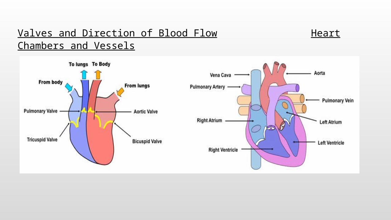
Valves and Direction of Blood Flow Heart Chambers and Vessels


6.2.2 State that coronary arteries supply heart muscle with oxygen and nutrients
▪ The heart is a muscle that must continually contract in order to pump blood around the body
▪ Coronary arteries form a network of vessels around the heart and supply the cardiac tissue with oxygen and nutrients (i.e. glucose)
▪ These are required to produce the necessary energy via aerobic respiration - if a coronary artery is blocked, a heart attack may occur

6.2.3 Explain the action of the heart in terms of collecting blood, pumping blood and opening and closing valves
▪ Blood returning from all parts of the body (except lungs) enter the right atrium via the vena cava - this blood is relatively deoxygenated
▪ The blood passes from the right atrium to the right ventricle and then via the pulmonary artery to the lungs (where blood is reoxygenated)
▪ The blood returns to the left atrium via the pulmonary vein and passes through the left ventricle to the aorta, where it is pumped around the body

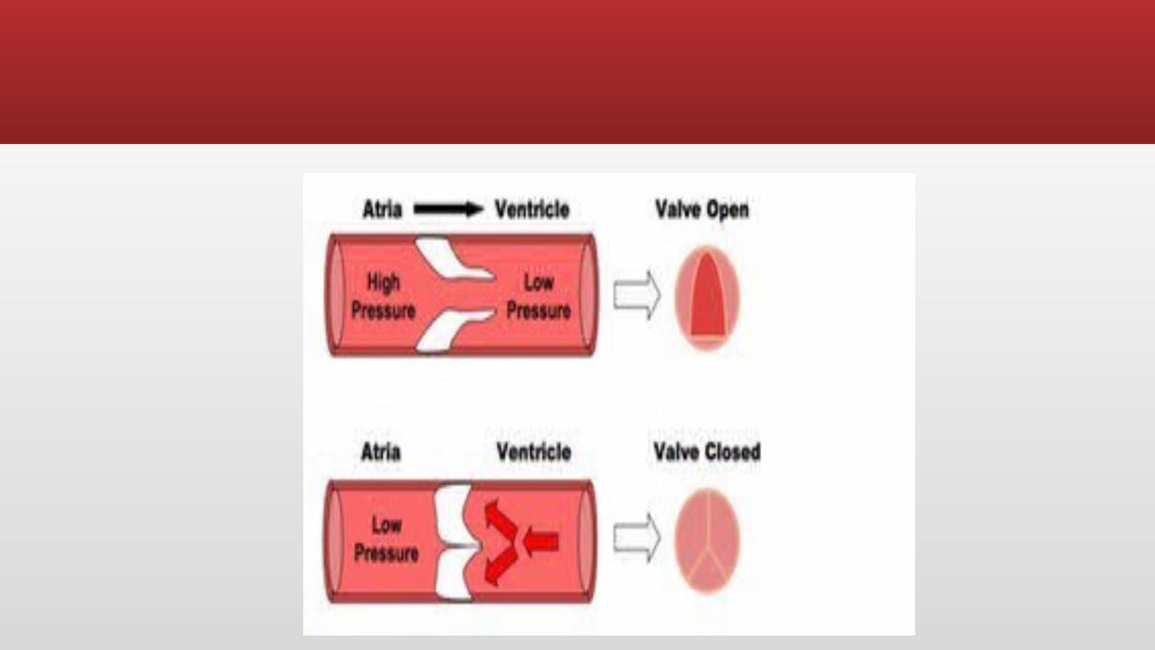
6.2.3 Cont.,
▪ The heart valves maintain the one-way flow of blood:
▪ When the atria contract, atrioventricular (AV) valves open
▪ Blood flows from the atria and into the ventricles
▪ When the ventricles contract, the AV valves close and semilunar valves open
▪ This forces blood out of the ventricles and into the arteries

Flow of BloodBlood Flow through LungsAnd Heart

IP Physiology: Anatomy Review: Blood vessel structure and function
6.2.5 Explain the relationship between the structure and function of arteries, capillaries and veins
▪ Arteries
▪ Arteries carry blood at high pressure (80 - 120 mm Hg)
▪ They have a narrower lumen (to maintain high pressure) surround by a thick wall made of two layers
▪ The middle layer (tunica media) contains muscle and elastin to help maintain pulse flow (it can contract and stretch)
▪ The outer layer (tunica adventitia) contains collagen prevents the artery rupturing due to the high pressure blood flow

Structure of Blood Vessels


▪ Veins
▪ Veins carry blood under low pressure (<10 mm Hg)
▪ They have a very wide lumen (keeps pressure low and allows greater flow of blood)
▪ The walls of tissue surrounding the vein are thin (blood is not travelling in rhythmic pulses)
▪ They have valves to prevent blood pooling at extremities (arteries do not have valves)
▪ Capillaries
▪ Capillaries are involved with material and gas exchange with the surrounding body tissue
▪ Blood pressure in the capillaries is relatively low (~15 mm Hg) and they have a very small diameter (~5 micrometers wide)
▪ Their wall is made up a a single layer of cells to allow for ease of diffusion
▪ Capillaries may contain pores to aid the transport of material

Capilarry

H.5.4 Outline atherosclerosis and the cause of coronary thrombosis
▪ Atherosclerosis is the hardening and narrowing of the arteries, due to the deposition of material commonly known as plaque
▪ Damage to the artery walls (e.g. due to high blood pressure) causes chronic inflammation, leading to the accumulation of lipids, cholesterol, cell debris and calcium
▪ Atheromas (fatty deposits) develop in the arteries and significantly reduce the diameter of the lumen (stenosis) and reduces the elasticity of the artery wall needed for pulse flow
▪ This may lead to the formation of clots and blockages in the artery, and if this occurs in the coronary arteries (coronary thrombosis), it may lead to a heart attack (acute myocardial infarction)

Atherosclerosis
H.5.5 Discuss factors that affect the incidence of coronary heart disease
▪ Risk factors for coronary heart disease (CHD) include:
▪ Exercise – sedentary life style or excessive exercise can both place a strain on normal heart activity
▪ Genetics – having hypertension (high blood pressure) or a family history of heart attacks increases the risk
▪ Gender – males are more at risk than females due to low estrogen levels (risk increases in women post-menopause as levels fall)
▪ Smoking – smoking raises blood pressure because nicotine causes vasoconstriction
▪ Obesity – being overweight places additional strain on the heart
▪ Diet – too much saturated fats and cholesterol promotes atherosclerosis, high salt levels and excessive alcohol intake are also risk factors
▪ Age – old age leads to less flexible blood vessels

Quiz
IP Physiology: Measuring Blood Pressure
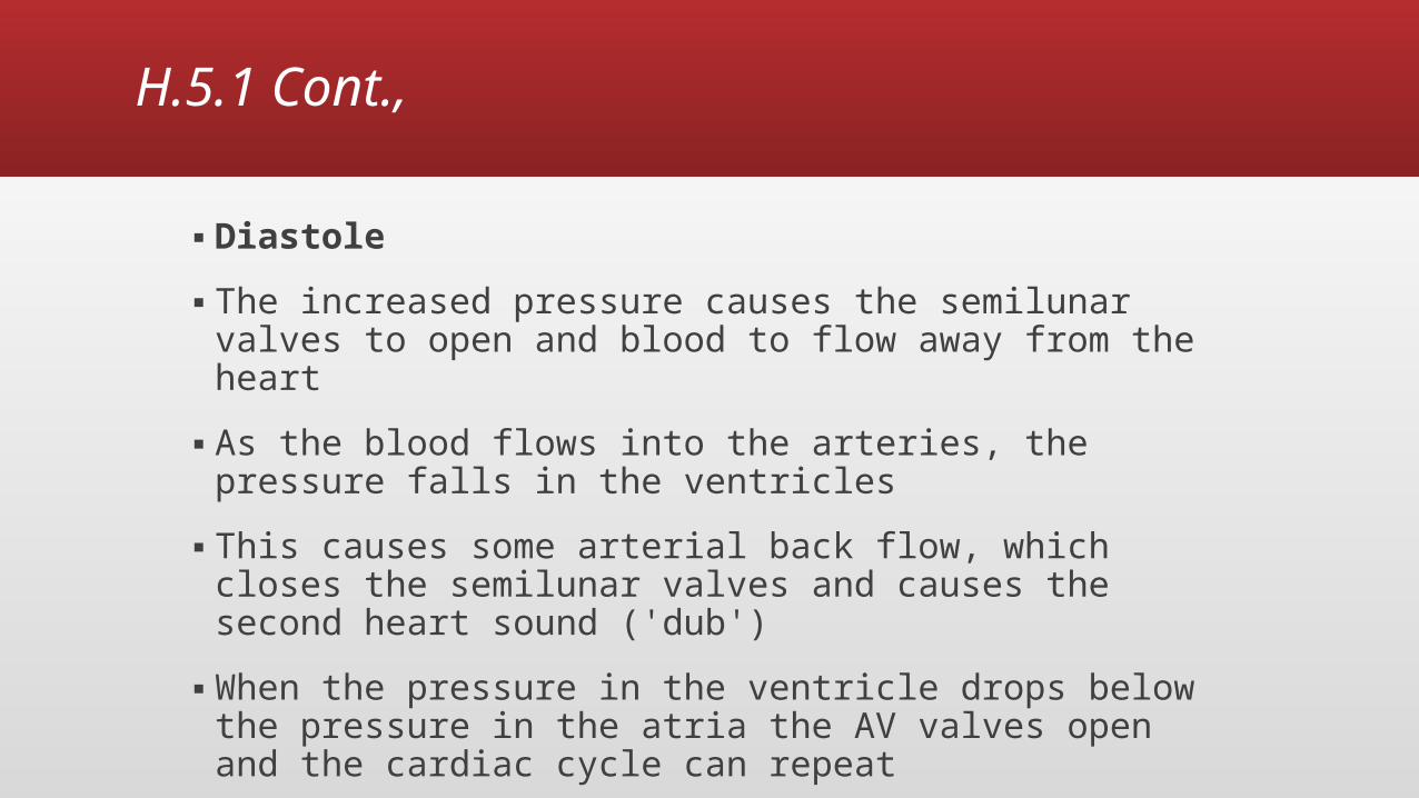
H.5.1 Explain the events of the cardiac cycle, including atrial and ventricular systole and diastole, and heart sounds
▪ The cardiac cycle describes the series of events that take place in the heart over the duration of one heart beat
▪ It is comprised of a period of contraction (systole) and relaxation (diastole)

H.5.1 Cont.,
▪ Systole
▪ Blood returning to the heart will freely flow from the atria to the ventricles as the AV valves kept open by the pressure in the atria
▪ The sinoatrial node (pacemaker) receives signals to fire when the ventricles are almost full (~70%)
▪ The contraction of the atria (atrial systole) causes blood to fill the ventricles to the maximum
▪ The signal from the SA node is transferred to the AV node and then via Purkinje fibres to cause the delayed contraction of the ventricles
▪ As the ventricles contract, the increase of pressure in the ventricles closes the AV valves, causing the first heart sound ('lub')
H.5.1 Cont.,
▪ Diastole
▪ The increased pressure causes the semilunar valves to open and blood to flow away from the heart
▪ As the blood flows into the arteries, the pressure falls in the ventricles
▪ This causes some arterial back flow, which closes the semilunar valves and causes the second heart sound ('dub')
▪ When the pressure in the ventricle drops below the pressure in the atria the AV valves open and the cardiac cycle can repeat

Blood Pressure

H.5.2 Analyze data showing pressure and volume changes in the left atrium, left ventricle and the aorta, during the cardiac cycle

IP Physiology: Intrinsic Conductive System

6.2.4 Outline the control of the heartbeat in terms of myogenic muscle contraction, the role of the pacemaker, nerves, the medulla of the brain and epinephrine (adrenaline)
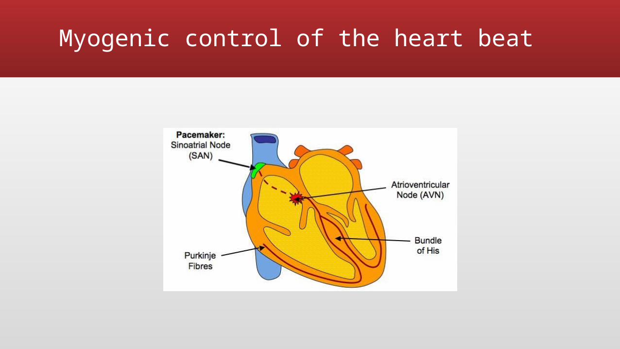
▪ The contraction of the heart tissue (myocardium) is myogenic, meaning the signal for cardiac contraction arises within the heart muscle itself
▪ Within the wall of the right atrium are a specialized plexus of nerves called the sinoatrial node (SAN)
▪ The sinoatrial node initiates contraction of the cardiac muscle and acts as a pacemaker, regulating normal sinus rhythm
6.2.4 Cont.,
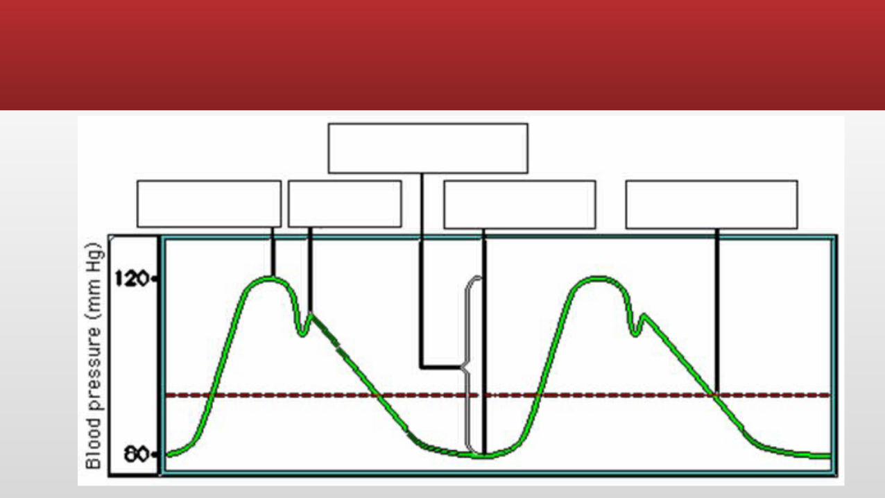
▪ SA node stimulates atria to contract and, when excitation reaches the junction between atria and ventricles, stimulates another node (atrioventricular node)
▪ The atrioventricular node (AVN) sends signals via the Bundle of His to Purkinje fibres, which cause ventricular contraction
▪ This sequence always ensures their is a delay between atrial and ventricular contractions, resulting in two heart sounds ('lub dub')
Myogenic Control of the Heart Beat
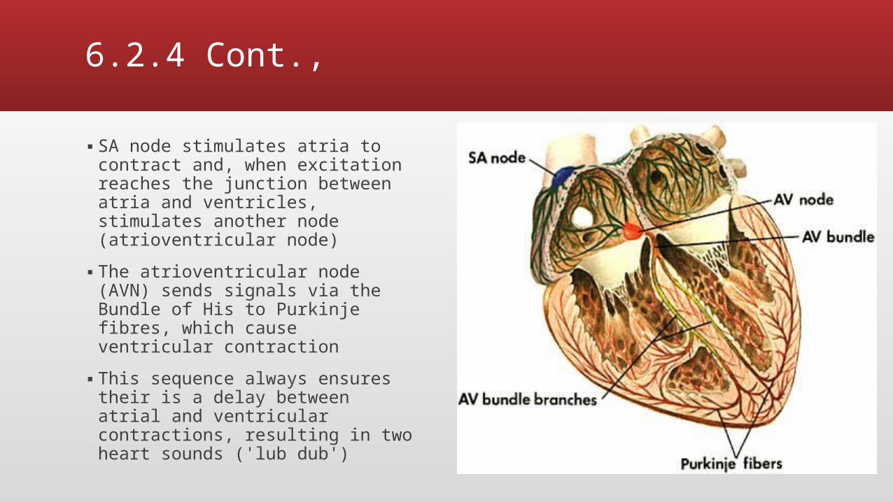
▪ The pacemaker is under autonomic control from the brain, specifically the medulla oblongata (brain stem)
▪ Sympathetic nerves speed up heart rate by releasing a neurotransmitter (noradrenaline) to increase the rate of myocardial contraction
▪ Parasympathetic nerves slows down heart rate by releasing a neurotransmitter (acetylcholine) to decrease the rate of myocardial contraction
▪ Additionally, the heart rate may be increased by the chemical release of the hormone adrenaline into the blood (from the adrenal gland)

H.5.3 Outline the mechanism that controls the heartbeat, including the roles of the SA node, AV node and conducting fibres in the ventricular walls
▪ The contraction of the heart tissue (myocardium) is myogenic, meaning the signal for cardiac contraction arises within the heart muscle itself
▪ Within the wall of the right atrium are a specialized plexus of nerves called the sinoatrial node (SAN)
▪ The sinoatrial node initiates contraction of the cardiac muscle and acts as a pacemaker, regulating normal sinus rhythm
▪ It stimulates atria to contract and, when excitation reaches the junction between atria and ventricles, stimulates another node (atrioventricular node)
H.5.3 Cont.,
▪ The atrioventricular node (AVN) sends signals via the Bundle of His to Purkinje fibres, which cause ventricular contraction▪ This sequence always ensures their is a delay between
atrial and ventricular contractions, resulting in maximum blood flow
▪ By having ventricular contractions start at the apex (bottom), it ensures blood is pushed up towards the arteries
Myogenic control of the heart beat

H.5.3 Cont.,
▪ The pacemaker is under autonomic control from the brain, specifically the medulla oblongata (brain stem)
▪ Sympathetic nerves speed up heart rate by releasing a neurotransmitter (noradrenaline) to increase the rate of myocardial contraction
▪ Parasympathetic nerves slow down heart rate by releasing a neurotransmitter (acetylcholine) to decrease the rate of myocardial contraction
▪ Additionally, the heart rate may be increased by the chemical release of the hormone adrenaline into the blood (from the adrenal gland)

Quiz

6.2.6 State that blood is composed of plasma, erythrocytes, leukocytes (phagocytes and lymphocytes) and platelets
▪ There are four main components to blood:
▪ Plasma - the fluid medium of the blood
▪ Erythrocytes - red blood cells (involved in oxygen transport)
▪ Leukocytes - white blood cells, such as phagocytes (non-specific immunity) and lymphocytes (specific immunity)
▪ Platelets - responsible for blood clotting (haemostasis)
6.2.7 State that the following are transported by blood: nutrients, oxygen, carbon dioxide, hormones, antibodies, urea and heat
▪ The following things are transported by blood:
▪ Nutrients (e.g. glucose)
▪ Antibodies
▪ Carbon dioxide
▪ Hormones
▪ Oxygen
▪ Urea
▪ Heat (not a molecules, unlike all the others