6-mercaptopurine and daunorubicin double drug liposomes—preparation, drug-drug interaction and...
TRANSCRIPT
Journal of Liposome Research, 15:141–155, 2005Copyright © Taylor & Francis Inc.ISSN: 0898-2104DOI: 10.1080/08982100500364081
141
LLPR0898-21041532-2394Journal of Liposome Research, Vol. 15, No. 3-4, October 2005: pp. 1–25Journal of Liposome Research
6-Mercaptopurine and Daunorubicin Double Drug Liposomes—Preparation, Drug-Drug Interaction
and Characterization
6-MP and DR Double Drug LiposomesV. Agrawal, M. K. Paul, and A. K. MukhopadhyayVINEET AGRAWAL, MANASH K. PAUL, AND ANUP K. MUKHOPADHYAY
National Institute of Pharmaceutical Education and Research, Sector-67, SASNagar, Punjab, Mohali, India
This article addresses and investigates the dual incorporation of daunorubicin (DR)and 6-mercaptopurine (6-MP) in liposomes for better chemotherapy. These drugs arepotential candidates for interaction due to the quinone (H acceptor) and hydroxyl(H donor) groups on DR and 6-MP, respectively. Interactions between the two drugsin solution were monitored by UV/Vis and fluorescence spectroscopy. Interactionbetween the two drugs inside the liposomes was evaluated by HPLC (for 6-MP) and byfluorescence spectroscopy (for daunorubicin) after phospholipase-mediated liposomelysis. Our results provide evidence for the lack of interaction between the two drugs insolution and in liposomes. The entrapment efficiencies of 6-MP in the neutral Phos-phatidyl choline (PC):Cholesterol (Chol):: 2:1 and anionic PC:Chol:Cardiolipin (CL) ::4:5:1 single and double drug liposomes were found to be 0.4% and 1.5% (on average),respectively. The entrapment efficiencies of DR in the neutral and anionic double drugliposomes were found to be 55% and 31%, respectively. The corresponding entrap-ment of daunorubicin in the single drug liposomes was found to be 62% on average.Our thin layer chromatography (TLC) and transmission electron microscopy (TEM)results suggest stability of lipid and liposomes, thus pointing plausible existence ofdouble drug liposomes. Cytotoxicity experiments were performed by using both singledrug and double drug liposomes. By comparing the results of phase contrast and fluo-rescence microscopy, it was observed that the double drug liposomes were internalizedin the jurkat and Hut78 (highly resistant cell line) leukemia cells as viewed by the fluo-rescence of daunorubicin. The cytotoxicity was dose dependent and had shown a syn-ergistic effect when double drug liposome was used.
Keywords daunorubicin (DR), 6-mercaptopurine (6-MP), double drug liposome,drug-drug interaction, pH gradient, absorbance and fluorescence spectroscopy, high-pressure liquid chromatography, phospholipase, lipolysis, cytotoxicity
Address correspondence to Dr. Anup Kumar Mukhopadhyay, Department of Biotechnology,National Institute of Pharmaceutical Education and Research (NIPER), Sector-67, S.A.S Nagar,Mohali, Punjab-160062, India; Fax: +91 172 2214692; E-mail: [email protected] [email protected]
Jour
nal o
f L
ipos
ome
Res
earc
h D
ownl
oade
d fr
om in
form
ahea
lthca
re.c
om b
y M
emor
ial U
nive
rsity
of
New
foun
dlan
d on
08/
03/1
4Fo
r pe
rson
al u
se o
nly.
142 V. Agrawal, M. K. Paul, and A. K. Mukhopadhyay
Introduction
Conventional cancer chemotherapy is seriously limited by dose-dependent toxicity andmultidrug resistance (MDR). MDR cells exhibit changes in the level or activity of mem-brane transporters causing energy dependent drug efflux, increased drug detoxification,DNA repair, and defects in the apoptotic pathways leading to reduced apoptosis (Mamot,2003a). Different drug combinations are used for escaping MDR conditions based onrationale; probability of two or more different drug resistances to arise spontaneously inthe same cell is very remote (Liotta, 1992). Drug combinations are useful in patients need-ing a prompt reduction in tumor burden and in treating drug-resistant states. Furthermore,a combination can lower the concentration of individual drugs reducing their toxicity.
Daunorubicin (anthracycline antibiotic) and 6-mercaptopurine (antimetabolite) are apart of antileukemic regimen. Daunorubicin is used for treating acute lymphocytic andgranulocytic leukemia, whereas 6-mercaptopurine (6-MP) is used for treating acute lym-phatic leukemia. Daunorubicin is primarily a DNA topoisomerase II inhibitor althoughother mechanisms like intercalation into DNA and, consequently, generation of free radi-cals (Gewirtz, 1999) have also been proposed. 6-Mercaptopurine is converted in vivo toan active form 6-thioinosine-5′-Phosphate (T-IMP) by the enzyme hypoxanthine guanylphosphoribosyltransferase (HGPRT). T-IMP inhibits the conversion of phosphoribosylpy-rophosphate to 5-phosphoribosylamine, a rate-controlling step in the de novo synthesis ofpurines (Chabner et al., 2000; Remers, 1998). Apart from causing bone marrow depres-sion and myelosuppressive effect, daunorubicin is cardiotoxic owing to the generation ofreactive oxygen species (ROS) by one electron transfer to molecular oxygen in the pres-ence of various oxidoreductases. 6-MP causes leucopenia and thrombocytopenia and alsogenerates ROS during its oxidation to 6-thiouric acid by xanthine oxidase. Apart from thesimilarity of active ROS generation, both the drugs have different mechanisms of action,similar therapeutic profile, and thus can be ideal candidates for a feasible combination.However, the cumulative effect of ROS can have a deleterious effect on the various cellu-lar systems.
Efforts have been made for reducing toxicity by incorporating these drugs in lipo-somes. But no attempts of incorporating these two drugs together in the liposomes havebeen reported. Here for the first time, we test the feasibility and existence of double drugliposome. Because the modes of action of these drugs are widely different, we hypothesizethat such a combination may lead to the development of a new regimen. Furthermore, it isknown that anticancer and toxicity effect of daunorubicin is due to oxidoreductase(s)-mediated superoxide formation. The quinone moiety of daunorubicin is reduced to semi-quinone by various oxido reductases as cytochrome P-450 reductase, xanthine oxidase,and NADH oxidoreductase. The semiquinone form transfers an electron to molecular oxygen-generating superoxide, which exerts cytotoxicity by variety of mechanisms. Therefore, itis expected that in the presence of 6-mercaptopurine (a substrate of xanthine oxidase) inthe double drug formulation, ROS load will be increased, leading to enhanced cytotoxicityat the site of cancer (Scheme 1). The use of double drug liposomes can reduce the nonspe-cific systemic toxicity and enhance the ROS content selectively in the vicinity of cancer-ous cells. This increment of ROS can markedly increase the antitumor effect (Pelicanoet al., 2004) by inducing apoptosis of cancerous cells. Thus, the combination can causemalignant cell death both by direct (inherent cytotoxic action of drugs) and indirect(enhanced ROS content) means. However, the therapeutic activity of double drug lipo-somes can be supported only in the absence of interaction between the two drugs. Dauno-rubicin has a quinone moiety that can act as a potential hydrogen bond acceptor, whereas
Jour
nal o
f L
ipos
ome
Res
earc
h D
ownl
oade
d fr
om in
form
ahea
lthca
re.c
om b
y M
emor
ial U
nive
rsity
of
New
foun
dlan
d on
08/
03/1
4Fo
r pe
rson
al u
se o
nly.
6-MP and DR Double Drug Liposomes 143
sulfydryl group of 6-MP can act as a hydrogen donor. Thus, there is a strong possibility ofmultiple hydrogen-bonded adduct formation between the two drugs. Furthermore, in lipo-somes, due to confined boundary of lipid vesicles, the two drugs molecules are in so closeproximity that chances of an interaction are significantly elevated.
With this background in mind, our objectives were twofold. Primarily, we intended tostudy the interaction between 6-MP and DR in solution, in liposomes and to check theefficacy of the dual drug liposome in terms of cytotoxicity. Second, we intended to ana-lyze the stability of the lipid, physical viability, and morphology of double drug-liposomefor the feasibility of such a combination and check the efficacy on leukemia cell lines.
Materials and Methods
Drugs and Chemicals
6-Mercaptopurine, phosphatidyl choline, cardiolipin, phospholipase A2, phospholipase C,and trypan blue were obtained from Sigma Chemical Company, St. Louis, Missouri, USA.Daunorubicin hydrochloride, cholesterol, and Sephadex G-50 were obtained from Calbio-chem, Germany. Methanol HPLC grade, lactose, citric acid, sodium dihydrogen phos-phate, disodium hydrogen phosphate, and dihydrogen potassium phosphate were obtainedfrom Merck India ltd. Acetonitrile HPLC grade was purchased from J. T. Baker, Mexico.Triple distilled water from an all-glass still or ELGA water was used.
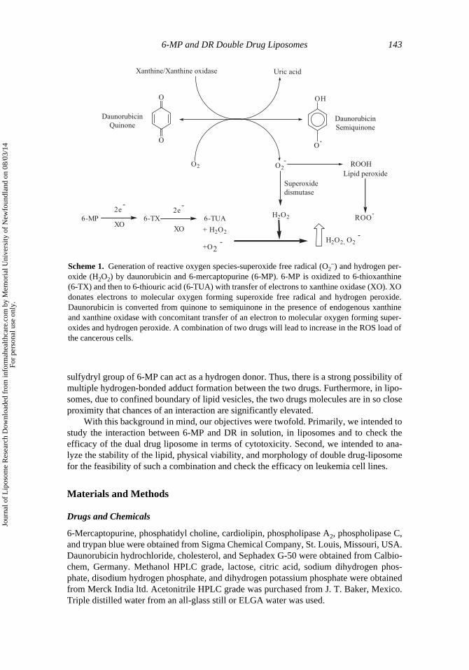
Scheme 1. Generation of reactive oxygen species-superoxide free radical (O2−) and hydrogen per-
oxide (H2O2) by daunorubicin and 6-mercaptopurine (6-MP). 6-MP is oxidized to 6-thioxanthine(6-TX) and then to 6-thiouric acid (6-TUA) with transfer of electrons to xanthine oxidase (XO). XOdonates electrons to molecular oxygen forming superoxide free radical and hydrogen peroxide.Daunorubicin is converted from quinone to semiquinone in the presence of endogenous xanthineand xanthine oxidase with concomitant transfer of an electron to molecular oxygen forming super-oxides and hydrogen peroxide. A combination of two drugs will lead to increase in the ROS load ofthe cancerous cells.
O
O
OH
O.
Xanthine/Xanthine oxidase Uric acid
O2 O2
-
Superoxide
dismutase
H2O2
ROOH
ROO.
Lipid peroxide
6-MP 6-TX 6-TUAXO
XO
2e-
2e-
+ H2O2
+O2- H2O2, O2
-
Daunorubicin
QuinoneDaunorubicin
Semiquinone
Jour
nal o
f L
ipos
ome
Res
earc
h D
ownl
oade
d fr
om in
form
ahea
lthca
re.c
om b
y M
emor
ial U
nive
rsity
of
New
foun
dlan
d on
08/
03/1
4Fo
r pe
rson
al u
se o
nly.

144 V. Agrawal, M. K. Paul, and A. K. Mukhopadhyay
Spectral Characterization of Individual Drugs
6-MP was characterized by UV/Vis spectroscopy. Fresh stock solution of 6-MP (1 mM)was prepared in the deionized water by adding 10–20 μL of 1N NaOH with constant stir-ring for 1–2 h. Absorbance scans for 1–40 μM were taken in 100 mM phosphate buffer pH7.5, and the standard curve was constructed from the optical density taken at 321 nm (λmaxof 6-MP). Daunorubicin was characterized by UV/Vis (Perkin Elmer Lambda 25 spectro-photometer) and fluorescence spectroscopy (A LS 50B luminescence spectrometer wasused). Fresh stock solution (1 mM) was made in deionized water. Absorption scans ofconcentrations ranging from 2 to 20 μM were taken and absorbance recorded at 490 nm.Fluorescence emission scans of daunorubicin from 2 to 30 μM were taken from 500 to 650nm with excitation fixed at 490 nm, keeping the excitation and emission slit width 5 and10 nm, respectively.
Drug Interaction Studies
Two different sets of experiments were performed: 1) by varying the concentration of6-MP (2–40 μM) at a fixed concentration of daunorubicin and 2) vice versa. Absorptionscans were taken from 200 to 800 nm. In the second, emission scan of 10 μM daunorubi-cin solution was recorded, and subsequent emission scans with increasing concentration of6-MP (5, 10, and 20 μM) added to daunorubicin were recorded.
Preparation of Liposomes
Film hydration method (Woodle and Papahadjopoulos, 1989) was used for the preparationof neutral and anionic liposomes. A 2:1 molar ratio of egg phosphatidyl choline (20 mg) andcholesterol (5.4 mg) or 1:4:5 molar ratio of Cardiolipin:Phosphatidylcholine:Cholesterolwas used for casting of dry film on the round bottom flask. A buchi rotary evaporator wasused for this purpose. The film was hydrated with 1.25 mL of 50 mM citric acid and 125mM lactose solution at pH 2–2.5, containing 1 mM 6-MP, along with a few glass beads. Thefilm was hydrated for a period of 1 h at 45°C, giving a milky white vesicular suspension.The suspension was then sonicated for 3–5 min (15-sec pulsing time and 30-sec rest) andcentrifuged at 5000 g for 10 min. The supernatant was collected and stored at 4°C.
Daunorubicin was loaded in the above preformed 6-MP liposomes by creating a pHgradient (Forssen, 1990; Harigan et al., 1993; Mayer et al., 1993). One milliliter of 125 mMsodium hydroxide was added to 1 mL of liposomal suspension. The suspension was thenwarmed at 37°C, and 2 mg of powdered daunorubicin hydrochloride was added to yield afinal concentration of 1 mg/mL. The suspension was then incubated at 37°C for 120 minwith vigorous intermittent mixing to load daunorubicin in the preformed liposomes.Sephadex G-50 mini-column centrfugation was used to separate free drugs from liposo-mal drug sample.
Drug Entrapment Efficiency Calculations
The amount of the drug entrapped inside the liposomes was quantified by high-performanceliquid chromatography (HPLC) for 6-mercaptopurine and by fluorescence spectroscopyfor daunorubicin.
6-Mercaptopurine: 200 μL of methanol was added to 100 μL (lipid concentration25 mg/mL) of purified liposomes. The solution was then warmed at 40°C for 10 min and
Jour
nal o
f L
ipos
ome
Res
earc
h D
ownl
oade
d fr
om in
form
ahea
lthca
re.c
om b
y M
emor
ial U
nive
rsity
of
New
foun
dlan
d on
08/
03/1
4Fo
r pe
rson
al u
se o
nly.

6-MP and DR Double Drug Liposomes 145
centrifuged at 5000 g for 5 min; 25 μL of the supernatant was injected into the C18 reversephase column equilibrated with mobile phase containing potassium phosphate buffer 50mM (pH 6.6):acetonitrile (96:4), maintained at a flow rate of 1 mL/min. The area of theplot was obtained and the corresponding concentration was quantified from the standardcurve plotted for pure 6-MP.
Daunorubicin: An initial scan of 10 μL (equivalent to 25 μg lipid) daunorubicin lipo-somes in 50 mM sodium phosphate buffer, pH 7.2, was taken, and mixture of Phospholi-pases C and A2 (300 mU/mL) was added to it. Increase in the fluorescence at 560 nm wasmonitored with a time interval of 2 min up to 10 min when a plateau was attained, and nofurther fluorescence increase was observed. The net increase in the fluorescence intensitywas quantified, and concentration of the entrapped drug was calculated from the standardcurve.
The entrapment efficiency was calculated as per the following formula: % Entrap-ment = (amount of drug analyzed from liposomes/total drug added) × 100
Cell Line and Culture
Jurkat and Hut78 (human T-cell lymphoma) cell lines were purchased from National Cen-tre for Cell Science culture collection (NCCS), Pune, India. Both the cell lines were main-tained in RPMI 1640 medium supplemented with 100 U/mL penicillin, 100 μg/mLstreptomycin, and 10% fetal bovine serum (FBS; Gibco-BRL, Grand Island, NY, USA) at37°C in an environment of 5% CO2.
Cellular Uptake and Efficacy of Double Drug Liposomes
The cellular uptake and efficacy of the double drug liposomes were tested on Hut78 andJurkat (human T-cell lymphoma) cell line. The cellular uptake of the liposomes was con-firmed by fluorescence microscopy. For this, approximately 106 cells were incubated intriplicate with Dauno-6-MP liposome in RPMI-1640 media for 1 h at 37°C. After incuba-tion, cells were washed three times with cold phosphate buffer saline (PBS). The cellswere finally pelleted down and fixed by using 4% paraformaldehyde for 10 min and againwashed with cold PBS and mounted on the glass slide. Fluorescence microscopy was car-ried out by using a Leica fluorescence microscope DMLB attached with a DMFC 320CCD camera (Leica) (Mamot, 2003b).
Determination of In Vitro Cytotoxicity of Dauno-6MP Liposome to Leukemia Cells
Jurkat and Hut78 cells were aliquoted into 96-well tissue culture plate at the cell den-sity of approximately 1 × 10⁴ cells in 200 μL per well. Cells were incubated in quadru-plicate with different concentrations of Dauno-6-MP liposome for 2 h at 37°C. Thesuspension was then centrifuged at 1000 g for 3 min, and pelleted cells were washedthree times with cold PBS and cultured for an additional 72 h in fresh media. Cell via-bility was then checked by using trypan blue assay followed by microscopic counting(Pan et al., 2002).
Results
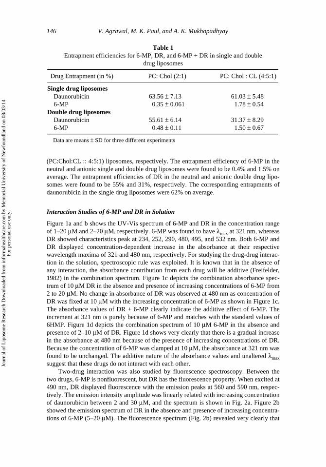
Table 1 shows the entrapment efficiencies of the daunorubicin (DR) and 6-mercaptopurine(6-MP) individually and in combination in the neutral (PC:Chol :: 2:1) and anionic
Jour
nal o
f L
ipos
ome
Res
earc
h D
ownl
oade
d fr
om in
form
ahea
lthca
re.c
om b
y M
emor
ial U
nive
rsity
of
New
foun
dlan
d on
08/
03/1
4Fo
r pe
rson
al u
se o
nly.
146 V. Agrawal, M. K. Paul, and A. K. Mukhopadhyay
(PC:Chol:CL :: 4:5:1) liposomes, respectively. The entrapment efficiency of 6-MP in theneutral and anionic single and double drug liposomes were found to be 0.4% and 1.5% onaverage. The entrapment efficiencies of DR in the neutral and anionic double drug lipo-somes were found to be 55% and 31%, respectively. The corresponding entrapments ofdaunorubicin in the single drug liposomes were 62% on average.
Interaction Studies of 6-MP and DR in Solution
Figure 1a and b shows the UV-Vis spectrum of 6-MP and DR in the concentration rangeof 1–20 μM and 2–20 μM, respectively. 6-MP was found to have λmax at 321 nm, whereasDR showed characteristics peak at 234, 252, 290, 480, 495, and 532 nm. Both 6-MP andDR displayed concentration-dependent increase in the absorbance at their respectivewavelength maxima of 321 and 480 nm, respectively. For studying the drug-drug interac-tion in the solution, spectroscopic rule was exploited. It is known that in the absence ofany interaction, the absorbance contribution from each drug will be additive (Freifelder,1982) in the combination spectrum. Figure 1c depicts the combination absorbance spec-trum of 10 μM DR in the absence and presence of increasing concentrations of 6-MP from2 to 20 μM. No change in absorbance of DR was observed at 480 nm as concentration ofDR was fixed at 10 μM with the increasing concentration of 6-MP as shown in Figure 1c.The absorbance values of DR + 6-MP clearly indicate the additive effect of 6-MP. Theincrement at 321 nm is purely because of 6-MP and matches with the standard values of6HMP. Figure 1d depicts the combination spectrum of 10 μM 6-MP in the absence andpresence of 2–10 μM of DR. Figure 1d shows very clearly that there is a gradual increasein the absorbance at 480 nm because of the presence of increasing concentrations of DR.Because the concentration of 6-MP was clamped at 10 μM, the absorbance at 321 nm wasfound to be unchanged. The additive nature of the absorbance values and unaltered λmaxsuggest that these drugs do not interact with each other.
Two-drug interaction was also studied by fluorescence spectroscopy. Between thetwo drugs, 6-MP is nonfluorescent, but DR has the fluorescence property. When excited at490 nm, DR displayed fluorescence with the emission peaks at 560 and 590 nm, respec-tively. The emission intensity amplitude was linearly related with increasing concentrationof daunorubicin between 2 and 30 μM, and the spectrum is shown in Fig. 2a. Figure 2bshowed the emission spectrum of DR in the absence and presence of increasing concentra-tions of 6-MP (5–20 μM). The fluorescence spectrum (Fig. 2b) revealed very clearly that
Table 1Entrapment efficiencies for 6-MP, DR, and 6-MP + DR in single and double
drug liposomes
Drug Entrapment (in %) PC: Chol (2:1) PC: Chol : CL (4:5:1)
Single drug liposomesDaunorubicin 63.56 ± 7.13 61.03 ± 5.486-MP 0.35 ± 0.061 1.78 ± 0.54
Double drug liposomesDaunorubicin 55.61 ± 6.14 31.37 ± 8.296-MP 0.48 ± 0.11 1.50 ± 0.67
Data are means ± SD for three different experiments
Jour
nal o
f L
ipos
ome
Res
earc
h D
ownl
oade
d fr
om in
form
ahea
lthca
re.c
om b
y M
emor
ial U
nive
rsity
of
New
foun
dlan
d on
08/
03/1
4Fo
r pe
rson
al u
se o
nly.

6-MP and DR Double Drug Liposomes 147
increasing concentration of 6-MP did not have any significant quenching or enhancingeffect on DR fluorescence. Furthermore, there was no shift of λmax emission in the fluores-cence spectrum. Thus, our absorbance and fluorescence experiments suggest very clearlythat these two drugs do not have any interaction in solution; hence, combination of thesetwo drugs can be entrapped safely in the liposomes.
Studies on the Interaction Between 6-MP and DR Inside the Liposomes
Characterization of Daunorubicin. To check the two-drug interaction inside the lipo-somes, a mixture of phospholipase C and A2 was added to the DR and DR-6MP lipo-somes. It was found that 300 mU of phospholipase mixture was optimum for themaximum release of DR as monitored by the fluorescence burst. Figure 3a shows theresult of Phosholipase-induced lysis of liposomes. On addition of Phospholipases, there
Figure 1. (a) Spectral characteristics of 6-MP with concentrations 1, 2, 5, 10, 20, and 40 μM. (b)Spectral characteristics of daunorubicin with concentrations 2, 5, 10, and 20 μM. (c) Spectra forfixed concentration of daunorubicin (DR 10 μM) and DR with increasing concentration of 6-MP (2,5,10, and 20 μM). (d) Spectra for fixed concentration of 6-MP (10 μM) and 6-MP with increasingconcentration of daunorubicin DR (2, 5, and 10 μM).
Jour
nal o
f L
ipos
ome
Res
earc
h D
ownl
oade
d fr
om in
form
ahea
lthca
re.c
om b
y M
emor
ial U
nive
rsity
of
New
foun
dlan
d on
08/
03/1
4Fo
r pe
rson
al u
se o
nly.

148 V. Agrawal, M. K. Paul, and A. K. Mukhopadhyay
was a time-dependent increase in the fluorescence, and even the addition of higher con-centration of phospholipase mixture did not further increase the fluorescence level, sug-gesting that all the liposomes in the suspension had been lipolysed, thus suggesting that theentire drug molecules have been released. The control experiment with the bare liposomesand the free drug did not yield any increment of the fluorescence on the addition of lipasemixture (Fig. 3b), suggesting that in our single or double drug liposome formulation, DRwas entrapped inside the liposomes. Percentage of entrapped DR was calculated from theextent of DR released as described in the methods section. The λmax and the entire natureof the fluorescence spectrum of the entrapped DR released from the double drug
Figure 2. (a) Fluorescence emission spectra of daunorubicin with concentrations 2, 5, 10, 20, and30 μM. (b) Fluorescence emission spectra of daunorubicin (10 μM) without and with increasingconcentration of 6-MP (5, 10, and 20 μM).
Figure 3. (a) Phospholipase (Phospholipase C and A2)-mediated lysis of double drug liposomes.Fluorescence release kinetics of entrapped daunorubicin with time interval of 2 min. (b) Emissionspectra for bare liposomes with free daunorubicin subjected to phospholipase (Phospholipase C andA2) treatment.
Jour
nal o
f L
ipos
ome
Res
earc
h D
ownl
oade
d fr
om in
form
ahea
lthca
re.c
om b
y M
emor
ial U
nive
rsity
of
New
foun
dlan
d on
08/
03/1
4Fo
r pe
rson
al u
se o
nly.
6-MP and DR Double Drug Liposomes 149
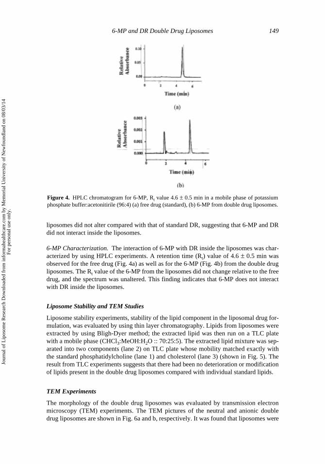
liposomes did not alter compared with that of standard DR, suggesting that 6-MP and DRdid not interact inside the liposomes.
6-MP Characterization. The interaction of 6-MP with DR inside the liposomes was char-acterized by using HPLC experiments. A retention time (Rt) value of 4.6 ± 0.5 min wasobserved for the free drug (Fig. 4a) as well as for the 6-MP (Fig. 4b) from the double drugliposomes. The Rt value of the 6-MP from the liposomes did not change relative to the freedrug, and the spectrum was unaltered. This finding indicates that 6-MP does not interactwith DR inside the liposomes.
Liposome Stability and TEM Studies
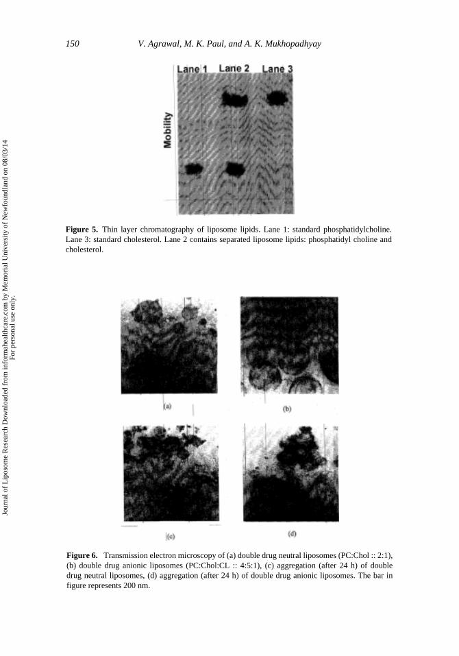
Liposome stability experiments, stability of the lipid component in the liposomal drug for-mulation, was evaluated by using thin layer chromatography. Lipids from liposomes wereextracted by using Bligh-Dyer method; the extracted lipid was then run on a TLC platewith a mobile phase (CHCl3:MeOH:H2O :: 70:25:5). The extracted lipid mixture was sep-arated into two components (lane 2) on TLC plate whose mobility matched exactly withthe standard phosphatidylcholine (lane 1) and cholesterol (lane 3) (shown in Fig. 5). Theresult from TLC experiments suggests that there had been no deterioration or modificationof lipids present in the double drug liposomes compared with individual standard lipids.
TEM Experiments
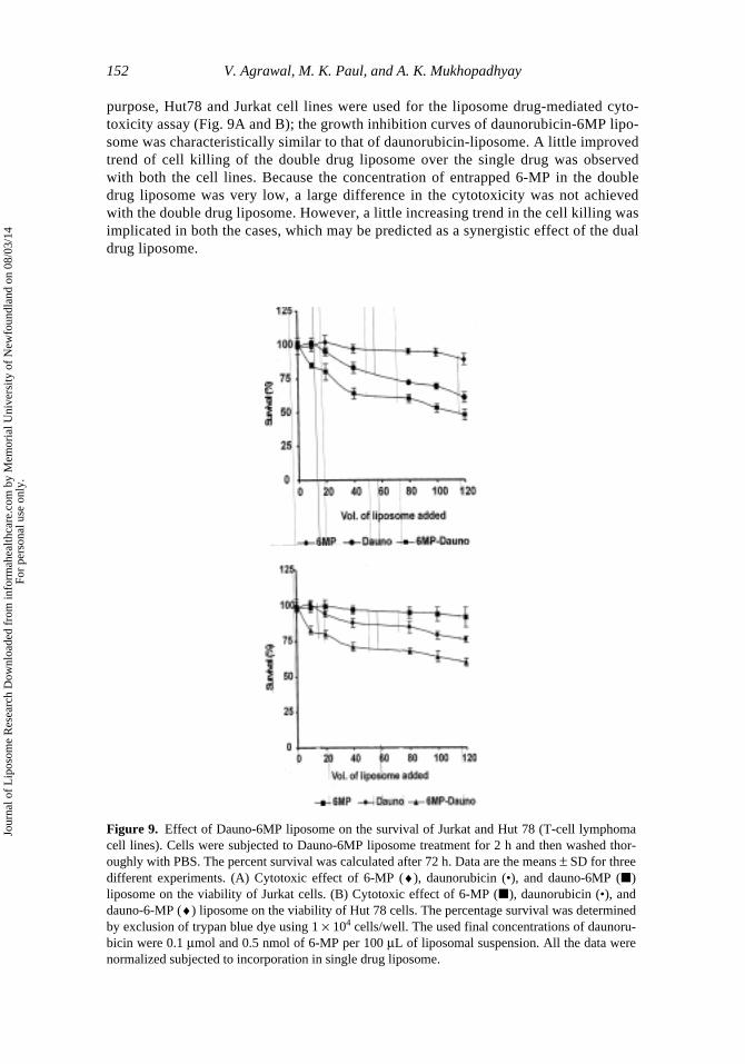
The morphology of the double drug liposomes was evaluated by transmission electronmicroscopy (TEM) experiments. The TEM pictures of the neutral and anionic doubledrug liposomes are shown in Fig. 6a and b, respectively. It was found that liposomes were
Figure 4. HPLC chromatogram for 6-MP, Rt value 4.6 ± 0.5 min in a mobile phase of potassiumphosphate buffer:acetonitirile (96:4) (a) free drug (standard), (b) 6-MP from double drug liposomes.
Jour
nal o
f L
ipos
ome
Res
earc
h D
ownl
oade
d fr
om in
form
ahea
lthca
re.c
om b
y M
emor
ial U
nive
rsity
of
New
foun
dlan
d on
08/
03/1
4Fo
r pe
rson
al u
se o
nly.
150 V. Agrawal, M. K. Paul, and A. K. Mukhopadhyay
Figure 5. Thin layer chromatography of liposome lipids. Lane 1: standard phosphatidylcholine.Lane 3: standard cholesterol. Lane 2 contains separated liposome lipids: phosphatidyl choline andcholesterol.
Figure 6. Transmission electron microscopy of (a) double drug neutral liposomes (PC:Chol :: 2:1),(b) double drug anionic liposomes (PC:Chol:CL :: 4:5:1), (c) aggregation (after 24 h) of doubledrug neutral liposomes, (d) aggregation (after 24 h) of double drug anionic liposomes. The bar infigure represents 200 nm.
Jour
nal o
f L
ipos
ome
Res
earc
h D
ownl
oade
d fr
om in
form
ahea
lthca
re.c
om b
y M
emor
ial U
nive
rsity
of
New
foun
dlan
d on
08/
03/1
4Fo
r pe
rson
al u
se o
nly.

6-MP and DR Double Drug Liposomes 151
unilamellar in nature, and most of them were in the size range of 100–400 nm. Stability studiesof fresh and 24 h stored liposomes indicated that anionic liposomes (Fig. 6d) were more stablethan neutral liposomes (Fig. 6c), which showed aggregation and phase separation.
Cellular Uptake and Efficacy of Daunorubicin-6MP Liposome
Daunorubicin-6MP-loaded liposomes bind to and are internalized into Hut78 cells, as evidentfrom fluorescence microscopy pictures (Figs. 7 and 8). Hut78 cell exposure to daunorubicin-6MP-loaded liposomes revealed that the liposomes gain access to the cell (evident by theintense fluorescence of daunorubicin molecules), as shown in Fig. 7B. Parallel photographunder the same field was taken by using a phase contrast microscope, and similar impressionwas obtained, suggesting that the fluorescence was coming from the cells that internalized theliposome-containing daunorubicin (Fig. 7A). The cellular uptake of daunorubicin-6-MP lipo-somes was time dependent, and the internalization of the liposome was detected after 30 minand well evident after 60 min of exposure as shown in Fig. 8A, B, and C.
In Vitro Cytotoxic Activity of Daunorubicin-6MP Loaded Liposomes in Hut78 and Jurkat Cell Lines
We explored whether double drug liposomes of daunorubicin-6MP would increase thedrug cytotoxicity compared with the counterpart single drug liposomes. For this
Figure 7. Internalization of double drug liposome in Hut 78 lymphoma cells. Hut 78 cells wereincubated in triplicate with Dauno-6MP liposome in RPMI-1640 media for 1 h at 37°C. The inher-ent fluorescence of daunorubicin entrapped in the liposome was visualized by using a Leica fluores-cence microscope DMLB attached with a DMFC 320 CCD camera (Leica). (A) Cells visualized byphase contrast microscopy. (B) Cells visualized by fluorescence microscopy under the same fieldshowing internalization of liposome (detected by daunorubicin fluorescence).
Figure 8. Fluorescence micrographs of Hut78 cells showing time-dependent internalization ofDauno-6-MP liposome. Images of the cells were collected by using a Leica DMLB microscopeattached with fluorescence module, as described in Materials and Methods. (A) Cells incubatedwith Dauno-6MP liposome for 30 min, (B) for 45 min and (C) for 60 min.
Jour
nal o
f L
ipos
ome
Res
earc
h D
ownl
oade
d fr
om in
form
ahea
lthca
re.c
om b
y M
emor
ial U
nive
rsity
of
New
foun
dlan
d on
08/
03/1
4Fo
r pe
rson
al u
se o
nly.
152 V. Agrawal, M. K. Paul, and A. K. Mukhopadhyay
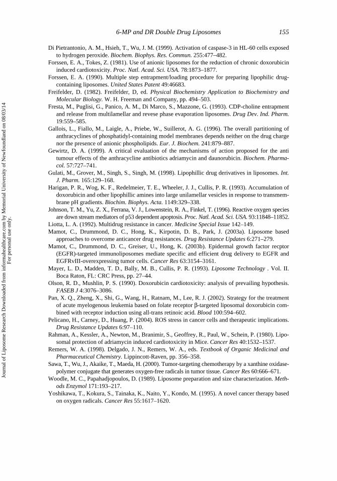
purpose, Hut78 and Jurkat cell lines were used for the liposome drug-mediated cyto-toxicity assay (Fig. 9A and B); the growth inhibition curves of daunorubicin-6MP lipo-some was characteristically similar to that of daunorubicin-liposome. A little improvedtrend of cell killing of the double drug liposome over the single drug was observedwith both the cell lines. Because the concentration of entrapped 6-MP in the doubledrug liposome was very low, a large difference in the cytotoxicity was not achievedwith the double drug liposome. However, a little increasing trend in the cell killing wasimplicated in both the cases, which may be predicted as a synergistic effect of the dualdrug liposome.
Figure 9. Effect of Dauno-6MP liposome on the survival of Jurkat and Hut 78 (T-cell lymphomacell lines). Cells were subjected to Dauno-6MP liposome treatment for 2 h and then washed thor-oughly with PBS. The percent survival was calculated after 72 h. Data are the means ± SD for threedifferent experiments. (A) Cytotoxic effect of 6-MP (♦), daunorubicin (•), and dauno-6MP (�)liposome on the viability of Jurkat cells. (B) Cytotoxic effect of 6-MP (�), daunorubicin (•), anddauno-6-MP (♦) liposome on the viability of Hut 78 cells. The percentage survival was determinedby exclusion of trypan blue dye using 1 × 10⁴ cells/well. The used final concentrations of daunoru-bicin were 0.1 μmol and 0.5 nmol of 6-MP per 100 μL of liposomal suspension. All the data werenormalized subjected to incorporation in single drug liposome.
Jour
nal o
f L
ipos
ome
Res
earc
h D
ownl
oade
d fr
om in
form
ahea
lthca
re.c
om b
y M
emor
ial U
nive
rsity
of
New
foun
dlan
d on
08/
03/1
4Fo
r pe
rson
al u
se o
nly.

6-MP and DR Double Drug Liposomes 153
Discussion
In our hand, the entrapment efficiency of the daunorubicin was almost the same in allkinds of liposomes. This is justifiable because the pH gradient method was used for theloading of DR, which is not limited to any particular lipid composition. Because thisapproach does not rely on any specific drug lipid interaction to enhance entrapment, it canbe used for any lipid mixture (Mayer et al., 1993), and high entrapment efficiencies can beobtained. 6-MP showed poor entrapment perhaps due to low drug to lipid ratio and itsbiphasic insolubility (Fresta et al., 1993; Gulati et al., 1998). Biphasic insoluble drugs aremost problematic to entrap; owing to insolubility either in the aqueous or lipid phase, theyshow only meager uptake by the liposomes.
The absorbance and fluorescence spectrum are characteristics of a compound, andany change in the chemical structure is marked by the corresponding change in the stan-dard spectrum. The combination spectrum of 6-MP and DR in Fig. 1c and d showed nosignificant visual changes compared with the standard spectrum of individual drugs (Fig.1a and b). The change in the absorbance was found to be additive due to the contributionby the other drug (we have confirmed this by adding the absorbances of each of the drugsat their respective λmax). The increase in the absorbance was similar as that of the standarddrugs, suggesting absence of any mechanistic interaction. Fluorescence spectroscopy fur-ther concluded the results that DR and 6-MP are noninteracting in solution. There couldhave been chances of an interaction inside the liposomes owing to the close vicinity of thetwo drugs. However, a similar DR spectrum and same Rt value of 6-MP ruled out the pos-sibility of any interaction. In our case, we found that fluorescence was quenched when theDR was entrapped inside the liposomes. It has been reported that DR fluorescence isreduced in the nonaqueous organic phase or in the lipid compared with that of the aqueousphase (Gallois et al., 1996). Thus, probably DR is partitioning in the lipid compartment.On the addition of lipase mixture, a burst of fluorescence emission was seen (Fig. 3), sug-gesting the release of DR in the bulk aqueous compartment. Thus, our observation regard-ing the quenching of the daunorubicin fluorescence inside liposomes and enhancement ofthe DR fluorescence (after the lipase treatment) due to release of drug from liposomes tosolution parallels the findings of Gallois et al. (1996). Studies of DR release from liposome-drug formulation were also done with detergent (Triton X 100), but Triton X was found tointerfere with the emission spectra of DR; hence, phospholipases were used for the lysis ofliposomes instead of Triton X. The stability studies of liposomes showed aggregation inneutral (PC: Chol) liposomes leading to the destabilization of liposomes followed byphase separation. However, aggregation was inhibited in the negative liposomes owing tothe repulsion between negatively charged vesicles compared with the neutral liposomes,hence, the anionic liposomes were more stable. Physical stability of liposomal preparationwith regard to lipid composition was revealed by TLC experiments. Equivalent Rf valuesof liposomal lipids as that of standards and lack of degradation products suggest absenceof any modification or oxidation of lipids. Thus, it can be said that double drug combina-tion is not having a deleterious effect on liposomal lipids.
Our results show quite low entrapment efficiency of 6-MP (0.5–1.5%); hence, anytherapeutic applicability due to purine biosynthesis inhibition is not much expected.Therefore, we are now trying to improve the incorporation efficiency of 6-MP by usinghydrophobic derivative of 6-MP. However, a combination of 6-MP and DR in vitro wasfound to produce more superoxides (measured by xanthine oxidase-cyt c method) thanonly DR or 6-MP (data not shown). It has been reported that ROS increases antitumoractivity by variety of mechanisms by causing apoptosis (Di Pietrantonio et al., 1999;
Jour
nal o
f L
ipos
ome
Res
earc
h D
ownl
oade
d fr
om in
form
ahea
lthca
re.c
om b
y M
emor
ial U
nive
rsity
of
New
foun
dlan
d on
08/
03/1
4Fo
r pe
rson
al u
se o
nly.

154 V. Agrawal, M. K. Paul, and A. K. Mukhopadhyay
Johnson et al., 1996). However, an increase in ROS raises the concern of enhanced toxic-ity of the combination especially cardiotoxicity due to daunorubicin. It has been reportedthat the cellular localization of xanthine oxidase in the heart is negligible (Olson andMushlin, 1990), and anionic liposomes reduce cardiotoxicity significantly (Forssen andTokes, 1981; Rahman et al., 1980). Therefore, it may be expected that an increase insuperoxide content would not take place in the heart tissue from our double drug anionicliposomes. On the other hand, the presence of xanthine oxidase in the tumor cells willselectively enhance superoxide formation at the tumor site exerting anticancer effects(Sawa et al., 2000; Yoshikawa et al., 1995).
We believe that 6-MP and DR are incorporated in different compartments inside theliposomes and DR, being lipophillic, has been partitioned preferentially in the lipid phase.Noninteraction of two drugs in solution and in liposomes is supposed to preserve the thera-peutic potential of individual drugs and enhance the therapeutic activity of this formulation.
When both the cell lines were incubated with the single and double drug liposomes, theinternalization of the liposome-drug into these cells was clearly visible. The release of thedrug was time dependent, and the cell killing was dependent on the amount of the drugpresent in the formulation. The pheno and genotypic nature of the two leukemia cell linesused in our experiments are entirely different. Although both Jurkat and Hut 78 cell line areT-cell lymphoma, Hut 78 cells are more resistant to daunorubicin-mediated apoptosis; there-fore, these cells were exposed to our double drug preparation for testing its efficacy. As pre-dicted, 6-MP containing mono drug liposome did not kill the cells significantly, and dual drugliposome was to some extent more efficient than only daunorubicin containing liposome, sug-gesting thereby the presence of a probable synergistic effect, which could have been muchbetter if 6-MP would have been incorporated more. The synergistic effect of the two drugsmight arise due to the differences of their mode of action and combined ROS production.Therefore, an effort was given to incorporate more 6-MP in the double dug liposome byincreasing the external concentration of 6-MP. But it was observed that increasing concentra-tion of 6-MP to as high as 50 mM could not even increase the percentage efficiency beyond1%. Therefore, at present we are trying to improve the efficiency of incorporation of 6-MP byusing different ester linked 6-MP (which are more hydrophobic and is susceptible to theaction of esterase inside the cell) and by trying our preparation with different kind of lipids.
The two-drug combination in liposomes may reduce the dose requirements loweringtoxicity and can aid in drug resistant states. Although the studies have been done with anti-cancer drugs, the double drug liposomes can be formulated with various drugs against dif-ferent diseases. Different drug combinations exerting therapeutic or modulating effect canbe encapsulated for therapeutic efficiency. The result from our study, although very pre-liminary, clearly and strongly indicates that two-drug liposomes are feasible and can opennew avenues for better therapy.
Acknowledgement
We thank Council of Scientific and Industrial Research (CSIR, New Delhi, Government of India)Grant # 01(1823)/ 02/EMR-II) for the funding of our research.
References
Chabner, A. B., Ryan, P. D., Ares, L., Carbonero, R., Calabresi, P. (2000). The PharmacologicalBasis of Therapeutics. New York: Mc-Graw Hill Medical Publishing Division, pp. 1411–1414.
Jour
nal o
f L
ipos
ome
Res
earc
h D
ownl
oade
d fr
om in
form
ahea
lthca
re.c
om b
y M
emor
ial U
nive
rsity
of
New
foun
dlan
d on
08/
03/1
4Fo
r pe
rson
al u
se o
nly.
6-MP and DR Double Drug Liposomes 155
Di Pietrantonio, A. M., Hsieh, T., Wu, J. M. (1999). Activation of caspase-3 in HL-60 cells exposedto hydrogen peroxide. Biochem. Biophys. Res. Commun. 255:477–482.
Forssen, E. A., Tokes, Z. (1981). Use of anionic liposomes for the reduction of chronic doxorubicininduced cardiotoxicity. Proc. Natl. Acad. Sci. USA. 78:1873–1877.
Forssen, E. A. (1990). Multiple step entrapment/loading procedure for preparing lipophilic drug-containing liposomes. United States Patent 49:46683.
Freifelder, D. (1982). Freifelder, D, ed. Physical Biochemistry Application to Biochemistry andMolecular Biology. W. H. Freeman and Company, pp. 494–503.
Fresta, M., Puglisi, G., Panico, A. M., Di Marco, S., Mazzone, G. (1993). CDP-choline entrapmentand release from multilamellar and revese phase evaporation liposomes. Drug Dev. Ind. Pharm.19:559–585.
Gallois, L., Fiallo, M., Laigle, A., Priebe, W., Suillerot, A. G. (1996). The overall partitioning ofanthracyclines of phosphatidyl-containing model membranes depends neither on the drug chargenor the presence of anionic phospholipids. Eur. J. Biochem. 241:879–887.
Gewirtz, D. A. (1999). A critical evaluation of the mechanisms of action proposed for the antitumour effects of the anthracycline antibiotics adriamycin and daunorubicin. Biochem. Pharma-col. 57:727–741.
Gulati, M., Grover, M., Singh, S., Singh, M. (1998). Lipophillic drug derivatives in liposomes. Int.J. Pharm. 165:129–168.
Harigan, P. R., Wog, K. F., Redelmeier, T. E., Wheeler, J. J., Cullis, P. R. (1993). Accumulation ofdoxorubicin and other lipophillic amines into large unilamellar vesicles in response to transmem-brane pH gradients. Biochim. Biophys. Acta. 1149:329–338.
Johnson, T. M., Yu, Z. X., Ferrana, V. J., Lowenstein, R. A., Finkel, T. (1996). Reactive oxygen speciesare down stream mediators of p53 dependent apoptosis. Proc. Natl. Acad. Sci. USA. 93:11848–11852.
Liotta, L. A. (1992). Multidrug resistance in cancer. Medicine Special Issue 142–149.Mamot, C., Drummond, D. C., Hong, K., Kirpotin, D. B., Park, J. (2003a). Liposome based
approaches to overcome anticancer drug resistances. Drug Resistance Updates 6:271–279.Mamot, C., Drummond, D. C., Greiser, U., Hong, K. (2003b). Epidermal growth factor recptor
(EGFR)-targeted immunoliposomes mediate specific and efficient drug delivery to EGFR andEGFRvIII-overexpressing tumor cells. Cancer Res 63:3154–3161.
Mayer, L. D., Madden, T. D., Bally, M. B., Cullis, P. R. (1993). Liposome Technology . Vol. II.Boca Raton, FL: CRC Press, pp. 27–44.
Olson, R. D., Mushlin, P. S. (1990). Doxorubicin cardiotoxicity: analysis of prevailing hypothesis.FASEB J 4:3076–3086.
Pan, X. Q., Zheng, X., Shi, G., Wang, H., Ratnam, M., Lee, R. J. (2002). Strategy for the treatmentof acute myelogenous leukemia based on folate receptor β-targeted liposomal doxorubicin com-bined with receptor induction using all-trans retinoic acid. Blood 100:594–602.
Pelicano, H., Carney, D., Huang, P. (2004). ROS stress in cancer cells and therapeutic implications.Drug Resistance Updates 6:97–110.
Rahman, A., Kessler, A., Newton, M., Branimir, S., Geoffrey, R., Paul, W., Schein, P. (1980). Lipo-somal protection of adriamycin induced cardiotoxicity in Mice. Cancer Res 40:1532–1537.
Remers, W. A. (1998). Delgado, J. N., Remers, W. A., eds. Textbook of Organic Medicinal andPharmaceutical Chemistry. Lippincott-Raven, pp. 356–358.
Sawa, T., Wu, J., Akaike, T., Maeda, H. (2000). Tumor-targeting chemotherapy by a xanthine oxidase-polymer conjugate that generates oxygen-free radicals in tumor tissue. Cancer Res 60:666–671.
Woodle, M. C., Papahadjopoulos, D. (1989). Liposome preparation and size characterization. Meth-ods Enzymol 171:193–217.
Yoshikawa, T., Kokura, S., Tainaka, K., Naito, Y., Kondo, M. (1995). A novel cancer therapy basedon oxygen radicals. Cancer Res 55:1617–1620.
Jour
nal o
f L
ipos
ome
Res
earc
h D
ownl
oade
d fr
om in
form
ahea
lthca
re.c
om b
y M
emor
ial U
nive
rsity
of
New
foun
dlan
d on
08/
03/1
4Fo
r pe
rson
al u
se o
nly.

Jour
nal o
f L
ipos
ome
Res
earc
h D
ownl
oade
d fr
om in
form
ahea
lthca
re.c
om b
y M
emor
ial U
nive
rsity
of
New
foun
dlan
d on
08/
03/1
4Fo
r pe
rson
al u
se o
nly.