6-br-5methylindirubin-3′oxime (5-me-6-bio) targeting the
TRANSCRIPT

International Journal for Parasitology 39 (2009) 1289–1303
Contents lists available at ScienceDirect
International Journal for Parasitology
journal homepage: www.elsevier .com/locate / i jpara
6-Br-5methylindirubin-30oxime (5-Me-6-BIO) targeting the leishmanialglycogen synthase kinase-3 (GSK-3) short form affects cell-cycle progressionand induces apoptosis-like death: Exploitation of GSK-3 for treating leishmaniasis
Evangelia Xingi a, Despina Smirlis a, Vassilios Myrianthopoulos b, Prokopios Magiatis b, Karen M. Grant c,Laurent Meijer d, Emmanuel Mikros b, Alexios-Leandros Skaltsounis b, Ketty Soteriadou a,*
a Laboratory of Molecular Parasitology, Department of Microbiology, Hellenic Pasteur Institute, 127 Vas. Sofias Ave., 11521 Athens, Greeceb Laboratories of Pharmacognosy and Pharmaceutical Chemistry, Department of Pharmacy, University of Athens, Panepistimiopolis-Zografou, Athens 15771, Greecec School of Health and Medicine, Lancaster University, LA1 4YB, UKd C.N.R.S., Cell Cycle Group, Station Biologique, B.P. 74, 29682 Roscoff Cedex, Bretagne, France
a r t i c l e i n f o a b s t r a c t
Article history:Received 11 February 2009Received in revised form 31 March 2009Accepted 3 April 2009
Keywords:Leishmania donovani glycogen synthasekinase-3 short5-Me-6-BIO6-BIOIndirubinsApoptosis-like deathDrug target
0020-7519/$36.00 � 2009 Australian Society for Paradoi:10.1016/j.ijpara.2009.04.005
* Corresponding author. Tel.: +30 2106478841; faxE-mail address: [email protected] (K. Soteria
Indirubins known to target mammalian cyclin-dependent kinases (CDKs) and glycogen synthase kinase(GSK-3) were tested for their antileishmanial activity. 6-Br-indirubin-30-oxime (6-BIO), 6-Br-indirubin-30acetoxime and 6-Br-5methylindirubin-30oxime (5-Me-6-BIO) were the most potent inhibitors of Leish-mania donovani promastigote and amastigote growth (half maximal inhibitory concentration (IC50) val-ues 61.2 lM). Since the 6-Br substitution on the indirubin backbone greatly enhances the selectivityfor mammalian GSK-3 over CDKs, we identified the leishmanial GSK-3 homologues, a short (LdGSK-3s)and a long one, focusing on LdGSK-3s which is closer to human GSK-3b, for further studies. Kinase assaysshowed that 5-Me-6-BIO inhibited LdGSK-3s more potently than CRK3 (the CDK1 homologue in Leish-mania), whilst 6-BIO was more selective for CRK3. Promastigotes treated with 5-Me-6-BIO accumulatedin the S and G2/M cell-cycle phases and underwent apoptosis-like death. Interestingly, these phenotypeswere completely reversed in parasites over-expressing LdGSK-3s. This finding strongly supports thatLdGSK-3s is: (i) the intracellular target of 5-Me-6-BIO, and (ii) involved in cell-cycle control and in path-ways leading to apoptosis-like death. 6-BIO treatment induced a G2/M arrest, consistent with inhibitionof CRK3 and apoptosis-like death. These effects were partially reversed in parasites over-expressingLdGSK-3s suggesting that in vivo 6-BIO may also target LdGSK-3s. Molecular docking of 5-Me-6-BIO inCRK3 and 6-BIO in human GSK-3b and LdGSK-3s active sites predict the existence of functional/structuraldifferences that are sufficient to explain the observed difference in their affinity. In conclusion, LdGSK-3sis validated as a potential drug target in Leishmania and could be exploited for the development of selec-tive indirubin-based leishmanicidals.
� 2009 Australian Society for Parasitology Inc. Published by Elsevier Ltd. All rights reserved.
1. Introduction
Leishmaniasis is an umbrella term for a group of protozoan vec-tor-borne parasitic diseases and manifests with three major forms,visceral, cutaneous and mucocutaneous. It is a significant cause ofmorbidity and mortality in developing countries, and affects about2 million people per year mostly in tropical and sub-tropical re-gions (Alvar et al., 2006). Leishmaniasis is also an important publichealth and veterinary concern in Mediterranean countries (Dujar-din, 2006). Chemotherapy for leishmaniasis is generally ineffective,mainly due to the emerging drug-resistance and severe toxic sideeffects (Croft et al., 2006). Antimonials are used as first-linetreatment despite their toxicity. In case of antimonial resistance,
sitology Inc. Published by Elsevier
: +30 2106423498.dou).
liposomal formulations of amphotericin B, not devoid of adverseside effects, are used (Croft et al., 2006). Miltefosine, the first oraldrug, has proved to be highly effective against visceral leishmani-asis. However, miltefosine-resistant parasites have been obtainedin vitro indicating that there is a risk of resistance emerging inthe field. Consequently there is an urgent need to discover new tar-geted drugs against leishmaniases (Croft et al., 2006).
Leishmania species are transmitted to mammals by the bite of asand fly vector. During a sandfly blood meal, Leishmania promastig-otes pass into the mammalian host where they penetrate macro-phages and, within their phagolysosomes, transform into thenon-flagellated, non-motile amastigote form and multiply (Chang,1983). These trypanosomatid protozoan parasites have developedunusual and unique features in their cell biology to ensure adapta-tion to the contrasting environments of their insect and mamma-lian hosts that are reflected in the complexity of their cell- cycle
Ltd. All rights reserved.

1290 E. Xingi et al. / International Journal for Parasitology 39 (2009) 1289–1303
control and during their differentiation. Therefore, differences be-tween cell-cycle control in Leishmania and mammals may lead tothe identification of essential molecules regulating the parasitecell-cycle that could be exploited for rational drug design (Naulaet al., 2005). Potential parasite candidate targets include cyclin-dependent kinases (CDKs), glycogen synthase kinases (GSK-3),Aurora kinases and mitogen activated protein kinases (MAPKs)(Naula et al., 2005). Recently, it was shown that Trypanosoma bru-cei GSK-3 ‘‘short” is a potential drug target for trypanosomiasistherapy (Ojo et al., 2008). Efforts are therefore focused on theexploitation of kinase inhibitor libraries for the identification andfurther development of inhibitors that selectively target parasitekinases without damaging the host.
Indirubin analogues (collectively referred to as indirubins), afamily of bis-indoles known for over a century as a minor constit-uent of plant, animal and microorganism-derived indigo, are pow-erful inhibitors of mammalian CDKs and GSK-3 by competing withATP for binding to their catalytic site (Meijer et al., 2003; Polychro-nopoulos et al., 2004). 6-Bromo substituted indirubins displayhigher selectivity for mammalian GSK-3 over CDKs (Meijer et al.,2003; Polychronopoulos et al., 2004). In cell-based assays, indiru-bins display anti-mitotic and anti-tumoral activity and induce ar-rest in G1 or G2/M phase of the cell-cycle, depending on the cellline (Hoessel et al., 1999; Damiens et al., 2001). Specifically, 6-bro-mo-indirubin-30-oxime (6-BIO) induces apoptotic death in neuro-blastoma cells (Ribas et al., 2006).
GSK-3 is a multifunctional serine/threonine kinase found in alleukaryotes. This enzyme is known to play a key role in many cellularand physiological events, including Wnt signalling, transcription,cell-cycle and differentiation, neuronal functions and circadianrhythm (Frame et al., 2001; Doble and Woodgett, 2003). These func-tions of GSK-3 and its implication in many human diseases such asAlzheimer’s disease, non-insulin-dependent diabetes mellitus andcancer have stimulated an active search for potent and selectiveGSK-3 inhibitors, such as indirubins (Meijer et al., 2004).
CRK3, a leishmanial CDK1 homologue, displaying 54% identityand 71% similarity with human CDK1, has been validated as a drugtarget (Grant et al., 1998; Hassan et al., 2001). Three indirubins (5-sulphonamide-indirubin-30oxime, 5-SO3Na-30oxime and 5-SO3H)have been shown to inhibit CRK3 with IC50 values of 11, 16 and47 nM, respectively, and Leishmania donovani infection of mousemacrophages with half maximal inhibitory concentration (IC50)values of 3.56, 5.8 and 7.6 lM, respectively (Grant et al., 2004;Wells et al., 2006). Leishmania mexicana promastigotes treatedwith indirubins displayed growth arrest and disruption of cell-cy-cle, in line with the inhibition of a CDK (Grant et al., 2004).
In this study, 16 indirubins were tested for their antileishmanialactivity and three, 6-BIO, 6-BIA and 5-Me-6-BIO, were found to bethe most powerful inhibitors of both L. donovani promastigotes andintracellular amastigote growth. Since the 6-Br substitution on theindirubin backbone greatly enhances the selectivity for mamma-lian GSK-3 over CDKs, we identified the leishmanial GSK-3 homo-logues, a short (LdGSK-3s) and a long one (LdGSK-3l). We theninvestigated whether our compounds target LdGSK-3s which is clo-ser to human GSK-3b. LdGSK-3s was identified as the predominantintracellular target of 5-Me-6-BIO. Evidence is also presented thatLdGSK-3s is involved in cell-cycle control as well as in pathwaysleading to apoptosis-like death.
2. Materials and methods
2.1. Cell culture
Leishmania donovani (strain LG13, MHOM/ET/0000/HUSSEN)promastigotes and the murine macrophage J774 cell line (ATCC)were cultured in medium 199 (M199) and RPMI 1640 (RPMI),
respectively, both supplemented with 10% heat inactivated FBS(HI-FBS), 10 mM Hepes and antibiotics. Axenic L. donovaniamastigotes were generated as previously described (Barak et al.,2005; Smirlis et al., 2006). Spleen-derived L. donovani amastigoteswere maintained in Schneider’s insect medium pH 5.5 supple-mented with 20% HI-FBS at 37 �C. Leishmania donovani transfec-tants with the Leishmania expression vector pLEXSY-sat, the L.donovani glycogen synthase kinase-3 short cloned in pLEXSY-sat(pLEXSY-sat-LdGSK-3s) and the LdGSK-3s kinase-dead mutantcloned in pLEXSY-sat (pLEXSY-sat-LdGSK-3s/K49R) were culturedin M199 supplemented with 100 lg/ml nourseothricin (Jena Bio-science). Leishmania mexicana CRK3his transfectants were culturedas described previously (Grant et al., 2004).
2.2. Chemical library
The chemical library consists of 16 indirubins (Table 1), syn-thesised as previously described, (Polychronopoulos et al., 2004;Ribas et al., 2006). The compounds were dissolved in DMSO at10 mM and serial dilutions in DMSO were made (1 mM and100 lV). Indirubins were diluted in culture medium to give the de-sired final concentrations.
2.3. In vitro testing of the antileishmanial activity of indirubins againstL. donovani promastigotes, intracellular amastigotes and axenicamastigotes
The Alamar blue assay (Mikus and Steverding, 2000) was ap-plied for determining the antileishmanial activity of indirubinsand Amphotericin B (Fungizone) was used as a reference drug. Sta-tionary-phase L. donovani promastigotes (2 � 107 cells/ml) wereseeded into 96-well flat bottom plates at a density of 2.5 � 106
cells/ml in 200 ll M199 without phenol red, containing increasingindirubin concentrations or the equivalent volume of the diluentDMSO, each in quadruplicate. The final concentration of DMSOwas always <1% (v/v) and did not affect the growth of parasites.Following indirubin treatment for 72 h, Alamar blue (20 ll/well)was added and the plates were incubated at 26 �C for a further12 h. Colorimetric readings were performed at a test wavelengthof 550 nm and a reference wavelength of 620 nm. Comparison ofDMSO-treated controls with samples allowed the calculation ofthe concentration of indirubin necessary to reduce the growth rateof promastigotes by 50% (IC50 values).
To evaluate the inhibitory activity of indirubins on intracellularamastigotes we treated infected macrophages for 72 h with indiru-bins and then assessed amastigote survival by lysing infected mac-rophages using the Alamar blue assay. Briefly, J774 macrophages(2 � 105 cells/ml in 200 ll RPMI) were left to adhere overnight at37 �C in 5% CO2 into 96-well flat bottom plates. Macrophages wereinfected with stationary-phase L. donovani promastigotes at a ratioof 10 parasites/macrophage and incubated for a further 24 h at37 �C in 5% CO2 as previously described (Papageorgiou and Soteria-dou, 2002). Then the overlying medium was removed and cellswere washed three times in fresh RPMI. Fresh RPMI was addedcontaining increasing concentrations of indirubins or the equiva-lent volume of the diluent DMSO, each in quadruplicate. After72 h, the medium was removed and infected cells were lysed byaddition of 100 ll 0.01% SDS in PBS for 30 min at 37 �C. Then100 ll Schneider’s medium was added to each well that containedthe liberated amastigotes (Papageorgiou and Soteriadou, 2002).Amastigote growth was assessed by the addition of Alamar blue(20 ll/well) and the plates were incubated for 48 h at 37 �C. Com-parison of DMSO-treated controls with samples enabled the calcu-lation of the degree to which infection had been inhibited by thepresence of indirubins and to calculate the concentration that re-duces the number of amastigotes by 50%.

Table 1Indirubin compounds tested in vitro for their antileishmanial activity against Leishmania donovani promastigotes (P), intracellular amastigotes (In. A) and axenic amastigotes (Ax.A) using the Alamar blue assay.
Compound Y X W R L. donovani IC50* (lM)
P In. A Ax. A
Indirubin O H H H n.i.a n.i. n.i.Indirubin-30oxime NOH H H H n.i. n.i. n.i.5-Br-indirubin O H Br H n.i. n.i. n.i.5-Br-indirubin-30oxime (5-DIJ) NOH H Br H 5.2 ± 1.6 1 ± 0.15 1 ± 0.25-Aminoindirubin-30oxime NOH H NH2 H >10 b >10 >106-Br-indirubin O Br H H n.i. n.i. n.i.6-Br-indirubin-30oxime (6-DIJ) NOH Br H H 0.8 ± 0.1 0.75 ± 0.05 0.9 ± 0.16-Br-N-methyl-indirubin-30-oxime NOH Br H CH3 n.i. n.i. n.i.6-Br-indirubin-30acetoxime (6-BIA) NOAc Br H H 0.9 ± 0.1 1 ± 0.05 1 ± 0.16-Br-N-methyl-indirubin-30acetoxime NOAc Br H CH3 n.i. n.i. n.i.6-Br-indirubin-30diethyl phosphatoxime NOPO(OEt)2 Br H H >10 >10 >10Indirubin-30-methoxime NOCH3 H H H n.i. n.i. n.i.6-Br-5nitroindirubin O Br NO2 H n.i. n.i. n.i.6-Br-5nitroindirubin-30-oxime NOH Br NO2 H >10 >10 >106-Br-5methylindirubin O Br CH3 H n.i. n.i. n.i.6-Br-5methylindirubin-30oxime (5-Me-6-BIO) NOH Br CH3 H 1.2 ± 0.2 1 ± 0.1 1 ± 0.2
* IC50 values were determined from dose–response curves and are expressed in lM.a n.i., no inhibition at 50 lV.b IC50 values ranging between 10 and 50 lV.
E. Xingi et al. / International Journal for Parasitology 39 (2009) 1289–1303 1291
Leishmania donovani axenic amastigotes were treated with theinhibitors and the percentages of growth inhibition were assessedby addition of Alamar blue after 72 h of treatment (Habtemariam,2003). In all cases, IC50 values were determined from dose–re-sponse curves via linear interpolation.
2.4. Analysis of indirubin-treated promastigotes by flow cytometry
Stationary-phase (2 � 107 cells/ml) L. donovani promastigoteswere seeded at 106 cells/ml in M199 and incubated at 26 �C inthe presence of DMSO (the diluent, used as control) or the testedindirubin. Preparation of samples for fluorescence-activated cellsorting (FACS) analysis of the cell-cycle was carried out as de-scribed by Smirlis et al. (Smirlis et al., 2006). Exposed phosphati-dylserine on the outer membrane of cells and plasma membraneintegrity of cells were assessed using Annexin V-FITC and propidi-um iodide (PI) staining (Apoptosis Detection kit, R&D Systems).Preparation of samples for FACS analysis was performed accordingto the manufacturer’s instructions. In all cases, 20,000 cells/samplewere analysed, using a Becton Dickinson FACSCalibur flow cytom-eter and data were analysed using the Cell Quest software. Allexperiments were performed at least three times.
2.5. Cellular and nuclear morphology
Stationary-phase L. donovani promastigotes were seeded at 106
cells/ml and incubated with either 2 lM indirubin or DMSO for 24and 48 h. Cells were fixed in 2% paraformaldehyde and treatedwith 50 lg/ml RNaseA and 10 lg/ml PI. Parasites were observedunder a TCSSP Leica Confocal fluorescence microscope. At least
100 cells from three independent experiments were recorded foreach condition.
2.6. Cell count and viability assay
Stationary-phase L. donovani promastigotes were seeded at 106
cells/ml and incubated with either 2 lM 5-Me-6-BIO or DMSO for24, 48 and 72 h. Cells were then washed twice in PBS, resuspendedin drug-free medium and allowed to recover for 24, 48 and 72 h.The viability assay at different time points after exposure of para-sites to 5-Me-6-BIO and drug removal was assessed using 0.4% Try-pan blue solution. Both total cell count and the percentages ofviable and non-viable cells were recorded. The experiment wasperformed three times.
2.7. In situ labelling of DNA fragments by TUNEL
In situ detection of DNA strand breaks was performed using theCell Death Fluorescein Detection kit (Roche Applied Science) fol-lowing the manufacturer’s instructions. Samples were analysedunder a Zeiss fluorescence microscope at 120� magnification.The ratio of apoptosis (apoptotic to total cells) was determinedby counting at least 400 cells per group in three independentexperiments.
2.8. Gene cloning and antibody production
The L. donovani GSK-3s gene was amplified by PCR from L. dono-vani (strain LG13, MHOM/ET/0000/HUSSEN) genomic DNA, using asense primer 50-ACC GCC ATG GAC ATG TCG CTC AAC GCT GC-30

1292 E. Xingi et al. / International Journal for Parasitology 39 (2009) 1289–1303
and an antisense primer 50-CCC CCT CGA GCT GCT TGC GAA CTAGCT T-30, that were designed based on the gene coding for theshorter of the two Leishmania major Friedlin GSK-3 proteins(LmjF18.0270). The amplified PCR product was cloned into theNcoI-XhoI site of pTriEx-1.1 vector (Novagen), a construct allowingthe addition of a poly-Histidine extention to the C-terminus of therecombinant protein (pTriEx-1.1-LdGSK-3s). The cloned gene wasthen sequenced and compared with the short sequence of Leish-mania infantum GSK-3 (LinJ18_V3.0270). The LdGSK-3s DNA andprotein sequences were found to be identical to the L. infantumGSK-3s sequences. LdGSK-3s nucleotide sequence was depositedin GenBank (EF620873). The pTriEx-1.1-LdGSK-3s construct wastransformed into bacteria and (His)6-tagged LdGSK-3 was purifiedby Metal-Affinity Chromatography (Qiagen Ni–NTA Superflow re-sin). LdGSK-3s was detected using a polyclonal IgG His-probe anti-body (1:500 dilution, stock solution 200 lg/ml, Santa CruzBiotechnology) and a polyclonal anti-ratGSK-3b antibody. The re-combinant protein was subsequently used to immunize a NewZealand white rabbit using the scheme described in a previousstudy (Smirlis et al., 2006). The immunization protocols were ap-proved by the Ethical Committee of Hellenic Pasteur Instituteand were in accordance with the European Directive 86/609/EECon the protection of animals used in research. Affinity purifiedanti-LdGSK-3s antibody was isolated by low pH elution of antibod-ies bound to purified LdGSK-3s on nitrocellulose strips, as previ-ously described (Smirlis et al., 2006).
2.9. Generation of transgenic promastigotes and purification of LdGSK-3s and CRK3
The DNA encoding (His)6-tagged LdGSK-3s was amplified byPCR from the construct pTriEx-1.1-LdGSK-3s, described above.Sense and antisense primers for the amplification were 50-ACCGCC ATG GAC ATG TCG CTC AAC GCT GC-30 and 50-GCA GGC GGCCGC TGA GGT TAA TCA CTT AGT G-30, respectively. The PCR productwas then cloned in the NcoI and NotI sites of the Leishmaniaexpression vector pLEXSY-sat (pF4X1.4sat) (Jena Bioscience) togenerate the pLEXSY-sat-LdGSK-3s plasmid. The cloned gene wassequenced to confirm the correct orientation. Site-directed muta-genesis was performed on LdGSK-3s in pLEXSY-sat-LdGSK-3s con-struct, using the Phusion� Site-Directed Mutagenesis Kit(Finnzymes) following the manufacturer’s protocol. Primers usedfor Lysine 49 to Arginine mutation (K49R) were as follows: For-ward 50-GAGCGTGGCGATCCGGAAGGTTATCCAGGAC-30 and Re-verse 50-ATGCCCGTCGACTTCTCCTTGCCTAGTTGCA-30. The K49Rmutation in the construct pLEXSY-sat-LdGSK-3s/K49R was con-firmed by sequencing.
Leishmania donovani transfectants with pLEXSY-sat, pLEXSY-sat-LdGSK-3s and pLEXSY-sat-LdGSK-3s/K49R plasmids (supercoiled,transfected as episomes) were generated as previously described(Smirlis et al., 2006). Selection of transgenic promastigotes was per-formed in M199 containing 100 lg/ml nourseothricin.
Purification of LdGSK-3s from L. donovani sat-LdGSK-3s trans-fectants and of LdGSK-3s/K49R from sat-LdGSK-3s/K49R transfec-tants as well as of CRK3 from transgenic L. mexicanapromastigotes was carried out as previously described (Grantet al., 2004). LdGSK-3s and CRK3 were stored with 10% glycerolat �80 �C for kinase assays.
2.10. Immunoblotting
Parasites were suspended in lysis buffer (50 mM 3-(N-morpho-lino)propanesulfonic acid (MOPs) pH 7.5, 100 mM NaCl, 1 mMEDTA, 1 mM EGTA, 1 mM Na3VO4, 10 mM NaF, 1% Triton X-100)supplemented with protease inhibitors (0.1 mg/ml leupeptin,1 mM phenylmethanesulphonylfluoride (PMSF), 5 lg/ml aprotinin,
5 lg/ml pepstatin A, 1 mM phenanthroline) and Laemmli samplebuffer was added. Whole cell lysates were resolved by 12% SDS–PAGE, transferred to nitrocellulose membranes and subsequentlyprobed with the appropriate primary antibodies: a polyclonalanti-ratGSK-3b antibody [directed against the C-terminal sequenceCAHSFFDELRDPNVK, residues identical between rat and LdGSK-3sare underlined] (1:100 dilution, stock solution 500 lg/ml, Abcam);the generated anti-LdGSK-3s rabbit polyclonal antibody (1:1000dilution) and the polyclonal IgG His-probe antibody (1:200 dilu-tion). After incubation with peroxidase-conjugated secondary anti-body, 3,30-diaminobenzidine was used for detection.
To demonstrate equal loading of cells, the blot was stripped andre-probed with antiserum against L. infantum myo-inositol-1-phosphate synthase (LinJ14_V3.1450, INO1), reported to be equallyexpressed in L. mexicana promastigotes and amastigotes (Ilg,2002). The INO1 gene was PCR-amplified from L. infantum genomicDNA, using a sense primer 50-CAAGGGATCCGATGACGCGTGACATGGACG-30 and an antisense primer 50-GGCACTC GAGCAGCATGTTGCTGTCGG-30, cloned into the BamHI and XhoI site of pTriEx-1.1,in frame with a C terminal his-tag, expressed in Escherichia coliand purified on Ni+2- nitrilotriacetate (Ni–NTA) resin. Anti-LinINO1 antibody was produced and purified, using nitrocellulosestrips with purified INO1, as previously described (Smirlis et al.,2006).
2.11. Immunofluorescence
Parasites were fixed in 2% formaldehyde and 0.05% glutaralde-hyde. Cells were blocked in 50 mM NH4Cl containing 3% BSA inPBS and treated with 50 lg/ml RNaseA. Nuclei were stained with10 lg/ml PI followed by incubation for 5 h with either anti-LdGSK-3s or the anti-ratGSK-3b antibodies (10 or 5 lg/ml, respec-tively) in PBS containing 0.1% Triton X-100 and 3% BSA. The boundantibody was detected with 1:100 diluted FITC conjugated anti-rabbit or anti-rat IgG antibody (Sigma). Cells were observed witha TCSSP Leica Confocal fluorescence microscope.
2.12. Kinase assays
Kinase assays were performed with purified LdGSK-3s and CRK3from L. donovani over-expressing transfectants and transgenic L.mexicana promastigotes, respectively (Supplementary Fig. S1).The kinase activity of LdGSK-3s was assayed using GS-1 peptide(YRRAAVPPSPSLSRHSSPHQSpEDEEE) as a substrate; GS-1 peptidewas patterned after the GSK-3 phosphorylation sites of mamma-lian glycogen synthase (Meijer et al., 2004). CRK3 kinase assayswere performed using histone H1 substrate as previously de-scribed (Grant et al., 2004). All assays were performed in the kinaseassay buffer (50 mM MOPS pH 7.2, 20 mM MgCl2, 10 mM EGTA,2 mM DTT) in the presence of [c-33] ATP (3000 Ci/mmol; 1 mCi/ml) in a final volume of 30 ll and incubated for 30 min at 30 �Cas previously described (Meijer et al., 2004). Initially the Km valuesfor ATP and substrate for each kinase were measured. The Km val-ues for ATP for both kinases were around 15 lM (15.2 lM forLdGSK-3s and 14.78 lM for CRK3). In order to determine the IC50
values with the inhibitors, we used ATP and substrate concentra-tions at the calculated Km values. For both LdGSK-3s and CRK315 lV ATP were used in the assays, in the presence of 8.3 lVGS-1 peptide and 5 lV histone H1, respectively. Ki values for eachinhibitor were calculated using the Cheng-Pursoff equation[Ki = IC50/(1 + S/Km)]. Kinase assays were also performed usingthe Kinase Luminescent Assay Kit (Promega), following the manu-facturer’s instructions, and gave comparable results.
LdGSK-3s activity (purified from L. donovani over-expressingtransfectants, 1 lg enzyme/reaction) was also determined usingpotential protein substrates: L. infantum histone H1 (LeishH1),

Table 2Amino-acid identity (%, above diagonal) and similarity (%, below diagonal) relation-ships of Leishmania infantum GSK-3 enzymes versus human and Trypanosoma bruceiproteinsa.
H.sapGSK-3b
H.sapGSK-3a
L.infGSK-3s
L.infGSK-3l
T.bruGSK-3s
T.bruGSK-3l
H.sap GSK-3b 67.1 39.5 22 40.7 32.2H.sap GSK-3a 74.4 34.3 21.5 35.4 30.5L.inf GSK-3s 55.8 48.1 19.5 65.4 29.8L.inf GSK-3l 30.5 30.6 29.2 20.1 26.5T.bru GSK-3s 55.9 48.1 80.6 27.9 31.2T.bru GSK-3l 47.8 45.7 43.1 36.4 44.7
a The table was constructed using BioEdit 7.0.5.3 Sequence Alignment Editor(pairwise global alignments using BLOSUM62 similarity matrix). Accession num-bers for the enzymes were: Homo sapiens (H.sap) GSK-3b (P49841), H. sapiens GSK-3a (P49840), L. infantum (L.inf) GSK-3s (XP_001464844, LinJ18_V3.0270), L. infan-tum GSK-3l (XP_001465568, LinJ22_V3.0370), T. brucei (T.bru) GSK-3short(Tb10.61.3140), T. brucei GSK-3long (Tb927.7.2420).
E. Xingi et al. / International Journal for Parasitology 39 (2009) 1289–1303 1293
mammalian histone H1 (Sigma), axin (recombinant, purified frombacteria), myelin basic protein and casein (dephosphorylated frombovine milk, Sigma), (approximately 1 lg substrate/reaction).LeishH1 was expressed in bacteria as a fusion protein with GSTand purified with Sepharose 4B-Glutathione beads as previouslydescribed (Smirlis et al., 2006). LeishH1 recombinant protein wascleaved from the GST moiety by thrombin treatment (Smirliset al., 2006). In vitro phosphorylation of protein substrates wasperformed in the kinase assay buffer. After 30-min incubation at30 �C, in the presence of [c-32P] ATP (6000 Ci/mmol, 10 mCi/ml)in a final volume of 30 ll, the kinase reaction was stopped by addi-tion of Laemmli buffer (Meijer et al., 2004). The protein substrateswere resolved by 12% SDS–PAGE, stained with Coomassie blue andtheir phosphorylation level was visualised by autoradiography.
2.13. Homology modelling
The homology model of parasite GSK-3s was based on the crystalstructure of the human GSK-3b complexed with 6-BIO (pdb 1Q41)and the respective of CRK3 on the template structure of humanCDK2-cyclin A (pdb 1E9H), (Davies et al., 2001). Model buildingwas performed with MODELLER v. 6 program (Sali and Blundell,1993) and stereochemical validation with PROCHECK program (Las-kowski et al., 1991). Docking was performed with a Monte Carlosearch algorithm. Ligand partial charges were calculated in a semi-empirical level by MOPAC6 (AM1 hamiltonian) (Stewart, 1990).
3. Results
3.1. Evaluation of the antileishmanial effect of indirubins on L.donovani promastigotes and amastigotes
The Alamar blue assay (Mikus and Steverding, 2000), in a 96-well format, was used for the primary screening and subsequentmonitoring of the growth of L. donovani promastigotes and axenicamastigotes exposed to indirubins. The same assay was adaptedand used for estimating the growth of intracellular amastigotes48 h after lysis of the infected macrophages treated for 72 h withthe indirubins. In the initial screening, 16 indirubins were testedat 10 lM. Nine of the compounds did not significantly affect para-site growth even when used at a higher concentration of 50 lM.Four out of the 16 compounds tested significantly inhibited Leish-mania growth and their IC50 values were determined, (Table 1).More specifically, 6-BIO, 6-BIA and 5-Me-6-BIO inhibited promas-tigote growth with an IC50 of 0.8 ± 0.1, 0.9 ± 0.1 and 1.2 ± 0.2 lM,respectively (Table 1). 5-BIO inhibited promastigote growth withan IC50 of 5.2 ± 1.6 lM (Table 1). Interestingly, 6-BIO, 6-BIA, 5-Me-6-BIO and 5-BIO were also found to significantly inhibit thegrowth of both L. donovani intracellular and axenic amastigoteswith IC50 values ranging from 0.75 ± 0.05 to 1 ± 0.2 lM, respec-tively (Table 1). N1-methyl derivatives of 6-BIO and 6-BIA didnot inhibit the growth of either promastigotes or amastigotes, con-sistent with the inactivation of indirubins as kinase inhibitors bythis modification (Meijer et al., 2003).
All four compounds did not affect the growth of macrophages atthe concentration used, but as determined using the same assay,they were toxic for host cells at significantly higher concentrations(IC50 values >25 lM). Amphotericin B used as a reference drug inhib-ited promastigote and amastigote (intracellular or axenic) growthwith IC50 values of 0.1 ± 0.01 and 0.2 ± 0.02 lM, respectively.
3.2. Molecular characterisation of LdGSK-3s: expression andlocalisation in the L. donovani life cycle
Since 6-bromo indirubins are powerful and selective inhibitorsof mammalian GSK-3 (Meijer et al., 2003; Polychronopoulos et al.,
2004) we searched in the Leishmania GeneDB database for GSK-3homologues (Parsons et al., 2005). Two GSK-3 encoding genes werefound in the Leishmania genome using BLAST homology searches, ashort and a long version. Comparison of the amino acid identitiesof the two human GSK-3 orthologues versus the two L. infantumforms revealed that GSK-3s is closer than GSK-3l to human GSK-3a and GSK-3b. However, GSK-3s has a slightly higher identity toGSK-3b than to GSK-3a (Table 2). This does not allow us to unam-biguously determine whether GSK-3s is equivalent to the GSK-3bmammalian form.
We focused on the GSK-3s isoform for further studies, since: (i)the GSK-3s homologues are almost identical in different Leishmaniaspecies, and (ii) GSK-3s is slightly closer to the mammalian GSK-3b,the most well-studied isoform. The GSK-3s gene in L. infantum, in L.major and in L. mexicana is located on chromosome 18 and encodesa protein of 355 amino acids with a predicted molecular mass of40.7 kDa. BLASTP analysis showed that the identified L. donovaniGSK-3s gene was identical to the L. infantum GSK-3 short geneand almost identical to L. major and L. mexicana GSK-3 short andGSK-3b genes, respectively (98% identity and 99% similarity), (Sup-plementary Fig. S2). LdGSK-3s shares 49% sequence identity and68% similarity with hGSK-3b. It also shares 65% identity and 80%similarity with T. brucei GSK-3 ‘‘short”, 47% identity and 67% simi-larity with Danio rerio GSK-3b, 42% identity and 64% similarity withPlasmodium falciparum GSK-3, and 49% identity and 68% similaritywith Mus musculus GSK-3b (Supplementary Fig. S2).
LdGSK-3s was detected in L. donovani extracts using an affinity-purified anti-LdGSK-3s polyclonal antibody (raised against the re-combinant protein expressed in E.coli) and a commercially availablepolyclonal anti-ratGSK-3b antibody (raised against the C-terminalsequence of rat GSK-3b). The�40 kDa protein detected by both anti-bodies is in line with the predicted molecular mass of LdGSK-3s.Western blot analysis indicated that the level of expression ofLdGSK-3s in L. donovani stationary and logarithmic-phase prom-astigotes (107 cells/lane) was comparable (Fig. 1A, lanes S and L,respectively). LdGSK-3s was also detected in spleen-derived L. dono-vani amastigotes and axenic amastigotes (107 cells/lane), (Fig. 1A,lanes Am and Ax, respectively). Scanning densitometry of the de-tected bands revealed that LdGSK-3s expression level was about 3-fold lower in amastigotes. As a control for loading equal number ofcells, the blot was stripped and re-probed with the antiserum againstL. infantum myo-inositol-1-phosphate synthase (LinINO1), a 46 kDaprotein whose level of expression is constitutive during promasti-gote growth and which is equally expressed in promastigotes andamastigotes, (Ilg, 2002; Rosenzweig et al., 2008) (Fig. 1B). No proteinband was detected when pre-immune serum was used as a negativecontrol (Fig. 2C, lane S, Ax). LdGSK-3s was also recognised by theanti-ratGSK-3b antibody (Fig. 2D, lane S). Similarly, the mouse

Fig. 1. Leishmania donovani glycogen synthase kinase-3 short (LdGSK-3s) expression in L. donovani. Western blot analysis (A) anti-LdGSK-3s antibody: stationary-phasepromastigotes (S), logarithimic-phase promastigotes (L), axenic amastigotes (Ax), spleen-derived amastigotes (Am) and J774 macrophages (J774) 107 parasites or 9 � 105
macrophage cells were loaded per lane. (B) anti-LinINO1 antibody, to confirm equal cell loading. (C) Pre-immune serum: stationary-phase promastigotes (S) and axenicamastigotes (Ax). (D) anti-ratGSK-3b antibody: stationary-phase promastigotes (107) and J774 cell extracts (9 � 105 cells/lane), used as positive controls. Evaluation of thelevel of expression of LdGSK-3s was analysed using the Alpha Imager Software and compared with that of INO1. (E) Localisation of LdGSK-3s in L. donovani logarithmic andstationary-phase promastigotes and logarithmic-phase axenic amastigotes, using the affinity-purified anti-LdGSK-3s antibody (5 lg/ml). Parasite nuclei and kinetoplastswere counterstained with propidium iodide (PI). Scale bars 4 lm.
Fig. 2. Kinase activity of Leishmania donovani glycogen synthase kinase-3 short (LdGSK-3s) purified from L. donovani over-expressing transfectants. The ability of LdGSK-3s(1 lg LdGSK-3s/reaction) to phosphorylate various substrates was assessed in vitro. Kinase reactions, containing approximately 1 lg substrate/reaction, were subjected toSDS–PAGE and phosphorylated substrates detected by autoradiography. Lane 1, LdGSK-3s; lane 2, Leishmania histone H1 (LeishH1); lane 3, LeishH1 plus 4 lV 6-Br-5methylindirubin-30oxime (5-Me-6-BIO); lane 4, LeishH1 plus the LdGSK-3s kinase-dead mutant (LdGSK-3s/K49R); lane 5, GST; lane 6, axin; lane 7, myelin basic protein,MBP; lane 8, mammalian histone H1 and lane 9, casein.
1294 E. Xingi et al. / International Journal for Parasitology 39 (2009) 1289–1303
GSK-3b (a 47 kDa protein) in J774 cell extracts (9 � 105 cells/lane)was detected using both antibodies (Fig. 1A and D, lane J774). Thisresult shows the cross-reactivity of the two antibodies with mam-malian and leishmanial GSK-3s.
The intracellular localisation of LdGSK-3s in L. donovani prom-astigotes and axenic amastigotes was detected by immunofluores-cence using both the affinity-purified anti-LdGSK-3s and theanti-ratGSK-3b antibodies. Immunostaining of L. donovani logarith-mic-phase promastigotes showed that LdGSK-3s is localised in theparasite cytoplasm and flagellum (Fig. 1E). FITC-staining in the
parasite nucleus or kinetoplast was detected at background levels.Interestingly, LdGSK-3s was localised mainly in the parasite nu-cleus and flagella in stationary-phase promastigotes (Fig. 1E).LdGSK-3s was also detected in logarithmic-phase axenic amastig-otes but the pattern of immunostaining was different from bothlogarithmic- and stationary-phase promastigotes: more condensedand localised immunostaining in the cytoplasm of axenic amastig-otes (Fig. 1E). In all cases staining with the two antibodies wassimilar and therefore only that with the affinity-purified anti-LdGSK-3s is shown.
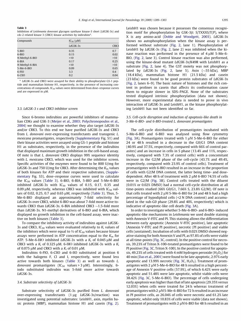
Table 3Inhibition of Leishmania donovani glycogen synthase kinase-3 short (LdGSK-3s) andcdc-2 related kinase 3 (CRK3) kinase activities by indirubinsa.
Compounds IC50 (lM)
LdGSK-3s CRK3
5-BIO 0.35 0.76-BIO 0.15 0.02N-Methyl-6-BIO >10 >106-BIA 0.17 0.25N-Methyl-6-BIA >10 >105-Me-6-BIO 0.09 0.656-FIO 3.3 0.46-ClIO 0.2 0.04
a LdGSK-3s and CRK3 were assayed for their ability to phosphorylate GS-1 pep-tide and mammalian histone H1, respectively, in the presence of increasing con-centrations of compounds. IC50 values were determined from dose–response curvesand are expressed in lM.
E. Xingi et al. / International Journal for Parasitology 39 (2009) 1289–1303 1295
3.3. LdGSK-3 s and CRK3 inhibitor screen
Since 6-bromo indirubins are powerful inhibitors of mamma-lian CDKs and GSK-3 (Meijer et al., 2003; Polychronopoulos et al.,2004) we thought to examine whether they also target LdGSK-3sand/or CRK3. To this end we have purified LdGSK-3s and CRK3from L. donovani over-expressing transfectants and transgenic L.mexicana promastigotes, respectively (Supplementary Fig. S1) andtheir kinase activities were assayed using GS-1 peptide and histoneH1 as substrates, respectively, in the presence of the indirubinsthat displayed maximum growth inhibition in the cell-based assay.Of note is that L. donovani CRK3 displays 99% sequence identitywith L. mexicana CRK3, which was used for the inhibitor screen.Specific activities of the enzymes were found to be 800 U/mg forLdGSK-3s and 750 U/mg for CRK3. After determining the Km valuesof both kinases for ATP and their respective substrates, (Supple-mentary Fig. S3), dose–response curves were used to calculatethe IC50 values (Table 3). 6-BIO, 6-BIA, 5-BIO and 5-Me-6-BIOinhibited LdGSK-3s with IC50 values of 0.15, 0.17, 0.35 and0.09 lM, respectively, whereas CRK3 was inhibited with IC50 val-ues of 0.02, 0.25, 0.7 and 0.65 lM, respectively (Table 3). Thus, 5-Me-6-BIO displayed an approximately 7-fold selectivity forLdGSK-3s over CRK3, whilst 6-BIO was about 7-fold more active to-wards CRK3 than LdGSK-3s. 6-BIA inhibited CRK3 �1.5-fold morethan LdGSK-3s. N1-methyl derivatives of 6-BIO and 6-BIA, whichdisplayed no growth inhibition in the cell-based assay, were inac-tive on both kinases (Table 3).
To compare the inhibitory activity of indirubins against LdGSK-3s and CRK3, IC50 values were evaluated relatively to Ki values ofthe inhibitors which were equal to ½ of IC50 values because kinaseassays were performed in ATP concentration equal to the Km forATP. 5-Me-6-DIJ inhibited LdGSK-3s with a Ki of 0.045 lM andCRK3 with a Ki of 0.325 lM. 6-DIJ inhibited LdGSK-3s with a Ki
of 0.075 lM and CRK3 with a Ki of 0.01 lM.Indirubins 6-FIO, 6-ClIO and 6-IIO substituted at position 6
with the halogens F, Cl and I, respectively, were found lessactive towards both kinases (Table 3) as well as towards L.donovani promastigotes (IC50 values > 3 lM). Interestingly, 6-iodo substituted indirubin was 5-fold more active towardsLdGSK-3s.
3.4. Substrate selectivity of LdGSK-3s
Substrate selectivity of LdGSK-3s purified from L. donovaniover-expressing transfectants (1 lg LdGSK-3s/reaction) wasinvestigated using potential substrates: LeishH1, axin, myelin ba-sic protein (MBP), mammalian histone H1 and casein (Fig. 2).
LeishH1 was chosen because it possesses the consensus recogni-tion motif for phosphorylation by GSK-3b: S/TXXXS/T(P), whereX is any amino-acid (Doble and Woodgett, 2003). LdGSK-3sshows no autophosphorylation when the kinase assay is per-formed without substrate (Fig. 2, lane 1). Phosphorylation ofLeishH1 by LdGSK-3s (Fig. 2, lane 2) was inhibited when the ki-nase reaction was performed in the presence of 4 lM 5-Me-6-BIO, (Fig. 2, lane 3). Control kinase reaction was also performed,using the kinase-dead mutant LdGSK-3s/K49R with LeishH1 as asubstrate (Fig. 2, lane 4). The GST moiety was not phosphory-lated by LdGSK-3s (Fig. 2, lane 5). Axin (�55 kDa), MBP(18.4 kDa), mammalian histone H1 (21.5 kDa) and casein(23 kDa) were found to be good protein substrates of LdGSK-3s(Fig. 2, lanes 6–9). The basic nature of histones and the rich con-tent in prolines in casein that affects its conformation causethem to migrate slower in SDS–PAGE. None of the substratestested displayed intrinsic phosphorylation (data not shown).However, more experimental data is needed to prove in vivointeraction of LdGSK-3s and LeishH1, as the kinase phosphorylat-ing LeishH1 has not been identified so far.
3.5. Cell-cycle disruption and induction of apoptosis-like death in5-Me-6-BIO- and 6-BIO-treated L. donovani promastigotes
The cell-cycle distribution of promastigotes incubated with5-Me-6-BIO and 6-BIO was analysed using flow cytometry(Fig. 3A). Promastigotes treated with 1 lV 5-Me-6-BIO for either24 or 48 h resulted in a decrease in the G0/G1 DNA content(40.9% and 37.5%, respectively, compared with 66% of control par-asites) and an increase in cells in S phase (13.4% and 11%, respec-tively, compared with 7.3% of control cells) with a concomitantincrease in the G2/M phase of the cell-cycle (43.7% and 49.4%,respectively, compared with 23.9% of control cells). Treatment ofpromastigotes with 6-BIO resulted in an increase in the proportionof cells with G2/M DNA content, the latter being time- and dose-dependent. After 48 h of treatment with 2 lM 6-BIO 74.5% of cellswere in G2/M (Fig. 3B). Control cells treated with the diluent(0.01% or 0.02% DMSO) had a normal cell-cycle distribution at alltime-points studied (66% G0/G1, 7.66% S, 23.8% G2/M). Of note isthat cells treated with 2 lV 5-Me-6-BIO for 24 and 48 h had a highpercentage of hypodiploid cells (<2 N DNA content) and accumu-lated in the sub-G0 phase (29.8% and 40%, respectively) which isindicative of apoptotic-like cell death (Fig. 3A).
In order to investigate whether 5-Me-6-BIO- and 6-BIO- inducedapoptotic-like mechanisms in Leishmania we used double stainingwith Annexin V-FITC and PI. This staining allows the differentiationbetween early apoptotic (Annexin V-FITC positive), late apoptotic(Annexin V-FITC and PI positive), necrotic (PI positive) and viablecells (unstained). Incubation of cells with 0.02% DMSO showed neg-ative staining for both Annexin V and PI, as 97.8% of cells were viableat all time-points (Fig. 3C, control). In the positive control for necro-sis, 39.23% of Triton X-100-treated promastigotes were found to bePI positive (Fig. 3C, Triton X-100). In the positive control for apopto-sis, 49.23% of cells treated with 4 mM hydrogen peroxide (H2O2) for40 min (Das et al., 2001) were found to be late apoptotic, 2.97% earlyapoptotic and 13.99% necrotic (Fig. 3C, H2O2). Treatment of prom-astigotes with 2 lV 5-Me-6-BIO for 48 h resulted in a high percent-age of Annexin V positive cells (57.9%), of which 6.42% were earlyapoptotic and 51.48% were late apoptotic, whilst viable cells were38.42% (Fig. 3C, 5-Me-6-BIO). The percentage of cells undergoingearly apoptosis was higher than that of late apoptosis (29.35% versus12.03%) when cells were treated for 24 h whereas treatment ofpromastigotes with 2 lV5-Me-6-BIO for 72 h resulted in an increasein PI positive cells, as 26.04% of cells were necrotic and 61.2% lateapoptotic, whilst only 10.83% of cells were viable (data not shown).Treatment of promastigotes with 2 lV 6-BIO for 48 h resulted in an
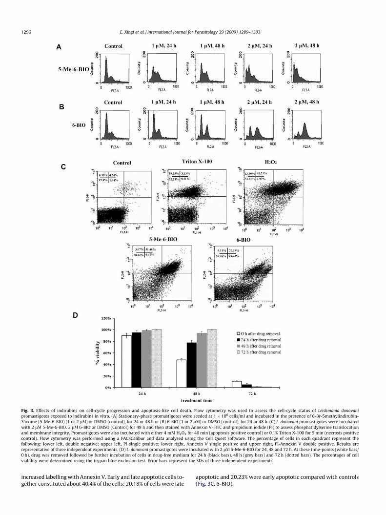
Fig. 3. Effects of indirubins on cell-cycle progression and apoptosis-like cell death. Flow cytometry was used to assess the cell-cycle status of Leishmania donovanipromastigotes exposed to indirubins in vitro. (A) Stationary-phase promastigotes were seeded at 1 � 106 cells/ml and incubated in the presence of 6-Br-5methylindirubin-30oxime (5-Me-6-BIO) (1 or 2 lV) or DMSO (control), for 24 or 48 h or (B) 6-BIO (1 or 2 lV) or DMSO (control), for 24 or 48 h. (C) L. donovani promastigotes were incubatedwith 2 lV 5-Me-6-BIO, 2 lV 6-BIO or DMSO (Control) for 48 h and then stained with Annexin V-FITC and propidium iodide (PI) to assess phosphatidylserine translocationand membrane integrity. Promastigotes were also incubated with either 4 mM H2O2 for 40 min (apoptosis positive control) or 0.1% Triton X-100 for 5 min (necrosis positivecontrol). Flow cytometry was performed using a FACSCalibur and data analysed using the Cell Quest software. The percentage of cells in each quadrant represent thefollowing: lower left, double negative; upper left, PI single positive; lower right, Annexin V single positive and upper right, PI-Annexin V double positive. Results arerepresentative of three independent experiments. (D) L. donovani promastigotes were incubated with 2 lV 5-Me-6-BIO for 24, 48 and 72 h. At these time-points (white bars/0 h), drug was removed followed by further incubation of cells in drug-free medium for 24 h (black bars), 48 h (grey bars) and 72 h (dotted bars). The percentages of cellviability were determined using the trypan blue exclusion test. Error bars represent the SDs of three independent experiments.
1296 E. Xingi et al. / International Journal for Parasitology 39 (2009) 1289–1303
increased labelling with Annexin V. Early and late apoptotic cells to-gether constituted about 40.4% of the cells: 20.18% of cells were late
apoptotic and 20.23% were early apoptotic compared with controls(Fig. 3C, 6-BIO).

Fig. 4. Cell and nuclear morphology of Leishmania donovani promastigotes exposed to indirubins. Leishmania donovani promastigotes were incubated with 2 lM 6-Br-5methylindirubin-30oxime (5-Me-6-BIO), 2 lM 6-BIO or 0.02% DMSO (control) for 24 and 48 h, in vitro, and then fixed and stained with propidium iodide. Confocalmicrographs are representative of three independent experiments. Scale bar 4 lm. N, nucleus; K, kinetoplast; white arrows indicate condensed nuclei; blue arrows indicatedisintegrated nuclei.
E. Xingi et al. / International Journal for Parasitology 39 (2009) 1289–1303 1297
To further study whether the observed effect of indirubins wasdue to apoptosis-like death we monitored morphological and nu-clear changes by confocal microscopy. Control cells displayed anormal elongated morphology with two discrete stained organ-elles, the nucleus and the kinetoplast (Fig. 4, control). Promastig-otes exposed to 5-Me-6-BIO for 24 h showed rounded forms, cellshrinkage and variations in the length of their flagella as well asnuclear changes characteristic of apoptosis-like death; apoptoticnuclei were identified by their bright red fluorescence, which in-cluded a certain degree of condensation of nuclear chromatin in36% of cells and breakdown of the nuclear material in 41% of cells(Fig. 4, 5-Me-6-BIO, 24 h). At the 48 h-time point, 78% of cellsexhibited a totally fragmented nucleus (Fig. 4, 5-Me-6-BIO, 48 h).Formation of zoids was not observed (Grant et al., 2004). Treat-ment with 2 lM of 6-BIO for 24 h had less pronounced morpholog-ical alterations. It resulted in cells with either a normalmorphology with a discrete kinetoplast and a nucleus (approxi-mately 38% of cells) or in rounded-shaped with short flagella andcondensed nuclear chromatin (�62%) (Fig. 4, 6-BIO, 24 h). Aftertreatment with 6-BIO for 48 h, the majority of parasites (75%) dis-played an aberrant morphology, with round body shape and shortflagella of which 36% displayed nuclear condensation and 39% nu-clear fragmentation (Fig. 4, 6-BIO, 48 h).
Since the cellular effects induced by 5-Me-6-BIO were morepronounced than those of 6-BIO we investigated whether theywere reversible after drug removal. To this end the recovery of cellsfollowing exposure to 5-Me-6-BIO for 24, 48 and 72 h was assessed24, 48 and 72 h after the drug removal. Control cells treated with
0.02% DMSO were viable at all time-points tested (100% viability).As shown in Fig. 3D treatment of cells with 5-Me-6-BIO for 24 hfollowed by incubation with fresh medium up to 72 h resulted infull recovery of cells. After 48 h of treatment, about 48% of cellswere viable. Further incubation in fresh medium for 24 h resultedin an increase in the percentage of viable cells (77.7%). At the 48 hand 72 h time-points, a full recovery of the cells was observed. Incontrast, incubation for 72 h with 5-Me-6-BIO resulted in about89% dead cells. Removal of the drug resulted in irreversiblecytotoxicity.
3.6. LdGSK-3s over-expression in L. donovani counteracts 5-Me-6-BIO-and 6-BIO-induced growth inhibition, cell-cycle progression andapoptosis-like death
To investigate whether LdGSK-3s is the intracellular target of 5-Me-6-BIO and whether all the observed phenotypes in the pres-ence of 5-Me-6-BIO could be attributed to inhibition of this kinase,we have generated transgenic L. donovani promastigotes over-expressing LdGSK-3s and compared their susceptibility to 5-Me-6-BIO with that of control transfectants bearing the plasmid alone.We also investigated whether over-expression of LdGSK-3s af-fected the growth inhibitory effect of 6-BIO. As a control, we gen-erated transgenic promastigotes overexpressing LdGSK-3s/K49R,which was a kinase-dead mutant as confirmed by kinase assays(Fig. 2, lane 4). The mutation of the catalytic residue Lys 49 toArg was designed based on the homology model of LdGSK-3s andon the widely used mutation of Lys 85 to Arg or Ala of mammalian

Fig. 5. Over-expression of Leishmania donovani glycogen synthase kinase-3 short (LdGSK-3s) counteracts the effects of 6-Br-5methylindirubin-30oxime (5-Me-6-BIO) and 6-BIO on Leishmania donovani promastigotes. (A) Western blot analysis of L. donovani sat, sat-LdGSK-3s/K49R and sat-LdGSK-3s promastigotes (107/lane) probed with His-probe,anti-LdGSK-3s or anti-LinINO1 antibodies. The intensity of the bands was analysed with the use of the Alpha Imager Software and the fold-over-expression estimated. (B)Growth inhibition of L. donovani sat (grey symbols, dotted lines), sat-LdGSK-3s/K49R (solid symbols) and sat-LdGSK-3s (open symbols) transfectants treated with different 5-Me-6-BIO concentrations after 24, 48 and 72 h of incubation. Results are depicted from four independent experiments performed in duplicate. (C) Flow cytometry analysis ofL. donovani sat, sat-LdGSK-3s/K49R and sat-LdGSK-3s transfectants incubated in the presence of 0.02% DMSO (control) or 2 lV 5-Me-6-BIO or 2 lV 6-BIO for 48 h. Results areindicative of three independent experiments. (D) Growth inhibition of L. donovani sat (grey symbols, dotted lines), sat-LdGSK-3s/K49R (solid symbols) and sat-LdGSK-3s (opensymbols) transfectants treated with different 6-BIO concentrations after 24, 48 and 72 h of incubation. Results are depicted from four independent experiments performed induplicate.
1298 E. Xingi et al. / International Journal for Parasitology 39 (2009) 1289–1303
GSK-3b, which results in a kinase-dead protein (He et al., 1995).Over-expression of LdGSK-3s and expression of LdGSK-3s/K49R inthe LdGSK-3s and LdGSK-3s/K49R transfectants was confirmed byimmunoblotting using both a His-probe antibody and the anti-LdGSK-3s antibody (Fig. 5A). The LinINO1 antibody was used as aloading control. Scanning densitometry showed that the level ofexpression of LdGSK-3s in LdGSK-3s and LdGSK-3s/K49R transfec-tants was about 2-fold higher in comparison with sat transfectants(Fig. 5A). Expression of the kinase-dead mutant did not cause anyapparent changes in parasite growth or morphology.
The susceptibility of stationary-phase L. donovani sat, sat-LdGSK-3s and sat-LdGSK-3s/K49R transfectants to increasingconcentrations of 5-Me-6-BIO was assessed after 24, 48 and72 h of treatment by cell counting. The IC50 of 5-Me-6-BIO after24, 48 and 72 h of treatment of the sat-transfectants was1.5 ± 0.2, 1.25 ± 0.1 and 1.2 ± 0.1 lM, respectively, which arecomparable with the IC50 against wild-type promastigotes (Table1), whereas its respective IC50 for the LdGSK-3s over-expressingtransfectants was 3.6 ± 0.3, 3.2 ± 0.2 and 2.8 ± 0.2 lM (Fig. 5B).As expected, the sat-LdGSK-3s/K49R transfectants were inhibitedby 5-Me-6-BIO with IC50 values of 1.7 ± 0.1 lM after 24 h,1.4 ± 0.1 lM after 48 h and 1.3 ± 0.05 lM after 72 h, which arecomparable with the respective IC50 values against sat-transfec-tants (Fig. 5B).
Treatment of the LdGSK-3s over-expressing transfectants with6-BIO resulted in a clear decrease in their sensitivity, with IC50 val-ues of 4.8 ± 0.5 lM after 24 h, 2.2 ± 0.4 lM after 48 h of treatmentand 1.57 ± 0.3 lM after 72 h of treatment, which were approxi-mately 2-fold higher compared with IC50 values of sat and sat-LdGSK-3s/K49R transfectants (Fig. 5D). Sat and sat-LdGSK-3s/K49R transfectants treated with different 6-BIO concentrations dis-played growth inhibition with approximately the same IC50 valuesas L. donovani wild-type promastigotes. Sat transfectants wereinhibited with IC50 values of 2.75 ± 0.45 lM after 24 h of treat-ment, 1.3 ± 0.3 lM after 48 h of treatment and 0.78 ± 0.25 lM after72 h of treatment and sat-LdGSK-3s/K49R transfectants wereinhibited with IC50 values of 2.9 ± 0.3 lM after 24 h, 1.3 ± 0.2 lMafter 48 h and 0.8 5 ± 0.2 lM after 72 h (Fig. 5D).
Flow cytometry analysis of the DNA content of control L. dono-vani sat, sat-LdGSK-3s and sat-LdGSK-3s/K49R over-expressingtransfectants, treated with 0.02% DMSO, showed that cells had anormal cell-cycle distribution: 67.9% G1, 6.9% S, 23.2% G2(Fig. 5C, control). Interestingly, sat-LdGSK-3s promastigotes incu-bated with 2 lV 5-Me-6-BIO for 48 h had a normal cell-cycle dis-tribution: 62% G1, 5.45% S and 30.5% G2 (Fig. 5C, 5-Me-6-BIO),whereas 40% of sat and sat-LdGSK-3s/K49R transfectants werehypodiploid and accumulated in the sub-G0 phase (Fig. 5C), aswas observed with wild-type promastigotes (Fig. 3A).

E. Xingi et al. / International Journal for Parasitology 39 (2009) 1289–1303 1299
However, as shown in Fig. 5C, LdGSK-3s over-expressing prom-astigotes treated with 6-BIO displayed a less pronounced increasein G2/M (41.7% compared with 74.5% in 6-BIO-treated wild-typeparasites) and a less pronounced decrease in G0/G1 (46% comparedwith 15.8% in 6-BIO-treated wild-type parasites) (Fig. 5C, sat-LdGSK-3s). In contrast, sat transfectants incubated with 2 lV 6-BIO for 48 h were comparable with wild-type promastigotes andarrested at G2/M (74.9% compared with 23.2% of control), with asignificant decrease in G0/G1 (19.3% compared with 67.9% of con-trol) (Fig. 5C, 6-BIO). Similar results with the latter were obtainedfor the sat-LdGSK-3s/K49R transfectants which had the followingcell-cycle distribution: 21.8% G0/G1, 4.4% S and 71.7% G2/M.
Since LdGSK-3s over-expression resulted in a significantly re-duced growth inhibition and a normal cell-cycle distribution upon5-Me-6-BIO-treatment, we investigated whether LdGSK-3s over-expression affected apoptosis-like death using the terminal deoxy-nucleotidyltransferase-mediated dUTP nick end labelling (TUNEL)assay, which detects apoptosis at a single-cell level. Control cellstreated with 0.02% DMSO (sat, sat-LdGSK-3s or sat-LdGSK-3s/K49R) containing intact genomic DNA were not stained, (Fig. 6,control). Promastigotes treated with 4 mM H2O2 for 6 h, servedas a TUNEL positive control, as about 99% of cells showed positivenuclear staining (Fig. 6, H2O2). Sat transfectants exposed to 2 lV 5-Me-6-BIO for 48 h were about 68% TUNEL positive and their mor-phology was dramatically affected in comparison to the normalelongated morphology of control cells (Fig. 6, sat/5-Me-6-BIO).Treated cells displayed an aberrant morphology, with round bodyshape and cell shrinkage. Sat-LdGSK-3s transfectants treated with2 lV 5-Me-6-BIO for 48 h were not positive for TUNEL reactivity,only a background staining of about 3% was detected and theirmorphology was not affected (Fig. 6, sat-LdGSK-3s/5-Me-6-BIO).Sat-LdGSK-3s/K49R transfectants were about 70.4% TUNEL positive(Fig. 6, sat-LdGSK-3s/K49R/5-Me-6-BIO).
The contribution of LdGSK-3s in the apoptosis-like deathobserved in 6-BIO treated promastigotes was also studied usingthe over-expressor lines. Whereas sat transfectants exposed to2 lV 6-BIO for 48 h were �50% TUNEL positive and their morphol-ogy was dramatically affected, LdGSK-3s over-expressing transfec-tants were resistant to the effects of 6-BIO and displayed a muchmilder phenotype. Only 20% of the cells were TUNEL positive andtheir morphology was not significantly affected compared withcontrol cells (Fig. 6, sat/6-BIO and sat-LdGSK-3s/6-BIO, respec-tively). As expected, sat-LdGSK-3s/K49R transfectants were about52.9% TUNEL positive (Fig. 6, sat-LdGSK-3s/K49R/6-BIO).
3.7. Structure activity relationships studies of indirubin-leishmanialkinases interactions using molecular simulations
Biological results imply that indirubins inhibit leishmanial ki-nases. Interestingly, the selectivity observed for 6-substitutedindirubins towards GSK-3 with respect to the CDKs in human is re-versed in the case of Leishmania and the homologous kinases (GSK-3 and CRK3) with the exception of the 6-iodo as well as the bisub-stituted 5-Me-6Br analogues. Whilst indirubins potently inhibitthe leishmanial GSK-3s (5-Me-6-BIO with an IC50 = 0.09 lM and6BIO with an IC50 = 0.150 lM), they are not as efficient as in thecase of the human homologue (5-Me-6-BIO with anIC50 = 0.006 lM and 6-BIO with an IC50 = 0.005 lM) (Meijer et al.,2003; Polychronopoulos et al., 2004).
Both pairs of homologous kinases are highly similar and the ob-served differences in affinity could possibly be explained by thekey residue differences of the binding cavity. In order to obtain in-sight in the inhibitor–protein interactions, we built homologymodels of the parasite kinases (Supplementary Data S1). Despitethe fact that important residues of the leishmanial GSK-3s seemto be well conserved (Supplementary Fig. S2), there were two ma-
jor differences between the two kinases located in the bindingpocket: (i) the replacement of Gln185hGSK-3b by His155LGSK-3s inthe sugar-binding region, and (ii) the replacement of the ‘‘gate-keeper” Leu132hGSK-3b by Met100LGSK-3s. The ‘‘gatekeeper” residuecontrols access to a hydrophobic cavity of the binding pocket andis considered as a selectivity determinant of most ATP competitivekinase inhibitors (Bohmer et al., 2003). In the majority of the mem-bers of the GSK-3 family (CMGC III), the gatekeeper is a leucine, ex-cept for MCK-1 kinase which has a methionine (Hanks and Quinn,1991). However, a methionine is present in Leishmania, T. bruceiand P. falciparum GSK-3s, (Supplementary Fig. S2).
Docking calculations were performed in order to study thebinding mode/interactions of indirubins in the binding cavity ofeach kinase. In each case the inhibitor was anchored at the kinasebackbone through the formation of three hydrogen bonds in theusually observed adenine type of interaction (Fig. 7A), whilst thesubstituent of position 6 was positioned in the hydrophobic cavityformed by the sidechain of the gatekeeper residue interacting withit. In human GSK-3b the leucine gatekeeper can form only hydro-phobic interactions with the position 6 substituent of indirubin.However, in the parasite kinase the mode of interactions accom-modated by the methionine gatekeeper is more complicated,resulting in a larger entropic and desolvation cost upon inhibitorbinding (Supplementary Data S1). Such a net effect for the replace-ment of the leucine gatekeeper to a methionine could be consid-ered as unfavourable for binding affinity, accounting for the lossof binding affinity in a common trend for all indirubins tested,which is in consistency with IC50 results obtained from kinaseassays.
The higher affinity for CRK3 (reversal of selectivity with respectto the human kinases) demonstrated by 6-substituted indirubinstested with the exception of 6-IIO, 5-Me-6-BIO and partially of 6-BIA compared to the affinity for LdGSK-3s could be explained bythe formation of a hydrogen bond between Tyr101CRK3 andGlu103CRK3 (Fig. 7B), which is not possible in the human CDK2homolog. The influence of this bonding interaction on the cavitysize and subsequently on the ligand affinity could explain the ob-served gain of selectivity of 6-BIO towards CRK3. The above holdswith the exception of 6-IIO, the bisubstituted 5-Me-6-BIO and theacetoxime 6-BIA, for which energy optimisation calculations dem-onstrated that the presence of the bulky substituent provoked adisplacement of the ligand and the pair of residues Tyr101-Glu103 (Fig. 7C) resulting in less favorable interactions and lossof affinity.
All aforementioned structural observations are in accordancewith previous studies showing that minor differences of the kinasebinding cavity elements induce major variations in affinity andshould be taken into account in designing new selective inhibitorsof the leishmanial GSK-3s and CRK3. One possible route of selec-tively improving affinity towards the parasite GSK-3 is by takingadvantage of the differential presence of the proton acceptingHis155LGSK-3 (instead of Gln185hGSK-3b of human). The replacementor extension of the oxime by a group with the potential to formattractive albeit selective interactions with the sidechain ofHis155 LGSK-3 would increase affinity towards the parasite protein.Combined with the obvious preference of LdGSK-3 for bisubstitut-ed or generally bulkier substituents with regard to CRK3, a moder-ate selectivity improvement can be achieved, resulting in anincrease of parasite killing whilst reducing toxicity to human cells.
4. Discussion
Herein we showed that three 6-bromo substituted indirubins,6-BIO, 6-BIA and 5-Me-6-BIO were powerful inhibitors of both L.donovani promastigote and intracellular amastigote growth. Leish-mania donovani axenic amastigotes were also inhibited by the

Fig. 6. DNA fragmentation in Leishmania donovani promastigotes incubated with indirubins. Leishmania donovani sat, LdGSK-3s kinase-dead mutant (sat-LdGSK-3s/K49R) andsat-LdGSK-3s transfectants were incubated with 0.02% DMSO (negative control), 2 lV 6-Br-5methylindirubin-30oxime (5-Me-6-BIO), 2 lV 6-BIO or 4 mM H2O2 (apoptosispositive control) and then subjected to the terminal deoxynucleotidyltransferase-mediated dUTP nick end labelling assay (TUNEL). Cells were visualised under a Zeissfluorescence microscope at 120� magnification. The experiment was performed three times.
1300 E. Xingi et al. / International Journal for Parasitology 39 (2009) 1289–1303
three indirubins with IC50 values61 lM, a finding that further sup-ports that indirubin-induced growth inhibition of intracellular
amastigotes is mediated through parasite-kinase(s) inhibitionand not through inhibition of the host-kinase(s).

Fig. 7. Docking calculations showing the binding mode/interactions of indirubins in the binding cavity of each kinase. (A) A superposition of the crystal structure of humanglycogen synthase kinase-3 (GSK-3) active site complexed with 6-Br-indirubin-30oxime (6-BIO) (green) and the complex of Leishmania donovani glycogen synthase kinase-3short (LdGSK-3s) with 6-BIO resulting from docking calculations (turquoise). Hydrogen bonds are depicted as yellow dotted lines. Residues within the active site which differbetween human and L. donovani GSK-3s are annotated, with the most important for 6-BIO affinity being the gatekeeper mutation of leucine132 in the human tomethionine100 in the parasite protein. (B) Overlay of the crystal structure of cyclin-dependent kinase 2 (CDK2) complexed with indirubin-5-sulphonate (turquoise) and thecdc-2 related kinase 3 (CRK3) homology model (orange). The double mutation of phe82 in CDK2 to tyr101 in CRK3 and of his84 in CDK2 to glu103 in CRK3 results in ahydrogen bond between tyrosine and glutamate (shown in yellow) that translates the paired sidechains towards the cavity reducing its volume and offering a rigid partner forstacking or charge dipole stabilizing interactions with the extended aromatic scaffold of indirubins to form. (C) Ligands 6-BIO (green) and 5-Me-6-BIO (orange) in the CRK3binding cavity as resulted from simple energy minimizations. Visible are the displacements of the bisubstituted 5-Me-6-BIO relative to 6-BIO and of the tyrosine-glutamatebonded pair.
E. Xingi et al. / International Journal for Parasitology 39 (2009) 1289–1303 1301
The adapted Alamar blue assay allows the rapid and easyscreening of the antileishmanial activity of compounds in 96-wellformat, although it is not very quantitative for measuring intra-macrophage Leishmania growth compared with luciferase-express-ing recombinant parasites (Roy et al., 2000). Also it does notmeasure the number of amastigotes at the point of lysis. However,contrary to Giemsa staining it takes into account only viable cells.
Since 6-bromo substitution on the indirubin backbone enhancesthe selectivity for mammalian GSK-3 over CDKs (Meijer et al.,2003) we investigated whether 6-BIO, 6-BIA and 5-Me-6-BIO tar-get GSK-3 in Leishmania and studied their selectivity over CRK3.To this end we identified and characterised one of the two GSK-3forms in L. donovani, namely LdGSK-3s, and found that its expres-sion pattern was comparable in logarithmic and stationary-phasepromastigotes, but it was about 3-fold down-regulated in amastig-otes, consistent with recent findings on LdGSK-3s expression in L.donovani axenic amastigotes (Rosenzweig et al., 2008). In addition,LdGSK-3s which had cytosolic and flagellar localisation in logarith-mic-phase promastigotes, displayed nuclear translocation in sta-tionary-phase promastigotes. In mammalian cells, GSK-3b is alsopredominately in the cytosol although under proapoptotic stimuli,a portion of GSK-3b is found within the nucleus (Meares and Jope,2007). LdGSK-3s translocation to the nucleus in stationary-phasepromastigotes, thought to be arrested in G1 phase of the cell-cycle(Wiesgigl and Clos, 2001), may reflect a role for LdGSK-3s in G1,consistent with accumulation of parasites in G1 when LdGSK-3sis inhibited with 5-Me-6-BIO. The observed differences in thelocalisation and expression level of LdGSK-3s may reflect divergentroles played by LdGSK-3s in the two parasite stages. It could bespeculated that in the intracellular amastigotes the function ofLdGSK-3s may be linked, among others, to their response and adap-
tation to stress conditions i.e. pH and temperature changes (Rich-ard et al., 2005). The finding that the level of inhibition ofpromastigotes and amastigotes by 5-Me-6-BIO is the samealthough LdGSK-3s is 3-fold less in amastigotes may suggest thatLdGSK-3s activity is higher in the latter or that 5-Me-6-BIO mayalso target other kinases in this stage.
Inhibitor screen assays against LdGSK-3s and CRK3 showed that5-Me-6-BIO, which is a 50-fold selective inhibitor of mammalianGSK-3 over CDK1/Cyclin B (Meijer et al., 2003; Polychronopouloset al., 2004) displayed an approximately 7-fold selectivity forLdGSK-3s over CRK3. However, 6-BIO was about 7-fold more activetowards CRK3 than LdGSK-3s, although it is a mammalian GSK-3selective inhibitor, with 64-fold less potency towards CDK1/CyclinB (Meijer et al., 2003). Molecular docking of the compounds inhGSK-3 and CDK1 active sites compared with LdGSK-3s andCRK3 support the higher inhibitory activity of 5-Me-6-BIO towardsLdGSK-3s compared with CRK3 and the lower inhibitory activity of6-BIO towards LdGSK-3s compared with that against its mamma-lian counterpart.
5-Me-6-BIO and 6-DIJ displayed a disparity between cellularactivity and enzyme activity (Ki values) (22- to 27-fold for 5-Me-6-BIO and 75- to 80-fold for 6-BIO), which can be attrib-uted to: (i) the ATP concentration in the kinase assays, that isseveral fold lower than the intracellular concentration, whichis in the mM range, (ii) the bioavailability of the inhibitors (cellpermeability of the compounds, rate of inhibitor efflux by cellefflux pumps), (iii) possible in vivo phosphatase activity, (iv)the intracellular concentration of the target kinase, (v) the pres-ence of the LdGSK-3l isoform, and (vi) the need for total inhi-bition of the enzyme to get cellular effect (Knight and Shokat,2005).

1302 E. Xingi et al. / International Journal for Parasitology 39 (2009) 1289–1303
We next investigated the effects on cell-cycle progression andthe death process induced by 5-Me-6-BIO treatment, using a num-ber of different techniques. Its effect on parasite growth appearedto be more dose- than time-dependent, as the IC50 values did notsignificantly vary with the incubation time. However, 5-Me-6-BIO treatment affected the recovery potential of treated cells afterremoval of the drug, as cells were able to recover after 48 h oftreatment, whereas 72 h of treatment caused an irreversible inhi-bition of cell growth.
In an effort to elucidate whether 5-Me-6-BIO targets LdGSK-3sin vivo and the potential role of LdGSK-3s in cell-cycle progressionand apoptosis-like death a sat-LdGSK-3s over-expressor mutantand a cell line expressing a kinase-dead mutant sat-LdGSK-3s/K49R were generated. Cells that over-express LdGSK-3s (about 2-fold) were about 2-fold less susceptible to growth inhibition thansat-LdGSK-3s/K49R and sat transfectants at all time-points, indi-cating that the observed growth inhibition was closely associatedwith inhibition of LdGSK-3s activity by 5-Me-6-BIO. In additionthese results imply that the wild-type kinase should be inhibitedby 5-Me-6-BIO with an IC50 value comparable with that of theHis-tagged LdGSK-3s. Also, the 2-fold increase in LdGSK-3s expres-sion in sat-LdGSK-3s transfectants completely reversed the cell-cy-cle disruption effect of 5-Me-6-BIO and abolished the induction ofapoptosis-like death. However, the LdGSK-3s/K49R expression re-sulted in similar phenotypes with those of the sat transfectants.The results provide strong evidence that LdGSK-3s is the intracellu-lar target of 5-Me-6-BIO and suggest the direct or indirect involve-ment of LdGSK-3s in cell-cycle control as well as in pathwaysleading to apoptosis-like death. Although there is evidence thatapoptosis-like death occurs in Leishmania (Das et al., 2001) thepathways and proteins involved remain to be elucidated. GSK-3is known to modulate apoptosis in mammalian cells by regulatingthe apoptotic pathways (Beurel and Jope, 2006). Therefore com-mon pathways may exist between Leishmania and mammaliancells in regulating apoptotic signalling pathways through GSK-3.
In contrast to 5-Me-6-BIO, 6-BIO induced a time-dependentgrowth inhibition accompanied with a dose- and time-dependentaccumulation of cells in G2/M. Moreover, 6-BIO induced apoptosis-like death in a lower proportion of promastigotes compared with5-Me-6-BIO and this death process progressed more slowly in para-sites exposed to 6-BIO. These differences between the cellular effectsinduced by 6-BIO compared with 5-Me-6-BIO suggest that in vivothe two indirubins may target different kinases and/or pathways.
The observation that over-expression of LdGSK-3s only partiallyreversed the effect of 6-BIO is not unexpected, since in vitro 6-BIOpreferentially inhibits CRK3. Moreover, the phenotype of prom-astigotes incubated with 6-BIO, especially the accumulation ofcells in G2/M, is consistent with inhibition of CRK3, which is essen-tial for cell-cycle progression at the G2/M phase transition (Grantet al., 1998; Hassan et al., 2001) and implies that CRK3 may bethe main intracellular target of 6-BIO. Although 6-BIO was a moreeffective inhibitor of CRK3, and despite the observed phenotypebeing consistent with CRK3-inhibition, the fact that LdGSK-3sover-expression partially reversed promastigote G2/M arrest andpartially protected cells from 6-BIO induced apoptosis-like death,implies that 6-BIO may also target LdGSK-3s in the parasite wherethe level of expression of the two kinases is not known. Since 6-BIOarrests promastigotes in G2/M phase of the cell-cycle, this maymean that LdGSK-3s also plays a role in G2/M phase transition,although this is difficult to reconcile with the results for 5-Me-6-BIO. Alternatively, over-expression of LdGSK-3s may influence 6-BIO activity by a non-specific mechanism (i.e. lower proportionof 6-BIO available for binding to and inactivating CRK3).
In conclusion, the complete reversal of the cellular effects in-duced by 5-Me-6-BIO in the over-expressing parasites strongly im-plies that LdGSK-3s is the main target of 5-Me-6-BIO and suggests
a potential role for LdGSK-3s in cell-cycle progression and in apop-tosis-like death. Moreover, the dramatic effect of LdGSK-3s inhibi-tors on Leishmania, especially the intramacrophage amastigotestage, suggests that LdGSK-3s has potential as a drug target inthese parasites. Future work would be required to develop para-site-selective inhibitors that do not target host GSK-3 since its inhi-bition may affect the balance between Th1 and Th2 rsponses(Ohtani et al., 2008). In addition, RNA interference studies of T. bru-cei GSK-3 lead to similar cellular phenotypes, such as growth inhi-bition and altered parasite morphology (Ojo et al., 2008), to thatcaused by GSK-3 inhibitors in Leishmania. Importantly, the very re-cent validation of TbruGSK-3 as a drug target for this protozoanparasite (Ojo et al., 2008), reinforces our claim that GSK-3 couldconstitute a trans-trypanosomatid as well as trans-protozoan tar-get, including P. falciparum and that it should be exploited foranti-protozoan drug development.
Acknowledgements
This work was supported by HPI (E.X. PhD fellowship), by theGeneral Secretariat of Research and Technology of Greece; PENEDprogramme (V.M.), University of Athens programme Kapodistriasand by the FP6-2002-Life Sciences and Health, PRO-KINASE Re-search Project (L.M.). We thank our colleagues Dr. Christos Hara-lambous for the generation of the phylogenetic tree and GeorgiaKonidou for technical assistance.
Appendix A. Supplementary data
Supplementary data associated with this article can be found, inthe online version, at doi:10.1016/j.ijpara.2009.04.005.
References
Alvar, J., Yactayo, S., Bern, C., 2006. Leishmaniasis and poverty. Trends Parasitol. 22,552–557.
Barak, E., Amin-Spector, S., Gerliak, E., Goyard, S., Holland, N., Zilberstein, D., 2005.Differentiation of Leishmania donovani in host-free system: analysis of signalperception and response. Mol. Biochem. Parasitol. 141, 99–108.
Beurel, E., Jope, R.S., 2006. The paradoxical pro- and anti-apoptotic actions of GSK3in the intrinsic and extrinsic apoptosis signaling pathways. Prog. Neurobiol. 79,173–189.
Bohmer, F.D., Karagyozov, L., Uecker, A., Serve, H., Botzki, A., Mahboobi, S., Dove, S.,2003. A single amino acid exchange inverts susceptibility of related receptortyrosine kinases for the ATP site inhibitor STI-571. J. Biol. Chem. 278, 5148–5155.
Chang, K.P., 1983. Cellular and molecular mechanisms of intracellular symbiosis inleishmaniasis. Int. Rev. Cytol. Suppl. 14, 267–305.
Croft, S.L., Sundar, S., Fairlamb, A.H., 2006. Drug resistance in leishmaniasis. Clin.Microbiol. Rev. 19, 111–126.
Damiens, E., Baratte, B., Marie, D., Eisenbrand, G., Meijer, L., 2001. Anti-mitoticproperties of indirubin-30-monoxime, a CDK/GSK-3 inhibitor: induction ofendoreplication following prophase arrest. Oncogene 20, 3786–3797.
Das, M., Mukherjee, S.B., Shaha, C., 2001. Hydrogen peroxide induces apoptosis-likedeath in Leishmania donovani promastigotes. J. Cell Sci. 114, 2461–2469.
Davies, T.G., Tunnah, P., Meijer, L., Marko, D., Eisenbrand, G., Endicott, J.A., Noble,M.E., 2001. Inhibitor binding to active and inactive CDK2: the crystal structureof CDK2-cyclin A/indirubin-5-sulphonate. Structure 9, 389–397.
Doble, B.W., Woodgett, J.R., 2003. GSK-3: tricks of the trade for a multi-taskingkinase. J. Cell Sci. 116, 1175–1186.
Dujardin, J.C., 2006. Risk factors in the spread of leishmaniases: towards integratedmonitoring? Trends Parasitol. 22, 4–6.
Frame, S., Cohen, P., Biondi, R.M., 2001. A common phosphate binding site explainsthe unique substrate specificity of GSK3 and its inactivation byphosphorylation. Mol. Cell 7, 1321–1327.
Grant, K.M., Hassan, P., Anderson, J.S., Mottram, J.C., 1998. The crk3 gene ofLeishmania mexicana encodes a stage-regulated cdc2-related histone H1 kinasethat associates with p12. J. Biol. Chem. 273, 10153–10159.
Grant, K.M., Dunion, M.H., Yardley, V., Skaltsounis, A.L., Marko, D., Eisenbrand, G.,Croft, S.L., Meijer, L., Mottram, J.C., 2004. Inhibitors of Leishmania mexicana CRK3cyclin-dependent kinase: chemical library screen and antileishmanial activity.Antimicrob. Agents Chemother. 48, 3033–3042.
Habtemariam, S., 2003. In vitro antileishmanial effects of antibacterial diterpenesfrom two Ethiopian Premna species: P. schimperi and P. oligotricha.. BMCPharmacol. 3, 6.

E. Xingi et al. / International Journal for Parasitology 39 (2009) 1289–1303 1303
Hanks, S.K., Quinn, A.M., 1991. Protein kinase catalytic domain sequence database:identification of conserved features of primary structure and classification offamily members. Methods Enzymol. 200, 38–62.
Hassan, P., Fergusson, D., Grant, K.M., Mottram, J.C., 2001. The CRK3 protein kinaseis essential for cell cycle progression of Leishmania mexicana. Mol. Biochem.Parasitol. 113, 189–198.
He, X., Saint-Jeannet, J.P., Woodgett, J.R., Varmus, H.E., Dawid, I.B., 1995. Glycogensynthase kinase-3 and dorsoventral patterning in Xenopus embryos. Nature 374,617–622.
Hoessel, R., Leclerc, S., Endicott, J.A., Nobel, M.E., Lawrie, A., Tunnah, P., Leost, M.,Damiens, E., Marie, D., Marko, D., Niederberger, E., Tang, W., Eisenbrand, G.,Meijer, L., 1999. Indirubin, the active constituent of a Chinese antileukaemiamedicine, inhibits cyclin-dependent kinases. Nat. Cell Biol. 1, 60–67.
Ilg, T., 2002. Generation of myo-inositol-auxotrophic Leishmania mexicana mutantsby targeted replacement of the myo-inositol-1-phosphate synthase gene. Mol.Biochem. Parasitol. 120, 151–156.
Knight, Z.A., Shokat, K.M., 2005. Features of selective kinase inhibitors. Chem. Biol.12, 621–637.
Laskowski, R., MacArthur, M., Moss, D., Thornton, J., 1991. PROCHECK: a programmto check the stereochemichal quality of protein structures. J. Appl. Crystallogr.26, 283–291.
Meares, G.P., Jope, R.S., 2007. Resolution of the nuclear localization mechanism ofglycogen synthase kinase-3: functional effects in apoptosis. J. Biol. Chem. 282,16989–17001.
Meijer, L., Skaltsounis, A.L., Magiatis, P., Polychronopoulos, P., Knockaert, M., Leost, M.,Ryan, X.P., Vonica, C.A., Brivanlou, A., Dajani, R., Crovace, C., Tarricone, C.,Musacchio, A., Roe, S.M., Pearl, L., Greengard, P., 2003. GSK-3-selective inhibitorsderived from Tyrian purple indirubins. Chem. Biol. 10, 1255–1266.
Meijer, L., Flajolet, M., Greengard, P., 2004. Pharmacological inhibitors of glycogensynthase kinase 3. Trends Pharmacol. Sci. 25, 471–480.
Mikus, J., Steverding, D., 2000. A simple colorimetric method to screen drugcytotoxicity against Leishmania using the dye Alamar Blue. Parasitol. Int. 48,265–269.
Naula, C., Parsons, M., Mottram, J.C., 2005. Protein kinases as drug targets intrypanosomes and Leishmania. Biochim. Biophys. Acta 1754, 151–159.
Ohtani, M., Nagai, S., Kondo, S., Mizuno, S., Nakamura, K., Tanabe, M., Takeuchi, T.,Matsuda, S., Koyasu, S., 2008. Mammalian target of rapamycin and glycogensynthase kinase 3 differentially regulate lipopolysaccharide-inducedinterleukin-12 production in dendritic cells. Blood 112, 635–643.
Ojo, K.K., Gillespie, J.R., Riechers, A.J., Napuli, A.J., Verlinde, C.L., Buckner, F.S., Gelb,M.H., Domostoj, M.M., Wells, S.J., Scheer, A., Wells, T.N., Van Voorhis, W.C., 2008.Glycogen Synthase Kinase 3 is a potential drug target for Africantrypanosomiasis therapy. Antimicrob. Agents Chemother. 52, 3710–3717.
Papageorgiou, F.T., Soteriadou, K.P., 2002. Expression of a novel Leishmania geneencoding a histone H1-like protein in Leishmania major modulates parasiteinfectivity in vitro. Infect. Immun. 70, 6976–6986.
Parsons, M., Worthey, E.A., Ward, P.N., Mottram, J.C., 2005. Comparativeanalysis of the kinomes of three pathogenic trypanosomatids: Leishmaniamajor, Trypanosoma brucei and Trypanosoma cruzi. BMC Genomics 6,127.
Polychronopoulos, P., Magiatis, P., Skaltsounis, A.L., Myrianthopoulos, V., Mikros, E.,Tarricone, A., Musacchio, A., Roe, S.M., Pearl, L., Leost, M., Greengard, P., Meijer,L., 2004. Structural basis for the synthesis of indirubins as potent and selectiveinhibitors of glycogen synthase kinase-3 and cyclin-dependent kinases. J. Med.Chem. 47, 935–946.
Ribas, J., Garrofe-Ochoa, X., Boix, J., 2006. Characterization of the cell deathprocesses triggered by indirubin derivatives in neuroblastoma cells. In: Meijer,L., Guyard, N., Skaltsounis, L.A., Eisenbrand, G. (Eds.), Indirubin, The Red Shadeof Indigo, Life in Progress Editions, Roscoff, France, pp. 215–226.
Richard, O., Paquet, N., Haudecoeur, E., Charrier, B., 2005. Organization andexpression of the GSK3/shaggy kinase gene family in the moss Physcomitrellapatens suggest early gene multiplication in land plants and an ancestralresponse to osmotic stress. J. Mol. Evol. 61, 99–113.
Rosenzweig, D., Smith, D., Opperdoes, F., Stern, S., Olafson, R.W., Zilberstein, D.,2008. Retooling Leishmania metabolism: from sand fly gut to humanmacrophage. FASEB J. 22, 590–602.
Roy, G., Dumas, C., Sereno, D., Wu, Y., Singh, A.K., Tremblay, M.J., Ouellette,M., Olivier, M., Papadopoulou, B., 2000. Episomal and stable expression ofthe luciferase reporter gene for quantifying Leishmania spp. infections inmacrophages and in animal models. Mol. Biochem. Parasitol. 110, 195–206.
Sali, A., Blundell, T.L., 1993. Comparative protein modelling by satisfaction of spatialrestraints. J. Mol. Biol. 234, 779–815.
Smirlis, D., Bisti, S.N., Xingi, E., Konidou, G., Thiakaki, M., Soteriadou, K.P., 2006.Leishmania histone H1 overexpression delays parasite cell-cycle progression,parasite differentiation and reduces Leishmania infectivity in vivo. Mol.Microbiol. 60, 1457–1473.
Stewart, J.J., 1990. MOPAC: a semiempirical molecular orbital program. J. Comput.Aided Mol. Des. 4, 1–105.
Wells, C., McNae, I., Walkinshaw, M., Westwood, N.J., Grant, K.M., 2006. Theselective biological activity of indirubin-based inhibitors: applications inparasitology. In: Meijer, L., Guyard, N., Skaltsounis, L. A., Eisenbrand, G (Eds.),Indirubin, The Red Shade of Indigo, Life in Progress Editions, Roscoff, France, pp.259–267.
Wiesgigl, M., Clos, J., 2001. Heat shock protein 90 homeostasis controls stagedifferentiation in Leishmania donovani. Mol. Biol. Cell 12, 3307–3316.