document
TRANSCRIPT

articles
The detailed structures of transient intermediates and of tran-sition states in the folding pathways of small monomeric pro-teins can be studied by various theoretical and experimentalapproaches. The latter set includes Φ-value analysis, whichinvokes a combination of biophysical and protein engineeringtechniques to determine the extent of specific tertiary and sec-ondary interactions1,2. However, the role of quaternary interac-tions and the structures of transient species in the foldingpathways of oligomeric proteins are not well understood at theatomic level. Renaturation of large oligomers is generally acomplex process that involves a number of essentially uncou-pled folding and association steps, some of them in competi-tion with non-productive aggregation (for reviews see refs3–5). In contrast, folding and association of several smalldimers and dimeric domains are coupled and conform to two-state transitions, with only denatured monomers and foldeddimers significantly populated at equilibrium (for review see
nature structural biology • volume 6 number 2 • february 1999 191
ref. 6). As with monomeric proteins, transient folding interme-diates have been detected with some of these dimers but not withothers7–13.
One of the smallest known protein tetramerization domainsis found in the multifunctional transcription factor p53, a pro-tein of biomedical significance because of its role as a tumorsuppressor14,15. The structure of the isolated human p53tetramerization domain (p53tet) has been determined by bothNMR spectroscopy and X-ray crystallography16–18. Eachmonomer (30 residues) lacks Pro or Cys residues that couldcomplicate kinetic studies, and is comprised of a β-strand (p53residues 326–333), a tight turn and an α-helix (residues335–353 or to 355, depending on the model) The tetramer canbe described as a dimer of dimers (Fig. 1), with each primarydimer formed by an antiparallel β-sheet and two antiparallelhelices. The two dimers are arranged in a roughly orthogonalmanner and the helices associate through a hydrophobic
Mechanism of folding and assembly of asmall tetrameric protein domain from tumorsuppressor p53Mauricio G. Mateu1,2, Manuel M. Sánchez Del Pino1 and Alan R. Fersht2
We have analyzed the folding pathway of the tetramerization domain of the tumor suppressor protein p53.Structures of transition states were determined from Φ-values for 25 mutations, including leucine to norvaline,and the analysis encompassed nearly every residue in the domain. Denatured monomers fold and dimerize,through a transition state with little native structure, to form a transient, highly structured dimericintermediate. The intermediate dimerizes, through a native-like transition state with the primary dimers fullyfolded but with interdimer interactions only partially formed, to form the native tetramer as a ‘dimer ofdimers’.
1Cambridge University Chemical Laboratory and Cambridge Centre for Protein Engineering, MRC Centre, Hills Road, Cambridge CB2 2QH, UK. 2On leave fromUniversidad Autónoma de Madrid, Cantoblanco 28049 Madrid, Spain.
Correspondence should be addressed to A.R.F. email: [email protected]
Fig. 1 Ribbon model of the p53tet structure.Two different orientations are shown on theleft and right sides. The 1sak coordinates17 forthe minimum tet domain (p53 residues326–356), obtained from the Protein DataBank, and the program Molscript29 were used.The two primary dimers are shown in differentshades of grey . The side chain of Tyr 327 at theintermonomer interfaces is also depicted.
a b
© 1999 Nature America Inc. • http://structbio.nature.com©
199
9 N
atu
re A
mer
ica
Inc.
• h
ttp
://s
tru
ctb
io.n
atu
re.c
om
articles
tetramer interface that involves mainly eight Leu side chains(Leu 344 and Leu 348 of each monomer). Truncation of eitherof these two residues to Ala leads to the formation of reason-ably stable dimers instead of tetramers19. The folding equilibri-um of p53tet is a fully reversible, two-state process that involvesonly unfolded monomers and folded tetramers, and in whichstable intermediates do not accumulate19,20. In addition, thecontribution of each side chain to the thermodynamic stabilityof this domain has been determined19. p53tet could thereforeprovide an excellent minimal model for the study of the foldingpathway of small tetrameric proteins. That p53 acts as an effi-cient tumor suppressor only in its oligomeric form14, adds fur-ther interest to such studies. Here, we describe the unfoldingand refolding kinetics of the normal tetrameric and mutantdimeric forms of p53tet, and the effect on the measuredunfolding and refolding rates of single truncations of almostany side chain in the tetramer. The results illustrate the powerof Φ-value analysis in determining pathways for folding andassociation. We find that the folding pathway of p53tet involvesa transient dimeric intermediate.
Folding kinetics of the tetramerization domainThe kinetics of unfolding by guanidinium chloride (GdmCl) ofp53tetS (p53 residues 311–367; ref. 19) were followed by
192 nature structural biology • volume 6 number 2 • february 1999
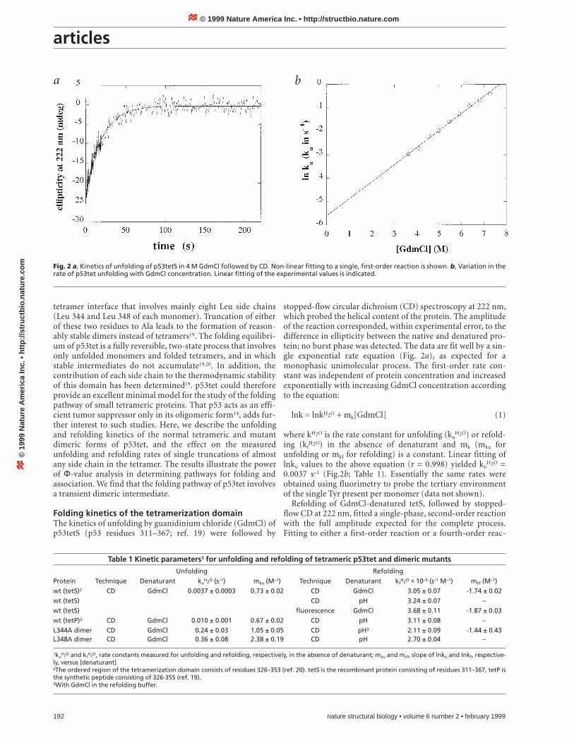
stopped-flow circular dichroism (CD) spectroscopy at 222 nm,which probed the helical content of the protein. The amplitudeof the reaction corresponded, within experimental error, to thedifference in ellipticity between the native and denatured pro-tein; no burst phase was detected. The data are fit well by a sin-gle exponential rate equation (Fig. 2a), as expected for amonophasic unimolecular process. The first-order rate con-stant was independent of protein concentration and increasedexponentially with increasing GdmCl concentration accordingto the equation:
lnk = lnkH2O + mk[GdmCl] (1)
where kH2O is the rate constant for unfolding (kuH2O) or refold-
ing (kfH2O) in the absence of denaturant and mk (mku for
unfolding or mkf for refolding) is a constant. Linear fitting oflnku values to the above equation (r = 0.998) yielded ku
H2O =0.0037 s–1 (Fig.2b; Table 1). Essentially the same rates wereobtained using fluorimetry to probe the tertiary environmentof the single Tyr present per monomer (data not shown).
Refolding of GdmCl-denatured tetS, followed by stopped-flow CD at 222 nm, fitted a single-phase, second-order reactionwith the full amplitude expected for the complete process.Fitting to either a first-order reaction or a fourth-order reac-
Fig. 2 a, Kinetics of unfolding of p53tetS in 4 M GdmCl followed by CD. Non-linear fitting to a single, first-order reaction is shown. b, Variation in therate of p53tet unfolding with GdmCl concentration. Linear fitting of the experimental values is indicated.
a b
Table 1 Kinetic parameters1 for unfolding and refolding of tetrameric p53tet and dimeric mutants
Unfolding RefoldingProtein Technique Denaturant ku
H2O (s–1) mku (M–1) Technique Denaturant kfH2O × 10–5 (s–1 M–1) mkf (M–1)
wt (tetS)2 CD GdmCl 0.0037 ± 0.0003 0.73 ± 0.02 CD GdmCl 3.05 ± 0.07 -1.74 ± 0.02wt (tetS) CD pH 3.24 ± 0.07 –wt (tetS) fluorescence GdmCl 3.68 ± 0.11 -1.87 ± 0.03wt (tetP)2 CD GdmCl 0.010 ± 0.001 0.67 ± 0.02 CD pH 3.11 ± 0.08 –L344A dimer CD GdmCl 0.24 ± 0.03 1.05 ± 0.05 CD pH3 2.11 ± 0.09 -1.44 ± 0.43L348A dimer CD GdmCl 0.36 ± 0.08 2.38 ± 0.19 CD pH 2.70 ± 0.04 –
1kuH2O and kf
H2O, rate constants measured for unfolding and refolding, respectively, in the absence of denaturant; mku and mkf, slope of lnku and lnkf, respective-ly, versus [denaturant]2The ordered region of the tetramerization domain consists of residues 326–353 (ref. 20). tetS is the recombinant protein consisting of residues 311–367, tetP isthe synthetic peptide consisting of 326-355 (ref. 19). 3With GdmCl in the refolding buffer.
© 1999 Nature America Inc. • http://structbio.nature.com©
199
9 N
atu
re A
mer
ica
Inc.
• h
ttp
://s
tru
ctb
io.n
atu
re.c
om
articles
tion was very poor (Fig. 3a). Within the range tested (9–70 µMmonomer), the kinetics of folding showed the dependence onprotein concentration expected for a second-order reaction. Nocurvature was observed in a plot of lnkf versus GdmCl concen-tration. Linear fitting of lnkf values, using equation 1 (r =0.9996) yielded kf
H2O = 3.05 × 10⁵ M–1 s–1 (Fig.3b; Table 1).Essentially the same value for kf
H2O was obtained with acid-denatured tetS in pH-jump refolding experiments. Very similarparameters were also obtained by stopped-flow fluorimetry(Table 1). The refolding rate constant was essentially indepen-dent of the viscosity of the medium, as determined using up to45% sucrose in both the protein sample and the refoldingbuffer (data not shown).
Double-jump experiments (see Methods) were also per-formed. GdmCl-denatured or acid-denatured tetS was allowedto refold for different times (from 50 ms to many seconds), andthen unfolded again. The reaction was followed by fluorimetry.The second-order kf
H2O values obtained from the amplitudesobserved at different delay times were similar to those obtainedin the same conditions in single-jump refolding experiments. Itwas not possible to detect whether or not there was a lag phasebecause of the experimental error inherent to these experi-
nature structural biology • volume 6 number 2 • february 1999 193
ments. Only a single, first-order unfolding phase was repro-ducibly observed at all delay times, and the values of ku
obtained were very similar to those obtained in single-jumpexperiments in the same conditions (data not shown).
A synthetic peptide (tetP) that represents the minimumtetramerization domain (p53 residues 326–355) is only slightlyless stable than tetS19. GdmCl denaturation of tetP yielded anunfolding rate constant only slightly higher than that of tetSand a similar value of mku. Renaturation of acid-denatured tetPgave a refolding rate very similar to that of tetS (Table 1). Thus,the unstructured N- and C-terminal tails present in tetS had nosubstantial effect on the kinetic constants.
Folding and association via a transient dimer The pathway 4U 2I2 N4 for folding and associa-tion of p53tetS is the simplest and most likely model consistentwith the kinetic results described above. U is the denaturedmonomeric form, I2 is a transient, unstable dimeric intermedi-ate, and N4 is the native tetramer. This model may be justifiedas follows: At least two kinetic steps and a transient dimericintermediate must occur in the folding pathway, because a sec-ond-order refolding step was observed, and not a fourth order.
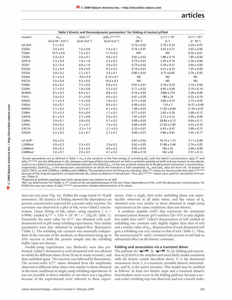
Table 2 Kinetic and thermodynamic parameters1 for folding of mutant p53tet
mutant meq ∆∆GuH2O ∆∆Gu
[GdmCl]50% mku kuH2O × 103 kf
H2O × 10–5
(kcal M–1 mol–1) (kcal mol–1) (kcal mol–1) (M–1) (s–1) (s–1 M–1)wt tetS 5.1 ± 0.2 – – 0.73 ± 0.02 3.70 ± 0.33 3.24 ± 0.07E326A 5.0 ± 0.3 1.6 ± 0.8 1.6 ± 0.1 0.76 ± 0.01 3.02 ± 0.21 3.02 ± 0.06F328A 10.5 ± 0.6 7.2 ± 0.7 11.7± 0.2 ND3 ND 0.36 ± 0.01T329A 5.4 ± 0.3 3.6 ± 0.7 4.6 ± 0.2 0.82 ± 0.02 1.88 ± 0.16 1.85 ± 0.04Q331A 5.3 ± 0.4 1.4 ± 1.0 2.3 ± 0.2 0.79 ± 0.01 2.45 ± 0.16 2.56 ± 0.06I332V 4.2 + 0.3 4.8 ± 1.0 2.9 ± 0.2 0.73 ± 0.02 5.34 ± 0.57 3.64 ± 0.09R333A 5.2 ± 0.4 3.9 ± 0.9 4.5 ± 0.2 0.79 ± 0.01 4.31 ± 0.25 1.95 ± 0.04R335A 5.8 ± 0.2 2.1 ± 0.7 3.9 ± 0.1 0.80 ± 0.02 4.73 ±0.60 2.76 ± 0.05E336A 6.1 ± 0.3 -3.0 ± 0.9 -0.14 ± 0.1 ND ND NDR337A 5.0 ± 0.4 9.3 ± 0.6 10.0 ± 0.3 ND ND NDF338A 6.0 ± 0.2 7.2 ± 0.6 9.0 ± 0.2 0.97 ± 0.01 5.10 ± 0.33 2.14 ± 0.04E339A 5.7 ± 0.3 1.8 ± 0.8 3.5 ± 0.2 0.71 ± 0.02 4.95 ± 0.69 5.19 ± 0.14M340A 6.5 ± 0.5 6.4 ± 0.7 8.8 ± 0.2 0.76 ± 0.02 3380 ± 276 3.30 ± 0.06F341L 5.4 ± 0.3 3.1 ± 0.8 4.0 ± 0.2 0.41 ± 0.05 189 ± 24 6.52 ± 0.16R342A 5.1 ± 0.3 1.4 ± 0.8 1.6 ± 0.2 0.71 ± 0.04 3.60 ± 0.75 2.73 ± 0.05E343A 5.0 ± 0.1 1.1 ± 0.5 0.9 ± 0.1 0.49 ± 0.01 119 ± 7 14.57 ± 0.49N345A 5.1 ± 0.2 3.7 ± 0.7 4.1 ± 0.2 1.44 ± 0.03 11.65 ± 0.60 11.54 ± 0.47E346A 5.5 ± 0.3 0.4 ± 0.9 1.7 ± 0.1 0.77 ± 0.01 2.85 ± 0.26 3.68 ± 0.12A347G 6.1 ± 0.3 2.7 ± 0.8 5.0 ± 0.2 1.07 ± 0.01 3.13 ± 0.12 2.90 ± 0.06E349A 5.9 ± 0.1 3.8 ± 0.6 5.7 ± 0.2 0.90 ± 0.03 28.84 ± 4.15 4.64 ± 0.11L350A 4.9 ± 0.2 2.1 ± 0.7 1.9 ± 0.2 0.84 ± 0.05 21.63 ± 3.83 4.32 ± 0.10K351A 5.2 ± 0.3 -2.5 ± 1.0 -2.1 ± 0.2 0.33 ± 0.01 6.43 ± 0.47 3.90 ± 0.15D352A 5.2 ± 0.2 2.2 ± 0.7 2.7 ± 0.1 0.60 ± 0.01 9.80 ± 0.65 5.45 ± 0.17
wt tetP2 4.6 ± 0.2 – – 0.67 ± 0.02 10.19 ± 1.13 3.11 ± 0.08L330Nva2 4.9 ± 0.2 2.9 ± 0.5 3.5± 0.2 0.62 ± 0.05 21.80 ± 5.46 2.74 ± 0.05L344Nva2 4.8 ± 0.2 3.3 ± 0.6 4.0 ± 0.2 0.95 ± 0.05 166 ± 32 2.60 ± 0.08L348Nva2 5.0 ± 0.1 2.9 ± 0.5 4.1 ± 0.2 0.84 ± 0.10 162 ± 62 3.57 ± 0.12
1Kinetic parameters are as defined in Table 1. meq is the variation in the free energy of unfolding ∆Gu with the GdmCl concentration; ∆∆GuH2O and
∆∆Gu[GdmCl]50% are the differences in ∆Gu between wild-type p53tet (recombinant wt tetS or synthetic peptide wt tetP) and any mutant at zero denat-
urant or at a GdmCl concentration intermediate between the [GdmCl]50% of the two proteins measured at 40 µM protein (monomer ) concentration,respectively; [GdmCl]50% is the denaturant concentration at which the transition is half-completed. meq and ∆∆Gu data are from ref. 19 except forI332V, F341L, wt tetP, L330Nva, L344Nva and L348Nva. The standard errors of fitting are indicated. ∆∆Gu
H2O values are less accurate than ∆∆Gu[GdmCl]50%
because of the long extrapolation of experimental ∆Gu values to absence of denaturant. Thus, ∆Gu[GdmCl]50% values were used for calculation of Φ-val-
ues. (Table 3).2Prepared as synthetic peptides (see text); values given are relative to wt tetP.3ND, not determined. For F328A, ku
H2O could not be calculated because of the non-linear dependence of lnku with the denaturant concentration; forE336A the very low value of ∆∆Gu
[GdmCl]50% prevented a reliable determination of Φ-values.
© 1999 Nature America Inc. • http://structbio.nature.com©
199
9 N
atu
re A
mer
ica
Inc.
• h
ttp
://s
tru
ctb
io.n
atu
re.c
om

articles
One of the steps must be spectroscopically silent, because onlyone phase was seen. The results indicate that the step observedis 2U → I2. The alternative (2I2 → N4 observed) would haverequired that three conditions were all met. (i) The 2U → I2
step should be extremely fast and lost in the dead time of theinstrument (a few ms), because the observed phase showed nolag. (ii) The dimeric intermediate should have the same ellip-ticity at 222 nm and intrinsic Tyr fluorescence as the dena-tured monomers, because the observed phase accounted forthe full amplitude of the process. This would require the inter-mediate to have no helical content and no tertiary environ-ment of Tyr 327, which equates to very little or no structure(Fig. 1). (iii) Some curvature should be detected in plots oflnkf versus GdmCl, which was not observed. On the otherhand, if the refolding step observed is 2U → I2, all of theresults can be easily explained provided this step is rate-limit-ing and/or the dimeric intermediate has essentially the sameellipticity and fluorescence than the native tetramer. Tyr 327is located at the intermonomer interface within each primarydimer, and far away from the interdimer (tetramer) interface(Fig. 1). Thus the change in fluorescence will probably occurduring dimerization of the monomers to form the primarydimers I2, and not during association of the dimers to formthe tetramer. In addition, the lack of curvature in logarithmicplots of kf versus GdmCl is consistent only with the 2U → I2
step being observed. Experiments on dimeric mutants areseen next to suggest an additional step.
Folding kinetics of dimeric p53tet variantsSome p53tet mutants, including L344A and L348A, form sta-ble dimers instead of tetramers at low (µM) concentrations,although they are able to tetramerize at very high (mM) con-centrations19 (M.G.M and A.R.F., unpublished results). Boththe molar ellipticity at 222 nm and the far-UV CD spectrumof the dimeric and tetrameric forms of L344A are indistin-guishable. Thus, these dimeric forms could provide a modelfor transient intermediates for folding of tetrameric, wild typetet. Unfolding of tetS dimeric mutants L344A and L348A wasfollowed by stopped-flow CD at 222 nm. A single first-order
194 nature structural biology • volume 6 number 2 • february 1999
phase with the full amplitude expected for the process wasobserved. Linear fitting of lnku values (r = 0.996) yieldedunfolding rates in the absence of denaturant about two ordersof magnitude faster than for tetrameric wild-type p53tet(Table 1).
As observed with tetrameric p53tet, refolding of acid-dena-tured L344A or L348A followed by stopped flow CD at 222 nmfitted a single-phase, second-order reaction with the fullamplitude expected for the complete process. The correspond-ing values for the rate constant for refolding were very similarto that observed for the tetramer (Table 1). For acid-denaturedL344A, the dependence of kf with GdmCl concentration was
Fig. 4 Variation in the rate of unfolding with GdmCl concentration forsome representative p53tet tetrameric mutants. (P), wild type tetS; (G),wild type tetP; (H), E326A; (p), L330Nva; (l), R333A; (R), M340A; (h),A347G; (L), L344Nva; (g), E349A (compare Table 2). For L344Nva only thevalues obtained at low and medium GdmCl concentrations were used inthe fitting, because of the curvature at high denaturant concentrations(see text).
Fig. 3 a, Kinetics of refolding of GdmCl-denatured p53tetS followed by CD. Fitting to a single, second-order phase (middle smooth tracing) is shown.Lack of fitting to first-order and fourth-order single reactions (upper and lower smooth tracings) is apparent. b, variation in the rate of p53tet refold-ing phase observed with GdmCl concentration. Linear fitting of the experimental values is indicated.
a b
© 1999 Nature America Inc. • http://structbio.nature.com©
199
9 N
atu
re A
mer
ica
Inc.
• h
ttp
://s
tru
ctb
io.n
atu
re.c
om

articles
also analyzed (Table 1). In spite of thenarrow range (0–0.2 M GdmCl) in whichthe dimer was in the native form, linearfitting of lnkf values showed a reasonablecorrelation (r = 0.89). Refolding of urea-denatured L344A within the range1.0–2.5 M urea was consistent with a lin-ear dependence of lnkf with denaturantconcentration (r = 0.96). The rate con-stant for refolding of GdmCl-denaturedL344A was nearly identical to that of theacid-denatured protein in the samerefolding conditions, and very similar kf
values were also obtained using fluo-rimetry instead of CD (not shown).
Clues from dimeric mutantsThe folding kinetics of the stable p53tetdimers described above does not appearto be two-state. In spite of the linear cor-relations observed outside the unfoldingtransition region between both lnku andlnkf and denaturant concentration, thevalues obtained for the ratio 2ku
H2O/kfH2O
(15 × 10–7 M or 27 × 10–7 M, respectively)did not equal the respective equilibriumconstants Ku (1.3 × 10⁻⁷ M or 9.7 × 10⁻⁷ M)determined from equilibrium unfoldingexperiments19. Also for L344A,RT(mku+mkf ) = 1.47 does not equal meq
= 3.0 (where meq is the dependence of thefree energy for unfolding with GdmClconcentration). This indicates the pres-ence of a transient intermediate thatcould, in principle, be either monomericor dimeric. A line of reasoning similar tothat used above for the tetrameric formindicates the second possibility is thecorrect one. The model 2U → I'2 → I2
(where I'2 and I2 are a dimeric intermedi-ate and the native dimer, respectively,and 2U → I'2 represents the observedrefolding step) would be consistent withthe data if 2U → I'2 is rate-limiting forrefolding and/or if the spectroscopicproperties of I'2 are similar to those ofthe native dimer.
The finding that refolding rates of tetrameric p53tet and thetwo dimeric forms are very similar (Table 1) suggests that thefolding/association step 2U → I'2 and the general features ofthe dimeric intermediate are probably shared by bothtetrameric and dimeric p53tet. Both native forms and theirtransient folding intermediates are also similar in that theyhave the same ellipticity at 222 nm, and in that they appear tolack exposed hydrophobic patches able to bind ANS (data notshown). Furthermore, the percentage of hydrophobic surfacearea buried in the transition state for dimerization ofmonomers, relative to the native, was also similar for bothdimeric L344A and wild-type p53tet (28% and 20%, respec-tively).
Φ-values from non-natural amino acidsOne of the strategies of Φ-value analysis is to make mutations
nature structural biology • volume 6 number 2 • february 1999 195
that are ‘non-disruptive’ — that is, to delete a moiety of a sidechain without altering its sterochemistry1. Mutation to Ala gen-erally does this, and we had previously constructed 24 Ala (andGly) single mutants of p53tet19. This set of mutants was usedhere to provide insights into the structure of transition statesduring folding of p53tet. Ala mutations at five positions weretoo destabilizing or led to formation of dimers instead oftetramers. For these positions, less disruptive mutations had tobe engineered. Particularly problematic is the side chain of Leu,because substitution of natural amino acids larger than Ala isdisruptive. We overcame this problem by replacing Leu at 330,344 or 348 with the non-natural amino acid norvaline (Nva) insynthetic tetP peptides. In this way only a methyl group wasdeleted nondisruptively. The recombinant tetS mutants I332Vand F341L were also constructed. All of the new mutants weretetramers as shown by analytical gel filtration using an initialprotein (monomer) concentration of 40 µM (data not shown).
Table 3 Φf values from refolding and unfolding data
Residue and mutation Interactions1 Φf12 Φf2
2
β-strandE326A solvent exposed4 0.05 1.07Y3273 intermonomer with 331, 333F328A dimer core 0.22 ND5
T329A solvent exposed 0.14 1.09L330Nva dimer core 0.04 0.87Q331A intermonomer with 327 0.12 1.10I332V dimer core -0.05 0.93R333A 327; mc intermonomer H-bond with sc345, sc349 0.13 0.98turnG334 – – –α-helixR335A solvent exposed4 0.05 0.96E336A solvent exposed ND5 ND5
R337A dimer core, intermonomer salt bridge with 352 ND NDF338A dimer core 0.05 0.98E339A solvent exposed4 -0.16 0.95M340A tetramer interface 0.00 0.54F341L dimer core, 344, 348 -0.20 0.42R342A dimer core, solvent exposed4 0.13 1.01E343A mc interdimer H-bond with sc351 -1.98 -1.28L344Nva tetramer interface 0.05 0.59N345A sc intermonomer H-bond with mc333 -0.36 0.83E346A solvent exposed4 -0.09 1.09A347G tetramer interface 0.03 1.02L348Nva tetramer interface -0.04 0.60E349A sc intermonomer H-bond with mc333 -0.07 0.79L350A tetramer interface -0.18 0.45K351A tet interface, sc interdimer H-bond with mc343 0.10 1.15D352A intermonomer salt bridge with 337 -0.22 0.79
1The list reflects only the main interactions. The tetramer is considered as a dimer of dimers. Eachprimary dimer consists of a β-sheet and two helices that stack antiparallel. The dimer–dimer(tetramer) interface is formed by the orthogonal packing of the helices of each dimer (Fig. 1).Intermonomer and interdimer refer to interactions within or between primary dimers, respective-ly; sc: side chain; mc: main chain.2Φf1 and Φf2 are Φ values for refolding. Φf1 was obtained from refolding data and refer to the tran-sition state for the U to I'2 step; Φf2 was obtained from unfolding data and refer to the transitionstate for the I'2 to N4 step. Fractional Φ values are highlighted in bold.3Tyr 327 was not mutated because it was used as a probe to determine protein concentration andin fluorescence assays.4The side chains of Glu 326, Arg 335, Glu 339, Arg 342, Glu 346 alternate on the p53tet surfacewith Oε and Nη atoms at distances from 4–6 Å .5Not determined (see Table 2).
© 1999 Nature America Inc. • http://structbio.nature.com©
199
9 N
atu
re A
mer
ica
Inc.
• h
ttp
://s
tru
ctb
io.n
atu
re.c
om

articles
Equilibrium unfolding CD experiments were carried out at thesame protein concentration, using GdmCl as denaturant, andthe relevant thermodynamic parameters were obtained (Table2). The destabilization caused by any of these five substitutionswas about 3–4 kcal mol–1 tetramer.
Folding kinetics of tetrameric p53tet mutantsThe effects on the kinetic constants of removing specific inter-actions in the native protein were determined using the aboveset of tetrameric mutants (Table 2). The interactions altered arelisted in Table 3. GdmCl-induced unfolding was followed bystopped-flow CD at 222 nm. A single, first-order phase with thefull expected amplitude was observed in all cases. For allmutants except F328A, a very good linear correlation betweenlnku and denaturant concentration (0.99 < r < 0.9999) wasfound (Fig. 4). For F341L, L344Nva (Fig. 4), L348Nva, L350Aand K351A, this linear dependence of lnku was seen at low andmedium denaturant concentrations only; at higher concentra-tions the unfolding rate became invariant or even decreasedslightly, as observed with some other proteins21,22. For F328A,non-linearity occurred at low GdmCl concentrations, close toits unusually sharp unfolding transition region19, which pre-vented determination of the corresponding ku
H2O value. Formost mutants, linear extrapolation of lnku data yielded ku
H2O
values close to (within 0.5–1.4x) that of parental p53tet (Table2). Some mutations in the α-helix caused a substantial increasein the unfolding rate. Truncations at the tetramer interface oradjacent to it (M340A, F341L, L344Nva, L348Nva and L350A)caused a remarkable increase (up to 900-fold; Table 2). A fewmutants (F341L, E343A, N345A and K351A) gave somewhatanomalous mku values (under study). However, the vast major-ity of mutants yielded mku values close to that obtained forwild-type tet (Fig. 4; Table 2).
196 nature structural biology • volume 6 number 2 • february 1999
Refolding of acid-denatured p53tet mutants was also moni-tored by stopped-flow CD at 222 nm (Table 2). Like the parentp53tet , refolding of all 23 mutants were fit well by a singlephase, second-order reaction with the amplitude expected forthe process. All but three mutants gave kf
H2O values similar(within 0.6–2×) to that of parental tet (Table 2). In brief,despite the fact that some of the side chain truncations probedhad severe destabilizing effects on the native protein, the rateconstants for the observed unfolding and refolding steps werelittle affected in most, though not all cases.
Transition state for folding/dimerization of monomers The rate constant for refolding/association of denaturedmonomers does not vary with solvent viscosity, which indicatesthat the step observed is not diffusion limited. In addition, thevariation of this rate with p53tet concentration indicates thatthe corresponding transition state is dimeric. The ratioRTmkf/meq = 0.20 suggests that this transition state has buriedonly 20% of the hydrophobic surface area buried in the nativetetramer23. More detailed information about this transitionstate was obtained by calculating the differences in free energy,relative to the denatured state, between the transition state ofeach mutant (mut) and that of wild-type (wt) p53tet (∆∆GU-‡,per dimer), as well as the corresponding Φf-values for folding(Table 3) using the equations:
∆∆GU-‡ = -RTln(kf(mut)H2O / kf (wt)
H2O) (2)
Φf = 2(∆∆GU-‡)/∆∆GU-N (3)
where ∆∆GU-N is the difference in the free energy of unfoldingbetween wild-type and mutant tetramers. Single truncations ofmost of the side chains within the tetramerization domain hada very small influence on the observed refolding rate. Thus, thefree energy of the transition state probed is not substantiallyaffected by mutation and most of the Φf-values were close to 0(Table 3), indicating that very little interaction energies areformed. The anomalous Φ-values obtained for E343A both in
Fig. 6 Proposed qualitative reaction coordinate diagram for the folding andunfolding of tetrameric wild type p53tet and most tetrameric mutants. U,denatured monomers; ‡2, transition state for folding/dimerization ofmonomers; I'2 and I2, transient dimeric intermediates; I2 would be stabilizedby mutations like L344A or L348A; ‡'4, transition state for dimerization of I2;N4, native tetramer.
Fig. 5 Residues with fractional Φ-values in analysis of p53tet unfolding. Aribbon model of p53tet (residues 326–352) oriented as in Fig. 1b is shown;A primary dimer is colored green and the other orange. Residues withnative interactions not fully formed in the rate-limiting step for unfolding(Φf = 0.4–0.6) are depicted as blue ball-and-stick models. The side chainsof residues with native interactions fully formed (Φf-values close to 1)have been omitted for clarity. The program Molscript29 was used.
© 1999 Nature America Inc. • http://structbio.nature.com©
199
9 N
atu
re A
mer
ica
Inc.
• h
ttp
://s
tru
ctb
io.n
atu
re.c
om
articles
refolding and unfolding may be due to the formation of non-native interactions and/or a different pathway of folding forthis particular mutant. The above results indicate that the tran-sition state for folding of denatured monomers to the dimericintermediate is not compact and most native interactions areessentially not formed. A somewhat similar situation wasobserved for the dimeric Arc repressor, in which only ~20% ofthe side chain interaction was used between the denatured stateand the dimeric transition state for folding11. Folding/dimer-ization of denatured p53tet monomers may begin with a gener-al hydrophobic collapse and the formation of very weakinteractions in the transition state. Alternatively, the transitionstate may involve the formation of main-chain hydrogen bondsbetween the β-strands of each monomer (Fig. 1). The presenceof helical secondary structure in the transition state is unlikely,because some of the mutations tested (such as A347G) arehelix-destabilizing and would have probably caused a signifi-cant decrease in the refolding rate, which was not observed.
A low Φ-value can be caused by interactions that are asprevalent in the denatured state as they are in the transitionstate. We calculated the helical content of the denatured state ofthe wild-type monomer in water from the program AGADIR24
to be ~4%. It is thus most unlikely that there is a significant for-mation of helical secondary structure in the first transitionstate.
Transition state for unfolding of the tetramer As expected, only a single, first order phase was observed dur-ing unfolding of p53tet. In strongly unfolding conditions nointermediates were populated and the observed rate corre-sponds to the rate-limiting step for unfolding. The value forRTmku/meq was 0.08, which indicates that, for this unfoldingstep, 92% of the hydrophobic surface buried in the native stateis still buried in the transition state. Additional insight into thestructure of this transition state was obtained by calculating thedifferences in the free energy, relative to the native state,between the transition state of each mutant and that of wild-type p53tet (∆∆G‡-N, per tetramer), as well as the correspond-ing Φf-values (in the direction of folding) (Table 3), using theequations:
∆∆G‡-N = -RTln(ku (wt)H2O/ku (mut)
H2O) (4)
Φf = 1- (∆∆G‡-N/∆∆GU-N) (5)
Most of the Φf-values for folding obtained from unfolding dataafter single truncation of almost any of the residues involvedmainly in native intradimer interactions were very close to 1,indicating that the interactions are essentially fully formed. Incontrast, for most of the residues involved in interdimer nativeinteractions (Leu 344, Leu 348, Met 340 and Leu 350 and theadjacent Phe 341), the Φf-values were close to 0.5 (Table 3; Fig.5). Thus, the rate-limiting transition state for unfolding ofp53tet closely resembles the native tetramer in that it isextremely compact and has most of the native interactionswithin each primary dimer fully formed; however, most inter-dimer interactions at the tetramer interface appear to be onlypartially formed. These results are consistent with the unfold-ing kinetics observed, and indicate that slow dissociation oftetramers to primary dimers is the rate-limiting step forunfolding of the tetramer.
The Φf-values are extremely clear cut and informative forboth steps of the reaction studied. For the initial association
nature structural biology • volume 6 number 2 • february 1999 197
step, they are very close to zero and for the final transition stateare very close to 1.0 except for hydrophobic residues at thetetramer interface. These are the simplest and most unam-bigous cases to analyze, strongly implying nearly completelyunformed and essentially fully formed interactions, respective-ly1. Fractional values for hydrophobic interactions tend to par-allel the degree of bond formation1. In the rate-limitingtransition state for unfolding the hydrophobic residues at thetetramer interface may be involved in native interactions with-in but not between the primary dimers, which could explainthe fractional Φf-values obtained.
Mechanism for the folding and assembly of p53tetThe simplest mechanism for folding of p53tet consistent withall of the above data is as follows (compare Fig. 6):
kf = 3.1 × 10⁵ S-1M-1 (silent)
4U 2 I'2 2 I2 N4fast ku = 0.0037 S-1
This tentative model involves the following: (i), a commonfolding/association step from denatured monomers to a tran-sient dimeric intermediate I'2, through a dimeric transitionstate with little native structure; (Formation of most nativeintradimer interactions would occur between this transitionstate and I'2. Truncation of any of the side chains involvedmainly in interactions within the primary dimers will destabi-lize I'2 and, as a consequence, also the native tetramer.); (ii), aunimolecular reorganization to form a second transient dimer-ic intermediate I2; (This step must be invoked only if we assumethat mutations like L344A or L348A act by stabilizing a tran-sient intermediate I2 in the folding of the normally tetramericp53tet) and (iii), a second dimerization step from the dimericintermediate to the native tetramer, through a highly compact,native-like tetrameric transition state with the primary dimersfully folded but with interdimer interactions only partiallyformed (Fig. 6). The model proposed is also consistent with thestructural organization of p53tet as a dimer of dimers; with thekey role in p53 stability of a cluster of hydrophobic residues inthe center of each primary dimer19; with the existence of stablep53tet dimeric mutants19,25; and with single mutations at thetetramer interface being able to change the oligomerization sta-tus of p53tet. All of these observations point to an ancestraldimeric protein module that evolved to a tetrameric domainwith a minimum of changes in its sequence, structure and fold-ing pathway.
General implicationsThe assembly of the highly helical tetramerization domainfrom its constituent small peptides that are estimated to be~4% helical would appear to be an ideal set up for a framework(or diffusion-collision) mechanism, involving the associationof preformed helices, as is suggested for a monomeric lambdarepressor26. But, instead, the initial association is between high-ly unstructured molecules. The nucleation-condensationmechanism27, in which elements of secondary structure areonly partly formed in the transition state, has been shown for afew small proteins. The initial step of the folding of thetetramerization domain may be thought of as a nucleation-condensation mechanism with an early transition state.Conversely, the 2I2 → N4 step is a clear example of a frameworkmechanism, because the preformed elements of structure that‘diffuse and collide’ are so stable27. These findings are consistent
© 1999 Nature America Inc. • http://structbio.nature.com©
199
9 N
atu
re A
mer
ica
Inc.
• h
ttp
://s
tru
ctb
io.n
atu
re.c
om

1. Fersht, A.R., Matouschek, A. & Serrano, L. The folding of an enzyme. I. Theoryof protein engineering analysis of stability and pathway of protein folding. J.Mol. Biol. 224, 771–782 (1992).
2. Fersht, A.R. Characterizing transition states in protein folding: an essential stepin the puzzle. Curr. Opin. Struct. Biol. 5, 79–84 (1994).
3. Jaenicke, R. Folding and association of proteins. Prog. Biophys. Mol. Biol. 49,117–237 (1987).
4. Garel, J.-R. Folding of large proteins: multidomain and multisubunit proteins.In Protein Folding (ed. T.E. Creighton) 405–454 (Freeman, New York; 1992).
5. Price, N.C. Assembly of multi-subunit structures. In Mechanisms of proteinfolding. (ed. R.H. Pain) 160–193 (IRL Press, Oxford; 1994).
6. Neet, K.E. & Timm, D.E. Conformational stability of dimeric proteins:quantitative studies by equilibrium denaturation. Prot. Sci. 3, 2167–2174(1994).
7. Gittelman, M.S. & Matthews, C.R. Folding and stability of trp aporepressorfrom Escherichia coli. Biochemistry 29, 7011–7020 (1990).
8. Milla, M.E. & Sauer, R.T. P22 Arc repressor: folding kinetics of a single-domain, dimeric protein. Biochemistry 33, 1125–1133 (1994).
9. Wendt, H., Baici, A. & Bosshard, H.R. Mechanism of assembly of a leucine zipperdomain. J. Am. Chem. Soc. 116, 6973–6974 (1994).
10. Wendt, H., Berger, C., Baici, A., Thomas, R.M. & Bosshard, H.R. Kinetics offolding of leucine zipper domains. Biochemistry 34, 4097–4107 (1995).
11. Milla, M.E., Brown, B.M., Waldburger, C.D. & Sauer, R.T. P22 Arc repressor:transition state properties inferred from mutational effects on the rates ofprotein unfolding and refolding. Biochemistry 34, 13914–13919 (1995).
12. Zitzewitz, J.A., Bilsel, O., Luo, J., Jones, B.E. & Matthews, C.R. Probing thefolding mechanism of a leucine zipper peptide by stopped-flow circulardichroism spectroscopy. Biochemistry 34, 12812–12819 (1995).
13. Mok, Y.-K., Bycroft, M. & dePrat-Gay, G. The dimeric DNA binding domain ofthe human papillomavirus E2 protein folds through a monomeric intermediatewhich cannot be native-like. Nature Struct. Biol. 3, 711–717 (1996).
14. Arrowsmith, C.H. & Morin, P. New insights into p53 function from structuralstudies. Oncogene 12, 1379–1385 (1996).
15. Levine, A.J. p53, the cellular gatekeeper for growth and division. Cell 88,323–331 (1997).
16. Lee, W. et al. Solution structure of the tetrameric minimum transformingdomain of p53. Nature Struct. Biol. 1, 877–890 (1994).
17. Clore, G.M. et al. Refined solution structure of the oligomerization domain ofthe tumor suppressor p53. Nature Struct. Biol. 2, 321–333 (1995).
18. Jeffrey, P.D., Gorina, S. & Pavletich, N.P. Crystal structure of the tetramerizationdomain of the p53 tumor suppressor at 1.7 angstroms. Science 267, 1498–1502(1995).
19. Mateu, M.G. & Fersht, A.R. Nine hydrophobic side chains are key determinantsof the thermodynamic stability and oligomerization status of tumorsuppressor p53 tetramerization domain. EMBO J. 17, 2748–2758 (1998).
20. Johnson, C.R., Morin, P.E., Arrowsmith, C.H. & Freire, E. Thermodynamicanalysis of the structural stability of the tetrameric oligomerization domain ofp53 tumor suppressor. Biochemistry 34, 5309–5316 (1995).
21. Zaidi, F.N., Nath, U. & Udgaonkar, J.B. Multiple intermediates and transitionstates during protein unfolding. Nature Struct. Biol. 4,1016–1024 (1997).
22. Jonsson, T., Waldburger, C.D. & Sauer, R.T. Nonlinear free energy relationshipsin Arc repressor unfolding imply the existence of unstable, native-like foldingintermediates. Biochemistry 35, 4795–4802 (1996).
23. Tanford, C. Protein denaturation. Adv. Prot. Chem. 24, 1–95 (1970).24. Muñoz, V. & Serrano, L. Elucidating the folding problem of helical peptides
using empirical parameters. Nature Struct. Biol. 1, 399–409 (1994).25. McCoy, M. et al. Hydrophobic side-chain size is a determinant of the three-
dimensional structure of the p53 oligomerization domain. EMBO J. 16,6230–6236 (1997).
26. Burton, R.E., Myers, J.K. & Oas, T.G. Protein folding dynamics: Quantitativecomparison between theory and experiment. Biochemistry 37, 5337–534(1998).
27. Fersht, A.R. Nucleation Mechanisms in Protein Folding. Curr. Opin. Struct. Biol.7, 3–9 (1997).
28. Itzhaki, L.S., Otzen, D.E. & Fersht, A.R. The structure of the transition state forfolding of chymotrypsin inhibitor 2 analysed by protein engineering methods:Evidence for a nucleation-condensation mechanism for protein folding J. Mol.Biol. 254, 260–288 (1995).
29. Kraulis, P. J. Molscript: A program to produce both detailed and schematic plotsof protein structures. J. Appl. Crystallogr. 24, 946–950 (1991).
articles
with the proposal that small folding domains tend to fold bynucleation type mechanisms and folding becomes more hierar-chical in nature as individual domains become more stable27,28.
MethodsSubcloning of human p53tetS, the construction of a set of tetSmutants with single residue truncations to Ala (or Gly) and theirexpression and purification have been described19. Construction ofadditional tetS mutants was performed using the QuickChangesite-directed mutagenesis kit (Stratagene) and pairs of 26-mer or31-mer oligonucleotides incorporating the desired mutations.
The synthesis of a 30-mer synthetic peptide representing the min-imum sequence of human p53tet (tetP) has been described19. 30-mer synthetic peptides representing variant tetP with Leu 330, Leu344 or Leu 348 replaced by the non-natural amino acid nor-valinewere custom-made by the Peptide Facility, University of Barcelona,Spain. All the peptides were amidated at the C-terminus and puri-fied, and characterized by HPLC and mass spectrometry.
Equilibrium denaturation of tetrameric tetS and tetP variantswas analyzed by far-UV CD spectroscopy in a Jasco-720 spectropo-larimeter using GdmCl as denaturant, and the data were fitted toa two-state transition from folded tetramers to denaturedmonomers as described19.
Stopped-flow spectropolarimeter and fluorimeters (AppliedPhotophysics SX17) were used to measure the kinetics of p53tetunfolding and refolding by CD and fluorescence spectroscopy.Protein ellipticity was followed at 222 nm. Tyr fluorescence wasmeasured above 320 nm using an excitation wavelength of 280nm and the appropriate cut-off filter. Unfolding was initiated bydilution of native p53tet solutions (in 25 mM sodium phosphate,pH = 7) into GdmCl solutions in the same buffer. Refolding ofGdmCl-denatured protein (generally in 4 M GdmCl, 25 mM phos-phate pH = 7) was initiated by dilution into 25 mM phosphate pH =7 with no or low concentrations of GdmCl. Refolding of acid-dena-tured tet (in 62 mM phosphate, pH = 2.1) was initiated by dilutioninto phosphate buffer with no or low concentrations of GdmCl togive a final pH = 7 in 25 mM phosphate. A 1:10 mixing ratio (pro-tein solution : unfolding or refolding buffer) was used in all cases.Total protein (monomer) concentration was generally 36 µM or 18
198 nature structural biology • volume 6 number 2 • february 1999
µM for unfolding or refolding experiments, respectively. The tem-perature was kept at 25 oC.
Double-jump (refolding-unfolding) stopped-flow experimentswere performed as follows: GdmCl-denatured or acid-denaturedp53tet were mixed 1:10 with the appropriate renaturing buffer,allowed to refold for variable lengths of time and then unfoldedby mixing 1:1 with either 8 M GdmCl in 25 mM phosphate pH = 7(to give a final GdmCl concentration above 4.2 M) or 100 mMH3PO4 (to give a final pH of 2.1). The change in Tyr fluorescencewas determined as described above.
Binding of ANS to p53tet during refolding was assayed instopped-flow experiments by 1:10 mixing of GdmCl-denaturedtetS with renaturing buffer (25 mM phosphate pH = 7 with 50 µMANS). The fluorescence due to ANS was measured above 400 nmusing an excitation wavelength of 350 nm and the appropriatecut-off filter. The binding of ANS to denatured and refoldedp53tet, and to BSA as a positive control, were also assayed in thesame conditions.
Unfolding and refolding data were fitted to a single first-orderrate equation (equation 6) and to a single second-order rate equa-tion (equation 7), respectively, using the program Kaleidagraph(Synergy Software):
I(t) = If + A e(–kt ) (6)
I(t) = If - (A /(Ptkt + 1)) (7)
where I(t) is the signal (ellipticity or fluorescence) recorded at timet, If is the signal at t = ∞, Pt is the total protein (monomer) concen-tration, A is the amplitude of the signal and k is the rate constantfor unfolding (ku ) or folding (kf ).
AcknowledgmentsWe gratefully acknowledge M. Bycroft, C. Johnson and J.L. Neira for scientificdiscussions and expert advice. This work was supported by the CRC of the UK.M.G.M. was supported by a grant from the European Union.
Received 21 July, 1998; accepted 3 November, 1998.
© 1999 Nature America Inc. • http://structbio.nature.com©
199
9 N
atu
re A
mer
ica
Inc.
• h
ttp
://s
tru
ctb
io.n
atu
re.c
om