5 neurobehaviour - world health organization 5.pdf118 5 neurobehaviour neurobehavioural studies...
TRANSCRIPT
118
5 NEUROBEHAVIOURNeurobehavioural studies encompass the effects of exposure to
ELF electromagnetic fields on the nervous system and its responses at differ-ent levels of organization. These include the direct stimulation of peripheraland central nerve tissue, perceptual effects resulting from sensory stimula-tion, and effects on central nervous system function. Effects on the latter canbe assessed both electrophysiologically by recording the electrical activity ofthe brain, and by tests of cognition, assessment of mood, and other studies.
The nervous system also has a central role in the control of otherbody systems, particularly the cardiovascular system, through direct nervouscontrol, and the endocrine system, through neural input into the pineal andpituitary glands. These glands in turn influence reproduction and develop-ment, and in a more general way, physiology and well-being.
The brain and nervous systems function by using electrical signals,and may therefore be considered particularly vulnerable to low frequencyEMFs and the resultant induced electric fields and currents. Substantial num-bers of laboratory experiments with volunteers and animals have investigatedthe possible consequences of exposure to weak EMFs on various aspects ofnervous system function, including cognitive, behavioural and neuroendo-crine responses. In addition, epidemiological studies have been carried outon the relationship between EMF exposure and both suicide and depression.
These studies have been reviewed by NRC (1997), NIEHS (1998),IARC (2002), ICNIRP (2003) and McKinlay et al. (2004). In particular,ICNIRP (2003) reviewed in detail some of the evidence summarized here.
In general, there are few effects for which the evidence is strong,and even the more robust field-induced responses seen in the laboratory stud-ies tend to be small in magnitude, subtle and transitory in nature (Crasson etal., 1999; Sienkiewicz et al., 1993).
5.1 Electrophysiological considerationsAn examination of the electrophysiological properties of the ner-
vous system, particularly the central nervous system (CNS: brain and spinalcord) gives an indication of its likely susceptibility to the electric fieldsinduced in the body by EMF exposure. Ion channels in cell membranes allowpassage of particular ionic species across the cell membrane in response tothe opening of a “gate” which is sensitive to the transmembrane voltage(Catterall, 1995; Hille & Anderson, 2001; Mathie, Kennard & Veale, 2003).It is well established that electric fields induced in the body either by directcontact with external electrodes, or by exposure to low frequency magneticfields, will, if of sufficient magnitude, excite nerve tissue through their inter-action with these voltage-gated ion channels. Sensitivity is therefore prima-rily to the transmembrane electric field and varies widely between differention channels (Hille & Anderson, 2001; Mathie, Kennard & Veale, 2003;Saunders & Jefferys, 2002). Many voltage-gated ion channels are associatedwith electrical excitability and electrical signalling. Such electrically excit-
119
able cells not only comprise neurons, glial and muscle cells, but also endo-crine cells of the anterior pituitary, adrenal medulla and pancreas, gametesand, with reservations, endothelial cells (Hille & Anderson, 2001).
All these cells generally express voltage-gated sodium and calciumchannels. Both of these ion channels are involved in electrical signaling andcalcium ions activate a number of crucial cellular processes including neu-rotransmitter release, excitation-contraction coupling in muscle cells andgene expression (Catterall, 2000; Hille & Anderson, 2001). Some ion chan-nels, for example voltage-gated potassium and chloride ion channels, alsoexist in other, non-excitable tissues such as those in the kidney and liver andshow slow electric potential changes but their voltage sensitivity is likely tobe lower (Begenisich & Melvin, 1998; Cahalan, Wulff & Chandy, 2001; Cat-terall, 2000; Jan & Jan, 1989; Nilius & Droogmans, 2001). Since voltage-gated ion channels in excitable cells are steeply sensitive to the transmem-brane electric potential, electric field strength in tissue is a more relevantparameter to relate to electrically excitable cell thresholds than current den-sity (Bailey et al., 1997; Blakemore & Trombley, 2003; Reilly, 2005; Shep-pard, Kavet & Renew, 2002). In fact, the relevant parameter in determiningthe transmembrane current and hence the excitability is the linear gradient inelectric field (Tranchina & Nicholson, 1986), which in turn relates to geo-metric parameters of the neuron, including the degree of bending of the axon.
Peripheral nerves comprise neurons whose cell bodies are locatedwithin the CNS with extended processes (axons) that lie outside the CNS.They conduct action potentials (impulses) towards (sensory nerves) or from(motor nerves) the spinal cord and nerve stimulation shows an all-or-nothingthreshold behaviour. Excitation results from a membrane depolarisation ofbetween 10–20 mV, corresponding to an electric field in tissue of 5–25V m-1 (McKinlay et al., 2004). Pulsed magnetic fields, where the rate ofchange of field induces large localised electric fields, can directly stimulateperipheral nerves and nerve fibres located within the brain (see below).
Cells of the central nervous system are considered to be sensitive toelectric fields induced in the body by exposure to ELF magnetic fields at lev-els that are below threshold for impulse initiation in nerve axons (Jefferys,1995; Jefferys et al., 2003; Saunders, 2003; Saunders & Jefferys, 2002).Such weak electric field interactions have been shown in experimental stud-ies mostly using isolated animal brain tissue to have physiological relevance.These interactions result from the extracellular voltage gradients generatedby the synchronous activity of a number of neurons, or from those generatedby applying pulsed or alternating currents directly through electrodes placedon either side of the tissue. Jefferys and colleagues (Jefferys, 1995; Jefferyset al., 2003) identified in vitro electric field thresholds of around 4–5 V m-1.Essentially, the extracellular gradient alters the potential difference acrossthe neuronal membrane with opposite polarities at either end of the neuron; atime-constant of a few tens (15–60) of milliseconds results from the capaci-tance of the neuronal membrane (Jefferys et al., 2003) and indicates a limitedfrequency response. Similar arguments concerning the limited frequency
120
response of weak electric field effects due to the long time-constants (25 ms)arising from cell membrane capacitance have been given by Reilly (2002)regarding phosphene data.
The CNS in vivo is likely to be more sensitive to induced low fre-quency electric fields and currents than are in vitro preparations (Saunders &Jefferys, 2002). Spontaneous activity is higher, and interacting groups or net-works of nerve cells exposed to weak electrical signals would be expected,on theoretical grounds, to show increased sensitivity through improved sig-nal-to-noise ratios compared with the response of individual cells (Adair,2001; Stering, 1998; Valberg, Kavet & Rafferty, 1997). Much of normal cog-nitive function of the brain depends on the collective activity of very largenumbers of neurons; neural networks are thought to have complex non-lineardynamics that can be very sensitive to small voltages applied diffusely acrossthe elements of the network (Adair, 2001; ICNIRP, 2003; Jefferys et al.,2003). Gluckman et al. (2001) placed the detection limit for network modula-tion in hippocampal slices by electric fields at around 100 mV m-1 . Recentexperimental work by Francis, Gluckman & Schiff (2003) confirms a neuralnetwork threshold of around 140 mV m-1, which the authors found was lowerthan single neuron thresholds, based on a limited number of measurements.A lower limit on neural network sensitivity to physiologically weak inducedelectric fields has elsewhere been considered on theoretical grounds to bearound 1 mV m-1 (Adair, Astumian & Weaver, 1998; Veyret, 2003). Thetime-course of the opening of the fastest voltage-gated ion channels can beless than 1 ms (Hille & Anderson, 2001), suggesting that effects at frequen-cies up to a few kilohertz should not be ruled out. Accommodation to aslowly changing stimulus resulting from slow inactivation of the sodiumchannels will raise thresholds at frequencies less than around 10 Hz.
Other electrically excitable tissues with the potential to show net-work behaviour include glial cells located within the CNS (e.g. Parpura etal., 1994), and the autonomic and enteric nervous systems (see Sukkar, El-Munshid & Ardawi, 2000), which comprise interconnected non-myelinatednerve cells and are distributed throughout the body and gut, respectively.These systems are involved in regulating the visceral or “housekeeping”functions of the body; for example, the autonomic nervous system isinvolved in the maintenance of blood pressure. Muscle cells also show elec-trical excitability; only cardiac muscle tissue has electrically interconnectedcells. However, Cooper, Garny & Kohl et al. (2003), in a review of cardiacion channel activity, conclude that weak internal electric fields much belowthe excitation threshold are unlikely to have any significant effect on cardiacphysiology. EMF effects on the heart could theoretically result from indirecteffects mediated via the autonomic nervous system and CNS (Sienkiewicz,2003). Effects on the endocrine system could potentially also be mediatedthis way, although the evidence from volunteer experiments indicates thatacute ELF magnetic field exposure up to 20 µT does not influence the circa-dian variation in circulating levels of the hormone melatonin (Warman et al.,2003b), nor other plasma hormone levels (ICNIRP, 2003).
121
5.2 Volunteer studiesAn electric charge is induced on the surface of a human (or other
living organism) exposed to a low frequency electric field that alternates inamplitude with the frequency of the applied field. The alternation of the sur-face charge with time induces an electric field and therefore current flowwithin the body; in addition, exposure to a low frequency magnetic fieldinduces circulating eddy currents and associated electric fields. If of suffi-cient magnitude, these induced electric fields and currents can interact withelectrically excitable nerve and muscle tissue. Generally, however, the sur-face charge effects of exposure to low frequency electric fields become pro-hibitive long before the internal electric fields become large enough to elicita response in the tissue.
5.2.1 Surface electric chargeThe surface electric charge can be perceived directly through the
induced vibration of body hair and tingling sensations in areas of the body,particularly the arms, in contact with clothing, and indirectly through sparkdischarges between a person and a conducting object within the field. In sev-eral studies carried out in the 1970’s and 1980’s (summarized by Reilly,1998a; 1999), the threshold for direct perception has shown wide individualvariation; 10% of the exposed subjects had detection thresholds of around 2–5 kV m-1 at 60 Hz, whereas 50% could detect fields of 7–20 kV m-1. Theseeffects were considered annoying by 5% of the test subjects exposed underlaboratory conditions above electric field strengths of about 15–20 kV m-1.In addition to showing a wide variation in individual sensitivity, theseresponses also vary with environmental conditions, particularly humidity;the studies referred to above, however, included both wet and dry exposureconditions.
It has been estimated that spark discharges would be painful to 7%of subjects who are well-insulated and who touch a grounded object within a5 kV m-1 field (Reilly, 1998a; Reilly, 1999) whereas they would be painful toabout 50% in a 10 kV m-1 field. Unpleasant spark discharges can also occurwhen a grounded person touches a large conductive object such as a largevehicle that is “well-insulated” from ground and is situated within a strongelectric field. Here, the threshold field strength required to induce such aneffect varies inversely with the size of the conductive object. In both cases,the presence in the well-insulated person or object of a conductive pathwayto ground would tend to mitigate the intensity of any effect (Reilly, 1998a;Reilly, 1999), as would the impedance to earth of the grounded object or per-son.
People can perceive electric currents directly applied to the bodythrough touching, for example, a conductive loop in which current is inducedby exposure to environmental electromagnetic fields. Thresholds for directlyapplied currents have also been characterised. At 50 to 60 Hz, the malemedian threshold for perception was between 0.36 mA (finger contact) and1.1 mA (grip contact), while pain occurred at 1.8 mA (finger contact).
122
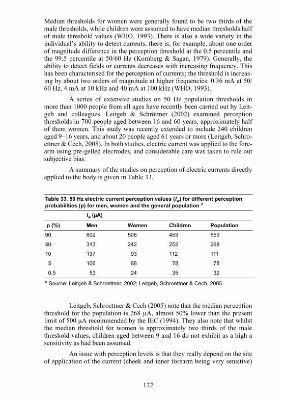
Median thresholds for women were generally found to be two thirds of themale thresholds, while children were assumed to have median thresholds halfof male threshold values (WHO, 1993). There is also a wide variety in theindividual’s ability to detect currents, there is, for example, about one orderof magnitude difference in the perception threshold at the 0.5 percentile andthe 99.5 percentile at 50/60 Hz (Kornberg & Sagan, 1979). Generally, theability to detect fields or currents decreases with increasing frequency. Thishas been characterised for the perception of currents; the threshold is increas-ing by about two orders of magnitude at higher frequencies: 0.36 mA at 50/60 Hz, 4 mA at 10 kHz and 40 mA at 100 kHz (WHO, 1993).
A series of extensive studies on 50 Hz population thresholds inmore than 1000 people from all ages have recently been carried out by Leit-geb and colleagues. Leitgeb & Schröttner (2002) examined perceptionthresholds in 700 people aged between 16 and 60 years, approximately halfof them women. This study was recently extended to include 240 childrenaged 9–16 years, and about 20 people aged 61 years or more (Leitgeb, Schro-ettner & Cech, 2005). In both studies, electric current was applied to the fore-arm using pre-gelled electrodes, and considerable care was taken to rule outsubjective bias.
A summary of the studies on perception of electric currents directlyapplied to the body is given in Table 33.
Leitgeb, Schroettner & Cech (2005) note that the median perceptionthreshold for the population is 268 µA, almost 50% lower than the presentlimit of 500 μA recommended by the IEC (1994). They also note that whilstthe median threshold for women is approximately two thirds of the malethreshold values, children aged between 9 and 16 do not exhibit as a high asensitivity as had been assumed.
An issue with perception levels is that they really depend on the siteof application of the current (cheek and inner forearm being very sensitive)
Table 33. 50 Hz electric current perception values (Iw) for different perception probabilities (p) for men, women and the general population a
Iw (µA)
p (%) Men Women Children Population
90 602 506 453 553
50 313 242 252 268
10 137 93 112 111
5 106 68 78 78
0.5 53 24 35 32a Source: Leitgeb & Schroettner, 2002; Leitgeb, Schroettner & Cech, 2005.
123
and the area of application of the current (i.e. current density). The lattermakes the comparison of current values difficult (Reilly, 1998a).
5.2.2 Nerve stimulationLarge, rapidly changing, pulsed magnetic fields used in various
specialised medical applications such as magnetic resonance imaging (MRI)and transcranial magnetic stimulation (TMS) can induce electric fields largeenough to stimulate nervous tissue in humans. Minimum, orientation-depen-dent stimulus thresholds for large diameter (20 µm) myelinated nerve axonshave been estimated to be approximately 6 V m-1 at frequencies up to about1–3 kHz (Reilly, 1998a; Reilly, 1999). In addition, accommodation to aslowly changing stimulus resulting from slow inactivation of sodium chan-nels will raise thresholds at low frequencies. In MRI, nerve stimulation is anunwanted side effect of a procedure used to derive cross-sectional images ofthe body for clinical diagnosis (see Shellock, 2001). Threshold rates ofchange of the switched gradient magnetic fields used in MRI for perception,discomfort and pain resulting from peripheral nerve stimulation are exten-sively reviewed by Nyenhuis et al. (2001). Generally, median, minimumthreshold rates of change of magnetic field (during periods of < 1 ms) forperception were 15–25 µT s-1 depending on orientation and showed consid-erable individual variation (Bourland, Nyenhuis & Schaefer, 1999). Thesevalues were somewhat lower than previously estimated by Reilly (1998a;1999), possibly due to the constriction of eddy current flow by high imped-ance tissue such as bone (Nyenhuis et al., 2001). Thresholds rose as the pulsewidth of the current induced by the switched gradient field decreased; themedian pulse width (the chronaxie) corresponding to a doubling of the mini-mum threshold (the rheobase) ranged between 360 and 380 µs but againshowing considerable individual variation (Bourland, Nyenhuis & Schaefer,1999). Numerical calculations of the electric field induced by pulses in the84 subjects tested by Nyenhuis et al. (2001) have been used to estimate themedian threshold for peripheral nerve stimulation at 60 Hz as 48 mT (Bailey& Nyenhuis, 2005). Furthermore, Nyenhuis et al. (2001)using data frommeasurements on human volunteers estimated a rheobase electric field of 2.2V m-1 in tissue.
In TMS, parts of the brain are deliberately stimulated in order toproduce a transient, functional impairment for use in the study of cognitiveprocesses (see Reilly, 1998a; Ueno, 1999; Walsh, Ashbridge & Cowey,1998). Furthermore, in TMS, brief, localised, suprathreshold stimuli aregiven, typically by discharging a capacitor through a coil situated over thesurface of the head, in order to stimulate neurons in a small volume (a fewcubic centimetres) of underlying cortical tissue (Reilly, 1998a). The inducedcurrent causes the neurons within that volume to depolarise synchronously,followed by a period of inhibition (Fitzpatrick & Rothman, 2000). When thepulsed field is applied to a part of the brain thought to be necessary for theperformance of a cognitive task, the resulting depolarisation interferes withthe ability to perform the task. In principle then, TMS provides cognitiveneuroscientists with the capability to induce highly specific, temporally and
124
spatially precise interruptions in cognitive processing – sometimes known as“virtual lesions”. Reilly (1998a) noted induced electric field thresholds to beof the order of 20 V m-1. However, Walsh & Cowey (1998) cited typicalrates of change of magnetic field of 30 kT s-1 over a 100 µs period transientlyinducing an electric field of 500 V m-1 in brain tissue.
People are likely to show variations in sensitivity to induced elec-tric fields. In particular, epileptic syndromes are characterised by increasedneuronal excitability and synchronicity (Engelborghs, D'Hooge & De Deyn,2000); seizures arise from an excessively synchronous and sustained dis-charge of a group of neurons (Engelborghs, D'Hooge & De Deyn, 2000; Jef-ferys, 1994). TMS is widely used, apparently without adverse effects.However, repetitive TMS has been observed to trigger epileptic seizure insome susceptible subjects (Fitzpatrick & Rothman, 2000; Wassermann,1998). These authors also reported short- to medium-term memory impair-ments and noted the possibility of long-term cognitive effects from alteredsynaptic activity or neurotransmitter balance. Contraindications for TMS useagreed at an international workshop on repetitive TMS safety (Wassermann,1998) include epilepsy, a family history of seizure, the use of tricyclic anti-depressants, neuroleptic agents and other drugs that lower seizure threshold.Serious heart disease and increased intracranial pressure have also been sug-gested as contraindications due to the potential complications that would beintroduced by seizure.
5.2.3 Retinal functionThe effects of exposure to weak low frequency magnetic fields on
human retinal function are well established. Exposure of the head to mag-netic flux densities above about 5 mT at 20 Hz, rising to about 15 mT at 50Hz, will reliably induce faint flickering visual sensations called magneticphosphenes (Attwell, 2003; Sienkiewicz, Saunders & Kowalczuk, 1991;Taki, Suzuki & Wake, 2003). It is generally agreed that these phosphenesresult from the interaction of the induced electric current with electricallysensitive cells in the retina. Several lines of evidence suggest the productionof phosphenes by a weak induced electric field does not involve the initialtransduction of light into an electrical signal. Firstly, the amplification of theinitial signal generated by the absorption of light takes place primarilythrough an intracellular “second-messenger cascade” of metabolic reactionsprior to any change in ion channel conductivity (Hille & Anderson, 2001).Secondly, the phosphene threshold appears unaffected by “dark” adaptationto low light levels (Carpenter, 1972). In addition, phosphenes have beeninduced in a patient with retinitis pigmentosa, a degenerative illness prima-rily affecting the pigment epithelium and photoreceptors (Lövsund et al.,1980).
There is good reason to view retinal circuitry as an appropriatemodel for induced electric field effects on CNS neuronal circuitry in general(Attwell, 2003). Firstly, the retina displays all the processes present in otherCNS areas, such as graded voltage signalling and action potentials, and has asimilar biochemistry. Secondly, in contrast to more subtle cognitive effects,

125
phosphenes represent a direct and reproducible perception of field interac-tion. A clear distinction can be made in this context between the detection ofa normal visual stimulus and the abnormal induction of a visual signal bynon-visual means (Saunders, 2003); the latter suggests the possibility ofdirect effects on cognitive processes elsewhere in the CNS.
Thresholds for electrically induced phosphenes have been esti-mated to be about 10–14 mA m-2 at 20 Hz (Adrian, 1977; Carstensen, 1985).A similar value (10 mA m-2 at 20 Hz), based on studies of magneticallyinduced phosphenes, has been derived by Wake et al. (1998). The equivalentelectric field threshold can be estimated as around 100–140 mV m-1 using atissue conductivity for brain tissue of about 0.1 S m-1 (Gabriel, Gabriel &Corthout, 1996). More recently, Reilly (2002) has calculated an approximate20 Hz electric field threshold in the retina of 53 mV m-1 for phosphene pro-duction. A similar value (60 mV m-1) has been reported elsewhere (see Saun-ders, 2003). Subsequently, however, Taki et al. (2003) indicated thatcalculations of phosphene thresholds suggested that electrophosphenethresholds were around 100 mV m-1, whereas magnetophosphene thresholdswere around 10 mV m-1 at 20 Hz.
More detailed calculation by Attwell (2003) based on neuroanatom-ical and physiological considerations, suggests that the phosphene electricfield threshold in the extracellular fluid of the retina is in the range 10–60mV m-1 at 20 Hz. There is however, considerable uncertainty attached tothese values. In addition, the extrapolation of values in the extracellular fluidto those appropriate for whole tissue, as used in most dosimetric models, iscomplex, depending critically on the extracellular volume and other factors.With regard to the frequency response, Reilly (2002) suggests that the nar-row frequency response is the result of relatively long membrane time con-stants of around 25 ms. However, at present, the exact mechanismunderlying phosphene induction is unknown. It is not clear whether the nar-row frequency response is due to intrinsic physiological properties of the ret-inal neurons, as suggested by Reilly (2002) above and by Attwell (2003)considering active amplification process in the retinal neuron synaptic termi-nals, or is the result of central processing of the visual signal (Saunders,2003; Saunders & Jefferys, 2002). This issue can only be resolved throughfurther investigation.
5.2.4 Brain electrical activity Since the first suggestion that occupational exposure to EMFs
resulted in clinical changes in the electroencephalogram (EEG) was pub-lished in 1966 (Asanova & Rakov, 1966; 1972), various studies have investi-gated if exposure to magnetic fields can affect the electrical activity of thebrain. Such methods can provide useful diagnostic information regarding thefunctional state of the brain, not only from recordings of the spontaneousactivity at rest but also from recording the sensory functions and subsequentcognitive processes evoked in response to specific stimuli (evoked or event-related potentials, ERPs). Nevertheless, neurophysiological studies usingmagnetic fields need to be performed with much care and attention since
126
they can be prone to many potential sources of error and artefact (NIEHS,1998). Changes in arousal and attention of volunteers, in particular, can sub-stantially affect the outcome of these studies.
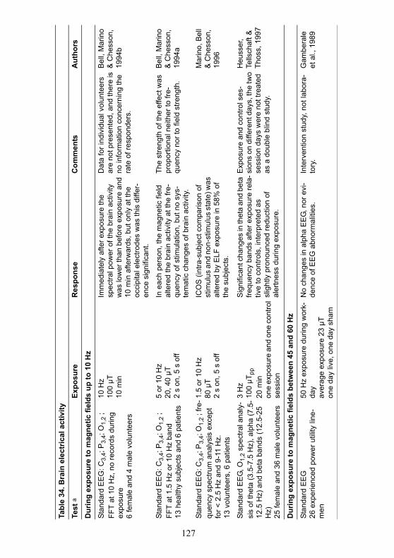
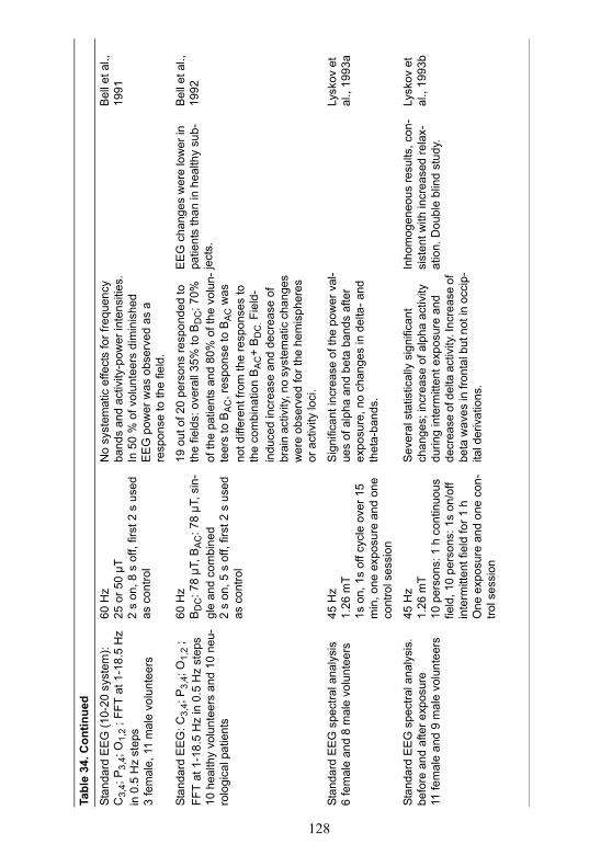
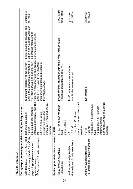
Various studies have investigated the effects of magnetic fields onbrain activity by analysing the spectral power of the main frequency bands ofthe EEG (Bell et al., 1992; Bell et al., 1991; Bell, Marino & Chesson, 1994a;Bell, Marino & Chesson, 1994b; Gamberale et al., 1989; Heusser, Tellschaft& Thoss, 1997; Lyskov et al., 1993b; Lyskov et al., 1993a; Marino, Bell &Chesson, 1996; Schienle et al., 1996; Silny, 1986). These studies have used awide variety of experimental designs and exposure conditions, as well ashealthy volunteers and patients with neurological conditions, and thus aredifficult to compare and evaluate. Despite some scattered field-dependentchanges, most notably in the alpha frequency band, and with intermittentexposure perhaps more effective than continuous exposure, these studieshave produced inconsistent and sometimes contradictory results.
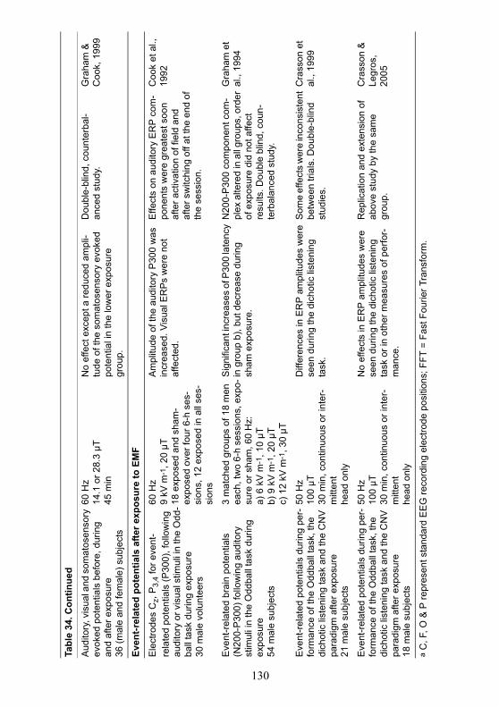
A difficulty with interpretation of the EEG in individuals at rest isthat the intra-individual variability is very high. The variability of ERPs ismuch lower, resulting in better reproducibility, and other studies have inves-tigated the effects of magnetic fields and combined electric and magneticfields on these potentials within the EEG waveform. There are some differ-ences between studies, but generally, the early components of the evokedresponse corresponding to sensory function do not appear affected by expo-sure (Graham & Cook, 1999; Lyskov et al., 1993b). In contrast, large andsustained changes on a later component of the waveform representing stimu-lus detection may be engendered by exposure at 60 mT (Silny, 1984; 1985;1986), with lesser effects occurring using fields of 1.26 mT (Lyskov et al.,1993b), and nothing below 30 µT (Graham & Cook, 1999). Finally, exposureduring the performance of some discrimination and attention tasks mayaffect the late major components of the EEG which are believed to reflectcognitive processes involved with stimulus evaluation and decision making(Cook et al., 1992; Crasson et al., 1999; Graham et al., 1994), although Cras-son and Legros (2005) were unable to replicate the effects they reported pre-viously. There also is some evidence that task difficulty and fieldintermittency may be important experimental variables. However, all thesesubtle effects are not well defined, and some inconsistencies between studiesrequire additional investigation and explanation.
A summary of studies on changes in brain electrical activity whileawake is given in Table 34.
127
Tabl
e 34
. Bra
in e
lect
rical
act
ivity
Test
a
Expo
sure
Res
pons
eC
omm
ents
Aut
hors
Dur
ing
expo
sure
to m
agne
tic fi
elds
up
to 1
0 H
z
Stan
dard
EE
G: C
3,4;
P3,
4; O
1,2
; FF
T at
10
Hz,
no
reco
rds
durin
g ex
posu
re6
fem
ale
and
4 m
ale
volu
ntee
rs
10 H
z10
0 µT
10 m
in
Imm
edia
tely
afte
r exp
osur
e th
e sp
ectra
l pow
er o
f the
bra
in a
ctiv
ity
was
low
er th
an b
efor
e ex
posu
re a
nd
10 m
in a
fterw
ards
, but
onl
y at
the
occi
pita
l ele
ctro
des
was
this
diff
er-
ence
sig
nific
ant.
Dat
a fo
r ind
ivid
ual v
olun
teer
s ar
e no
t pre
sent
ed, a
nd th
ere
is
no in
form
atio
n co
ncer
ning
the
rate
of r
espo
nder
s.
Bel
l, M
arin
o &
Che
sson
, 19
94b
Stan
dard
EE
G: C
3,4;
P3,
4; O
1,2
; FF
T at
1.5
Hz
or 1
0 H
z ba
nd
13 h
ealth
y su
bjec
ts a
nd 6
pat
ient
s
5 or
10
Hz
20, 4
0 µT
2 s
on, 5
s o
ff
In e
ach
pers
on, t
he m
agne
tic fi
eld
alte
red
the
brai
n ac
tivity
at t
he fr
e-qu
ency
of s
timul
atio
n, b
ut n
o sy
s-te
mat
ic c
hang
es o
f bra
in a
ctiv
ity.
The
stre
ngth
of t
he e
ffect
was
pr
opor
tiona
l nei
ther
to fr
e-qu
ency
nor
to fi
eld
stre
ngth
.
Bel
l, M
arin
o &
Che
sson
, 19
94a
Stan
dard
EE
G: C
3,4;
P3,
4; O
1,2
; fre
-qu
ency
spe
ctru
m a
naly
sis
exce
pt
for <
2.5
Hz
and
9-11
Hz.
13 v
olun
teer
s, 6
pat
ient
s
1.5
or 1
0 H
z80
µT
2 s
on, 5
s o
ff
ICO
S (i
ntra
-sub
ject
com
paris
on o
f st
imul
us a
nd n
on-s
timul
us s
tate
) was
al
tere
d by
ELF
exp
osur
e in
58%
of
the
subj
ects
.
Mar
ino,
Bel
l &
Che
sson
, 19
96
Stan
dard
EE
G, O
1,2
spec
tral a
naly
-si
s of
thet
a (3
.5-7
.5 H
z), a
lpha
(7.5
-12
.5H
z) a
nd b
eta
band
s (1
2.5-
25
Hz)
25 fe
mal
e an
d 36
mal
e vo
lunt
eers
3 H
z10
0 µT
pp20
min
one
expo
sure
and
one
con
trol
sess
ion
Sig
nific
ant c
hang
es in
thet
a an
d be
ta
frequ
ency
ban
ds a
fter e
xpos
ure
rela
-tiv
e to
con
trols
, int
erpr
eted
as
slig
htly
pro
noun
ced
redu
ctio
n of
al
ertn
ess
durin
g ex
posu
re.
Exp
osur
e an
d co
ntro
l ses
-si
ons
on d
iffer
ent d
ays,
the
two
sess
ion
days
wer
e no
t tre
ated
as
a d
oubl
e bl
ind
stud
y.
Heu
sser
, Te
llsch
aft &
Th
oss,
199
7
Dur
ing
expo
sure
to m
agne
tic fi
elds
bet
wee
n 45
and
60
Hz
Stan
dard
EE
G26
exp
erie
nced
pow
er u
tility
line
-m
en
50 H
z ex
posu
re d
urin
g w
ork-
day
aver
age
expo
sure
23
µTon
e da
y liv
e, o
ne d
ay s
ham
No
chan
ges
in a
lpha
EE
G, n
or e
vi-
denc
e of
EE
G a
bnor
mal
ities
.In
terv
entio
n st
udy,
not
labo
ra-
tory
.G
ambe
rale
et
al.,
198
9
128
Tabl
e 34
. Con
tinue
d
Stan
dard
EE
G (1
0-20
sys
tem
): C
3,4;
P3,
4; O
1,2
; FFT
at 1
-18.
5 H
z in
0.5
Hz
step
s3
fem
ale,
11
mal
e vo
lunt
eers
60 H
z25
or 5
0 µT
2 s
on, 8
s o
ff, fi
rst 2
s u
sed
as c
ontro
l
No
syst
emat
ic e
ffect
s fo
r fre
quen
cy
band
s an
d ac
tivity
-pow
er in
tens
ities
. In
50
% o
f vol
unte
ers
dim
inis
hed
EE
G p
ower
was
obs
erve
d as
a
resp
onse
to th
e fie
ld.
Bel
l et a
l.,
1991
Stan
dard
EE
G: C
3,4;
P3,
4; O
1,2
; FF
T at
1-1
8.5
Hz
in 0
.5 H
z st
eps
10 h
ealth
y vo
lunt
eers
and
10
neu-
rolo
gica
l pat
ient
s
60 H
zB
DC
: 78
µT, B
AC
: 78
µT, s
in-
gle
and
com
bine
d2
s on
, 5 s
off,
firs
t 2 s
use
d as
con
trol
19 o
ut o
f 20
pers
ons
resp
onde
d to
th
e fie
lds:
ove
rall
35%
to B
DC
; 70%
of
the
patie
nts
and
80%
of t
he v
olun
-te
ers
to B
AC
, res
pons
e to
BA
C w
as
not d
iffer
ent f
rom
the
resp
onse
s to
th
e co
mbi
natio
n B
AC
+ B
DC
. Fie
ld-
indu
ced
incr
ease
and
dec
reas
e of
br
ain
activ
ity, n
o sy
stem
atic
cha
nges
w
ere
obse
rved
for t
he h
emis
pher
es
or a
ctiv
ity lo
ci.
EE
G c
hang
es w
ere
low
er in
pa
tient
s th
an in
hea
lthy
sub-
ject
s.
Bel
l et a
l.,
1992
Stan
dard
EE
G s
pect
ral a
naly
sis
6 fe
mal
e an
d 8
mal
e vo
lunt
eers
45 H
z1.
26 m
T1s
on,
1s
off c
ycle
ove
r 15
min
, one
exp
osur
e an
d on
e co
ntro
l ses
sion
Sig
nific
ant i
ncre
ase
of th
e po
wer
val
-ue
s of
alp
ha a
nd b
eta
band
s af
ter
expo
sure
, no
chan
ges
in d
elta
- and
th
eta-
band
s.
Lysk
ov e
t al
., 19
93a
Stan
dard
EE
G s
pect
ral a
naly
sis.
be
fore
and
afte
r exp
osur
e11
fem
ale
and
9 m
ale
volu
ntee
rs
45 H
z1.
26 m
T10
per
sons
: 1 h
con
tinuo
us
field
, 10
pers
ons:
1s
on/o
ff in
term
itten
t fie
ld fo
r 1 h
One
exp
osur
e an
d on
e co
n-tro
l ses
sion
Sev
eral
sta
tistic
ally
sig
nific
ant
chan
ges;
incr
ease
of a
lpha
act
ivity
du
ring
inte
rmitt
ent e
xpos
ure
and
decr
ease
of d
elta
act
ivity
. Inc
reas
e of
be
ta w
aves
in fr
onta
l but
not
in o
ccip
-ita
l der
ivat
ions
.
Inho
mog
eneo
us re
sults
, con
-si
sten
t with
incr
ease
d re
lax-
atio
n. D
oubl
e bl
ind
stud
y.
Lysk
ov e
t al
., 19
93b
129
Tabl
e 34
. Con
tinue
d
Dur
ing
expo
sure
to m
agne
tic fi
elds
at h
ighe
r fre
quen
cies
Stan
dard
EE
G, F
3,4,
P3,
4,O
1,2,
trad
i-tio
nal f
requ
ency
ban
ds 0
.1 -
30 H
z,
psyc
holo
gica
l par
amet
ers
and
ques
tionn
aire
s26
fem
ale
and
26 m
ale
volu
ntee
rs
Sphe
rics
sim
ulat
ion:
10
kH
z50
0 µs
dur
atio
n, ra
ndom
in
terv
als
betw
een
50 a
nd 1
50
ms
38 A
m-1
pea
k va
lue
elec
tric
field
shi
elde
dex
posu
re 1
0 m
in a
nd c
ontro
l se
ssio
n
Sig
nific
ant r
educ
tion
of th
e po
wer
on
ly in
the
alph
a fre
quen
cy b
and
(8-
13 H
z) in
par
ieta
l and
occ
ipita
l der
i-va
tions
, whe
n an
alys
ing
sub-
grou
ps
only
in 1
0 - 1
0.75
Hz.
P /
O re
cord
-in
gs s
how
sig
nific
ant r
educ
tions
in
the
volta
ge p
ower
.
Fact
ors
such
as
phys
ical
con
-di
tion
and
neur
otic
s w
ere
con-
side
red
as m
edia
tors
of
sphe
rics
effe
ctiv
enes
s.
Sch
ienl
e et
al
., 19
96
Evok
ed p
oten
tials
afte
r exp
osur
e to
EM
F
Visu
al e
voke
d po
tent
ials
100
subj
ects
5 –
50 H
z pu
lsed
mag
netic
fie
ldup
to 1
00 m
T
Pha
se re
vers
al o
f com
pone
nts
of th
e vi
sual
evo
ked
pote
ntia
l at 6
0 m
T.
Very
inte
nse
field
s.S
ilny,
198
4;
1985
; 198
6
Aud
itory
evo
ked
pote
ntia
ls6
fem
ale
and
8 m
ale
volu
ntee
rs45
Hz
1.26
mT
1s o
n, 1
s o
ff, 1
5 m
inon
e ex
posu
re a
nd o
ne c
ontro
l se
ssio
n
N10
0 co
mpo
nent
s w
ere
shor
ter,
ampl
itude
s w
ere
redu
ced.
Ly
skov
et
al.,
1993
a
Aud
itory
evo
ked
pote
ntia
ls11
fem
ale
and
9 m
ale
volu
ntee
rs45
Hz
1.26
mT
10 p
erso
ns: 1
h c
ontin
uous
fie
ld10
per
sons
: 1 h
1 s
on/
off
inte
rmitt
ent f
ield
one
expo
sure
and
one
con
trol
sess
ion
Not
affe
cted
.Ly
skov
et
al.,
1993
b
130
Tabl
e 34
. Con
tinue
d
Aud
itory
, vis
ual a
nd s
omat
osen
sory
ev
oked
pot
entia
ls b
efor
e, d
urin
g an
d af
ter e
xpos
ure
36 (m
ale
and
fem
ale)
sub
ject
s
60 H
z 14
.1 o
r 28.
3 µT
45 m
in
No
effe
ct e
xcep
t a re
duce
d am
pli-
tude
of t
he s
omat
osen
sory
evo
ked
pote
ntia
l in
the
low
er e
xpos
ure
grou
p.
Dou
ble-
blin
d, c
ount
erba
l-an
ced
stud
y.G
raha
m &
C
ook,
199
9
Even
t-rel
ated
pot
entia
ls a
fter e
xpos
ure
to E
MF
Ele
ctro
des
Cz,
P3,
4 fo
r eve
nt-
rela
ted
pote
ntia
ls (P
300)
, fol
low
ing
audi
tory
or v
isua
l stim
uli i
n th
e O
dd-
ball
task
dur
ing
expo
sure
30 m
ale
volu
ntee
rs
60 H
z9
kV m
-1, 2
0 µT
18 e
xpos
ed a
nd s
ham
-ex
pose
d ov
er fo
ur 6
-h s
es-
sion
s, 1
2 ex
pose
d in
all
ses-
sion
s
Am
plitu
de o
f the
aud
itory
P30
0 w
as
incr
ease
d. V
isua
l ER
Ps
wer
e no
t af
fect
ed.
Effe
cts
on a
udito
ry E
RP
com
-po
nent
s w
ere
grea
test
soo
n af
ter a
ctiv
atio
n of
fiel
d an
d af
ter s
witc
hing
off
at th
e en
d of
th
e se
ssio
n.
Coo
k et
al.,
19
92
Eve
nt-r
elat
ed b
rain
pot
entia
ls
(N20
0-P
300)
follo
win
g au
dito
ry
stim
uli i
n th
e O
ddba
ll ta
sk d
urin
g ex
posu
re54
mal
e su
bjec
ts
3 m
atch
ed g
roup
s of
18
men
ea
ch, t
wo
6-h
sess
ions
, exp
o-su
re o
r sha
m, 6
0 H
z:a)
6 k
V m
-1, 1
0 µT
b) 9
kV
m-1
, 20
µT
c) 1
2 kV
m-1
, 30
µT
Sig
nific
ant i
ncre
ases
of P
300
late
ncy
in g
roup
b),
but d
ecre
ase
durin
g sh
am e
xpos
ure.
N20
0-P
300
com
pone
nt c
om-
plex
alte
red
in a
ll gr
oups
, ord
er
of e
xpos
ure
did
not a
ffect
re
sults
. Dou
ble
blin
d, c
oun-
terb
alan
ced
stud
y.
Gra
ham
et
al.,
1994
Eve
nt-r
elat
ed p
oten
tials
dur
ing
per-
form
ance
of t
he O
ddba
ll ta
sk, t
he
dich
otic
list
enin
g ta
sk a
nd th
e C
NV
pa
radi
gm a
fter e
xpos
ure
21 m
ale
subj
ects
50 H
z10
0 µT
30 m
in, c
ontin
uous
or i
nter
-m
itten
t he
ad o
nly
Diff
eren
ces
in E
RP
ampl
itude
s w
ere
seen
dur
ing
the
dich
otic
list
enin
g ta
sk.
Som
e ef
fect
s w
ere
inco
nsis
tent
be
twee
n tri
als.
Dou
ble-
blin
d st
udie
s.
Cra
sson
et
al.,
1999
Eve
nt-r
elat
ed p
oten
tials
dur
ing
per-
form
ance
of t
he O
ddba
ll ta
sk, t
he
dich
otic
list
enin
g ta
sk a
nd th
e C
NV
pa
radi
gm a
fter e
xpos
ure
18 m
ale
subj
ects
50 H
z10
0 µT
30 m
in, c
ontin
uous
or i
nter
-m
itten
t he
ad o
nly
No
effe
cts
in E
RP
am
plitu
des
wer
e se
en d
urin
g th
e di
chot
ic li
sten
ing
task
or i
n ot
her m
easu
res
of p
erfo
r-m
ance
.
Rep
licat
ion
and
exte
nsio
n of
ab
ove
stud
y by
the
sam
e gr
oup.
Cra
sson
&
Legr
os,
2005
a C
, F, O
& P
repr
esen
t sta
ndar
d E
EG
reco
rdin
g el
ectro
de p
ositi
ons;
FFT
= F
ast F
ourie
r Tra
nsfo
rm.
131
5.2.5 SleepSleep is a complex biological process controlled by the central ner-
vous system and is necessary for general health and well-being. The possibil-ity that EMFs may exert a detrimental effect on sleep has been examined intwo studies. Using the EEG to assess sleep parameters, Åkerstedt et al.(1999) reported that continuous exposure of healthy volunteers to 50 Hz at 1µT at night caused disturbances in sleep. In this study, total sleep time, sleepefficiency, slow-wave sleep (stage III and IV), and slow-wave activity weresignificantly reduced by exposure, as was subjective depth of sleep. Graham& Cook (1999) reported that intermittent, but not continuous, exposure to 60Hz, 28 µT magnetic fields at night resulted in less total sleep time, reducedsleep efficiency, increased time in stage II sleep, decreased time in rapid eyemovement (REM) sleep and increased latency to first REM period. Consis-tent with a pattern of poor and broken sleep, volunteers exposed to the inter-mittent field also reported sleeping less well and feeling less rested in themorning.
A comparison between these two studies is made difficult becauseof the differences in the exposure levels used, 1 µT (Åkerstedt et al., 1999)vs. 28 µT (Graham & Cook, 1999) and also of other differences in thedesign. As to the results, in the Åkerstedt study, results were apparentlyobtained by low-level continuous exposure, whereas the Graham study failedto elicit such results by continuous exposure, but did produce similar resultswith intermittent exposures. Further studies with similar designs are neededbefore any conclusions can be drawn.
A summary of studies on brain electrical activity during sleep isgiven in Table 35.
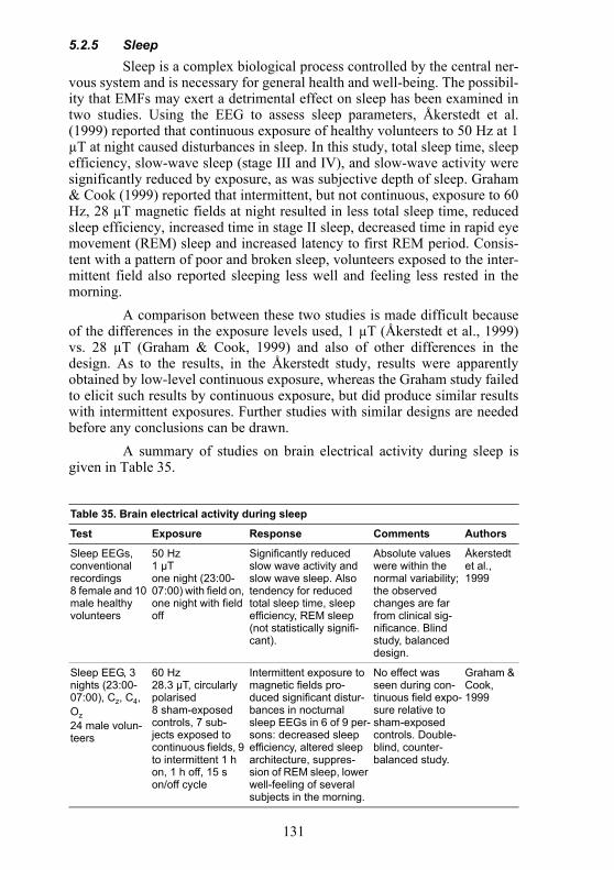
Table 35. Brain electrical activity during sleep
Test Exposure Response Comments Authors
Sleep EEGs, conventional recordings8 female and 10 male healthy volunteers
50 Hz1 µTone night (23:00-07:00) with field on, one night with field off
Significantly reduced slow wave activity and slow wave sleep. Also tendency for reduced total sleep time, sleep efficiency, REM sleep (not statistically signifi-cant).
Absolute values were within the normal variability; the observed changes are far from clinical sig-nificance. Blind study, balanced design.
Åkerstedt et al., 1999
Sleep EEG, 3 nights (23:00-07:00), Cz, C4, Oz 24 male volun-teers
60 Hz28.3 µT, circularly polarised8 sham-exposed controls, 7 sub-jects exposed to continuous fields, 9 to intermittent 1 h on, 1 h off, 15 s on/off cycle
Intermittent exposure to magnetic fields pro-duced significant distur-bances in nocturnal sleep EEGs in 6 of 9 per-sons: decreased sleep efficiency, altered sleep architecture, suppres-sion of REM sleep, lower well-feeling of several subjects in the morning.
No effect was seen during con-tinuous field expo-sure relative to sham-exposed controls. Double-blind, counter-balanced study.
Graham & Cook, 1999
132
5.2.6 Cognitive effectsDespite the potential importance of field-induced effects on atten-
tion, vigilance, memory and other information processing functions, rela-tively few studies have looked for evidence of changes in cognitive abilityduring or after exposure to low frequency EMFs. These have been reviewedby NIEHS (1998), Cook, Thomas & Prato (2002), Bailey (2001), Crasson(2003) and ICNIRP (2003). While few field-dependent changes have beenobserved, it is important to consider that this type of study may be particu-larly susceptible to various environmental and individual factors which mayincrease the variance of the experimental endpoint and decrease the power todetect a small effect. This may be particularly important, since any field-dependent effects are likely to be small with fields at environmental levels(Sienkiewicz et al., 1993; Whittington, Podd & Rapley, 1996).
The effects of acute exposure to magnetic fields on simple andchoice reaction time have been investigated in several recent studies using awide range of magnetic flux densities (20 µT – 1.26 mT) and experimentalconditions. Some studies did not find any field-dependent effects (Gam-berale et al., 1989; Kurokawa et al., 2003b; Lyskov et al., 1993b; Lyskov etal., 1993a; Podd et al., 2002; Podd et al., 1995), although modest effects onspeed (Crasson et al., 1999; Graham et al., 1994; Whittington, Podd & Rap-ley, 1996) and accuracy during task performance (Cook et al., 1992;Kazantzis, Podd & Whittington, 1998; Preece, Wesnes & Iwi, 1998) havebeen reported. However, Crasson & Legros (2005) were unable to replicatethese observations. These data also suggest that effects may depend on thedifficulty of the task (Kazantzis, Podd & Whittington, 1998; Whittington,Podd & Rapley, 1996) and that exposure may attenuate the usual improve-ment seen with practice in reaction time (Lyskov et al., 1993b; Lyskov et al.,1993a; Stollery, 1986)
A few studies have reported subtle field-dependent changes in othercognitive functions, including memory and attention. Using a battery of neu-ropsychological tests, Preece, Wesnes & Iwi (1998) found that exposure to a50 Hz magnetic field at 0.6 mT decreased accuracy in the performance ofnumerical working memory task and decreased sensitivity of the perfor-mance in a word recognition task. Similarly Keetley et al. (2001) investi-gated the effects of exposure to 28 µT, 50 Hz fields using a series ofcognitive tests. A significant decrease in performance was seen with oneworking memory task (the trail-making test, part B) that involves visual-motor tracking and information processing within the prefrontal and parietalareas of the cortex. Podd et al. (2002) reported delayed deficits in the perfor-mance of a recognition memory task following exposure to a 50 Hz field at100 µT. Trimmel & Schweiger (1998) investigated the effects of acute expo-sure to 50 Hz magnetic fields at 1 mT. The fields were produced using apower transformer, and volunteers were exposed in the presence of a 45 dBsound pressure level noise. Compared with a no-field, no-noise condition andnoise alone (generated using a tape recording) significant reductions in visualattention, perception and verbal memory performance were observed during
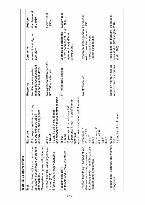
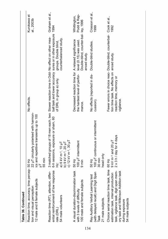
133
Tabl
e 36
. Cog
nitiv
e ef
fect
s
Test
Expo
sure
Res
pons
eC
omm
ents
Aut
hors
Rea
ctio
n tim
e, v
igila
nce,
mem
ory
and
perc
eptio
n sp
eed
test
ed b
efor
e an
d af
ter e
ach
day
26 e
xper
ienc
ed p
ower
util
ity li
nem
en.
50 H
z ex
posu
re d
urin
g w
orkd
ayav
erag
e ex
posu
re 2
3 µT
one
day
live,
one
day
sha
m
No
diffe
renc
e in
per
for-
man
ce b
etw
een
expo
sed
and
non-
expo
sed
days
.
Inte
rven
tion
stud
y, n
ot
labo
rato
ry.
Gam
bera
le e
t al
., 19
89
Rea
ctio
n tim
e (R
T) a
nd ta
rget
-del
e-tio
n te
st (T
DT)
6 fe
mal
e an
d 8
mal
e vo
lunt
eers
45 H
z1.
26 m
T1
s on
, 1 s
off
cycl
e, 1
5 m
inon
e ex
posu
re a
nd o
ne c
ontro
l ses
sion
No
sign
ifica
nt d
iffer
ence
s fo
r R
T, T
DT
not a
ffect
ed.
Lysk
ov e
t al.,
19
93a
Rea
ctio
n tim
e (R
T)11
fem
ale
and
9 m
ale
volu
ntee
rs45
Hz
1.26
mT
10 p
erso
ns: 1
h c
ontin
uous
fiel
d10
per
sons
: 1 h
our 1
s o
n/of
f int
erm
it-te
nt fi
eld
one
expo
sure
and
one
con
trol s
essi
on
RT
not d
irect
ly a
ffect
ed.
Lear
ning
to p
erfo
rm th
e R
T te
st (d
ecre
ase
of R
T in
re
peat
ed tr
ials
) affe
cted
by
exp
osur
e .
Lysk
ov e
t al.,
19
93b
Rea
ctio
n tim
e to
ligh
t fla
shed
at v
ari-
able
inte
rval
s du
ring
expo
-sur
e12
sub
ject
s (e
xpt 1
) and
24
subj
ects
(e
xpt 2
), m
ale
and
fem
ale
Exp
erim
ent 1
:10
.1 o
r 0.2
Hz
1.1
mT
300
sE
xper
imen
t 2:
0.2
or 4
3 H
z1.
8m
T 30
0 s
No
effe
cts
foun
d.E
xper
imen
t 2 d
esig
ned
to
test
for p
ossi
ble
para
met
-ric
reso
nanc
e th
eory
. D
oubl
e bl
ind
stud
ies.
Pod
d et
al.,
19
95
Rea
ctio
n tim
e, a
ccur
acy
and
mem
ory
reco
gniti
on60
Hz
100
µT1
s on
, 1 s
off
for 1
1 m
in
Effe
ct o
n m
emor
y, n
ot o
n re
actio
n tim
e or
acc
urac
y.R
esul
ts d
iffer
ent f
rom
pre
-vi
ous
stud
ies
(Whi
tting
ton
et a
l., 1
996)
Pod
d et
al.,
20
02
134
Tabl
e 36
. Con
tinue
d
Rea
ctio
n tim
e, a
ccur
acy,
tim
e pe
rcep
-tio
n an
d vi
sual
per
cept
ion
12 m
ale
and
8 fe
mal
e su
bjec
ts
50 H
z22
µT
circ
ular
ly p
olar
ised
with
har
mon
-ic
s an
d re
petit
ive
trans
ient
s up
to 1
00
µT 55 m
in
No
effe
cts.
Kur
okaw
a et
al
., 20
03b
Rea
ctio
n tim
e (R
T), a
ttent
ion,
diff
er-
entia
l rei
nfor
cem
ent o
f low
resp
onse
ra
te (D
RL)
54 m
ale
volu
ntee
rs
3 m
atch
ed g
roup
s of
18
men
eac
h, tw
o 6-
h se
ssio
ns, e
xpos
ure
or s
ham
, 60
Hz:
a) 6
kV
m-1
, 10
µTb)
9 k
V m
-1, 2
0 µT
c) 1
2 kV
m-1
, 30
µT
Slo
wer
reac
tion
time
in O
dd-
ball t
ask
and
low
er a
ccur
acy
of D
RL
in g
roup
a) o
nly.
No
effe
ct o
n ot
her m
ea-
sure
s or
in o
ther
exp
osur
e gr
oups
. Dou
ble
blin
d,
coun
terb
alan
ced
stud
y.
Gra
ham
et a
l.,
1994
A v
isua
l dur
atio
n-di
scrim
inat
ion
task
w
ith 3
leve
ls o
f diff
icul
ty10
0 m
ale
and
fem
ale
subj
ects
50 H
z 10
0 µT
inte
rmitt
ent
9 m
in
Dec
reas
ed re
actio
n tim
e fo
r th
e ha
rdes
t lev
el o
f per
for-
man
ce.
A re
laxe
d si
gnifi
canc
e le
vel (
0.15
) was
use
d.
Dou
ble-
blin
d, c
ount
er-b
al-
ance
d st
udy.
Whi
tting
ton,
P
odd
& R
ap-
ley,
199
6
Rey
Aud
itory
Ver
bal L
earn
ing
test
(w
ith d
elay
ed re
call)
and
Dig
it Sp
an
Task
21 m
ale
subj
ects
.
50 H
z10
0 µT
con
tinuo
us o
r int
erm
itten
t30
min
head
onl
y
No
effe
cts
(rep
orte
d in
dis
-cu
ssio
n).
Dou
ble-
blin
d st
udie
s.C
rass
on e
t al.,
19
99
Cho
ice
seria
l rea
ctio
n tim
e ta
sk, t
ime
estim
atio
n ta
sk, i
nter
val p
rodu
ctio
n ta
sk, v
igila
nce
task
, dig
it sp
an m
em-
ory
task
and
Wilk
inso
n A
dditi
on ta
sk
durin
g ex
posu
re54
mal
e su
bjec
ts
60 H
z9
kV m
-1 a
nd 2
0 µT
2 x
3 h
/ day
for 4
day
s
Few
er e
rror
s in
cho
ice
reac
-tio
n tim
e ta
sk. N
o ef
fect
s on
re
actio
n tim
e, m
emor
y or
vi
gila
nce.
Dou
ble-
blin
d, c
ount
erba
l-an
ced
stud
y.C
ook
et a
l.,
1992
135
Tabl
e 36
. Con
tinue
d
A v
isua
l dur
atio
n-di
scrim
inat
ion
task
w
ith 3
leve
ls o
f diff
icul
ty.
40 m
ale
and
59 fe
mal
e su
bjec
ts
50 H
z 10
0 µT
inte
rmitt
ent
7.9
min
Impr
oved
acc
urac
y fo
r the
ha
rdes
t lev
el o
f per
for-
man
ce.
A re
laxe
d si
gnifi
canc
e le
vel (
0.3)
was
use
d. D
ou-
ble-
blin
d, c
ount
er-b
al-
ance
d st
udy.
Kaz
antz
is,
Pod
d &
Whi
t-tin
gton
, 199
8
Imm
edia
te w
ord
reca
ll, re
actio
n tim
e,
digi
t vig
ilanc
e ta
sk, c
hoic
e re
actio
n tim
e, s
patia
l wor
king
mem
ory,
nu
mer
ic w
orki
ng m
emor
y, d
elay
ed
wor
d re
call
and
reco
gniti
on a
nd p
ic-
ture
reco
gniti
on d
urin
g ex
posu
re.
16 (m
ale
and
fem
ale)
sub
ject
s
50 H
z or
sta
tic m
agne
tic fi
elds
at 0
.6
mT
appl
ied
to th
e he
ad. D
urat
ion
not
spec
ified
. Cur
rent
den
sity
in h
ead
esti-
mat
ed a
s 2–
6 m
A m
-2
Red
uced
acc
urac
y of
wor
d an
d nu
mbe
r rec
all a
nd p
er-
form
ance
of c
hoic
e re
actio
n tim
e ta
sk.
Ran
dom
ised
blin
d cr
oss-
over
des
ign.
Pre
ece,
W
esne
s &
Iwi,
1998
Dur
atio
n D
iscr
imin
atio
n Ta
sk a
nd
Stro
op C
olou
r Wor
d te
st.
18 m
ale
subj
ects
50 H
z10
0 µT
con
tinuo
us o
r int
erm
itten
t30
min
head
onl
y
No
effe
ct o
n re
actio
n tim
e an
d pe
rform
ance
acc
urac
y.D
oubl
e bl
ind
with
cou
nter
-ba
lanc
ed e
xpos
ure
orde
r.C
rass
on &
Le
gros
, 200
5
Syn
tact
ic a
nd s
eman
tic v
erba
l rea
-so
ning
task
s, 5
-cho
ise
seria
l rea
ctio
n tim
e ta
sk, a
nd v
isua
l sea
rch
task
s du
ring
expo
sure
76 m
ale
subj
ects
50 H
z cu
rren
t50
0 µA
dire
ctly
app
lied
to h
ead
and
shou
lder
s5.
5 h
/ day
for 2
day
s
Incr
ease
d la
tenc
y in
syn
tac-
tic re
ason
ing
task
. P
ossi
ble
diffe
renc
es
betw
een
grou
ps. D
oubl
e-bl
ind
proc
edur
es w
ith
cros
s ov
er d
esig
n.
Stol
lery
, 198
6;
1987
Rey
Aud
itory
Ver
bal L
earn
ing
test
; D
igit
Span
Mem
ory
Task
; Dig
it S
ym-
bol S
ubst
itutio
n te
st; S
peed
of C
om-
preh
ensi
on T
est a
nd T
rail
Mak
ing
Test
30 s
ubje
cts,
bot
h se
xes
50 H
z28
µT
50 m
inve
rbal
and
writ
ten
test
s ad
min
iste
red
20 m
in fr
om e
xpos
ure
onse
t
Mos
t res
ults
indi
cate
d no
ef
fect
, but
dat
a su
gges
tive
of d
etrim
enta
l effe
ct o
n sh
ort-t
erm
lear
ning
and
ex
ecut
ive
func
tioni
ng.
Dou
ble-
blin
d cr
oss-
over
de
sign
.K
eetle
y et
al.,
20
01
Visu
al d
iscr
imin
atio
n, p
erce
ptio
n, v
er-
bal m
emor
y an
d m
ood
and
sym
ptom
ch
eckl
ist
66 (m
ale
and
fem
ale)
sub
ject
s
50 H
z1
mT
45dB
noi
se c
ompa
red
to n
oise
alo
ne
Sig
nific
ant r
educ
tion
in
visu
al a
ttent
ion,
per
cept
ion
and
verb
al m
emor
y pe
rfor-
man
ce.
Dou
ble
blin
d st
udie
s.Tr
imm
el &
S
chw
eige
r, 19
98
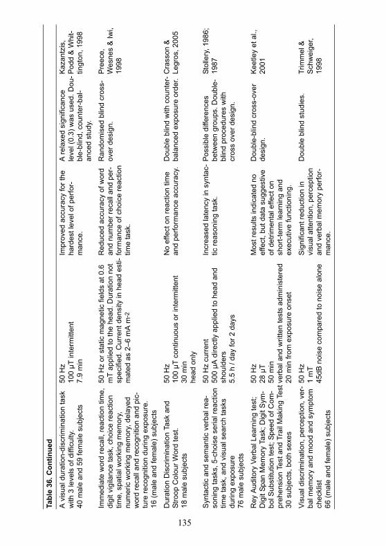
136
field exposure. The presence of the noise during exposure, however, compli-cates interpretation of this study.
Generally, while electrophysiological considerations suggest thatthe central nervous system is potentially susceptible to induced electricfields; cognitive studies have not revealed any clear, unambiguous finding.There is a need for a harmonisation of methodological procedures used indifferent laboratories, and for dose-response relationships to be investigated.The studies on various cognitive effects from ELF field exposure are summa-rized in Table 36.
5.2.7 HypersensitivityIt has been suggested that some individuals display increased sensi-
tivity to levels of EMFs well below recommended restrictions on exposure.People self-reporting hypersensitivity may experience a wide range of severeand debilitating symptoms, including sleep disturbances, general fatigue, dif-ficulty in concentrating, dizziness, and eyestrain. In extreme forms, everydayliving may become problematical. A number of skin problems such aseczema and sensations of itching and burning have also been reported, espe-cially on the face, and, although there may be no specific symptom profile(see Hillert et al., 2002), increased sensitivity to chemical and other factorsoften occurs (Levallois et al., 2002). The responses to EMFs are reported tooccur at field strengths orders of magnitude below those required for conven-tional perception of the field (Silny, 1999). These data have been reviewedby Bergqvist & Vogel (1997) and more recently by Levallois (2002),ICNIRP (2003) and Rubin et al. (2005).
In contrast to anecdotal reports, the evidence from double-blindprovocation studies (Andersson et al., 1996; Flodin, Seneby & Tegenfeldt,2000; Lonne-Rahm et al., 2000; Lyskov, Sandström & Hansson Mild,2001b; Swanbeck & Bleeker, 1989) indicate that neither healthy volunteersnor self-reporting hypersensitive individuals can reliably distinguish fieldexposure from sham-exposure. In addition, subjective symptoms and circu-lating levels of stress-related hormones and inflammatory mediators couldnot be related to field exposure. Similar results were reported in a survey ofoffice workers (Arnetz, 1997). In studies reported by Keisu (1996) and byToomingas (1996), the outcome of tests on an individual was used therapeu-tically in the medical handling of the patient. In none of these series wasthere any reproducible association between exposure and symptoms. Furthertest series have been performed in Sweden, the UK and in Germany, includ-ing an unsuccessful repetition of the Rea et al. (1991) study (see below), butthese have not been published in a peer reviewed form. For a review, seeBergqvist & Vogel (1997). These results are consistent with the view thathypersensitivity to EMFs is a psychosomatic syndrome, suggested by Gothe,Odoni & Nilsson (1995).
Not all studies dismiss the possibility of EMF hypersensitivity,however. Two studies have reported weak positive field discrimination(Mueller, Krueger & Schierz, 2002; Rea et al., 1991) and another two studies
137
reported subtle differences in heart rate, visual evoked potentials, electroret-inogram amplitudes and electrodermal activity between normal and hyper-sensitive volunteers (Lyskov, Sandström & Hansson Mild, 2001a; Sandströmet al., 1997). The study by Rea et al. (1991) has, however, been criticised onseveral methodological grounds (ICNIRP, 2003): the selection of individu-als, the exposure situation and whether the test was blind or not. There issome morphological evidence to suggest that the numbers and distribution ofmast cells in the dermis of the skin on the face may be increased in individu-als displaying hypersensitive reactions (Gangi & Johansson, 2000; Johans-son et al., 1994; Johansson, Hilliges & Han, 1996). Increased responsivenesswas attributed to changes in the expression of histamine and somatostatinand other inflammatory peptides. Similar effects in the dermis have also beenreported following provocation tests to VDU-type fields in normal, healthyvolunteers (Johansson et al., 2001).
EMF hypersensitivity was addressed by the World Health Organi-zation (WHO) at a workshop held in Prague in October 2004 (WHO, 2005).It was proposed that this hypersensitivity, which has multiple recurrentsymptoms and is associated with diverse environmental factors tolerated bythe majority of people, should be termed “idiopathic environmental intoler-ance (IEI) with attribution to EMF”. The workshop concluded that IEI incor-porates a number of disorders sharing similar nonspecific symptoms thatadversely affect people and cause disruptions in their occupational, social,and personal functioning. These symptoms are not explained by any knownmedical, psychiatric or psychological disorder, and the term IEI has no med-ical diagnostic value. IEI individuals cannot detect EMF exposure any moreaccurately than non-IEI individuals, and well-controlled and conducted dou-ble-blind studies have consistently shown that their symptoms are not relatedto EMF exposure per se. A summary of hypersensitivity studies is given inTable 37.
5.2.8 Mood and alertnessThe possible impact of EMFs on mood and arousal has also been
assessed in double-blind studies in which volunteers completed mood check-lists before and after exposure. No field-dependent effects have beenreported using a range of field conditions (Cook et al., 1992; Crasson et al.,1999; Crasson & Legros, 2005; Graham et al., 1994). However, in contrastStollery (1986) reported decreased arousal in one of two participating groupsof subjects when mild (500 µA) 50 Hz electric current was passed throughthe head, upper arms, and feet. This was done to simulate the internal electricfields generated by exposure to an external electric field strength of 36 kVm-1. Also Stevens (2001) reported that exposure to a 20 Hz, 50 µT magneticfield increased positive affective responses displayed to visual stimuli com-pared with sham-exposure. Arousal, as measured by skin conductance, gavevariable results. Table 38 summarizes the studies on effects of ELF fieldexposure on mood and alertness.
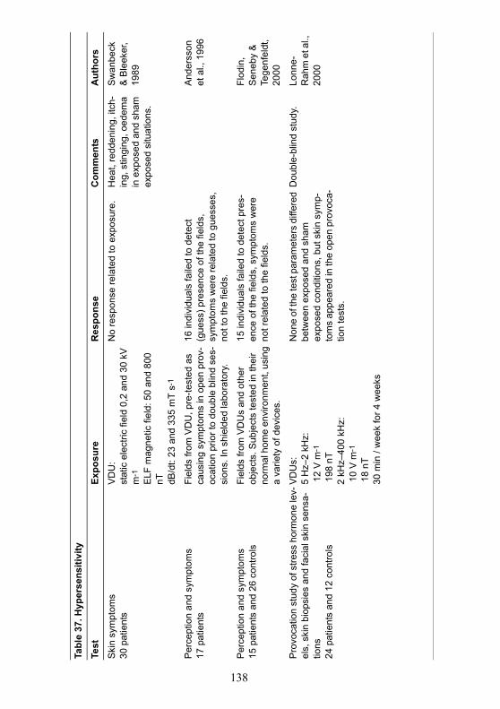
138
Tabl
e 37
. Hyp
erse
nsiti
vity
Test
Expo
sure
Res
pons
eC
omm
ents
Aut
hors
Ski
n sy
mpt
oms
30 p
atie
nts
VD
U:
stat
ic e
lect
ric fi
eld
0,2
and
30 k
V
m-1
ELF
mag
netic
fiel
d: 5
0 an
d 80
0 nT dB
/dt:
23 a
nd 3
35 m
T s-
1
No
resp
onse
rela
ted
to e
xpos
ure.
Hea
t, re
dden
ing,
itch
-in
g, s
tingi
ng, o
edem
a in
exp
osed
and
sha
m
expo
sed
situ
atio
ns.
Sw
anbe
ck
& B
leek
er,
1989
Per
cept
ion
and
sym
ptom
s17
pat
ient
s Fi
elds
from
VD
U, p
re-te
sted
as
caus
ing
sym
ptom
s in
ope
n pr
ov-
ocat
ion
prio
r to
doub
le b
lind
ses-
sion
s. In
shi
elde
d la
bora
tory
.
16 in
divi
dual
s fa
iled
to d
etec
t (g
uess
) pre
senc
e of
the
field
s,
sym
ptom
s w
ere
rela
ted
to g
uess
es,
not t
o th
e fie
lds.
And
erss
on
et a
l., 1
996
Per
cept
ion
and
sym
ptom
s15
pat
ient
s an
d 26
con
trols
Fiel
ds fr
om V
DU
s an
d ot
her
obje
cts.
Sub
ject
s te
sted
in th
eir
norm
al h
ome
envi
ronm
ent,
usin
g a
varie
ty o
f dev
ices
.
15 in
divi
dual
s fa
iled
to d
etec
t pre
s-en
ce o
f the
fiel
ds, s
ympt
oms
wer
e no
t rel
ated
to th
e fie
lds.
Flod
in,
Sen
eby
&
Tege
nfel
dt,
2000
Pro
voca
tion
stud
y of
stre
ss h
orm
one
lev-
els,
ski
n bi
opsi
es a
nd fa
cial
ski
n se
nsa-
tions
24 p
atie
nts
and
12 c
ontro
ls
VD
Us:
5 H
z–2
kHz:
12
V m
-1
198
nT
2 kH
z–40
0 kH
z: 1
0 V
m-1
18
nT30
min
/ w
eek
for 4
wee
ks
Non
e of
the
test
par
amet
ers
diffe
red
betw
een
expo
sed
and
sham
ex
pose
d co
nditi
ons,
but
ski
n sy
mp-
tom
s ap
pear
ed in
the
open
pro
voca
-tio
n te
sts.
Dou
ble-
blin
d st
udy.
Lonn
e-R
ahm
et a
l.,
2000
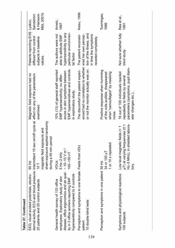
139
Tabl
e 37
. Con
tinue
d
EE
G, v
isua
l evo
ked
pote
ntia
ls, e
lect
ro-
derm
al a
ctiv
ity, E
CG
and
blo
od p
ress
ure.
20 p
atie
nts
and
20 c
ontro
l sub
ject
s
60 H
zin
term
itten
t 15
sec
on/o
ff cy
cle
at
10 T
mag
netic
fiel
d ex
posu
re a
nd
sham
exp
osur
e ap
plie
d ra
ndom
ly
durin
g a
40 m
in p
erio
d
Mag
netic
fiel
d ex
posu
re h
ad n
o ef
fect
on
any
of th
e pa
ram
eter
s ex
amin
ed.
Pat
ient
s re
porti
ng E
HS
di
ffere
d fro
m c
ontro
l su
bjec
ts in
bas
elin
e va
lues
.
Lysk
ov,
San
dströ
m
& H
anss
on
Mild
, 200
1b
Gen
eral
hea
lth s
urve
y of
133
offi
ce
empl
oyee
s. E
xplo
rato
ry s
tudy
of s
kin
dise
ase,
offi
ce e
rgon
omic
s an
d ai
r qua
l-ity
in 3
offi
ce w
orke
rs re
porti
ng E
MF
hype
rsen
titiv
ity c
ompa
red
to 5
con
trols
VD
Us:
5 H
z–2
kHz
~ 10
–15
V m
-1
100–
150
nT
10%
(13)
of g
ener
al s
taff
repo
rted
EM
F hy
pers
ensi
tivity
; no
diffe
r-en
ces
in s
kin
sym
ptom
s be
twee
n E
MF
hype
rsen
sitiv
es a
nd c
ontro
ls
in e
xplo
rato
ry s
tudy
.
The
auth
ors
wer
e no
t ab
le to
attr
ibut
e E
MF
hype
rsen
sitiv
ity to
any
pa
rticu
lar e
nviro
nmen
-ta
l fac
tor.
Arn
etz,
19
97
Per
cept
ion
and
sym
ptom
s in
one
fem
ale
patie
nt10
dou
ble-
blin
d te
sts
Fiel
ds fr
om V
DU
Th
e di
scom
fort
the
patie
nt e
xper
i-en
ced
had
no c
orre
latio
n to
whe
ther
or
not
the
mon
itor a
ctua
lly w
as o
n.
The
patie
nt re
cons
id-
ered
her
ow
n pe
rcep
-tio
n of
the
illne
ss, a
nd
in ti
me
the
sym
ptom
s re
cede
d co
mpl
etel
y.
Kei
su, 1
996
Per
cept
ion
and
sym
ptom
s in
one
pat
ient
50 H
z 34
or 1
00 µ
T1
or 1
0 s
repe
ated
Pos
itive
resp
onse
whe
n hu
mm
ing
of th
e co
ils a
udib
le, d
isap
pear
ed
whe
n “c
amou
flage
d” b
y m
aski
ng
nois
e.
. To
omin
gas,
19
96
Sym
ptom
s an
d ph
ysio
logi
cal r
eact
ions
100
subj
ects
Low
leve
l mag
netic
fiel
ds (<
1
µT) a
t var
ying
freq
uenc
ies
(0.1
H
z–5
MH
z), i
n sh
ield
ed la
bora
-to
ry.
16 o
ut o
f 100
indi
vidu
als
reac
ted
repe
ated
ly to
fiel
ds b
y se
vera
l pa
ram
eter
s (s
ympt
oms,
pup
il di
am-
eter
cha
nges
etc
.).
Not
sur
e w
heth
er fu
lly
blin
d st
udy.
Rea
et a
l.,
1991
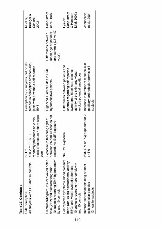
140
Tabl
e 37
. Con
tinue
d
EM
F pe
rcep
tion
49 s
ubje
cts
with
EH
S a
nd 1
4 co
ntro
ls50
Hz
100
V m
-1
6 µ
Tra
ndom
ly p
rese
nted
as
2 m
in
bloc
k of
exp
osur
e / s
ham
exp
o-su
re
Per
cept
ion
by 7
sub
ject
s, b
ut n
o di
f-fe
renc
e in
per
cept
ion
betw
een
sub-
ject
s w
ith o
r with
out s
elf-r
epor
ted
EH
S.
Mue
ller,
Kru
eger
&
Sch
ierz
, 20
02
Ele
ctro
card
iogr
am, v
isua
l evo
ked
pote
n-tia
ls (V
EP)
and
ele
ctro
retin
ogra
ms
10 s
ubje
cts
repo
rting
EM
F hy
pers
ensi
v-ity
and
10
cont
rols
Exp
osur
e to
flic
kerin
g lig
ht a
t be
twee
n 25
and
75
flash
es p
er
seco
nd. N
o E
MF
expo
sure
Hig
her V
EP
ampl
itude
s in
EM
F hy
pers
ensi
tive
patie
nts.
Diff
eren
ces
betw
een
mea
n ag
e of
pat
ient
s an
d co
ntro
ls (3
7 vs
47
year
).
San
d-st
röm
et
al.,
199
7
Sel
f-rep
orte
d sy
mpt
oms,
blo
od p
ress
ure,
he
art r
ate,
(ski
n) e
lect
rode
rmal
act
ivity
, E
EG
s an
d vi
sual
evo
ked
pote
ntia
ls10
sub
ject
s re
porti
ng h
yper
sens
itivi
ty
and
10 c
ontro
ls
No
EM
F ex
posu
reD
iffer
ence
s be
twee
n pa
tient
s an
d co
ntro
ls re
gard
ing
self-
repo
rted
sym
ptom
s, h
eart
rate
, ele
ctric
al
activ
ity o
f the
ski
n, a
nd v
isua
l ev
oked
pot
entia
l am
plitu
des.
Lysk
ov,
San
d-st
röm
&
Han
sson
M
ild, 2
001a
Imm
uno-
fluor
esce
nt s
tain
ing
of m
ast
cells
from
ski
n bi
opsi
es13
hea
lthy
subj
ects
VD
U (T
V o
r PC
) exp
osur
e fo
r 2
or 4
hIn
crea
se in
num
ber o
f mas
t cel
ls in
pa
pilla
ry a
nd re
ticul
ar d
erm
is in
5
subj
ects
.
Joha
nsso
n et
al.,
200
1
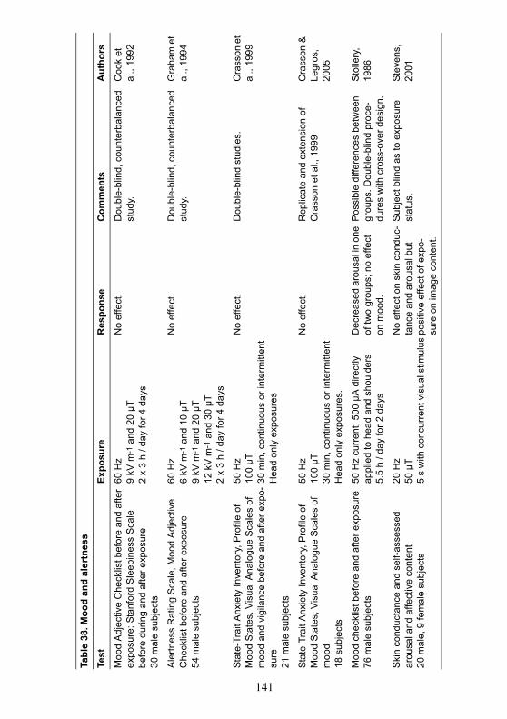
141
Tabl
e 38
. Moo
d an
d al
ertn
ess
Test
Expo
sure
Res
pons
eC
omm
ents
Aut
hors
Moo
d A
djec
tive
Che
cklis
t bef
ore
and
afte
r ex
posu
re; S
tanf
ord
Sle
epin
ess
Sca
le
befo
re d
urin
g an
d af
ter e
xpos
ure
30 m
ale
subj
ects
60 H
z9
kV m
-1 a
nd 2
0 µT
2 x
3 h
/ day
for 4
day
s
No
effe
ct.
Dou
ble-
blin
d, c
ount
erba
lanc
ed
stud
y.C
ook
et
al.,
1992
Ale
rtnes
s R
atin
g S
cale
, Moo
d A
djec
tive
Che
cklis
t bef
ore
and
afte
r exp
osur
e54
mal
e su
bjec
ts
60 H
z6
kV m
-1 a
nd 1
0 µT
9 kV
m-1
and
20
µT12
kV
m-1
and
30
µT2
x 3
h / d
ay fo
r 4 d
ays
No
effe
ct.
Dou
ble-
blin
d, c
ount
erba
lanc
ed
stud
y.G
raha
m e
t al
., 19
94
Stat
e-Tr
ait A
nxie
ty In
vent
ory,
Pro
file
of
Moo
d St
ates
, Vis
ual A
nalo
gue
Sca
les
of
moo
d an
d vi
gila
nce
befo
re a
nd a
fter e
xpo-
sure
21 m
ale
subj
ects
50 H
z10
0 µT
30 m
in, c
ontin
uous
or i
nter
mitt
ent
Hea
d on
ly e
xpos
ures
No
effe
ct.
Dou
ble-
blin
d st
udie
s.C
rass
on e
t al
., 19
99
Stat
e-Tr
ait A
nxie
ty In
vent
ory,
Pro
file
of
Moo
d St
ates
, Vis
ual A
nalo
gue
Sca
les
of
moo
d18
sub
ject
s
50 H
z10
0 µT
30 m
in, c
ontin
uous
or i
nter
mitt
ent
Hea
d on
ly e
xpos
ures
.
No
effe
ct.
Rep
licat
e an
d ex
tens
ion
of
Cra
sson
et a
l., 1
999
Cra
sson
&
Legr
os,
2005
Moo
d ch
eckl
ist b
efor
e an
d af
ter e
xpos
ure
76 m
ale
subj
ects
50 H
z cu
rren
t; 50
0 µA
dire
ctly
ap
plie
d to
hea
d an
d sh
ould
ers
5.5
h / d
ay fo
r 2 d
ays
Dec
reas
ed a
rous
al in
one
of
two
grou
ps; n
o ef
fect
on
moo
d.
Pos
sibl
e di
ffere
nces
bet
wee
n gr
oups
. Dou
ble-
blin
d pr
oce-
dure
s w
ith c
ross
-ove
r des
ign.
Stol
lery
, 19
86
Ski
n co
nduc
tanc
e an
d se
lf-as
sess
ed
arou
sal a
nd a
ffect
ive
cont
ent
20 m
ale,
9 fe
mal
e su
bjec
ts
20 H
z50
µT
5 s
with
con
curr
ent v
isua
l stim
ulus
No
effe
ct o
n sk
in c
ondu
c-ta
nce
and
arou
sal b
ut
posi
tive
effe
ct o
f exp
o-su
re o
n im
age
cont
ent.
Sub
ject
blin
d as
to e
xpos
ure
stat
us.
Stev
ens,
20
01
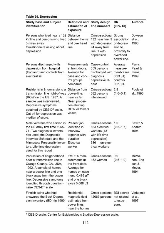
142
Table 39. Depression
Study base and subject identification
Definition and estimation of exposure
Study design and numbers
RR(95% CI)
Authors
Persons who lived near a 132 kV line and persons who lived 3 miles awayQuestionnaire asking about depression
Distance between home and overhead line
Cross-sectional132 near line, 9 with depression94 away from line, 1 with depression
Strong association of depres-sion to proximity to overhead power line.
Dowson et al., 1988
Persons discharged with depression from hospital (England) and controls from electoral list
Measurements at front doors. Average for case and con-trol groups compared
Case-control359 persons discharged with diagnosis depressive ill-ness
Average measure-ment cases: 0.23 µT; controls 0.21 µT.
Perry, Pearl & Binns, 1989
Residents in 8 towns along a transmission line right-of-way (ROW) in the US, 1987. A sample was interviewed. Depressive symptoms obtained by CES-Da scale. Cut-off for depression was median of score.
Distance from power-line: near vs farNear: proper-ties abutting ROW or towers visible
Cross-sectional382 persons interviewed
2.8 (1.6–5.1)
Poole et al., 1993
Male veterans who served in the US army first time 1965-71. Two diagnostic invento-ries used: the Diagnostic Interview Schedule and the Minnesota Personality Inven-tory. Life time depression used for this report
Present job identified in interview together with durationElectrical worker
Cross-sectional183 electrical workers (13 with life-time depression)3861 non-elec-trical workers
1.0 (0.5–1.7)
Savitz & Ananth, 1994
Population of neighborhood near a transmission line in Orange County, CA, USA, 1992. A sample of homes near a power line and one block away from the power line. Depressive symptoms identified through question-naire CES-D* scale
EMDEX mea-surements at the front door. Average for homes on ease-ment: 0.486 µT and one block away 0.068 µT
Cross-sectional152 women
0.9 (0.5–1.9)
McMa-han, Eric-son & Meyer, 1994
Finnish twins who had answered the Beck Depres-sion Inventory (BDI) in 1990
Residential magnetic field estimated from power lines near the homes
Cross-sectional12063 persons
BDI scores not related to expo-sure.
Verkasalo et al., 1997
a CES-D scale: Centre for Epidemiologic Studies-Depression scale.
143
5.3 Epidemiological studiesWith regard to neurobehavioural effects, epidemiological studies
have focussed on depression and suicide. Studies of an association betweenEMF exposure and neurodegeneration are covered in Chapter 6.
5.3.1 DepressionTwo early studies relating ELF EMF exposure to depression (Dow-
son et al., 1988; Perry, Pearl & Binns, 1989) are difficult to interpret becauseof methodological limitations to the procedures for selection of study sub-jects, because they did not use validated scales for identification of depres-sive symptoms (ICNIRP, 2003). Moreover, Perry, Pearl & Binns (1989) alsoreported unusually high average EMF levels that remain unexplained.
More recent studies used validated depression scales. One of thesestudies showed a clear association between proximity to power lines anddepression (Poole et al., 1993), whereas three more recent studies (McMa-han, Ericson & Meyer, 1994; Savitz & Ananth, 1994; Verkasalo et al., 1997)provided little evidence for such an association. The study by Poole et al.(1993) is well designed: it compares subjects on properties abutting a powerline right-of-way to subjects further away, and the results appear internallyconsistent. The investigators report a relative risk of 2.8 (95% CI: 1.6–5.1).McMahan, Ericson & Meyer (1994) employed a similar design and measure-ments to confirm that the homes close to the line have considerably higherEMF levels than those further away. This study also appears valid but yieldsa relative risk of 0.9 (95% CI: 0.5–1.9). McMahan, Ericson & Meyer (1994)offer a number of possible explanations for the lack of consistency betweenthese two studies but none of the explanations is convincing (ICNIRP, 2003).
Overall, ICNIRP (2003) conclude that the literature on depressivesymptoms and EMF exposure is difficult to interpret because the findings arenot consistent. This complexity cannot easily be resolved by suggesting thatone type of result can be confined to a group of studies with methodologicalproblems or some other limitation.
A summary of studies on the effects of ELF field exposure ondepression is given in Table 39.
5.3.2 SuicideAn early case control study carried out in England (Perry et al.,
1981; Reichmanis et al., 1979) found significantly higher EMFs in casehomes than control homes. However, ICNIRP (2003) considers the studymethodologically limited both for the way subjects were selected and for thestatistical analysis employed. Subsequent studies have used a range of differ-ent approaches to assess exposure, varying from crude techniques based ondistance between home and power lines, or on job titles, to more sophisti-cated approaches based on detailed information about cohorts of utilityworkers (Baris et al., 1996a; Baris et al., 1996b; Baris & Armstrong, 1990;Johansen & Olsen, 1998a; McDowall, 1986; van Wijngaarden et al., 2000).Only the latter study provides some support for the original findings,
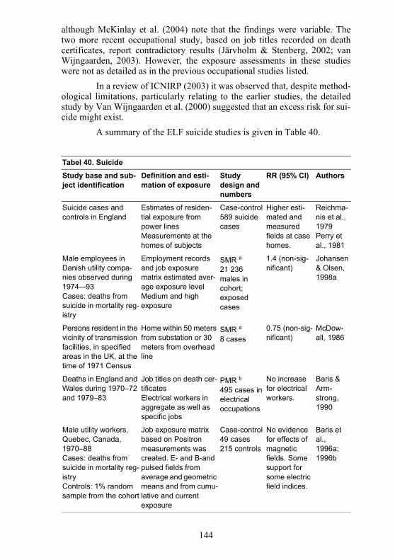
144
although McKinlay et al. (2004) note that the findings were variable. Thetwo more recent occupational study, based on job titles recorded on deathcertificates, report contradictory results (Järvholm & Stenberg, 2002; vanWijngaarden, 2003). However, the exposure assessments in these studieswere not as detailed as in the previous occupational studies listed.
In a review of ICNIRP (2003) it was observed that, despite method-ological limitations, particularly relating to the earlier studies, the detailedstudy by Van Wijngaarden et al. (2000) suggested that an excess risk for sui-cide might exist.
A summary of the ELF suicide studies is given in Table 40.
Tabel 40. Suicide
Study base and sub-ject identification
Definition and esti-mation of exposure
Study design and numbers
RR (95% CI) Authors
Suicide cases and controls in England
Estimates of residen-tial exposure from power linesMeasurements at the homes of subjects
Case-control589 suicide cases
Higher esti-mated and measured fields at case homes.
Reichma-nis et al., 1979Perry et al., 1981
Male employees in Danish utility compa-nies observed during 1974–-93Cases: deaths from suicide in mortality reg-istry
Employment records and job exposure matrix estimated aver-age exposure levelMedium and high exposure
SMR a21 236 males in cohort; exposed cases
1.4 (non-sig-nificant)
Johansen & Olsen, 1998a
Persons resident in the vicinity of transmission facilities, in specified areas in the UK, at the time of 1971 Census
Home within 50 meters from substation or 30 meters from overhead line
SMR a8 cases
0.75 (non-sig-nificant)
McDow-all, 1986
Deaths in England and Wales during 1970–72 and 1979–83
Job titles on death cer-tificatesElectrical workers in aggregate as well as specific jobs
PMR b495 cases in electrical occupations
No increase for electrical workers.
Baris & Arm-strong, 1990
Male utility workers, Quebec, Canada, 1970–88Cases: deaths from suicide in mortality reg-istryControls: 1% random sample from the cohort
Job exposure matrix based on Positron measurements was created. E- and B-and pulsed fields from average and geometric means and from cumu-lative and current exposure
Case-control49 cases215 controls
No evidence for effects of magnetic fields. Some support for some electric field indices.
Baris et al., 1996a; 1996b
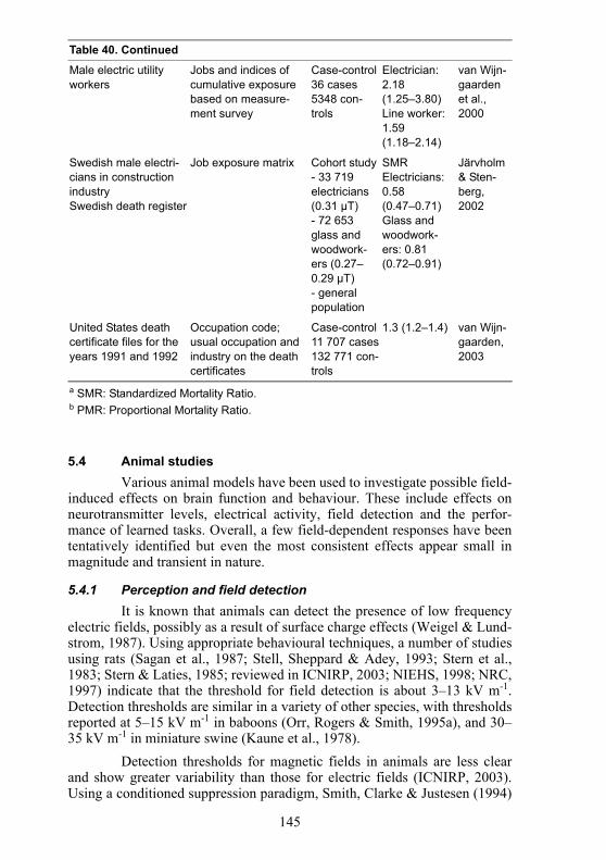
145
5.4 Animal studiesVarious animal models have been used to investigate possible field-
induced effects on brain function and behaviour. These include effects onneurotransmitter levels, electrical activity, field detection and the perfor-mance of learned tasks. Overall, a few field-dependent responses have beententatively identified but even the most consistent effects appear small inmagnitude and transient in nature.
5.4.1 Perception and field detectionIt is known that animals can detect the presence of low frequency
electric fields, possibly as a result of surface charge effects (Weigel & Lund-strom, 1987). Using appropriate behavioural techniques, a number of studiesusing rats (Sagan et al., 1987; Stell, Sheppard & Adey, 1993; Stern et al.,1983; Stern & Laties, 1985; reviewed in ICNIRP, 2003; NIEHS, 1998; NRC,1997) indicate that the threshold for field detection is about 3–13 kV m-1.Detection thresholds are similar in a variety of other species, with thresholdsreported at 5–15 kV m-1 in baboons (Orr, Rogers & Smith, 1995a), and 30–35 kV m-1 in miniature swine (Kaune et al., 1978).
Detection thresholds for magnetic fields in animals are less clearand show greater variability than those for electric fields (ICNIRP, 2003).Using a conditioned suppression paradigm, Smith, Clarke & Justesen (1994)
Table 40. Continued
Male electric utility workers
Jobs and indices of cumulative exposure based on measure-ment survey
Case-control36 cases5348 con-trols
Electrician:2.18 (1.25–3.80)Line worker:1.59 (1.18–2.14)
van Wijn-gaarden et al., 2000
Swedish male electri-cians in construction industrySwedish death register
Job exposure matrix Cohort study- 33 719 electricians (0.31 µT)- 72 653 glass and woodwork-ers (0.27–0.29 µT)- general population
SMRElectricians: 0.58 (0.47–0.71)Glass and woodwork-ers: 0.81 (0.72–0.91)
Järvholm & Sten-berg, 2002
United States death certificate files for the years 1991 and 1992
Occupation code; usual occupation and industry on the death certificates
Case-control11 707 cases132 771 con-trols
1.3 (1.2–1.4) van Wijn-gaarden, 2003
a SMR: Standardized Mortality Ratio.b PMR: Proportional Mortality Ratio.
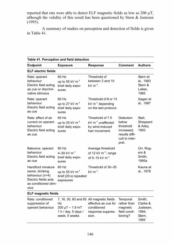
146
reported that rats were able to detect ELF magnetic fields as low as 200 µT,although the validity of this result has been questioned by Stern & Justesen(1995).
A summary of studies on perception and detection of fields is givenin Table 41.
Table 41. Perception and field detection
Endpoint Exposure Response Comment Authors
ELF electric fields
Rats: operant behaviourElectric field acting as cue or discrimi-native stimulus
60 Hzup to 55 kV m-1
brief daily expo-sures
Threshold of between 3 and 10 kV m-1.
Stern et al., 1983Stern & Laties, 1985
Rats: operant behaviourElectric field acting as cue
60 Hzup to 27 kV m-1
brief daily expo-sures
Threshold of 8 or 13 kV m-1 depending on the test protocol.
Sagan et al., 1987
Rats: effect of air current on operant behaviour Electric field acting as cue
60 Hzup to 25 kV m-1
brief daily expo-sures
Threshold of 7.5 kV m-1 unaffected by wind-induced hair movement.
Detection below threshold increased; results diffi-cult to inter-pret.
Stell, Sheppard & Adey, 1993
Baboons: operant behaviourElectric field acting as cue
60 Hz4–50 kV m-1
brief daily expo-sures
Average threshold of 12 kV m-1; range of 5–15 kV m-1.
Orr, Rog-ers & Smith, 1995a
Handford minature swine: drinking behaviour (n=4)Electric fields acts as conditioned stim-ulus
60 Hzup to 55 kV m-1
brief (20 s) repeated exposures
Threshold of 30–35 kV m-1.
Kaune et al., 1978
ELF magnetic fields
Rats: conditioned suppression of operant behaviour
7, 16, 30, 60 and 65 Hz200 µT – 1.9 mT1 h / day, 5 days / week, 5 weeks
All magnetic fields effective as cue for conditioned response suppres-sion.
Temporal rather than magnetic field condi-tioning?
Smith, Clarke & Justesen, 1994; Stern, 1995
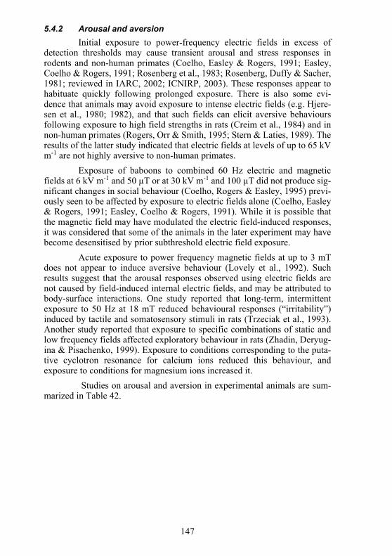
147
5.4.2 Arousal and aversionInitial exposure to power-frequency electric fields in excess of
detection thresholds may cause transient arousal and stress responses inrodents and non-human primates (Coelho, Easley & Rogers, 1991; Easley,Coelho & Rogers, 1991; Rosenberg et al., 1983; Rosenberg, Duffy & Sacher,1981; reviewed in IARC, 2002; ICNIRP, 2003). These responses appear tohabituate quickly following prolonged exposure. There is also some evi-dence that animals may avoid exposure to intense electric fields (e.g. Hjere-sen et al., 1980; 1982), and that such fields can elicit aversive behavioursfollowing exposure to high field strengths in rats (Creim et al., 1984) and innon-human primates (Rogers, Orr & Smith, 1995; Stern & Laties, 1989). Theresults of the latter study indicated that electric fields at levels of up to 65 kVm-1 are not highly aversive to non-human primates.
Exposure of baboons to combined 60 Hz electric and magneticfields at 6 kV m-1 and 50 µT or at 30 kV m-1 and 100 µT did not produce sig-nificant changes in social behaviour (Coelho, Rogers & Easley, 1995) previ-ously seen to be affected by exposure to electric fields alone (Coelho, Easley& Rogers, 1991; Easley, Coelho & Rogers, 1991). While it is possible thatthe magnetic field may have modulated the electric field-induced responses,it was considered that some of the animals in the later experiment may havebecome desensitised by prior subthreshold electric field exposure.
Acute exposure to power frequency magnetic fields at up to 3 mTdoes not appear to induce aversive behaviour (Lovely et al., 1992). Suchresults suggest that the arousal responses observed using electric fields arenot caused by field-induced internal electric fields, and may be attributed tobody-surface interactions. One study reported that long-term, intermittentexposure to 50 Hz at 18 mT reduced behavioural responses (“irritability”)induced by tactile and somatosensory stimuli in rats (Trzeciak et al., 1993).Another study reported that exposure to specific combinations of static andlow frequency fields affected exploratory behaviour in rats (Zhadin, Deryug-ina & Pisachenko, 1999). Exposure to conditions corresponding to the puta-tive cyclotron resonance for calcium ions reduced this behaviour, andexposure to conditions for magnesium ions increased it.
Studies on arousal and aversion in experimental animals are sum-marized in Table 42.
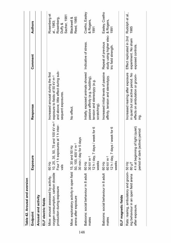
148
Tabl
e 42
. Aro
usal
and
ave
rsio
n
Endp
oint
Expo
sure
Res
pons
eC
omm
ent
Aut
hors
Aro
usal
and
act
ivity
ELF
elec
tric
fiel
ds
Mic
e: a
rous
al a
sses
sed
by a
ctiv
ity,
oxyg
en c
onsu
mpt
ion,
car
bon
diox
ide
prod
uctio
n du
ring
expo
sure
60 H
z10
, 25,
35,
50,
75
and
100
kV m
-1
Four
1 h
exp
osur
es a
t 1 h
inte
r-va
ls
Incr
ease
d ar
ousa
l dur
ing
the
first
ex
posu
re to
fiel
ds o
f 50
kV m
-1
and
abov
e; li
ttle
effe
ct d
urin
g su
b-se
quen
t exp
osur
es.
Ros
enbe
rg e
t al
., 19
83;
Ros
enbe
rg,
Duf
fy &
S
ache
r, 19
81
Mic
e: e
xplo
rato
ry a
ctiv
ity in
ope
n fie
ld
aren
a af
ter e
xpos
ure
15, 3
0 an
d 50
Hz
50, 1
00, 4
00 V
m-1
30 m
in /
day
for 5
day
s
No
effe
ct.
Bla
ckw
ell &
R
eed,
198
5
Bab
oons
: soc
ial b
ehav
iour
in 8
adu
lt m
ales
60 H
z30
kV
m-1
12 h
/ da
y, 7
day
s / w
eek
for 6
w
eeks
Inita
lly, e
xpos
ed a
nim
als
show
ed
pass
ive
affin
ity (e
.g. h
uddl
ing)
, te
nsio
n an
d st
ereo
typy
(e.g
. sc
ratc
hing
).
Indi
cativ
e of
stre
ss.
Coe
lho,
Eas
ley
& R
oger
s,
1991
Bab
oons
: soc
ial b
ehav
iour
in 8
adu
lt m
ales
60 H
z60
kV
m-1
12 h
/ da
y, 7
day
s / w
eek
for 6
w
eeks
Incr
ease
d in
itial
leve
ls o
f pas
sive
af
finity
, ten
sion
and
ste
reot
ypy.
Rep
eat o
f pre
viou
s st
udy
usin
g hi
gher
ele
c-tri
c fie
ld s
treng
th.
Eas
ley,
Coe
lho
& R
oger
s,
1991
ELF
mag
netic
fiel
ds
Rat
s: re
arin
g, a
mbu
lato
ry a
nd g
room
-in
g be
havi
our i
n an
ope
n fie
ld a
rena
af
ter e
xpos
ure.
50 H
z80
µT
4 h
at b
egin
ning
of l
ight
(qui
et)
perio
d or
dar
k (a
ctiv
e) p
erio
d
Incr
ease
d re
arin
g af
ter e
xpos
ure
in q
uiet
but
not
act
ive
perio
d. N
o ef
fect
s on
am
bula
tion
or g
room
-in
g.
Effe
ct re
plic
ated
in 2
nd
expe
rimen
t. N
o sh
am
expo
sed
cont
rols
.
Rud
olph
et a
l.,
1985
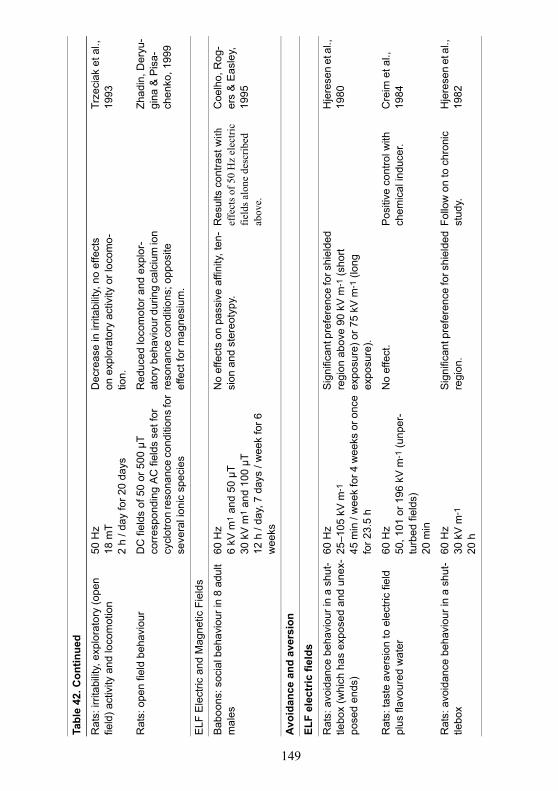
149
Tabl
e 42
. Con
tinue
d
Rat
s: ir
ritab
ility
, exp
lora
tory
(ope
n fie
ld) a
ctiv
ity a
nd lo
com
otio
n50
Hz
18 m
T2
h / d
ay fo
r 20
days
Dec
reas
e in
irrit
abilit
y, n
o ef
fect
s on
exp
lora
tory
act
ivity
or l
ocom
o-tio
n.
Trze
ciak
et a
l.,
1993
Rat
s: o
pen
field
beh
avio
urD
C fi
elds
of 5
0 or
500
µT
corr
espo
ndin
g AC
fiel
ds s
et fo
r cy
clot
ron
reso
nanc
e co
nditi
ons
for
seve
ral i
onic
spe
cies
Red
uced
loco
mot
or a
nd e
xplo
r-at
ory
beha
viou
r dur
ing
calc
ium
ion
reso
nanc
e co
nditi
ons;
opp
osite
ef
fect
for m
agne
sium
.
Zhad
in, D
eryu
-gi
na &
Pis
a-ch
enko
, 199
9
ELF
Ele
ctric
and
Mag
netic
Fie
lds
Bab
oons
: soc
ial b
ehav
iour
in 8
adu
lt m
ales
60 H
z6
kV m
1 an
d 50
µT
30 k
V m
1 an
d 10
0 µT
12
h /
day,
7 d
ays
/ wee
k fo
r 6
wee
ks
No
effe
cts
on p
assi
ve a
ffini
ty, t
en-
sion
and
ste
reot
ypy.
Res
ults
con
trast
with
ef
fect
s of 5
0 H
z el
ectri
c fie
lds a
lone
des
crib
ed
abov
e.
Coe
lho,
Rog
-er
s &
Eas
ley,
19
95
Avoi
danc
e an
d av
ersi
on
ELF
elec
tric
fiel
ds
Rat
s: a
void
ance
beh
avio
ur in
a s
hut-
tlebo
x (w
hich
has
exp
osed
and
une
x-po
sed
ends
)
60 H
z25
–105
kV
m-1
45 m
in /
wee
k fo
r 4 w
eeks
or o
nce
for 2
3.5
h
Sign
ifica
nt p
refe
renc
e fo
r shi
elde
d re
gion
abo
ve 9
0 kV
m-1
(sho
rt ex
posu
re) o
r 75
kV m
-1 (l
ong
expo
sure
).
Hje
rese
n et
al.,
19
80
Rat
s: ta
ste
aver
sion
to e
lect
ric fi
eld
plus
flav
oure
d w
ater
60 H
z50
, 101
or 1
96 k
V m
-1 (u
nper
-tu
rbed
fiel
ds)
20 m
in
No
effe
ct.
Pos
itive
con
trol w
ith
chem
ical
indu
cer.
Cre
im e
t al.,
19
84
Rat
s: a
void
ance
beh
avio
ur in
a s
hut-
tlebo
x 60
Hz
30 k
V m
-1
20 h
Sign
ifica
nt p
refe
renc
e fo
r shi
elde
d re
gion
.Fo
llow
on
to c
hron
ic
stud
y.H
jere
sen
et a
l.,
1982
150
Tabl
e 42
. Con
tinue
d
Bab
oons
: beh
avio
ural
ave
rsio
n th
roug
h op
eran
t res
pons
es (l
ever
pr
ess)
to te
rmin
ate
expo
sure
60 H
zup
to 6
5 kV
m-1
dur
ing
test
ing
Fiel
d pe
rcei
ved
but d
id n
ot a
ct a
s ne
gativ
e (a
vers
ive)
re-in
forc
er.
No
supp
ort f
or s
tress
ef
fect
.R
oger
s, O
rr &
S
mith
, 199
5
Bab
oons
: beh
avio
ural
ave
rsio
n th
roug
h op
eran
t res
pons
es (l
ever
pr
ess)
to te
rmin
ate
expo
sure
60 H
z90
or 1
00 k
V m
-1
brie
f exp
osur
es
Expo
sure
did
not
indu
ce le
ver
pres
sing
(fie
ld te
rmin
atio
n) b
ehav
-io
ur.
Inca
ndes
cent
lam
p as
po
sitiv
e co
ntro
l.St
ern
& L
atie
s,
1989
ELF
mag
netic
fiel
ds
Rat
s: a
void
ance
beh
avio
ur in
a s
hut-
tlebo
x 60
Hz
3.03
mT
1 h
No
effe
ct.
Love
ly e
t al.,
19
92
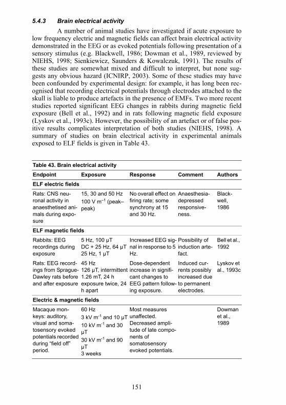
151
5.4.3 Brain electrical activityA number of animal studies have investigated if acute exposure to
low frequency electric and magnetic fields can affect brain electrical activitydemonstrated in the EEG or as evoked potentials following presentation of asensory stimulus (e.g. Blackwell, 1986; Dowman et al., 1989, reviewed byNIEHS, 1998; Sienkiewicz, Saunders & Kowalczuk, 1991). The results ofthese studies are somewhat mixed and difficult to interpret, but none sug-gests any obvious hazard (ICNIRP, 2003). Some of these studies may havebeen confounded by experimental design: for example, it has long been rec-ognised that recording electrical potentials through electrodes attached to theskull is liable to produce artefacts in the presence of EMFs. Two more recentstudies reported significant EEG changes in rabbits during magnetic fieldexposure (Bell et al., 1992) and in rats following magnetic field exposure(Lyskov et al., 1993c). However, the possibility of an artefact or of false pos-itive results complicates interpretation of both studies (NIEHS, 1998). Asummary of studies on brain electrical activity in experimental animalsexposed to ELF fields is given in Table 43.
Table 43. Brain electrical activity
Endpoint Exposure Response Comment Authors
ELF electric fields
Rats: CNS neu-ronal activity in anaesthetised ani-mals during expo-sure
15, 30 and 50 Hz100 V m–1 (peak–peak)
No overall effect on firing rate; some synchrony at 15 and 30 Hz.
Anaesthesia-depressed responsive-ness.
Black-well, 1986
ELF magnetic fields
Rabbits: EEG recordings during exposure
5 Hz, 100 µTDC + 25 Hz, 64 µT25 Hz, 1 µT
Increased EEG sig-nal in response to 5 Hz.
Possibility of induction arte-fact.
Bell et al., 1992
Rats: EEG record-ings from Sprague-Dawley rats before and after exposure
45 Hz126 µT, intermittent1.26 mT, 24 h exposure twice, 24 h apart
Dose-dependent increase in signifi-cant changes to EEG pattern follow-ing exposure.
Induced cur-rents possibly increased due to permanent electrodes.
Lyskov et al., 1993c
Electric & magnetic fields
Macaque mon-keys: auditory, visual and soma-tosensory evoked potentials recorded during “field off” period.
60 Hz3 kV m-1 and 10 µT10 kV m-1 and 30 µT30 kV m-1 and 90 µT3 weeks
Most measures unaffected. Decreased ampli-tude of late compo-nents of somatosensory evoked potentials.
Dowman et al., 1989
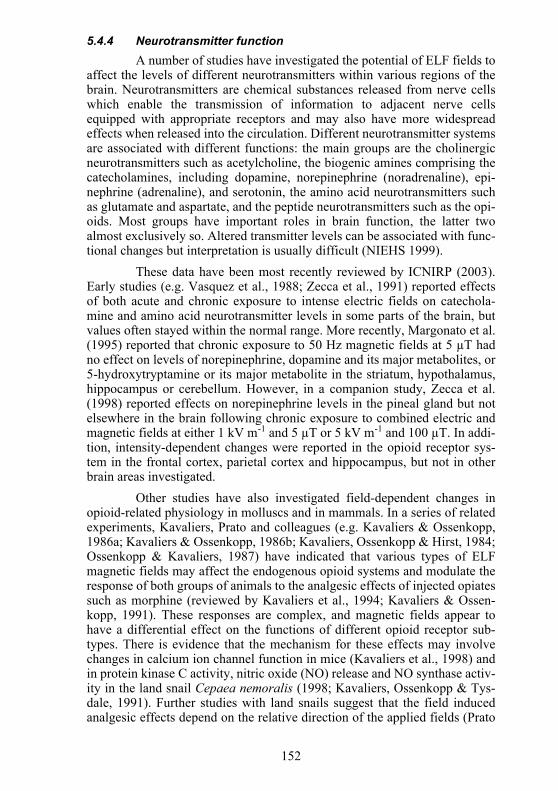
152
5.4.4 Neurotransmitter functionA number of studies have investigated the potential of ELF fields to
affect the levels of different neurotransmitters within various regions of thebrain. Neurotransmitters are chemical substances released from nerve cellswhich enable the transmission of information to adjacent nerve cellsequipped with appropriate receptors and may also have more widespreadeffects when released into the circulation. Different neurotransmitter systemsare associated with different functions: the main groups are the cholinergicneurotransmitters such as acetylcholine, the biogenic amines comprising thecatecholamines, including dopamine, norepinephrine (noradrenaline), epi-nephrine (adrenaline), and serotonin, the amino acid neurotransmitters suchas glutamate and aspartate, and the peptide neurotransmitters such as the opi-oids. Most groups have important roles in brain function, the latter twoalmost exclusively so. Altered transmitter levels can be associated with func-tional changes but interpretation is usually difficult (NIEHS 1999).
These data have been most recently reviewed by ICNIRP (2003).Early studies (e.g. Vasquez et al., 1988; Zecca et al., 1991) reported effectsof both acute and chronic exposure to intense electric fields on catechola-mine and amino acid neurotransmitter levels in some parts of the brain, butvalues often stayed within the normal range. More recently, Margonato et al.(1995) reported that chronic exposure to 50 Hz magnetic fields at 5 µT hadno effect on levels of norepinephrine, dopamine and its major metabolites, or5-hydroxytryptamine or its major metabolite in the striatum, hypothalamus,hippocampus or cerebellum. However, in a companion study, Zecca et al.(1998) reported effects on norepinephrine levels in the pineal gland but notelsewhere in the brain following chronic exposure to combined electric andmagnetic fields at either 1 kV m-1 and 5 µT or 5 kV m-1 and 100 µT. In addi-tion, intensity-dependent changes were reported in the opioid receptor sys-tem in the frontal cortex, parietal cortex and hippocampus, but not in otherbrain areas investigated.
Other studies have also investigated field-dependent changes inopioid-related physiology in molluscs and in mammals. In a series of relatedexperiments, Kavaliers, Prato and colleagues (e.g. Kavaliers & Ossenkopp,1986a; Kavaliers & Ossenkopp, 1986b; Kavaliers, Ossenkopp & Hirst, 1984;Ossenkopp & Kavaliers, 1987) have indicated that various types of ELFmagnetic fields may affect the endogenous opioid systems and modulate theresponse of both groups of animals to the analgesic effects of injected opiatessuch as morphine (reviewed by Kavaliers et al., 1994; Kavaliers & Ossen-kopp, 1991). These responses are complex, and magnetic fields appear tohave a differential effect on the functions of different opioid receptor sub-types. There is evidence that the mechanism for these effects may involvechanges in calcium ion channel function in mice (Kavaliers et al., 1998) andin protein kinase C activity, nitric oxide (NO) release and NO synthase activ-ity in the land snail Cepaea nemoralis (1998; Kavaliers, Ossenkopp & Tys-dale, 1991). Further studies with land snails suggest that the field inducedanalgesic effects depend on the relative direction of the applied fields (Prato
153
et al., 1995) as well as the presence of light (1997; Prato, Kavaliers & Car-son, 1996; 2000).
In another series of experiments, it was reported that the acuteexposure of rats to a 60 Hz magnetic field at 0.75 mT decreased activity inthe cholinergic pathways in the frontal cortex and hippocampus (Lai et al.,1993). These effects were blocked by naltrexone, but not by naloxonemethiodide, which was taken as evidence that magnetic fields affectedendogenous opioids only within the central nervous system. Further studiesshowed the changes in cholinergic activity appeared to be mediated by acti-vation of endogenous opioids (Lai, Carino & Ushijima, 1998). There alsoappears to be some interaction between exposure duration and field intensity,such that longer exposures (3 hours) at lower intensity fields (0.05 mT) couldinduce changes in cholinergic activity similar to those induced by shorterexposure at higher intensity (Lai & Carino, 1999).
Overall, limited changes in neurotransmitter levels in different partsof the rodent brain have been reported. Although of less direct relevance tohuman health, similar results have been reported in the molluscan nervoussystem. The biological significance of the changes seen in mammals is diffi-cult to assess without corroborative changes in brain function and behaviour.However, several studies suggest possible EMF effects on the opioid andcholinergic systems which can be modulated by appropriate antagonists andshould be studied further. Studies on the effects of ELF fields on neurotrans-mitters and analgesia are summarized in Table 44.
5.4.5 Cognitive functionEarly studies with macaque monkeys reported that exposure to ELF
electric fields at well below detection thresholds may affect operant perfor-mance (IARC, 2002; ICNIRP, 2003). However, well-conducted studiesusing baboons found that exposure to 60 Hz electric fields at 30 and 60 kVm-1 had no sustained effect on the performance of two operant schedules(1995b; Rogers et al., 1995a) although initial exposure may contributetowards producing a temporary interruption in responding.
Similarly, studies using 60 Hz electric and magnetic fields (Orr,Rogers & Smith, 1995b) indicated that combined exposure to 6 kV m-1 and50 µT or to 30 kV m-1 and 100 µT had no effect on operant performance on adelayed match-to-sample task in baboons. This result is generally consistentwith earlier results from other research groups using non-human primates(reviewed by ICNIRP, 2003; NIEHS, 1998; Sienkiewicz, Saunders & Kow-alczuk, 1991). However, one study using rats (Salzinger et al., 1990) sug-gested exposure to 60 Hz fields of 30 kV m-1 and 100 µT may exert subtleeffects on performance that depend on the time of testing within the light-dark cycle.
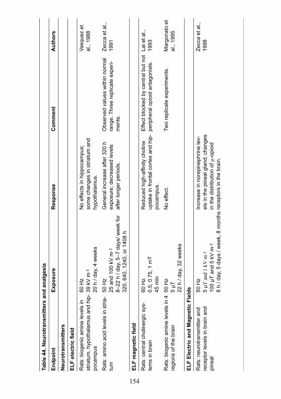
154
Tabl
e 44
. Neu
rotr
ansm
itter
s an
d an
alge
sia
Endp
oint
Expo
sure
Res
pons
eC
omm
ent
Aut
hors
Neu
rotr
ansm
itter
s
ELF
elec
tric
fiel
d
Rat
s: b
ioge
nic
amin
e le
vels
in
stria
tum
, hyp
otha
lam
us a
nd h
ip-
poca
mpu
s
60 H
z39
kV
m-1
20 h
/ da
y, 4
wee
ks
No
effe
cts
in h
ippo
cam
pus;
so
me
chan
ges
in s
triat
um a
nd
hypo
thal
amus
.
Vasq
uez
et
al.,
1988
Rat
s: a
min
o ac
id le
vels
in s
tria-
tum
50 H
z25
and
100
kV
m-1
8–22
h /
day,
5–7
day
s/ w
eek
for
320,
640
, 124
0, o
r 140
8 h
Gen
eral
incr
ease
afte
r 320
h
expo
sure
; dec
reas
ed le
vels
af
ter l
onge
r per
iods
.
Obs
erve
d va
lues
with
in n
orm
al
rang
e. T
hree
repl
icat
e ex
peri-
men
ts.
Zecc
a et
al.,
19
91
ELF
mag
netic
fiel
d
Rat
s: c
entra
l cho
liner
gic
sys-
tem
s in
bra
in60
Hz
0.5,
0.7
5, 1
mT
45 m
in
Red
uced
hig
h-af
finity
cho
line
upta
ke in
fron
tal c
orte
x an
d hi
p-po
cam
pus.
Effe
ct b
lock
ed b
y ce
ntra
l but
not
pe
riphe
ral o
pioi
d an
tago
nist
s.La
i et a
l.,
1993
Rat
s: b
ioge
nic
amin
e le
vels
in 4
re
gion
s of
the
brai
n50
Hz
5 µT
22 h
/ da
y, 3
2 w
eeks
No
effe
ct.
Two
repl
icat
e ex
perim
ents
.M
argo
nato
et
al.,
1995
ELF
Elec
tric
and
Mag
netic
Fie
lds
Rat
s: n
euro
trans
mitt
er a
nd
rece
ptor
leve
ls in
bra
in a
nd
pine
al
50 H
z5
µT a
nd 1
kV
m-1
100
µT a
nd 5
kV
m-1
8 h
/ day
, 5 d
ays
/ wee
k, 8
mon
ths
Incr
ease
in n
orep
inep
hrin
e le
v-el
s in
the
pine
al g
land
; cha
nges
in
the
dist
ribut
ion
of µ
-opi
oid
rece
ptor
s in
the
brai
n.
Zecc
a et
al.,
19
98
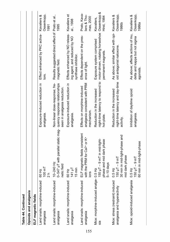
155
Tabl
e 44
. Con
tinue
d
Opi
oids
and
ana
lges
ia
ELF
mag
netic
fiel
ds
Land
sna
ils: m
orph
ine-
indu
ced
anal
gesi
a60
Hz
100
mT
2 h
Exp
osur
e-in
duce
d re
duct
ion
in
anal
gesi
a.E
ffect
enh
ance
d by
PK
C a
ctiv
a-to
rs.
Kav
alie
rs &
O
ssen
kopp
, 19
91
Land
sna
ils: m
orph
ine-
indu
ced
anal
gesi
a10
–240
Hz
0–54
7 µT
with
par
alle
l sta
tic m
ag-
netic
fiel
d
Non
-line
ar d
ose-
resp
onse
; fre
-qu
ency
resp
onse
rela
tions
hips
se
en in
ana
lges
ia re
duct
ion.
Res
ults
sug
gest
ed d
irect
effe
ct o
f m
agne
tic fi
eld.
Pra
to e
t al.,
19
95
Land
sna
ils: m
orph
ine-
indu
ced
anal
gesi
a60
Hz
141
µT15
min
Exp
osur
e-in
duce
d re
duct
ion
in
anal
gesi
a.E
ffect
s en
hanc
ed b
y N
O re
leas
-in
g ag
ent,
and
redu
ced
by N
O
synt
hase
inhi
bito
r.
Kav
alie
rs e
t al
., 19
98
Land
sna
ils: m
orph
ine-
indu
ced
anal
gesi
aE
LF m
agne
tic fi
elds
con
sist
ent
with
the
PR
M fo
r Ca2
+ or
K+
ions
Effe
cts
on m
orph
ine-
indu
ced
anal
gesi
a co
nsis
tent
with
PR
M
mec
hani
sm.
Effe
cts
depe
nden
t on
the
pres
-en
ce o
f lig
ht.
Pra
to, K
ava-
liers
& T
ho-
mas
, 200
0
Mic
e: m
orph
ine-
indu
ced
anal
ge-
sia
0.5
Hz
150
µT –
9 m
T in
mid
-ligh
t-ph
ase
and
mid
dar
k ph
ase
5–10
day
s
Red
uctio
n in
the
incr
ease
d ni
ght-t
ime
late
ncy
to re
spon
d to
ho
t-pla
te.
Exp
osur
e sy
stem
com
pris
ed
mot
or-d
riven
, rot
atin
g ho
rses
hoe
perm
anen
t mag
nets
.
Kav
alie
rs,
Oss
enko
pp &
H
irst,
1984
Mic
e: s
tress
(res
train
t) in
duce
d an
alge
sia
and
hype
ract
ivity
0.5
Hz
150
µT –
9 m
T30
min
in m
id-li
ght-p
hase
and
m
id d
ark
phas
e
Red
uctio
n in
the
incr
ease
d ni
ght-t
ime
late
ncy
and
day-
time
activ
ity.
As
abov
e; s
imila
r effe
ct w
ith o
pi-
oid
anta
goni
st n
alox
one.
Kav
alie
rs &
O
ssen
kopp
, 19
86b
Mic
e: o
pioi
d-in
duce
d an
alge
sia
0.5
Hz
150
µT –
9 m
T60
min
in m
id-li
ght-p
hase
Inhi
bitio
n of
day
time
opio
id
anal
gesi
a.A
s ab
ove;
affe
cts
actio
ns o
f mu,
de
lta a
nd k
appa
but
not
sig
ma
agon
ists
.
Kav
alie
rs &
O
ssen
kopp
, 19
86a
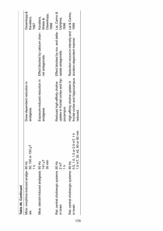
156
Tabl
e 44
. Con
tinue
d
Mic
e: m
orph
ine-
indu
ced
anal
ge-
sia
60 H
z50
, 100
or 1
50 µ
T1
h
Dos
e-de
pend
ant r
educ
tion
in
anal
gesi
a.O
ssen
kopp
&
Kav
alie
rs,
1987
Mic
e: s
tero
id-in
duce
d an
alge
sia
60 H
z14
1 µT
30 m
in
Exp
osur
e-in
duce
d re
duct
ion
in
anal
gesi
a.E
ffect
blo
cked
by
calc
ium
cha
n-ne
l ant
agon
ists
.K
aval
iers
, W
iebe
&
Oss
enko
pp,
1998
Rat
: cen
tral c
holin
ergi
c sy
stem
s in
bra
in60
Hz
2 m
T1
h
Red
uced
hig
h-af
finity
cho
line
upta
ke in
fron
tal c
orte
x an
d hi
p-po
cam
pus.
Effe
ct b
lock
ed b
y m
u- a
nd d
elta
-op
iate
ant
agon
ists
.La
i, C
arin
o &
U
shiji
ma,
19
98
Rat
: cen
tral c
holin
ergi
c sy
stem
s in
bra
in60
Hz
0.5,
1.0
, 1.5
or 2
.0 m
T, 1
h1.
0 m
T, 3
0, 4
5, 6
0 or
90
min
Hig
h-af
finity
cho
line
upta
ke in
fro
ntal
cor
tex
and
hipp
ocam
pus
redu
ced.
Red
uctio
n oc
curs
in in
tens
ity a
nd
dura
tion-
depe
nden
t man
ner.
Lai &
Car
ino,
19
99
157
Several recent studies using the Morris water maze or radial armmaze have investigated the effects of magnetic fields on spatial memory andplace learning. These studies provide evidence that exposure of rats, mice orvoles to power frequency fields at ~100 µT and above may modulate taskperformance (Kavaliers & Ossenkopp, 1993 Kavaliers et al., 1996; Lai,1996; Lai, Carino & Ushijima, 1998; Sienkiewicz et al., 1998; Sienkiewicz,Haylock & Saunders, 1998). Exposure to complex pulsed magnetic fieldsmay also affect performance (McKay & Persinger, 2000; Thomas & Pers-inger, 1997). In addition, much evidence has accrued over the last decadethat effects may also occur using specific combinations of static and time-varying fields (see Sienkiewicz, Haylock & Saunders, 1998). The mecha-nism for these effects has been partly explored and the changes in behaviourhave been attributed to decreases in cholinergic functions caused by field-induced changes in endogenous opioid activity (Lai, 1996; Lai, Carino &Ushijima, 1998; Thomas & Persinger, 1997).
The conditions to produce any of these phenomena are not welldefined, and both deficits and enhancements in performance have beenobserved and one study did not report any field-dependent effects (Sienk-iewicz, Haylock & Saunders, 1996). It is feasible that these differences inoutcome may depend on experimental or other variables including the timingand duration of exposure relative to learning (McKay & Persinger, 2000;Sienkiewicz et al., 2001). While these results suggest that the neural repre-sentations or processes underlying the performance of spatial memory tasksmay be vulnerable to the effects of magnetic fields, some part of theobserved outcome may be attributable to changes in arousal (IARC, 2002;ICNIRP, 2003) or in motivation (Thomas & Persinger, 1997). Nevertheless,the transient nature and small magnitudes of the responses do not suggest anobvious deleterious effect.
Two studies using rodents have investigated the effects of magneticfields on recognition memory. Using the field conditions putatively identi-fied as having an acute effect of spatial memory, Sienkiewicz et al. (2001)found no effects on the performance of an object recognition task by mice.Animals were exposed for 45 minutes to a 50 Hz field at 7.5, 75 or 750 µT.However, Mostafa, Mostafa & Ennaceur et al. (2002) reported that discrimi-nation between familiar and novel objects was impaired in rats followingchronic exposure at 200 µT for 2 weeks.
Stern et al. (1996) failed to replicate the results of earlier studies(Liboff, McLeod & Smith, 1989; Thomas, Schrot & Liboff, 1986) suggest-ing exposure to combined static and power frequency magnetic fields,arranged to simulate the cyclotron resonance conditions for lithium ions, sig-nificantly impaired operant performance. The earlier positive results wereattributed to possible confounding.
A summary of studies on cognitive function in animals is given inTable 45.
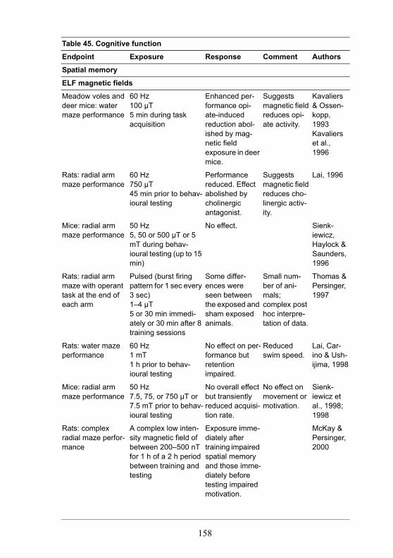
158
Table 45. Cognitive function
Endpoint Exposure Response Comment Authors
Spatial memory
ELF magnetic fields
Meadow voles and deer mice: water maze performance
60 Hz100 µT5 min during task acquisition
Enhanced per-formance opi-ate-induced reduction abol-ished by mag-netic field exposure in deer mice.
Suggests magnetic field reduces opi-ate activity.
Kavaliers & Ossen-kopp, 1993Kavaliers et al., 1996
Rats: radial arm maze performance
60 Hz750 µT45 min prior to behav-ioural testing
Performance reduced. Effect abolished by cholinergic antagonist.
Suggests magnetic field reduces cho-linergic activ-ity.
Lai, 1996
Mice: radial arm maze performance
50 Hz5, 50 or 500 µT or 5 mT during behav-ioural testing (up to 15 min)
No effect. Sienk-iewicz, Haylock & Saunders, 1996
Rats: radial arm maze with operant task at the end of each arm
Pulsed (burst firing pattern for 1 sec every 3 sec)1–4 µT5 or 30 min immedi-ately or 30 min after 8 training sessions
Some differ-ences were seen between the exposed and sham exposed animals.
Small num-ber of ani-mals; complex post hoc interpre-tation of data.
Thomas & Persinger, 1997
Rats: water maze performance
60 Hz1 mT1 h prior to behav-ioural testing
No effect on per-formance but retention impaired.
Reduced swim speed.
Lai, Car-ino & Ush-ijima, 1998
Mice: radial arm maze performance
50 Hz7.5, 75, or 750 µT or 7.5 mT prior to behav-ioural testing
No overall effect but transiently reduced acquisi-tion rate.
No effect on movement or motivation.
Sienk-iewicz et al., 1998; 1998
Rats: complex radial maze perfor-mance
A complex low inten-sity magnetic field of between 200–500 nT for 1 h of a 2 h period between training and testing
Exposure imme-diately after training impaired spatial memory and those imme-diately before testing impaired motivation.
McKay & Persinger, 2000
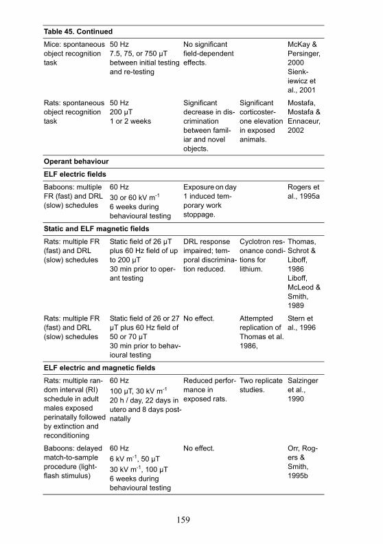
159
Table 45. Continued
Mice: spontaneous object recognition task
50 Hz7.5, 75, or 750 µTbetween initial testing and re-testing
No significant field-dependent effects.
McKay & Persinger, 2000Sienk-iewicz et al., 2001
Rats: spontaneous object recognition task
50 Hz200 µT1 or 2 weeks
Significant decrease in dis-crimination between famil-iar and novel objects.
Significant corticoster-one elevation in exposed animals.
Mostafa, Mostafa & Ennaceur, 2002
Operant behaviour
ELF electric fields
Baboons: multiple FR (fast) and DRL (slow) schedules
60 Hz30 or 60 kV m-1
6 weeks during behavioural testing
Exposure on day 1 induced tem-porary work stoppage.
Rogers et al., 1995a
Static and ELF magnetic fields
Rats: multiple FR (fast) and DRL (slow) schedules
Static field of 26 µT plus 60 Hz field of up to 200 µT30 min prior to oper-ant testing
DRL response impaired; tem-poral discrimina-tion reduced.
Cyclotron res-onance condi-tions for lithium.
Thomas, Schrot & Liboff, 1986Liboff, McLeod & Smith, 1989
Rats: multiple FR (fast) and DRL (slow) schedules
Static field of 26 or 27 µT plus 60 Hz field of 50 or 70 µT30 min prior to behav-ioural testing
No effect. Attempted replication of Thomas et al. 1986,
Stern et al., 1996
ELF electric and magnetic fields
Rats: multiple ran-dom interval (RI) schedule in adult males exposed perinatally followed by extinction and reconditioning
60 Hz100 µT, 30 kV m-1
20 h / day, 22 days in utero and 8 days post-natally
Reduced perfor-mance in exposed rats.
Two replicate studies.
Salzinger et al., 1990
Baboons: delayed match-to-sample procedure (light-flash stimulus)
60 Hz6 kV m-1, 50 µT30 kV m-1, 100 µT6 weeks during behavioural testing
No effect. Orr, Rog-ers & Smith, 1995b
160
5.5 ConclusionsExposure of volunteers to power frequency electric fields causes
well-defined biological responses, ranging from perception to annoyance,through surface electric-charge effects. These responses depend on fieldstrength, ambient environmental conditions, and individual sensitivity. Thethresholds for direct perception by 10% of volunteers varied between 2 and20 kV m-1, while 5% found 15–20 kV m-1 annoying. The spark dischargefrom a person to ground is found to be painful by 7% of volunteers in a fieldof 5 kV m-1. Thresholds for the discharge from an object through a groundedperson depend on the size of the object and therefore require specific assess-ment.
High field strength, rapidly pulsed magnetic fields can stimulateperipheral or central nerve tissue; such effects can arise during MRI exposureand are used in transcranial magnetic stimulation. Threshold induced electricfield strengths for direct nerve stimulation could be as low as a few volts permetre. The threshold is likely to be constant over a frequency range betweena few hertz and a few kilohertz. People suffering from or predisposed to epi-lepsy are likely to be more susceptible to induced ELF electric fields in theCNS. Furthermore, sensitivity to electrical stimulation of the CNS seemslikely to be associated with a family history of seizure and the use of tricyclicantidepressants, neuroleptic agents and other drugs that lower the seizurethreshold.
The function of the retina, which is part of the CNS, can be affectedby exposure to much weaker ELF magnetic fields than those that cause directnerve stimulation. A flickering light sensation, called magnetic phosphenesor magnetophosphenes, results from the interaction of the induced electricfield with electrically excitable cells in the retina. Threshold electric fieldstrengths in the extracellular fluid of the retina have been estimated to liebetween about 10–100 mV m-1 at 20 Hz. There is, however, considerableuncertainty attached to these values.
The evidence for other neurobehavioural effects in volunteer stud-ies, such as the effects on brain electrical activity, cognition, sleep, hypersen-sitivity and mood, is less clear. Generally, such studies have been carried outat exposure levels below those required to induce effects described above,and have produced evidence only of subtle and transitory effects at best. Theconditions necessary to elicit such responses are not well defined at present.There is some evidence suggesting the existence of field-dependent effectson reaction time and reduced accuracy in the performance of some cognitivetasks, which is supported by the results of studies on the gross electricalactivity of the brain. Studies investigating EMF-induced changes in sleepquality have reported inconsistent results. It is possible that these differencesmay be attributable in part to differences in the design of the studies.
Some people claim to be hypersensitive to EMF. However, the evi-dence from double blind provocation studies suggests that the reportedsymptoms are unrelated to EMF exposure.
161
There is only inconsistent and inconclusive evidence that exposureto ELF electric and magnetic fields causes depressive symptoms or suicide.Thus, the evidence is considered inadequate.
In animals, the possibility that exposure to ELF fields may affectneurobehavioral functions has been explored from a number of perspectivesusing a range of exposure conditions. Few robust effects have been estab-lished. There is convincing evidence that power-frequency electric fields canbe detected by animals, most likely as a result of surface charge effects, andmay induce transient arousal or mild stress. In rats, the detection range isbetween 3 and 13 kV m-1. Rodents have been shown to be aversive to fieldstrengths greater than 50 kV m-1. Other possible field-dependent changes areless well-defined and generally laboratory studies have only produced evi-dence of subtle and transitory effects. There is some evidence that exposureto magnetic fields may modulate the functions of the opioid and cholinergicsystems, and this is supported by the results of studies investigating theeffects on analgesia and on the acquisition and performance of spatial mem-ory tasks.