4474-4481-nox1 promotes colon cancer cell metastasis via ... · nox1 promotes colon cancer cell...
TRANSCRIPT

4474
world1. The traditional curative treatment for patients with colon cancer is still surgical re-section. However, within five years post-oper-ative, about 25 to 35% of colon cancer patients with node-positive develop either local recur-rence or distant metastasis, because of the re-lease of inflammatory mediators in response to surgical trauma2. The aberrant expression of inflammatory mediators can potentiate the metastatic ability to circulate cancer cells and residual cancer cells, which contribute to the significant reduction of disease-free and overall survival for patients with colorectal cancer3-6.
The cancer cell metastatic cascade is com-posed of several steps, among which the cancer cell adherence is an essential and critical step7. On one hand, the high level of exogenous ROS can enhance the ability to circulate cancer cells to adhere to the endothelial lining and preferen-tially to the exposed extracellular matrix. On the other hand, the low level of endogenous ROS can promote cell survival through regulation of re-dox-sensitive survival pathways to facilitate can-cer cell metastasis8. Endogenous ROS is mainly produced by NADPH oxidase 1 (Nox) enzymes, which compose a family of seven enzymes, Nox1, Nox2, Nox3, Nox4, Nox5, Duox1, and Duox29. The expression of Nox enzymes in cancer cells has recently been documented, and Nox-derived ROS has been shown to facilitate the metastatic process in several cancer cells, including colon, gastric, melanoma, and pancreatic cancer cells10-12. However, the underlying molecular mechanism of Nox1 in the regulation of colon cancer progres-sion remains largely unknown.
Tumor necrosis factor-α (TNF-α) converting enzyme (TACE)/a disintegrin and metalloprote-ase domain 17 (ADAM17) is the primary secre-
Abstract. – OBJECTIVE: Reactive oxygen spe-cies (ROS) generated by endogenous metabol-ic enzymes are involved in a variety of pathology processes, including cancer. In particular, super-oxide-generating NADPH oxidase 1 (Nox1), a mem-ber of Nox enzyme family, is highly expressed in the colon tissue and has been implicated in phys-iological and pathophysiological states of colon cancer. However, the underlying molecular mech-anism of Nox1 in the regulation of colon cancer progression remains largely unknown.
MATERIALS AND METHODS: In vitro scratch wound healing and invasion assays were used to compare the migration and invasion abilities of HT29 cells in which Nox1 protein levels were ma-nipulated. Western blot assay was performed to detect the expression of key proteins of the EG-FR-PI3K-AKT signaling pathway. Immunoprecip-itation assay was performed to detect the inter-action between Nox1 and ADAM17.
RESULTS: Nox1 overexpression promoted co-lon cancer cell growth, migration, and invasion through the EGFR-PI3K-AKT signaling pathway. At the molecular level, Nox1 regulated the ex-pression of tumor necrosis factor-α (TNF-α) con-verting enzyme (TACE)/a disintegrin and metal-loprotease domain 17 (ADAM17). Furthermore, Nox1 interacted with and stabilized ADAM17 from ubiquitin-mediated degradation, leading to the activation of the ADAM17 signaling pathway.
CONCLUSIONS: This study suggests that Nox1 promotes colorectal cancer metastasis by modulating the stability of ADAM17.
Key Words: Nox1, Colorectal cancer, ADAM17, Migration, Inva-
sion.
Introduction
Colorectal cancer is the second most common cause of cancer-related mortality in the western
European Review for Medical and Pharmacological Sciences 2016; 20: 4474-4481
H.-P. WANG, X. WANG, L.-F. GONG, W.-J. CHEN, Z. HAO, S.-W. FENG, Y.-B. WU, T. YE, Y.-K. CAI
Department of General Surgery, Shanghai Fifth People Hospital Affiliated to Fudan University, Shanghai, China
Hui-Peng Wang and Xin Wang are the co-first author
Nox1 promotes colon cancer cell metastasis via activation of the ADAM17 pathway
Corresponding Author: Tao Ye, MD, e-mail: [email protected] Cai, MD, [email protected]

Nox1 promotes colon cancer cell metastasis via activation of the ADAM17 pathway
4475
tase responsible for the release of the soluble form of TNF-α from the plasma membrane13. Accumu-lating evidence shows that the ADAM17-EGFR-PI3K-Akt pathway plays a critical role in cancer cell migration and invasion14,15. Herein, we show that Nox1 interacts with and stabilises ADAM17 protein to activate the ADAM17-EGFR-PI3K-Akt signaling pathway in colon cancer cells.
Materials and Methods Cell culture and reagents
HCT116, HCT8, HT29, LS174T, LOVO, SW480, and SW620 colon cancer cell lines were obtained from the American Type Culture Collection (Manas-sas, VA, USA). Cells were cultured in Dulbecco’s modified Eagle’s medium (DMEM) or RPMI 1640 medium supplemented with 10% foetal bovine se-rum and maintained at 37C° in a humidified atmo-sphere with 5% CO2. Flag M2 beads, mouse IgG, DMSO, and cycloheximide were purchased from Sigma-Aldrich (St. Louis, MO, USA).
Lentivirus-based shRNA, RNA extraction, and real-time PCR
Nox-1 shRNA (h) was purchased from OriGene Technologies (TF311135, Rockville, MD, USA), TACE (ADAM17) shRNA (h) lentiviral particles (sc-36604-V) and control shRNA lentiviral parti-cles (sc-108080) were purchased from Santa Cruz Biotechnology (Santa Cruz, CA, USA). Total RNA was isolated from cells with TRIzol reagent (Invi-trogen, Carlsbad, CA, USA) and reverse transcrip-tion was performed by using Takara RNA PCR kit (Takara, Dalian, China) to generate cDNA, fol-lowing the manufacturer’s instructions. Real-time PCR was performed using a SYBR Green Premix Ex Taq (Takara) on an ABI 7500 machine to deter-mine the expression of individual transcripts. Prim-er sequences were obtained from the qPrimer Depot website (http://primerdepot.nci.nih.gov/).
Cell growth curveHT29 cells were plated at 1 × 10⁴ cells/well in
24-well plates containing a complete medium. Every day, the medium was removed and the ad-herent cells were trypsinised and quantified using a haemocytometer. Cell counts of three wells per day and per group were averaged.
Western blottingCells were harvested and washed with cold PBS.
The cells were lysed with ice-cold RIPA lysis buf-
fer for 30 min. After centrifugation at 20000 × g for 15 min at 4gation at 20000d with cold PBS. The cells were by using the bicinchoninic acid (BCA as-say (Pierce, Rockford, IL, USA), separated by 12% sodium dodecyl sulphate-polyacrylamide gel elec-trophoresis (SDS-PAGE), and transferred to PVDF membranes. Membranes were blocked with 10% non-fat milk in PBS and then immunoblotted with the indicated antibodies, followed by HRP-linked secondary antibodies (Cell Signaling Technology, Danvers, MA, USA). The primary antibodies against Nox1, AKT, p-AKT, MEK, p-MEK, VEGF, EGFR, and p-EGFR were purchased from Cell Signaling Technology. ADAM17 antibody was purchased from Santa Cruz Biotechnology. Flag and HA an-tibodies were purchased from Sigma-Aldrich. The signals were detected by Millipore™ Immobilon™ Western Chemiluminescent HRP Substrate (ECL) kit (Millipore, Fisher Scientific, Waltham, MA, USA) according to the manufacturer’s instructions. Protein levels were normalized to total GAPDH, using a mouse anti-GAPDH antibody (Santa Cruz Biotechnology, Santa Cruz, CA, USA).
Immunoprecipitation (IP)Co-IP was performed on lysates from cells trans-
fected with the indicated plasmids. Cells were lysed in RIPA buffer, and supernatants were clarified and precipitated with Flag-M2 agarose beads for 3 h at 4C°. Beads with immune complexes were washed six times with RIPA buffer and boiled in SDS-PAGE sample buffer. For endogenous IP, 1 × 10⁷ cells were lysed in RIPA buffer, and supernatants were clari-fied and precipitated with Nox1 antibody or mouse IgG overnight at 4C°. Protein G beads were added and incubated for 2 h. Beads with immune com-plexes were washed six times with RIPA buffer and boiled in SDS-PAGE sample buffer.
Migration AssayFor in vitro scratch wound healing assays,
monolayers of HT29 cells were cultured in 12-well plates and then physically wounded by using a 200-µL yellow pipette tip. The area devoid of cells was imaged immediately and 36 h later. The Nikon Eclipse TS100 microscope (Nikon, Singa-pore) was used to visualize and image cultured cells. The NIS-Elements F.300 software was used to acquire the imaging data.
Invasion AssayThe invasion ability of HT29 cells was tested
by matrigel invasion assays. Cells were cultured in 24-well BD invasion chambers at a concentra-
H.-P. Wang, X. Wang, L.-F. Gong, W.-J. Chen, Z. Hao, S.-W. Feng, Y.-B. Wu, T. Ye, Y.-K. Cai
4476
tion of 1 × 10⁵cells/well without serum and im-mediately placed onto the upper compartment of the plates. The lower compartment was filled with complete medium containing 10% serum. Cells in the chambers were incubated for 36 h, and cells that migrated on the lower surface of the mem-brane were stained with crystal violet and fixed in 4% paraformaldehyde. The invasion ability was expressed as a percentage of control.
Statistical AnalysisResults were statistically evaluated by using two-
way ANOVA, and Bonferroni comparison post-test or t-test using GraphPad Prism 5.0 software (Graph-Pad, San Diego, CA, USA). Results are shown as means ± S D. Statistical significance is displayed as *(p < 0.05), **(p < 0.01), or *** (p < 0.001).
Results
Screening of Colon Cancer Cell Lines to Identify Cells Expressing the Highest Level of Endogenous Nox1
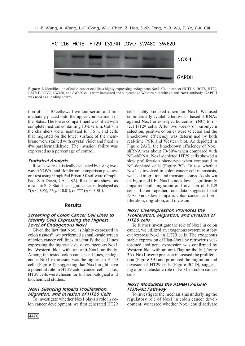
Given the fact that Nox1 is highly expressed in colon tissues16, we performed a small-scale screen of colon cancer cell lines to identify the cell lines expressing the highest level of endogenous Nox1 by Western blot with an anti-Nox1 antibody. Among the tested colon cancer cell lines, endog-enous Nox1 expression was the highest in HT29 cells (Figure 1), suggesting that Nox1 might have a potential role in HT29 colon cancer cells. Thus, HT29 cells were chosen for further biological and biochemical studies.
Nox1 Silencing Impairs Proliferation, Migration, and Invasion of HT29 Cells
To investigate whether Nox1 plays a role in co-lon cancer development, we first generated HT29
cells stably knocked down for Nox1. We used commercially available lentivirus-based shRNAs against Nox1 or non-specific control (NC) to in-fect HT29 cells. After two weeks of puromycin selection, positive colonies were selected and the knockdown efficiency was determined by both real-time PCR and Western blot. As depicted in Figure 2A-B, the knockdown efficiency of Nox1 shRNA was about 70-80% when compared with NC-shRNA. Nox1-depleted HT29 cells showed a slow proliferation phenotype when compared to NC-depleted cells (Figure 2C). To test whether Nox1 is involved in colon cancer cell metastasis, we used migration and invasion assays. As shown in Figure 2D-E, Nox1 knockdown significantly impaired both migration and invasion of HT29 cells. Taken together, our data suggested that Nox1 knockdown impairs colon cancer cell pro-liferation, migration, and invasion.
Nox1 Overexpression Promotes the Proliferation, Migration, and Invasion of HT29 cells
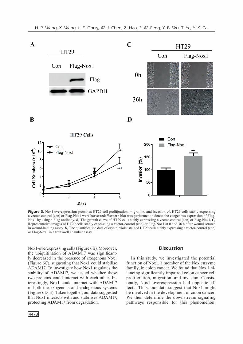
To further investigate the role of Nox1 in colon cancer, we utilized an exogenous system to stably overexpress Nox1 in HT29 cells. The exogenous stable expression of Flag-Nox1 by retrovirus vec-tor-mediated gene expression was confirmed by Western blot with an anti-Flag antibody (Figure 3A). Nox1 overexpression increased the prolifera-tion (Figure 3B) and promoted the migration and invasion of HT29 cells (Figure 3C-D), suggest-ing a pro-metastatic role of Nox1 in colon cancer cells.
Nox1 Modulates the ADAM17-EGFR-PI3K-Akt Pathway
To investigate the mechanisms underlying the regulatory role of Nox1 in colon cancer devel-opment, we tested whether Nox1 could activate
Figure 1. Identification of colon cancer cell lines highly expressing endogenous Nox1. Colon cancer HCT116, HCT8, HT29, LS174T, LOVO, SW480, and SW620 cells were harvested and subjected to Western blot with an anti-Nox1 antibody. GAPDH was used as a loading control.

Nox1 promotes colon cancer cell metastasis via activation of the ADAM17 pathway
4477
signaling pathways known to be involved in co-lon cancer development. The EGFR-PI3K-Akt pathway was impaired in Nox1-depleted cells, while it was activated in Nox1-overexpressing cells (Figure 4A-B). Moreover, ADAM17 was decreased in Nox1-depleted cells (Figure 4B). These data suggest that Nox1 may act as a mod-ulator upstream of the ADAM17-EGFR-PI3K-Akt signaling pathway.
ADAM17 Inhibition Prevents Nox1-in-duced Colon Cancer Cell Proliferation, Migration, and Invasion
Then, we tested whether the ADAM17-EGFR-PI3K-Akt signaling pathway contributed to the biological function of Nox1 in colon cancer cells. ADAM17 inhibition largely abolished Nox1-in-duced colon cancer cell proliferation, migration, and invasion (Figure 5A-D). Thus, our data sug-
gested a role of Nox1 in the regulation of the AD-AM17-EGFR-PI3K-Akt signaling pathway.
Nox1 Interacts with and Stabilises ADAM17
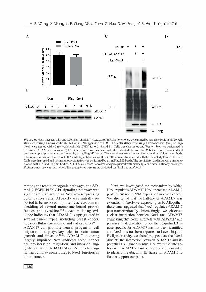
To further investigate how Nox1 regulates the expression of ADAM17 to activate its downstream signaling pathway, we tested whether Nox1 could induce ADAM17 mRNA expression. However, Nox1 inhibition only induced subtle changes in ADAM17 mRNA levels, suggesting that Nox1 does not regulate ADAM17 mRNA expression (Figure 6A). In contrast, we observed a clear de-crease in ADAM17 protein levels in Nox1-deplet-ed cells, while ADAM17 protein levels increased in Nox1-overexpressing cells (Figure 4A-B), sug-gesting that Nox1 post-transcriptionally regulates ADAM17 protein levels. Consistently, we found that the half-life of ADAM17 was extended in
Figure 2. Nox1 silencing impairs proliferation, migration, and invasion of HT29 cells. A, Nox1 mRNA levels determined by real time PCR in HT29 cells stably expressing a non-specific shRNA or a shRNA against Nox1. B, Nox1 protein levels determined by Western blot in HT29 cells stably expressing a non-specific shRNA or a shRNA against Nox1. GAPDH was used as a loading control. C, Growth curve of HT29 cells stably expressing a non-specific shRNA or a shRNA against Nox1. D, Representative images of HT29 cells stably expressing a non-specific shRNA or a shRNA against Nox1 at 0 and 36 h after wound scratch in wound-healing assay. E, Cell migration ability was analyzed by transwell chamber assay. The representative images of crystal violet stained HT29 cells stably expressing a non-specific shRNA or a shRNA against Nox1.
H.-P. Wang, X. Wang, L.-F. Gong, W.-J. Chen, Z. Hao, S.-W. Feng, Y.-B. Wu, T. Ye, Y.-K. Cai
4478
Nox1-overexpressing cells (Figure 6B). Moreover, the ubiquitination of ADAM17 was significant-ly decreased in the presence of exogenous Nox1 (Figure 6C), suggesting that Nox1 could stabilise ADAM17. To investigate how Nox1 regulates the stability of ADAM17, we tested whether these two proteins could interact with each other. In-terestingly, Nox1 could interact with ADAM17 in both the exogenous and endogenous systems (Figure 6D-E). Taken together, our data suggested that Nox1 interacts with and stabilises ADAM17, protecting ADAM17 from degradation.
Discussion
In this study, we investigated the potential function of Nox1, a member of the Nox enzyme family, in colon cancer. We found that Nox 1 si-lencing significantly impaired colon cancer cell proliferation, migration, and invasion. Consis-tently, Nox1 overexpression had opposite ef-fects. Thus, our data suggest that Nox1 might be involved in the development of colon cancer. We then determine the downstream signaling pathways responsible for this phenomenon.
Figure 3. Nox1 overexpression promotes HT29 cell proliferation, migration, and invasion. A, HT29 cells stably expressing a vector-control (con) or Flag-Nox1 were harvested; Western blot was performed to detect the exogenous expression of Flag-Nox1 by using a Flag antibody. B, The growth curve of HT29 cells stably expressing a vector-control (con) or Flag-Nox1. C, Representative images of HT29 cells stably expressing a vector-control (con) or Flag-Nox1 at 0 and 36 h after wound scratch in wound-healing assay. D, The quantification data of crystal violet stained HT29 cells stably expressing a vector-control (con) or Flag-Nox1 in a transwell chamber assay.

Nox1 promotes colon cancer cell metastasis via activation of the ADAM17 pathway
4479
Figure 4. Nox1 modulates the ADAM17-EGFR-PI3K-Akt path-way. A, HT29 cells stably ex-pressing a non-specific shRNA or a shRNA against Nox1 were harvested; Western blot was per-formed by using the indicated an-tibodies. B, HT29 cells stably ex-pressing a vector-control (con) or Flag-Nox1 were harvested; West-ern blot was performed by using the indicated antibodies.
Figure 5. ADAM17 inhibition prevents Nox1-induced colon cancer cell proliferation, migration, and invasion. A, Flag-Nox1-HT29 cells were infected with lentivirus expressing a non-specific shRNA or a shRNA against ADAM17. Cells were harvested and ADAM17 knockdown efficiency was determined by Western blot. B, The growth curve of Flag-Nox1-HT29 cells stably expressing a non-specific shRNA or a shRNA against ADAM17. C, Representative images of Flag-Nox1-HT29 cells stably expressing a non-specific shRNA or a shRNA against ADAM17 at 0 and 36 h after wound scratch in wound-healing assay. D, The quantification data of crystal violet stained Flag-Nox1-HT29 cells stably expressing a non-specific shRNA or a shRNA against ADAM17 in a transwell chamber assay.
H.-P. Wang, X. Wang, L.-F. Gong, W.-J. Chen, Z. Hao, S.-W. Feng, Y.-B. Wu, T. Ye, Y.-K. Cai
4480
Among the tested oncogenic pathways, the AD-AM17-EGFR-PI3K-Akt signaling pathway was significantly activated in Nox1-overexpressing colon cancer cells. ADAM17 was initially re-ported to be involved in proteolytic ectodomain shedding of several membrane-bound growth factors and cytokines17,18. Accumulating evi-dence indicates that ADAM17 is upregulated in several cancer types, including breast cancer, hepatocellular carcinoma, and colon cancer19-21. ADAM17 can promote neural progenitor cell migration and plays key roles in brain tumor growth and invasion22,23. ADAM17 silencing largely impaired Nox1-induced colon cancer cell proliferation, migration, and invasion, sug-gesting that the ADAM17-EGFR-PI3K-Akt sig-naling pathway contributes to Nox1 function in colon cancer.
Next, we investigated the mechanism by which Nox1 regulates ADAM17. Nox1 increased ADAM17 protein, but not mRNA expression in colon cancer. We also found that the half-life of ADAM17 was extended in Nox1-overexpressing cells. Altogether, these data suggested that Nox1 regulates ADAM17 post-transcriptionally. Interestingly, we observed a clear interaction between Nox1 and ADAM17, suggesting that Nox1 interacts with ADAM17 and prevents its degradation. Since the ubiquitin E3 li-gase specific for ADAM17 has not been identified and Nox1 has not been reported to have ubiquitin E3 ligase activity, we, therefore, speculate that Nox1 disrupts the interaction between ADAM17 and its potential E3 ligase via mutually exclusive interac-tion with ADAM17. Further studies are warranted to identify the ubiquitin E3 ligase for ADAM17 to further support our point.
Figure 6. Nox1 interacts with and stabilises ADAM17. A, ADAM17 mRNA levels were determined by real time PCR in HT29 cells stably expressing a non-specific shRNA or shRNA against Nox1. B, HT29 cells stably expressing a vector-control (con) or Flag-Nox1 were treated with 40 mM cycloheximide (CHX) for 0, 2, 4, and 8 h. Cells were harvested and Western blot was performed to determine ADAM17 expression. C, HT29 cells were co-transfected with the indicated plasmids for 36 h. Cells were harvested and co-immunoprecipitation was performed by using Flag M2 beads. The precipitates were immunoblotted with an ubiquitin antibody. The input was immunoblotted with HA and Flag antibodies. D, HT29 cells were co-transfected with the indicated plasmids for 36 h. Cells were harvested and co-immunoprecipitation was performed by using Flag M2 beads. The precipitates and input were immuno-blotted with HA and Flag antibodies. E, HT29 cells were harvested and precipitated with mouse IgG or a Nox1 antibody overnight. Protein G agarose was then added. The precipitates were immunoblotted for Nox1 and ADAM17.
Nox1 promotes colon cancer cell metastasis via activation of the ADAM17 pathway
4481
Conclusions
Our data clearly indicate a pro-oncogenic role of Nox1 in colon cancer. Additionally, we showed that Nox1 regulates colon cancer cell proliferation and invasion through the ADAM17-EGFR-PI3K-Akt signaling pathway. Our data support the no-tion that Nox1 might be a potential drug target for the treatment of colon cancer.
AcknowledgementsThe study was supported by the project of Shanghai Fifth People s Hospital Affiliated to Fudan Universi-ty (No 2015WYYJ01) AND the project of Science and Technology Commission of Minhang District (No. 2015MH2027).
Conflicts of interestThe authors declare no conflicts of interest.
References
1) Jemal a, Siegel R, WaRd e, Hao Y, Xu J, THun mJ. Cancer statistics 2009. CA Cancer J Clin 2009; 59: 225-249.
2) KobaYaSHi H, mocHizuKi H, SugiHaRa K, moRiTa T, KoTaKe K, TeRamoTo T, KameoKa S, SaiTo Y, TaKa-HaSHi K, HaSe K, oYa m, maeda K, HiRai T, KameYama m, SHiRouzu K, muTo T. Characteristics of recur-rence and surveillance tools after curative resec-tion for colorectal cancer: a multicenter study. Surgery 2007; 141: 67-75.
3) moRSe ma, naiR S, FeRnandez-caSal m, deng Y, ST Pm, WilliamS R, HobeiKa a, moSca P, claY T, cum-ming Ri, FiSHeR e, clavien P, PRoia ad, niedzWiecKi d, caRon d, lYeRlY HK. Preoperative mobilization of circulating dendritic cells by Flt3 ligand admin-istration to patients with metastatic colon cancer. J Clin Oncol 2000; 18: 3883-3893.
4) Silva Jm, RodRiguez R, gaRcia Jm, munoz c, Silva J, dominguez g, PRovencio m, eSPana P, bonilla F. Detection of epithelial tumor RNA in the plasma of colon cancer patients is associated with ad-vanced stages and circulating tumor cells. Gut 2002; 50: 530-534.
5) Huang HY, zHang zJ, cao cb, Wang n, liu FF, Peng JQ, Ren XJ, Qian J. The TLR4/NF-kappaB signaling pathway mediates the growth of colon cancer. Eur Rev Med Pharmacol Sci 2014; 18: 3834-3843.
6) Tang J, cHen JX, cHen l, Tang JY, cui z, liu cH, Wang z. Metastasis associated in colon cancer 1 (MACC1) promotes growth and metastasis pro-cesses of colon cancer cells. Eur Rev Med Phar-macol Sci 2016; 20: 2825-2834.
7) maSSague J, obenauF ac. Metastatic colonization by circulating tumor cells. Nature 2016; 529: 298-306.
8) niSHiKaWa m. Reactive oxygen species in tumor metastasis. Cancer Lett 2008; 266: 53-59.
9) PeTRY a, WeiTnaueR m, goRlacH a. Receptor acti-vation of NADPH oxidases. Antioxid Redox Signal 2010; 13: 467-487.
10) JuHaSz a, ge Y, maRKel S, cHiu a, maTSumoTo l, van balgooY J, RoY K, doRoSHoW JH. Expression of NA-DPH oxidase homologues and accessory genes in human cancer cell lines, tumors and adjacent nor-mal tissues. Free Radic Res 2009; 43: 523-532.
11) Kim eY, Seo Jm, Kim c, lee Je, lee Km, Kim JH. BLT2 promotes the invasion and metastasis of aggressive bladder cancer cells through a reac-tive oxygen species-linked pathway. Free Radic Biol Med 2010; 49: 1072-1081.
12) WeaveR am. Regulation of cancer invasion by reactive oxygen species and Tks family scaffold proteins. Sci Signal 2009; 2: e56.
13) RoSe-JoHn S. ADAM17, shedding, TACE as thera-peutic targets. Pharmacol Res 2013; 71: 19-22.
14) zHeng X, Jiang F, KaTaKoWSKi m, zHang X, Jiang H, zHang zg, cHoPP m. Sensitization of cerebral tissue in nude mice with photodynamic therapy induces ADAM17/TACE and promotes glioma cell invasion. Cancer Lett 2008; 265: 177-187.
15) lin P, Sun X, Feng T, zou H, Jiang Y, liu z, zHao d, Yu X. ADAM17 regulates prostate cancer cell pro-liferation through mediating cell cycle progression by EGFR/PI3K/AKT pathway. Mol Cell Biochem 2012; 359: 235-243.
16) KRauSe KH. Tissue distribution and putative physi-ological function of NOX family NADPH oxidases. Jpn J Infect Dis 2004; 57: S28-S29.
17) ceRReTTi dP. Characterization of the tumor ne-crosis factor alpha-converting enzyme, TACE/ADAM17. Biochem Soc Trans 1999; 27: 219-223.
18) boRland g, muRPHY g, ageR a. Tissue inhibitor of metalloproteinases-3 inhibits shedding of L-selectin from leukocytes. J Biol Chem 1999; 274: 2810-2815.
19) zHeng X, Jiang F, KaTaKoWSKi m, lu Y, cHoPP m. ADAM17 promotes glioma cell malignant pheno-type. Mol Carcinog 2012; 51: 150-164.
20) KennY Pa, biSSell mJ. Targeting TACE-dependent EGFR ligand shedding in breast cancer. J Clin In-vest 2007; 117: 337-345.
21) blancHoT-JoSSic F, JaRRY a, maSSon d, bacH-ngo-Hou K, Paineau J, deniS mg, laboiSSe cl, moSnieR JF. Up-regulated expression of ADAM17 in hu-man colon carcinoma: co-expression with EGFR in neoplastic and endothelial cells. J Pathol 2005; 207: 156-163.
22) li Q, zHang z, li z, zHou m, liu b, Pan l, ma z, zHeng Y. ADAM17 is critical for multipolar exit and radial migration of neuronal intermediate progeni-tor cells in mice cerebral cortex. PLoS One 2013; 8: e65703.
23) zHeng X, Jiang F, KaTaKoWSKi m, KalKaniS Sn, Hong X, zHang X, zHang zg, Yang H, cHoPP m. Inhibition of ADAM17 reduces hypoxia-induced brain tumor cell invasiveness. Cancer Sci 2007; 98: 674-684