4 556789 $: ; > >$99
TRANSCRIPT
Acta Otolaryngology 93: 31-41, 1982
PATTERNS OF HAIR CELL LOSS IN CHICK BASILAR PAPILLAAFTER INTENSE AUDITORY STIMULATION
Exposure duration and survival time!
Edwin W. Rubel and Brenda M. Ryals
From the Laboratory of Developmental Neuro-Otology , Departments of Otolaryngology, Physiologyand Speech Pathology & Audiology, University of Virginia Medical School, Charlottesville,
Virginia, USA
(Received February 17, 1981)
Abstract. Ten-day-old chicks were exposed to a puretone (1.5 kHz) or white noise at 125 dB SPL avg.I RMS for 4 to 24 hours, and were sacrificed either 10,! 30 or 60 days after exposure. The basilar papillae were; embedded in plastic, sectioned, and hair cells wereI counted at 100-/Lm intervals throughout the length ofj the papilla. As sound exposure duration increased,iboth the maximum number of hair cells lost, and the1 extent of the damaged area along the basilar membraneI increased. Short hair cells situated on the free area ofI the basilar papilla were more susceptible to damagei than were tall hair cells. The location of hair .cell lossi varied as a function of frequency band of exposure;.the pure tone produced a well localized basal lesion,\.while wide-band noise produced a more general lesion),which extending toward the apex. Degeneration con-l tinued with increased survival time up to 30 days. Itlis concluded that avians respond to acoustic over-i stimulation in a manner very similar to mammals. The;convenience of this preparation along with the relativeIsimplicity of its cochlea may render it useful for futurednvestigations of the mechanisms of acoustic trauma.
Wor many years it has been known thatexposure to intense sound can cause per-imanent structural changes to the sensory'epithelium of the mammalian cochlea (Haber-,mann, 1890; Guild, 1921; Miller, Watson &Covell, 1963). While the use of other verte-brate classes has proven quite rewarding forstUdies of peripheral auditory function(Wever, 1974), central processing (Capranica,:'1976), and auditory development (Rubel,~1978), experimental models to study the pe-ripheral and central effects of acoustic over-stimulation have generally been limited tomammals.
A great deal of literature is available de-scribing both the development and structureof the avian middle ear (Smith, 1905; Borg,et al., 1979), inner ear (Takasaka & Smith,1971; Tanaka & Smith, 1975; Cohen &Fermin, 1978; Hirokawa, 1978; Tanaka &Smith, 1978) and central auditory pathways(Rubel & Parks, 1975; Rubel, Smith &Miller, 1976; Parks & Rubel, 1978; Smith &Rubel, 1979). Moreover, the avian basilarpapilla is convenient for histological prepara-tion and analysis due to its short, uncoilednature which allows serial section recon-structions (Guild, 1921) within a reasonabletime frame. Finally, since avian auditorysystem development has been extensivelydocumented, the developmental effects ofperturbations of the auditory environment,such as noise exposure, can be related toother aspects of auditory system ontogeny.Two previous reports concerning the re-
sponse of the avian inner ear to acoustictrauma (Bohne & Dooling, 1974; Dooling &Saunders, 1974) used surface preparationtechniques and suggested that there wasessentially no hair cell degeneration afterintense· acoustic stimulation (106 dB SPL for12 hours). Further investigation into thesusceptibility of avian inner ear hair cells to
1 A preliminary account of this study was. presented atthe Association for Research in Otolaryngology, Mid-winter Research Meeting, 1979.
Acta Otolaryngol 93
32 E. W. Rubel and B. M. Ryals
Table I. Summary of groups
Stimulation conditionDurationof Pure Plugged
Survival exposure tone White ortime (hours) (1.5 kHz) noise none
4 4 Ears10 Days 8 4 Ears
12 4 Ears 4 Ears} 8 Ears"24 4 Ears 4 Ears
30 Days 24 2 Ears 2 Ears
60 Days 24 2 Ears 2 Ears
Total N=40 ears.a Data from ears that were plugged during sound expo-sure (n =4) were combined with data from normal ears(n =4) since there was no difference between these controlgroups.
acoustic trauma using serial sections andvariations in acoustic intensity level, fre-quencies of stimulation, exposure durationsand survival was felt to be warranted. Inthe next paper (Ryals & Rubel, 1981) wedescribe the location of hair cell damage as afunction of the frequency spectra of acousticoverstimulation.
METHODS
l. SubjectsEight experimental and four control groupsof to-day-old Hubbard x Hubbard chickswere used (see Table I). The 2-8 chicks ineach group were obtained from randomhatching groups. Normal control subjects notexposed to the acoustic trauma conditionsor papillae from ears that had been protectedby an ear plug during exposure served ascontrols. Since the intensity level at whichhair cell destruction first occurred was un-known, a 125 dB SPL intensity level waschosen to maximize effects. One pure tone(1.5 kHz) chosen to be near the middle ofthe animals' frequency range and near theirmaximum sensitivity (Saunders et aI., 1974;Kerr et aI., 1979) was chosen. This allowedassessment of the specificity of damage. In
Acta Oto/aryngo/93
addition, a white noise stimulus was usedto produce a widespread pattern of stimula-tion throughout the basilar membrane. Expo-sure durations extended from 4 to 24 hoursof continuous stimulation. Chicks were sacrificed either 10, 30 or 60 days after noiseexposure. A summary of all conditions isshown in Table I.
2. Sound exposureSignal generation. Audio signals were gene-rated by either a noise generator (GS 90lB)or an audio oscillator (Wavetek Model 134),amplified (Southwest Technical Products2071A) and led to a freefield speaker (IRSPower Horn 40-1238). Acoustic signals werecalibrated using a 1/2-inch electret micro-phone (GR 19-72) with preamplifier(l5600P42) and a wave analyser (GR 1521-B).The acoustic environment of the chamber inwhich animals were confined during noiseexposure was calibrated with a constantvoltage input and was shown to be acoustically flat through 2000 Hz. Spectral analysis,was performed with a microphone positionel]near the animal's ear canal before, duringIand after auditory exposure and showed a1 .
flat frequency response for the white noiseI I
condition up to 4000 Hz. Sound pressurellevels for the pure tone stimulation were!measured at the level of the ear canal and'
1. twere calibrated to 125±2 dB. Levels for tbel.experimental tone and its harmonics were!C
. h' Inthen measured at 10 other positions Wit In1,d' Ithe chamber. Both the 1.5 kHz tone an Ibroad band noise were within 3 dB througb·!!out the chamber. The first and second haq:monies of the 1.5 kHz tone were >30 dB!below the fundamental throughout tbelJ
chamber. 'AProcedure. Ten-day-old chicks werel
placed, in pairs, in a small wire mesh tubuJalltrt e
chamber (5 x5 1/2 inch) directly beneath tbe;:IRS power horn inside an acoustic chamberi:'(lAC) and were continuously exposed !CI.hieither the pure tone or white noise stimuJUlla;for the desired duration. Decibel levels near!~n,
i:'

10
II
It
InF, IT' I 'IJ Eg. . ransverse sectton through basi ar papi la (appr.~3.3 mm from proximal tip) in a normal 40-day-oldjchicken. At this point tall hair cells predominate the
I.fhe ear canal were monitored continuouslyI,during exposure using a l-inch condenseri/nicrophone (B&K 4145) attached to a soundlevel meter (B&K 2204) set either on a linear,scale (for white noise stimuli) or the appro-I,pnate octave band during pure tone stimula-tion. ',
l. Ear plugging,",caustic over-stimulation was provided,Illonaurally in some animals so that the otherear could be used for within-animal com-.Jarisons. The ear plugging procedure used toj)rotect one ear from over-stimulation hasI)een described elsewhere (Kerr et al., 1979)lind has been shown to provide at least 40 dB
-812955
ACOUSlic trauma and hair eel/loss in chicken 33
".. ,~\~" ~,', "-. .'\ ~,,.. ,
"-;' 1\
width of the basilar membrane. TV = tegmentum vasculo-sum, EM = basilar membrane, H = habenula, TM =tectorial membrane. Bar indicates 100 ,urn.
attenuation from 125 to 4500 Hz. Afteracoustic over-stimulation earplugs wereremoved. It has previously been shown (Kerret al., 1979) that the plugs can be left inplace for as long as 10 days with no adverseeffect on cochlear or eighth nerve function.
4. Fixation and tissue preparationAfter 10, 30 or 60 days survival durationthe chicks were deeply anesthetized by intra-venous injection of Nembutal. A direct intra-labyrinthine perfusion of I % paraforrnal-dehyde 0.75 % glutaraldehyde mixture wasperformed bilaterally 'immediately after de-capitation. The entire head was then im-mersed in cold fixative for 8-12 hours. The
Acta Oto!aryngo! 93

34 E. W. Rubel and B. M. Ryals
:3 30-'wu
"~ 20:J:
..J
;:~ 10
z<w
'" o 25%.97mm
50%1.94mm
75%2.91 mm
100%3.8 Bmm
DISTANCE FROM BASEFig. 2. Shaded area shows mean total hair cell counts(± I S.E.M.) in 20-day-old chicks (N=8). Black lineshows mean total hair cell counts (± I S.E.M.) in 40-day-old subjects (N=2). Note decrease in older chicksat the distal (apical) end.
basilar papillae were removed from the skull,washed in phosphate buffer and post-fixedin 2% osmium tetroxide (in PO" pH=7.3)for 2 hours. Following osmication the papil-lae were dehydrated in a graded methanolseries and embedded in Epon. The em-bedded papillae were sectioned transverseto the longitudinal axis in the proximal todistal (basal to apical) direction using an LKBUltramicrotome. A group of three or four,3-j.l.m thick sections were collected at each100-j.l.minterval throughout the length of thepapilla, mounted in serial order, and stainedwith toluidine blue.
5. Quantitative analysis of number ofhair cellsQuantitative analysis of the number of haircells at each level of the basilar papilla wasperformed by viewing each section under40x planapochromatic oil immersion objec-tive (NA= 1.0) at a total magnification ofx640. In order to maintain consistency andreliability between hair cell counts, countingcriteria were established. A hair cell wascounted when the following criteria weremet: presence of cuticular plate, cilia, andcell body. The average counts from the threesections at each 10Q.j.l.minterval were then
Acta Ototuryngol 93
RESULTS
plotted as a function of normalized distance(% of total membrane length) from the proximal tip (base) of the basilar membrane.
I. Normative baseline hair cell countsFig. I is a photomicrograph showing thegeneral appearance of the basilar papillaseen in a 3-j.l.m thick section taken 85%(appr. 3.3 mm) from the proximal or basaltip. As described by Retzius (1884), twotypes of hair cell are found in the papilla:tall hair cells located primarily on thesuperior edge of the fibro-cartilaginous plate,and short hair cells located toward the freeedge of the basilar membrane. For the purposes of this study both cell types were combined for total cell counts. In general, cellcounts revealed a small number (5-6) of haircells across the basilar membrane at theproximal tip of the basilar papilla, gradualhincreasing in number as the distal tip warapproached until a maximum of 25-30 eel]were seen at approximately 90% of length.Fig. 2 shows the mean total hair cell counsobtained throughout the length of the normalpapilla. Since qualitative and quantitativeanalysis showed no difference in cell appearaance or number (lob,.=1.15, df=158) tht,plugged ear counts (4 ears) were combine'with the counts from the non-exposed su~jects (4 ears).
A comparison of the number and distri·bution of hair cells along the basilar papilllin 2Q.day-old chicks vis-a-vis the numbelfound in 40-day-old chicks is also shown ilFig. 2. In older chicks the general appear·
Fig. 3. (a) p'hotomicrograph taken appro 1.35 mm fro~the proximal tip of a normal basilar papilla. (b) Ph?lµgraph taken in same area (1.41 mm from prOX'fI\1'lti~) of an animal expo.sed to 1.5 ~Hz for 4 hO~I~S.(I;Higher power photomicrograph of damaged hall eelseen in a section taken 1.5 mm from basal rip of I~Isame animal shown in 3b (1.5 kHz, 125 dB, 4 hourtPointers indicate cells which were considered abno~and did not meet the criteria for counting. a & b, bars- IfLm; C, bar=20 fLm.

Acoustic trauma and hair cell loss ill chicken 35
A •
B .!.
,c-..~
.'. 'M'lIk:''''''... ".4Ir I
.'
c •
AC/(l Ototaryngol 93

36 E. W. Rubel and B. M. Ryals
~ 30..Jw 4 hruc;<i 20r..J
~o 10...z«ur
"~ 30..JWU
c;< 20r..J
~010...z«w
"
12 hr
..
o 25% 50%,97mm 1.94mm
75%2.91 mm
DISTANCE FROM BASE
Fig. 4. Mean total hair cell counts in animals exposedto a 1.5 kHz pure (one at 125 dB SPL for 4, 8, 12or 24 hours (± I S.E.M.) and allowed to survive 10days. Shaded area shows mean total hair cell counts
ance of the hair cells along the papilla wasthe same as in 10-day-olds, though therewere fewer hair cells in the distal portionof the papilla. The hair cell number wasreduced by 16% at the distal tip (from 65 to100% length) in the 4Q-day-old chicks repre-senting a reduction in total hair cell numberof 4 %. There were no subsequent changesin older (67 days) chickens,
Before turning to the quantitative analysisof hair cell loss, qualitative observationsregarding changes in hair cell morphologyafter noise exposure should be noted. Fig. 3shows a comparison of a typical region ofhair cell damage in a traumatized papilla(3b) and the same region in a normal papilla(3a). It is apparent that fewer cells arepresent and in particular, short hair cellsseem lost. In addition, the remaining hair
Acta Otolaryngol 93
1500 Hz
24 hr
100% 03.88 mm
25%.97mm
75%2.91 mm
100%3.8 8 rn~
50%1.94mm
01 STANCE FROM BASE
in control animals (±l S.E.M.). Black arrows indicatemidpoint of damage, white arrows indicate point dmaximum hair cell 105s.
cell bodies are distorted, cilia are clurnpel I
or missing and a general tlattening of somesupportive cells can be seen. An exampleof the damaged cells from a noise-exposed Iear is shown at higher magnification in IFig.3c. I
I3. Quantitative assessment of hair cell loss I
Duration of exposure. Fig. 4 and 5 shoWIthe mean number of hair cell counts fromIanimals surviving 10 days following exposu~to the 1500-Hz and white noise at all exp~ Isure durations. As can be seen in Fig. 4 theIposition of hair cell loss resulting from exp~ isure to 1500 Hz was remarkably discrete Iand constant across animals and exposure Idurations. With pure tone exposure, IhI Ilonger the duration of the acoustic stimLlI~Ition the larger the degree of total hair ce~I
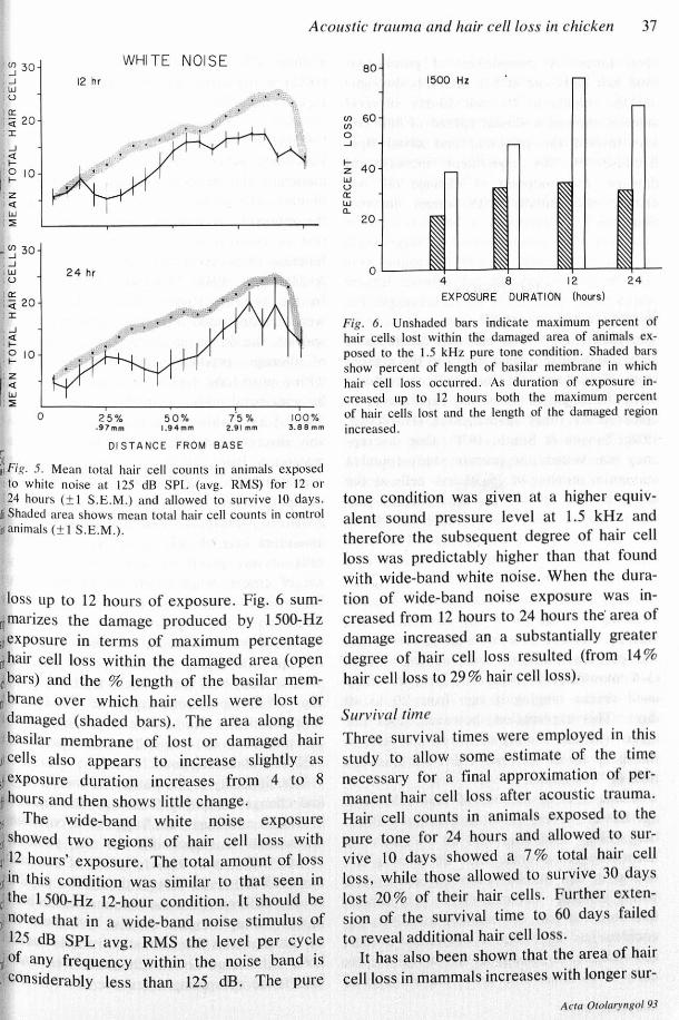
Fig. 6. Unshaded bars indicate maximum percent ofhair cells lost within the damaged area of animals ex-posed to the 1.5 kHz pure tone condition. Shaded barsshow percent of length of basilar membrane in whichhair cell loss occurred. As duration of exposure in-creased up to 12 hours both the maximum percent
100% of hair cells lost and the length of the damaged region3.88mm increased.
.~ 30
.-'
.W
.U
100'<r. 20I
-'a'f-I'~10IZ1«,WI:>
WHITE NOISE12 hr
-:~ 30.-',WU
'00i <r. 20II
-'«.....,~ 10
I;:i,WI:>
24 hr
a i5%2.91 mm
25%.97mm
50%1.94mm
DISTANCE FROM BASE,II .iIF1g.5. Mean total hair cell counts in animals exposedto white noise at 125 dB SPL (avg. RMS) for 12 or24 hours (± I S.E.M.) and allowed to survive 10 days.
at Shaded area shows mean total hair cell counts in controlirianimals (± I S.E.M.).
loss Up to 12 hours of exposure. Fig. 6 sum-~marizes the damage produced by 1500-HzIlexposure in terms of maximum percentage~hair cell loss within the damaged area (open,ibars) and the % length of the basilar mern-n brane over which hair cells were lost ordamaged (shaded bars). The area along thebasilar membrane of lost or damaged hair
sl cells also appears to increase slightly asIIexposure duration increases from 4 to 8~hours and then shows little change.II The wide-band white noise exposure:.~showed two regions of hair cell loss with,,12 hours' exposure. The total amount of loss:1 in this condition was similar to that seen inc'the 1500-Hz 12-hour condition. It should beinoted that in a wide-band noise stimulus of125 dB SPL avg. RMS the level per cycle
n' of any frequency within the noise band is. considerably less than 125 dB. The pure
Acoustic trauma and hair cell loss in chicken 37
801500 Hz
r-U) 60U)
0-'
f- 40Zwo00wa. 20
0 '--- ~4 8 12 24EXPOSURE DURATION (hours)
tone condition was given at a higher equiv-alent sound pressure level at 1.5 kHz andtherefore the subsequent degree of hair cellloss was predictably higher than that foundwith wide-band white noise. When the dura-tion of wide-band noise exposure was in-creased from 12 hours to 24 hours the' area ofdamage increased an a substantially greaterdegree of hair cell loss resulted (from 14%hair cell loss to 29% hair cell loss).
Survival timeThree survival times were employed in thisstudy to allow some estimate of the timenecessary for a final approximation of per-manent hair cell loss after acoustic trauma.Hair cell counts in animals exposed to thepure tone for 24 hours and allowed to sur-vive 10 days showed a 7% total hair cellloss, while those allowed to survive 30 dayslost 20% of their hair cells. Further exten-sion of the survival time to 60 days failedto reveal additional hair cell loss.It has also been shown that the area of hair
cell loss in mammals increases with longer sur-
Acta Oro/arYlIgo/93

38 E. W. Rubel and B. M. Ryals
vival times. A comparison of percentagetotal hair cells lost at 5% intervals through-out the papilla in 10- and 30-day survivalanimals showed a similar spread of hair cellloss toward the proximal and distal tips.Because of this equivalent increase indamage, the midpoint of damage did notchange substantially with longer survivaltimes.
DISCUSSION
Normative hair cell countsThe pattern of normal hair cell distributionsalong the basilar papilla found in the presentstudy closely resemble hair cell counts takenalong the length of the basilar papilla in adultchickens by other investigators (Hirokawa,1978; Tanaka & Smith, 1978). One discrep-ancy was noted; the present study found amaximum number of 25-30 hair cells at thedistal portion of the papilla, whereas pre-vious studies found a maximum of 45-46hair cells at the distal portion. The mostlikely explanation for this discrepancy is ageneral difference in criteria used in cellcounting. No definition of criteria for cellcounting was given in the aforementionedstudies. Another possible explanation is thatthe previous authors used adult chickens(3-4 months old) while the present studyused chicks ranging in age from 20 to 40days. This explanation, however, does notagree with our finding of fewer hair cells inthe distal tip of 40- and 70-day-old chickensthan in 20-day-olds.A decrease in number of hair cells with
age has also been shown in mammals. Bred-berg (1968) used fetal ears as the basis fornormative cell counts, since he found a de-crease in cell number between fetuses andyoung children. Coleman (1976) in a norma-tive study of hair cell number in guinea pigcochlea has also shown decreases in haircell number, especially at the apical tip, asearly as 24 hours after birth. Our findings of
Acta Otolaryngol 93
!a total. 4 % n~ducti?n,. w.ith its maxim~rnl(16%) m the distal tip, IS III agreement WIth,these results.
Damage due to acoustic overstimulationFor many years it has been known that inmammals the general location and amountof hair cell damage varies as a function ofthe intensity, frequency spectra, and duration of acoustic over-stimulation, and as afunction of survival time (e.g. Lurie, Davis& Hawkins, 1944; Stockwell et al., 1969).In the present study similar relationshipswere demonstrated in a representative avianspecies, the domestic chick. The comparisonof damage produced by over-stimulationlwith a pure tone versus the injury producedby wide-band noise exemplifies this parallel.The 1.5 kHz white noise produced a remarkably discrete and non-variable lesion with amaximum hair cell loss of over 80% atapproximately 1/3 of the way from the baseto apex. White noise, on the other hand,produced widespread damage resulting in almaximum loss of only 1/3 to 1/2 of the hairlcells at any given location. These results',lare of course what would be expected on,the basis of mammalian data and/or a con"sideration of the amplitude and shape of thntravelling wave produced by these stimuli]Perhaps the most notable result of the!present study was the lack of variability seenlafter 1.5 kHz pure-tone exposure. The exte~tito which this is due to differences in analytl.,\cal methods, or true differences between.birds and mammals is unclear. IThe similarity in the pattern of morpholog·!
ical changes resulting from acoustic traumaIin avians and mammals "suggests a commonImechanism is responsible for the position!of hair cell loss at intense levels of stimulaJtion (125 dB), even though the basilar paPilla!I".is very different from the mammalian cochlea.in many morphological details. Two theoreti-Ical mechanisms for acoustic trauma, meta'bolic exhaustion and mechanical stress, aresaid to be competing when the level of the

xilstimulus is greater than 120 dB (Spoendlin,1(11976). It has been postulated that these twomechanisms may interact differently as afunction of the frequency spectra, duration'lor survival time of the subjects. Differencesn~!withrespect to the frequency of overstimula-ffiltionwill be discussed in the next paper,iowhile the possible effects of exposure dura-Ition and survival time are briefly consideredIhere.n As exposure time increased, the region oflhair cell loss did not change appreciablynllneither conditions. With the 1.5 kHz pureatone cell loss increased with increases in'&exposure duration up to 12 hours. As ex-JllOectedby the relative intensities there was'Ill marked increase in cell loss between 12mnd 24 hours of exposure to broad-band:nOoise. Instead of analysing percentage hairwir.ellloss with exposure duration, some in-I%vestigatorshave considered length of damage:~long the basilar membrane as the dependent~Ineasure. Spoendlin & Brun (1973), usinglProad-band noise, have shown a consistenterelationship between logarithmic increases,~!ntime of exposure and linear increases in~~helength of damage along the basilar mem-Iprane in the guinea pig. They saw an initiald'ather sharp increase in damage length when'itXposure time was increased with a gradualrevelling off of damage length. Our resultsI~how a similar trend. With 1.5 kHz exposure~lhere was an initial increase in the length~I~fdamage from 4 to 8 hours, with the lengthI;>fdamage at 8, 12 and 24 hours remaining{elatively constant. Twenty-four hour expo-n~urewith wide-band noise produced reliable~amage throughout the papilla which was$10t true in the 12 hour exposure condition.,~ith respect to the proposed dichotomy~etween mechanical and metabolic factors,It seems reasonable that increases in expo-I~ureduration would have a progressive effect:~n metabolic factors, whereas an asympto-flatic effect on mechanical factors would1100n be reached. If this is indeed the caselit is not clear why increasing exposure dura-
Acoustic trauma and hair cell/oss in chicken 39
tion from 12 to 24 hours in the pure-tonecondition did not eliminate all of the haircells at the position of maximum basilarmembrane movement. Possibly the couplingof mechanical and metabolic factors reducesprogressive hair cell injury after major injuryhas been sustained.Longer post-exposure survival times up to
30 days revealed a continuation of hair celldegeneration. Investigations of mammalshave suggested that it takes from one toseveral months for the full extent of haircell degeneration to occur after noise expo-sure. In the chick it appears that hair cellloss can be detected by 10 days followingsound exposure; it then continues, at a dimin-ished rate, during the succeeding 20 daysby which time it appears maximum. Longersurvival times also caused hair cell degenera-tion to spread toward the base and apex.This general spread might be predicted as afunction of continuing degeneration due tometabolic factors which were not revealedat short survival times.
CONCLUSIONS
In the present report we have shown that thepattern of damage produced by acousticoverstimulation of the avian basilar papillais very similar to that reported for mammals.Hair cell loss varies systematically with theacoustic spectra, exposure time and survivaltime in ways that are apparently identicalwith what has been found in mammals. Inthis respect it is clear that avian prepara-tions, because of the accessibility of theorgan and the relative ease of quantitativehistological analysis, may provide an excel-lent model for future studies of acoustictrauma. In the following paper we willfurther develop this model by assessing therelationship between the frequency ofacoustic over-stimulation and the positionof hair cell loss. We will then further con-sider the implications for. understandingacoustic trauma.
Acta Otolaryngol 93

40 E. W. Rubel and B. M. Ryals
ACKNOWLEDGEMENTSThe authors gratefully acknowledge the assistance ofthe following people: Patricia Palmer provided excellenthistological assistance; R. Bruce Masterton and ZahrlG. Schoeny provided critiques of earlier drafts of themanuscript; Marilyn Engelhardt and Deborah Steinprovided secretarial and editorial assistance. The re-search was supported by NIH grant no. NS 15395 andfunds from the Deafness Research Foundation; salarysupport was provided by NIH RCDA no. NS 00305(E. W. R.) and Office of Education Fellowship (B. R.).
ZUSAMMENFASSUNGZehn Tage alte Hiihnchen wurden entweder mit einemreinen Ton von 1,5 Hz oder mit weiBem Rauschenbeschallt. Den Untersuchungen wurden Reize mit denfolgenden Eigenschaften zugrunde gelegt: 125 dB SPLdurchschnittliche RMS mit einer Reizdauer von 4 bis24 Stunden. Die Versuchstiere wurden 10, 30 oder60 Tage nachher geopfert. Nachdern die Partes basilaresin Kunststoff eingebettet und durchgeschnitten wurden,konnte man die Haarzellen in histologischen Probe-schnitten jeweils nach Abteilungen von 100 JLm durchdie Lange der Partis basilaris zahlen, Indem man dieReizlange erhohte, vergroflerte sich die Maximalnummerder verschwundenen Haarzellen und es brei tete sich derUmfang des Beschadigung entlang der Partis basilarisaus. Kurze Haarzellen, die sich in dem freien Teil derPartis basilaris befinden, schienen empfindlicher ge-geniiber grofseren Haarzellen. Die Stellung des Haarzel-lenverlusts konnte auf die Breite des Reiztonbereichszuruckgefuhrt werden: Reine Tone veranlaBten klarumgrenzte Verletzungen, wahrend breitere Rauschfre-quenzbande eine verbreitete Verletzung, die sich zurSpitze ausdehnte, verursachten. Es wird vorgeschlagen,daB die Reaktion der Vogel auf akustische Uberrei-zungen mit der von Saugetieren verglichen werden kann,Wegen der Bequemlichkeit und verhaltnisrnallig un-komplizierten Struktur der Cochlea eignet sich diesesPraparat fur weitere Untersuchungen des akustischenTraumas.
REFERENCESBohne, B. A. & Dooling, R. J. 1974. Morphologicalchanges in the ears of noise-exposed parakeets.J Acoust Soc Am, Supp!. 55, 577.
Borg, E., Counter, S. A. & Rydqvist, B. 1979. Con-traction properties and functional morphology of theavian stapedius muscle. Acta Otolaryngol (Stockh),88,20.
Bredberg, G. 1968. Cellular pattern and nerve supplyof the human organ of Corti. Acta Otolaryngol(Stockh), Suppl. 236.
Capranica, R. R. 1976. Morphology and Physiology ofthe Auditory System in Frog Neurobiology (ed.R. Llinas &W. Precht). Springer-Verlag, Berlin.
Cohen, G. M. & Fermin, D. C. 1978. The development
Acta Otolaryngol 93
of hair cells in the embryonic chick's basilar papillalActa Otolaryngol (Stockh), 86, 342. i
Coleman, J. W. 1976. Hair cell loss as a function oftage in the normal cochlea of the guinea pig. ActaOtolaryngol (Stockh), 82, 33. I
Dooling, R. J. & Saunders, J. C. 1974. Threshold Shift"produced by continuous noise exposure in the para]keet.] Acoust Soc Am 55, 577 (A).
Guild, S. R. 1921. A graphic reconstruction methoo,for the study of the organ of Corti. Anat Rec 22".141.
Habermann, J. 1890. Uber die Schwerhorigkeit dellKesselschmiede. Arch Ohrenheilk 30, I. 1
Hirokawa, N. 1978. The ultrastructure of the basilaripapilla of the chick.u Com Neurol lSl , 361. !
Kerr, L. M., Ostapoff', E. M. & Rubel, E. W. I979,!Influence of acoustic experience on the ontogeny offrequency generalization gradients in the chickenlJ Exp Psy: Anim Behav Proc 5,97. i
Lurie, M. H., Davis, H. & Hawkins, J. E. 19441Acoustic trauma of the organ of Corti in the guinea!pig. Laryngoscope 54, 375. !
Miller, J. D., Watson, C. & Covell, W. 1963. Deafeniol!effects of noise in the cat. Acta Otolaryngolf(Stockh), Supp!. /76. f
Parks, T. N. & Rubel, E. W. 1978. Organization aoo!development of brain stem auditory nuclei of thelchicken: Primary afferent projections. J COnlPiNeurol/80,439. !
Retzius, G. 1884. Das Gehororgan der Wirbeltiere IflDas Gehororgan der Reptilien, der Vogel und Saug~itiere. Samson & Wallin, Stockholm. ii'
Rubel, E. W. 1978. Ontogeny of structure and function{in the vertebrate auditory system. In Handbook aj!Sensory Physiology (ed. M. Jacobson), Vol. IXISpringer-Verlag, New York. i
Rubel, E. W. & Parks, T. M. 1975. Organization anJrdevelopment of brain stem auditory nuclei of the;chicken: Tonotopic organization of n, magnocel/u'ilaris andn. laminarisi J Camp Neurol/64, 411. f
Rubel, E. W., Smith, D. J. & Miller, L. C. 1976,Organization and development of brain stem aUdiftory nuclei of the chicken: Ontogeny of n, magn&,cellularis and n. laminaris. J Camp Neural 166'1469. J
Ryals, B. M. & Rubel, E. W. 1981. Patterns of halllcell loss in chick basilar papilla after intense aUdl'I••tory stimulation: Frequency organization. Acta 01&,laryngol (Stockh). In press. I
Saunders, J. C. & Dooling, R. J. 1974. Noise induC~tthreshold shift in the parakeet (MelopsitfOCl/ilundulutusi, Proc Nat Acad Sci (SA) 7/, 1962-1965. f
Smith, D. J. & Rubel, E. W. 1979. Organization anl~development of brain stem auditory nuclei in (hll'chicken: Dendritic gradients in n. laminaris. J Cowl!!eurol/86,213. . .. 1
Smith, G. 1905. The middle ear and columella of blrd1fQuart] Micros Sci, 48, 11. , tSpoendlin, H. 1976. Anatomical changes. fOIlOWlmj.•...••....•various noise exposures. In Effects of Noise 1/
Hearing (ed. D. Henderson). Raven Press. Nell
York; f
I

Spoendlin, H. & Brun, J. P. 1973. Relation of structuraldamage to response time and intensity in acoustictrauma. Acta Otolaryngol (Stockh) 75, 220.
Stockwell, W. W., Ades, H. W. & Engstrom, H. 1969.Patterns of hair cell damage after intense auditorystimulation. Ann Otol Rhinol Laryngol 78, 1144.
Takasaka, T. & Smith, C. A. 1971. The structure andinnervation of the pigeon's basilar papilla. J Ultra-struct Res 35,20.
Tanaka, K. & Smith, C. A. 1975. Structure of the aviantectorial membrane. Ann Otol Rhinal Laryngol 84,287.
Tanaka, K. & Smith, C. A. 1978. Structure of the
Acoustic trauma and hair cell/ass in chicken 41
chicken's inner ear: SEM and TEM study. Am JAnat /53,251.
Wever, E. G. 1974. The evolution of vertebrate hearing.In Handbook of Sensory Physiology (ed. W. Keidel&W. Neff), vol VII. Springer-Verlag, New York.
Dr Edwin W. RubelDept of OtolaryngologyBox 430University of Virginia Medical SchoolCharlottesvilleVirginia 22908USA
Acta Otolaryngol 93