3t mr imaging of the pediatric cartilage using 3d dual...
TRANSCRIPT
Introduction
Hyaline cartilage development is essential in pediatric musculoskeletal growth. Although in an adult patient, we focus solely on the articular cartilage, in the pediatric population, the articular, epiphysial and physial cartilage play an important role in skeletal maturation. The epiphysial and physial cartilage participate in enchondral ossification and contrib-ute to a child’s longitudinal growth while the articular cartilage buffers and transmits forces across joints. Although functionally and anatomi-cally different, the three different types of cartilage in the pediatric population are histologically alike.
Magnetic resonance imaging (MRI) is the ideal modality for the assess-ment of pediatric* cartilage due to its lack of ionizing radiation and inher-ent excellent soft tissue contrast [1].
3T MR Imaging of the Pediatric Cartilage Using 3D Dual Echo Steady State (DESS)Shivani Ahlawat, M.D.1; Abraham Padua, Jr2; Thierry A G M Huisman, M.D.3; John A Carrino, M.D. MPH1
1 The Russel H. Morgan Department of Radiology and Radiological Science, Johns Hopkins University School of Medicine, Baltimore, MD, USA 2 Siemens Medical Solutions, Malvern, PA, USA
3Division of Pediatric Radiology, Russell H. Morgan Department of Radiology and Radiological Science, Bloomberg Children’s Center, The Johns Hopkins Hospital, Baltimore, MD, USA
An optimal MRI sequence designed specifically for imaging hyaline carti-lage should be able to accurately detect cartilage thickness and volume, characterize subtle morphological alterations as well as assess the adja-cent bone. Three-dimensional (3D) MRI with isotropic, high resolution voxels is suitable for the pediatric population because it enables the radiologist to perform multiplanar reformats after a single sequence acquisition. This approach will also reduce scan time, which enhances the child’s compliance.
3T MR scanners with multichannel dedicated coils allow obtaining image data with high signal-to-noise ratio (SNR) and contrast-to-noise ratio (CNR) [2]. Conventional spin echo and turbo spin echo sequences (T1, T2, and intermediate-weighted
Proton Density sequences with or without fat suppression) as well as gradient echo techniques (incoherent GRE sequence, such as FLASH and coherent or steady state sequence, such as DESS) have been used for many years in cartilage imaging.
In this article, we will discuss the technical considerations as well as various clinical applications of Dual Echo Steady State (DESS) in imaging of the pediatric articular, epiphysial and physial cartilage on the 3T MAGNETOM Verio (Siemens Healthcare, Erlangen, Germany).
* Siemens disclaimer: MR scanning has not been established as safe for imaging fetuses and infants less than two years of age. The responsible physician must evaluate the benefits of the MR examination compared to
those of other imaging procedures.
Clinical Orthopedic Imaging
54 MAGNETOM Flash | 2/2014 | www.siemens.com/magnetom-world
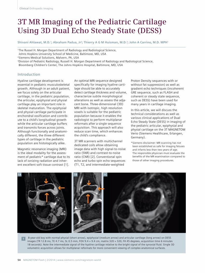
8-year-old boy with normal physial (short arrow), epiphysial (medium arrow) and articular cartilage (long arrow) on DESS images (TR 12.8 ms, TE 4.7 ms, SL 0.5 mm, FOV 9.4 × 9.4 cm, matrix 320 × 320, FA 45 degrees, acquisition time 6 minutes 18 seconds). Note the intermediate signal of the hyaline cartilage relative to the bright signal of the synovial fluid. Single 3D volumetric acquisition allows for multiplanar reformats for more convenient viewing of complex anatomical surfaces.
1
1A 1C1B

Technical considerations
Dual Echo Steady State (DESS) is a 3D coherent (steady state) gradient echo sequence. Steady state sequences (FISP, TrueFISP, DESS, PSIF, CISS) share two major characteristics. Firstly, a short repetition time (TR) prevents transverse magnetization to decay before the next radiofrequency (RF) pulse is applied. Secondly, the slice or slab selective RF pulse is evenly spaced. When phase-coherent RF pulses with the same flip angle are applied with a constant TR that is shorter than the T2 of the tissue, a dynamic equilibrium is achieved between transverse magnetization (TM) and longitudinal magnetization (LM) [3]. Once this equilibrium is reached, two types of echoes and therefore two types of MR images are produced. The first type is post excita-tion signal (S+ or FISP (fast imaging steady state precession)) that consists of free induction decay (FID) arising from the most recent RF pulse. The second type is an echo reformation that occurs prior to excitation (S− or PSIF (reversed FISP)) and results when residual echo is refocused at the time of the subsequent RF pulse [4, 5]. DESS combines S+ and S− signals into one. High T2 contrast is obtained due to the PSIF contribution; whereas, morphological images are obtained by the FISP contribution. The variable T2-weightings of both echoes allow the calculation of quantitative T2. Therefore, the DESS sequence com-
bines morphological and functional analysis from the same data set with high resolution in a relatively short imaging time [6].
In DESS, two or more gradient echoes are acquired. Each of these group of echoes are separated by a refocusing pulse and the combined data results in higher T2*-weighting, creating high signal in cartilage and synovial fluid [7]. The most important param-eter which needs to be kept in mind while acquiring a DESS is the flip angle (FA). According to Hardy et al. [8], the appropriate flip angle for the 3D DESS sequence is 60 degrees which allows for high SNR and CNR.
Although hyaline cartilage is inter-mediate signal intensity and synovial fluid is high signal intensity in both 2D fat suppressed turbo spin echo proton density and 3D DESS, slice thickness is thinner in 3D DESS sug-gesting that this technique may detect smaller cartilage defects than the 2D technique (Fig. 1). Water excited or fat suppressed 3D gradient echo imaging is recommended for measuring the exact cartilage thick-ness without partial volume artifacts, even though the contrast-to-noise ratio between cartilage and bone marrow is relatively poor [9]. Chemi-cal shift artifact can affect the carti-lage-bone interface and fat suppres-sion helps to reduce this, of particular importance in the physial cartilage.
The disadvantages of 3D gradient echo imaging techniques are relatively long scan time. In addition, fat sup-pressed turbo spin echo proton den-sity is superior for intra-articular and periarticular structures and is obtained in short imaging time, but it has the previously mentioned disadvantage of partial volume effects.
Clinical applications
3D DESS allows quantitative assess-ment of cartilage thickness and vol-ume with good accuracy and preci-sion [10]. Although there have been longitudinal studies in adults regard-ing accuracy of DESS sequence in assessing the articular cartilage, there are no large comparative studies in the pediatric population [11]. We will discuss the potential applications of the DESS technique in assessment of pediatric epiphsyial, physial and articular hyaline cartilage.
Congenital: Skeletal dysplasias affect the normal epiphysial cartilage zonal architecture [12, 13] and may mani-fest on MRI as disruptions in the zone of provisional calcification. Neonates with cartilage abnormalities, such as achondrogenesis and type II hypo-chondrogenesis, have shown a signif-icant increase in the number and size of the epiphysial vascular channels [14, 15].
Orthopedic Imaging Clinical
MAGNETOM Flash | 2/2014 | www.siemens.com/magnetom-world 55
12-year-old boy with prior history of Salter Harris II fracture of the distal tibia with DESS sequence (TR 12.8 ms, TE 4.7 ms, SL 0.5 mm, FOV 9.4 × 10 cm, matrix 320 × 320, Flip angle 45 degrees, acquisition time 7 minutes 30 seconds). Note the central loss of physial cartilage and devel-opment of physial bar (arrow).
2
2A 2B
In the setting of tarsal coalition, DESS can potentially be useful in dis-tinguishing fibrous from cartilaginous fusion. In addition the volumetric 3D acquisition allows for multiplanar reformats which can be useful in assessment of complex subtalar joint morphology.
Metabolic: Metabolic disturbances, such as rickets and scurvy, can result in abnormal epiphysial cartilage development.
Trauma: The physis is a relatively weak region and is easily damaged by trauma, infection, tumor invasion, ischemia, radiation, metabolic and hematologic disorders, electrical and thermal burns, and frostbite [11, 12, 16–22]. Traumatic physial injuries may be seen in the various types of Salter-Harris fractures [22]. Generally, the frequency of growth arrest is directly proportional to the increasing Salter-Harris number [17]. Angular deformity such as in Blount disease, altered joint mechanics, leg-length discrepancy, and long-term disability can also result from injury to the physis at weight-bearing sites, such as the knees [16, 17].
56 MAGNETOM Flash | 2/2014 | www.siemens.com/magnetom-world
Clinical Orthopedic Imaging
Vertical physial injuries crossing growth plates can result in trans-physial vascular communication. This communication can lead to the formation of bony bridges (Fig. 2). The DESS is ideal for assessment of physial bridges due to isotropic acquisition which can be used to generate multiplanar reformations and axial maximum-intensity-projec-tion maps of the physial plate, all of which show high-signal cartilage interrupted by a low-intensity physial bridge [16–18]. Metaphysial vascular injury can result in the arrest of endo-chondral ossification and thickening of the injured physes (as in gymnast’s wrist) [17].
Osteochondritis dissecans (OCD) is an acquired disorder of bone in which there is fragmentation of the sub-chondral bone with varying degrees of articular cartilage involvement. The etiology is likely related to indi-rect trauma in most cases. The most common locations for OCD are the femoral condyles, talar dome and capitellum. In the growing child, irreg-ular but normal epiphysial cartilage ossification must be distinguished from an OCD. The variants will have normal
overlying cartilage and no associated marrow edema and are commonly located in the posterior lateral femoral condyle. Capitellar OCD is frequently found in young teenagers, particularly pitchers and gymnasts. OCD usually occurs in the anterolateral portion and must be distinguished from the normal pseudodefect in the posterior capitel-lum. Fluid signal at the bone-osteo-chondral lesion interface as well as a loose intra-articular body herald lesion instability.
Idiopathic: Legg Calve Perthes disease (LCP) is an idiopathic cause of hip pain and limp in preadolescent children due to osteonecrosis (or osteochondrosis) of the femoral epiphysis. In the avas-cular phase, DESS can be used to assess acetabular cartilage and labral hyper-trophy (Fig. 3). In the revascularization and reparative phase, findings that suggest possible physial involvement by LCP disease include increased undu-lation of the growth plate (W- or M-shaped), deepening of the growth plate or ‘cupping’, epiphysial–meta-physeal osseous fusion (bone bridge or bar formation across the physis), or physial cystic change [23].
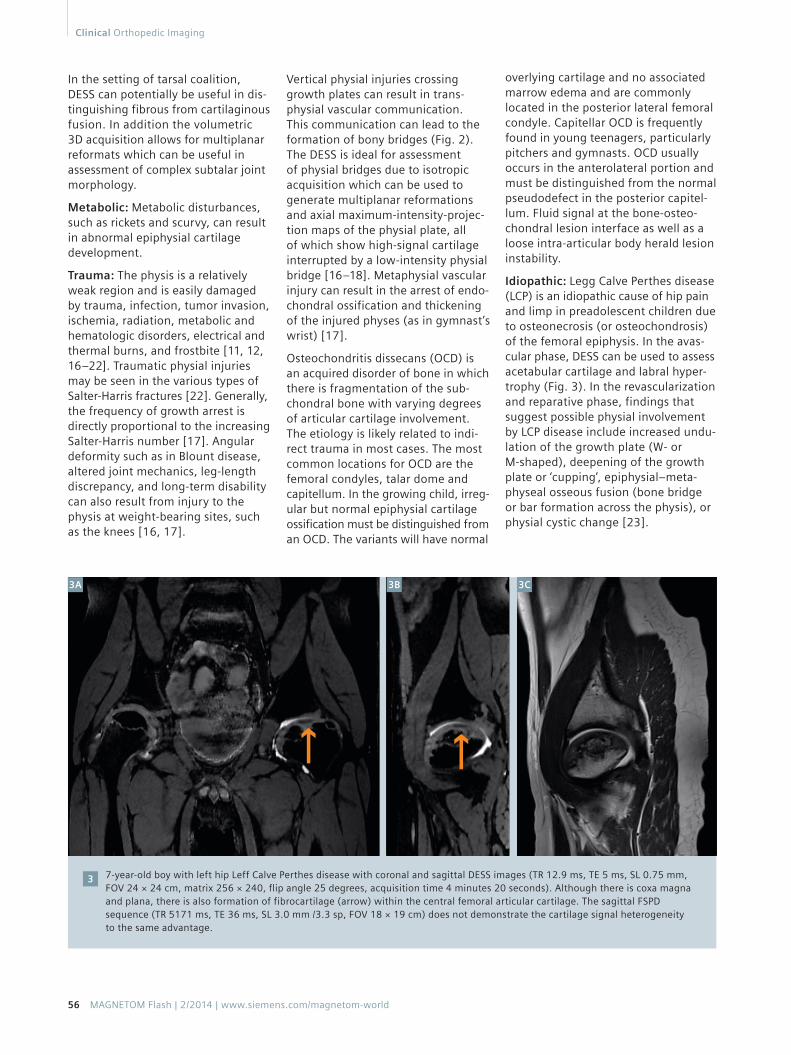
7-year-old boy with left hip Leff Calve Perthes disease with coronal and sagittal DESS images (TR 12.9 ms, TE 5 ms, SL 0.75 mm, FOV 24 × 24 cm, matrix 256 × 240, flip angle 25 degrees, acquisition time 4 minutes 20 seconds). Although there is coxa magna and plana, there is also formation of fibrocartilage (arrow) within the central femoral articular cartilage. The sagittal FSPD sequence (TR 5171 ms, TE 36 ms, SL 3.0 mm /3.3 sp, FOV 18 × 19 cm) does not demonstrate the cartilage signal heterogeneity to the same advantage.
3
3C3A 3B
MAGNETOM Flash | 2/2014 | www.siemens.com/magnetom-world 57
Orthopedic Imaging Clinical
19-year-old young woman with remote history of septic arthritis on DESS sequence (TR 12.8 ms, TE 4.69 ms, SL 0.5 mm, FOV 9.4 × 10 cm, FA 45 degrees, acquisition time 6 minutes 18 seconds). The entire tibiotalar hyaline cartilage demonstrates diffuse intermediate grade thinning with large central lateral talar dome subchndral cyst (arrow).
4
4A 4B
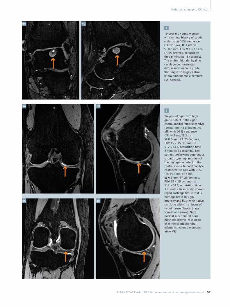
16-year-old girl with high grade defect in the right central medial femoral condyle (arrow) on the preoperative MRI with DESS sequence (TR 14.1 ms, TE 5 ms, SL 0.6 mm, FA 25 degrees, FOV 15 × 15 cm, matrix 512 × 512, acquisition time 3 minutes 36 seconds). The patient underwent auto logous chondrocyte implantation of the high grade defect in the central medial femoral condyle. Postoperative MRI with DESS (TR 14.1 ms, TE 5 ms, SL 0.6 mm, FA 25 degrees, FOV 15 × 15 cm, matrix 512 × 512, acquisition time 3 minutes 36 seconds) shows repair cartilage tissue that is homogeneous in signal intensity and flush with native cartilage with small focus of hypointense fibrocartilage formation (arrow). Note normal subchondral bone plate and interval resolution of minimal subchondral edema noted on the preoper-ative MRI.
5
5A
5C 5D
5B

58 MAGNETOM Flash | 2/2014 | www.siemens.com/magnetom-world
Clinical Orthopedic Imaging
Contact
John A. Carrino, M.D., M.P.H.Associate Professor of Radiology and Orthopaedic SurgeryThe Russel H. Morgan Department of Radiology and Radiological ScienceJohns Hopkins University School of Medicine601 North Caroline St. / JHOC 5165Baltimore, [email protected]
References 1 Gold GE, Chen CA, Koo S, Hargreaves BA,
Bangerter NK. Recent advances in MRI of articular cartilage. AJR Am J Roentgenol 2009 Sep;193(3):628-638.
2 Kornaat PR, Reeder SB, Koo S, Brittain JH, Yu H, Andriacchi TP, et al. MR imaging of articular cartilage at 1.5T and 3.0T: comparison of SPGR and SSFP sequences. Osteoarthritis Cartilage 2005 Apr;13(4):338-344.
3 Chavhan GB, Babyn PS, Jankharia BG, Cheng HM, Shroff MM. Steady-State MR Imaging Sequences: Physics, Classifi-cation, and Clinical Applications1. Radio-graphics July-August 2008 July-August 2008;28(4):1147-1160.
4 Gyngell ML. The application of steady-state free precession in rapid 2DFT NMR imaging: FAST and CE-FAST sequences. Magn Reson Imaging 1988 Jul-Aug;6(4):415-419.
5 Bruder H, Fischer H, Graumann R, Deimling M. A new steady-state imaging sequence for simultaneous acquisition of two MR images with clearly different contrasts. Magn Reson Med 1988 May;7(1):35-42.
6 Welsch GH, Scheffler K, Mamisch TC, Hughes T, Millington S, Deimling M, et al. Rapid estimation of cartilage T2 based on double echo at steady state (DESS) with 3 Tesla. Magn Reson Med 2009 Aug;62(2):544-549.
7 Crema MD, Roemer FW, Marra MD, Burstein D, Gold GE, Eckstein F, et al. Articular Cartilage in the Knee: Current MR Imaging Techniques and Applications in Clinical Practice and Research. Radio-graphics January-February 2011 January- February 2011;31(1):37-61.
8 Hardy PA, Recht MP, Piraino D, Thomasson D. Optimization of a dual echo in the steady state (DESS) free-precession sequence for imaging cartilage. J Magn Reson Imaging 1996 Mar-Apr;6(2):329-335.
9 Gold GE, McCauley TR, Gray ML, Disler DG. Special Focus Session. Radiographics 2003 September 01;23(5):1227-1242.
10 Eckstein F, Hudelmaier M, Wirth W, Kiefer B, Jackson R, Yu J, et al. Double echo steady state magnetic resonance imaging of knee articular cartilage at 3 Tesla: a pilot study for the Osteoarthritis Initiative. Ann Rheum Dis 2006 Apr;65(4):433-441.
11 Wirth W, Nevitt M, Hellio Le Graverand MP, Benichou O, Dreher D, Davies RY, et al. Sensitivity to change of cartilage morphometry using coronal FLASH, sagittal DESS, and coronal MPR DESS protocols – comparative data from the Osteoarthritis Initiative (OAI). Osteoarthritis Cartilage 2010 Apr;18(4): 547-554.
12 Jaramillo D, Connoly SA, Mulkern RV, et al. Developing epiphysis: MR imaging characteristics and histologic correlation in the newborn lamb. Radiology 1998; 207:637–645.
13 Li X, Wang R, Li Y, Tang L, Xu A, Hu J. MRI characteristics and transverse relaxation time measurements in normal growing cartilage. J Huazhong Univ Sci Technolog Med Sci 2004; 24:411–413.
14 Cairns R. Magnetic resonance imaging of the growth plate: pictorial essay. Can Assoc Radiol J 2003; 54:234–242.
15 Gruber HE, Lachman RS, Rimoin DL. Quantitative histology of cartilage vascular canals in the human rib: findings in normal neonates and children, and in achondrogenesis II-hypochondrogenesis. J Anat 1990; 173:69–75.
16 Oeppen RS, Jaramillo D. Sports injuries in the young athlete. Top Magn Reson Imaging 2003; 14:199–208.
17 12. Li X, Wang R, Li Y, et al. Epiphysial and physial cartilage: normal gadolinium-enhanced MR imaging. J Huazhong Univ Sci Technolog Med Sci 2005; 25:209–211.
18 Sailhan F, Chotel F, Guibal AL, et al. Three-dimensional MR imaging in the assessment of physial growth arrest. Eur Radiol 2004; 14:1600–1608.
19 Ogden JA. The evaluation and treatment of partial physial arrest. J Bone Joint Surg Am 1987; 69:1297–1302.
20 Peterson HA. Partial growth plate arrest and its treatment. J Pediatr Orthop 1984; 4:246–258.
21 Ogden JA. Injury to the growth mecha-nisms. In: , Ogden JA, ed. Skeletal injury in the child, 2nd ed. Philadelphia, PA: Saunders, 1990: 97–173 .
22 Salter R, Harris W. Injuries involving the epiphysial plate. J Bone Joint Surg Am 1963; 45:587–622.
23. Dillman J and Hernandez R. MRI of Legg-Calve-Perthes disease. AJR 2009 193:5, 1394-1407.
Inflammation/Infection: Hyaline cartilage damage can also result from inflammatory conditions such as juvenile idiopathic arthritis as well as infection (Fig. 4). Sequelae of osteo-chondral injury include total-thick-ness loss of articular and epiphysial cartilage, cartilage contour irregularity, and intrinsic signal heterogeneity.
Post-operative: MRI is also used to follow cartilage repair procedures, such as microfracture, osteochondral autografts and allografts, and auto-
logous chondrocyte implantation. The DESS sequence can assess carti-lage repair via evaluation of repair tissue signal characteristics, border integration, thickness relative to native cartilage, chondral clefts and changes within the subchondral bone plate (Fig. 5).
Conclusions
The physial, epiphysial and articular hyaline cartilage is affected in various congenital, post-traumatic, idiopathic
and inflammatory conditions in chil-dren. 3D DESS techniques have many advantages in assessment of pediatric cartilage pathology including higher SNR, increased cartilage-to-fluid con-trast and isotropic resolution, which helps to reduce partial volume effects. 3D DESS techniques are also of essential importance in the pediatric population due to the lack of ionizing radiation.