3.preparation and characterization of gelatin hyaluronic acid cryogels
TRANSCRIPT

Acta Biomaterialia 9 (2013) 9012–9026
Contents lists available at SciVerse ScienceDirect
Acta Biomaterialia
journal homepage: www.elsevier .com/locate /ac tabiomat
Preparation and characterization of gelatin/hyaluronic acid cryogelsfor adipose tissue engineering: In vitro and in vivo studies
1742-7061/$ - see front matter � 2013 Acta Materialia Inc. Published by Elsevier Ltd. All rights reserved.http://dx.doi.org/10.1016/j.actbio.2013.06.046
⇑ Corresponding author. Tel.: +886 3 2118800; fax: +886 3 2118668.E-mail address: [email protected] (J.-P. Chen).
1 These authors contributed equally to this work.
Kun-Hung Chang a,1, Han-Tsung Liao a,b,1, Jyh-Ping Chen a,⇑a Department of Chemical and Materials Engineering, Chang Gung University, Kwei-San, Taoyuan 333, Taiwan, ROCb Department of Plastic and Reconstructive Surgery, Chang Gung Memorial Hospital, Craniofacial Research Center, Chang Gung University, Kwei-San, Taoyuan 333, Taiwan, ROC
a r t i c l e i n f o
Article history:Received 18 March 2013Received in revised form 14 June 2013Accepted 28 June 2013Available online 10 July 2013
Keywords:CryogelAdipose tissue engineeringGelatinHyaluronic acidAdipose-derived stem cells
a b s t r a c t
Macroporous elastic scaffolds containing gelatin (4% or 10%) and 0.25% hyaluronic acid (HA) werefabricated by cryogelation for application in adipose tissue engineering. These cryogels have intercon-nected pores (�200 lm), high porosity (>90%) and a high degree of cross-linking (>99%). The highergelatin concentration reduced the pore size, porosity and swelling ratio of the cryogel but improved itsswelling kinetics. Compressive mechanical testing of cryogel samples demonstrated non-linear stress–strain behavior and hysteresis loops during loading–unloading cycles, but total recovery from largestrains. The presence of more gelatin increased the elastic modulus, toughness and storage modulusand yielded a cryogel that was highly elastic, with a loss tangent equal to 0.03. Porcine adipose-derivedstem cells (ADSCs) were seeded in the cryogel scaffolds to assess their proliferation and differentiation. Invitro studies demonstrated a good proliferation rate and the adipogenic differentiation of the ADSCs inthe cryogel scaffolds, as shown by their morphological change from a fibroblast-like shape to a sphericalshape, decreased actin cytoskeleton content, growth arrest, secretion of the adipogenesis marker proteinleptin, Oil Red O staining for triglycerides and expression of early (LPL and PPARc) and late (aP2 andleptin) adipogenic marker genes. In vivo studies of ADSCs/cryogel constructs implanted in nude miceand pigs demonstrated adipose tissue and new capillary formation, the expression of PPARc, leptinand CD31 in immunostained explants, and the continued expression of adipocyte-specific genes. Boththe in vitro and in vivo studies indicated that the gelatin/HA cryogel provided a structural and chemicalenvironment that enabled cell attachment and proliferation and supported the biological functions andadipogenesis of the ADSCs.
� 2013 Acta Materialia Inc. Published by Elsevier Ltd. All rights reserved.
1. Introduction
Soft tissue deficiency is a common problem after traumaticinjury, tumor ablation, congenital underdevelopment and thenatural changes of the aging process. An autologous fat graft iscurrently considered the gold standard in soft tissue augmentationprocedures. The advantages are that the graft is autogenous, elim-inating the risk of immunogenic reactions, and the harvest of thefat graft is simple and safe, with minimal complications. However,autologous fat grafts have been found to have a 40–60% resorptionrate [1,2]. Therefore, multiple procedures are required to achievesatisfactory results. Sometimes central necrosis results in micro-calcifications that can be palpated and are difficult to differentiatefrom tumors in the patients. In addition, the donor source is lim-ited in slim patients when a large amount of fat graft is needed.Adipose tissue engineering is a promising method to solve these
problems [3]. In tissue engineering, cells can be seeded onto or intoan artificial structure that is referred to as the scaffold, whichmainly gives mechanical support in the formation of a three-dimensional (3-D) tissue engineered cells/scaffold construct thatis subsequently implanted into the host tissue.
Scaffolds have an important role in tissue engineering. For suc-cessful tissue engineering, the scaffold must be biodegradable andbiocompatible, with a 3-D porous architecture and inter-poreconnections and the appropriate mechanical properties similar tothose of the reconstructed tissue [4]. Scaffolds must also be pro-duced to allow cell distribution and to direct their growth intothe 3-D structure. Several techniques have been developed toproduce 3-D porous scaffolds. These techniques include solvent-casting/particulate-leaching, gas foaming, phase separation, elec-trospinning, melt molding, rapid prototyping and freeze-drying[5]. However, most of those traditional production methods arecomplex, require specific equipment, use high temperatures or in-volve hazardous organic solvents.
Cryogelation is a simple approach for producing macroporousscaffolds [6]. This method allows effective control over the pore size
K.-H. Chang et al. / Acta Biomaterialia 9 (2013) 9012–9026 9013
by using ice crystals as templates and produces a porous structurewithout the involvement of organic solvents or any additives dur-ing the production process. The method involves the cryogenictreatment of polymeric gel precursors in the moderate freezing,storage (in the frozen state) and subsequent thawing steps. Thecryogel matrices are cross-linked at sub-zero temperatures,whereas part of the solution remains unfrozen in a liquid micro-phase, in which the phases are separated after undergoing chemicalreactions [7]. The reactions that occur in the liquid microphase leadto gel formation, with the ice crystal acting as porogen substrates.In thawing the ice crystals, a system of large interconnected poresis formed within the gel. The compulsory displacement of the poly-meric precursors into a non-frozen microphase results in the poly-mer gel being concentrated in the pore walls, which augments themechanical strength of the cryogel. Wide, interconnected poresform in the cryogel as each ice crystal grows until it contacts adja-cent crystals during the freezing of the initial aqueous solution, pro-viding a labyrinthine system of interconnected channels after thefrozen sample is thawed. Freezing at moderate temperatures thatdo not compromise the scaffold’s mechanical properties could offera more cost-efficient process than freeze-drying [8]. Hence, cryo-gels have some important general characteristics, including aninterconnected, highly porous structure, mechanical stability, elas-ticity, reversible and very rapid size changes induced by externalforces and good swelling in aqueous media [9]. They are very toughand can withstand high levels of deformation, including tensile,compressive or flexural strains [10]. However, all of these proper-ties depend on the materials used.
Gelatin, a partially degraded product of collagen, is believed tohave a lower antigenicity than collagen. Moreover, gelatin is notonly more economical than collagen, but it also contains aminoacid sequences, such as the RGD of collagen, which can enhancecell attachment. Therefore, gelatin has been blended with othernatural or synthetic biomaterials to fabricate 3-D scaffolds usingvarious methods for different tissue engineering applications. Gel-atin microspheres containing basic fibroblast growth factor wereshown to induce preadipocytes to form adipose tissue at the im-plant site [11]. Macroporous gelatin spheres were reported to besuitable transplantation vehicles and biodegradable scaffolds forthe guided regeneration of soft tissues [12]. Gelatin sponges havebeen used for adipose tissue engineering employing human mar-row stromal cells [13,14].
Hyaluronic acid (HA) is a naturally polysaccharide composed ofrepeating disaccharide units of D-glucuronic acid and N-acetyl-D-glucosamine linked by alternating (1 ? 4) and (1 ? 3) linkages.As an important component of the extracellular matrix (ECM),HA can increase cell attachment and cell migration into the scaf-fold due to its high water retention and intrinsic swelling property.Therefore, HA has been used alone or cross-linked with other bio-materials for applications in tissue engineering, such as skin, carti-lage, bone and fat. Borzacchiello et al. reported using an esterderivative of HA, HYAFF II, as a potential 3-D scaffold for adiposetissue engineering, both in vitro and in vivo [15]. An aminatedhyaluronic acid-g-poly(N-isopropylacrylamide) thermosensitivehydrogel was shown to have a 3-D porous structure able to encap-sulate adipose-derived stem cells (ADSCs) to form adipose tissuein vivo [16]. Recent studies also demonstrated the utility of HA-based scaffolds in enhancing adipose tissue development in vitro[17,18] and in vivo [19–21].
In view of the successful use of gelatin and HA-based scaffoldsin adipose tissue engineering, we hypothesized that a cryogel scaf-fold fabricated from those precursors would be suitable for in vitroand in vivo adipogenesis using ADSCs. To the best of our knowl-edge, there are no reports discussing the optimal scaffold for adi-pose tissue engineering from the aspect of its mechanicalproperties. For adipose tissue engineering, the designed scaffold
should not be as hard as bone tissue but must be elastic and capa-ble of withstanding cyclic mechanical strains. We conjectured thatgelatin/HA cryogels could withstand static and dynamic deforma-tional loading without cracking or permanent deformation. Thecharacteristics of the gelatin/HA cryogels were analyzed in termsof pore size, porosity, swelling kinetics and mechanical propertiesusing static and dynamic compressive testing. To test the biocom-patibility and potential for applications in adipose tissue engineer-ing, the cryogels were seeded with porcine ADSCs to study the cellproliferation, adipogenic gene expression and protein synthesisboth in vitro and in vivo using biochemical analyses, quantitativereal-time polymerase chain reactions (qRT-PCR) and histology.
2. Materials and methods
2.1. Materials
1-ethyl-3-(3-dimethylaminopropyl) carbodiimide (EDC) wasobtained from Acros. Hyaluronic acid (sodium salt from Streptococ-cus equi, molecular weight 1500–1750 kDa), gelatin (type A fromporcine skin, 300 bloom), 2-morpholinoethane sulfonic acid(MES) and 2,4,6-trinitrobenzene sulfonic acid (TNBS) were all pur-chased from Sigma. Dulbecco’s modified Eagle’s medium (DMEM,Sigma) and fetal bovine serum (FBS, HyClone) were used for cellculture. Rhodamine-phalloidin and the 40, 6-diamidino-2-phenyl-indole dihydrochloride (DAPI) solution for cell staining were pur-chased from Life Technologies.
2.2. Preparation of gelatin-hyaluronic acid scaffold by cryogelation
Cryogels containing 4% gelatin/0.25% HA (GH4) and 10% gelatin/0.25% HA (GH10) were prepared. Gelatin (8% and 20% (w/v)) andHA (0.5% (w/v)) solutions were prepared separately in 0.1 M MESbuffer (pH 6.0). 1-ethyl-3-(3-dimethylaminopropyl) carbodiimidewas added to the HA solution, which had been prepared at 37 �C,to a final concentration of 2% (w/v) and mixed for 1 h. The solutionwas mixed gently with an equal volume of gelatin solution, whichhad been prepared in a 60 �C water bath, and immediately injectedinto the wells of a 48-well cell culture plate, then placed in a�20 �C deep freezer. The whole mixing and transfer step was com-pleted within 1 min. The culture plate was incubated in the freezerfor 12 h to complete the cross-linking reaction and was thenthawed at room temperature. The cryogels were removed andthoroughly washed with deionized water to remove any residualreactants. Each cryogel was cut with a sharp blade into 5 mm thickdisks with a diameter of 10 mm.
2.3. Characteristics of cryogel
2.3.1. Physico-chemical properties of scaffoldThe morphology of cryogel samples was examined with a scan-
ning electron microscope (SEM; JEOL ISM-5410). Scaffold porositywas determined using an ethanol displacement method [22]. Cryo-gel samples were individually immersed in a cylinder containing aknown volume of ethanol (V1), and the total volume of ethanol andscaffold was recorded (V2). After sonication for 5 min to eliminateair from the scaffold and induce pore filling, the ethanol-filledscaffold was removed from the cylinder and the residual ethanolvolume was recorded (V3). Cryogel porosity was calculated asporosity (%) = (V1 � V3)/(V2 � V3) � 100.
The degree of cross-linking was determined using the TNBSmethod [23]. The available free amino groups within a samplewere determined from a standard curve generated using glucosa-mine. The degree of cross-linking (DC) was expressed as the per-centage of free amino groups in the original non-cross-linked
9014 K.-H. Chang et al. / Acta Biomaterialia 9 (2013) 9012–9026
sample that was cross-linked by EDC after normalization with theweight of the sample used in the reaction. It was calculated asfollows:
DC ð%Þ¼ ½1�ðfree amino groups=massÞc=ðfree amino groups=massÞnc��100ð1Þ
where the subscripts c and nc stand for the cross-linked and non-cross-linked samples, respectively.
Fourier transform infrared (FTIR) spectra were obtained using aHoriba 730 FTIR spectrometer. The cryogel sample was ground to afine powder, mixed with KBr powder (1:8), dried in a 70 �C oven for24 h and compressed into pellets for FTIR examination over awavenumber range between 400 and 4000 cm�1 with a scanningspeed of 2.5 cm s�1 and a resolution of 2 cm�1. A CFP-1100-AI cap-illary flow porometer (Porous Materials Inc.) was used to measurethe pore diameters and the pore size distribution. Calwick, whichhas a defined surface tension of 21 dyne cm�1, was used as thewetting agent for the porometry measurements. The pore diameterwas calculated using the following equation [24]:
D ¼ 4c coshP
ð2Þ
where D is the pore diameter, c is the surface tension of the wettingliquid, h is the contact angle of the wetting liquid and P is the differ-ential pressure.
2.3.2. Swelling ratio and swelling kineticsThe swelling ratios of the cryogels were determined following
the conventional gravimetric procedure [25]. The as-preparedcryogels were cut into round disks (10 cm diameter � 5 mmheight) and were vacuum-dried at 60 �C for 24 h. The swellingratios were measured by weighing the swollen cryogel samples,which were immersed in deionized water at room temperature,at intervals. The masses of the swollen gel samples (Mt) weredetermined immediately after removing the excess water withpaper tissues. The equilibrium masses of the swollen gel samples(Meq) were similarly obtained until no measurable weight increasewas observed. The swelling ratio (SR) was calculated as follows:
SR ¼ ðMt �MdÞ=Md ð3Þ
where Md is the mass of the dried samples. To investigate the diffu-sion of water in the cryogels, the swelling kinetics was fitted to thefollowing equation [26]:
ðMt �MdÞ=ðMeq �MdÞ ¼ ktn ð4Þ
where k is a characteristic constant of the gel, t is time and n is acharacteristic exponent of the transport mode of water.
2.3.3. Mechanical propertiesThe quasi-static and dynamic compression behavior of the cryo-
gels was investigated using unconfined compression tests. Thecompression tests were conducted in phosphate-buffered saline(PBS) at 37 �C using an ElectroForce�5200 BioDynamic™ testingmachine (Bose). The compression load was applied with a 250 Nload cell at a cross-head speed of 0.05 mm s�1. Cryogel samples(5 mm height � 10 mm diameter) were soaked in PBS at 37 �C for24 h prior to testing. A stress–strain curve was recorded with uniax-ial stress. The strain was defined as e (mm mm�1) = displacement(mm)/height (mm). The stress was defined as r (MPa) = force (N)/cross sectional area (mm2). The ultimate stress and ultimate strainvalues were taken as the point where the cryogel failure occurred.The stress–strain data from the unconfined compression test upto failure were curve-fitted according to the following non-linearequation [27]:
r ¼ AeBe�1 ð5Þ
where A and B are elastic constants.The tangential Young’s (elastic) modulus (slope of the tangent
to the stress–strain curve) at 10% and 30% strain was calculatedusing the non-linear elastic model. The toughness (compressivestrain energy to failure) of a cryogel, defined as the energy neces-sary to deform a specimen to failure, was obtained from the areaunder the stress–strain curve.
A cyclic compression test was similarly performed by loadingthe samples to r = 0.3 with five cycles at a frequency of 1 Hz. Theenergy absorption in the cryogel was derived from the stress–strain relations. A hysteresis loop was shown to exist in thestress–strain relations, bounded by the loading and unloadingcurves, indicating the dissipation of energy or energy absorbeddue to the viscous properties of the cryogel. The dissipation energy(kJ m�3) loss during the hysteresis cycle was calculated from thearea bounded within the hysteresis loop. The percentage of energydissipation (%) was calculated by dividing the dissipative energy bythe area bounded between the loading curve and the horizontalaxis (compression energy), which indicated the total energyapplied to the sample during compression.
For the dynamic compression testing, the sample was dynami-cally tested with sinusoidal compressions from 0.2 to 5 Hz at 30%compression. The complex modulus (E⁄) is the ratio of the stressto the strain, which can be divided into the energy stored per cycle,or storage modulus (E0), and the energy lost per cycle, or loss mod-ulus (E00). The ratio of these two quantities is referred to as the losstangent (tan d), or viscous damping [28], as follows:
tan d ¼ E00=E0 ð6Þ
For each testing frequency, the dynamic mechanical analysissoftware measured the values of E⁄, E0, E00 and tan d.
2.4. Isolation and harvest of porcine adipose-derived stem cells
Porcine ADSCs were harvested and isolated according to a mod-ification of the procedure of Zuk et al. [29]. Briefly, the fat tissuewas harvested from the lower abdomen of the pig after sterilizingthe lower abdomen with a beta-iodine solution. The fat was dicedinto very small pieces using scissors in a sterile procedure. Thediced fat tissue sample was washed extensively with equal vol-umes of PBS, and the ECM was digested with 0.05% collagenasein a 37 �C water bath shaken at 165 rpm for 30 min. The enzymeactivity was neutralized by adding an equal volume of controlmedium consisting of DMEM and 10% FBS, and centrifuged at250g for 10 min to obtain a high-density cell pellet. The superna-tant was discarded, and the cell pellet was resuspended in160 mM NH4Cl and incubated at room temperature for 10 min tolyse the contaminating red blood cells. The cell pellet was re-col-lected by centrifugation as described above, filtered through a100 lm nylon mesh to remove the cellular debris and incubatedovernight in control medium in culture dishes at 37 �C in a 5%CO2 environment. Following the incubation, the culture disheswere washed extensively with PBS to remove the residual non-adherent red blood cells, and the ADSC-enriched density fractionwas collected.
2.5. Adipose tissue engineering in vitro study
2.5.1. In vitro cell cultureDisk-shaped cryogel scaffolds (GH10) (5 mm in thickness and
10 mm in diameter) were sterilized with 75% ethanol for 24 hand rinsed three times with PBS before being placed in 48-well cul-ture plates (Nunc) for cell seeding. An aliquot of 1 ml of a cell sus-pension (5 � 104 cells ml�1) was seeded onto the surface of thecryogel scaffold in each well. Cell-seeded cryogels were incubatedat 37 �C for 4 h to allow cell adhesion and were then transferred to
K.-H. Chang et al. / Acta Biomaterialia 9 (2013) 9012–9026 9015
a new well with the addition of 2 ml of control medium or adipo-genic differentiation medium to each well. The cells were culturedat 37 �C in 5% CO2 with a medium change every 2 days. The controlmedium was DMEM, 10% FBS and 1% antibiotic/antimycotic solu-tion. The adipogenic differentiation medium was DMEM, 10%FBS, 1% antibiotic/antimycotic solution, 1 lM dexamethasone,0.5 mM isobutyl-methylxanthine, 10 lM insulin and 200 lMindomethacin.
2.5.2. Cell proliferation in cryogelsThe proliferation rate of the ADSCs in the GH10 cryogel was
determined using CellTiter 96 AQueous One Solution Reagent(Promega), which contains a novel tetrazolium salt (MTS). TheMTS tetrazolium compound was bioreduced by living cells into acolored formazan product that is soluble in tissue culture medium.The quantity of formazan product was directly proportional to thenumber of viable cells. MTS assays were performed by adding MTSsolution to each specimen and incubating at 37 �C for 4 h with pro-tection from light. The colorimetric measurement of the amount offormazan dye was performed at a wavelength of 492 nm using anELISA microplate reader (Bio-Tek Synergy HT).
2.5.3. SEM analysisAfter 21 days of cell culture, the ADSCs/cryogel constructs were
examined by SEM. The constructs were fixed with 2.5% glutaralde-hyde for 24 h at room temperature. After thorough washing with0.1 M PBS (pH 7.4), the constructs were dehydrated in ethanol ina sequential manner (40%, 60%, 80% and 100%) for 10 min eachand allowed to dry on a clean Petri plate at room temperature.The constructs were observed by SEM (JEOL ISM-5410) after goldcoating.
2.5.4. Cell viability and cytoskeleton expressionThe viability of the ADSCs was assessed using the live/dead via-
bility/cytotoxicity assay kit (Invitrogen) after 21 days of culture.The kit contains two molecular probes, calcein AM and ethidiumhomodimer-1 (EthD-1). The ADSCs/cryogel constructs were incu-bated in 300 ll of staining solution (5 ll of 2 mM EthD-1 and3 ll of 4 mM calcein AM in 10 ml of PBS solution) in the dark for5 min and observed using a confocal laser scanning microscope(Zeiss LSM 510 Meta). The excitation wavelengths for EthD-1 andcalcein AM are 528 and 494 nm, respectively. The emission wave-lengths for EthD-1 and calcein AM are 617 and 517 nm,respectively.
To assess the cytoskeletal structure of the ADSCs within thecryogels, the ADSCs/cryogel constructs were fixed in 0.5% parafor-maldehyde in PBS for 30 min at room temperature. The cells werepermeabilized in 0.1% Triton-X100 in PBS for 1 min. The constructswere then immersed in rhodamine–phalloidin solution (diluted1:100 in PBS) for 30 min and washed twice with PBS. The nucleiwere stained with 0.1 lg ml�1 DAPI in PBS for 5 min. The actincytoskeleton fluoresced red and the nuclei were stained blue. Thefluorescence-stained cells were visualized using a confocal laserscanning microscope (Zeiss LSM 510 Meta). The excitation wave-lengths for rhodamine–phalloidin and DAPI are 540 and 340 nm,respectively. The emission wavelengths for rhodamine–phalloidinand DAPI are 573 and 488 nm, respectively.
2.5.5. Oil Red O stainingFollowing 21 days of adipogenic induction, the ADSCs/cryogel
constructs were fixed in 4% paraformaldehyde/PBS, embeddedand frozen in optimal cutting temperature (OCT) compound andcryosectioned at 30 lm using a cryostat (LEICA CM3050S). Theintracellular lipid was stained with 3 mg ml�1 of Oil Red O in iso-propanol for 15 min and viewed using an inverted optical micro-scope (Olympus IX71) [30].
2.5.6. Leptin quantificationThe leptin concentration in the cell culture medium was deter-
mined using a sandwich ELISA and normalized to the protein con-centration. The cell culture medium was removed on days 12–14,19–21 or 40–42, centrifuged for 5 min at 13,200 rpm to removethe cellular debris and then the supernatant was frozen at�80 �C before use. An ELISA kit for porcine leptin obtained fromUscn Life Science Inc. was used, following the manufacturer’s pro-tocol [31]. The protein concentration was determined using theProtein Assay Kit from Bio-Rad.
2.5.7. Quantitative evaluation of adipogenic specific gene expressionThe expression of adipogenic marker genes was analyzed by
qRT-PCR using SYBR green chemistry. The total RNA of eachspecimen was isolated using Trizol reagent (Invitrogen), accord-ing to the manufacturer’s protocol. The isolated RNA was dis-solved in RNase-free water and the amount of RNA wasdetermined by measuring the absorbance at 260 nm (OD260)using a spectrophotometer (NanoDrop 2000, Thermo Scientific).The RNA quality was verified by the OD260/OD280 measurement.cDNA was prepared from 2 lg of total RNA using a RevertAidFirst Strand cDNA Synthesis Kit (Thermo Scientific) in a final vol-ume of 20 ll. For a single PCR reaction in 20 ll, 0.2 ll of cDNAwas used. To allow the visualization of the PCR products in realtime, the SYBR Green I supermix (Bio-Rad) was used. A three-temperature cycle, consisting of a denaturation step at 95 �Cfor 30 s, an annealing step at 57.6 �C for 30 s and an extensionstep at 72 �C for 30 s, was conducted using an MiniOpticonreal-time PCR system (Bio-Rad CFD-3120). The specificity of eachPCR reaction was assessed by performing a melting curve analy-sis after each reaction. Glyceraldehyde-3-phosphate dehydroge-nase (GAPDH) acted as a housekeeping control. The expressionof the early (peroxisome proliferator-activated receptor-gamma(PPARc) and lipoprotein lipase (LPL)) and late (adipocyte-specificfatty acid binding protein (aP2) and leptin) adipogenic markergenes were quantified using the 2�DDCt relative quantificationmethod with respect to the control time point of day 0. Theexpression of each gene was evaluated in triplicate. The primersequences are shown in Table 1.
2.6. Adipose tissue engineering in vivo study
The animal protocols were approved by the Institutional AnimalCare and Use Committee of Chang Gung University.
2.6.1. Nude mouse modelBALB/c nude mice (4 weeks olds) were anesthetized with isoflu-
rane. The back was sterilized with chlorohexidine and a 1 cm inci-sion was made. The mice were randomly divided into two groups.The experimental group received cryogels seeded with 1 � 106
ADSCs and cultured in adipogenic differentiation medium for21 days. The control group received acellular cryogels. An acellularcryogel or a cell-seeded cryogel was inserted into the dorsal subcu-taneous pocket and sutured in place using 6–0 suture (Ethicon). Themice were housed individually and fed standard mouse chow afterthe operation. The specimens were harvested at 2, 4 and 8 weekspost-implantation for qRT-PCR analysis of the expression of adipo-cyte-specific genes (PPARc, LPL, aP2 and leptin), hematoxylin andeosin (H&E) staining, immunohistochemical (IHC) staining (PPARc,leptin and CD31) and immunocytochemical (ICC) staining (PPARc).
2.6.2. Porcine modelSurgery was performed under sterile conditions by using endo-
tracheal isoflurane anesthesia after induction with intravenous 4%sodium pentobarbital (1 ml/10 kg). The surgical area of the abdo-men of each pig was shaved before the procedure, and the surgical

Table 1Primer sequences used in gene expression analysis.
Gene Gene ID Forward/reverse
Lipoprotein lipase (LPL) AF102859 GCAGGAAGTCTGACCAATAAG/GGTTTCTGGATGCCAATAC
Peroxisome proliferator-activated receptorgamma 2 (PPARc2)
AF103946 GCGCCCTGGCAAAGCACT/TCCACGGAGCGAAACTGACAC
Adipocyte-specific fattyacid-binding protein(aP2)
AF102872 GGCCAAACCCAACCTGA/GGGCGCCTCCATCTAAG
Leptin AF052691 TTGAAGGCCTCTCTCCCCACAGTCG/CTTTCCCGGTGGGCGCGGAA
Glyceraldehyde 3-phosphatedehydrogenase(GAPDH)
AF017079 GCTTTGCCCCGCGATCTAATGTTC/GCCAAATCCGTTCACTCCGACCTT
9016 K.-H. Chang et al. / Acta Biomaterialia 9 (2013) 9012–9026
field was prepared with betadine solution. Six 1 cm incisions weremade in the abdominal skin of each pig. A subcutaneous pocketwas dissected in each incision using scissors. The pigs were ran-domly divided into two groups. Cryogels seeded with 1 � 106 ADS-Cs and cultured in adipogenic differentiation medium for 21 daysand acellular cryogels were buried within subcutaneous pocketsin the experiment and control groups, respectively. After achievinghemostasis, the wound was sutured primarily with 4–0 nylon. Thespecimens were harvested at 2, 4 and 8 weeks post-implantationand subjected to the same analysis as those from the nude mousemodel.
2.6.3. Immunohistochemical (IHC) and immunocytochemical (ICC)staining
For IHC staining, the harvested specimens were fixed in a 10%formaldehyde solution, dehydrated and embedded in paraffin. Se-rial sections (4 lm thick) were deparaffinized using the Trilogy™Pretreatment Solution (Cell Marque Co.). The deparaffinized sec-tions were washed three times in Tris-buffered saline containing0.1% Tween 20 (TBST) for 5 min. The non-specific binding siteswere blocked with Ultra V Block (Thermo Scientific) for 5 min,and the sections were then incubated for an additional 120 minat room temperature in rabbit anti-PPARc primary antibody (Ab-cam ab19481) at 1:200, rabbit anti-leptin primary antibody (Ab-cam ab16227) at 1:100, or rabbit anti-CD31 primary antibody(Abbiotec 250590) at 1:200 in a humid environment. After rinsingwith TBST, the sections were incubated in UltraVision ONE HRPPolymer (Thermo Scientific) for 30 min. The peroxidase activitywas visualized using AEC (PPARc and leptin) or DAB (CD31) asthe substrate by incubation with AEC or DAB solution for 10 min.The sections were then counterstained with hematoxylin for5 min and observed under an inverted optical microscope [30].
For ICC staining, sections incubated with PPARc primary anti-body were treated with rhodamine-conjugated chicken anti-rabbitIgG-R secondary antibody (Santa Cruz sc-2862) overnight at 4 �C.The sections were counterstained with DAPI for 5 min and exam-ined under an inverted fluorescence microscope.
2.7. Statistical analysis
All of the data are reported as the mean values ± standard devi-ation (SD). Statistical analysis among the multiple group data fromthe cell proliferations and biochemical assays were conductedusing a one-way ANOVA test to determine the significant differ-ences. Tukey’s post hoc test was used to determine the differencebetween any two groups with p < 0.05 considered statisticallysignificant.
3. Results
3.1. Synthesis and characterization of the gelatin/hyaluronic acidcryogels
Initially, different gelatin to HA mass ratios ranging from 1:0.25to 15:0.25 were used to prepare cryogels with a fixed mass of HA.The selection of the optimum ratio of gelatin to HA was based onthe cryogel properties, which should fulfill the minimal criteriaset for scaffolds intended for soft tissue engineering. The final gel-atin to HA ratio was determined to be between 4:0.25 (GH4) and10:0.25 (GH10), whereas other combinations did not yield goodmechanical or morphological properties. Cryogels with low gelatincontent were very soft and elastic but mechanically inadequate.Because the cryogelation process depends upon the rate of cross-linking, increasing the gelatin concentration beyond 10% wasfound to offer an unsatisfactorily high cross-linking rate at thecross-linking temperature (�20 �C) and with the cross-linker(EDC) concentration used. This resulted in completion of the gela-tion process in the polymer solution before porogens (ice crystals)were formed. The cryogel formed under these conditions was verybrittle and had low porosity and inferior mechanical strength.
The morphology of cryogel samples was evaluated using SEM.Both cryogels exhibited a similar open interconnected pore mor-phology with pore diameters ranging from 100 to 400 lm,although GH4 appeared to have larger pores than GH10 (Fig. 1aand b). A more accurate measurement of the mean pore diameterand pore size distribution was provided by capillary flow porome-try (Fig. 1c and d). Both cryogels showed a sharp pore-size distribu-tion, with minimum pore diameters of 150 lm and 100 lm forGH4 and GH10, respectively. The mean pore diameter decreasedfrom 245.3 lm (GH4) to 170.3 lm (GH10) with the increased gel-atin content used during cryogel synthesis. All synthesized cryogelscaffolds showed high porosity, greater than 90%, which was con-sidered beneficial for cell ingrowth and survival (Table 2). How-ever, while similar trends were observed for the pore diameters,GH4 exhibited higher porosity than GH10, which also resulted inlower density. The degree of cross-linking was as high as 99%and was independent of the gelatin concentration (Table 2). Judg-ing from the macroporous structure and the high degree of cross-linking, EDC can serve as an efficient cross-linker for GH cryogelfabrication, catalyzing intermolecular covalent linkages betweengelatin and HA at �20 �C, simultaneously with micropore forma-tion from the growth of ice crystals.
The results of the water-uptake studies, shown in Fig. 2, wereobtained by measuring the swelling ratio of the cryogels. In bothcryogels, the majority of the water uptake occurred within the first2 min and seemed to have stabilized after 20 min of soaking time(Fig. 2a). GH10 showed a higher swelling rate than GH4 becausethe swelling ratio at 2 min was �60% and 85% of the equilibriumvalue for GH4 and GH10, respectively. In contrast, the values ofthe equilibrium swelling ratio followed the opposite trend, withthe GH4 value (11.9 ± 1.2) significantly higher than that of GH10(8.7 ± 1.7). The swelling kinetics was further analyzed using Eq.(4) and the fitted curves shown in Fig. 2b indicate that the modelcan satisfactorily predict water diffusion into the cryogels. The fit-ted parameters k and n values are 0.523 and 0.226 for GH4(r2 = 0.989), and 0.824 and 0.051 for GH10 (r2 = 0.999). The valuesof n can be used to determine the mechanism of water diffusion[26]. Because the n values for both cryogels are less than 0.5, thediffusion of water within the pores of cryogel occurs by Fickian-type mechanism.
The FTIR spectra of the cryogels together with their individualpolymer components are shown in Fig. 3. In the FTIR spectrum ofpure gelatin, the peaks at 1634 cm�1 and 1524 cm�1 correspond

% o
f tot
al p
ores
0
10
20
30
40
50
60
70c
100-00-150 150-20-200 2
Pore diameter (μm) Pore diameter (μm)
200-2
Me
-250 25
Mean p
250-30
n pore d
-300 30
re diam
300-35
ameter
350 350
ter = 24
350-400
245 μm
00
m
% o
f tot
al p
ores
1
2
3
4
5
6
7
0
10
20
30
40
50
60
70
10100-15
d
a b
-150 15150-20200 200200-250
Me
50 250
ean po
50-300
pore d
00 300-
e diame
00-350
meter =
350-4
er = 170
0-400
170 μmm
Fig. 1. (a, b) Scanning electron micrographs (bar = 500 lm) and (c, d) pore size distribution of the cryogels measured using capillary flow porometry. (a, c) GH4; (b, d) GH10.
Table 2Porosity, density and degree of cross-linking of synthesized cryogel. Values are themean ± SD of five independent measurements.
Cryogel Porosity (%) Density (g cm�3) Degree of cross-linking (%)
GH4 95.2 ± 2.2% 0.0893 ± 0.0051 99.3% ± 0.6GH10 90.1 ± 3.5% 0.1989 ± 0.0132 99.2% ± 0.1
K.-H. Chang et al. / Acta Biomaterialia 9 (2013) 9012–9026 9017
to amide I for the C@O stretching vibration and amide II for the N–H bending vibration, respectively, whereas the peaks at 1455 cm�1
and 1080 cm�1 are assigned to the bands for CH2 bending and C–Ostretching, respectively. In the spectrum of pure HA, the peak at1044 cm�1 is attributed to the C–O–C stretching. The absorptionpeaks at 1417 cm�1 and 1614 cm�1 are assigned to the symmetricand asymmetric stretching vibration bands of the carboxyl groups.All of the characteristic peaks of gelatin and HA were detected inthe spectra of the cryogel scaffolds, indicating the presence of bothpolymers in the hybrid scaffolds. EDC can react with the abundantcarboxyl groups in HA to form an activated intermediate acidanhydride, which can readily react with an amino group of gelatinto form an amide linkage, or with a hydroxyl group of HA to yieldan ester bond, resulting in gelatin–HA and HA–HA cross-linking[12]. In the spectra of the cross-linked GH cryogels, due to the exis-tence of amide groups in gelatin and ester bonds in HA, the absorp-tion peaks assigned to the amide group and the ester bond in thetwo host polymers highly overlap with those of the newly formedfunctional groups resulting from EDC cross-linking. Therefore, noobvious new absorption peaks could be observed in the spectraof GH4 and GH10.
Fig. 4a shows the compressive stress–strain (r vs. e) curves ofthe cryogels. The stress–strain behavior of gels with ionic cross-linking can be fitted using a power law, whereas covalentlycross-linked gels usually exhibit exponential behavior. The com-pressive stress–strain behavior of the cryogels was non-linear
and could be fitted satisfactorily with the empirical non-linearmodel shown in Eq. (5). GH10 displayed a rapid increase in theelastic modulus at high strain values (>30%). In contrast, GH4exhibited a more gradual increase in the modulus leading up tofailure. Because the elastic modulus (slopes of the stress–straincurves) changed continuously from start to failure during compres-sion, Eq. (5) was used to calculate the elastic moduli at 10% and30% deformation (Table 3). The elastic moduli of GH10 were 1.9and 3.9 times those of GH4 at 10% and 30% strain, respectively.At failure, GH10 could withstand 3.6 times more stress than couldGH4 (Table 3). The failure strain of GH10 was 44%, which is 17%less than that of GH4. The strain energy to failure or the toughnessof GH10 was 25.2 kJ m�3, which is 2.3 times higher than that ofGH4. These measurements revealed that the mechanical propertiesof GH10 were significantly better than those of GH4. This result canbe attributed to the fact that GH10 possesses smaller pores andmore cross-linked biopolymers in the cryogel strut, resulting inhigher resistance to mechanical compression. According to the re-sults of cyclic compression testing, a hysteresis loop exists in thestress–strain curve, indicating the dissipation of energy or energyabsorbed due to the viscous properties of the cryogel (Fig. 4b andc). The area bounded by the rising and falling curves representsthe dissipation energy during one loading–unloading cycle. Thedissipation energy can be calculated from the difference betweenthe compression energy (the energy applied during loading) andthe relaxation energy (the energy recovered during relaxation),which are represented by the area between the loading curve orthe unloading curve and the horizontal axis, respectively(Table 3). The dissipation energy loss of the cryogels varied signif-icantly and decreased with the gelatin content, with the value forGH4 being 1.72 times that of GH10. The percentage of energy dis-sipation was calculated by dividing the dissipation energy by thecompression energy, which is the loop area divided by the area

Time (min)
0 5 10 15 20 25
Swel
ling
ratio
0
2
4
6
8
10
12
14
GH10GH4
Time (min)
0 5 10 15 20 25
Mt/M
e
0.0
0.2
0.4
0.6
0.8
1.0
GH10GH4
a
b
Fig. 2. Swelling kinetics of the cryogels. (a) Swelling ratio (Ws/Wd), where Ws andWd are the weights of swollen and dry gels, respectively. The swelling rate in (b)was determined from the water uptake ratio (Mt/Me), where Mt and Me are the massof the water absorbed at time t and at equilibrium, respectively. The lines shown in(b) are curves fitted using Mt/Me = ktn.
Wavenumber (cm-1)
50015002500 10003005 200030004000
% T
rans
mitt
ance
(T%
)
gelatinhyaluronic acidGH4GH10
16341524
14551080
104414171614
Fig. 3. FTIR spectra of gelatin, hyaluronic acid and gelatin/hyaluronic acid (GH)cryogels.
9018 K.-H. Chang et al. / Acta Biomaterialia 9 (2013) 9012–9026
under the loading curve. As shown in Table 3, the percentage of en-ergy dissipation was 37.2% and 19.1% for GH4 and GH10, respec-tively. Therefore, the cryogels were found to be resilient,returning �63% to 81% of the energy used to deform them. Asshown in Fig. 4b and c, when the stress–strain curves resultingfrom cyclic compressive loading are plotted, the first cycle andall subsequent cycles with the same strain were fully reversible,
highly reproducible and encompass a similar area. From the dissi-pation energy calculated during each compression cycle, the dissi-pative energy of the fifth cycle was 95.4% and 96.8% that of the firstcycle for GH4 and GH10, respectively (Fig. 4d). Furthermore, therewas no significant difference among the dissipation energies dur-ing the cycles (Fig. 4d), indicating that the GH cryogels can fully re-cover from compressive loading–unloading cycles and that 30%strain does not cause permanent bond breakage.
Fig. 5a shows the viscoelastic behavior of GH cryogels with thestorage (E0) and viscous (E00) moduli measured using a frequencyscan from 0.2 to 5 Hz. The values of both viscoelastic parametersare higher for the small-pore cryogel (GH10) than for the large-pore one (GH4), due to the higher presence of voids in the latter.In particular, the value of the storage modulus (E0) at the frequencyof 5 Hz for GH10 is 480 kPa, whereas the corresponding value forGH4 is 270 kPa. The storage modulus represents the elastic compo-nent of a material and is an indicator of the capability of a materialto store energy during deformation [32]. Fig. 5b shows the varia-tion of the loss tangent (tan d) with the frequency. The loss tangentis the ratio between the amount of energy dissipated by viscousmechanisms and the energy stored in the elastic component,which provides information about the viscoelastic properties ofthe material [33]. The smaller the loss tangent is, the more elasticis the material. For both of the cryogels, the tan d decreased withthe increase of the frequency and reached 0.03 (d � 2�), indicatingthat the gelatin content did not influence the elasticity of the cryo-gel. Judging from the overall better physico-chemical and mechan-ical properties offered by GH10, this cryogel was chosen as ascaffold for adipose tissue engineering using porcine ADSCs inthe following studies.
3.2. In vitro cell culture
From the result of the proliferation test, the ADSCs proliferatedgradually from 0 to 28 days, both in the adipogenic differentiationmedium and in the control medium. However, at each time pointafter the initial cell seeding, more cells were observed in the con-trol medium than in the adipogenic differentiation medium. TheADSCs showed continued growth in the control medium; in con-trast, the ADSCs exhibited growth arrest on day 21 in the adipo-genic differentiation medium (Fig. 6). Generally speaking, ADSCsare fusiform or fibroblast-like in appearance before they differenti-ate [29]. During adipogenesis, the spindle-shaped ADSCs changetheir morphology, becoming sphere-shaped adipocytes due to theaccumulation of lipids, which is accompanied by extensive ECMremodeling, changes in cell–ECM interactions and cytoskeletalrearrangements. Therefore, the adipogenic differentiation of ADSCsis expected to result in morphologic, biochemical and molecularchanges, wherein the cells will become round-shaped, accumulateintracellular lipids and begin to express adipocyte-specific genes[1,34,35]. From the SEM observations, the undifferentiated ADSCsin the control medium were fibroblast-like in appearance and theywere flattened and adhered to the pore walls, suggesting that theGH10 cryogel provided an ECM environment for the spreadingand attachment of the ADSCs (Fig. 7a). In contrast, the cellular mor-phology changed into a rounded shape after being cultured in adi-pogenic differentiation medium for 21 days (Fig. 7b). The live/deadstaining also showed more fibroblast-like cells in the control groupthan spherical cells in the adipogenic induction group (Fig. 7c andd), which is consistent with the difference in the cell proliferationrate observed using the MTS assay (Fig. 6). During the early stage ofadipocyte differentiation, dramatic changes occur in the cytoskele-tal components due to the decrease in actin and tubulin expression[36]. Indeed, the phalloidin–DAPI staining demonstrated the richexpression of the actin cytoskeleton (red color) in the control

Strain (mm/mm)
0.0 0.1 0.2 0.3 0.4 0.5 0.6
Stre
ss (M
Pa)
0.00
0.05
0.10
0.15
0.20
0.25
GH10, fittedGH4, fittedGH4GH10
Strain (mm/mm)0.00 0.05 0.10 0.15 0.20 0.25 0.30 0.35
Stre
ss (M
Pa)
0.00
0.01
0.02
0.03
0.04
0.05
cycle 1cycle 2cycle 3cycle 4cycle 5
Strain (mm/mm)0.00 0.05 0.10 0.15 0.20 0.25 0.30 0.35
Stre
ss (M
Pa)
0.000
0.005
0.010
0.015
0.020
0.025
0.030
0.035
cycle 1cycle 2cycle 3cycle 4cycle 5
Cycle1 2 3 4 5
Dis
sipa
ton
erne
rgy
durin
g hy
ster
esis
(kJ/
cm3 )
0
20
40
60
80
100
120
140GH4GH10
a b
c d
Fig. 4. Compressive mechanical properties of the cryogels. (a) Stress–strain curves. Hysteresis of loading ± unloading curves during five successive compressions to amaximum strain of 0.3 for GH10 (b) and GH4 (c). (d) The dissipation energy calculated from the loop area in the loading–unloading hysteresis curve during each successivecompressive cycle. No significant differences were found among the dissipation energies during each cycle for GH4 and GH10.
Table 3Mechanical properties of gelatin/hyaluronic acid cryogels. Values are the mean ± SDof five independent measurements.
GH4 GH10
Compressive elastic modulus @ e = 0.1 (kPa) 22.4 ± 2.3 43.9 ± 6.2Compressive elastic modulus @ e = 0.3 (kPa) 139.6 ± 21.7 545.3 ± 67.1Compressive strain to failure, emax (%) 53.50 ± 3.54 43.98 ± 3.47Compressive stress to failure, rmax (kPa) 66.5 ± 6.9 236.4 ± 50.0Compressive strain energy to failure (kJ m�3) 11.10 ± 1.56 25.19 ± 3.11Compression energy (kJ m�3) 2.872 ± 0.162 3.231 ± 0.371Relaxation energy (kJ m�3) 1.805 ± 0.210 2.613 ± 0.454Dissipation energy (kJ m�3) 1.067 ± 0.181 0.618 ± 0.088Percentage of energy dissipation (%) 37.2 ± 2.4 19.1 ± 1.3
K.-H. Chang et al. / Acta Biomaterialia 9 (2013) 9012–9026 9019
medium but less of it in the adipogenic differentiation medium(Fig. 7e and f).
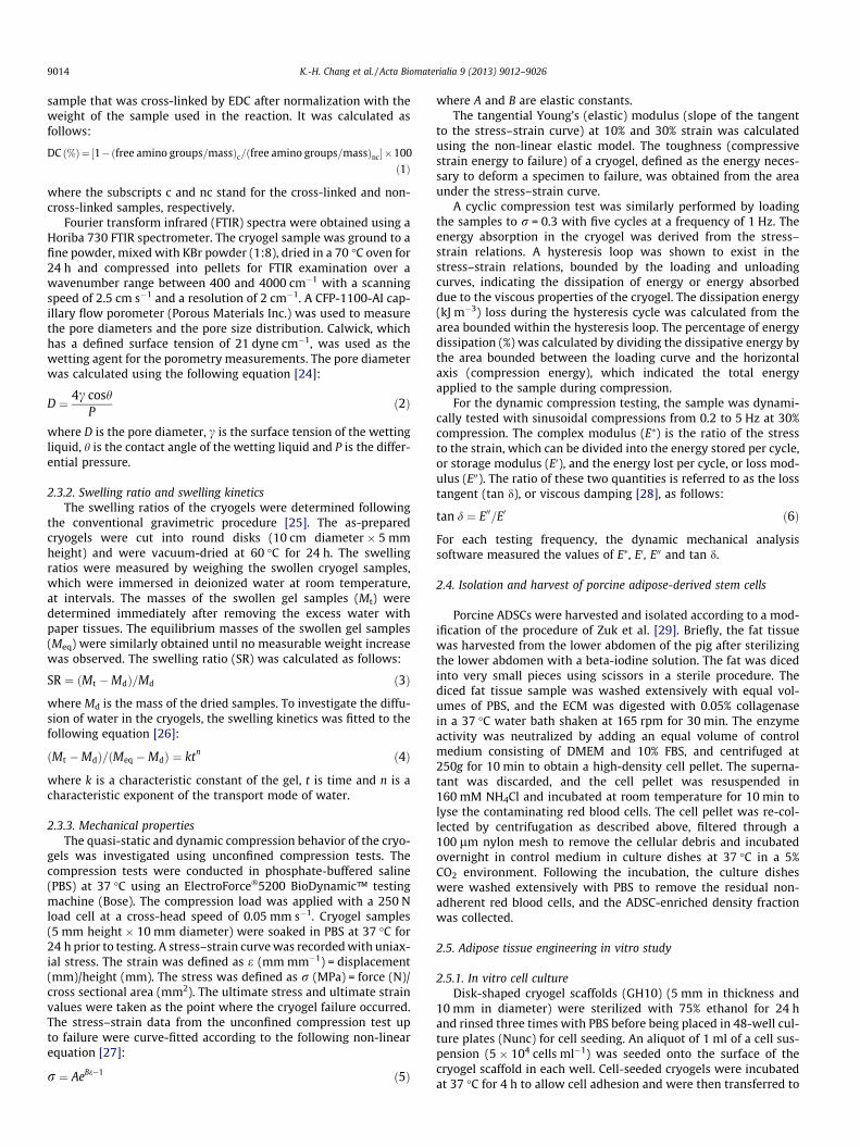
The amount of leptin, an adipogenic marker protein, in the cul-ture medium under adipogenic induction conditions (normalizedto total protein) was not significantly different in the groups cul-tured for 12–14 days or 19–21 days (Fig. 8a). However, in thegroup cultured for 40–42 days, the amount of secreted leptin in-creased significantly and the value was 2.5-fold that at 12–14and 19–21 days. The end-stage of adipogenesis was furtherconfirmed by Oil Red O staining of the lipid-containing vesiclesin the differentiated ADSCs in the GH10 cryogels (Fig. 8b).
As shown in Fig. 9, consistent with early adipogenic geneexpression, PPARc and LPL genes were significantly highlyexpressed at 3 and 7 days post-induction compared to 14 to28 days post-induction. Regarding the expression of the late genes,the aP2 gene expression was highest at 28 days, followed by21 days and worst at 3 to 14 days post-induction; the expression
of the leptin gene was low from 3 to 21 days but significantly ele-vated at 28 days post-induction.
3.3. In vivo animal studies
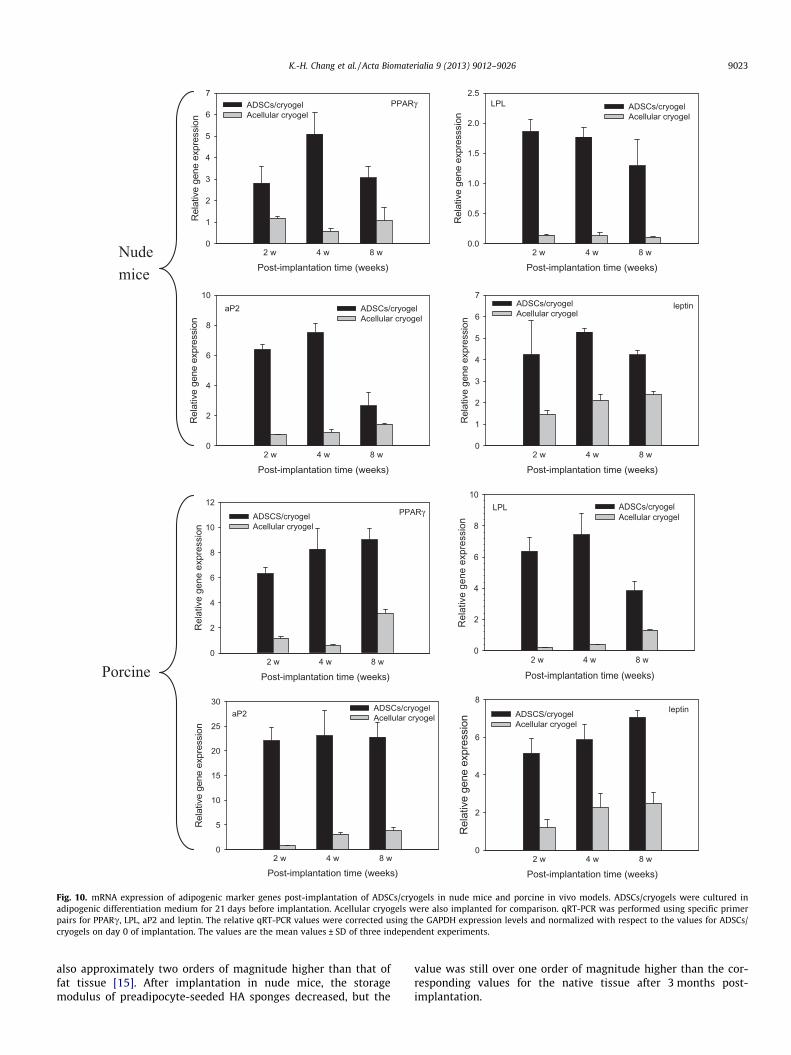
Encouraged by successful in vitro studies demonstrating that theGH10 cryogel is a good scaffold for adipose tissue engineering,in vivo animal studies were subsequently conducted with nudemouse and porcine models for translational research. In both ani-mal studies, the animals were healthy during the experimental per-iod. No inflammation, skin necrosis or any signs of infection wasnoted at the implant sites. This result demonstrates the in vivo bio-compatibility of the synthetic GH10 cryogels, which did not exerttoxic effects. The specimens were harvested at 2, 4 and 8 weekspost-implantation. The qRT-PCR results showed significant expres-sion of all of the early and late adipogenic marker genes in theexperimental groups (ADSCs/cryogel) compared with the controlgroups (acellular cryogel) at all of the time points in both animalmodels (Fig. 10). The slow increase of the background gene expres-sion in acellular cryogel groups may involve the penetration of sub-cutaneous fat cells into the cryogels at the implantation site.Nonetheless, after 3 weeks of adipogenic induction in vitro, the im-planted constructs showed a stable and consistent expression ofadipocyte-specific genes throughout the whole course of in vivostudy, indicating that the induced ADSCs in a cryogel can preservetheir adipocyte genotype in vivo for up to 8 weeks. Notably, the val-ues of relative gene expression in the ADSCs/cryogel were alwayslarger than 1, and the values were higher for the porcine model thanfor the nude mouse model. These results indicate that the cryogelcan provide a suitable environment for continued differentiationof the implanted ADSCs down the adipocyte lineage in diverse host

Frequency (Hz)0 1 2 3 4 5
E' &
E" (
MPa
)
0.0
0.1
0.2
0.3
0.4
0.5
0.6
E', GH4 E', GH10 E", GH4 E", GH10
Frequency (Hz)0 1 2 3 4 5
tan
δ
0.00
0.02
0.04
0.06
0.08
0.10
GH4GH10
a
b
Fig. 5. Viscoelastic properties of gelatin/hyaluronic the (GH) cryogel measuredusing dynamic compression testing. (a) Storage modulus (E0) and loss modulus (E00),(b) loss tangent (tan d). Strain = 30%.
Culture time (days)0 7 14 21 28
OD
492
0.0
0.5
1.0
1.5
2.0
2.5
3.0
3.5
AdipogenicControl
**
* *
Fig. 6. Comparison of the proliferation of ADSCs in the gelatin/hyaluronic cryogel(GH10) when cultured in adipogenic differentiation medium or control medium.The number of viable cells was determined by an MTS assay and reported asabsorbance at 492 nm (OD492). ⁄p < 0.05 compared with control on each day.
9020 K.-H. Chang et al. / Acta Biomaterialia 9 (2013) 9012–9026
environments, although the extent of differentiation may dependon the interplay of the source of the ADSCs and the host.
Histology using H&E staining showed adipose-like tissue forma-tion with round cells, the typical phenotype of adipocytes, withinthe pores of the cryogels in the ADSCs/cryogel group (Fig. 11aand c). In contrast, only fibrotic tissue or empty pores were foundfor the cryogels in the acellular cryogel group (Fig. 11b and d).Using antibodies specific for porcine PPARc and leptin, IHC and
ICC staining positively identified the production of PPARc and lep-tin, proteins specifically found in adipose tissue, in the ADSCs/cryo-gel group (Fig. 11e–p). In contrast, no PPARc or leptin was found inthe acellular cryogel group. CD31, also called platelet endothelialcell adhesion molecule, plays a key role in removing aged neutro-phils from the body and is used primarily to demonstrate the pres-ence of endothelial cells in histological tissue sections. In our study,positive CD31 staining was observed in the acellular cryogel groupin addition to the ADSCs/cryogel group (Fig. 11s–v). However,cryogels seeded with differentiated ADSCs exhibited more angio-genesis (larger CD31-positive areas and greater staining intensity)than did the acellular cryogels. This phenomenon might be due tothe angiogenic potential of the differentiating preadipocytes or themature adipose cells.
4. Discussion
GH cryogels were synthesized using EDC as the cross-linkingagent at �20 �C. EDC and glutaraldehyde are two of the most com-monly used chemical agents, but they work in distinctly differentmanners. EDC is limited to cross-link gelatin/HA molecules thatare directly adjacent to each other (1 nm), whereas glutaraldehydecan cross-link molecules that are more separated. However, theincorporation of glutaraldehyde into scaffolds can have implica-tions for biocompatibility. EDC, conversely, is known as a non-toxicand a biocompatible cross-linker because it generates peptide-likebonds. During the fabrication of the GH cryogels, EDC first reactswith HA to activate its carboxyl groups, and the activated groupssubsequently react with the primary amino groups of gelatin. Thisstrategy maximizes the intermolecular cross-linking between gel-atin and HA, minimizes the intramolecular covalent bond forma-tion within gelatin and generates a robust cryogel with highporosity and a high degree of cross-linking (Table 1). For scaffoldsused in tissue engineering applications, high levels of porosity playa critical role in in vitro and in vivo tissue formation [37]. The GHcryogels also had a highly interconnected 3-D structure, which isdesirable because it enhances the diffusion of nutrients and metab-olites inside the scaffold.
Other than SEM observation of the porous structure, which indi-cated pore diameters between 100 and 400 lm, we used capillaryflow porometry to obtain a more reliable estimation of the poresizes and distributions (Fig. 1). Previously, mercury intrusionporosimetry was primarily used for characterizing pore size incryogels. Because mercury intrusion porometry measures the pres-sure required to push mercury, a non-wetting liquid, through thepores in a material, the high pressure required to force viscousmercury through tiny pores could distort the structure of flexiblecryogels. In contrast, capillary flow porometry can provide repro-ducible pore size and distribution measurements with negligibledistortion errors. Indeed, the pore size of chitosan–gelatin cryogelsmeasured using mercury intrusion porosimetry (30–100 lm) ismuch lower than the values reported (�200 lm) here [8]. An ade-quate porous structure is extremely important for cellular penetra-tion into the scaffold and differentiation after transplantation.During differentiation, the adipocytes store lipids in cellular vacu-oles, thereby increasing their volume up to 20-fold. Hence, appro-priate porous structures are required so as not to inhibit thegrowth process and differentiation of the seeded ADSCs. Earlierexperiments showed that collagen sponges with a pore size of40 lm restricted the growth and differentiation of preadipocytes[38]. Using HA sponges with pore diameters varying from 50 to340 lm substantially improved cellular penetration and preadipo-cyte differentiation [17]. However, when modified HA spongeswith a pore size of 400 lm were loaded with preadipocytes andimplanted into nude mice, only minor amounts of adipose tissue
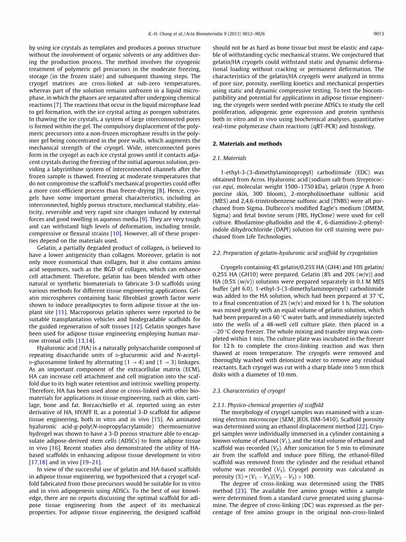
Fig. 7. Scanning electron micrographs, live/dead staining and phalloidin/DAPI cytoskeleton staining of ADSCs/cryogel constructs cultured for 21 days in control medium oradipogenic differentiation medium. Live and dead cells were stained green and red with calcein AM and ethidium homodimer-1, respectively. Actin and nuclei were stainedred and blue with phalloidin and DAPI, respectively. (a) bar = 100 lm; (b) bar = 30 lm; (c, d) bar = 100 lm; (e, f) bar = 50 lm.
Culture time (days)12-14 19-21 40-42
Lept
in s
ecre
tion
(ng/
μg to
tal p
rote
in)
0.0
0.2
0.4
0.6
0.8
1.0
1.2
**a b
Fig. 8. (a) The amount of secreted leptin at different time points and (b) Oil Red O staining (bar = 50 lm) of lipid vesicles in ADSCs/cryogels cultured for 21 days in adipogenicdifferentiation medium. ⁄p < 0.05.
K.-H. Chang et al. / Acta Biomaterialia 9 (2013) 9012–9026 9021
were found in the constructs. Hemmrich et al. hypothesized that400 lm is too large a diameter to adequately stimulate preadipo-cytes to undergo adipogenic conversion because contact inhibitionby neighboring cells is a key stimulus of differentiation by adiposetissue progenitors [19]. They proposed that the solution for achiev-ing the necessary cellular contact inhibition was to generate 3-Dstructures in which the pore diameter varies between 100 and400 lm [19]. The pore diameters determined for GH4 and GH10are within in this range. This pore size can be compared with thevalues found in other types of scaffolds for adipose tissue engineer-ing, including 135–633 lm for poly(lactic-co-glycolic) acidscaffolds [39], 450 ± 30 lm for silk fibroin scaffolds [40] and100–220 lm for collagen–hyaluronic acid scaffolds [41].
The water-binding ability of the scaffolds is another importantfeature to evaluate when assessing their potential to be applied intissue engineering. Swelling increases the pore sizes, thus maximiz-ing the surface area/volume ratio, and facilitates the infiltration ofcells into the 3-D scaffolds during cell culture [42]. The swellingstudies of the GH cryogels indicated their very high swelling capac-ity and the ability to retain more water than their original weight.The equilibrium swelling was reached after�20 min. This fast swell-ing behavior is a characteristic response that has been observed withporous and hydrophilic materials [43]. Because of the highly porousnature of the cryogel structure (porosity > 90%), porosity does notplay a significant role in the overall water uptake. The decrease in
the swelling capacity of the cryogel in the presence of more gelatin(GH10) suggests that gelatin enhances the cross-linking density ofthe cryogel and improves the structural stability of the porous matri-ces (Fig. 2a). Because no long and wide aligned channels formed inthe cryogels, no capillary effects were found and water entered theinner region of the cryogel freely throughout the swelling process,judging from the Fickian mechanism of water diffusion during theswelling stage (Fig. 2b).
One of the key issues of scaffold design for tissue engineering istheir mechanical performance. Different tissues have different tis-sue regeneration rates and different mechanical properties. For in-stance, the elastic moduli of solid tissues in the body range from1 kPa for the liver to 100 kPa for collagenous bone [44]. Therefore,the materials used in a tissue engineering approach must havetheir mechanical properties finely tuned to match those of the na-tive tissues. It was shown that the compressive properties, such asthe Young’s modulus and the compressive strength, are of greaterinterest when studying the impact of the substrate properties oncellular activity because cells tend to bend and buckle individualstruts within a scaffold [45]. Therefore, in this work, compressivetesting in the wet stage was conducted to determine the effect ofthe cryogel composition on the compressive modulus and otherimportant mechanical characteristics of cryogels. The GH cryogelswere found to mimic the non-linear elasticity found in natural softtissues (Fig. 4a). The inclusion of more gelatin in the cryogel

Culture time (days)3 7 14 21 28
Rel
ativ
e ge
ne e
xpre
ssio
n
0
20
40
60LPL*
Culture time (days)3 7 14 21 28
Rel
ativ
e ge
ne e
xpre
ssio
n
0
20
40
60
80
100aP2*
*
Culture time (days)3 7 14 21 28
Rel
ativ
e ge
ne e
xpre
ssio
n
0
5
10
15
20
25
30PPARγ*
Culture time (days)3 7 14 21 28
Rel
ativ
e ge
ne e
xpre
ssio
n
0
5
10
15
20
25
30
35Leptin*
Fig. 9. mRNA expression of adipogenic marker genes by ADSCs in the ADSCs/cryogels during in vitro culture in adipogenic differentiation medium at different time points.qRT-PCR was performed using specific primer pairs for PPARc, LPL, aP2 and leptin. The relative qRT-PCR values were corrected using the GAPDH expression levels andnormalized with respect to the values on day 0 of culture. The values are the mean values ± SD of three independent experiments. ⁄p < 0.05.
9022 K.-H. Chang et al. / Acta Biomaterialia 9 (2013) 9012–9026
composition increased the Young’s modulus (or stiffness) andtoughness (Table 2). This finding can be attributed to the formationof additional cross-links between gelatin and HA molecules, whichcontributes to the resistance of the struts to bending under com-pression. A more cross-linked material (GH10) also has diminishedultimate strain (Table 2). The elastic modulus of the GH cryogelwith 10% gelatin compared favorably with that of similar gelatin-containing cryogels reported in the literature, which are 36–39 kPa for gelatin/chitosan cryogels and 39–44 kPa for chitosan/agarose/gelatin cryogels [8,46]. In a collagen/hyaluronic scaffoldmade by freeze-drying and cross-linked by EDC, a lower elasticmodulus of 4–7 kPa at 10% strain was reported under similar testconditions [41]. Nearly all of the reported mechanical propertiesof white adipose tissue used specimens taken from the humanbreast and heel pad. Using mechanical testing, the instantaneouselastic modulus for adipose tissue was found to be in the rangeof 3–24 kPa for breast samples [47] and in the range of 24–180 kPa for heel pad samples [48]. The minimal modulus require-ment could be met by the GH cryogels, although only the GH10cryogel could attain the upper limit (Table 2). Overall, GH4 dissi-pated more energy, on an absolute scale, and exhibited a higherpercentage of energy dissipation than did GH10 (Table 2). The per-centage of energy dissipation reported in the literature for humanheel pad fat is from 21% to 24%, and it changed little over the com-plete range of frequencies. Heel pads were found to be resilient,returning �80% of the energy used to deform them, independentof the loading frequency [49]. GH10 is thus expected to performbetter than GH4 in terms of energy dissipation.
One interesting characteristic of the GH cryogels was that nofailure or crack was found when they were compressed to morethan 60% of their original lengths and retained their shape when
the compression stress was removed. This property of superiorrecovery from large strains was shown by the same hysteresiscurves being obtained from the cryogels during five successiveloading–unloading cycles at 30% maximum strain (Fig. 4b andc). The constant dissipation energy during each cycle also pro-vides quantitative evidence to support this unique property(Fig. 4d). This unique feature contributes to the potential ofthe GH10 cryogel as a tough scaffold for soft tissue engineering,which can recover from large strains and absorb impacts withoutpermanent damage. The viscoelastic properties of the GH cryo-gels obtained from dynamic compression testing indicated veryhigh elasticity (Fig. 5), which could be compared to that of adi-pose tissue. The constitutive properties of adipose tissue havebeen measured over a wide range of frequencies, from2 � 10�3 s�1 to 5700 s�1. The storage modulus (E0) is almostinsensitive to the frequency and has a value of 1 kPa at a fre-quency below 10 s�1. However, at a higher frequency, above10 s�1, the storage modulus of adipose tissue increases sharply,and at a frequency of 3000 s�1, the storage modulus of adiposetissue is comparable to that of the dermis (�4 MPa) [50]. Krou-skop et al. investigated the viscoelastic properties of breast andprostate tissues (normal, benign and malignant) at low frequen-cies [51]. The group reported storage modulus values of 18–66 Pa for breast fat tissue under various amounts of pre-com-pression loading [51]. Although the values for the loss moduluswere not reported, they noted that the phase angle (d) of fatbreast tissue was less than 10� [51]. Such a small phase angleindicates a very small viscous response in the tissues at thefrequencies reported, which could be met by the highly elasticGH cryogel (d = 2�). It should be noted that the storage modulusof HA sponges used successfully for adipose tissue engineering is
PPARγ
Post-implantation time (weeks)2 w 4 w 8 w
Rel
ativ
e ge
ne e
xpre
ssio
n
0
1
2
3
4
5
6
7ADSCs/cryogelAcellular cryogel
LPL
Post-implantation time (weeks)2 w 4 w 8 w
Rel
ativ
e ge
ne e
xpre
sssi
on
0.0
0.5
1.0
1.5
2.0
2.5ADSCs/cryogelAcellular cryogel
aP2
Post-implantation time (weeks)2 w 4 w 8 w
Rel
ativ
e ge
ne e
xpre
ssio
n
0
2
4
6
8
10ADSCs/cryogelAcellular cryogel
leptin
Post-implantation time (weeks)2 w 4 w 8 w
Rel
ativ
e ge
ne e
xpre
ssio
n
0
1
2
3
4
5
6
7ADSCs/cryogelAcellular cryogel
PPARγ
Post-implantation time (weeks)2 w 4 w 8 w
Rel
ativ
e ge
ne e
xpre
ssio
n
0
2
4
6
8
10
12ADSCS/cryogelAcellular cryogel
LPL
Post-implantation time (weeks)2 w 4 w 8 w
Rel
ativ
e ge
ne e
xpre
ssio
n
0
2
4
6
8
10ADSCs/cryogelAcellular cryogel
aP2
Post-implantation time (weeks)2 w 4 w 8 w
Rel
ativ
e ge
ne e
xpre
ssio
n
0
5
10
15
20
25
30ADSCs/cryogelAcellular cryogel
leptin
Post-implantation time (weeks)2 w 4 w 8 w
Rel
ativ
e ge
ne e
xpre
ssio
n
0
2
4
6
8
ADSCS/cryogelAcellular cryogel
Nudemice
Porcine
Fig. 10. mRNA expression of adipogenic marker genes post-implantation of ADSCs/cryogels in nude mice and porcine in vivo models. ADSCs/cryogels were cultured inadipogenic differentiation medium for 21 days before implantation. Acellular cryogels were also implanted for comparison. qRT-PCR was performed using specific primerpairs for PPARc, LPL, aP2 and leptin. The relative qRT-PCR values were corrected using the GAPDH expression levels and normalized with respect to the values for ADSCs/cryogels on day 0 of implantation. The values are the mean values ± SD of three independent experiments.
K.-H. Chang et al. / Acta Biomaterialia 9 (2013) 9012–9026 9023
also approximately two orders of magnitude higher than that offat tissue [15]. After implantation in nude mice, the storagemodulus of preadipocyte-seeded HA sponges decreased, but the
value was still over one order of magnitude higher than the cor-responding values for the native tissue after 3 months post-implantation.
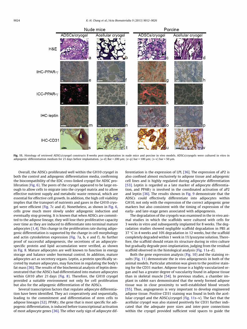
Fig. 11. Histology of retrieved ADSCs/cryogel constructs 8 weeks post-implantation in nude mice and porcine in vivo models. ADSCs/cryogels were cultured in vitro inadipogenic differentiation medium for 21 days before implantation. (a–d) Bar = 200 lm; (e–p) bar = 100 lm; (s–v) bar = 50 lm.
9024 K.-H. Chang et al. / Acta Biomaterialia 9 (2013) 9012–9026
Overall, the ADSCs proliferated well within the GH10 cryogel inboth the control and adipogenic differentiation media, confirmingthe biocompatibility of the EDC cross-linked cryogel for ADSC pro-liferation (Fig. 6). The pores of the cryogel appeared to be large en-ough to allow cells to migrate into the cryogel matrix and to alloweffective nutrient supply and metabolic waste removal, which areessential for effective cell growth. In addition, the high cell viabilityimplies that the transport of nutrients and gases in the GH10 cryo-gel were efficient (Fig. 7c and d). Nonetheless, as shown in Fig. 6,cells grow much more slowly under adipogenic induction andeventually stop growing. It is known that when ADSCs are commit-ted to the adipose lineage, they will lose their proliferative capacityover time as they are induced to differentiate into terminal matureadipocytes [1,4]. This change in the proliferation rate during adipo-genic differentiation is supported by the change in cell morphologyand actin cytoskeleton expression (Fig. 7a, b, e and f). As furtherproof of successful adipogenesis, the secretions of an adipocyte-specific protein and lipid accumulation were verified, as shownin Fig. 8. Mature adipocytes are well known to function in energystorage and balance under hormonal control. In addition, matureadipocytes act as secretory organs. Leptin, a protein specifically se-creted by mature adipocytes, may function in regulating the body’sfat mass [36]. The results of the biochemical analysis of leptin dem-onstrated that the ADSCs had differentiated into mature adipocyteswithin GH10 after 21 days (Fig. 8). Therefore, the GH10 cryogelprovided a suitable environment not only for cell proliferationbut also for the adipogenic differentiation of the ADSCs.
Several transcription factors that regulate adipocyte differentia-tion have been identified. They act cooperatively and sequentially,leading to the commitment and differentiation of stem cells toadipose lineages [52]. PPARc, the gene that is most specific for adi-pogenic differentiation, is induced before transcriptional activationof most adipocyte genes [36]. The other early sign of adipocyte dif-
ferentiation is the expression of LPL [36]. The expression of aP2 isalso confined almost exclusively to adipose tissue and adipogeniccell lines and is highly regulated during adipocyte differentiation[53]. Leptin is regarded as a late marker of adipocyte differentia-tion, and PPARc is involved in the coordinated activation of aP2and leptin [36]. The results shown in Fig. 9 demonstrate that theADSCs could effectively differentiate into adipocytes withinGH10, not only with the expression of the correct adipogenic genemarkers but also consistent with the timing of expression of theearly- and late-stage genes associated with adipogenesis.
The degradation of the cryogels was examined in the in vivo ani-mal studies in which the scaffolds were cultured with cells for3 weeks in vitro and subsequently implanted for 8 weeks. The deg-radation studies showed negligible scaffold degradation in PBS at37 �C in 4 weeks and 10% degradation in 12 weeks, but the scaffoldcompletely degraded within 1 week in 1% lysozyme solution. There-fore, the scaffold should retain its structure during in vitro culturebut gradually degrade post-implantation, judging from the residualscaffold observed in the histological analysis (Fig. 11a–d).
Both the gene expression analysis (Fig. 10) and the staining re-sults (Fig. 11) demonstrate the in vivo adipogenesis in both of theanimal models. Particular attention was given to the positive stain-ing for the CD31 marker. Adipose tissue is a highly vascularized or-gan and has a greater degree of vascularity found in adipose tissuethan in skeletal muscle [54]. In previous studies, a chamber im-plant in rabbit ears demonstrated that the newly formed adiposetissue was in close proximity to well-established blood vessels[55]. Thus, angiogenesis is very important to develop engineeredadipose tissue. Positive CD31 staining was found in both the acel-lular cryogel and the ADSCs/cryogel (Fig. 11s–v). The fact that theacellular cryogel was also stained positively for CD31 further indi-cated that the adequate pore sizes and inter-pore connectionswithin the cryogel provided sufficient void spaces to guide the

K.-H. Chang et al. / Acta Biomaterialia 9 (2013) 9012–9026 9025
ingrowth of the surrounding vasculature. Considering the angio-genic potential of differentiating preadipocytes or mature adiposecells in the GH10 cryogel, the differentiating preadipocytes couldexpress bFGF, which further enhanced angiogenesis by dictatingcapillary and preadipocyte growth [56]. Subcutaneous or omentaladipose tissue was proven to enhance wound healing and corneaneovascularization [57]. Therefore, it is believed that differentiat-ing or mature adipocytes secrete angiogenic factors. Recently, lep-tin has been found to generate a growth signal involving a tyrosinekinase-dependent intracellular pathway and promote angiogenicprocesses in vitro and in vivo [58]. The angiogenic role of leptin,whose gene expression and protein secretion in the in vitro cul-tured ADSCs in the cryogels were confirmed by PCR and an ELISA,further contributed to angiogenesis and the CD31-staining inten-sity of the implanted ADSCs/cryogel constructs in vivo.
5. Conclusion
We demonstrated that macroporous elastic cryogels could befabricated from gelatin and hyaluronic acid, two biopolymers suit-able as adipose tissue engineering scaffolding materials. The GHcryogel is endowed with unique mechanical properties, such ashigh extensibility, moderate toughness and total recovery fromlarge strains. Furthermore, the superior physico-chemical proper-ties, such as high porosity, large pore size, fast swelling kineticsand high swelling ratio, combined with mechanical properties sim-ilar to those of adipose tissue, make the GH10 cryogel a suitablescaffold for adipose tissue engineering. The growth arrest, morpho-logical changes, actin cytoskeleton expression and leptin secretionof the ADSCs in GH cryogel are consistent with the differentiationprocess that occurs under adipogenic induction. The mRNA expres-sion of adipogenic marker genes also supported the adipogenesis ofADSCs in the GH cryogel in vitro. Based on the IHC and ICC stainingof the phenotypic markers for adipocytes (PPARc and leptin) andthe angiogenic marker CD31, and the expression of adipogenicmarker genes, successful adipose tissue engineering using commit-ted ADSCs in the GH10 cryogel was also demonstrated in the nudemouse and porcine animal models.
Acknowledgements
This work was supported by Chang Gung Memorial Hospital(CMRPD2C0081) and National Science Council, Taiwan, ROC.
Appendix A. Appendix: Figures with essential colourdiscrimination
Certain figures in this article, particularly Figs. 3, 4, 7, 8 and 11,are difficult to interpret in black and white. The full colour imagescan be found in the on-line version, at http://dx.doi.org/10.1016/j.actbio.2013.06.046).
Reference
[1] Patrick CW. Adipose tissue engineering: the future of breast and soft tissuereconstruction following tumor resection. Semin Surg Oncol 2000;19:302–11.
[2] Kaufman MR et al. Autologous fat transfer national consensus survey: trends intechniques for harvest, preparation, and application, and perception of short-and long-term results. Plast Reconstr Surg 2007;119:323–31.
[3] Tanzi MC, Farè S. Adipose tissue engineering: state of the art, recent advancesand innovative approaches. Expert Rev Med Devices 2009;6:533–51.
[4] Beahm EK, Walton RL, Patrick CW. Progress in adipose tissue constructdevelopment. Clin Plast Surg 2003;30:547–58.
[5] Garg T, Singh O, Arora S, Murthy RSR. Scaffold: A novel carrier for cell and drugdelivery. Crit Rev Ther 2012;29:1–63.
[6] Bencherif SA, Sands RW, Bhatta D, Arany P, Verbeke CS, Edwards DA, et al.Injectable preformed scaffolds with shape-memory properties. Proc Natl AcadSci U S A 2012;109:19590–5.
[7] Dainiak MB, Kumar A, Galaev IY, Mattiasson B. Detachment of affinity capturedbioparticles by elastic deformation of a macroporous hydrogel. Proc Natl AcadSci U S A 2006;103:849–54.
[8] Kathuria N, Tripathi A, Kar KK, Kumar A. Synthesis and characterization ofelastic and macroporous chitosan-gelatin cryogels for tissue engineering. ActaBiomater 2009;5:406–18.
[9] Lozinsky VI, Galaev IY, Plieva FM, Savinal IN, Jungvid H, Mattiasson B.Polymeric cryogels as promising materials of biotechnological interest. TrendsBiotechnol 2003;21:445–51.
[10] Ozmen MM, Dinu MV, Dragan ES, Okay O. Preparation of macroporousacrylamide-based hydrogels: cryogelation under isothermal conditions. JMacromol Sci A 2007;44:1195–202.
[11] Kimura Y, Ozeki M, Inamoto T, Tabata Y. Adipose tissue engineering based onhuman preadipocytes combined with gelatin microspheres containing basicfibroblast growth factor. Biomaterials 2003;24:2513–21.
[12] Huss FRM, Junker JPE, Johnson H, Kratz G. Macroporous gelatin spheres asculture substrate, transplantation vehicle, and biodegradable scaffold forguided regeneration of soft tissues. In vivo study in nude mice. J Plast ReconstrRes 2007;60:543–55.
[13] Lin SD, Wang KH, Kao AP. Engineered adipose tissue of predefined shape anddimensions from human adipose-derived mesenchymal stem cells. Tissue EngPart A 2008;14:571–81.
[14] Hong L, Peptan I, Clark P, Mao JJ. Ex vivo adipose tissue engineering by humanmarrow stromal cell seeded gelatin sponge. Ann Biomed Eng 2005;33:511–7.
[15] Borzacchiello A, Mayol L, Ramires PA, Pastorello A, Di Bartolo C, Ambrosio L,et al. Structural and rheological characterization of hyaluronic acid-basedscaffolds for adipose tissue engineering. Biomaterials 2007;28:4399–408.
[16] Tan HP, Ramirez CM, Miljkovic N, Li H, Rubin JP, Marra KG. Thermosensitiveinjectable hyaluronic acid hydrogel for adipose tissue engineering.Biomaterials 2009;30:6844–53.
[17] Halbleib M, Skurk T, de Luca C, von Heimburg D, Hauner H. Tissue engineeringof white adipose tissue using hyaluronic acid-based scaffolds. I. In vitrodifferentiation of human adipocyte precursor cells on scaffolds. Biomaterials2003;24:3125–32.
[18] Flynn L, Prestwich G, Semple J, Woodhouse K. Adipose tissue engineering withnaturally derived scaffolds and adipose-derived stem cells. Biomaterials2007;28:3834–42.
[19] Hemmrich K, von Heimburg D, Rendchen R, Di Bartolo C, Milella E, Pallua N.Implantation of preadipocyte-loaded hyaluronic acid-based scaffolds intonude mice to evaluate potential for soft tissue engineering. Biomaterials2005;26:7025–37.
[20] Hemmrich K, Van de Sijpe K, Rhodes NP, Hunt JA, Di Bartolo C, Pallua N, et al.Autologous in vivo adipose tissue engineering in hyaluronan–based gels–apilot study. J Surg Res 2008;144:82–8.
[21] Flynn L, Prestwich GD, Semple JL, Woodhouse KA. Adipose tissue engineeringin vivo with adipose-derived stem cells on naturally derived scaffolds. JBiomed Mater Res 2009;89A:929–41.
[22] Zhang RY, Ma PX. Poly(a-hydroxyl acids)/hydroxyapatite porous compositesfor bone tissue engineering. I. Preparation and morphology. J Biomed MaterRes 1999;44:446–55.
[23] Habeeb AFSA. Determination of free amino groups in proteins bytrinitrobenzenesulfonic acid. Anal Biochem 1966;14:328–36.
[24] Jena A, Gupta K. Characterization of pore structure of filter media. Fluid/PartSep J 2002;14:227–41.
[25] Xue W, Champ S, Huglin MB, Jones TGJ. Rapid swelling and deswelling incryogels of crosslinked poly(N-isopropylacrylamide-co-acrylic acid). EurPolym J 2004;40:467–76.
[26] Khare AR, Peppas NA. Swelling/deswelling of anionic copolymer gels.Biomaterials 1995;16:559–67.
[27] Woo SL, Gomez MA, Akeson WH. The time and history dependent viscoelasticproperties of the canine medial collateral ligament. J Biomech Eng1981;1981(103):293–8.
[28] Lakes R. Viscoelastic Solids. Boca Raton, FL: CRC Press; 1999. pp. 1–72.[29] Zuk PA et al. Multilineage cells from human adipose tissue: implications for
cell-based therapies. Tissue Eng 2001;7:211–8.[30] Choi YS, Cha SM, Lee YY, Kwon SW, Park CJ, Kim M. Adipogenic differentiation
of adipose tissue derived adult stem cells in nude mouse. Biochem Biophys ResCommun 2006;345:631–7.
[31] Fischbach C, Spruss T, Weiser B, Neubauer M, Becker C, Hacker M, et al.Generation of mature fat pads in vitro and in vivo utilizing 3-D long-termculture of 3T3-L1 preadipocytes. Exp Cell Res 2004;300:54–64.
[32] Torres FG, Nazhat SN, Fadzullah SH, Maquet V, Boccaccini AR. Mechanicalproperties and bioactivity of porous PLGA/TiO2 nanoparticle-filled compositesfor tissue engineering scaffolds. Compos Sci Technol 2007;67:1139–47.
[33] Santawisuk W, Kanchanavasita W, Sirisinha C, Harnirattisai C. Dynamicviscoelastic properties of experimental silicone soft lining materials. DentMater J 2010;29:454–60.
[34] Beahm EK, Walton RL, Patrick Jr CW. Progress in adipose tissue constructdevelopment. Clin Plast Surg 2003;30:547–58.
[35] Gregoire FM, Smas CM, Sul HS. Understanding adipocyte differentiation.Physiol Rev 1998;78:783–809.
[36] Speigelman BM, Farmer SR. Decreases in tubulin and actin gene expressionprior to morphological differentiation of 3T3 adipocytes. Cell 1982;29:53–60.
[37] Karageorgiou V, Kaplan D. Porosity of 3D biomaterial scaffolds andosteogenesis. Biomaterials 2005;26:5474–91.

9026 K.-H. Chang et al. / Acta Biomaterialia 9 (2013) 9012–9026
[38] von Heimburg D, Zachariah S, Heschel I, Kuhling H, Schoof H, Hafemann B,et al. Human preadipocytes seeded on freeze-dried collagen scaffoldsinvestigated in vitro and in vivo. Biomaterials 2001;22:429–38.
[39] Patrick CW, Chauvin PB, Hobley J, Reece JP. Preadipocyte seeded PLGA scaffoldsfor adipose tissue engineering. Tissue Eng 1999;5:139151.
[40] Mauney JR, Nguyen T, Gillen K, Kirker-Head C, Gimble JM, Kaplan DL.Engineering adipose-like tissue in vitro and in vivo utilizing human bonemarrow and adipose-derived mesenchymal stem cells with silk fibroin 3Dscaffolds. Biomaterials 2007;28:5280–90.
[41] Davidenko N, Campbell JJ, Thian ES, Watson CJ, Cameron RE. Collagen–hyaluronic acid scaffolds for adipose tissue engineering. Acta Biomater2010;6:3957–68.
[42] Jayakumar R et al. Fabrication of chitin–chitosan/nano ZrO2 compositescaffolds for tissue engineering applications. Int J Biol Macromol2011;49:274–80.
[43] Jain E, Srivastava A, Kumar A. Macroporous interpenetrating cryogel networkof poly(acrylonitrile) and gelatin for biomedical applications. J Mater Sci MaterMed 2009;20:173–9.
[44] Engler AJ, Sen S, Sweeney HL, Discher DE. Matrix elasticity directs stem celllineage specification. Cell 2006;126:677–89.
[45] Harley BA, Leung JH, Silva ECCM, Gibson LJ. Mechanical characterization ofcollagen–glycosaminoglycan scaffolds. Acta Biomater 2007;3:463–74.
[46] Bhat S, Tripathi A, Kumar A. Supermacroprous chitosan agarose gelatincryogels: in vitro characterization and in vivo assessment for cartilage tissueengineering. J R Soc Interface 2011;8:540–54.
[47] Samani A, Plewes D. A method to measure the hyperelastic parameters ofex vivo breast tissue samples. Phys Med Biol 2004;49:4395–405.
[48] Gefen A, Megido-Ravid M, Itzchak Y. In vivo biomechanical behavior of thehuman heel pad during the stance phase of gait. J Biomech 2001;34:1661–5.
[49] Bennett MB, Ker RF. The mechanical properties of the human subcalcaneal fatpad in compression. J Anat 1990;171:131–8.
[50] Comley K, Fleck N. The compressive response of porcine adipose tissue fromlow to high strain rate. Int J Impact Eng 2012;46:1–10.
[51] Krouskop TA, Wheeler TM, Kallel F, Hall T. The elastic moduli of breast andprostate tissues under compression. Ultrason Imaging 1998;20:260–74.
[52] Mandrup S, Lane MD. Regulating Adipogenesis. J Biol Chem1997;272:5367–70.
[53] Coe NR, Bernlohr DA. Physiological properties and functions of intracellularfatty acid-binding proteins. Biochim Biophys Acta 1998;1391:287–306.
[54] Crandall DL, Hausman GJ, Kral JG. A review of the microcirculation of adiposetissue: anatomic, metabolic and angiogenic perspectives. Microcirculation1997;4:211–32.
[55] Clark E, Clark EL. Microscopic studies of the new formation of fat in living adultrabbits. Am J Anat 1940;675:255–85.
[56] Teichertkuliszewska K, Hamilton BS, Deitel M, Roncari DAK. Augmentedproduction of heparin-binding mitogenic proteins by preadipocytes frommassively obese persons. J Clin Invest 1992;90:1226–31.
[57] Silverman KJ et al. Angiogenic activity of adipose tissue. Biochem Biophys ResCommun 1988;153:347–52.
[58] Park HY et al. Potential role of leptin in angiogenesis: leptin inducesendothelial cell proliferation and expression of matrix metalloproteinasesin vivo and in vitro. Exp Mol Med 2001;33:95–102.