376138
TRANSCRIPT
8/13/2019 376138
http://slidepdf.com/reader/full/376138 1/17
Hindawi Publishing CorporationInternational Journal of Alzheimer’s DiseaseVolume 2012, Article ID 376138, 17 pagesdoi:10.1155/2012/376138
Research Article Vaccine Development to Treat Alzheimer’s DiseaseNeuropathology in APP/PS1 Transgenic Mice
Iv an Carrera,1 Ignacio Etcheverrıa,1 Lucıa Fernandez-Novoa,1 Valter Lombardi,1
Ramon Cacabelos,2 and Carmen Vigo3
1 Department of Neurosciences, EuroEspes Biotechnology, Polıgono de Bergondo, Nave F, 15165 A Coru˜ na, Spain 2 EuroEspes Biomedical Research Center, Institute for CNS Disorders and Genomic Medicine and EuroEspes Foundation,
15166 La Coru˜ na, Spain3 Neuroscience Division, Atlas Pharmaceuticals, Sunnyvale, CA 94089, USA
Correspondence should be addressed to Iv an Carrera, [email protected]
Received 15 May 2012; Revised 14 June 2012; Accepted 28 June 2012
Academic Editor: Francesco Panza
Copyright © 2012 Iv an Carrera et al. This is an open access article distributed under the Creative Commons Attribution License,which permits unrestricted use, distribution, and reproduction in any medium, provided the original work is properly cited.
A novel vaccine addressing the major hallmarks of Alzheimer’s disease (AD), senile plaque-like deposits of amyloid beta-protein(A β), neurofibrillary tangle-like structures, and glial proinflammatory cytokines, has been developed. The present vaccine takesa new approach to circumvent failures of previous ones tested in mice and humans, including the Elan-Wyeth vaccine (AN1792),which caused massive T-cell activation, resulting in a meningoencephalitis-like reaction. The EB101 vaccine consists of A β1–42
delivered in a novel immunogen-adjuvant composed of liposomes-containing sphingosine-1-phosphate (S1P). EB101 wasadministered to APPswe/PS1dE9 transgenic mice before and after AD-like pathological symptoms were detectable. Treatment withEB101 results in a marked reduction of A β plaque burden, decrease of neurofibrillary tangle-like structure density, and attenuationof astrocytosis. In this transgenic mouse model, EB101 reduces the basal immunological interaction between the T cellsandimmune activation markers in the aff ected hippocampal/cortical areas, consistent with decreased amyloidosis-induced inflam-mation. Therefore, immunization with EB101 prevents and reverses AD-like neuropathology in a significant manner by haltingdisease progression without developing behavioral spatial deficits in transgenic mice.
1. Introduction
Alzheimer’s disease (AD) is the most frequent form of dementia in developed countries, with a prevalence of about
1% at the age of 65 and over 25% in people older than 85 years of age. Clinically, it is characterized by progressive cog-nitive deterioration, behavioral disturbances, and functionaldecline. It is known that AD is a polygenic/complex disorderin which hundreds of genes, distributed across the humangenome, might be involved in close cooperation withenvironmental inducers, cerebrovascular dysfunction, andepigenetic phenomena [1]. The main neuropathologicalhallmarks of AD include accumulation of amyloid- β (A β)peptide, neurofibrillary tangle-like structures (NFTs) com-posed largely of paired helical filaments with phosphorylatedtau proteins, neuronal and synaptic loss [2], reduced overallbrain volume with specific damage of entorhinal cortex and
hippocampus [3], neuroinflammation, gliosis, free radicalformation [4], neurotransmitter and neurotrophic factordeficits, metabolic impairment [5], and proteasome andchaperone dysregulation [6]. In the past decade, A β-based
immunotherapy has been shown to be the most promis-ing therapeutic approach [7–12], and there are over10,000 patients currently enrolled in active or passiveA β immunotherapy, showing so far a certain degree of success in decreasing beta amyloid [13–17] and revers-ing memory deficits [18–20]. However, the first clinicaltrial in 2001 sponsored by Elan and Wyeth with activeimmunization, consisting of aggregated synthetic A β42 pep-tide delivered in QS21 adjuvant, resulted in 6% patientdeath due to a meningoencephalitis-like reaction [13, 21],probably induced by an over-extensive T-cell-mediatedimmune response [22, 23]. Remarkably, patients with abbre-viated treatment generated anti-A β antibodies, reducing
8/13/2019 376138
http://slidepdf.com/reader/full/376138 2/17
2 International Journal of Alzheimer’s Disease
cerebrospinal levels of tau and reported a slower cognitivedecline [24, 25]. Prior to clinical trials, numerous experi-mental therapeutic immunization strategies were developedusing transgenic mice models of AD-like pathology, includ-ing APPswe/PS1dE9 double transgenic mice, to investigateemergent therapies focused on preventing and/or treating
AD neuropathology [26, 27]. Transgenic mice expressingmutated forms of the gene for the human amyloid precursorprotein (hAPP ) prematurely show marked elevation of A β-protein levels and deposition in the cerebral cortex andhippocampus [28–30], as seen in the brain of AD patients[31]. Presenilin-1 (PS1-) mutant transgenic mice also show an increase in A β1–42 (A β42) peptide generation, potentiatingamyloid deposition in the brain at about 6 months of age[32]. In the present study we used APPswe/PS1dE9 double-transgenic mice derived from the coexpression of mutated APP and PS1 genes, which have demonstrated a prematurely accelerated accumulation of A β deposits in the brain,compared with those expressing singly APP or PS1 mutationsalone [33–37]. This feature, together with a variety of otherclinically relevant AD-like alterations, points out this modelas a valuable tool in the development of new AD therapeuticapproaches [38]. In the present study, we present the EB101vaccine, which is designed to become cost-eff ective and long-lasting, being targeted toward the reduction of A β burdenand the slowing of the main AD-like pathological alterations,including the inflammatory reaction, by the induction of an anti-inflammatory T-helper (Th) 2 immune response.All these requirements will be achieved by designing aphysiological adjuvant composed of naturally occurringphospholipids, proven safe and efficacious in other typeof vaccines, including influenza, with an added biologically active phospholipid, S1P, known to stimulate an anti-inflammatory reaction and act as a neuronal regeneratingagent in in vitro and in vivo studies [39].
The purpose of this study was to investigate the eff ectand safety of an A β vaccine (EB101) in APPswe/PS1dE9transgenic mice elicited by a novel immunogenic adjuvant,designed to reduce A β deposition but avoiding the massiveactivation of T-cell-mediated immune response that poten-tially caused severe adverse eff ects.
2. Materials and Methods
2.1. Animals. APPswe/PS1dE9 double-transgenic mice
(B6C3F1/J), expressing a chimeric mouse/human amyloidprecursor protein (Mo/HuAPP695swe) and human preseni-lin 1 (PS1-dE9) mutants, both directed to neurons of thecentral nervous system (CNS), were used in this study. Thesetwo constructs were coinjected into B6C3HF2 pronucleiand insertion of the transgenes occurred at a single mouselocus. The transgenic mice used were purchased from TheJackson Laboratory. All experimental procedures conformedto the guidelines established by the European CommunitiesCouncil Directive (86/609/EEC) and by the Spanish RoyalDecree 1201/2005 for animal experimentation and wereapproved by the Ethical Committee of the EuroEspes Bio-medical Research Center.
2.2. Experimental Design. Two experimental treatment stud-ies were carried out; one before AD onset, starting at 7weeks of age (preventive treatment) and the second after ADonset, at 35 weeks of age, when neuropathological AD chara-cteristics were well established (therapeutic treatment). Micewere randomly divided into these two experimental groups,
and each group was subdivided into another three, as follows.
2.2.1. Preventive Treatment. Group A consists of 6 transgenicmice that were immunized with a cocktail of A β42/S1P/liposome (EB101); group B, 6 transgenic mice immunizedwith liposomes without A β42 (EB102); group C, 4 transgenicmice inoculated with vehicle, PBS. Two wild-type mice werealso included in each group as a safety immunization control,both in preventive and therapeutic treatments.
2.2.2. Therapeutic Treatment. Group A consists of 6 trans-genic mice immunized with EB101; group B, 6 mice immu-nized with EB102; group C, 4 transgenic mice inoculated
with PBS. Mice were immunized with nine injections forseven months and then kept for 2 additional months beforesacrifice. Mice were 11 months old at the end of preventivetreatment and 18 months old at the end of therapeutictreatment.
2.3. Experimental Procedure
2.3.1. Preparation of A β. Two mg of human synthetic A β42
(TOCRIS bioscience; Tocris Cookson Ltd.) were dissolved in0.9 mL water, and 0.1 mL of PBS was added. The mixture wasvortexed, aliquoted, lyophilized, and stored as a dry powderat −20◦C until use.
2.3.2. Preparation of Liposomes. Liposomes were preparedfrom 1,2-Dioleoyl-sn-Glycero-3-Phosphocholine (DOPC),1-Palmitoyl-2-oleoyl-sn-glycero-3-phosphatidylglycerol,Sodium Salt (POPG), cholesterol (CH; Northern lipidsINC.), plus/minus D-erythro-Sphingosine-1-Phosphate(S1P), (AVANTI at 0.3/0.3/0.39/0.01, molar ratio resp.). Onehundred milligrams of each lipid were dissolved in 1 mL of chloroform and stored at −20◦C until the preparation of theS1P liposomes, 10 mg was dissolved in 1.5 mL (2 : 1 chloro-form/methanol) and stored at −20◦C until use. Lipids, inthe final proportion indicated above, were thoroughly mixedin the organic solvent, evaporated under nitrogen. The
corresponding mixture, containing total lipid 100 mg and0.6 mg of S1P, was resuspended in autoclaved ultrapurewater and thoroughly vortex mixed until a milky solutionwas formed or the so-called “multilamellar vesicles” (MLVs).Small (SLV) or single unilamellar vesicles (SUVs) were sub-sequently prepared by sonicating the MLV for 2 minutes at30-second intervals in an ice bath until the solution was clearand centrifuged at 2500 g/15 minutes to eliminate debris.The SUV preparation was freeze dried and resuspended inPBS-containing A β (10 mg) and thoroughly mixed. Thefreeze-dried mixture was resuspended in the correspondingamount of autoclaved ultrapure water, ready for immuniza-tion. 100 µL of EB101 was used for the immunization step.
8/13/2019 376138
http://slidepdf.com/reader/full/376138 3/17
International Journal of Alzheimer’s Disease 3
Liposomes containing S1P and A β are referred as EB101 andwithout S1P and Ab are referred as EB102.
2.3.3. Immunization Procedures. Forty-four APPswe/PS1dE9transgenic mice were inoculated intraperitoneally with100 µL per injection of EB101 (group A), only liposome
complex EB102 (group B) or PBS (group C), during sevenmonths (9 injections) for each treatment period. Theimmunization protocol was systematically used in previousstudies [45, 47], consisting of 3 injections every two weeks, inthe first month and then one injection each following monthregimen followed in the preventive and therapeutic groups.
2.3.4. Spleen Cell Preparation. Splenocytes from these micewere harvested from spleen tissue mechanically disruptedwith forceps. Lysis of red blood cells was carried out in100 mM Tris buff er, pH 7.5, containing 140 mM ammoniumchloride. Splenocytes were washed twice with PBS pH 7.0and centrifuged at 1600 rpm for 5 minutes. Cells were
counted and resuspended in complete RPMI-1640 mediumcontaining 10% FSC, 50 µM β-mercaptoethanol, and40 µg/mL−1 gentamycin at 106 cells/mL.
2.3.5. Preparation of B-Cell-Enriched Suspensions. B-cell-enriched suspensions were obtained as follows: macrophagesfrom spleen cells were depleted by selective adherence to glassPetri dishesfor 2 h at 37◦C, and nonadherent cell suspensionswere depleted of T cells by magnetic cell sorting using anti-Thy-1.2-coated magnetic beads (Dynal Biotech, France),following the manufacturer’s instructions. This procedure yielded an enriched B-cell population of >90% CD19+ cellswith <1% CD3+ cells and <5% CD11c+ cells, as determined
by flow cytometry analysis, with >95% viable cells, as deter-mined by trypan blue exclusion.
2.3.6. B- and T-Cell Phenotypic Analysis Was Carried Out by Flow Cytometry. Briefly, 1 × 10⁶ red-cell-depleted spleno-cytes were incubated with anti-mouse CD32/CD16 antibody (control antibody) to block nonspecific Ig trapping throughFc receptors. The following PE-conjugated anti-mouse mAbswere used for analysis: anti-CD4, anti-CD19, anti-IgM, andanti-CD138 (BD PharMingen, San Diego, CA, USA). Datafrom stained samples were acquired using FACS flow cytom-etry. A total of 15,000 events were analyzed using Lysis II soft-ware.
2.3.7. Lymphoproliferation Assay. Proliferation assays wereset up in triplicate in 96-well flat bottom plates (Nunc,Naperville, IL, USA). Each culture consisted of 1× 10⁵ puri-fied B and T cells in a final volume of 200 µL and incubatedwith LPS (20 µg/mL−1), IL-4 (100 U/mL−1), anti-CD40(5 µg/mL−1), and A β (1 µg/mL). After 48 h incubation at37◦Cin5%CO2, the proliferative response was monitored by MTT assay, based on the cleavage of the yellow tetrazoliumsalt MTT to purple formazan crystal by metabolic active cells.The formazan was then solubilized and the concentrationdetermined by optical density at 570 nm. Cells were seededinto 96-well tissueculture plates at a density of approximately
5000 cells per well. After stimulation, 10 µL of MTT solutionwas added to each well. Plates were mixed by briefly shakingon an orbital shaker and incubated at 37◦C for 4 hours.Medium was removed and 200 µL DMSO was added intoeach well to dissolve the formazan. Absorbances weremeasured in an ELISA plate reader at a wavelength of 570 nm.
The results obtained from triplicate assays were expressedas stimulation indexes (ratio between mean OD from stimu-lated cultures and mean OD from unstimulated ones). Valuesare representative of at least three independent experiments.
2.3.8. Measurement of Antibody Titers. 96-well microtiterplates (Costar EIA plates) were coated with 1 µg/mLof A β1–42
in well-coating buff er (0.1 M sodium phosphate, pH 8.5,0.1% sodium azide) and incubated overnight at room tem-perature (RT). The wells were aspirated and sera added to thewells at a starting dilution of 1/100 in specimen diluent(0.014 M sodium phosphate, pH 7.4, 0.15 M NaCl, 0.6%bovine serum albumin, 0.05% thimerosal). Six serial dilu-
tions (1/400, 1/1.600, 1/6.400, 1/25.600), of the samples weremade directly in the plates in twofold steps to reach a finaldilution of 1/102.400. The samples were incubated in thecoated wells for one hour at RT. The plates were then washedfive times with PBS containing 0.05% Tween 20, and a secondantibody, a goat anti-mouse Ig conjugated to horseradishperoxidase (obtained from Boehringer Mannheim), wasadded to the wells (100 µL/well at a dilution of 1/4.000 inspecimen diluent) and incubated for one hour at RT. Plateswere again washed five times in PBS/Tween 20 and developedwith chromogen, 100 µL TMB (3,3,5,5-tetramethyl benzi-dine obtained from Pierce Chemicals), which was added toeach well and incubated for 15 minutes at RT. The reaction
was stopped by the addition of 50 µL 0.5 M H2SO4. The colorintensity was then read on an ELISA plate reader (BioRad680 ELISA Reader). Titers were defined as the reciprocal of the dilution of serum giving one half the maximum OD.Maximal OD was generally taken from an initial 1/100dilution.
2.4. Immunohistochemistry. Animals under anesthesia wereperfused transcardially with NaCl solution followed by 4%paraformaldehyde. Their brains were then excised andimmersed in paraformaldehyde for 48 hrs, followed by immersion in phosphate buff er 0.1 M for 12 hrs and cryopro-tected with 30% sucrose in PB, embedded in OCT com-
pound (Tissue Tek, Torrance, CA, USA) and frozen with liq-uid nitrogen-cooled isopentane. Parallel series of transversesections (18/20 µm thick) were cut on cryostat and mountedon Superfrost Plus (Menzel Glasser, Madison, WI, USA)slides. For immunohistochemical analysis, parallel sectionswere pretreated with H2O2 in PBS at 37◦C for 15 minutesto eliminate endogenous peroxidase, rinsed twice in 0.05 MTrizma buff ered saline (TBS) containing 0.1% Tween-20at pH 7.4 (TBS-T) for 10 minutes each, pretreated withblocking avidin/biotin kit and then incubated overnight withthe primary antibodies (Table 1). The procedure providedby the mouse-on-mouse peroxidase immunodetection sys-tem (M.O.M. Kit; Vector) was used to eliminate any
8/13/2019 376138
http://slidepdf.com/reader/full/376138 4/17
4 International Journal of Alzheimer’s Disease
Table 1: Antibodies used for immunohistochemistry.
Antibody Antigen Type Source Dilut. Reference
β-amyloid A β1–42 (mouse) Mouse monoclonal Millipore 1 : 1000 [40, 41]
Neurofibrillar y tangle-like structure. NFTs (rabbit) Rabbit polyclonal Millipore 1 : 300 [42, 43]
Glial fibrillary acidic p GFAP (mouse) Mouse monoclonal Sigma 1 : 400 [14, 16, 44]
CD45RA B-cells (mouse) Mouse monoclonal Dako 1 : 100 [15, 17, 45]
CD3 T-cells (rabbit) Rabbit polyclonal Dako 1 : 100 [17, 46]
nonspecific binding of anti-mouse secondary antibodieswith the endogenous mouse immunoglobulins in the tissue,according to the manufacturer’s instructions. The sectionswere successively rinsed in TBS-T, incubated in goat IgGanti-rabbit (Dako) or goat IgG anti-mouse (Dako) for 1 hr,rinsed in TBS-T, and then incubated for 30 minutes in ABCkit system (Vectastain, Vector). Peroxidase reaction wasperformed with 3,3-diaminobenzidine as chromogen and
hydrogen peroxide as oxidant. In several adjacent sections,negative controls performed by omitting the primary, sec-ondary, or tertiary antibodies showed no immunostaining.Sections were then dehydrated in graded ethanol and coveredwith a rapid embedding agent (Eukitt, Fluka).
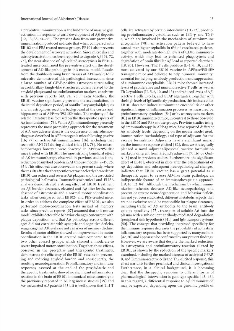
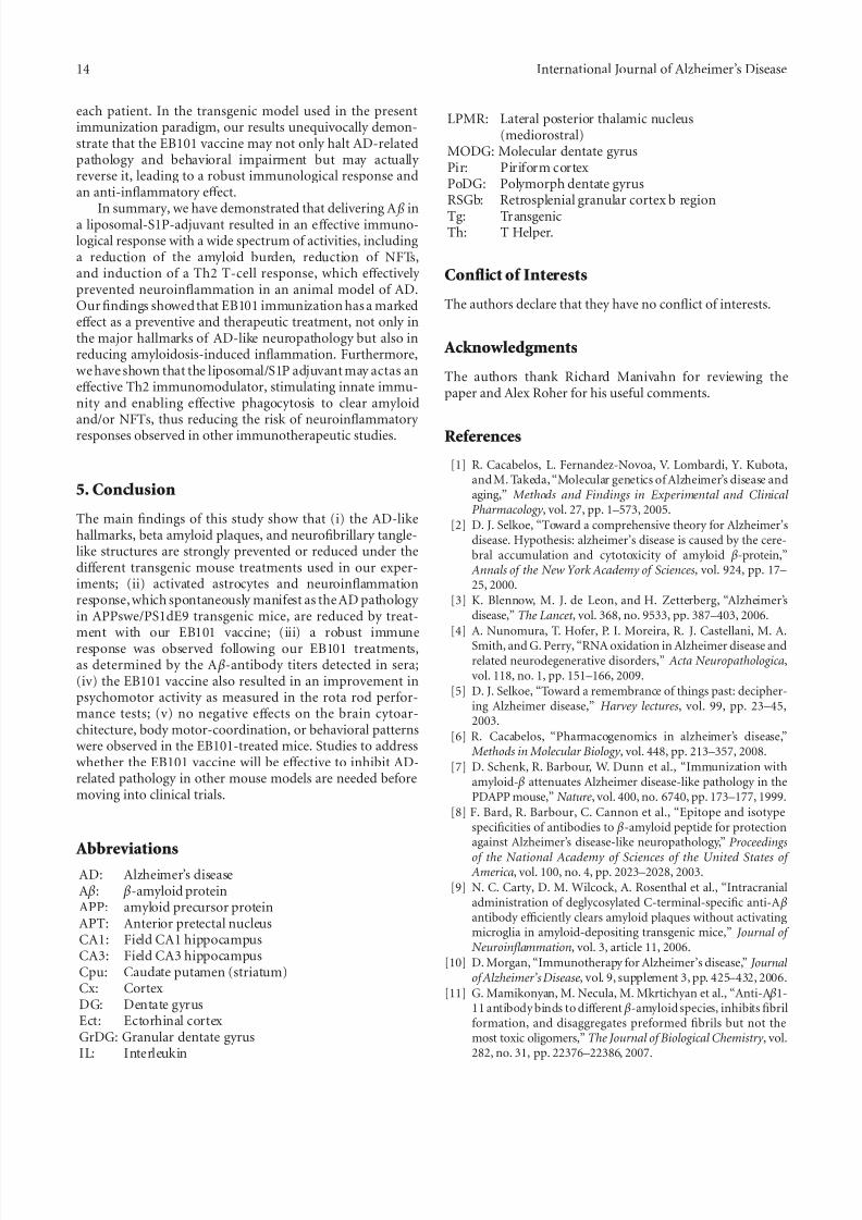
2.5. Neuropathology Marker Quantification. A β plaque quan-tification was determined in 7 randomly selected micro-scopic transverse sections per treated animal (Figures 1 and2), as defined by the stereotaxic Bregma coordinates (−0.94;−1.34; −1.70; −2.30; −2.70; −3.64; −4.60 mm). A total of 7selected sections per animal were evaluated on a PC using
the HIH Image J program by defining region of interestand setting a threshold to discriminate nonspecific staining.The same procedure was undertaken for the neurofibrillary tangle-like structure sections and activated astroglia, B andT cells. All data analysis and measurements were blindly performed by investigators unaware of the treatment proto-col, and in cases of significant measurement discrepanciesbetween two investigators, the evaluation was repeated by a third. Quantitative analysis of amyloid burden area wasalso performed in the hippocampal regions (oriens, pyra-midal layer, stratum radiatum, and dentate gyrus) andparietal/temporal cortical regions in the three experimentalgroups (Figures 1 and 2). Area/pixel analysis software (Pix-cavator 4) was used to quantify the number of pixels insidethe outer boundary of each A β plaque for one brain section.Thus, Pixcavator imaging was used to analyze the areaoccupied by β-amyloid (A β load) relative to the backgroundand expressed in percentage units. The area of A β plaques of the brain of three treatment groups is represented in graphics(Figure 2).
2.6. Imaging. Images were visualized using a microscope(Olympus BX50) and digitized using a digital camera (DP-10, Olympus). The photographs were adjusted for bright-ness and contrast with Corel Photo-Paint (Corel, Ottawa,Canada) and plates were composed with Corel Draw.
2.6.1. Confocal Imaging Analysis. Each individual immuno-fluorescent section was photographed with a Spectral Con-focal Laser Scanning Microscope (Leica TCS-SP2). Plaqueswere imaged at the level of their largest cross-section, andtheir size was determined by using the Leica processing soft-ware. This methodological approach was used to compareNFTs-A β, NFTs-GFAP, and NFTs-Bcell immunostainings.Two brain regions (hippocampus and entorhinal cortex) per
animal were analyzed. Imaging of the NFTs immunostainingwas revealed with a fluorescein isothiocyanate filter (excita-tion at 488 nm), and A β/GFAP/B-cell staining was imagedwith a Texas Red filter (excitation 568 nm).
2.7. Evaluation of Motor Coordination and Balance. Themotor strength, ability, balance, and coordination skills of allthe experimental mice in the two treatment periods wereevaluated in a rota-rod operated at 10 rpm (ColumbusInstruments, Columbus, OH, USA) beginning at 7 weeksof age (preventive group) and 35 weeks of age (therapeuticgroup), respectively. The animals were adapted to the appa-ratus by receiving training sessions in two trials, sufficient
to reach a baseline level of performance [48]. This procedurewas designed to assess motor behavior. If the mouseremained on the rod for 1 minute, the test was completedand scored as 1 minute. Each animal was tested during six weeks for three sessions, with each session separated by 15minutes, the time each mouse remained on the rod wasregistered automatically and stopped when the animals fellor inverted (by climbing) from the top of the rotating barrel.Two investigators performed the experiment, one evaluatedrota-rod performance, unaware of the mice treatments, andthe second treated the mice.
2.8. Statistical Analyses. All statistical parameters were per-
formed by SPSS (version 11.0; SPSS Inc, Chicago); a P value< 0.05 indicated statistical significance. Average range of A βplaque density, burden area, and antibody titers developedby mice during treatments were analyzed using a two-factorrepeated-measures analysis of variance (ANOVA) followedby a post hoc analysis when relevant. All data were expressedas the mean ± SEM.
2.9. Limitations of the Study. Preclinical research in AD islinked to the use of a wide range of transgenic mouse modelsthat may recapitulate some of the pathological features of clinical human AD. Despite the fact that to date, none of the models developed fully the entire disease background
8/13/2019 376138
http://slidepdf.com/reader/full/376138 5/17
International Journal of Alzheimer’s Disease 5
CA1
PoDG
DG
MoDG
P
r e v e n t i v e t r e a t m e n t
A βGrp A
EB101
(a)
CA1
MoDG
PoDG
A βGrp A
EB101
(b)
PoDG
MoDG
CA1
A βGrp A
EB101
(c)
DG
MoDG
PoDG
CA1
P r e v e n t i v e t r e a t m e n t A βGrp B
EB102
(d)
PoDG
MoDG
CA1
A βGrp BEB102
(e)
CA1
A βGrp BEB102
(f)
P r e v e n t i v e t r e a t m e n t
DG
CA1Control wt
MoDG
DG
CA1
MoDG
PoDG
PoDG
P r e v e n t i v e t r e a t m e n t Grp C
PBS
A β
(g)
MoDG
DG
PoDG
CA1
Control wt
A βGrp C
PBS
MoDG
(h)
CA1
A βGrp CPBS
CA1
Control wt
(i)
Cx
CA1
DG
T h e r a p e u t i c t r e a t m e n t A β
Grp AEB101
(j)
Cx
CA1
Grp AEB101
A β
(k)
Cx
CPuEct
A β
Grp AEB101
(l)
Cx CA1
DG
CA3
T h e r a p e u t i c t r e a t m e n t
A βGrp BEB102
(m)
Cx
CA1
DG
A βGrp BEB102
(n)
Cx
CPuEct
A βGrp BEB102
(o)
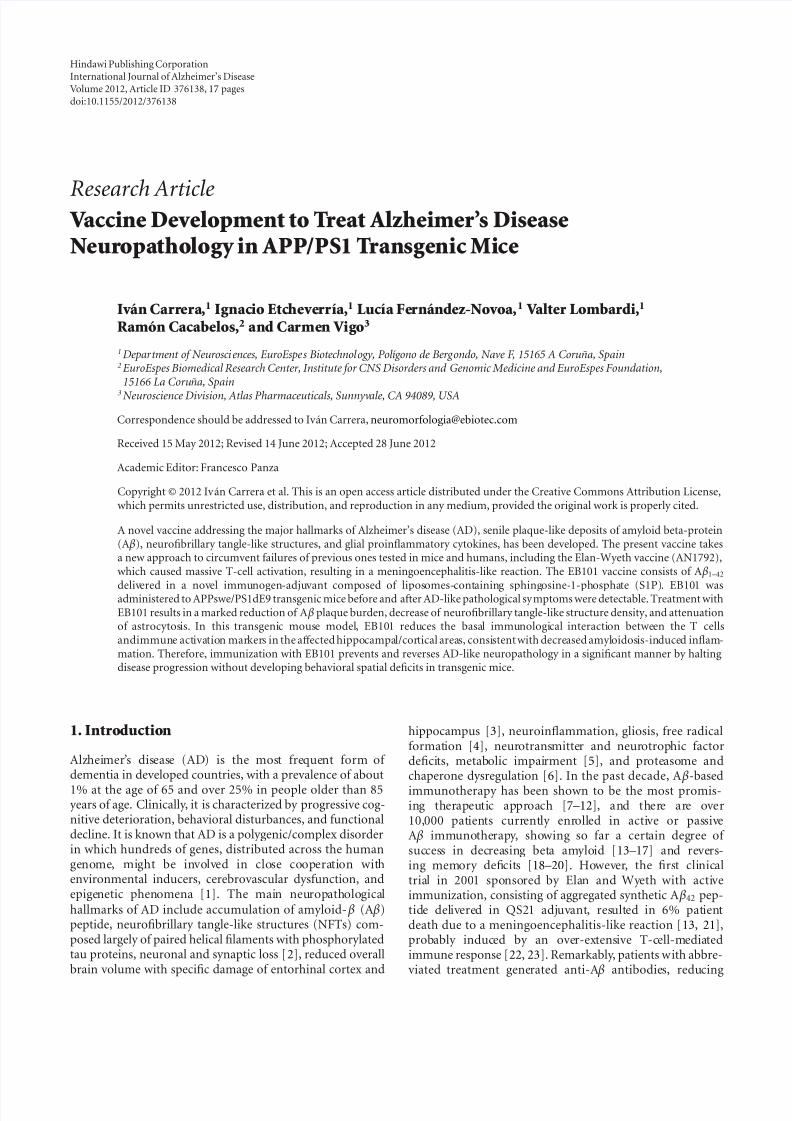
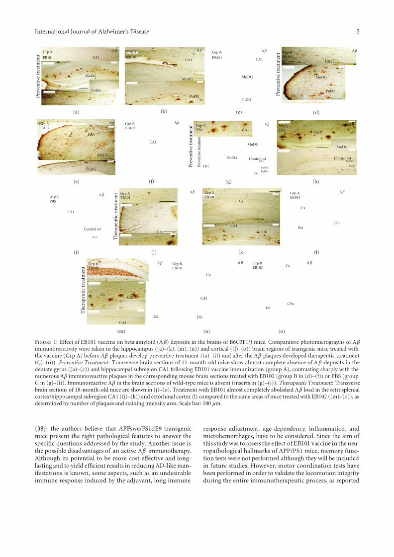
Figure 1: Eff ect of EB101 vaccine on beta amyloid (A β) deposits in the brains of B6C3F1/J mice. Comparative photomicrographs of A βimmunoreactivity were taken in the hippocampus ((a)–(k), (m), (n)) and cortical ((l), (o)) brain regions of transgenic mice treated withthe vaccine (Grp A) before A β plaques develop preventive treatment ((a)–(i)) and after the A β plaques developed therapeutic treatment((j)–(o)). Preventive Treatment : Transverse brain sections of 11-month-old mice show almost complete absence of A β deposits in thedentate gyrus ((a)–(c)) and hippocampal subregion CA1 following EB101 vaccine immunization (group A), contrasting sharply with thenumerous A β immunoreactive plaques in the corresponding mouse brain sections treated with EB102 (group B in (d)–(f)) or PBS (groupC in (g)–(i)). Immunoreactive A β in the brain sections of wild-type mice is absent (inserts in (g)–(i)). Therapeutic Treatment : Transverse
brain sections of 18-month-old mice are shown in (j)–(o). Treatment with EB101 almost completely abolished A β load in the retrosplenialcortex/hippocampal subregion CA1 ((j)–(k)) and ectorhinal cortex (l) compared to the same areas of mice treated with EB102 ((m)–(o)), asdetermined by number of plaques and staining intensity area. Scale bar: 100 µm.
[38]; the authors believe that APPswe/PS1dE9 transgenicmice present the right pathological features to answer thespecific questions addressed by the study. Another issue isthe possible disadvantages of an active A β immunotherapy.Although its potential to be more cost eff ective and long-lasting and to yield efficient results in reducing AD-like man-ifestations is known, some aspects, such as an undesirableimmune response induced by the adjuvant, long immune
response adjustment, age-dependency, inflammation, andmicrohemorrhages, have to be considered. Since the aim of this study was to assess the eff ect of EB101 vaccine in the neu-ropathological hallmarks of APP/PS1 mice, memory func-tion tests were not performed although they will be includedin future studies. However, motor coordination tests havebeen performed in order to validate the locomotion integrity during the entire immunotherapeutic process, as reported
8/13/2019 376138
http://slidepdf.com/reader/full/376138 6/17
6 International Journal of Alzheimer’s Disease
M e a
n A β
b u r d e n p l a q u e s
0
20
40
60
80
100
120
140
160
Grp A(EB101)
Grp B(EB102)
Grp C(PBS)
Preventive treatment
Therapeutic treatment
(a)
0
24
6
8
10
12
14
16
18
Preventive treatment
Therapeutic treatment
M e a n
A β
p l a q u e a r e a / b r a i n
s e c t i o n
Grp A(EB101)
Grp B(EB102)
Grp C(PBS)
(b)
M e a n f a l l i n g l a t e
n c y ( s e c o n d s )
0
5
10
15
20
25
30
Preventive treatment
Therapeutic treatment
Grp A/tg Grp A/wt Grp B/tg Grp B/wt Grp C/wtGrp C/tg
(c)
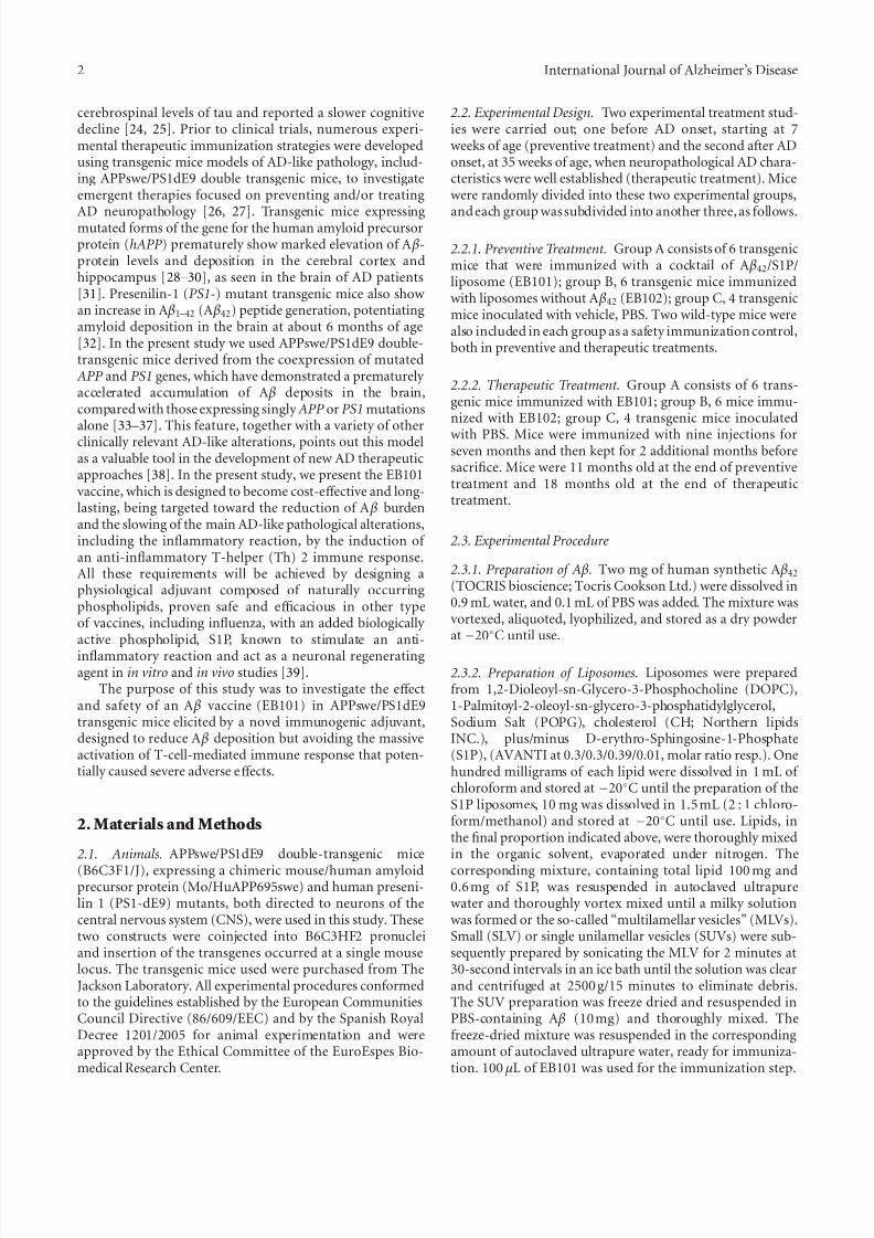
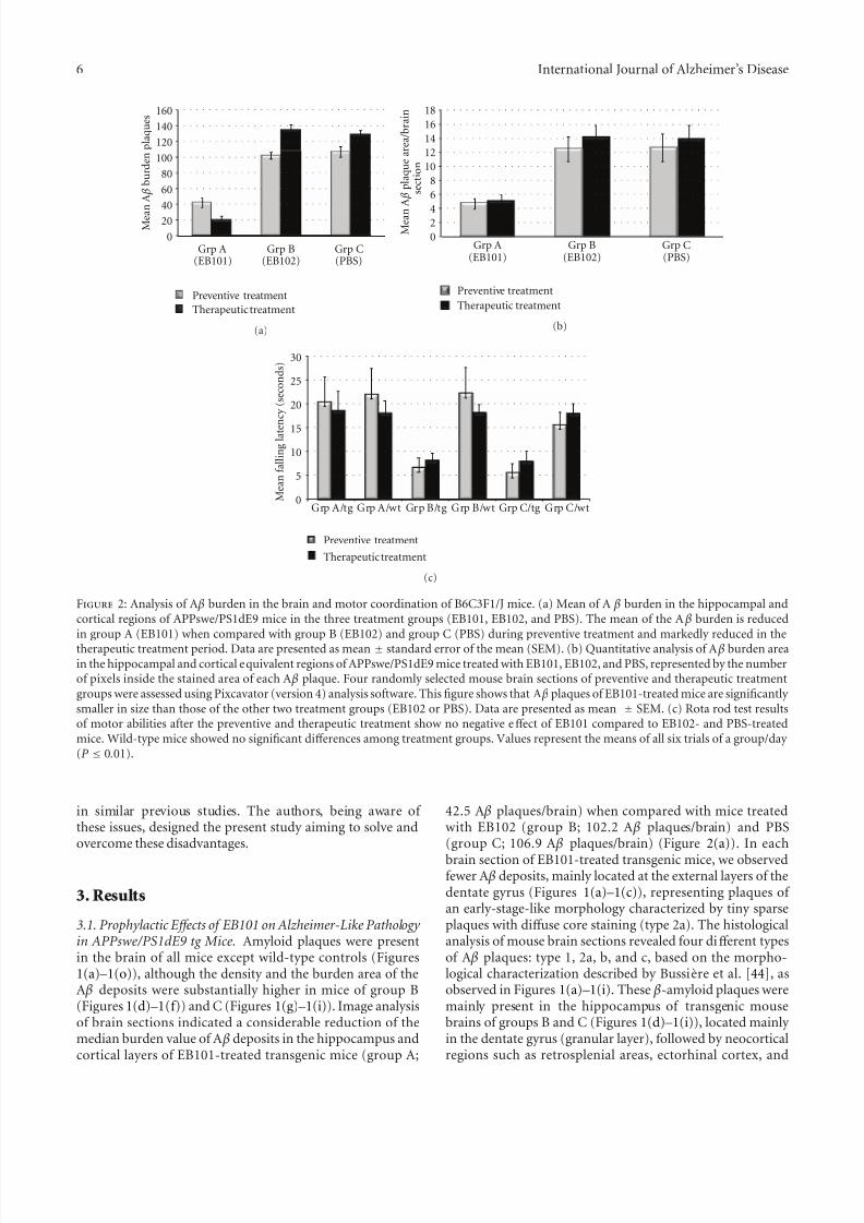
Figure 2: Analysis of A β burden in the brain and motor coordination of B6C3F1/J mice. (a) Mean of A β burden in the hippocampal andcortical regions of APPswe/PS1dE9 mice in the three treatment groups (EB101, EB102, and PBS). The mean of the A β burden is reducedin group A (EB101) when compared with group B (EB102) and group C (PBS) during preventive treatment and markedly reduced in thetherapeutic treatment period. Data are presented as mean ± standard error of the mean (SEM). (b) Quantitative analysis of A β burden areain the hippocampal and cortical equivalent regions of APPswe/PS1dE9 mice treated with EB101, EB102, and PBS, represented by the numberof pixels inside the stained area of each A β plaque. Four randomly selected mouse brain sections of preventive and therapeutic treatmentgroups were assessed using Pixcavator (version 4) analysis software. This figure shows that A β plaques of EB101-treated mice are significantly smaller in size than those of the other two treatment groups (EB102 or PBS). Data are presented as mean ± SEM. (c) Rota rod test resultsof motor abilities after the preventive and therapeutic treatment show no negative eff ect of EB101 compared to EB102- and PBS-treatedmice. Wild-type mice showed no significant diff erences among treatment groups. Values represent the means of all six trials of a group/day (P ≤ 0.01).
in similar previous studies. The authors, being aware of these issues, designed the present study aiming to solve andovercome these disadvantages.
3. Results
3.1. Prophylactic E ff ects of EB101 on Alzheimer-Like Pathology in APPswe/PS1dE9 tg Mice. Amyloid plaques were presentin the brain of all mice except wild-type controls (Figures1(a)–1(o)), although the density and the burden area of theA β deposits were substantially higher in mice of group B(Figures 1(d)–1(f)) and C (Figures 1(g)–1(i)). Image analysisof brain sections indicated a considerable reduction of themedian burden value of A β deposits in the hippocampus andcortical layers of EB101-treated transgenic mice (group A;
42.5 A β plaques/brain) when compared with mice treatedwith EB102 (group B; 102.2 A β plaques/brain) and PBS(group C; 106.9 A β plaques/brain) (Figure 2(a)). In each
brain section of EB101-treated transgenic mice, we observedfewer A β deposits, mainly located at the external layers of thedentate gyrus (Figures 1(a)–1(c)), representing plaques of an early-stage-like morphology characterized by tiny sparseplaques with diff use core staining (type 2a). The histologicalanalysis of mouse brain sections revealed four diff erent typesof A β plaques: type 1, 2a, b, and c, based on the morpho-logical characterization described by Bussiere et al. [44], asobserved in Figures 1(a)–1(i). These β-amyloid plaques weremainly present in the hippocampus of transgenic mousebrains of groups B and C (Figures 1(d)–1(i)), located mainly in the dentate gyrus (granular layer), followed by neocorticalregions such as retrosplenial areas, ectorhinal cortex, and
8/13/2019 376138
http://slidepdf.com/reader/full/376138 7/17
International Journal of Alzheimer’s Disease 7
piriform cortex layers. The area occupied by A β depositsin both the hippocampus and cerebral cortex of transgenicmouse brain sections of group A (EB101; Figures 1(a)–1(c))after preventive treatment was markedly reduced (4.83%,P < 0.05; Figure 2(b)) and diff ered significantly from theelevated A β deposit area observed in the correspondent brain
sections of group B (12.6%, P < 0.05; Figure 2(b)) and groupC (12.81%, P < 0.05; Figure 2(b)). All wild-type mice of each group showed no A β deposits in any brain region (seesquared area in Figures 1(g)–1(i)).
The balance, coordination, and motor planning in thismouse study were assessed at the end of the 7-month treat-ment using a rota-rod test. Results of motor abilities showedan improvement in motor coordination in the EB101-treated mice compared to the two other controls, whichshowed a moderate-to-severe impaired motor coordination(Figure 2(c)).
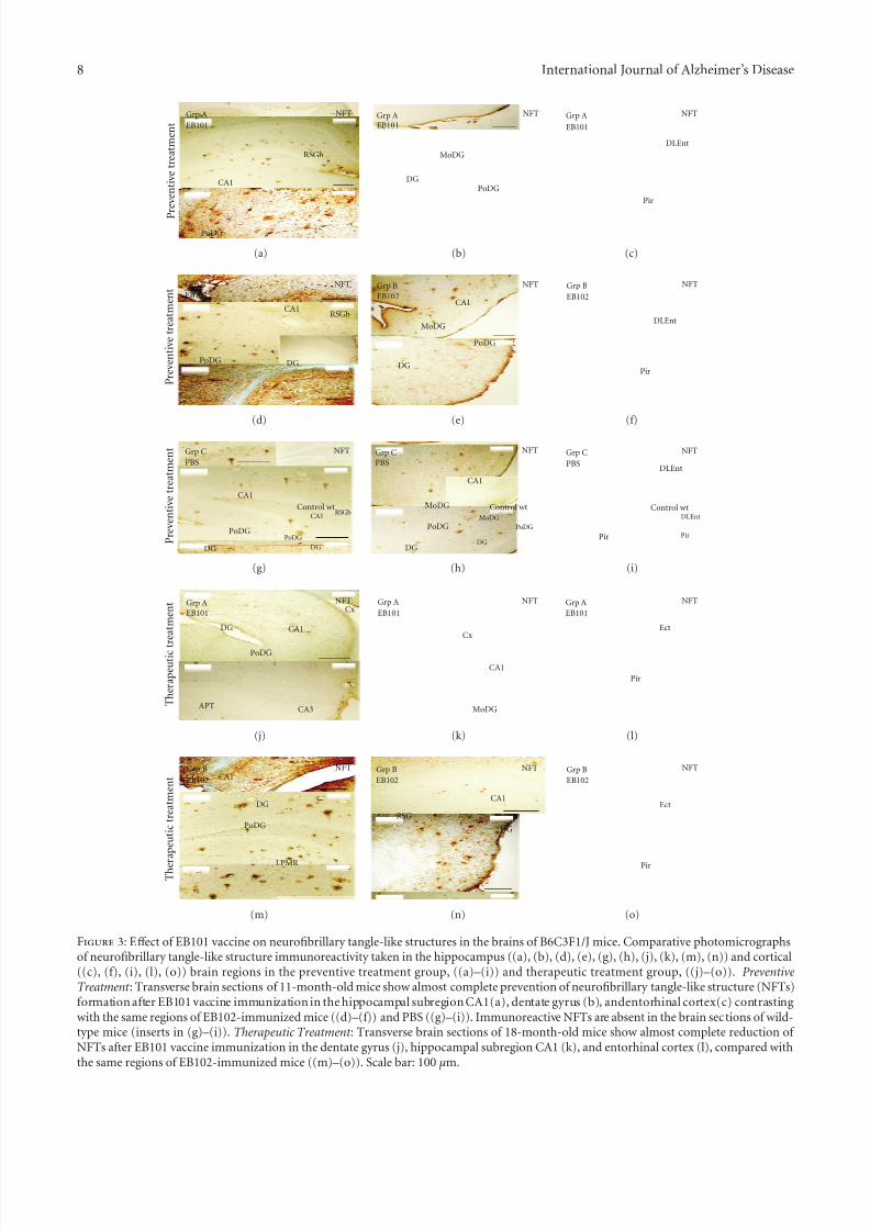
The eff ect of the EB101 vaccine on the development of neurofibrillary tangle-like structures (NFTs) was also studiedusing specific antibody against NFTs (Table 1). StainedNFTs were observed in all transgenic mice except wild-typecontrols (Figures 3(a)–3(i)). Similar to that observed with A βdeposits, the density of these stained tangles was substantially higher in mouse brains of group B (Figures 3(d)–3(f))and C (Figures 3(g)–3(i)) compared to the EB101-treatedgroup A. These immunopositive structures were mainly located in the hippocampal brain regions of the mice treatedwith EB102 and PBS and were composed of aggregatedhyperphosphorylated tau protein located along the neuronalhelical filaments with a plaque-like immunoreactive coreand an apical variable dendrite extension (often having aflame-shape appearance, Figure 3). The brain regions of micetreated with EB101 (group A) were mostly devoid of NFTs(Figures 3(a)–3(c)), with scattered immunoreactive tanglesin the dentate gyrus (Figure 3(b)) and entorhinal cortex layers (Figure 3(c)). The same regions of group B (Figures3(d)–3(f)) were notably similar to those observed in mice of group C (Figures 3(g)–3(i)) and showed an extensive density of NFTs occupying all hippocampal regions, retrosplenialareas, ectorhinal, and piriform cortex. The stained intensity of the NFTs observed in groups B and C, including density and burden area, contrasts markedly with the scarcity andalmost absence of these neuropathological features in micetreated with EB101.
3.2. Therapeutic E ff ects of EB101 on Alzheimer-Like Pathology in APPswe/PS1dE9 tg Mice. The therapeutic eff ect of EB101vaccine in APPswe/PS1dE9 tg mice was initiated in 35-week-old mice after the appearance of the Alzheimer-like neu-ropathology in defined brain regions. To determine whetherEB101 vaccine reverses the massive development of β-amyloid plaque, NFTs and reactive glia, brain sections fromwild-type and transgenic mice of all experimental groupswere immunostained with their specific antibodies (Table 1).The results obtained after the therapeutic treatment showedthat A β deposits were almost absent in brain sections of the EB101-treated mice of group A (20.3 A β plaques/brain)(Figures 1(j)–1(l) and 2(a)), markedly diff erent from the
A β burden levels observed in the mice treated with EB102(134.3 A β plaques/brain; Figures 1(m)–1(o) and 2(a)) andPBS (128.6 A β plaques/brain). The few A β plaques observedin group A mice presented a tiny central core surroundedby scarce fibrillar material (type 2a), with lower A β depositarea (5.27%, P < 0.05; Figure 2(b)), mainly located at the
external cortical layers (Figures 1(j)–1(l)) and almost totally absent in the hippocampal regions. In the brain section of the same transverse regions, group B mice presented anextensive A β plaque area (14.32%, P < 0.05; Figures 1(m)–1(o) and 2(b)), showing a scattered distribution throughoutthe hippocampal and cortical layers, similar to that observedin group C (14.07%, P < 0.05; Figure 2(b)).
The rota-rod latency test, addressing motor coordina-tion during the therapeutic treatment, showed no negativeperformance by the EB101-treated-group (Figure 2(c)), butmice treated with EB102 and PBS (Figure 2(c)) showedmoderately-to-severely impaired motor abilities and coordi-nation.
Histological analysis of mice in group A showed a lessdense distribution pattern of NFTs (Figures 3(j)–3(l)) thanthat observed in the preventive treatment. However, micefrom group B showed a similarly elevated density of NFTs(Figures 3(m)–3(o)) to those seen in mice in the preventivetreatment.
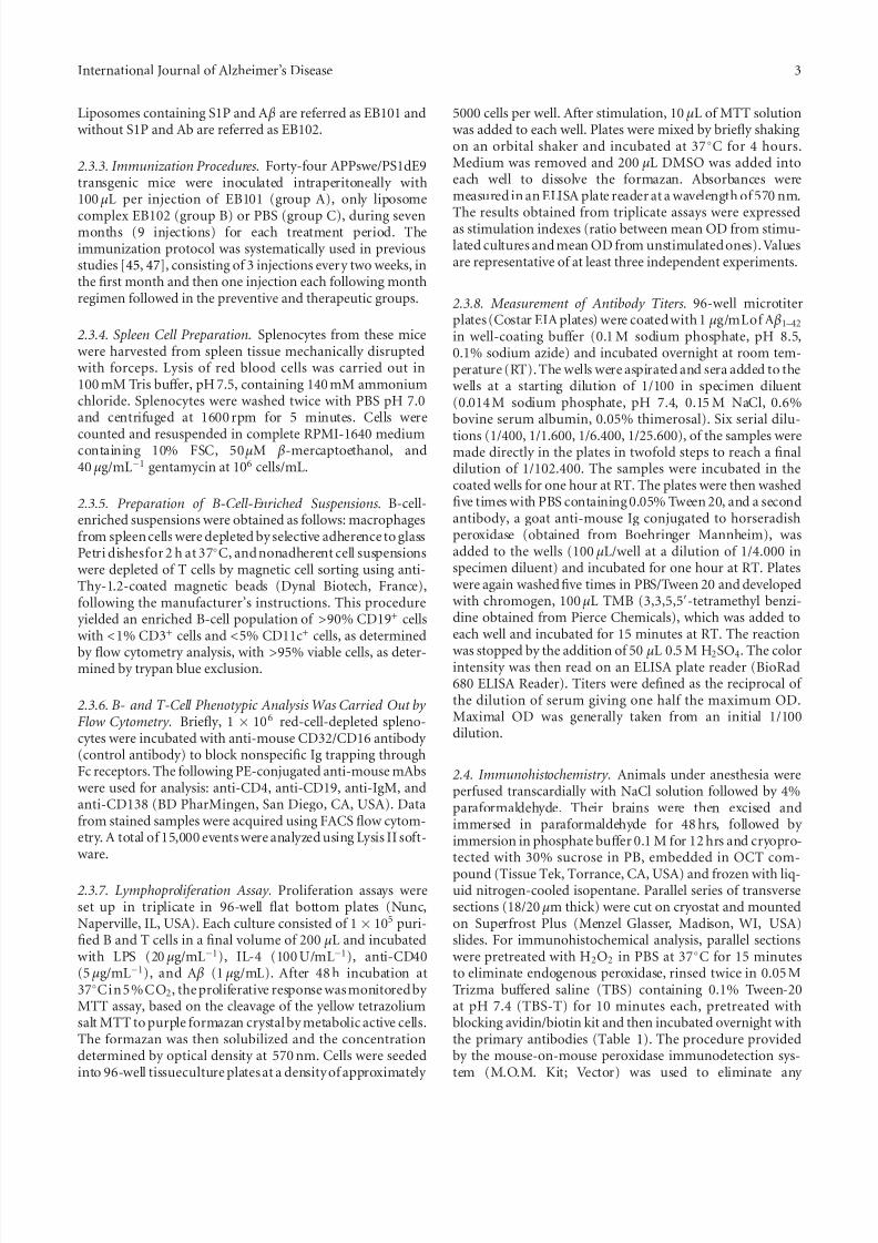
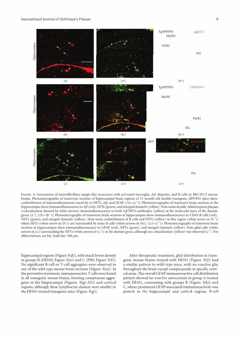
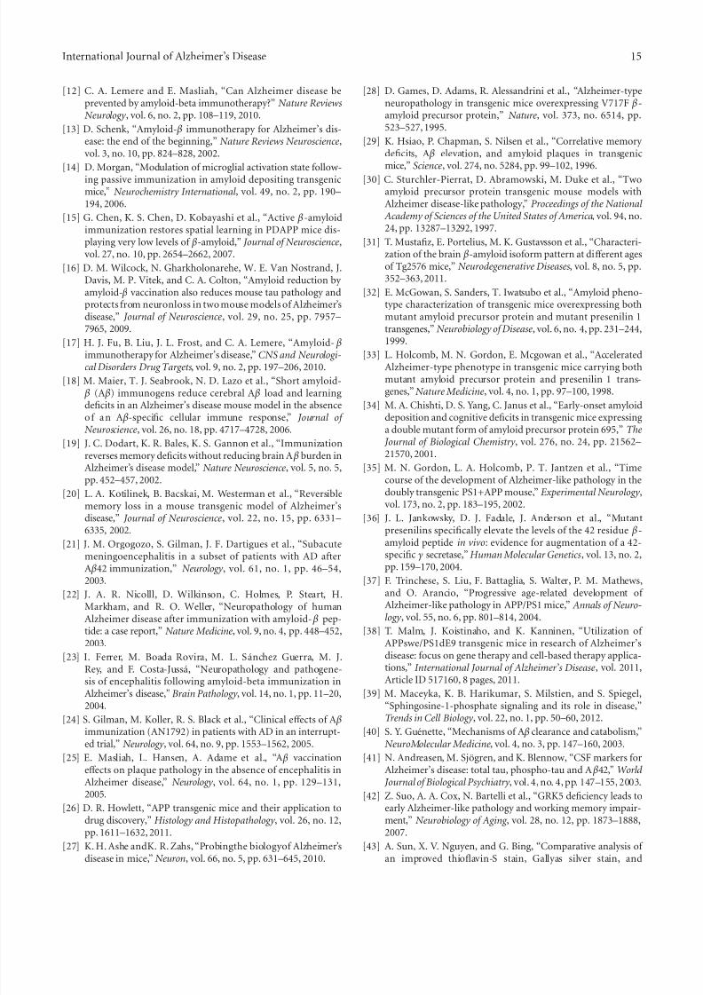
3.3. Double Immunofluorescence Detection of Neuropathologi-cal Markers in APPswe/PS1dE9 tg Mice. Simultaneous doubleimmunofluorescence was performed to detect colocalizationof A β plaques and neurofibrillary tangle-like structures inmouse brains (Figures 4(a)–4(a)). Some sparse plaquesin the hippocampus and entorhinal regions of mice were
observed, colocalized in the molecular layer of the dentategyrus (Figures 4(a)–4(a)) and at the entorhinal cortex.Immunofluorescence to CD45RA (B cells) and NFTs wasalso analyzed by confocal microscopy, showing B cells sur-rounding the NFTs where some codistribution was observedin the polymorph and molecular layers of the dentate gyrus(Figures 4(b)–4(b)). Double immunofluorescence to GFAPand NFTs showed that glial cells were surrounding NFTs inthe dentate gyrus (Figures 4(c)–4(c)) and entorhinal cortex,although no colocalization was observed.
3.4. E ff ect of EB101 on the Immune Response. To assess theeff ect of immunization on glial reactivity (astrocytosis) and
active lymphocytes, the distribution of glial fibrillary acidicprotein (GFAP), B-cell (CD45RA) and T-cell (CD3) surfacemarkers was analyzed in the transverse section of the mousebrains (Figures 5(a)–5(r)). After preventive treatment, acti-vated or reactive glia, astrocytosis-like morphology withaggregated astrocytes replacing dead neurons was observedsurrounding A β plaques and NFTs in the cortical andhippocampal brain regions of transgenic mice of groupsB (Figure 5(b)) and C (Figure 5(c)). The same regions of transgenic mouse brains of group A were almost devoid of reactive glia (Figure 5(a)). No astrocytosis was observed inwild-type mice (Figure 5(l)). Brain sections of EB101-treatedtransgenic mice showed some immunoreactive B cells in the
8/13/2019 376138
http://slidepdf.com/reader/full/376138 8/17
8/13/2019 376138
http://slidepdf.com/reader/full/376138 9/17
International Journal of Alzheimer’s Disease 9
Aβ A β/NFT
GrDG
MoDG
DG
GrDG
MoDG
DG
GrDG
MoDG
DG
TgAPP/PS1
MoDG
PoDG
MoDG
DG DG
PoDG
MoDG
DG
PoDG
NFT
NFT
NFT
CD45/NFT
H
i p p o c a m p u s
H i p p o c a m p u s
H i p p o c a m p u s
TgAPP/PS1TgAPP/PS1
TgAPP/PS1TgAPP/PS1TgAPP/PS1
TgAPP/PS1TgAPP/PS1
MoDG
DG
MoDG
DG
MoDG
GFAP
CD45
DG
GFAP/NFTTgAPP/PS1
(a) (a) (a)
(b) (b) (b)
(c) (c) (c)
Figure 4: Association of neurofibrillary tangle-like structures with activated microglia, A β deposits, and B cells in B6C3F1/J mousebrains. Photomicrographs of transverse sections of hippocampal brain regions of 11-month-old double-transgenic APP/PS1 mice show codistribution of immunofluorescent reactivity to NFTs, A β, and GFAP. ((a)–(a)): Photomicrographs of transverse brain sections at thehippocampus show immunofluorescence to A β (red), NFTs (green), and merged channels (yellow). Note some double-labeled sparse plaques(colocalization showed by white arrows) immunofluorescence to both A β/NFTs antibodies (yellow) at the molecular layer of the dentategyrus (a). ((b)–(b)): Photomicrographs of transverse brain sections at hippocampus show immunofluorescence to CD45-B cells (red),NFTs (green), and merged channels (yellow). Note some codistribution of B cells and NFTs (yellow) in this region (white arrow in (b))where NFTs (white arrow in (b)) are surrounded by some B cells (white arrows in (b)). ((c)–(c)): Photomicrographs of transverse brainsections at hippocampus show immunofluorescence to GFAP (red), NFTs (green), and merged channels (yellow). Note glial cells (whitearrows in (c)) surrounding the NFTs (white arrows in (c )) at the dentate gyrus, although no colocalization (yellow) was observed (c). For
abbreviations, see list. Scale bar: 100 µm.
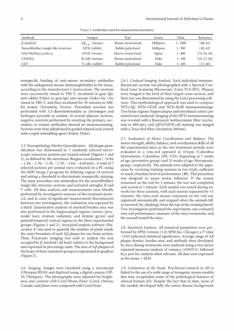
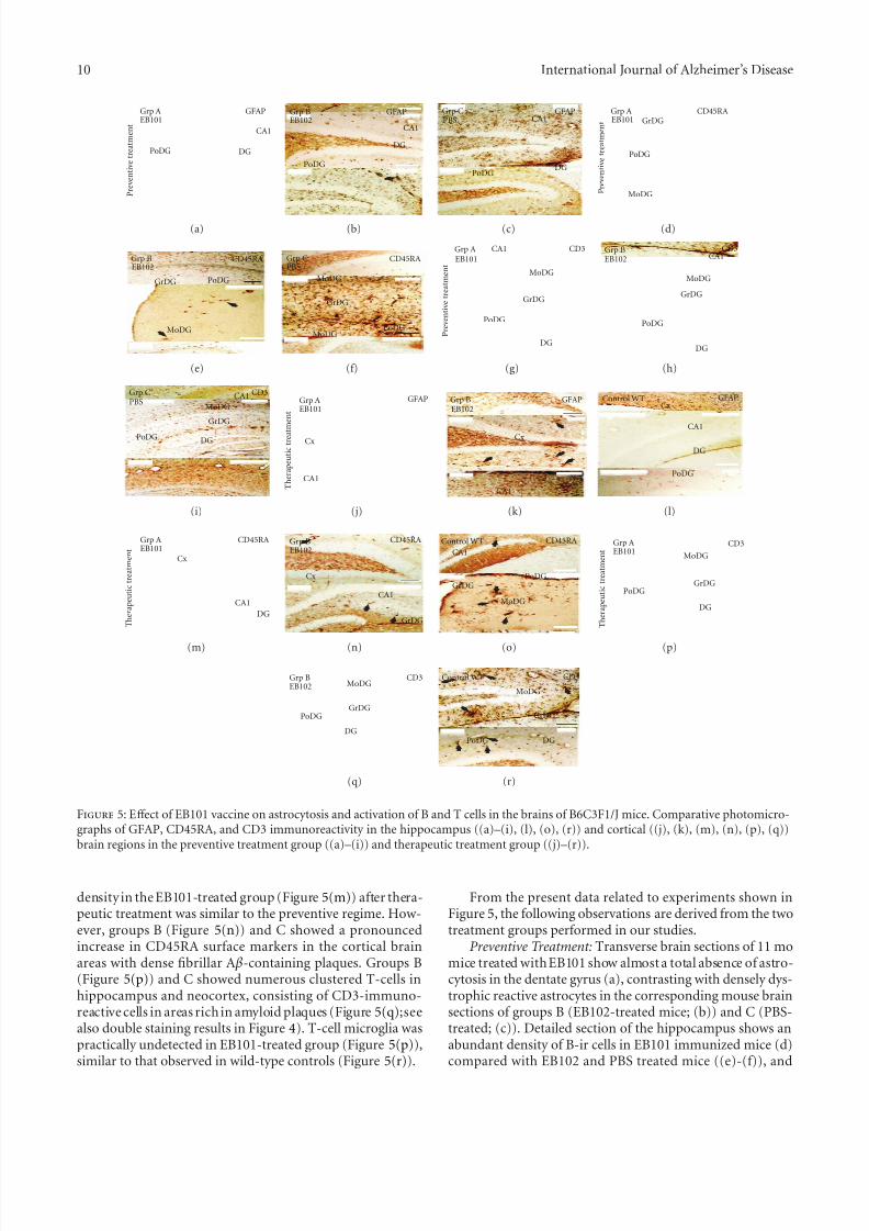
hippocampal regions (Figure 5(d)), with much lower density in groups B (EB102; Figure 5(e)) and C (PBS; Figure 5(f)).No significant B-cell or T-cell aggregates were observed inany of the wild-type mouse brain sections (Figure 5(o)). Inthe preventive treatment, immunoreactive T cells were foundin all transgenic mouse brains, forming conspicuous aggre-gates in the hippocampal (Figures 5(g)–5(i)) and corticalregions, although these lymphocyte clusters were smaller inthe EB101-treated transgenic mice (Figure 5(g)).
After therapeutic treatment, glial distribution in trans-genic mouse brains treated with EB101 (Figure 5(j)) hada similar pattern to wild-type mice, with no reactive gliathroughout the brain except conspicuously in specific corti-cal areas. The overall GFAP immunoreactive cell distributionpattern showed no reactive astrocytosis in group A treatedwith EB101, contrasting with groups B (Figure 5(k)) andC, where prominent GFAP-associated immunoreactivity waspresent in the hippocampal and cortical regions. B-cell
8/13/2019 376138
http://slidepdf.com/reader/full/376138 10/17
10 International Journal of Alzheimer’s Disease
PoDG DG
GFAP
CA1
Grp AEB101
P r e v e n t i v e t r e a t m e n t
(a)
CA1
DG
Grp B GFAP
PoDG
EB102
(b)
Grp C GFAPCA1
DGPoDG
PBS
(c)
PoDG
GrDGGrp A CD45RA
MoDG
EB101
P r e v e n t i v e t r e a t m e n t
(d)
PoDGGrDG
MoDG
Grp B CD45RAEB102
(e)
PoDG
MoDG
MoDG
GrDG
CD45RAGrp CPBS
(f)
Grp A
PoDG
CA1
DG
GrDG
MoDG
EB101
CD3
P r e v e n t i v e t r e a t m e n t
(g)
DG
PoDG
Grp BCA1
CD3
MoDG
GrDG
EB102
(h)
PoDG
MoDG
GrDG
DG
Grp CPBS
CD3CA1
(i)
CA1
Cx
GFAPGrp AEB101
T h e r a p e u t i c t r e a t m e n t
(j)
Grp B
Cx
GFAPEB102
CA1
(k)
GFAPControl WTCx
PoDG
DG
CA1
(l)
Grp A
DG
EB101CD45RA
T h e r a p e u t i c t r e a t m e n t
Cx
CA1
(m)
Cx
CD45RA
GrDG
EB102Grp B
CA1
(n)
CA1
CD45RAControl WT
PoDG
MoDG
GrDG
(o)
DG
MoDG
Grp A
PoDG
EB101CD3
T h e r a p e u t i c t r e a t m e n t
GrDG
(p)
Grp B
DG
CD3EB102
PoDG
MoDG
GrDG
(q)
GrDG
PoDG
MoDG
DG
CD3Control WT
(r)
Figure 5: Eff ect of EB101 vaccine on astrocytosis and activation of B and T cells in the brains of B6C3F1/J mice. Comparative photomicro-graphs of GFAP, CD45RA, and CD3 immunoreactivity in the hippocampus ((a)–(i), (l), (o), (r)) and cortical ((j), (k), (m), (n), (p), (q))brain regions in the preventive treatment group ((a)–(i)) and therapeutic treatment group ((j)–(r)).
density in the EB101-treated group (Figure 5(m)) after thera-peutic treatment was similar to the preventive regime. How-ever, groups B (Figure 5(n)) and C showed a pronouncedincrease in CD45RA surface markers in the cortical brainareas with dense fibrillar A β-containing plaques. Groups B(Figure 5(p)) and C showed numerous clustered T-cells inhippocampus and neocortex, consisting of CD3-immuno-reactive cells in areas rich in amyloid plaques (Figure 5(q);seealso double staining results in Figure 4). T-cell microglia waspractically undetected in EB101-treated group (Figure 5(p)),similar to that observed in wild-type controls (Figure 5(r)).
From the present data related to experiments shown inFigure 5, the following observations are derived from the twotreatment groups performed in our studies.
Preventive Treatment: Transverse brain sections of 11 momice treated with EB101 show almost a total absence of astro-cytosis in the dentate gyrus (a), contrasting with densely dys-trophic reactive astrocytes in the corresponding mouse brainsections of groups B (EB102-treated mice; (b)) and C (PBS-treated; (c)). Detailed section of the hippocampus shows anabundant density of B-ir cells in EB101 immunized mice (d)compared with EB102 and PBS treated mice ((e)-(f)), and
8/13/2019 376138
http://slidepdf.com/reader/full/376138 11/17
International Journal of Alzheimer’s Disease 11
O D v a l u e s
0
0.2
0.4
0.6
0.8
1
1.2
1.4
EB101 EB102 Control (PBS)
Preventive treatment
Therapeutic treatment
(a)
S t i m u l a t i o n i n d e x
LPS
Lymphocyte B proliferationpreventive treatment
Lymphocyte B proliferationtherapeutic treatment
Lymphocyte T proliferationpreventive treatment
Lymphocyte T proliferationtherapeutic treatment
0
20
40
60
80
100
120
EB101 EB102 Control (PBS)
(b)
EB101 EB102 Control(PBS)0
10
20
3040
50
60
70
80
IL-5
IL-6
IL-10
IL-13
p g / m L
±
S D
(c)
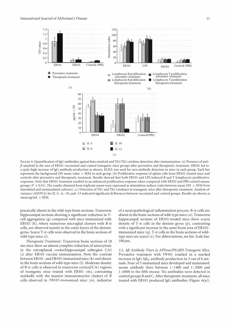
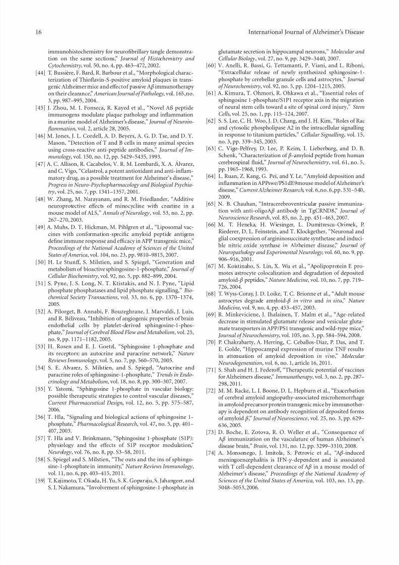
Figure 6: Quantification of IgG antibodies against beta-amyloid and Th1/Th2 cytokine detection after immunization. (a) Presence of anti- β-amyloid in the sera of EB101-vaccinated and control transgenic mice groups after preventive and therapeutic treatment. EB101 led to
a quite high increase of IgG antibody production as shown. ELISA was used for sera antibody detection in mice in each group. Each barrepresents the background OD mean value ± SEM in each group. (b) Proliferative response of spleen cells from EB101-treated mice andcontrols after preventive and therapeutic treatment. Results showed that both EB101 and LPS induced B and T lymphocyte proliferativeresponses. Note that EB101 treatment resulted in an enhanced proliferation response when compared with EB102 and PBS control mousegroups (P ≤ 0.01). The results obtained from triplicate assays were expressed as stimulation indices (ratio between mean OD ± SEM fromstimulated and unstimulated cultures). (c) Detection of Th1 and Th2 cytokines in transgenic mice after therapeutic treatment. Analysis of variance (ANOVA) for IL-5, -6, -10, and -13 indicated significant diff erences between vaccinated and control groups. Results are shown asmean pg/mL ± SEM.
practically absent in the wild-type brain sections. Transversehippocampal sections showing a significant reduction in T-cell aggregation (g) compared with mice immunized withEB102 (h), where numerous microglial clusters with B-ir
cells, are observed mainly in the outer layers of the dentategyrus. Scarce T-ir cells were observed in the brain sections of wild-type mice (i).
Therapeutic Treatment: Transverse brain sections of 18mo mice show an almost complete reduction of astrocytosisin the retrosplenial cortex/hippocampal subregion CA1(j) after EB101 vaccine immunization. Note the contrastbetween EB101- and EB102-immunized mice (k) and absentin the brain sections of wild-type mice (l). Moderate density of B-ir cells is observed in transverse cortical/CA1 regionsof transgenic mice treated with EB101 (m), contrastingmarkedly with the massive immunoreactive clusters of Bcells observed in EB102-immunized mice (n), indicative
of a neuropathological inflammation process. B-ir cells areabsent in the brain sections of wild-type mice (o). Transversehippocampal sections of EB101-treated mice show scarcedensity of T-ir cells in the dentate gyrus (p), contrasting
with a significant increase in the same brain area of EB102-immunized mice (q). T-ir cells in the brain sections of wild-type mice are scarce (r). For abbreviations, see list. Scale bar:100 µm.
3.5. A β Antibody Titers in APPswe/PS1dE9 Transgenic Mice.Preventive treatment with EB101 resulted in a markedincrease in IgG A β42 antibody production in 5 out of 6 ani-mals. Four of 5 immunized mice developed and maintainedserum antibody titers between 1 : 1400 and 1 : 2000 and1 : 6000 in the fifth mouse. No antibodies were detected incontrol groups B and C. After therapeutic treatment, all micetreated with EB101 produced IgG antibodies (Figure 6(a)).
8/13/2019 376138
http://slidepdf.com/reader/full/376138 12/17
12 International Journal of Alzheimer’s Disease
Titers were similar to those detected after the preventivetreatment; one out of 6 mice showed titers higher than1/35,000. In both preventive and therapeutic groups, EB101induced a strong lymphocyte proliferative response of B-and T cells (Figure 6(b)), while practically no response wasobserved in EB102- and PBS-treated groups. Data obtained
from the lymphoproliferation assay indicated that EB101resulted in an enhanced proliferative response, comparedwith EB102 and PBS control groups (P < 0.01; Figure 6(b)).
Detection of Th1 and Th2 cytokine types in all treatedmice was carried out by analyzing the cytokine profile in micesera (Figure 6(c)). Results from the multiple analyte detec-tion indicate significant diff erences between vaccinated andcontrol groups (EB102 and PBS) in sera cytokines IL-5 andIL13, whereas IL-6 and IL-10 showed significant diff erencesbetween vaccinated and the control group, suggesting thatthe antibody-mediated immunity observed in the presentstudy induces a T-cell type Th2 reaction.
4. Discussion
We have developed a novel anti-A ß liposome-formulatedvaccine that is cost eff ective, with long-lasting eff ects andthat not only induces a strong anti-A ß antibody reaction, butalso decreases neurofibrillary tangle-like structure formationand the inflammatory immune response previously seenwith other vaccines. Driven by the great eff ective potentialof liposome-based vaccines in preventing or treating AD[54] and the eff ectiveness of the first active vaccine inreducing A ß levels [21], we have undertaken a new approachto circumvent the AN1792 failure by judiciously selecting anadjuvant that addresses all the above targets. The liposomaladjuvant used consisted of naturally occurring phospho-lipids: phosphatidyl choline, phosphatidyl glycerol, and chol-esterol, proven safe and efficacious in other types of vaccines.To this phospholipid mixture, a biologically active sphin-golipid, sphingosine-1-phosphate (S1P) was added. A ß wasincorporated in the phospholipid/S1P-liposomes using thehydration-rehydration method commonly used for liposo-mal/protein formulation. S1P, a phosphorylated product of sphingosine, has been implicated as an important lipid medi-ator acting both inside and outside the cells [55, 56]. Extra-cellularly, S1P binds to members of the GTP-binding protein(G-protein-) coupled S1P receptor family (S1P15), trigger-
ing diverse cellular eff ects including angiogenesis, cardiacdevelopment, immunity, cell motility, and neurite42 exten-sion [25, 57], by acting in an autocrine and paracrine manner[53–55]. Intracellularly, S1P has been shown to functionby mediating mobilization of cellular calcium, cell growth,and suppression of apoptosis [56–58], while exogenously-added S1P itself causes glutamate secretion from presy-naptic sites and potentiates glutamate-induced transmittersecretion in primary hippocampal neurons [64], whichmay facilitate the formation of a positive activation cycle inexcitatory neurons such as glutaminergic neurons. In fact,S1P appears to have a therapeutic potential as a regenerativeagent in the nervous system since it has recently been
reported that intracellular S1P enhances nerve growth factor-induced excitability in rat sensory neurons [65]and controlsmigration of neuronal stem cells toward a site of spinal cordinjury as well [66]. Moreover, S1P1activation is also knownto induce cytoskeletal rearrangements through small G-protein Rac activation [67], which seems to facilitate synaptic
vesicle fusion to plasma membranes inducing transmittersecretion. Therefore, it would not be unexpected that sucha potent biologically active sphingolipid may play a very important role in neuronal regeneration, cell growth, sup-pression of apoptosis, and glutamate secretion from presy-naptic sites in AD patients. Taking advantage of these prop-erties, we incorporated S1P with the phospholipid mixtureto form a liposomal matrix used as adjuvant to deliver theactive antigen, A ß [68]. This adjuvant added regenerativeand anti-inflammatory properties to the A ß42 vaccine,having a considerable advantage over the previously devel-oped saponin-based A ß vaccines, key elements to increaseneuronal activity and prevent inflammation in the brainof APPswe/PS1dE9 transgenic mice (data not published),improving the previously reported A ß-based immunother-apy studies [7–17]. This new vaccine overcame the inflam-matory response encountered with AN1792, which poten-tially was due to the use of a saponin adjuvant (QS-21) andthe detergent polysorbate 80 used to deliver A ß, which arebelieved to have induced a proinflammatory Th1 response[33]. We have shown that EB101, a liposomal-based vaccineformulation, fulfills a long-standing need for a safe and42eff ective therapeutic vaccine for preventing or amelioratingAD-like neuropathology, improving the beneficial eff ectsreported in previous similar studies [7, 10, 19]. The resultsobserved in the preventive and therapeutic treatments indi-cate that EB101 vaccine is eff ective not only in preventingAD-like neuropathology but also in reducing it. PreviousAPPswe/PS1dE9 transgenic mouse studies reported that inthe first 6 months, brain A ß plaques were tiny but compact,increasing with age [31, 69]. Our results are also consistentwith these observations. APPswe/PS1dE9 transgenic miceshowed tiny, compact nonfibrillary amyloid plaque accumu-lation in the initial deposition period, mainly at the cortex and hippocampus, whereas at later stages diff use fibrillarplaques were increasingly represented also at nearby brainregions. After the preventive immunization with EB101, wedetected a significantly low density of A ß plaques in APP-swe/PS1dE9 mouse brains, as well as reduced burden areaswhen compared with APPswe/PS1dE9 mice treated with
EB102 or PBS. The fact that image analysis confirmed thisefficient reduction of early A ß deposits in the EB101 mousebrain suggests that EB101 vaccine acts as a preventivetreatment in the onset of the amyloid plaques of Alzheimer’sdisease in mice. A similar preventive immunization approachhas been already reported in PDAPP mice by Schenk et al.[7], although diff erences in the immunization efficiency andvaccine conformation have been improved in the presentstudy. During the early period, immunization with EB101also significantly prevented the massive development of other AD pathological hallmarks such as neurofibrillary tangle-like structures, astrocyte activation, and impairmentin motor coordination. One of the keys in the success of
8/13/2019 376138
http://slidepdf.com/reader/full/376138 13/17
International Journal of Alzheimer’s Disease 13
a preventive immunization is the hindrance of massive glialactivation in response to early development of A ß deposits[12, 13, 35, 64–66]. The present data from our preventiveimmunization protocol indicate that when compared withEB102 and PBS treated mouse groups, EB101 also preventsthe development of astrocyte activation. Since microglia and
astrocyte activation has been reported to degrade A ß [69, 72,73], the near absence of A ß-related astrocytosis in EB101-treated mice confirmed the preventive eff ect on the devel-opment of AD-like pathology in this mouse model. Resultsfrom the double-staining brain tissues of APPswe/PS1dE9mice also demonstrated this pathological interaction, sincea large number of GFAP-positive astrocytes surroundingneurofibrillary tangle-like structures, closely related to theamyloid plaques and neuroinflammation markers, consistentwith previous reports [49, 74, 75]. Taken together, theEB101 vaccine significantly prevents the accumulation, inthe initial deposition period, of nonfibrillary amyloidplaquesand an astrogliosis reaction background at the cortex andhippocampus of APPswe/PS1dE9 mice. The majority of therelated literature has focused on the therapeutic aspects of A ß immunization [76]. While A ß immunotherapy presently represents the most promising approach for the treatmentof AD, one adverse eff ect is the occurrence of microhemor-rhages as described in APP transgenic mice following passive[16, 77] or active A ß immunization [16], including thatseen with AN1792 during clinical trials [21, 78]. No micro-hemorrhages however, were observed in APPswe/PS1dE9mice treated with EB101. The most striking beneficial eff ectof A ß immunotherapy observed in previous studies is thereduction of amyloid burden in AD mouse models [7–19, 26,65]. This eff ect was also observed in the present study, wherethe results after the therapeutic treatment clearly showed thatEB101 can reduce and reverse A ß plaques and the associatedpathological hallmarks. Immunohistochemical and ELISAanalysis demonstrated a strong eff ect of EB101 treatmenton A ß burden clearance, elevated anti-A ß titer levels, nearabsence of astrocytosis and a normal motor coordinationtasks when compared with EB102- and PBS-treated mice.In order to address the complete eff ect of EB101, we alsoperformed motor-coordination tests instead of memory tasks, since previous reports [37] assumed that this mousemodel exhibits detectable behavior changes concurrent withplaque deposition, and that A ß pathology across diff erentages did not correlate with synaptic and cognitive deficits,suggesting that A ß levels are not a marker of memory decline.
Results of motor abilities showed an improvement in motorcoordination in the EB101-treated mice compared to thetwo other control groups, which showed a moderate-tosevere impaired motor coordination. Together, these eff ects,observed in the preventive and therapeutic treatments,demonstrate the efficiency of the EB101 vaccine in prevent-ing and reducing amyloid burden and consequently, theresulting neurodegeneration. Proinflammatory and immuneresponses, assessed at the end of the prophylactic andtherapeutic treatments, showed no significant inflammatory reaction in the brain of EB101-immunized mice, contrary tothe previously reported in APP tg mouse studies [79] andA ß-vaccinated AD patients [21]. It is well known that Th1 T
cells are activated by certain interleukins (IL-12), produc-ing proinflammatory cytokines such as IFN-γ and TNF-a, which are involved in the mechanism of autoimmuneencephalitis [78], an activation pattern believed to havecaused meningoencephalitis in 6% of vaccinated patients,together with moderate-to-high levels of CD45 immunore-
activity, which may lead to enhanced phagocytosis anddegradation of brain fibrillar A ß load as reported elsewhere[18, 80]. However, Th2 T cells produce IL-4, 6, 10, and 13,most activated by our EB101 vaccine in APPswe/PS1dE9transgenic mice and believed to help humoral immunity,essential for helping antibody production and suppressionof autoimmune encephalitis. EB101 mice showed increasedlevels of proliferative and immunoreactive T cells, as well asTh 2 cytokines (IL-5, 6, 10, and 13) and reduced levels of A ß-reactive T cells, B cells, and GFAP aggregates. Together withthe high levels of IgG antibody production, this indicates thatEB101 does not induce autoimmune encephalitis or othersignificant signs of inflammation processes, as measured by proinflammatory cytokines [54] or by astrocytosis markers[81] in EB101immunized mice, in contrast to those observedin the EB102 and PBS mouse groups. Previous studies usingA ß immunotherapy in APP-tg mice have reported diff erentA ß antibody levels, depending on the mouse model used,immunization methodology, and type of adjuvant for thevaccine formulation. Adjuvants have a significant impacton the immune response elicited [82], thus we strategically planned a novel adjuvant-liposomal vaccine formulationmarkedly diff erent from Freund’s adjuvant [7, 16] or Quil-A [6] used in previous studies. Furthermore, the significanteff ect of EB101, observed in mice after the establishment of A ß deposition and subsequent neuropathological changes,indicates that EB101 vaccine has a great potential as atherapeutic agent to reverse AD-like brain pathology, anindispensable feature of an immunotherapeutic approach[19, 40, 52, 84]. Although the mechanism by which immu-nization schemes decrease AD-like neuropathology andprevent or reverse neurodegeneration and behavior deficitshas not yet been elucidated, diff erent metabolic aspects thatare not exclusive could be responsible for plaque clearance,including traffic o f A ß antibodies to the brain, antibody epitope specificity [77], transport of soluble A ß into theplasma with a subsequent antibody-mediated degradation(peripheral sink hypothesis) [41], and IgG transport systems[50]. The concept that providing increased specificity forthe immune response decreases the probability of activating
inflammatory response has been supported by many authors[42, 50] and appears to be confirmed by our present findings.However, we are aware that despite the marked reductionin astrocytosis and proinflammatory reaction elicited by EB101, as shown by the reduction of the specific markersexamined, including the marked decrease of activated GFAP,B, and Timmunoreactive cells and Th2-elicited response, thiseff ect warrants further preclinical and clinical investigations.Furthermore, in a clinical background, it is becomingclear that the therapeutic response to diff erent forms of pharmacological intervention is genotype-specific [43, 46].In this regard, a diff erential response to A ß immunizationmay be expected, depending upon the genomic profile of
8/13/2019 376138
http://slidepdf.com/reader/full/376138 14/17
14 International Journal of Alzheimer’s Disease
each patient. In the transgenic model used in the presentimmunization paradigm, our results unequivocally demon-strate that the EB101 vaccine may not only halt AD-relatedpathology and behavioral impairment but may actually reverse it, leading to a robust immunological response andan anti-inflammatory eff ect.
In summary, we have demonstrated that delivering A ß ina liposomal-S1P-adjuvant resulted in an eff ective immuno-logical response with a wide spectrum of activities, includinga reduction of the amyloid burden, reduction of NFTs,and induction of a Th2 T-cell response, which eff ectively prevented neuroinflammation in an animal model of AD.Our findings showed that EB101 immunization has a markedeff ect as a preventive and therapeutic treatment, not only inthe major hallmarks of AD-like neuropathology but also inreducing amyloidosis-induced inflammation. Furthermore,we have shown that the liposomal/S1P adjuvant may actas aneff ective Th2 immunomodulator, stimulating innate immu-nity and enabling eff ective phagocytosis to clear amyloidand/or NFTs, thus reducing the risk of neuroinflammatory responses observed in other immunotherapeutic studies.
5. Conclusion
The main findings of this study show that (i) the AD-likehallmarks, beta amyloid plaques, and neurofibrillary tangle-like structures are strongly prevented or reduced under thediff erent transgenic mouse treatments used in our exper-iments; (ii) activated astrocytes and neuroinflammationresponse, which spontaneously manifest as the AD pathology in APPswe/PS1dE9 transgenic mice, are reduced by treat-ment with our EB101 vaccine; (iii) a robust immuneresponse was observed following our EB101 treatments,as determined by the A β-antibody titers detected in sera;(iv) the EB101 vaccine also resulted in an improvement inpsychomotor activity as measured in the rota rod perfor-mance tests; (v) no negative eff ects on the brain cytoar-chitecture, body motor-coordination, or behavioral patternswere observed in the EB101-treated mice. Studies to addresswhether the EB101 vaccine will be eff ective to inhibit AD-related pathology in other mouse models are needed beforemoving into clinical trials.
AbbreviationsAD: Alzheimer’s diseaseA β: β-amyloid proteinAPP: amyloid precursor proteinAPT: Anterior pretectal nucleusCA1: Field CA1 hippocampusCA3: Field CA3 hippocampusCpu: Caudate putamen (striatum)Cx: Cortex DG: Dentate gyrusEct: Ectorhinal cortex GrDG: Granular dentate gyrusIL: Interleukin
LPMR: Lateral posterior thalamic nucleus(mediorostral)
MODG: Molecular dentate gyrusPir: Piriform cortex PoDG: Polymorph dentate gyrusRSGb: Retrosplenial granular cortex b region
Tg: TransgenicTh: T Helper.
Conflict of Interests
The authors declare that they have no conflict of interests.
Acknowledgments
The authors thank Richard Manivahn for reviewing thepaper and Alex Roher for his useful comments.
References
[1] R. Cacabelos, L. Fernandez-Novoa, V. Lombardi, Y. Kubota,and M. Takeda, “Molecular genetics of Alzheimer’s disease andaging,” Methods and Findings in Experimental and Clinical Pharmacology , vol. 27, pp. 1–573, 2005.
[2] D. J. Selkoe, “Toward a comprehensive theory for Alzheimer’sdisease. Hypothesis: alzheimer’s disease is caused by the cere-bral accumulation and cytotoxicity of amyloid β-protein,”
Annals of the New York Academy of Sciences, vol. 924, pp. 17–25, 2000.
[3] K. Blennow, M. J. de Leon, and H. Zetterberg, “Alzheimer’sdisease,” The Lancet , vol. 368, no. 9533, pp. 387–403, 2006.
[4] A. Nunomura, T. Hofer, P. I. Moreira, R. J. Castellani, M. A.
Smith, and G. Perry, “RNA oxidation in Alzheimer disease andrelated neurodegenerative disorders,” Acta Neuropathologica,vol. 118, no. 1, pp. 151–166, 2009.
[5] D. J. Selkoe, “Toward a remembrance of things past: decipher-ing Alzheimer disease,” Harvey lectures, vol. 99, pp. 23–45,2003.
[6] R. Cacabelos, “Pharmacogenomics in alzheimer’s disease,” Methods in Molecular Biology , vol. 448, pp. 213–357, 2008.
[7] D. Schenk, R. Barbour, W. Dunn et al., “Immunization withamyloid- β attenuates Alzheimer disease-like pathology in thePDAPP mouse,” Nature, vol. 400, no. 6740, pp. 173–177, 1999.
[8] F. Bard, R. Barbour, C. Cannon et al., “Epitope and isotypespecificities of antibodies to β-amyloid peptide for protectionagainst Alzheimer’s disease-like neuropathology,” Proceedings
of the National Academy of Sciences of the United States of America, vol. 100, no. 4, pp. 2023–2028, 2003.
[9] N. C. Carty, D. M. Wilcock, A. Rosenthal et al., “Intracranialadministration of deglycosylated C-terminal-specific anti-A βantibody efficiently clears amyloid plaques without activatingmicroglia in amyloid-depositing transgenic mice,” Journal of
Neuroinflammation, vol. 3, article 11, 2006.[10] D. Morgan, “Immunotherapy for Alzheimer’s disease,” Journal
of Alzheimer’s Disease, vol. 9, supplement 3, pp. 425–432, 2006.
[11] G. Mamikonyan, M. Necula, M. Mkrtichyan et al., “Anti-A β1-11 antibody binds to diff erent β-amyloid species, inhibits fibrilformation, and disaggregates preformed fibrils but not themost toxic oligomers,” The Journal of Biological Chemistry , vol.282, no. 31, pp. 22376–22386, 2007.
8/13/2019 376138
http://slidepdf.com/reader/full/376138 15/17
International Journal of Alzheimer’s Disease 15
[12] C. A. Lemere and E. Masliah, “Can Alzheimer disease beprevented by amyloid-beta immunotherapy?” Nature Reviews
Neurology , vol. 6, no. 2, pp. 108–119, 2010.
[13] D. Schenk, “Amyloid- β immunotherapy for Alzheimer’s dis-ease: the end of the beginning,” Nature Reviews Neuroscience,vol. 3, no. 10, pp. 824–828, 2002.
[14] D. Morgan, “Modulation of microglial activation state follow-ing passive immunization in amyloid depositing transgenicmice,” Neurochemistry International , vol. 49, no. 2, pp. 190–194, 2006.
[15] G. Chen, K. S. Chen, D. Kobayashi et al., “Active β-amyloidimmunization restores spatial learning in PDAPP mice dis-playing very low levels of β-amyloid,” Journal of Neuroscience,vol. 27, no. 10, pp. 2654–2662, 2007.
[16] D. M. Wilcock, N. Gharkholonarehe, W. E. Van Nostrand, J.Davis, M. P. Vitek, and C. A. Colton, “Amyloid reduction by amyloid- β vaccination also reduces mouse tau pathology andprotects from neuronloss in two mouse models of Alzheimer’sdisease,” Journal of Neuroscience, vol. 29, no. 25, pp. 7957–7965, 2009.
[17] H. J. Fu, B. Liu, J. L. Frost, and C. A. Lemere, “Amyloid- βimmunotherapy for Alzheimer’s disease,” CNS and Neurologi-cal Disorders Drug Targets, vol. 9, no. 2, pp. 197–206, 2010.
[18] M. Maier, T. J. Seabrook, N. D. Lazo et al., “Short amyloid- β (A β) immunogens reduce cerebral A β load and learningdeficits in an Alzheimer’s disease mouse model in the absenceo f a n A β-specific cellular immune response,” Journal of
Neuroscience, vol. 26, no. 18, pp. 4717–4728, 2006.
[19] J. C. Dodart, K. R. Bales, K. S. Gannon et al., “Immunizationreverses memory deficits without reducing brain A β burden inAlzheimer’s disease model,” Nature Neuroscience, vol. 5, no. 5,pp. 452–457, 2002.
[20] L. A. Kotilinek, B. Bacskai, M. Westerman et al., “Reversiblememory loss in a mouse transgenic model of Alzheimer’s
disease,” Journal of Neuroscience, vol. 22, no. 15, pp. 6331–6335, 2002.
[21] J. M. Orgogozo, S. Gilman, J. F. Dartigues et al., “Subacutemeningoencephalitis in a subset of patients with AD afterA β42 immunization,” Neurology , vol. 61, no. 1, pp. 46–54,2003.
[22] J. A. R. Nicolll, D. Wilkinson, C. Holmes, P. Steart, H.Markham, and R. O. Weller, “Neuropathology of humanAlzheimer disease after immunization with amyloid- β pep-tide: a case report,” Nature Medicine, vol. 9, no. 4, pp. 448–452,2003.
[23] I. Ferrer, M. Boada Rovira, M. L. Sanchez Guerra, M. J.Rey, and F. Costa-Jussa, “Neuropathology and pathogene-sis of encephalitis following amyloid-beta immunization in
Alzheimer’s disease,” Brain Pathology , vol. 14, no. 1, pp. 11–20,2004.
[24] S. Gilman, M. Koller, R. S. Black et al., “Clinical eff ects of A βimmunization (AN1792) in patients with AD in an interrupt-ed trial,” Neurology , vol. 64, no. 9, pp. 1553–1562, 2005.
[25] E. Masliah, L. Hansen, A. Adame et al., “A β vaccinationeff ects on plaque pathology in the absence of encephalitis inAlzheimer disease,” Neurology , vol. 64, no. 1, pp. 129–131,2005.
[26] D. R. Howlett, “APP transgenic mice and their application todrug discovery,” Histology and Histopathology , vol. 26, no. 12,pp. 1611–1632, 2011.
[27] K. H. Ashe andK. R. Zahs, “Probingthe biologyof Alzheimer’sdisease in mice,” Neuron, vol. 66, no. 5, pp. 631–645, 2010.
[28] D. Games, D. Adams, R. Alessandrini et al., “Alzheimer-typeneuropathology in transgenic mice overexpressing V717F β-amyloid precursor protein,” Nature, vol. 373, no. 6514, pp.523–527, 1995.
[29] K. Hsiao, P. Chapman, S. Nilsen et al., “Correlative memory deficits, A β elevation, and amyloid plaques in transgenicmice,” Science, vol. 274, no. 5284, pp. 99–102, 1996.
[30] C. Sturchler-Pierrat, D. Abramowski, M. Duke et al., “Twoamyloid precursor protein transgenic mouse models withAlzheimer disease-like pathology,” Proceedings of the National
Academy of Sciences of the United States of America, vol. 94, no.24, pp. 13287–13292, 1997.
[31] T. Mustafiz, E. Portelius, M. K. Gustavsson et al., “Characteri-zation of the brain β-amyloid isoform pattern at diff erent agesof Tg2576 mice,” Neurodegenerative Diseases, vol. 8, no. 5, pp.352–363, 2011.
[32] E. McGowan, S. Sanders, T. Iwatsubo et al., “Amyloid pheno-type characterization of transgenic mice overexpressing bothmutant amyloid precursor protein and mutant presenilin 1transgenes,” Neurobiology of Disease, vol. 6, no. 4, pp. 231–244,1999.
[33] L. Holcomb, M. N. Gordon, E. Mcgowan et al., “AcceleratedAlzheimer-type phenotype in transgenic mice carrying bothmutant amyloid precursor protein and presenilin 1 trans-genes,” Nature Medicine, vol. 4, no. 1, pp. 97–100, 1998.
[34] M. A. Chishti, D. S. Yang, C. Janus et al., “Early-onset amyloiddeposition and cognitive deficits in transgenic mice expressinga double mutant form of amyloid precursor protein 695,” The
Journal of Biological Chemistry , vol. 276, no. 24, pp. 21562–21570, 2001.
[35] M. N. Gordon, L. A. Holcomb, P. T. Jantzen et al., “Timecourse of the development of Alzheimer-like pathology in thedoubly transgenic PS1+APP mouse,” Experimental Neurology ,vol. 173, no. 2, pp. 183–195, 2002.
[36] J. L. Jankowsky, D. J. Fadale, J. Anderson et al., “Mutantpresenilins specifically elevate the levels of the 42 residue β-amyloid peptide in vivo: evidence for augmentation of a 42-specific γ secretase,” Human Molecular Genetics, vol. 13, no. 2,pp. 159–170, 2004.
[37] F. Trinchese, S. Liu, F. Battaglia, S. Walter, P. M. Mathews,and O. Arancio, “Progressive age-related development of Alzheimer-like pathology in APP/PS1 mice,” Annals of Neuro-logy , vol. 55, no. 6, pp. 801–814, 2004.
[38] T. Malm, J. Koistinaho, and K. Kanninen, “Utilization of APPswe/PS1dE9 transgenic mice in research of Alzheimer’sdisease: focus on gene therapy and cell-based therapy applica-tions,” International Journal of Alzheimer’s Disease, vol. 2011,Article ID 517160, 8 pages, 2011.
[39] M. Maceyka, K. B. Harikumar, S. Milstien, and S. Spiegel,“Sphingosine-1-phosphate signaling and its role in disease,”Trends in Cell Biology , vol. 22, no. 1, pp. 50–60, 2012.
[40] S. Y. Guenette, “Mechanisms of A β clearance and catabolism,” NeuroMolecular Medicine, vol. 4, no. 3, pp. 147–160, 2003.
[41] N. Andreasen, M. Sjogren, and K. Blennow, “CSF markers forAlzheimer’s disease: total tau, phospho-tau and A β42,” World
Journal of Biological Psychiatry , vol. 4, no. 4, pp. 147–155, 2003.
[42] Z. Suo, A. A. Cox, N. Bartelli et al., “GRK5 deficiency leads toearly Alzheimer-like pathology and working memory impair-ment,” Neurobiology of Aging , vol. 28, no. 12, pp. 1873–1888,2007.
[43] A. Sun, X. V. Nguyen, and G. Bing, “Comparative analysis of an improved thioflavin-S stain, Gallyas silver stain, and
8/13/2019 376138
http://slidepdf.com/reader/full/376138 16/17
16 International Journal of Alzheimer’s Disease
immunohistochemistry for neurofibrillary tangle demonstra-tion on the same sections,” Journal of Histochemistry and Cytochemistry , vol. 50, no. 4, pp. 463–472, 2002.
[44] T. Bussiere, F. Bard, R. Barbour et al., “Morphological charac-terization of Thioflavin-S-positive amyloid plaques in trans-genic Alzheimer mice and eff ectof passive A β immunotherapy on their clearance,” American Journal of Pathology , vol. 165,no.
3, pp. 987–995, 2004.[45] J. Zhou, M. I. Fonseca, R. Kayed et al., “Novel Aß peptide
immunogens modulate plaque pathology and inflammationin a murine model of Alzheimer’s disease,” Journal of Neuroin-
flammation, vol. 2, article 28, 2005.
[46] M. Jones, J. L. Cordell, A. D. Beyers, A. G. D. Tse, and D. Y.Mason, “Detection of T and B cells in many animal speciesusing cross-reactive anti-peptide antibodies,” Journal of Im-munology , vol. 150, no. 12, pp. 5429–5435, 1993.
[47] A. C. Allison, R. Cacabelos, V. R. M. Lombardi, X. A. Alvarez,and C. Vigo, “Celastrol, a potent antioxidant and anti-inflam-matory drug, as a possible treatment for Alzheimer’s disease,”Progress in Neuro-Psychopharmacology and Biological Psychia-try , vol. 25, no. 7, pp. 1341–1357, 2001.
[48] W. Zhang, M. Narayanan, and R. M. Friedlander, “Additiveneuroprotective eff ects of minocycline with creatine in amouse model of ALS,” Annals of Neurology , vol. 53, no. 2, pp.267–270, 2003.
[49] A. Muhs, D. T. Hickman, M. Pihlgren et al., “Liposomal vac-cines with conformation-specific amyloid peptide antigensdefine immune response and efficacy in APP transgenic mice,”Proceedings of the National Academy of Sciences of the United States of America, vol. 104, no. 23, pp. 9810–9815, 2007.
[50] H. Le Stunff , S. Milstien, and S. Spiegel, “Generation andmetabolism of bioactive sphingosine-1-phosphate,” Journal of Cellular Biochemistry , vol. 92, no. 5, pp. 882–899, 2004.
[51] S. Pyne, J. S. Long, N. T. Ktistakis, and N. J. Pyne, “Lipidphosphate phosphatases and lipid phosphate signalling,” Bio-
chemical Society Transactions, vol. 33, no. 6, pp. 1370–1374,2005.
[52] A. Pilorget, B. Annabi, F. Bouzeghrane, J. Marvaldi, J. Luis,and R. Beliveau, “Inhibition of angiogenic properties of brainendothelial cells by platelet-derived sphingosine-1-phos-phate,” Journal of Cerebral Blood Flow and Metabolism, vol. 25,no. 9, pp. 1171–1182, 2005.
[53] H. Rosen and E. J. Goetzl, “Sphingosine 1-phosphate andits receptors: an autocrine and paracrine network,” NatureReviews Immunology , vol. 5, no. 7, pp. 560–570, 2005.
[54] S. E. Alvarez, S. Milstien, and S. Spiegel, “Autocrine andparacrine roles of sphingosine-1-phosphate,” Trends in Endo-crinology and Metabolism, vol. 18, no. 8, pp. 300–307, 2007.
[55] Y. Yatomi, “Sphingosine 1-phosphate in vascular biology:
possible therapeutic strategies to control vascular diseases,”Current Pharmaceutical Design, vol. 12, no. 5, pp. 575–587,2006.
[56] T. Hla, “Signaling and biological actions of sphingosine 1-phosphate,” Pharmacological Research, vol. 47, no. 5, pp. 401–407, 2003.
[57] T. Hla and V. Brinkmann, “Sphingosine 1-phosphate (S1P):physiology and the eff ects of S1P receptor modulation,”
Neurology , vol. 76, no. 8, pp. S3–S8, 2011.
[58] S. Spiegel and S. Milstien, “The outs and the ins of sphingo-sine-1-phosphate in immunity,” Nature Reviews Immunology ,vol. 11, no. 6, pp. 403–415, 2011.
[59] T. Kajimoto, T. Okada, H. Yu, S. K. Goparaju, S. Jahangeer, andS. I. Nakamura, “Involvement of sphingosine-1-phosphate in
glutamate secretion in hippocampal neurons,” Molecular and Cellular Biology , vol. 27, no. 9, pp. 3429–3440, 2007.
[60] V. Anelli, R. Bassi, G. Tettamanti, P. Viani, and L. Riboni,“Extracellular release of newly synthesized sphingosine-1-phosphate by cerebellar granule cells and astrocytes,” Journal of Neurochemistry , vol. 92, no. 5, pp. 1204–1215, 2005.
[61] A. Kimura, T. Ohmori, R. Ohkawa et al., “Essential roles of
sphingosine 1-phosphate/S1P1 receptor axis in the migrationof neural stem cells toward a site of spinal cord injury,” StemCells, vol. 25, no. 1, pp. 115–124, 2007.
[62] S. S. Lee, C. H. Woo, J. D. Chang, and J. H. Kim, “Roles of Racand cytosolic phospholipase A2 in the intracellular signallingin response to titanium particles,” Cellular Signalling , vol. 15,no. 3, pp. 339–345, 2003.
[63] C. Vigo-Pelfrey, D. Lee, P. Keim, I. Lieberburg, and D. B.Schenk, “Characterization of β-amyloid peptide from humancerebrospinal fluid,” Journal of Neurochemistry , vol. 61, no. 5,pp. 1965–1968, 1993.
[64] L. Ruan, Z. Kang, G. Pei, and Y. Le, “Amyloid deposition andinflammation in APPswe/PS1dE9mouse model of Alzheimer’sdisease,” Current Alzheimer Research, vol. 6,no. 6,pp. 531–540,
2009.[65] N. B. Chauhan, “Intracerebroventricular passive immuniza-
tion with anti-oligoA β antibody in TgCRND8,” Journal of Neuroscience Research, vol. 85, no. 2, pp. 451–463, 2007.
[66] M. T. Heneka, H. Wiesinger, L. Dumitrescu-Ozimek, P.Riederer, D. L. Feinstein, and T. Klockgether, “Neuronal andglial coexpression of argininosuccinate synthetase and induci-ble nitric oxide synthase in Alzheimer disease,” Journal of
Neuropathology and Experimental Neurology , vol. 60, no. 9, pp.906–916, 2001.
[67] M. Koistinaho, S. Lin, X. Wu et al., “Apolipoprotein E pro-motes astrocyte colocalization and degradation of depositedamyloid- β peptides,” Nature Medicine, vol. 10, no. 7, pp. 719–726, 2004.
[68] T. Wyss-Coray, J. D. Loike, T. C. Brionne et al., “Adult mouseastrocytes degrade amyloid- β in vitro and in situ,” Nature
Medicine, vol. 9, no. 4, pp. 453–457, 2003.[69] R. Minkeviciene, J. Ihalainen, T. Malm et al., “Age-related
decrease in stimulated glutamate release and vesicular gluta-mate transporters in APP/PS1 transgenic and wild-type mice,”
Journal of Neurochemistry , vol. 105, no. 3, pp. 584–594, 2008.[70] P. Chakrabarty, A. Herring, C. Ceballos-Diaz, P. Das, and T.
E. Golde, “Hippocampal expression of murine TNF resultsin attenuation of amyloid deposition in vivo,” Molecular
Neurodegeneration, vol. 6, no. 1, article 16, 2011.[71] S. Shah and H. J. Federoff , “Therapeutic potential of vaccines
for Alzheimers disease,” Immunotherapy , vol. 3, no. 2, pp. 287–298, 2011.
[72] M. M. Racke, L. I. Boone, D. L. Hepburn et al., “Exacerbation
of cerebral amyloid angiopathy-associated microhemorrhagein amyloid precursor protein transgenic mice by immunother-apy is dependent on antibody recognition of deposited formsof amyloid β,” Journal of Neuroscience, vol. 25, no. 3, pp. 629–636, 2005.
[73] D. Boche, E. Zotova, R. O. Weller et al., “Consequence of A β immunization on the vasculature of human Alzheimer’sdisease brain,” Brain, vol. 131, no. 12, pp. 3299–3310, 2008.
[74] A. Monsonego, J. Imitola, S. Petrovic et al., “A β-inducedmeningoencephalitis is IFN-γ-dependent and is associatedwith T cell-dependent clearance of A β in a mouse model of Alzheimer’s disease,” Proceedings of the National Academy of Sciences of the United States of America, vol. 103, no. 13, pp.5048–5053, 2006.
8/13/2019 376138
http://slidepdf.com/reader/full/376138 17/17
International Journal of Alzheimer’s Disease 17
[75] H. L. Weiner and D. Frenkel, “Immunology and immunother-apy of Alzheimer’s disease,” Nature Reviews Immunology , vol.6, no. 5, pp. 404–416, 2006.
[76] M. Kitazawa, S. Oddo, T. R. Yamasaki, K. N. Green, and F. M.LaFerla, “Lipopolysaccharide-induced inflammation exacer-bates tau pathology by a cyclin-dependent kinase 5-mediatedpathway in a transgenic model of Alzheimer’s disease,” Journal
of Neuroscience, vol. 25, no. 39, pp. 8843–8853, 2005.[77] D. H. Cribbs, A. Ghochikyan, V. Vasilevko et al., “Adjuvant-
dependent modulation of Th1 and Th2 responses to immu-nization with β-amyloid,” International Immunology , vol. 15,no. 4, pp. 505–514, 2003.
[78] V. Vasilevko, F. Xu, M. L. Previti, W. E. Van Nostrand, and D.H. Cribbs, “Experimental investigation of antibody-mediatedclearance mechanisms of amyloid- β in CNS of Tg-SwDItransgenic mice,” Journal of Neuroscience, vol. 27, no. 49, pp.13376–13383, 2007.
[79] M. P. Lambert, A. K. Barlow, B. A. Chromy et al., “Diff usible,nonfibrillar ligands derived from A β1-42 are potent centralnervous system neurotoxins,” Proceedings of the National
Academy of Sciences of the United States of America, vol. 95, no.
11, pp. 6448–6453, 1998.[80] A. Boutajangout, D. Quartermain, and E. M. Sigurdsson,
“Immunotherapy targeting pathological tau prevents cogni-tive decline in a new tangle mouse model,” Journal of Neuro-science, vol. 30, no. 49, pp. 16559–16566, 2010.
[81] R. B. DeMattos, K. R. Bales, D. J. Cummins, J. C. Dodart, S. M.Paul, andD. M. Holtzman, “Peripheral anti-A β antibody altersCNS and plasma A β clearance and decreases brain A β burdenin a mouse model of Alzheimer’s disease,” Proceedings of the
National Academy of Sciences of the United States of America,vol. 98, no. 15, pp. 8850–8855, 2001.
[82] P. Vollmar, J. S. Kullmann, B. Thilo et al., “Active immu-nization with amyloid- β 1-42 impairs memory performancethrough TLR2/4-dependent activation of the innate immunesystem,”
Journal of Immunology , vol. 185, no. 10, pp. 6338–
6347, 2010.[83] R. Cacabelos, “Pharmacogenomics in alzheimer’s disease,”
Methods in Molecular Biology , vol. 448, pp. 213–357, 2008.[84] R. Cacabelos, “Pharmacogenomics and therapeutic strategies
for dementia,” Expert Review of Molecular Diagnostics, vol. 9,no. 6, pp. 567–611, 2009.