327-hollow organ perforation)
TRANSCRIPT
• Name:葉X平
• Age:16
• Gender:Female
• Body Weight:67kg
• Occupation:student
• Date of admission:
950522
• Zone of Residency:
Taipei
Basic data

Chief Complaint
• Vomiting and diarrhea for one week
Present Illness
• Vomiting ,watery diarrhea and bloody stool
• Fever and chills
• Whole body artharagia , oral ulcer
• Persistent dull pan-abdominal pain
Physical examination
• General appearance : acute
• Abdomen : soft and mild distention
diffuse tenderness
hypoactive bowel sound

Laboratory Data
• WBC [4.0-11.0x10.e3/uL] = 21.96
• % Neut [40-74 %] = 84.1
• Amylase [25-125 U/L] = 164
• MCV [80-99 fL] = 77.5
• CRP [0.0-0.8 mg/dl] = 6.20
EKG
• Sinus tachycardia

Image
• Pneumoperitoneum
• Subphrenic free gas

Image
• Rigler’s sign
(double wall)
• Dilated bowel loop
• Subhepatic free gas
Impression
Hollow organ perforation• R/o Perforated Peptic Ulcer
• R/o Toxic Megacolon
• R/o Acute Colonic Pseudo-obstruction (ACPO)
• R/o Acute Megacolon
• R/o Malignancy
Perforated Peptic Ulcer
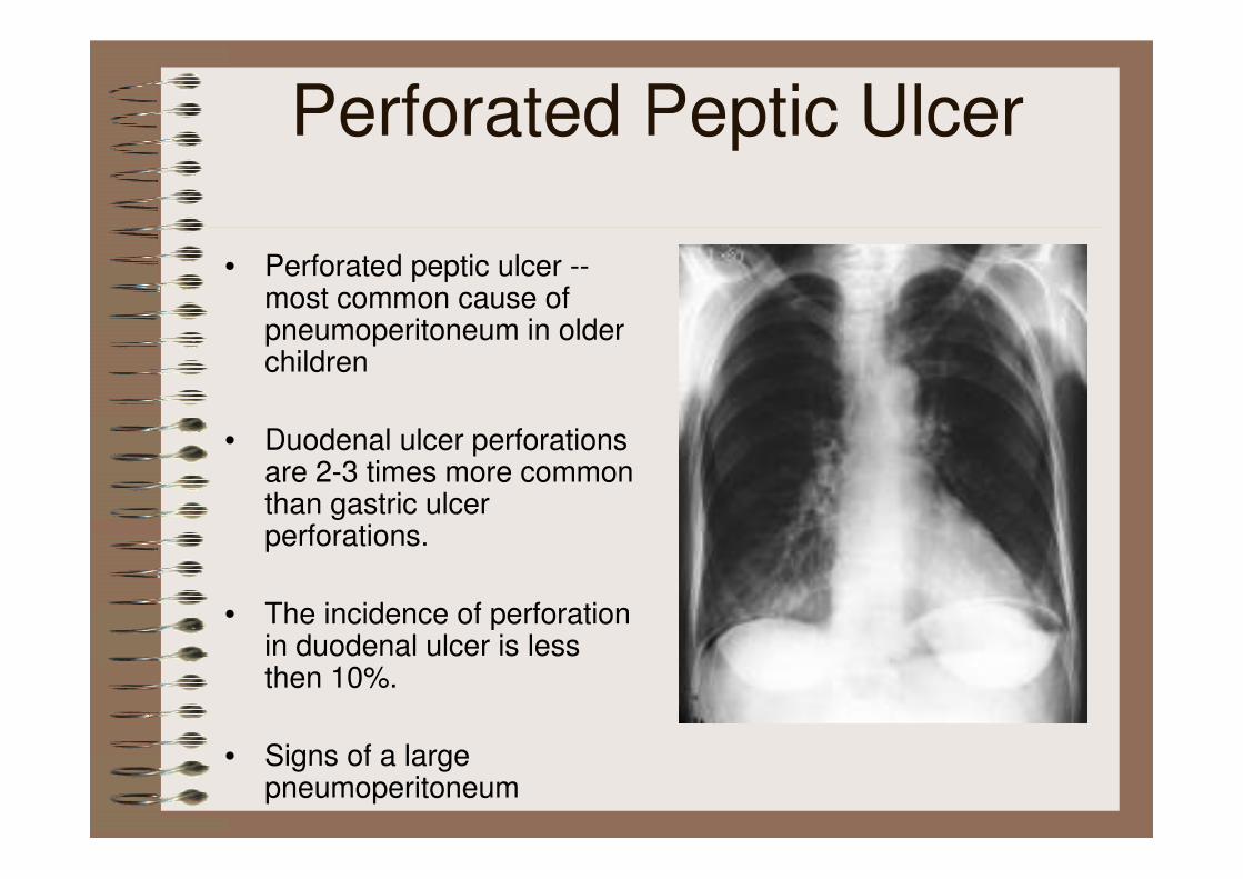
• Perforated peptic ulcer --most common cause of pneumoperitoneum in older children
• Duodenal ulcer perforations are 2-3 times more common than gastric ulcer perforations.
• The incidence of perforation in duodenal ulcer is less then 10%.
• Signs of a large pneumoperitoneum

Toxic megacolon• nonobstructive colonic dilatation larger than 6
cm and signs of systemic toxicity
• Colitides: inflammatory, ischemic, infectious
• Diagnostic criteria (Jalan et al):
1. radiographic evidence of colonic dilatation
2. any 3 of the following:
fever (>101.5°F), tachycardia (>120),
leukocytosis (>10.5), or anemia
3. any 1 of the following:
dehydration, altered mental status,
electrolyte abnormality, or hypotension.
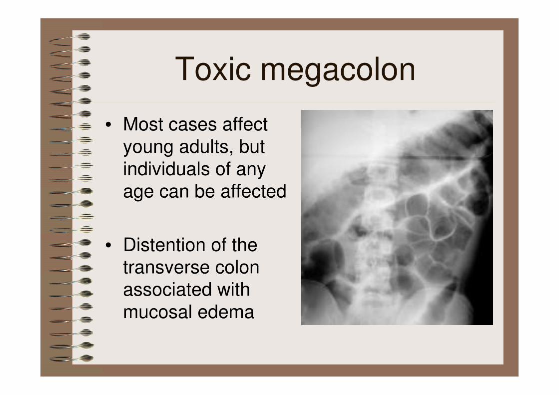
Toxic megacolon
• Most cases affect
young adults, but
individuals of any
age can be affected
• Distention of the
transverse colon
associated with
mucosal edema
Acute Colonic Pseudo-obstruction (ACPO)
• Ogilvie syndrome
• is a clinical disorder with the signs,
symptoms, and radiographic appearance of
an acute large bowel obstruction with no
evidence of distal colonic obstruction.
• The risk of perforation for ACPO ranges from
3-15% and carries a 50% mortality rate
• Age
1. generally a disease of elderly patients
2. may occur in younger patients, particularly those with underlying spinal cord disorders or primary cancer of or metastatic cancer to spinal region
• Clinical presentation:Abdominal pain(80%), Nausea and vomiting(80%),
Obstipation(40%), Fever(37%), Abdominal distention(90-100%),
Abdominal tenderness(64%)
• Imaging Studies:Specific attention to the diameter of the colon is important
If the colonic diameter exceeds 10 cm, decompression of the
colon must be considered and expedited.
Acute Colonic Pseudo-obstruction (ACPO)
Acute megacolon
• May be idiopathic, electrolyte abnormality,
metabolic abnormality, or certain medications,
including anticholinergics, opiates, digitalis,
and certain antipsychotic drugs
• can occur in any age group, the typical patient is an elderly person
• spontaneous perforation from nontoxic megacolon is 3%.
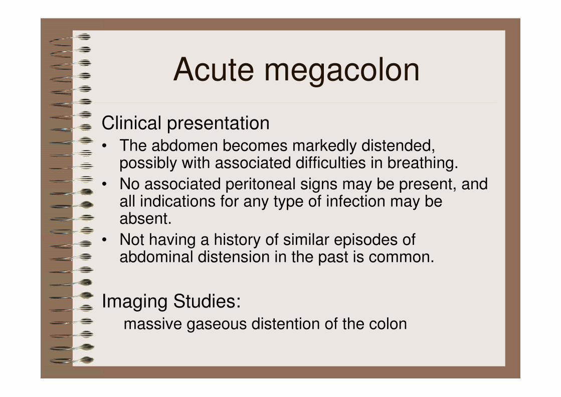
Clinical presentation• The abdomen becomes markedly distended,
possibly with associated difficulties in breathing.
• No associated peritoneal signs may be present, and all indications for any type of infection may be absent.
• Not having a history of similar episodes of abdominal distension in the past is common.
Imaging Studies:massive gaseous distention of the colon
Acute megacolon
Malignancy
• Colorectal cancer is rare in children with an incidence of 1.3 to 2 cases per million children
• Most of the cases in children occur in the second decade of life (13-18 y/o)
• abdominal pain, constipation, vomiting, nausea, rectal bleeding, abdomial distension, diarrhea
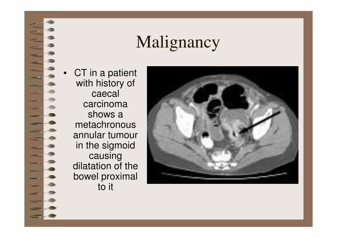
• CT in a patient with history of
caecalcarcinoma shows a
metachronousannular tumourin the sigmoid
causing dilatation of the bowel proximal
to it
Malignancy
• Arrange an emergent operation
• Keep vital sign
Plan
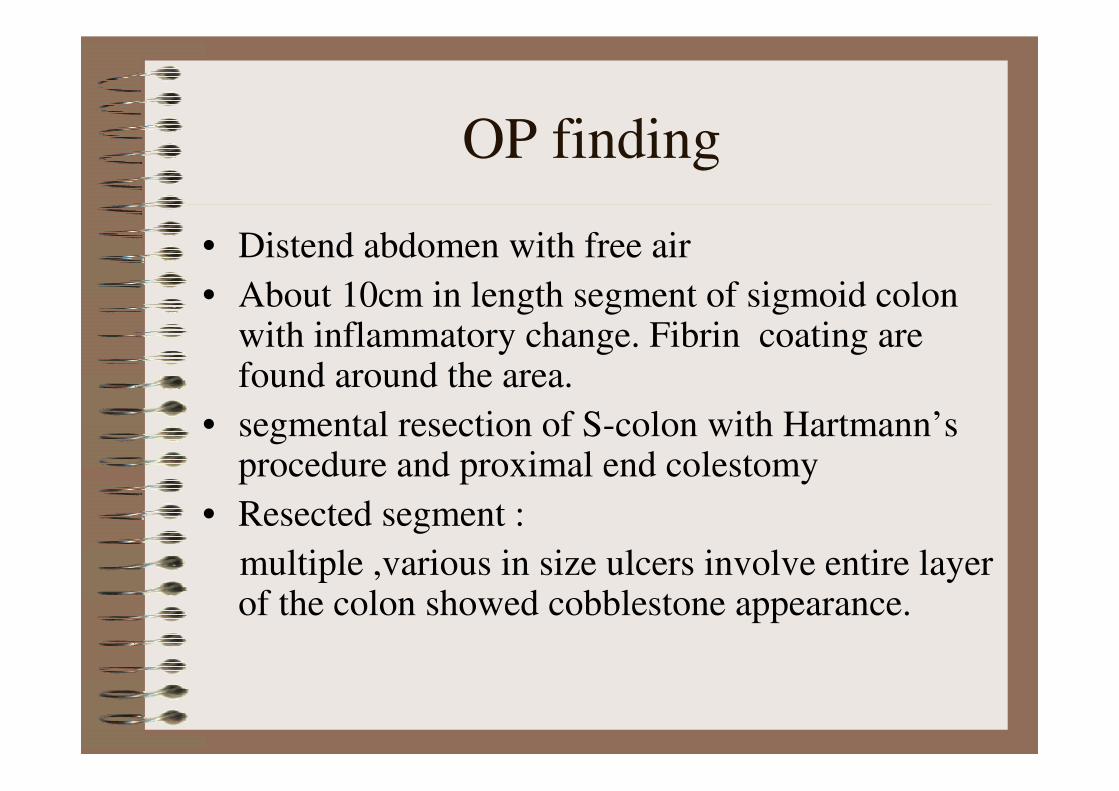
• Distend abdomen with free air
• About 10cm in length segment of sigmoid colon with inflammatory change. Fibrin coating are found around the area.
• segmental resection of S-colon with Hartmann’s procedure and proximal end colestomy
• Resected segment :
multiple ,various in size ulcers involve entire layer of the colon showed cobblestone appearance.
OP finding
Post op D/D
Sigmoid Colon Perforation
• R/o Crohn’s disease
• R/o viral infection
• R/o bacterial infection
Stool culture
• Salmonella(-)
• Clostridium difficile(-)
• Rota virus(-)

Pathhology(1)
• Specimen:
- Sigmoid colon, 20cm above the anal verge, segmental
resection
- 11.2 cm. in length and 4.8 - 8.2 cm in circumference
• Gross
- the serosa is coated by several small patches of fibrinous
exduate
- the intestinal wall in slightly thinner than usual
- the mucosa surface shows irregular, geographic, and
confluent ulcers, predominant arranged along the long
axis of the large intestine
- mucosa is reddening and edematous
• Microscopic
- multiple ulcers predominantly involving to the submucosa
- transmural ulcers with bowel perforation accompanied by
peritonitis
- ulcers are broad-based on aggregation of histiocytes
and scattered multinucleated giant cells
- granuloma formation is seen within or near the ulcers
• The features are compatible with Crohn's disease
Pathhology(2)

Final diagnosis
• Crohn’s disease
Treatment
• Sulfasalazine
Discussion
Crohn’s disease
• idiopathic, chronic, transmural inflammatory process of the bowel
• can affect any part of the GI tract from the mouth to the anus
• Most cases involve the small bowel, particularly the terminal ileum
• Skip lesions
• Case report in 1976: a 35y/o patient with Crohn'sdisease involved the entire colon without skip lesions and without terminal ileitis ( Acta Hepatogastroenterol (Stuttg) 1976 Jun;23(3):227-31 )
• Location
small intestine only (35%)
small and large intestine ( 45%)
large intestine (20%)
• The onset of Crohn disease has a bimodal distribution. The first peak occurs between the ages of 15-30 years; the second, between 60-80 years.
However, most cases begin before age 30 years.25-30% of all patients with CD present when younger than 20 years

Risk factors
• family history (HLA-DR1 and DQw5 )
• Smoking
• use of oral contraceptives ( 2 : 1 )
• Diet
• Ethnicity (Jewish )
• female-to-male ratio of 1.1-1.8 : 1

Clinical Presentation(1)
• GI symptoms
diarrhea, abdominal pain, weight loss,
rectal bleeding
• may be complicated by intestinal fistulization, obstruction, or both

• Extraintestinal manifestation- chronic intermittent fever
- Iron deficiency anemia
- arthritis and arthralgia
- Aphthous ulceration in the mouth
- erythema nodosum, pyoderma gangrenosum
Clinical Presentation(2)

Perforation rate
• the incidence of colonic and small bowel
perforation were 1.6 and 0.7%
• the highest frequency occurring in diseased
segments of jejunum (4%)
• Fistula leads to spontaneous intestinal
perforation in 1-2% of patients.
Clinical Presentation(3)
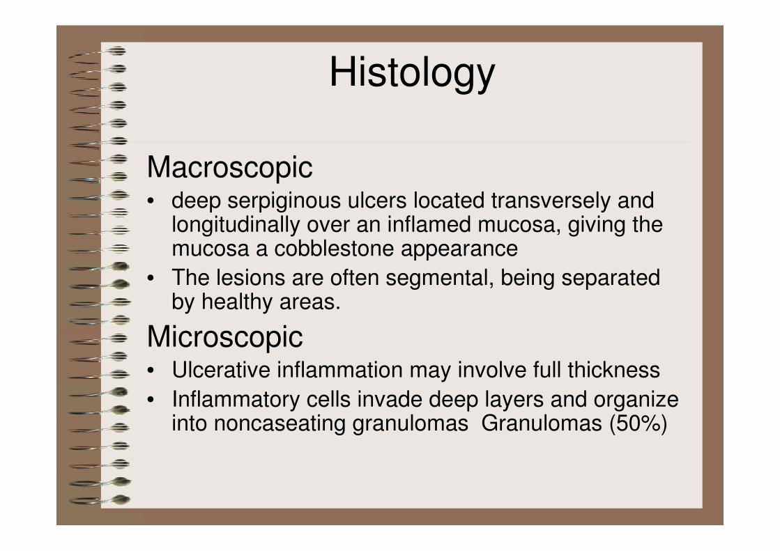
Histology
Macroscopic• deep serpiginous ulcers located transversely and
longitudinally over an inflamed mucosa, giving the mucosa a cobblestone appearance
• The lesions are often segmental, being separated by healthy areas.
Microscopic• Ulcerative inflammation may involve full thickness
• Inflammatory cells invade deep layers and organize into noncaseating granulomas Granulomas (50%)

Lab Data
• nonspecific
• microcytic anemia , leukocytosis
• ESR , CRP elevated in 90%
• Hypoalbuminemia, iron, and vitamin deficiencies
• Stool studies
• Anti-Saccharomyces cerevisiae antibody (ASCA)
-- positive in 70% in CD
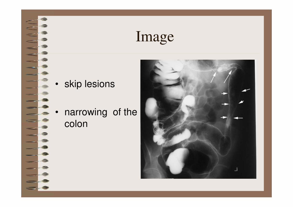
• skip lesions
• narrowing of the
colon
Image
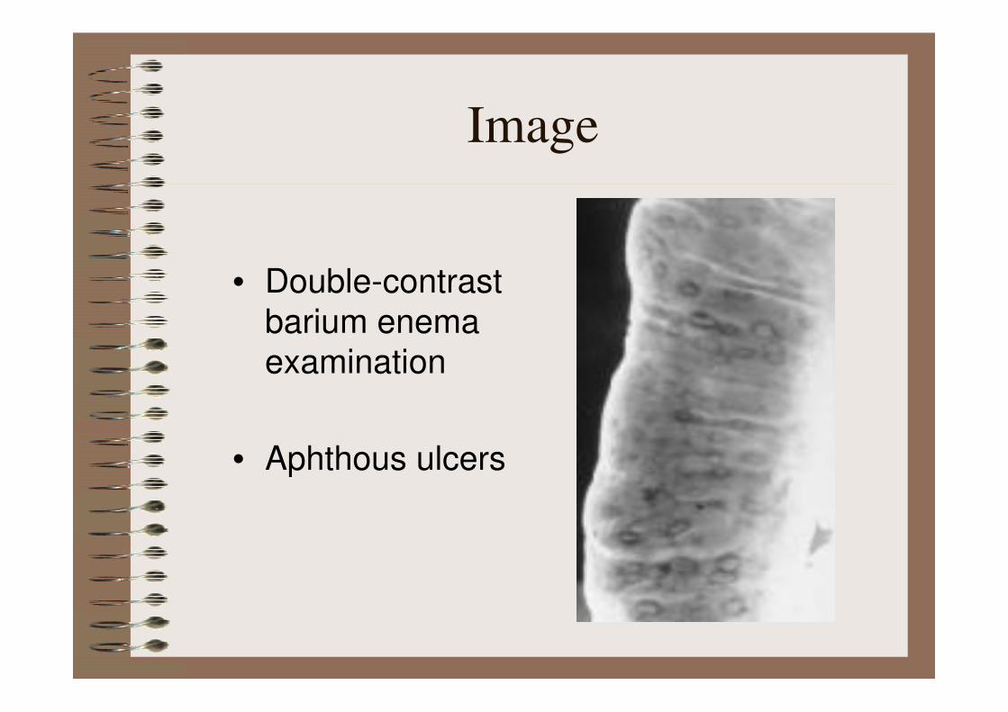
• Double-contrast
barium enema
examination
• Aphthous ulcers
Image

• narrowing and stricturing
• string sign
• sinus tract
Image

• multiple fistulous
tracts between the
terminal ileum and the
right colon adjacent to
the ileocecal valve
Image
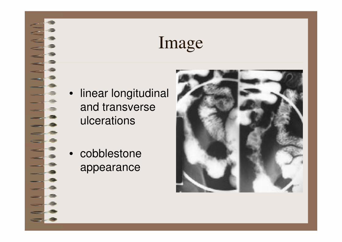
• linear longitudinal
and transverse
ulcerations
• cobblestone
appearance
Image

• Crohn's colitis with toxic megacolon
• marked dilatation of the large bowel, from the caecum on the right to a loop of sigmoid seen centrally in the pelvis
Image

• terminal ileal-wall
thickening
• adjacent mesenteric
inflammatory
stranding.
Image
Treatment
• Anti-inflammatory agents :
Sulfasalazine ,Mesalamine
• Corticosteroids : ,
Budesonide
• Antibiotics :
metronidazole and ciprofloxacin
• Immunosuppressive agents :
Azathioprine

• Nutritional therapies
omega-3 fatty acids (fish oil)
• Surgical Care
obstruction, abscess, fistula,
hemorrhage, perforation
Treatment

Prognosis
• may have a large impact on the life
• with the appropriate treatment and support, the prognosis is very good
• Colonic malignancy
- 10-25 years after diagnosis of colitis
is estimated to be 8%