326 chapter 27

1
326 Chapter 27 costal cartilages allow movement during inhalation and exhalation. The spaces between the ribs are known as the intercostal spaces and contain intercostal musculature and a neuro- vascular bundle that resides in a groove on the internal surface of the lower border of the each rib. Abnormalities of rib cage development may include pectus excavatum, also called sunken chest, and pectus carinatum, also called pigeon chest. In males, the rib cage expands as an effect of the testosterone surge that characterizes puberty. This con- tributes to the generally broader shoulders and expanded chest that allow adult males to inhale more to supply adequate oxygen to their increased muscle mass. The rib cage is the structure from which musculature acts in order to accomplish respiration. Inhalation is accomplished when the diaphragm contracts and flattens, moving the floor of the thorax inferiorly, and the intercostal muscles lift the rib cage up and outward. Exhalation is gen- erally a passive event, except in cases of forced exhalation. Muscles of Respiration Various muscles of respiration aid in both inspiration and expiration, which require changes in the pressure within the thoracic cavity (Figure 27-6). The primary muscles of inspiration are the diaphragm, the upper and more lateral external intercostals, and the parasternal portion of the internal intercostal muscles. Both the external intercostal muscles and the parasternal portion of the internal intercos- tal muscles elevate the ribs. This increases the width of the thoracic cavity, while the diaphragm contracts to increase the vertical dimensions of the thoracic cavity. The external intercostal muscles also aid in the elevation of the lower ribs. The levatores costarum are primary muscles of respiration that probably serve mostly a proprioceptive role. The accessory muscles of inspiration include the scalenes, SCM, levator scapulae, and upper fibers of the trapezius, the iliocostalis thoracis, subclavius, and omohyoid. The serratus anterior and latissimus dorsi are accessory muscles of respira- tion with the upper extremities elevated. Accessory muscles of expiration include the interosseous internal intercostals, abdominal muscles, transversus thoracis, subcostalis, ilio- costalis lumborum, quadratus lumborum, serratus posterior inferior, and latissimus dorsi. The accessory respiratory mus- cles are typically only used when the body needs to process energy quickly; for example, during heavy exercise, during the stress response, or during an asthma attack. When acces- sory muscles of respiration are habitually utilized and become part of the primary muscle recruitment pattern of respiration, dysfunction will follow. For example, decreased excursion of the diaphragm because of improper position will require the upper accessory musculature to increase activity for normal Figure 27-6. Muscles of inspiration and expiration.

Upload: others

Post on 12-Dec-2021

1 views

Category:

Documents


0 download

TRANSCRIPT

326  Chapter 27

costal cartilages allow movement during inhalation and exhalation.

The spaces between the ribs are known as the intercostal spaces and contain intercostal musculature and a neuro-vascular bundle that resides in a groove on the internal surface of the lower border of the each rib. Abnormalities of rib cage development may include pectus excavatum, also called sunken chest, and pectus carinatum, also called pigeon chest. In males, the rib cage expands as an effect of the testosterone surge that characterizes puberty. This con-tributes to the generally broader shoulders and expanded chest that allow adult males to inhale more to supply adequate oxygen to their increased muscle mass.

The rib cage is the structure from which musculature acts in order to accomplish respiration. Inhalation is accomplished when the diaphragm contracts and flattens, moving the floor of the thorax inferiorly, and the intercostal muscles lift the rib cage up and outward. Exhalation is gen-erally a passive event, except in cases of forced exhalation.

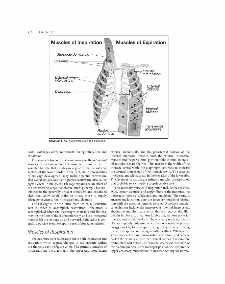
Muscles of RespirationVarious muscles of respiration aid in both inspiration and

expiration, which require changes in the pressure within the thoracic cavity (Figure 27-6). The primary muscles of inspiration are the diaphragm, the upper and more lateral

external intercostals, and the parasternal portion of the internal intercostal muscles. Both the external intercostal muscles and the parasternal portion of the internal intercos-tal muscles elevate the ribs. This increases the width of the thoracic cavity, while the diaphragm contracts to increase the vertical dimensions of the thoracic cavity. The external intercostal muscles also aid in the elevation of the lower ribs. The levatores costarum are primary muscles of respiration that probably serve mostly a proprioceptive role.

The accessory muscles of inspiration include the scalenes, SCM, levator scapulae, and upper fibers of the trapezius, the iliocostalis thoracis, subclavius, and omohyoid. The serratus anterior and latissimus dorsi are accessory muscles of respira-tion with the upper extremities elevated. Accessory muscles of expiration include the interosseous internal intercostals, abdominal muscles, transversus thoracis, subcostalis, ilio-costalis lumborum, quadratus lumborum, serratus posterior inferior, and latissimus dorsi. The accessory respiratory mus-cles are typically only used when the body needs to process energy quickly; for example, during heavy exercise, during the stress response, or during an asthma attack. When acces-sory muscles of respiration are habitually utilized and become part of the primary muscle recruitment pattern of respiration, dysfunction will follow. For example, decreased excursion of the diaphragm because of improper position will require the upper accessory musculature to increase activity for normal

Figure 27-6. Muscles of inspiration and expiration.