)3. the 2.0cm tissue from the surgeon became...
TRANSCRIPT
Collected: 04/18/2013 9:09:00
Clinical History I Notes:
Procedure: Preop Diagnosis: Clinical History:
Gross Description:
Pathology Reports
Received: 04/18/2013 9:28:00
Laparoscopic liver biopsy Acute liver failure None given
Accession Number: SP-13-02373
Pathologist: Rudzinski, Erin R, MD
Received fresh labeled LIVER BIOPSY is a single needle core biopsy fragment measuring 0.1 cm in diameter x 1.3 cm in length. The specimen is placed in formalin then wrapped and submitted in its entirety in a single cassette designated A 1. [MAJ
Microscopic Description:
H&E stained sections show a cirrhotic liver with bridging fibrosis and marked bile ductular proliferation. There is moderate infiammation in the portal tracts with scattered neutrophils, lymphocytes, and eosinophils. Bile ducts are present, although difficult to see with the marked portal expansion and ductal proliferation. Lobular inflammation is variable and predominantly lymphocytic in nature. There is variable cholestasis, with some nodules showing canalicular plugging and others showing only mild intracellular cholestasis. Acidophil bodies are readily identified. Hepatocytes are swollen with lacy cytoplasm and reactive appearing nucleoli. Rare areas show features of early hepatocyte necrosis with the blurring of cell junctions and hypereosinophilia. There is patchy mild macrosteatosis (<5%). A trichrome stain highlights the presence of bridging fibrosis, as well as pericellular fibrosis. Periodic acid shift stains with a without diastase reveal no diastase resistant globules.
Slide and stain summary: H&E 3 slides; PAS 1 slide; PAS-0 1 sfide: trichrome 1 slide
Electron Microscopy Findings:
Toluidine blue stained thick sections reveal cirrhotic liver .with both periportal and pericellular fibrosis. There is mild portal infiammation. Examination by transmission electron microscopy shows mild cholestasis, and occassional hepatocytes with membranous debris but no evidence of storage disease. Bile canaliculi show normal microvillous architecture, without Bylers-type bile. Probable peroxisomes are identified. Mitochondria are present in normal numbers, and are without circular or paracrystalline arrays of cristae.
Final Diagnosis:
LIVER, NATIVE, CORE BIOPSY: CIRRHOSIS AND BILE DUCTULAR PROLIFERATION, SEE COMMENT
Comment The biopsy shows established cirrhosis with marked bile ductular proliferation and variable cholestasis. There is ongoing hepatocyte injury, with scattered apoptotic cells, as well. PASd stain reveals no evidence of alpha-1 antitrypsin deficiency, and no abnormal mitochondria, bile or storage-type material are seen on electron microscopy. A definitive etiology for the patient's chronic liver failure is not evident from this biopsy.
Seattle Children's Hospital
PO Box 5371
Seattle, Washington 98105-0371
NAME: XIE, JIANHUA DRACO
DOB: 09/16/2012
MR: 1275567
Print Date 6/18/2013
I
Collected: 04/18/2013 9:09:00
Pathology Reports
Received: 04/18/2013 9:28:00
Accession Number: SP-13-02373
Pathologist: Rudzinski, Erin R, MD
Preliminary findings were reviewed with Dr Simon Horslen on 4/19/13 at approximately 14:30h.
T-62000, T-60650, M-76000, D5-80600, Pl-03100
Diagnosis by: Erin R Rudzinski, MD Electronic Signature
04/25/2013 ED: ERR
Seattle Children's Hospital
PO Box 5371
Seattle, Washington 98105-0371
NAME:
DOB:
XIE, JIANHUA DRACO
09/16/2012
MR: 1 275567
Print Date 6/18/2013
I

I Date of Service: Authored By:
Operative Report
04/18/2013 0:00:00 Healey, Patrick J, MD
Op Note
Document may Not be Signed/Finalized. See End of report for Electronic Authentication of Signature.
OPERATIVE REPORT
XIE, JIANHUA DRACO DOB: 09/16/2012 M -MR#: 01-27-55-67 PATIENT LOCATION: SUR-R6
DATE OF OPERATION: 04/18/2013
PREOPERATIVE DIAGNOSIS: Liver failure.
POSTOPERATIVE DIAGNOSIS: Liver failure.
OPERATION: Laparoscopy with liver biopsy.
SURGEONS: Patrick Healey, MD, Attending; Janice Kang, MD, Resident in Surgery.
INDICATIONS: Jianhua is a 7-month-old boy who presented to the Intensive Care Unit this past week with profound liver failure. He had hyperammonemia, severe coagulopathy, jaundice with ascites and splenomegaly. With intensive care medical support, diuresis, and appropriate management, he has clinically improved, though remains with borderline hypoglycemia, INR of 2.8, and ammonia of over 100. His diagnostic workup has not yielded a definitive diagnosis to date and liver biopsy was requested. Because of his ascites and degree of coagulopathy, percutaneous biopsy was deemed to be at unreasonable risk so surgical biopsy was requested. After description of the procedure and discussion of the potential risks and complications, informed consent was obtained.
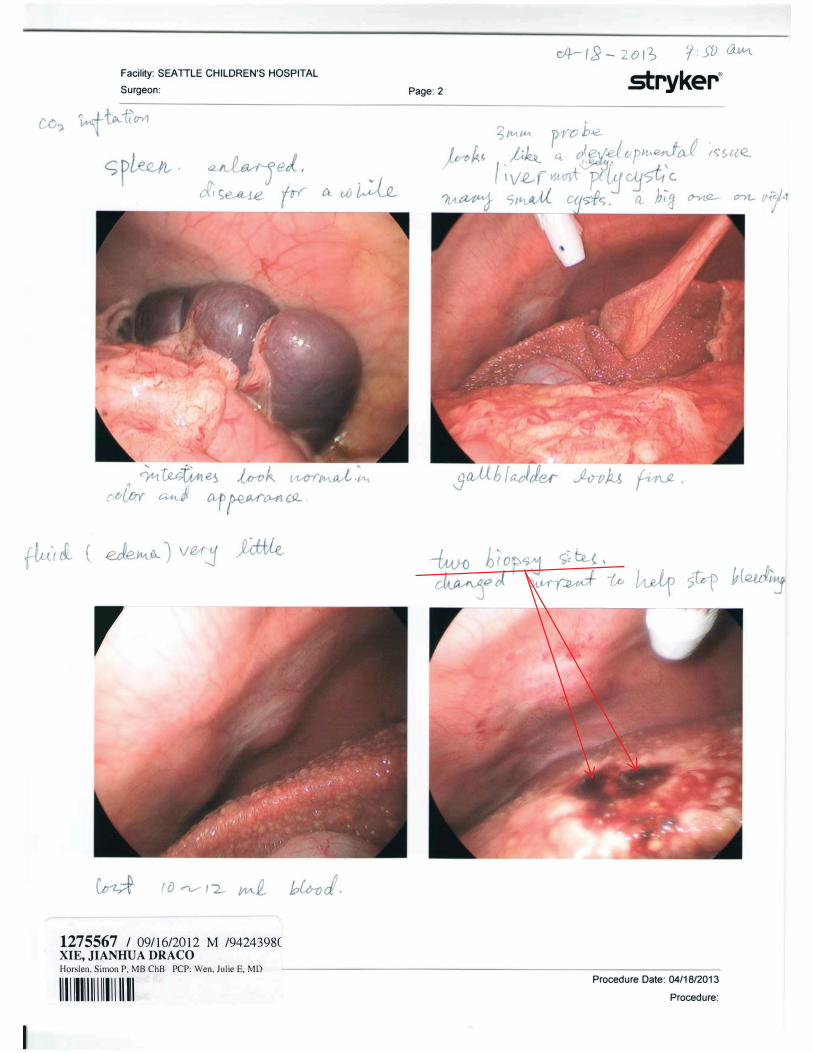
FINDINGS: Liver was small and firm with diffuse micronodular cystic change visible subcapsular. There were also some slightly larger macrocysts which correlated with those seen on a CT scan earlier. A 2 cm core liver biopsy was obtained from the right lobe.
PROCEDURE: The patient was taken to the operating room and positioned supine. General anesthesia was administered and the baby was then intubated endotracheally. A separate IV was then started. A surgical timeout was done confirming patient identification and consent of procedure, among other standard elements. The abdomen was then widely prepped and draped in a sterile fashion.
The baby had a large umbilical hernia. A small 5 mm transverse incision was made just to the right of the umbilical hernia overlying the rectus. Using an open Hassan technique, the dissection was carried down through the layers of the abdominal wall, identifying the anterior sheath, posterior sheath, and peritoneum. The peritoneum was entered sharply and a PDS suture was then placed around the defect. A 5 mm port with this Veress step introducer was then placed into the peritoneal cavity and pneumoperitoneum established to 8 cm
Seattle Children's Hospital PO Box 5371
Seattle, Washington 98105-0371
NAME: DOB:
MR:
XIE, JIANHUA DRACO
0911612012 1275567
Print Date 611812013
I
I Op Note
Date of Service: 04/18/2013 0:00:00 Authored By: Healey, Patrick J, MD
of pressure. Laparoscopy was then performed and the above findings noted. Under laparoscopic guidance, a second port, 3 mm, was then placed in the right upper quadrant at roughly the anterior axillary line. Through this working port, the omentum was moved off of the liver revealing a chronic-appearing contracted firm liver with multiple small and medium size cysts visible on its surface. The spleen was enlarged and lobulated. Under laparoscopic guidance then using a spring-loaded Tru-Cut needle, a biopsy was taken in the right lobe just to the right of the gallbladder fossa. A sample was a good sample and this was sent then fresh to Pathology. The biopsy site bled briskly initially and this was controlled with electrocautery without too much difficulty. The abdomen was then irrigated and excess fluid and blood clot evacuated. Biopsy site was reinspected and noted to remain hemostatic. After pathologic confirmation of adequate biopsy, the pneumoperitoneum was released and the ports removed. The wounds were closed in layers with PDS in the muscle fascia, Vicryl in the subcutaneous layer and in the subcuticular skin. Steri-Strips were applied to both incisions.
The patient tolerated this procedure well, was extubated in the operating room, and transferred to the recovery room in stable condition.
Please note, I was present for and performed this entire procedure with Dr. Kang.
Electronically Authenticated by Patrick J Healey, MD 04/20/2013 12:24 P
Patrick J Healey, MD , Attending Physician, Surgery
PJH/srl Doc #2944494 d: 0411912013 08:46 P t: 0411912013 09:12 P {1562796-) cc: cc Pediatric Assoc Redmo
Julie E Wen, MD
Seattle Children's Hospital
PO Box 5371
Seattle, Washington 98105-0371
NAME: XIE, JIANHUA DRACO
DOB: 09/1612012 MR: 1275567 Print Date 6118/2013
I