2nd revision nuclear receptors in drug metabolism, drug ... · genes involved in drug metabolism...
TRANSCRIPT

1
2nd REVISION
Nuclear Receptors in Drug Metabolism, Drug Response and Drug Interactions
Authors: Chandra Prakash1, 2, ¶, Baltazar Zuniga1,3, ¶, Chung Seog Song1, ¶,
Shoulei Jiang1, Jodie Cropper1, Sulgi Park1 and Bandana Chatterjee1,4,*
1 Department of Molecular Medicine/Institute of Biotechnology The University of Texas Health Science Center at San Antonio Texas Research Park, 15355 Lambda Drive, San Antonio, Texas 78245 2 William Carey University College of Osteopathic Medicine 498 Tucsan Ave, Hattiesburg, Mississipi 39401 3 University of Texas at Austin 2100 Comal Street, Austin, Texas 78712 4 South Texas Veterans Health Care System Audie L Murphy VA Hospital, 7400 Merton Minter Boulevard
San Antonio, Texas 78229 Key words: Nuclear receptors, PXR, CAR, Xenobiotic-response element, Gene induction, Phase 0-III
mediators, Genetic polymorphism, Epigenetics, Drug interactions, Drug screening
¶ Joint First Authors *Corresponding Author Bandana Chatterjee, Ph.D. Department of Molecular Medicine/Institute of Biotechnology 15355 Lambda Drive, San Antonio, Texas 78245 E-mail: [email protected]; Tel:210-567-7218, FAX: 210-567-7222
Abbreviations NR, nuclear receptor; DBD, DNA-binding domain; LBD, ligand-binding domain; XRE, xenobiotic response element, PXR, pregnane X receptor; CAR, constitutive androstane receptor; VDR, vitamin D receptor; FXR, farnesoid X receptor; LXR, liver X receptor; CYP, cytochrome P450; DME, drug-metabolizing enzyme; ADME, absorption, distribution, metabolism, excretion; DDI, drug-drug interaction; PTM, post-translational modification; MDR, multi-drug resistance; ABC, ATP-binding cassette; HDAC, histone deacetylase; HAT, histone acetyltransferase; HMT, histone methyltansferase; HMD, histone demethylase; DNMT, DNA methyltransferase; SNP, single nucleotide polymorphism

2
ABSTRACT
Orally delivered small-molecule therapeutics are metabolized in the liver and intestine by phase I
and phase II drug-metabolizing enzymes (DMEs), and transport proteins coordinate drug influx (phase
0) and drug/drug-metabolite efflux (phase III). Genes involved in drug metabolism and disposition are
induced by xenobiotic-activated nuclear receptors (NRs), i.e. PXR (pregnane X receptor) and CAR
(constitutive androstane receptor), and by the 1α, 25-dihydroxy vitamin D3-activated vitamin D receptor
(VDR), due to transactivation of xenobiotic-response elements (XREs) present in phase 0-III genes.
Additional NRs, like HNF4-α, FXR, LXR-α play important roles in drug metabolism in certain settings,
such as in relation to cholesterol and bile acid metabolism. The phase I enzymes CYP3A4/A5, CYP2D6,
CYP2B6, CYP2C9, CYP2C19, CYP1A2, CYP2C8, CYP2A6, CYP2J2, and CYP2E1 metabolize >90%
of all prescription drugs, and phase II conjugation of hydrophilic functional groups (with/without phase I
modification) facilitates drug clearance. The conjugation step is mediated by broad-specificity
transferases like UGTs, SULTs, GSTs. This review delves into our current understanding of
PXR/CAR/VDR-mediated regulation of DME and transporter expression, as well as effects of single
nucleotide polymorphism (SNP) and epigenome (specified by promoter methylation, histone
modification, microRNAs, long non coding RNAs) on the expression of PXR/CAR/VDR and phase 0-
III mediators, and their impacts on variable drug response. Therapeutic agents that target epigenetic
regulation and the molecular basis and consequences (overdosing, underdosing, or beneficial outcome)
of drug-drug/drug-food/drug-herb interactions are also discussed. Precision medicine requires
understanding of a drug’s impact on DME and transporter activity and their NR-regulated expression in
order to achieve optimal drug efficacy without adverse drug reactions. In future drug screening, new
tools such as humanized mouse models and microfluidic organs-on-chips, which mimic the physiology
of a multicellular environment, will likely replace the current cell-based workflow.

3
INTRODUCTION
Drug metabolism, which occurs primarily in the liver and intestine, refers to the enzymatic
modification and subsequent disposal of medicinally active compounds, originating either endogenously
(as steroids, neurotransmitters, metabolic products like bile acids) or exogenously (as natural products or
synthetic/semi-synthetic chemicals). Upon conversion to hydrophilic metabolites, drugs are eliminated
from the body following biliary excretion and renal clearance by glomerular filtration and tubular
secretion. Drug metabolism is also integral to the biotransformation of pro-drugs to pharmaco-active
agents. Drug metabolism and disposition is coordinated by an array of liver- and intestine-expressed
drug-metabolizing enzymes (DMEs) and drug-transporting proteins whose tissue abundance is
transcriptionally regulated by specific nuclear receptors (NRs), which are ligand-activated transcription
factors (Willson & Kleiwer, 2002).
Of the 48 distinct receptors comprising the NR superfamily in humans, pregnane X receptor
(PXR, NR1I2) and constitutive androstane receptor (CAR, NR1I3) are primary transcriptional regulators
of the genes involved in the metabolism and elimination of drugs/drug metabolites (Xie & Evans, 2001;
Tzameli & Moore, 2001; Evans & Mangelsdorf, 2014). PXR and CAR are generically referred as
xenobiotic NRs, since structurally diverse drugs and environmental xenobiotics activate these two NRs.
PXR and CAR are also activated by a number of endobiotics (steroids, sterols, retinoids, thyroid
hormones, bile acids). In addition, PXR and CAR activation can result from receptor phosphorylation
by various kinases that are activated in response to drug-mediated induction of specific intracellular
signal cascades; in this case, drugs may not directly interact with the xenobiotic NRs (Smutny et al,
2013). Vitamin D receptor (VDR, NR1I1), beyond its classic role in calcium and phosphate
homeostasis, has the ability to transcriptionally induce drug transporters and DMEs, especially in the
enterocytes of intestinal tissue (Makishima et al, 2002; Chatterjee et al, 2005). In certain contexts,

4
additional NRs, such as the bile acid-activated farnesoid X receptor (FXR, NR1H4); oxysterol-activated
liver X receptor (LXR-α, NR1H3); fatty acid/eicosanoid-activated peroxisome proliferator activated
receptor (PPAR-α, NR1C1), and retinoid-related orphan receptors (ROR-α, ROR-γ) regulate genes
linked to drug absorption, distribution, metabolism and excretion (ADME) (Xie & Chiang, 2013).
Hepatocyte nuclear factor (HNF4-α, NR2A1), a member of the NR superfamily, plays a synergizing
role in the PXR- and CAR-mediated transactivation of DME- and transporter-encoding genes (Tirona et
al, 2003; Echchgadda et al, 2007; Hwang-Verslues & Sladek, 2010).
Altered activities of polymorphic variants of NRs and ADME-related gene products account for
variable response to prescription medicine between individuals. Amino acid changes due to nucleotide
polymorphisms in the coding region can influence protein stability or activity, while polymorphism at
upstream, downstream or intragenic regulatory loci can alter NR-mediated ADME gene transactivation.
Epigenetic, transcriptional and posttranslational regulation of xenobiotic NRs can further impact drug
metabolism and clearance. Evaluation of drug-drug interactions (DDI), which result from changes in the
level or activity of DMEs and/or transporters due to a second drug, is an essential component of drug
development workflow.
In this review, we describe various classes of DMEs and transporters, present an overview of the
molecular underpinnings for NR-mediated genetic and epigenetic regulation of ADME genes and
consider roles for various NRs (especially PXR/CAR/VDR) and their target genes in differential drug
response. Illustrative examples highlighting critical roles of xenosensing NRs, DMEs and transporters in
DDI are also examined. Finally, we discuss current drug-screening platforms and their potential future
improvements.

5
1. Drug metabolizing enzymes (DMEs) and drug transporters: The four phases of drug metabolism
entail cellular uptake of therapeutic molecules (phase 0); their enzymatic oxidation (phase I) and
conjugation (phase II), and efflux of drug metabolites for clearance (phase III). PXR and CAR activate
genes involved in all four phases. General steps in drug metabolism and elimination are shown in Fig. 1.
1.a Phase 0 uptake proteins:
Basolaterally located uptake proteins guide cellular entry of drugs from circulation; drug influx
can be a rate-limiting step for drug metabolism and clearance (KÖnig et al, 2013; Döring and Petzinger,
2014). All uptake proteins are members of the solute carrier (SLC) protein family of which there are
more than 300 members grouped under 52 subfamilies. Liver, intestine and kidneys are major sites of
SLC expression. Most SLC proteins localize to cell membrane, although some may localize to
mitochondria and other organelles. Proteins from nineteen SLC gene subfamilies have drug uptake
activity. Most significant gene families of uptake transporters are SLC15 (oligopeptide transporter),
SLC22 (organic anion/cation/zwitterion transporter), SLCO (organic anion transporting polypeptide) and
SLC47 (organic cation transporter) (Russell, 2010; Nigam, 2015). For example, OCT1 is a SLC22A1
encoded organic cation uniporter involved in the influx of the antiviral agent acyclovir, ganciclovir and
the anti-diabetic drug metformin. Drug substrates for proteins encoded by SLC15, SLC22, SLCO and
SLC47 families have been described (Russel 2010; KÖnig et al, 2013; Nigam, 2015). SLCs either serve
as channels (uniporter) to guide drug diffusion down an electrochemical gradient, or drive drug transport
against a diffusion gradient that is coupled to the symport or antiport of small inorganic or organic ions.
1.b Phase I DMEs
Heme-containing cytochrome P450s (CYPs), flavin-containing monooxygenases, monoamine
oxidases and xanthine oxidase/aldehyde oxidases are examples of phase I DMEs, which generally

6
localize to the endoplasmic reticulum of cells. CYP enzymes play the most prominent role in phase I
metabolism. Liver is the first pass and primary site of phase I metabolism, along with the
gastrointestinal tract, kidneys, skin, and lung serving as additional sites; most tissues, however, express
phase I DMEs. Addition or exposure of polar functional groups (e.g. -OH, hydroxyl; -COOH, carboxyl;
-NH2, amine; -SH, sulfhydryl) to drug substrates enhances their bioavailability and solubility and
promotes pro-drug biotransformation. Polar groups also arise by reduction of a ketone or aldehyde group
to an alcohol; oxidation of an alcohol to an acidic group; hydrolysis of esters and amides; reduction of
azo and nitro groups; oxidative dealkylation of N-alkyl, O-alkyl , and S-alkyl groups. When sufficiently
polar, phase I metabolites can be pumped out of cells without additional modification.
CYPs are products of a multigene family, which for humans include 57 CYP genes (Zanger & Schwab,
2013). Individual CYP is specified by the family (with an Arabic numeral), then the subfamily (with a
letter) followed by the isozyme within the subfamily (with another Arabic numeral) and the allele
number (with a preceding asterisk) of an individual gene within the subfamily. As an example, for
CYP2D6*1, the *1 allele is wild type CYP2D6 with normal activity. Additional alleles of CYP2D6,
marked with higher numbers preceded by *, exhibit aberrant functions (Wijnen et al, 2007). CYPs are
expressed in practically all tissues, with liver exhibiting the highest abundance and expressing largest
number of individual CYPs. Enzymes of the CYP-1, -2 and-3 families metabolize majority of drugs and
non-drug xenobiotics. The fraction of clinical drugs that are substrates for individual CYPs is
schematically presented as Figure 2. CYP3A4, the most abundant CYP enzyme in human liver, acts on
the greatest number of drugs and other xenobiotics. CYP2D6, although present at lower abundance,
metabolizes numerous drugs. Substrate specificity is narrower for other members of the CYP family
that are expressed in hepatic and extrahepatic tissues. They target endogenous substrates like sterols,
fatty acids, eicosanoids and vitamins. A comprehensive list of drug substrates for CYPs has been

7
reported (Lynch & Price, 2007; www. pharmacologyweekly.com/cytochrome-cyp-p450-enzyme-
medication-herbs-substrates, updated May, 2015).
1.c. Phase II DMEs
Broad-specificity phase II transferases catalyze conjugative reactions. Common phase II
modifications are glucuronidation by UDP-glucuronyltransferase (UGT), sulfonation by sulfotransferase
(SULT), glutathionylation by glutathione S-transferase (GST), acetylation by N-acetyl transferase
(NAT) and methylation by methyltransferase (MT). For any given transferase family, individual family
members show predilection for a distinct set of substrates. Cofactors of phase II transferases react with
functional groups that are either part of the parent drug or generated by phase I modification. In contrast
to the enhanced potency of many phase I metabolites, phase II modified drug metabolites normally
exhibit diminished function. PXR- and CAR-mediated gene regulation for a number of phase II
transferases has been studied (Echchgadda et al, 2007; Tolson & Wang, 2010).
1.d. Phase III Efflux Proteins
Members of the ATP binding cassette (ABC) superfamily, encoded by the ABCB, ABCC, and
ABCG gene subfamilies, are broad-specificity exporters that pump drugs out of cells using energy from
ATP hydrolysis. In hepatocytes, efflux proteins reside either in canalicular/apical membranes or in
blood-facing basolateral/sinusoidal membranes, guiding drugs, endobiotics and their metabolites for
biliary excretion and efflux into systemic circulation. Multidrug-resistance associated proteins MRP2
(ABCC2), the bile salt export pump BSEP (ABCB11), the breast cancer resistance protein BCRP
(ABCG2) are examples of ABC cassette family transporters which mediate apical efflux of drugs,
steroids, bile acids and their conjugates. P-glycoprotein (MDR1, ABCB1) is an apical membrane
transporter in hepatocytes (Nigam, 2015). Basolateral efflux of unconjugated and phase II-conjugated

8
drugs, steroids, prostaglandin and bile acids from hepatocytes into sinusoidal blood is assisted by ABC
transporters such as the multi-drug resistance associated proteins MRP3, MRP4, MRP5 and, also, by the
ATP-independent OSTα/OSTβ complex that functions as an organic solute and steroid transporter.
OSTα contains seven transmembrane domains and OSTβ has a single transmembrane domain
(Ballatori, 2005); neither is part of the ABC transporter superfamily. MATE (multidrug and toxin
extrusion) efflux transporters are H+-coupled antiporters, which transport structurally unrelated
organic cations out of cells. They are members of the solute carrier subfamily SLC47,
expressed primarily in the liver and kidney, and they localize at apical membranes of renal
tubular epithelia and bile canaliculi. MATE1 (product of SLC47A1) mediates extrusion of
organic cations into urine and bile. In the human kidney, the uptake transporter OCT2 (organic
cation transporter, SLC22A2 encoded) promotes the import of cationic drugs (such as
metformin, cisplatin, imatinib) from the blood at the basolateral membrane of the proximal
tubule epithelial cells. MATE-1 and the isoform MATE-2K mediate secretion of cationic drugs
across the brush-border membrane into the proximal tubule lumen (Motohashi & Inui, 2013).
2. Nuclear receptors (NRs), response elements, gene regulation by PXR/ CAR/VDR:
NRs, upon association with DNA response elements, induce a cascade of protein-protein interactions
that lead to the assembly of multiple classes of regulatory proteins (coactivators, corepressors, histone
modifiers, chromatin remodeling complex) at the NR-bound chromatin region. Signal transmission
from the coregulator assembly to the basal transcription machinery via a multi-protein mediator complex
culminates in altered RNA polymerase II activity and transcriptional response of NR-regulated genes.
9
The NR superfamily of ligand-activated transcription factors in humans is defined by 48 receptors
grouped into four classes (Type I-IV) based on the nature of activating ligands, preferred sequence
organization of NR-binding DNA response elements in target genes and dimerization partner of the
activated NR (Mangelsdorf et al, 1995; Sever & Glass, 2013). Type I NRs reside in the cytoplasm in an
inactive state in association with chaperone proteins. They are activated upon binding cognate steroid
hormone ligands, translocated to the nuclear compartment and bind target gene response elements as
homodimers to mediate gene regulation. Type II receptors, such as the vitamin D receptor (VDR), are
activated by non-steroid endocrine ligands (1α,25-dihydroxy vitamin D3 (1,25-D3, in short) for VDR;
retinoic acid-all trans, for RAR-α/-β/-γ; thyroid hormone for TR-α/-β). Several Type II receptors are
activated by intracrine ligands (e.g. bile acids activating FXR-α; oxysterols activating LXR-α/-β; fatty
acids/eicosanoids activating PPAR-α/-γ/-δ). Type II NRs in an inactive state remain tethered to
corepressors as heterodimers with the obligate partner retinoid X receptor (RXR). Exchange of
corepressors for coactivators initiates activation of ligand-bound Type II NRs. PXR and CAR,
comprising the Type III subgroup, are transported from cytoplasm to the nucleus upon activation by
chemical inducers. Nuclear PXR and CAR bind to DNA response elements as dimers with RXR to set
the stage for subsequent regulation of target gene transcription. Activation of CAR in most cases entails
a ligand-binding independent mechanism, as reported for phenobarbital-like chemicals, which induce
dephosphorylation of CAR at threonine-38, thereby activating CAR and promoting its nuclear
translocation. Direct ligand binding and activation of CAR has also been reported for some xenobiotic
compounds (Mutoh et al, 2013; Yang & Wang, 2014). Type IV NRs (e.g. LRH1, NGF1-B/NUR77,
RORs) bind to DNA elements as a monomer, homodimer, or even as a heterodimer, partnering with
RXR or another member of the same subfamily (Mullican et al, 2013). Although PXR, CAR and, to a
significant extent VDR, are primary regulators of drug metabolism and disposition, NRs from all four
classes are known to influence drug/xenobiotic response, as discussed under section 2c.

10
For all NRs, the primary structure specifies a common generalized organization based on functional
domains (Sever & Glass, 2013; Evans & Mangelsdorf, 2014). The highly variable amino-terminal A/B
domain harbors constitutively active transactivation function (AF-1) and multiple autonomous
transactivation domains. This is followed by a DNA-binding domain (DBD, C domain), through which
an activated NR binds to a DNA response element. The ligand-binding domain (LBD, E domain) at the
carboxyl end encompasses the activation function-2 region (AF-2). A less conserved flexible hinge
domain (D) connects DBD and LBD. The hinge region contains a nuclear localization signal (NLS)
sequence, which extends to the 3’ end of DBD. A variable F domain follows the LBD E domain in
some but not all NRs. X-ray crystallography, cryo electron microscopy and solution structure
determination by various methods including small-angle X-ray scattering and hydrogen-deuterium
exchange, revealed DBD and LBD structures of several NRs, such as the first and second zinc finger
modules and DNA-binding specificity motif of DBD; receptor dimerization motif; twelve α-helices of
LBD and ligand-induced helix-12 repositioning that creates an interaction surface for coactivator or
corepressor recruitment (Pawlak et al, 2012; Helsen & Claessens, 2013; Carlberg & Campbell, 2013).
DNA elements cognate to Type I-III NRs constitute repeats of the half-site consensus sequence
RG(G/T)TCA (R: purine), configured as a direct repeat (DR), inverted repeat (IR), or everted repeat
(ER) and separated by a varying number of nucleotides. Type I NRs recognize IR3-type palindromic
elements; Type II and III NRs recognize specific repeat motifs of the consensus half site. Type IV NRs
bind to a single hexamer consensus RG(G/T)TCA, which may contain a short preferred sequence 5’ to
the hexameric site (Mullican et al, 2013).
Preferred response elements for PXR, CAR and VDR are 3- or 4- nucleotide spaced direct repeats
(DR3, DR4), as concluded from in vitro DNA-binding studies and response element-induced promoter
activity in transfected cells. Numerous PXR/CAR/VDR target genes are also found to contain ER or IR

11
motifs as response elements. Nevertheless, genome-wide chromatin immunoprecipitation (ChIP) and
deep sequencing of immunoprecipitated DNAs (ChIP-Seq) identified DR4 as the most frequent PXR-
associated recruitment sites in mouse liver (Cui et al, 2010). DR4 in the human genome is a preferred
DNA-binding site for the CAR/RXR heterodimer as well, as recently observed in a modified yeast one-
hybrid assay (Hosoda et al, 2015). DR3 is the prevalent VDR-binding site at genomic regions that
contain primary VDR target genes. These genomic regions are induced for chromatin opening in
response to 1,25-D3 signaling (Seuter et al, 2014).
2a. Regulation of PXR, CAR, VDR expression
PXR and CAR are the primary mediators of transcription regulation of ADME relevant genes.
Pathological conditions negatively impact drug metabolism due to reduction of PXR and CAR activity.
As an example, CYP3A4 expression is suppressed by inflammation in part due to interference of
inflammation-activated NF-κB with PXR’s transactivation function, since the p65 subunit of NF-κB
was found to disrupt DNA binding of the PXR/RXRα complex in the CYP3A4 gene (Gu et al, 2006).
Reduced PXR and CAR activity impairs drug metabolism under conditions of hepatic steatosis as well,
since SREBP-1 (sterol regulatory element binding protein-1), activated in hepatocytes by lipogenesis-
stimulated LXR-α, prevented p160 coactivator interaction with CAR or PXR, which curtailed
phenobarbital-induced, PXR/CAR-mediated CYP3A4/CYP2B6 gene transactivation (Roth et al, 2008).
PXR and CAR gene expression is regulated by many transcription factors including various NRs
(Aouabdi et al, 2006; Kumari et al, 2012; Yang & Wang, 2014). Cholic acid-activated FXR robustly
induced the mouse Pxr gene in the liver via four FXR-binding elements in the Pxr promoter (Jung et al,
2006). HNF4α regulates xenobiotic response in mice during fetal liver development through Pxr gene
activation (Kamiya et al, 2003). GR regulated rat Pxr promoter in transfected primary hepatocytes and

12
in hepatoma cells (Shi et al, 2010). Human PXR expression in liver is transcriptionally regulated by
PPARα (Aouabdi et al, 2006) and HNF4-α (Iwazaki et a, 2008). Expression of CAR is induced by
agonist ligands for GR, PPARα and functional binding sites for these NRs as well as a binding site for
HNF4α were identified in the upstream sequence of the CAR promoter (Ding et al, 2006; Yang &Wang,
2014). Furthermore, in animal studies, CAR mRNA expression was induced by fasting and calorie
restriction (Ding et al, 2006). Additional mechanisms entailing genetic polymorphism, changes in the
epigenetic landscape, post-transcriptional regulation by micro RNAs, and functional modulation through
posttranslational modification (PTM) can have major impacts on the expression and activity of PXR and
CAR. These examples are discussed under sections 3a and 3b.
VDR, upon activation by cognate ligands (i.e. 1,25-D3 and lithocholic acid, LCA), can also induce
ADME relevant genes, especially in the intestine. Examples for VDR-mediated induction of DMEs and
drug transporters in 1,25-D3- or LCA-treated cells include CYP3A4, CYP2B6, CYP2C9 (Makishima et al,
2002; Drocort et al, 2002), SULT2A1 (Echchgadda et al, 2004), OATP1A2 (Eloranta et al, 2012),
ABCA1 (Tachibana et al, 2009), and MRP3, MRP2 (Fan et al, 2009). Crystal structures of VDR bound
to LCA- and 3-ketoLCA have been determined (Masuno et al, 2013). Seasonal differences in intestinal
CYP3A4 levels are attributed to season-related fluctuations in sunlight exposure that lead to variations
in serum levels of 25-hydroxy-D3 and 1,25-D3 (Thirumaran et al, 2012).
Transcription of the VDR gene is under auto-regulation; 1,25-D3 can increase VDR gene expression.
Various endocrine factors including parathyroid hormone, retinoic acid, and glucocorticoids also
regulate VDR expression (Pike & Meyer, 2010). Like PXR/CAR, VDR expression/activity is influenced
by gene polymorphism, micro RNAs and by post-translational modification, as discussed in section 3.
2.b. Xenobiotic response element (XRE):
13
At the chromatin level, XREs serve as sensors of xenobiotic (or endobiotic) signals by recruiting
activated PXR/RXR and CAR/RXR to target genes. XRE activation is demonstrated by its activity in cis
to induce promoter-directed reporter gene expression in transfected cells. Screening for XRE activation
by synthetic or semi-synthetic chemicals is an integral part of the workflow for drug development.
XREs also help identify other regulatory factors, which modulate PXR- and CAR-mediated expression
of phase 0-III mediators. XREs in the phase II DME genes UGT1A1 (for the glucuronidating enzyme
UDP glucuronosyltransferase, isoform A1, subfamily-1) and SULT2A1 (for the sulfotransferase enzyme,
isoform A1, subfamily-2) are briefly described:
The phenobarbital responsive enhancer module (PBREM) in the human UGT1A1 includes three
CAR-responsive XREs that are required for the optimal induction of UGT1A1 by phenobarbital (PB)
(Sugatani et al, 2001; Frank et al, 2003). Protein-DNA interaction, analyzed by electrophoretic gel
mobility shift assay (EMSA), revealed that CAR binds as a monomer to one of the functional XREs in
the UGT1A1 PBREM, and similar to the CAR/RXR dimer, the DNA-bound CAR monomer can interact
with coactivators and corepressors. Furthermore, binding of the monomeric CAR or CAR/RXR dimer
to XRE is most favored when the hexamer repeat of the response element is preceded at the 5’ end by
the dinucleotide AG. Arginine residues at positions 90 and 91, located within the carboxy-terminal
extension of CAR’s DBD, mediate the dinucleotide-dependent binding preference (Frank et al, 2003).
XRE-dependent and PXR- and CAR-mediated induction of human SULT2A1 was investigated in
our laboratory (Echchgadda et al, 2007). Preferred substrates for SULT2A1 are bile acids and
dehydroepiandrosterone (DHEA) -- the latter is the steroid precursor for testosterone and
dihydrotestosterone. A major role for SULT2A1 in the enterohepatic tissue is to promote bile acid
clearance as the sulfate conjugate. Notably, the prostate cancer drug Zytiga® (abiraterone acetate) is

14
hydrolyzed in vivo to the therapeutic metabolite abiraterone, which is cleared from the body after
conversion by SUL2A1 to the inactive abiraterone sulfate, and by CYP3A4 to the inactive N-oxide
abiraterone, which is then converted to a sulfated derivative (PubChem database, CID 132971).
SULT2A1 expression is induced by VDR and the bile acid receptor FXR as well, which is in keeping
with its role in bile acid homeostasis (Song et al, 2001; Echchgadda et al, 2004). A PXR/CAR-
responsive composite XRE in the human SULT2A1 promoter and a synergizing role of HNF4-α in XRE-
induced SULT2A1 expression is described below and is summarized schematically in Figure 3.
A composite XRE and HNF4-α-responsive DR1 element in the human SULT2A1 promoter:
Induction of the SULT2A1 promoter by ligand-activated PXR and CAR in transfected liver and
intestinal cells was shown to be mediated by an upstream xenobiotic-responsive composite element
(XRE). Specific interaction of XRE with PXR/RXRα and CAR/RXRα was demonstrated by DNAse1
footprinting and EMSA. The XRE from -190 to -131 positions, was defined by an inverted repeat and a
direct repeat of the AG(G/T)TCA element, which are configured as IR2 (-190AACGCAAGCTCA-
GATGACCCCTAA-167) and DR4 (-155GATAAGTTCATGATTGCTCAACATC-131) (Echchgadda et al,
2007). XRE-mediated stimulation required both IR2 and DR4 elements; neither by itself was sufficient
to cause robust SULT2A1 promoter induction. Thus XRE is a composite element. The composite XRE
spanning -190 to -131 positions stimulated a heterologous promoter. Point mutations in the XRE
prevented its interaction with PXR and CAR and abrogated induction of the SULT2A1 and the
heterologous thymidine kinase promoter.
HNF4-α plays a modifying role in the PXR- and CAR-mediated target gene transcription, since
HNF4-α potentiated PXR- and CAR-mediated transactivation of the SULT2A1 promoter. A DR1
element (-63GTGACATGCTGGGACAAGGTTAAAGATCG-35) in the SULT2A1 gene promoter, located

15
upstream of -30 nucleotide position, serves as an HNF4-α-binding element. A schema on the regulation
of SULT2A1 by PXR and CAR via the composite XRE, and the synergizing influence of DR1-bound
HNF4-α on xenobiotic-induced SULT2A1 expression is presented as schema in Figure 3.
ii) Sult2A1 induction by FXR via an IR0 element: Apart from xenobiotic chemicals, bile acid
overload induces SULT2A1 expression. For example, Sult2A1 mRNAs were induced in the mouse liver
when animals were fed a cholic acid containing diet (Fig. 4), and bile acid activated FXR robustly
induced the Sult2A1 promoter through an FXR-bound IR0 element (Song et al, 2001). However, IR1 is
the most abundantly encountered FXR-responsive element. An IR1 element drives FXR-mediated
transactivation of ABCB11, the gene for the human bile salt export pump (Plass et al, 2002). A number
of other repeat motifs of the half site RG(G/T)TCA including DR1, ER6, ER8 are known to be FXR-
responsive functional elements in FXR target genes. Assessment of genome-wide FXR binding in the
mouse hepatic chromatin showed an IR1-type sequence as the preferred chromatin occupancy site for
FXR in vivo (Chong et al, 2010). FXR-occupied IR1 sites are frequently juxtaposed to a hexameric
half-site consensus sequence, which binds a monomeric NR such as LRH-1 (liver receptor homolog-1).
Positive interplay between FXR and LRH-1 for the gene encoding the small heterodimer partner (SHP),
which is an atypical NR devoid of a DBD, as well as several other FXR target genes has been
demonstrated (Schapp et al, 2014). The FXR/LRH-1/SHP axis plays a key role in bile acid homeostasis,
as discussed in the next section.
In summary, above examples of XREs demonstrate that PXR, CAR, FXR bind a variety of repeat
motifs of the consensus half site RG(G/T)TCA to induce genes involved in drug metabolism and
disposition.

16
2.c. NR, a drug target for diseases from disrupted bile acid/cholesterol homeostasis:
Bile acid synthesis is the primary pathway for cholesterol catabolism in liver, accounting for ∼50%
of daily cholesterol turnover. Cholesterol overload, the underlying cause for cholesterol stone, results
from insufficient bile acid synthesis when bile acid saturation with cholesterol leads to the formation of
cholesterol stone. On the other hand, bile acid accumulation leads to cholestasis due to reduction or
stoppage of bile flow. Oral bile acid therapy is given to patients with cholesterol stones, and
ursodeoxycholic acid is used to treat cholestasis of pregnancy and primary biliary cirrhosis (PBC), the
autoimmune disease causing bile duct destruction. FXR and other NRs, such as LRH-1, HNF4-α, LXR-
α, SHP, PXR and VDR maintain bile acid/cholesterol homeostasis (Kir et al, 2012; Schaap et al, 2014).
CYP7A1 (cholesterol 7α hydroxylase) is the rate-limiting enzyme for bile acid production from the
catabolic breakdown of cholesterol. HNF4-α and LRH-1 are positive regulators of CYP7A1 expression.
Some aspects of CYP7A1 regulation are, however, species-specific -- a prominent example being the
positive regulation of the basal expression of CYP17A1 by oxysterol-activated LXR-α in the rodent liver
but not in human liver, since the LXR-binding site in the human promoter is mutated (Handschin et al,
2004). Toxic accumulation of bile acids, on the other hand, is prevented by FXR-imposed negative
feedback regulation of CYP7A1. In this case, bile acid activated FXR induces SHP (Goodwin et al,
2000), and interference from SHP due to protein-protein interaction inhibits positive regulation of the
CYP7A1 promoter by LRH-1 (an NR activated by phospholipids) (Kir et al, 2012). SHP also interferes
with the stimulatory interaction between HNF4-α and the coactivator PGC1-α (peroxisome proliferator
activated receptor γ coactivator 1-α) on the CYP7A1 promoter (Schaap et al, 2014). In the ileum part of
intestine, SHP plays a role in the CYP7A1 repression by the fibroblast growth factor-19 which, like SHP,
is induced by FXR (Kir et al, 2012). PXR blocks CYP7A1 expression by disrupting the PGC1α →

17
HNF4 stimulatory axis (Li & Chiang, 2005). Thus, like FXR, PXR also regulates bile acid homeostasis
upon activation by drugs and certain bile acids. Drugs targeting FXR, SHP, LRH-1, PXR and HNF4-α
have therapeutic potential against liver and biliary disorders. Small molecules that augment SHP
activity may robustly reduce CYP7A1 expression to prevent bile acid overload. Small molecules, which
elevate LRH-1 activity or increase PGC1-α ↔ HNF4-α interaction, would be useful in enhancing
CYP7A1 expression, which then would promote cholesterol breakdown and reduce cholesterol build up.
Apart from FXR, TGR5, a transmembrane G protein coupled receptor, mediates bile acid signaling.
TGR5 is located in intestinal epithelium, Kupffer cells, sinusoidal endothelium and bile duct cells. Both
TGR5 and FXR are hotly pursued drug targets for diseases of errant bile acid and cholesterol
metabolism (Schaap et al, 2014). The athero-protective effect of LXR-α arises in part from the LXR-α-
mediated induction of efflux transporters in resident macrophages of the arterial wall, and this in turn
promotes cholesterol efflux and reverse cholesterol transport to the liver and intestinal tissue and
subsequent removal of cholesterol as part of excreta. Therefore, small molecule activators of LXR-α
may normalize cholesterol homeostasis. Finally, VDR can regulate bile acid and cholesterol
homeostasis, since agonist-activated VDR promotes cholesterol catabolism by repressing SHP and
increasing CYP7A1 expression (Chow et al, 2014).
3. Genetics, epigenetics and interindividual differences in drug response
3.a. Gene polymorphism and NR-regulated variable DME/drug transporter activity:
Single nucleotide polymorphism (SNP) at regulatory loci of ADME related genes, or non-
synonymous SNPs in the coding region of NR itself, alter NR-mediated DME/transporter expression. A
non-coding SNP at an HNF4-α binding site in the CYP2B6 promoter contributes to the interindividual
variations in CYP2B6 expression (Lamba et al, 2003), and a common African haplotype for an SNP at a

18
PXR-binding enhancer in GSTA (encoding glutathione S-transferase A) causes hypersensitivity for
GSTA induction by the human PXR ligand rifampin (Smith et al, 2014). CYP2D6, which metabolizes a
large number of drugs including antidepressants and β blockers, shows wide interindividual differences
in expression and activity. An HNF4-α variant having reduced binding to the CYP2D6 promoter and
causing decreased CYP2D6 expression has been identified. The variant HNF4-α arises from a non-
synonymous SNP, which yields glycine→aspartic acid substitution at the position 60 (G60D).
Compared to this variant, the wild-type HNF4α genotype is associated with higher CYP2D6 activity in
the human liver (Lee et al, 2008; Huang-Verslues & Sladek, 2010). The G60D HNF4-α appears at low
frequency in Asian populations; it has not been detected in Africans or Caucasians (Lee et al, 2008).
Variable CYP2D6 expression also results from gene amplification that ranges from 3 to 13 gene copies.
CYP2D6 deficiency is an autosomal recessive trait in ~7% Caucasians and ~1% Orientals, making these
individuals poor metabolizers of CYP2D6 drug substrates (Bertillson et al, 2002). Pharmacogenomic
tests for CYP2D6 variants are common practice for assessing the appropriateness and efficacy of a
CYP2D6 drug substrate. Interindividual differences in drug response are managed by dosage adjustment
based on the patient’s pharmacogenetic profile.
The basal level of CYP3A4 in the liver varies up to 60-fold between individuals, although SNPs in
coding sequences and regulatory loci of CYP3A4 do not explain this variability (Hustert et al, 2001).
Association analysis suggests that non-synonymous SNPs of PXR and FOXA2 (aka HNF3-β, a liver-
enriched transcription factor) contribute to CYP3A4 variation in the human liver, since the mRNA
expression level for CYP3A4 in the human liver significantly relates to SNPs of PXR and FOXA2, and
PXR expression itself is regulated by FOXA2. Binding sites for FOXA2 and PXR in the human
CYP3A4 distal promoter were identified (Lamba et al, 2010). VDR polymorphism accounts for
disparate intestinal CYP3A4 levels and variable first pass intestinal absorption and metabolism of
CYP3A4-targeted drugs (Thirumaran et al, 2013).

19
FXR, which regulates the expression of many uptake and efflux transporters, shows a common non-
coding -1G>T polymorphism, where T replaces G at the -1 position of the translation start site causing
reduced FXR expression. The FXR-1G>T SNP is associated with increased efficacy of the statin drug
rosuvastatin in lowering hepatic cholesterol biosynthesis, thus affording greater LDL-cholesterol
response (Hu et al, 2012). Rosuvastain remains un-metabolized in hepatocytes and ABCG2 (BCRP), an
apical ABC cassette efflux transporter, plays a major role in the biliary clearance of rosuvastatin.
ABCC2 (MRP2) and possibly ABCC11 (BSEP) also contribute to rosuvastatin disposition from human
liver. Mechanistically, low expression of the variant FXR accounts for reduced expression of the
transporters ABCG2, ABCC2, ABCC11, which leads to a blockade in the biliary clearance of
rosuvastatin and longer residency of the drug in hepatocytes – hence a more potent effect of this statin
on hypercholesterolemic patients who carry the FXR-1G>T SNP (Hu et al, 2012).
A large number of SNPs for PXR (NR1I2)- and CAR (NR1I3)- encoding genes are known, several
of which are associated with altered expression and/or function of these receptors (Schwabedissen &
Kim, 2009; Swart et al, 2012). For the NR1I2 SNP 63396C>T, located in a putative transcription factor
binding site, the 63396T variant associates with elevated PXR expression, increased CYP3A4 expression
and decreased plasma levels of the CYP3A4 substrate atazanavir (an anti-retroviral drug). Natural PXR
variants, which harbor single amino acid changes, confer altered transactivation response of the CYP3A4
promoter (Hustert et al, 2001). Among the 22 naturally occurring splice variants of CAR, some are
non-functional due to nonsense mutations. For the CAR (NR1I3) SNP rs2307424C>T, the T allele is
associated with a low plasma level of the anti-retroviral drug efavirenz, which is a CYP2B6 and
CYP3A4 substrate (Wyen et al, 2011). Extensive VDR gene polymorphism has been reported
(Uitterlinden et al, 2004), and it has been reported that intestinal CYP3A4 expression levels are
functions of VDR polymorphisms (Thirumaran et al, 2012).

20
3.b. Epigenetic machinery and drug response
Roles for DNA methylation, histone modification and microRNAs in the regulation of a large number of
mediators of phase 0-III processes and their NR regulators (PXR, VDR, HNF4-α) have been reported
(Ingelman-Sundberg et al, 2013; Ivanov et al, 2014). Epigenetic factors confer heritable changes in
chromatin structure and function, caused by mechanisms other than DNA sequence alteration at the
coding or non-coding region of a gene. An integral role of epigenetics in health and disease is revealed
by the tragic history of the synthetic estrogen diethyl stilbesterol (DES) as a birth control pill. In utero
DES exposure caused vaginal tumors and breast cancer in adult females. In mice, DES altered gene-
specific DNA methylation, expression of epigenetic enzymes (DNMT3A, MBD2, HDAC2, EZH2), and
the abundance of HOTAIR, a lncRNA (Nilsson & Skinner, 2014). Epigenetic systems are briefly
discussed and current information on their roles in drug metabolism and drug response is presented.
3.b.1. DNA methylation, ADME gene activity, interindividual differences: DNA methylation at the
5’ cytosine of the CpG sequence (5mC) is an epigenetic mark for gene activity (Sharma et al, 2010).
Gene repression is linked to hypermethylated promoters when 5mC methylation occurs within long
stretches of CpG repeats (CpG island) at proximal promoters, although 5C-methylation at low CpG
density (CpG shores) or even at single CpG sites can mark reduced gene expression. Of 3 major DNA
methyltransferases (DNMTs) in mammals, DNMT1 is the maintenance methyltransferase; DNMT-3a
and -3b are de novo enzymes, essential for the genome-wide methylation of DNA following embryo
implantation. Gene repression by DNA hypermethylation is aided by the interaction of DNMTs with the
polycomb repressor complex (PRC2), especially with EZH2 (Enhancer of Zeste homolog 2), the histone
methyltransferase component of PRC2 (Sharma et al, 2010). Cancer development is associated with
genome-wide DNA hypomethylation, which activates proto-oncogenes. For many tumor suppressors,
site-specific hypermethylation contributes to gene silencing (Sharma et al, 2010). 5-

21
hydroxymethylcytosine (5hmc) modification of DNA, on the other hand, is an activation mark, linked to
active gene transcription (Ivanov et al, 2010). Notably, the paternal sperm DNA methylation pattern has
been linked to autism risks in an autism-dense cohort (Feinberg et al, 2015). Extensive interindividual
differences in the genome-wide DNA methylation pattern have been reported (Zhang et al, 2010).
Acquired drug resistance has been linked to altered DNA methylation of NRs and NR-regulated
ADME genes, as observed in i) drug-induced demethylation of MDR1 and BCRP, which leads to their
overexpression causing multidrug resistance (MDR) of cancer cells (Bram et al, 2009; Ivanov et al,
2012); ii) drug-induced methylation of the estrogen receptor (ER-α) encoding ESR1 gene promoter,
causing reduced ER-α expression and tamoxifen resistance in breast cancer (Pathiraja et al, 2010); and
iii) resistance to progesterone therapy in endometrial cancer due to reduced expression caused by
enhanced methylation of the gene encoding progesterone receptor isoform A (PR-A) (Shao, 2013).
Methylation of the PXR gene promoter attenuated PXR expression and reduced CYP3A4 expression in
colon cancer cells (Kacevska et al, 2012). In colon and endometrial cancers, the VDR gene is aberrantly
methylated (Kacevska et al, 2012); differential methylation of PXR and FXR at CpG promoter sites has
been reported in cholestatic pregnancy versus normal healthy pregnancy (Cabrerizo et al, 2014).
A role for DNA methylation in the expression of a number of DMEs and drug transporters has been
reviewed (Gomez & Ingelman-Sundberg, 2009; Ivanov et al, 2014). A few representative examples are
discussed here. 1) CYP3A4/5/7 expression is dependent upon the methylation status of these genes, since
their expression was altered when human hepatoma cells were treated with 5-aza-2′-deoxycytidine (a
DNA demethylating agent). CYP3A4 induction is associated with reduced 5mC at CpG-rich regions
located at or near the binding sites for PXR, CAR and VDR, which are well-known regulators of
CYP3A4 (Ivanov et al, 2014). 2) Altered CYP1A1 expression in response to cigarette smoking is
associated with changes in the methylation status of CYP1A1. 3) Development stage-dependent

22
CYP2E1 expression is influenced by the methylation status of this gene. 4) Phase II genes including
UGT1A1, GSTP1, SULT1A1 and genes for efflux transporters like MDR1, BCRP and members of the
OATP family of uptake transporters are epigenetically regulated due to DNA methylation (Reed et al,
2009; Imai et al, 2013; Imai et al, 2013; Xie et al, 2014).
3.b.2. Histone marks; impact on NR-regulated ADME genes: Post-translational modification (PTM)
of histones (methylation, acetylation, phosphorylation, ubiquitinylation, sumoylation, ADP-
ribophosphorylation and several other modifications), especially acetylation and methylation at the
amino-terminal histone tails for histone H3 and H4, are well-characterized epigenetic signatures that
influence gene activity. PTMs are also known for histone H2A and H2B and the linker histone H1.
More than 10 different PTMs at ~80 sites on histone tails, histone core domains and on the H1 linker
histone have been identified (Cohen et al, 2011). Gene-activating histone marks include H4 lysine-16
acetylation (H4K16ac); H3 trimethylation at lysine-4 (H3K4me3) and lysine 36 (H3K36me3), and H3
phosphorylation at serine-10. Among repression marks, trimethylated histone H3 at lysine-9, lysine-27
and lysine-20 are most well characterized (Kouzarides, 2007). Histone deacetylases (HDACs,
subgrouped as class I to IV), histone acetyltransferases (HATs), histone methyltransferases (HMTs) and
histone demethylases (HDMs) are important drug targets. Numerous lysine-specific HMTs (SET7/9,
MLL, EZ2H2) and lysine-specific HDMs (LSD1/KDM-1, JMJD2A/KDM4A, JARID1A/KDM5A) have
been characterized. Arginine methylation of histones mediated by protein arginine methyltransferases
(PRMTs) also regulates gene activity, as seen for histone H3 Arg-17, H4 Arg-3. Histone marks are the
“codes” that are “read” by chromatin remodelers (such as SWI/SNF containing complexes) and histone-
modifying enzyme complexes (such as PRC2) in order to prepare chromatin for positive or negative
transcriptional response. Enzymes that mark histones through PTM are “writers”; those involved in

23
removing histone marks are “erasers”; and protein/enzyme complexes, which recognize histone codes,
are “readers”. Crosstalk of HMTs with DNMT1 influences epigenetic regulation (Sharma et al, 2010).
Among the ADMEs whose genes are known to be regulated by histone modification include the
phase I DMEs CYP3A4, CYP2E1, phase II DMEs SULT2B1, UGT1A1, the efflux transporters MDR1,
BCRP, the OATP family of uptake transporters and the SLC5A5 encoded iodine uptake transporter
sodium/iodide symporter (Imai et al, 2013; Ivanov et al, 2014). Histone modification can have long-
lasting effects on ADME genes. For example, the CAR target genes Cyp2b10 and Cyp2c37, when
neonatally exposed to the CAR ligand TCPOBOP, remained induced in adult mouse livers; as a result,
adult mice were much less sensitive to the Cyp2b10 substrate zoxazolamine (Chen et al, 2012). H3K4
methylation and H3K9 demethylation at CAR-responsive elements in the Cyp genes, detected in the
neonatal liver, persisted in the adult mouse liver. It was concluded that the early-life methylation status
of histone H3 played a role in the Cyp gene induction in the adult liver, since hepatocytes isolated from
the livers of mice receiving neonatal CAR activation were significantly more sensitive to low
TCPOBOP concentrations for Cyp gene induction than hepatocytes from control mice lacking neonatal
exposure to this inducer (Chen et al, 2012). Also, the possibility remains that the long-lasting stability
and biological potency of TCPOBOP in vivo, with activity persisting in mouse liver for 6 months or
more (Smith et al, 1993), contributed to the hepatic Cyp induction in adult mice when receiving a single
dose of TCPOBOP as neonates. PXR-mediated CYP3A4 induction was regulated by PRMT1, which
methylated histone H4 at arginine-3 that is located within a PXR-responsive chromatin region in the
CYP3A4 gene (Xie et al, 2009). In a rat model of chronic kidney disease, reduced Cyp2C and Cyp3A
expression, and associated reduction in PXR and HNF4-α binding to cognate sites in the Cyp2C11 and
Cyp3A2 promoters, was accompanied by reduced histone H4 acetylation at the Cyp3A2 promoter

24
regulatory region and reduced histone H3 acetylation at the PXR- and HNF4-α-bound regulatory loci of
Cyp2C11 and Cyp3A2 promoters (Velenosi et al, 2014).
Given the reversible nature of chromatin modifications by DNA methylation and histone PTM,
drugs targeting epigenetic enzymes (“epi drugs”), especially DNMT, HAT, HDAC, HMT and HDM, are
being developed. 5-azacytidine (Azacitidine) and 5-aza-2’-deoxycytidine (Decitabine) are nucleoside
analogs and DNA demethylating agents that are clinically used against myelodysplastic syndromes,
chronic myelomonocytic leukaemia and acute myeloid leukaemia. Second-generation DNA de-
methylating agents (SGI-110, CP-4200) are under development (Jordheim et al, 2013).
Valproic acid, a class I HDAC inhibitor and an anticonvulsant, activates CAR and PXR to induce
CYP3A4, CYP2B6 and MDR1 expression (Cerveny et al, 2007; Takizawa et al, 2010; Ni et al, 2015).
Valproic acid also enhances tissue sensitivity to estrogen and progesterone by potentiating estrogen
receptor (ER) and progesterone receptor (PR) activity due to HDAC1 inhibition (Jansen et al, 2004).
Vorinostat (a hydroxamate) and romidepsine (a depsipeptide) are orally administered pan-HDAC
inhibitors, which are used in combination therapy with chemotherapeutics like paclitaxel, doxorubicin.
Vorinostat resistance is thought to develop from increased expression of efflux transporters, since
MDR1, BCRP and MRPs were detected at elevated levels in vorinostat-treated cells (Ivanov et al, 2014).
Inhibition of ABC transporters in this case may improve vorinostat’s therapeutic efficacy. Epi drugs,
which target histone methylation, are at various stages of development (Wei et al, 2011).
3.b.3. Non-coding RNA-mediated regulation of PXR, CAR, VDR and ADME gene expression:
Non-coding RNAs (ncRNAs), best characterized for micro RNAs, are integrally linked to epigenetic
machinery. Transcripts of more than 90% of the human genome represent ncRNAs, many of which

25
regulate gene expression at transcriptional and posttranscriptional levels. Short (<30 nucleotides)
ncRNAs, best characterized for micro RNAs (miRNAs), and long ncRNAs (lnc RNAs) with > 200-
nucleotide lengths are two major categories of ncRNAs. The list for micro RNAs, which influence
ADME gene expression, is steadily growing (Kacevska et al, 2012; Yu & Pan, 2012; Rieger et al, 2013).
The miRbase lists as many as 2555 unique mature human miRNAs (database version 20; June 2013
release). Base pairing of the miRNA nucleotide sequence with a cognate sequence in the 3’
untranslated region (3’-UTR) of a target messenger RNA (mRNA) within the RNA-induced silencing
complex leads to either mRNA degradation (in the case of a perfectly complemented base pairing), or
translational suppression of the target mRNA when base pairing is not 100% complementary. A single
miRNA can target 3’UTRs of multiple messenger RNAs.
Studies in cell culture show that the messenger RNAs for ADME related genes are targeted directly
or, via upstream regulatory NR and other transcription factors, by one or more miRNAs. Regulation of
CYP1B1 and CYP3A4 mRNAs by miR-27b; CYP2E1 mRNA by miR-378; the MDR1 transporter by
miR-451; and the BCRP transporter by miR-328, miR-519C, miR-520h underscores the impact that
miRNAs may have on drug metabolism and disposition, provided these miRNA-dependent regulations
are upheld in vivo. While miR-27b directly regulates CYP3A4 expression, the VDR level is regulated by
this micro RNA as well, so that miR-27b can both directly and indirectly influence CYP3A4 expression.
PXR expression is regulated also by miR-148a. The miRNA-dependent regulation of several epigenetic
enzymes including DNMTs, HDACs, EZH2, and epigenetic enzymes that regulate miRNA-specifying
genes (which produce miRNA precursor transcripts) have been reported (Chung & Jones, 2007; Wang et
al, 2014). Such cross talks provide a miRNA-dependent additional regulatory cascades that may alter
DME/transporter expression. The abundance of specific miRNAs may predict drug response, since miR-

26
21 levels in pancreatic cancer biopsies correlated with gemcitabine responsiveness, and ectopic miR-21
expression caused gemcitabine resistance in pancreatic cancer cells (Giovanetti et al, 2010).
A lncRNA known as AIR is indirectly involved in the inactivation of the mouse organic cation
transporter (OCT) genes Slc22a2 and Slc22a3, since AIR plays a role in silencing the Igf2R gene cluster
and Slc22a2 and Slc22a3 are located within this cluster (Sleutels et al, 2002). LncLSTR, a recently
reported liver-enriched lncRNA, is a regulator of Cyp8b1, which is involved in bile acid biosynthesis (Li
et al, 2015). The lncRNAs PCA3 and PCGEM1 are elevated in human prostate cancer. PCGEM1
coactivates activities of the androgen receptor and cMYC oncoprotein. (Hung et al, 2014). Whether
lncRNAs directly regulate ADME-relevant genes remains to be determined.
4. Drug interactions: a role for xenosensing NRs
4.a. Drug-drug, drug-food, drug-herb interaction
Drug-drug interaction (DDI) reflects changes in target drug pharmacokinetics or bioavailability in
the presence of a co-administered drug. By activating PXR and CAR, the interfering drug renders
changes in one or more components of the drug metabolizing and disposition machinery. DDI is
assessed quantitatively by the pharmacokinetic parameters Cmax, which refers to the peak plasma drug
concentration at post-dosing; and AUC (area under the time-plasma drug concentration curve), which
defines total serum drug levels over time. DDI has three possible outcomes: i) overdosing and potential
toxicity due to increased half-life of a target drug caused by one or more of the following -- excessive
pro-drug bioactivation; attenuated DME activity; increased uptake activity and reduced efflux activity of
transporters; ii) underdosing resulting in low drug efficacy, which is due to reduced drug uptake and/or
reduced bioactivation; enhanced metabolism and/or accelerated drug efflux; iii) a boost in medicinal
potency. CYP-mediated DDI led to the withdrawal of numerous drugs from clinical use, such as

27
terfenadine (the antihistamine Seldane®) and cerivastatin (a cholesterol-lowering statin). Dietary
ingredients (e.g. furanocoumarins in grapefruit juice) or phytochemicals in medicinal herbs (e.g.
hyperforin in St John’s Wort) can modulate a drug’s efficacy and engender potentially fatal drug-food
and drug-herb interactions. CYP3A4/3A5 and CYP2D6 are most frequent participants in DDI (Lynch
& Price, 2007; Shirasaka et al, 2013).
Desirable outcomes may also result from drug interactions, as seen in the hepato-protective effect of
ginger extracts against diverse drugs including high-dose acetaminophen (Haniadka et al, 2013). DDI is
not a concern for peptide or antibody based therapeutics, since they do not activate PXR and CAR.
Recently approved PCSK9 inhibitors are antibody-based drugs, which aid in LDL-cholesterol clearance
from circulation by preventing PCSK9-mediated degradation of the LDL receptor (Roth et al, 2012).
4.b. Linking xenobiotic NRs to drug interactions
Association of the xenobiotic NR activity with clinical DDI has been reported (Willson & Kliewer,
2002; Ihunnah et al, 2011; Yu et al, 2015). Orally delivered drugs, which are CYP3A4 and/or MDR1
transporter substrates, can exhibit markedly altered pharmacokinetics in response to rifampicin co-
administration. For example, increased enterohepatic expression of CYP3A4, triggered by the long-term
treatment with the human PXR agonist rifampicin caused a 96% decrease in the oral bioavailability of
the CYP3A4 substrate (S)-verapamil and loss of the anti-hypertensive effect of this drug in patients
(Fuhr, 2000). Cyp2C9 induction by rifampicin-activated PXR reduced plasma concentrations of
CYP2C9 substrates such as warfarin (anticoagulant) and sulfonylurea (antidiabetic) (Niemi et al, 2003).
Binding and activation of PXR by hyperforin, a bioactive component of St. John’s Wort, leads to the
transcriptional induction of CYP3A4 and widely prevalent clinical DDI due to increased metabolism
and hence decreased efficacy of numerous drugs including oral contraceptives, the immune suppressant
drug cyclosporine and the anti-HIV protease inhibitor indinavir (Moore et al, 2000).

28
Apart from acting as direct ligands, certain drugs induce phosphorylation of PXR and CAR by
activating signal pathways that lead to activation of kinases such as PKA, PKC, CDK2, CDK5 and
p70S6K (Smutny et al, 2013). One such PXR activator is forskolin, a diterpene constituent of the Indian
plant C. forskohlii, which is used for the treatment of glaucoma, asthma and various other diseases.
Forskolin induces PXR phosphorylation through PKA activation, and enhances PXR-coactivator
interaction upon its direct binding to the PXR LBD (Staudinger, 2006). Additionally, forskolin is a
constituent of an herbal mixture marketed over-the-counter for weight loss. DDI/drug-herb interaction
may interfere with forskolin’s therapeutic value.
In the case of CAR, metformin induces phosphorylation of this xenosensing NR at threonine-38,
mediated by activated AMPK and the MAP kinase ERK1/2. Thr-38 phosphorylation restricts nuclear
translocation of CAR and disruption of coactivator-CAR interaction, thereby preventing CAR-mediated
induction of target genes such as CYP2B6 (Yang et al, 2014). As a result, co-administration of
metformin is known to cause altered pharmacokinetics for CYP2B6 drug substrates (Zamek-Gliszczynski
et al, 2014). Reciprocally, reduced CYP2B6 activity may render a negative impact on the renal
clearance of metformin as a result of reduced expression of the renal OCT2/MATE transporter system.
Pronounced DDI may be expected as a result of such negative interplays.
Additional examples below underscore how PXR and CAR may play roles in clinical DDI events.
Several reviews on NR-regulated drug interactions provide further elaborations on this subject (Harmsen
et al, 2007; Neuvonen, 2010; Kӧnig et al, 2013; Yu et al, 2015).
4.c. Drug-drug interaction
i) Statin-induced myopathy, DDI: a likely role for NRs: Uptake transporters of the OATP family are
predominantly involved in the hepatic import of statins (Kӧnig et al, 2013), and common variants in
SLCO1B1, which encodes OATP1B1, are strongly linked to an increased risk for statin-induced

29
myopathy (Search Collaborative group, 2008). As an example of adverse DDI, cyclosporine A, which
competitively inhibits OATP-mediated hepatic statin uptake, caused skeletal muscle statin overload and
muscle damage upon co-administration with pitavastatin or rosuvastatin. Extreme statin overload is
linked to the fatal condition of rhabdomyolysis (Tomaszewski et al, 2011).
Various statins, however, differ significantly in pharmacokinetic characteristics due to differences in
ADME. It is, therefore, conceivable that additional to altered uptake activity, the PXR-/CAR-regulated
expression of uptake transporters may influence the hepatic uptake of some form of statins. Indeed,
long-term treatment of rifampin reduced atorvastatin bioavailability due to induced expression of
CYP3A4 and efflux transporters by rifampin-activated PXR (Backman et al, 2005), whereas short-term
rifampicin administration caused inhibition of OATP-mediated hepatic uptake of atorvastatin and caused
elevated AUC for this statin (Lau et al, 2007). PXR- and CAR-responsive functional XREs are present
in genes for many drug transporters including OATPs and various efflux transporters (Tirona, 2011).
ii) Prostate cancer, ZYTIGA®, DDI with rifampicin: Zytiga® (abiraterone acetate), the anti-
androgen drug against recurrent metastatic prostate cancer, is a CYP3A4 and SULT2A1 substrate. In a
DDI trial, serum Zytiga® exposure decreased by 55% in the presence of rifampin, indicating a need for
higher dosage of this drug when a PXR activator is co-administered (Beckett et al, 2012). DDI is likely
caused by increased CYP3A4 and SULT2A1 expression by rifampicin-activated PXR, since inhibition
of CYP3A4 activity by co-administered ketoconazole (a strong CYP3A4 inhibitor) did not significantly
alter Zytiga® pharmacokinetics (Clinical Pharmacol 12.3; FDA Drug Safety Reporting, 2015).
4d. Drug-food, drug-herb interactions
i) Grapefruit juice, CYP3A4, drug transporters, PXR/CAR: Grapefruit and several other citrus
fruits contain furanocoumarins in addition to other phytochemicals. Furanocoumarins, which inhibit
OATPBs and CYP3A4, elevate the bioavailability of CYP3A4/OATPB substrates including

30
cyclosporine, midazolam, calcium channel blockers and certain statins (Hanley et al, 2011). Although in
humans furanocoumarins predominantly inhibit CYP3A4 activity, Cyp 1, 2, 3 expression and activity in
mice was induced by isopimpinellin (a furanocoumarin) in a Pxr- and Car-dependent manner (Kleiner et
al, 2008). To settle whether species specificity explains such differences, effects of furanocoumarins on
PXR and CAR activity should be re-assessed in humanized PXR-CAR-CYP3A4/5 mice where human
counterparts replace rodent Pxr, Car and Cyp genes (Xie et al, 2000; Cheng et al, 2011).
ii) Pomegranate juice, SULT2A1, Zytiga® activity: Punicalagin, a polyphenol constituent of
pomegranate, impairs sulfoconjugation of drugs in the intestine (Saruwatari et al, 2008), which leads to
reduced clearance and thus overdosing of orally delivered Zytiga® (abiraterone acetate) which, as a
CYP3A4 and SULT2A1 substrate, is normally metabolized to abiraterone sulfate and N-oxide
abiraterone sulfate. Inhibition of CYP2C9 by punicalagin has also been reported. It is not known
whether this polyphenol influences PXR or CAR activity.
iii) St. John’s Wort, PXR, CAR, CYP3A4: Hyperforin, which confers the anti-depressant activity of
St. John’s wort, is a ligand for human PXR and CAR (Moore et al, 2000; Chang, 2009). Hyperforin-
activated PXR/CAR induces CYP3A4, other CYP genes (CYP2B6, CYP2C9, CYP2C19), as well as
MDR1. Acute rejection of transplanted hearts in patients due to self-medication with St. John’s Wort is
an example of serious drug-herb interactions. Rejection was caused by a drop in plasma levels of
cyclosporine, which is a CYP3A4 and MDR1 substrate (Ruschitzka et al 2000).
iv) Garlic, CYP2C9, Warfarin: Garlic extracts suppressed CYP2C9 mRNA expression and activity
in the human hepatocyte-derived Fa2N-4 cell line; furthermore, garlic extract can competitively inhibit
CYP2C9 activity (Ho et al, 2010). Increased systemic exposure of CYP2C9 drug substrates such as
warfarin in the presence of garlic extract has been reported. Reduced warfarin metabolism may enhance
the possibility for uncontrolled bleeding. Since the diallyl sulfide in garlic extracts can activate CAR
(Sueyoshi et al, 2011), CYP2C9 gene suppression may be driven by a CAR-dependent mechanism.

31
v) Protection from acetaminophen toxicity by garlic extracts: a role for CAR-induced SULT: The
hepato-protective effect of organo-sulfers in garlic extracts against acetaminophen-induced liver injury
is due to two mechanisms: 1) reduction of hepatic CYP2E1 expression and inhibition of CYP2E1-
mediated acetaminophen biotransformation to a toxic metabolite (Park et al, 2002); 2) increased
acetaminophen clearance as a sulfate metabolite by SULT activity. It has been reported that CAR,
activated by diallyl sulfide (a garlic constituent), promotes acetaminophen conversion to a sulfated
metabolite by inducing SULT2A1 and other SULTs (SULT1A1, SULT1A3/4, SULT1E1) (Adjdi et al,
2008; McGill and Jaeschke, 2013). Reduced build up of acetaminophen prevents GSTpi induction by
acetaminophen-activated CAR. The net result is diminished oxidative stress from glutathione depletion
and consequent reduction of oxidant-induced liver injury (Zhang et al, 2002).
Additional NRs can potentially generate drug interactions. VDR-mediated regulation of DMEs and
transporters and a modifier role of HNF4 in the expression of ADME-relevant genes have been reported
(Makishima et al, 2000; Echchgadda et al, 2004; Echchgadda et al, 2007; Tirona, 2011; Knauer et al,
2013). Whether long-term use of vitamin D supplements would cause adverse drug interactions should
be evaluated. Drug interaction from activated glucocorticoid receptor (GR) is a distinct possibility,
since ligand-activated GR induces CAR and PXR expression; a GR-responsive element has been
identified in the CAR gene promoter (Pascussi et al, 2003). Dexamethasone, a synthetic glucocorticoid,
promotes nuclear translocation of CAR and PXR and induces PXR/CAR target genes (Pacussi et al,
2003; Sugatani et al, 2005). Ketoconazole, an anti-fungal agent and GR antagonist, prevented rifampin-
and phenobarbital-mediated PXR/CAR activation and induction of their target genes (Duret et al, 2006).
Thus, under ketoconazole co-medication, a primary drug may respond with altered pharmacokinetics.
4.e. Platforms for screening drug candidates:
32
Early assessment of drug candidates can avoid late-stage failure of clinical trials due to DDI and help
minimize costs for developing and marketing a new drug. Candidate drugs are routinely screened in a
cell based workflow for their impact on DME activity and PXR/CAR-mediated transactivation of XREs.
Humanized mouse models, where Pxr, Car and Cyp rodent genes are replaced by corresponding human
genes, are better suited for drug testing since these models provide in vivo relevance and they
approximate as human surrogates. The humanized PXR-CAR-CYP3A4/3A7 mouse strain is
commercially available. A new hPXR-hCAR-hCYP3A4/3A7-hCYP2C9-hCYP2D6 mouse strain, with
human PXR and CAR genes substituted for the rodent Pxr and Car genes and the gene clusters Cyp3a,
Cyp2c and Cyp2d replaced by counterpart human genes, has been reported (Kapelyukh et al, 2014).
In the not-to-distant future, microfluidic organs-on-chips may be adopted as a preferred platform for
drug testing, replacing animal models. In a microfluidic device, live cells on chips, organized in
continuously perfused chambers, mimic the complex multicellular environment so that bioavailability,
efficacy and toxicity of test molecules could be assessed in a context which, in part, recapitulates human
tissue and organ physiology (Bhatia & Ingber, 2014; Reardon, 2015). The future drug discovery
pipeline may also include a workflow that assesses drug-induced PTM profiles of PXR and CAR
determined through liquid chromatography-coupled-tandem mass spectrometry, and examines how PTM
alters PXR/CAR activity using an approach similar to that reported recently for PXR (Elias et al, 2014).
Summary and Perspectives
PXR and CAR, the two nuclear receptors that are activated by drugs and other xenobiotics,
coordinate both metabolism of orally administered drugs in the liver and intestine and excretion of drug
metabolites by mediating transcriptional induction of genes encoding phase I/phase II drug-metabolizing
enzymes (DMEs) and transporters which regulate drug influx (phase 0) and efflux (phase III) of drug
metabolites. Phase 0-III mediators are also induced by ligand-activated VDR, especially in the

33
enterocytes of intestine. Additional nuclear receptors, especially FXR, HNF4-α, LRH-1 and SHP
regulate expression of the enzymes and transporters involved in cholesterol and bile acid homeostasis.
More than 90% of all known drugs are metabolized by a subset of cytochrome P450s (CYPs) --
CYP3A4/3A5, CYP2D6, CYP2B6, CYP2C9, CYP2C19, CYP1A2, CYP2C8, CYP2A6, CYP2J2 and
CYP2E1. In the human liver and intestinal epithelium, CYP3A4 and its functionally indistinguishable
isoform CYP3A5 are the most abundant CYP enzymes and together, they metabolize more than half of
all prescription medicines. Overdosing or underdosing leading to drug toxicity or reduced drug efficacy,
respectively, is the consequence of interference from a co-administered second drug (DDI, i.e. drug-drug
interaction) or from a dietary or herbal agent (drug-food/drug-herb interaction). Adverse (or beneficial)
drug interaction results from i) enhanced gene transactivation for DMEs or transporters due to
PXR/CAR activation by the interfering drug or other agent; and/or ii) altered DME or transporter
activity. In order to minimize late-stage failure of clinical trials, an essential routine at early stages of
drug development is to evaluate candidate molecules for effects on the activities and expression of a
select set of CYP isozymes; for PXR and CAR activation and for DDI. Humanized mouse strains, as in
hPXR-hCAR-hCYP3A4-hCYP3A7 mice (available commercially), or recently reported hPXR-hCAR-
hCYP3A4/3A7-hCYP2C9-hCYP2D6 mice, may replace a cell-based workflow for screening candidate
drugs. A humanized mouse model provides human-like drug metabolism machinery and in vivo
relevance. A microfluidic organ-on-a chip platform, which mimics human physiology at tissue and
organ levels, may be used in the near future as a preferred alternative to animal models for screening
drug candidates (Fig. 5).
Disparate drug response among individuals results from altered activity or expression of DMEs/
transporters due to single nucleotide polymorphisms (SNPs) in coding regions or in PXR-/CAR-
/VDR/HNF4-α-regulated genomic loci; it can also be due to SNPs of PXR/CAR/VDR/HNF4-α that

34
lead to variable expression or activity of these nuclear receptors. An epigenome signature is specified by
DNA methylation, chromatin histone marks for transcription activation/repression (largely defined by
lysine acetylation and lysine/arginine methylation of the amino-terminal tails of H3 and H4 histones),
and by non-coding regulatory RNAs (microRNAs, long non-coding RNAs). The signature can have a
profound impact on drug metabolism and disposition due to changes in PXR/CAR/VDR mediated
transactivation of phase 0-III genes. The epigenome landscape also contributes to interindividual
variations in drug response, since such a landscape is shaped by endogenous regulatory molecules and
exogenous factors that are as varied as lifestyle, food habits, pollution and psychological disposition.
An integrated scheme linking genetic and epigenetic factors to drug metabolism/disposition, and
interindividual variations in drug response is presented (Fig. 6). In the era of personalized medicine, all
of these regulatory factors must be taken into consideration before deciding on a medicinal regimen that
provides optimal therapeutic efficacy and minimal toxicity, while preventing adverse drug reactions.
Acknowledgement
This work was supported by a VA Research Career Scientist (RCS) Award to BC; a DOD Grant (W81XWH-14-1-0606); a VA Merit-Review grant (2I01BX000280-05A1); and Morrison Trust Foundation, San Antonio. B Zuniga was supported by a summer undergraduate research fellowship from Cancer Prevention Research Institute in Texas (CPRIT). We thank past members of our laboratory for their contributions and Ms. Deborah Siller for assistance in manuscript preparation. Conflict of Interest: No conflict of interest exists for any of the authors REFERENCES Adjei AA, Gaedigk A, Simon SD, Weinshilboum RM and Leeder JS, Interindividual variability in acetaminophen sulfation by human fetal liver: implications for pharmacogenetic investigations of drug-induced birth defects. Birth Defects Res: A Clin Mol Teratol 82(3):155-165, 2008 Aouabdi S, Gibson G and Plant N, Transcriptional regulation of the PXR gene: identification and characterization of a functional peroxisome proliferator-activated receptor alpha binding site within the proximal promoter of PXR. Drug Metab Dispos 34:138-44, 2006

35
Backman JT, Luurila H, Neuvonen M and Neuvonen PJ, Rifampin markedly decreases and gemfibrozil increases the plasma concentrations of atorvastatin and its metabolites. Clin Pharmacol Ther 78: 154-167, 2005 Ballatori N, Biology of a novel organic solute and steroid transporter, OST alpha-OST beta. Exp Biol Med 230:689–698, 2005 Beckett RD, Rodeffer KM and Snodgrass R , Abiraterone for the Treatment of Metastatic Castrate-Resistant Prostate Cancer. The Annals of Pharmacotherapy 46: 1016-1024, 2012 Bertilsson L, Dahl ML, Dalen P and Al-Shurbaji A, Molecular genetics of CYP2D6: Clinical relevance with focus on psychotropic drugs. Br J Clinc Pharmacol 53:111-122, 2002. Bhatia SN and Ingber DE, Microfluidic organs-on-chips. Nature Biotech 32:760-772, 2014 Bram EE, Stark M, Raz S and Assaraf YG, Chemotherapeutuc drug-induced ABCG2 promoter demethylation as a novel mechanism of acquired multidrug resistance. Neoplasia 11:1359-1370, 2009 Cabrerizo R, Castaño G, Burgueño AL, Gianotti TF, Ledesma MMLG, Flichman D, Pirola CJ and Sookoian S, Promoter DNA methylation of farnesoid X receptor and pregnane X receptor modulates the intrahepatic cholestasis of pregnancy phenotype. PLOS ONE 9: e87697, 2014 Carlberg C and Campbell MJ, Vitamin D receptor signaling mechanisms: Integrated actions of a well-defined transcription factor. Steroids 78: 127–136, 2013 Cerveny L, Svecova L, Anzenbacherova E, Vrzal R, Staud F, Dvorak Z, Ulrichova J, Anzenbacher P and Pavek P, Valproic Acid induces CYP3A4 and MDR1 gene expression by activation of constitutive androstane receptor and pregnane X receptor pathways. Drug Metab Dis 35:1032–1041, 2007 Chang TKH, Activation of pregnane X receptor (PXR) and constitutive androstane receptor (CAR) by herbal medicines. The AAPS Journal, 11(3) 590-601, 2009 Chatterjee B, Echchgadda I and Song CS, Vitamin D receptor regulation of the steroid/bile acid sulfotransferase SULT2A1. Methods Enzymol. 400: 165-191, 2005 Chen WD, Fu X, Dong B, Wang YD, Shiah S, Moore DD and Huang W, Neonatal activation of the nuclear receptor CAR results in epigenetic memory and permanent change of drug metabolism in mouse liver. Hepatology 56:1499-1509, 2012 Cheng J, Ma X and Gonzalez FJ, Pregnane X receptor- and CYP3A4-humanized mouse models and their applications. Br J Pharmacol 163: 461–468, 2011 Chong HK, Infante AM, Seo YK, Jeon TI, Zhang Y, Edwards PA, Xie X and Osborne TF, Genome-wide interrogation of hepatic FXR reveals an asymmetric IR-1 motif and synergy with LRH-1. Nucleic Acid Res 1-11, 2010, doi: 10.1093/nar/gkq397

36
Chow EC, Magomedova L, Quach HP, Patel R, Durk MR, Fan J, Maeng HJ, Irondi K, Anakk S, Moore DD, Cummins CL and Pang KS, Vitamin D receptor activation down-regulates the small heterodimer partner and increases CYP7A1 to lower cholesterol. Gastroenterology. 146(4):1048-1059, 2014 Chuang JC and Jones PA, Epigenetics and MicroRNAs. Pediatric Res 61: 24R-29R, 2007 Cohen I, Poręba E, Kamieniarz K and Schneider R, Histone Modifiers in Cancer: Friends or Foes? Genes & Cancer 2: 631-647, 2011 Cosentino L, Masrour N, Burns J, Fang W, Brown R, Burns P and Pors K, Co-treatment with decitabine and paclitaxel is affected by the re-expression of drug-metabolizing enzymes. In: Proceedings of the 103rd Annual Meeting of the American Association for Cancer Researh 2012; 72(8 Suppl) Abstract nr 4088 doi: 1538-7445AM2012-4088, 2012 Cui JY, Gunewardena SS, Rockwell CE and Klaassen CD, ChIPing the cistrome of PXR in mouse liver. Nucleic Acids Res. 38: 7943–7963. 2010 Ding X and Staudinger JL, Induction of drug metabolism by forskolin: the role of pregnane X receptor and the protein kinase a signal transduction pathway. J Pharmacol Exp Ther. 312:849-856, 2005 Döring B and Petzinger E, Phase 0 and phase III transport in various organs: Combined concept of phases in xenobiotic transport and metabolism. Drug Metab Rev 46:261-282, 2014. Drocort L, Ourlin J-C, Pascussi J-M, Maurel P and Vilarem M-J, Expression of CYP3A4, CYP2B6, andCYP2C9 Is Regulated by the Vitamin D Receptor pathway in primary human hepatocytes. J Biol Chem. 277: 25125–25132, 2002 Duret C, Daujat-Chavanieu M, Pacussi JM, Pichard-Garcia L, Belaguer P, Fabre JM et al, Ketoconazole and miconazole are antagonists of the human glucocorticoid receptor: consequences on expression and function of constitutive androstane receptor and pregnane X receptor Mol Pharmacol 70: 329-339, 2006 Echchgadda I, Song CS, Roy AK and Chatterjee B, DHEA-sulfotransferase is a target for transcriptional induction by the vitamin D receptor. Mol Pharmacol, 65: 720-9, 2004 Echchgadda I, Song CS, Oh T, Ahmed M, De La Cruz IJ and Chatterjee B, The xenobiotic-sensing nuclear receptors pregnane X receptor, constitutive androstane receptor, and orphan nuclear receptor hepatocyte nuclear factor 4alpha in the regulation of human steroid-/bile acid-sulfotransferase. Mol Endocrinol 21:2099-2111, 2007 Elias A, High AA, Mishra A, Ong SS, Wu J, Peng J and Chen T, Identification and Characterization of Phosphorylation Sites within Pregnane X Receptor Protein. Biochem Pharmacol 87: 360–370, 2014 Eloranta JJ, Hiller C, Jüttner M and Kullak-Ublick GA, The SLCO1A2 Gene, Encoding Human Organic Anion-Transporting Polypeptide 1A2, Is Transactivated by the Vitamin D Receptor. Mol Pharmacol 82:37–46, 2012 Evans RM and Mangelsdorf DJ, Nuclear receptors, RXR and the big bang. Cell 157: 255-266, 2014

37
Fan J, Liu S, Du Y, Morrison J, Shipman R and Pang KS, Up-regulation of transporters and enzymes by the vitamin D receptor ligands, 1α,25-dihydroxy-vitamin D3 and vitamin D analogs, in the Caco-2 cell monolayer. J Pharmacol Exp Ther 330:389–402, 2009 Feinberg JI, Bakulski KM, Jaffe AE, Tryggvadottir R, Brown SC, Goldman LR, Croen LA, Hertz-Picciotto I, Newschaffer CJ, Fallin MD and Feinberg AP, Paternal sperm DNA methylation associated with early signs of autism risk in an autism-enriched cohort. Int J Epidemiol doi: 10.1093/ ije/ dyv 028, 2015 Frank C, Gonzalez MM, Oinonen C, Dunlop TW and Carlberg C, Characterization of DNA complexes formed by the nuclear receptor constitutive androstane receptor. J Biol Chem 278: 43299-43310, 2003 Fuhr U, Induction of drug metabolising enzymes: pharmacokinetic and toxicological consequences in humans, Clin. Pharmacokinet. 38: 493–504, 2000 Fujino H, Yamada I, Shimada S, Yoneda M and Kojima J, Metabolic fate of pitavastatin, a new inhibitor of HMG-CoA reductase: human UDP-glucuronosyltransferase enzymes involved in lactonization. Xenobiotica 33: 27-41, 2003 Giovannetti E , Funel N, Peters GJ, Chiaro MD, Erozenci LA, Vasile E, Leon LG, Pollina LE, Groen A, Falcone A, Danesi R, Campani D, Verheul HM, and Boggi Ugo, MicroRNA-21 in Pancreatic Cancer: Correlation with Clinical Outcome and Pharmacologic Aspects Underlying Its Role in the Modulation of Gemcitabine Activity. Cancer Res 70: 4528-4538, 2010 Gomez, A. and Ingelman-Sundberg, M., Pharmacoepigenetics: Its role in interindividual differences in drug response. Clin Pharmacol 85: 426-430, 2009 Goodwin B, Jones SA, Price RR, Watson MA, McKee DD, Moore LB, Galardi C, Wilson JG, Lewis MC, Roth ME, Maloney PR, Willson TM and Kliewer SA, A regulatory cascade of the nuclear receptors FXR, SHP-1, and LRH-1 represses bile acid biosynthesis. Mol Cell 6: 517-526, 2000 Gu X, Ke S, Sheng T, Thomas PE, Rabson AB, Gallo MA, Xie W and Tian Y, Role of NF-κB in Regulation of PXR-mediated Gene Expression: A mechanism for the suppression of cytochrome P-450 3A4 by proinflammatory agents. J Biol Chem 281:17882-17889, 2006 Haniadka R, Saxena A, Shivashankara AR, Fayad R, Palatty PL, Nazreth N, Francis A, Arora R and Baliga MS, Ginger Protects the liver against the toxic effects of xenobiotic Compounds: Preclinical observations. J Nutr Food Sci 3(5) 1000226, 2013 Hanley MJ, Cancalon P, Widmer WW and Greenblatt DJ, The effect of grapefruit juice on drug disposition. Expert Opin Drug Metab Toxicol 7(3): 267-286, 2011 Harmsen S, Meijerman I, Beijen JH and Schellens JHM, The role of nuclear receptors in pharmaco-kinetic drug-drug interactions in oncology. Cancer Treatment Reviews 33: 369-380, 2007 Helsen C and Claessens F, Looking at nuclear receptors from a new angle. Mol Cell Endocrinol 382: 97-106, 2013

38
Ho BE, Shen DD, McCune JS, Bui T, Risler L, Yang Z and Ho RJY, Effects of garlic on cytochromes P450 2C9- and 3A4-mediated drug metabolism in human hepatocytes. Sci Pharm., 78: 473–481, 2010 Hosoda K, Kanno Y, Sato M, Inajima J, Inouye Y and Yanai K, Identification of CAR/RXRα heterodimer binding sites in the human genome by a modified yeast one-hybrid Assay. Adv Biol Chem, 5, 83-97, 2015 Hu M, Lui SH , Tam L-S , Li EK and Tomlinson B, The farnesoid X receptor -1G>T polymorphism influences the lipid response to rosuvastatin. J Lipid Res 53: 1384–1389, 2012
Huang-Verslues WW and Sladek FM, HNF4α - role in drug metabolism and potential drug target? Curr Opinion Pharmacol 10:698-705, 2010
Hung C-L, Wang L-Y, Yu Y-L, Chen H-W, Srivastava S, Petrovicsc G and Kung H-J, A long non-coding RNA connects c-Myc to tumor metabolism. Proc. Natl. Acad. Sci USA 111: 18697-18702, 2014 Hustert E, Zibat A, Presecan-Siedel E, Eiselt R, Mueller R, Fuss C, Brehm I, Brinkmann U, Eichelbaum M, Wojnowski L and Burk O, Natural protein variants of pregnane X receptor with altered transactivation activity toward CYP3A4. Drug Metab Dispos 29:1454–1459, 2001 Imai S, Kikuchi R, Kusuhara H and Sugiyama Y, DNA methylation and histone modification profiles of mouse organic anion transporting polypeptides. Drug Metab Dispos 41; 72-78, 2013 Imai S, Kikuchi R, Tsuruya Y, Naoi S, Nishida S, Kusuhara H and Sugiyama Y, Epigenetic Regulation of Organic Anion Transporting Polypeptide 1B3 in Cancer Cell Lines. Pharmaceut Res 30: doi 10.1007/s11095-013-1117-1, 2013 Ihunnah CA, Jiang M and Xie W, Nuclear receptor PXR, transcriptional circuits and metabolic relevance. Biochim Biophys Acta 1812: 956-963, 2011 Ingelman-Sundberg M, Zhong X-B, Hankinson O, Beedanagari S, Yu A-M, Peng L and Osawa Y, Potential role of epigenetic mechanisms in the regulation of drug metabolism and transport. Drug Metab Dispos 41:1725–1731, 2013 Ivanov M, Kacevska M and Ingelman-Sundberg M, Epigenomics and interindividual differences in drug response. Clin Pharmacol Thera 92: 727-736, 2012 Ivanov M, Barragan I and Ingelman-Sundberg M, Epigenetic mechanisms of importance for drug treatment, Trends Pharmacol Sc 35: 384-396, 2014 Iwazaki N, Kobayashi K, Morimoto K, Hirano M, Kawashima S and Furihata T, Involvement of hepatocyte nuclear factor 4 alpha in transcriptional regulation of the human pregnane X receptor gene in the human liver. Drug Metab Pharmacokinetics 23: 59-66, 2008 Jansen MS, Nagel SC, Miranda PJ, Lobenhofer EK, Afshari CA and McDonnell DP, Short-chain fatty acids enhance nuclear receptor activity through mitogen-activated protein kinase activation and histone deacetylase inhibition. Proc Natl Acad Sci USA 101: 7199–7204, 2004
39
Jordheim LP, Durantel D, Zoulim F and Dumontet C, Advances in the development of nucleoside and nucleotide analogues for cancer and viral diseases. Nat Rev Drug Discovery 12: 447-464, 2013 Jung D, Mangelsdorf DJ and Meyer UA, Pregnane X receptor is a target of farnesoid X receptor. J Biol Chem 281: 19081–19091, 2006 Kacevska M, Ivanov M and Ingelman-Sundberg M, Epigenetic-dependent regulation of drug transport and metabolism. Pharmacogenomics 13: 1373-1385, 2012 Kamiya A, Inoue Y and Gonzalez FJ, Role of the hepatocyte nuclear factor 4α in control of the pregnane X receptor during fetal liver development. Hepatology 37: 1375-1384, 2003 Kapelyukh Y, Scheer N, McLaughin L, McMahon M, Rode A, Henderson C and Wolf R, Charaterization of hPXR-hCAR-hCYP3A4/A7-hCYP2C9-hCYP2D6 mouse model. Abstract, NCRI Cancer Conference, 2-5 November, The BT Convention Center Liverpool UK, 2014 Kir S, Zhang Y, Gerard RD, Kliewer SA and Mangelsdorf DJ, Nuclear Receptors HNF4-α and LRH-1 Cooperate in Regulating Cyp7a1 in Vivo. J Biol Chem 287: 41334-41341, 2012 Kleiner HE, Xia X, Sonoda J, Zhang J, Pontius E, Abey J, Evans RM, Moore DD and DiGiovanni J, Effects of naturally occurring coumarins on hepatic drug-metabolizing enzymes in mice. Toxicol Appl Pharmacol 232 :337-50, 2008 König J, Müller F and Fromm M, Transporters and drug-drug interactions: important determinants of drug disposition and effects. Pharmacol Rev 65: 944-966, 2013 Kouzarides T, Chromatin modifications and their function. Cell 128:693-705, 2007 Lamba V, Lamba J, Yasuda K, Strom S, Davila J, Hancock ML, Fackenthal JD, Rogan PK, Ring B, Wrighton SA, et al. Hepatic CYP2B6 expression: gender and ethnic differences and relationship to CYP2B6 genotype and CAR (constitutive androstane receptor) expression. J Pharmacol Exp Ther 307:906–922, 2003 Lamba V, Panetta JC, Strom S and Schuetz EG, Genetic predictors of interindividual variability in hepatic CYP3A4 expression. J Pharmacol Exp Therapeut 332:1088–1099, 2010 Lau YY, Huang Y, Frassetto L, and Benet LZ, The effect of OATP1B transporter inhibition on the pharmacokinetics of atorvastatin in healthy volunteers. Clin Pharmacol Ther 81:194–204, 2007 Lee SS, Cha E-Y, Jung H-J, Shon J-H, Kim E-Y, Yeo CW and Shin J-G, Genetic polymorphism of hepatocyte nuclear factor-4α influences human cytochrome P450 2D6 activity. Hepatology 48:635-645, 2008 Li P, Ruan X, Yang L, Kiesewetter K, Zhao Y, Luo H, Chen Y, Gucek M, Zhu J, Cao H. , A liver-enriched long non-coding RNA, lncLSTR, regulates systemic lipid metabolism in mice. Cell Metab 21: 455-467, 2015

40
Li T and Chiang JY, Mechanism of rifampicin and pregnane X receptor inhibition of human cholesterol 7 alpha-hydroxylase gene transcription. Am J Physiol Gastrointest Liver Physiol 288: G74-84, 2005 Lynch T and Price A, The Effect of cytochrome P450 metabolism on drug response, interactions, and adverse effects. Am Fam Physician. 76:391-396. 2007 Makishima M, Lu TT, Xie W, Whitfield GK, Domoto H, Evans RM, Haussler MR and Mangelsdorf DJ, Vitamin D receptor as an intestinal bile acid sensor. Science 296: 1313-1316, 2002 Mangelsdorf DJ, Thummel C, Beato M, Herrlich P, Schutz G, Umesono K, Blumberg B, Kastner P, Mark M, Chambon P and Evans RM. The nuclear receptor superfamily: The second decade. Cell 83: 835-839, 1995 Masuno H, Ikura T, Morizono D, Orita I, Yamada S, Shimizu M and Ito N, Crystal structures of complexes of vitamin D receptor ligand-binding domain with lithocholic acid derivatives. J Lipid Res 54: 2206-2213, 2013 McGill MR and Jaeschke H, Metabolism and disposition of acetaminophen: Recent advances in relation to hepatotoxicity and diagnosis. Pharm Res 30(9): 2174–2187, 2013 Moore LB, Goodwin B, Jones SA, Wisely GB, Serabjit-Singh C, Willson TM, Collins JL and Kliewer SA, St. John's wort induces hepatic drug metabolism through activation of the pregnane X receptor Proc. Natl Acad Sci USA 97: 7500–7502, 2000 Motohashi H and Inui K, Organic cation transporter OCTs (SLC22) and MATES (SLC47) in the human kidney. Am Assoc Pharmaceut Sc (AAPS) 15: 581-588, 2013 Mullican SE, DiSpirito JR and Lazar MA, The orphan nuclear receptors at their 25-year reunion. J Mol Endocrinol 51: T115-140, 2013 Mutoh S, Sobhany M, Moore R, Perera L, Pederson L, Sueyoshi T and Negishi M, Phenobarbital indirectly activates the constitutive active androstane receptor (CAR) by inhibition of epidermal growth factor receptor signaling. Sci Signal 6(274) pp. ra31, 2013, DOI: 10.1126/scisignal.2003705 Ni X, Li L and Pan G, HDAC inhibitor-induced drug resistance involving ATP-binding cassette transporters. Oncology Letts 9: 515-521, 2015 Niemi M, Backman JT, Fromm MF, Neuvonen PJ and Kivistö KT, Pharmacokinetic interactions with rifampicin : clinical relevance. Clin Pharmacokinet.;42:819-50, 2003 Nigam SK, What do drug transporters really do? Nature Reviews Drug Discovery 14: 29-44, 2015 Nilsson EE and Skinner MK, Environmentally induced epigenetic transgenerational inheritance of disease susceptibility. Transl. Res. http://dx.doi.org/10.1016/j.trsl.2014.02.003, 2014 Neuvonen PJ, Drug interactions with HMG-CoA reductase inhibitors (statins): the importance of CYP enzymes, transporters and pharmacogenetics. Curr Opin Investig Drugs 11: 323-332, 2010

41
Pacussi JM, Busson-Le Coniat M, Maurel P and Vilarel MJ, Transcriptional analysis of the orphan nuclear receptor constitutive androstane receptor (NR1I3) gene promoter: identification of a distal glucocorticoid response element. Mol Endocrinol 17: 42-55, 2003 Park KA, Kweon S and Choi H, Anticarcinogenic effect and modification of CYP2E1 by dietary garlic powder in diethylnitrosamine-initiated rat hepatocarcinogenesis. J Biochem Mol Biol 35(6):615-622, 2002 Pathiraja TN, Stearns V and Oesterreich S, Epigenetic regulation in estrogen receptor positive breast cancer--role in treatment response. J Mammary Gland Biol Neoplasia 15(1): 35-47, 2010 Pawlak M, Lefebvre P and Staels B, General molecular biology and architecture of nuclear receptors. Curr Top Med Chem 12(6): 486–504, 2012
Pike JW and Meyer MB, The Vitamin D Receptor: New paradigms for the regulation of gene expression by 1,25-dihydroxy vitamin D. Endocrinol Metab Clin North Am. 39: 255–269, 2010
Plass JR, Mol O, Heegsma J, Geuken M, Faber KN, Jansen PL and Mȕller M, Farnesoid X receptor and bile salts are involved in transcriptional regulation of the gene encoding the human bile salt export pump. Hepatology 35: 589-596, 2002 Qin T, Castoro R, Ahdab SE, Jelinek J, Wang X, Si J, Shu J, He R, Zhang N, Chung W, Kantarjian HP and Issa J-PJ, Mechanisms of Resistance to Decitabine in the Myelodysplastic Syndrome. PLOS One 6 ( 8 ): e23372, 2011 Reardon S, ‘Organs-on-chips’ go mainstream, Nature 523: 266, 2015 Reed K, Hembruff SL, Sprowl JA and Parissenti AM, The temporal relationship between ABCB1 promoter hypomethylation, ABCB1 expression and acquisition of drug resistance. The Pharmacogenomics Journal doi:10.1038/tpj.2010.1, 2010
Rieger JK, Klein K, Winter S and Zanger UM, Expression variability of absorption, distribution, metabolism, excretion-related microRNAs in human liver: influence of nongenetic factors and association with gene expression. Drug Metab Dispos 41:1752-1762, 2013
Roth A, Looser R, Kaufmann H and Meyer UA, Sterol regulatory element binding protein 1 interacts with pregnane X receptor and constitutive androstane receptor and represses their target genes. Pharmacogenet Genomics 18: 325-337, 2008 Roth EM, McKenney JM, Hanotin C, Asset G and Stein EA, Atorvastatin with or without an Antibody to PCSK9 in Primary Hypercholesterolemia. N Eng J Med 367: 1891-1900, 2012 Ruschitzka F, Meier PJ, Turina M, Lüscher TF and Noll G., Acute heart transplant rejection due to Saint John's wort. Lancet 355:548-549, 2000 Russel FGM, Transporters: Importance in Drug Absorption, Distribution, and Removal. In Enzyme- and Transporter-Based Drug–Drug Interactions (Pang KS, Rodrigues AD, Peter RM eds), pp 27-49, DOI

42
10.1007/978-1-4419-0840-7_2, Springer, 2010 Saruwatari A, Okamura S, Nakajima Y, Narukawa Y, Takeda T and Tamura H, Pomegranate juice inhibits sulfoconjugation in Caco-2 human colon carcinoma cells. J Med Food 11(4): 623-628, 2008 Schaap FG, Trauner M and Jansen PL, Bile acid receptors as targets for drug development. Nat Rev Gastroenterol Hepatol 11(1): 55-67, 2014 Schwabedissen HE Meyer and Richard B. Kim, Hepatic OATP1B Transporters and Nuclear Receptors PXR and CAR: Interplay, regulation of drug disposition genes, and single nucleotide polymorphisms. Mol Pharmaceutics 6: 1644–1661, 2009 Search Collaborative Group, SLCO1B1 variants and statin-induced myopathy- a genomewide study N Engl J Med 359:789-799, 2008 Seuter S, Neme A and Carlberg C, Characterization of genomic vitamin D receptor binding sites through chromatin looping and opening. PLOS One 9(4): e96184, 2014 Sever R and Glass CK, Signaling by nuclear receptors. Cold Spring Harb Perspect Biol 5:a016709, 2013 Shao R, Progesterone receptor isoforms A and B: new insights ibto the mechanism of progesterone resistance for the treatment of endometrial carcinoma. ecancer (open acces) 7: 381 DOI: 10.3332/ ecancer.2013381, 2013 Sharma S, Kelly TK and Jones PA, Epigenetics in cancer. Carcinogenesis 31:27–36, 2010 Shi D, Yang D and Yan B, Dexamethasone transcriptionally increases the expression of the pregnane X receptor and synergistically enhances pyrethroidesfenvalerate in the induction of cytochrome P450 3A23. Biochem Pharmacol 80: 1274–1283, 2010 Shirasaka Y, Chang S-Y, Grubb MF, Peng C-C, Thummel KE, Isoherranen N and Rodrigues AD, Effect of CYP3A5 Expression on the Inhibition of CYP3A-Catalyzed Drug Metabolism: Impact on Modeling CYP3A-Mediated Drug-Drug Interactions. Drug Metab Dispos 41:1566–1574, 2013 Sleutels F, Zwart R and Barlow DP, The non-coding Air RNA is required for silencing autosomal imprinted genes. Nature 415: 810-813, 2002 Smith G, Henderson CJ, Parker MG, White R, Bars RG and Wolf CR, 1,4-Bis[2-(3,5-dichloro-pyridyloxy)]benzene, an extremely potent modulator of mouse hepatic cytochrome P-450 gene expression. Biochem. J 289: 807-813, 1993 Smith RP, Eckalbar WL, Morrissey KM, Luizon MR, Hoffmann TJ, Sun X, Jones SL, Aldred SF, Ramamoorthy A, Desta Z, Liu Y, Skaar TC, Trinklein ND, Giacomini KM and Ahituv N, Genome-wide discovery of drug-dependent human liver regulatory elements. PLOS Genet 10: 1-11, 2014

43
Smutny T, Mani S and Pavek P, Post-translational and post-transcriptional modifications of pregnane X receptor (PXR) in regulation of the cytochrome P450 superfamily. Curr Drug Metab 14(10): 1059-1069, 2013 Song CS, Echchgadda I, Baek BS, Ahn SC, Oh T, Roy AK and Chatterjee B, Dehydroepiandrosterone sulfotransferase (SULT2A1) gene induction by bile acid activated farnesoid X receptor. J Biol Chem 276: 42549-42556, 2001 Staudinger JL, Ding X and Lichti K, Pregnane X receptor and natural products: beyond drug–drug interactions. Expert Opin Drug Metab Toxicol 2: 847-857, 2006 Sueyoshi T, Green WD, Vinal K, Woodrum TS, Moore R and Negishi M, Garlic extract diallyl sulfide (DAS) activates nuclear receptor CAR to induce the Sult1e1 gene in mouse Liver. PLOS One, 6: e21229, 2011 Sugatani J, Kojima H, Ueda A, Kakizaki S, Yoshinari K, Gong Q-H, Owens IS, Negishi M and Sueyoshi T, The phenobarbital response enhancer module in human bilirubin UDP glucuronosyl-transferase UGT1A1 gene and regulation by nuclear receptor CAR. Hepatology 33: 1232–1238, 2001 Sugatani J, Nishitani S, Yamakawa K, Yoshinari K, Sueyoshi T, Negishi M and Miwa M, Transcriptional regulation of human UGT1A1 gene expression: Activated glucocorticoid receptor enhances constitutive androstane receptor/pregnane X receptor-mediated UDP-glucuronosyl transferase 1A1 regulation with glucocorticoid receptor-interacting protein 1. Mol Pharmacol 67:845-855, 2005 Swart M, Whitehorn H, Ren Y, Smith P, Ramesar RS and Dandara C, PXR and CAR single nucleotide polymorphisms influence plasma efavirenz levels in South African HIV/AIDS patients. BMC Med Genet 13: 112. doi: 10.1186/1471-2350-13-112, 2012 Tachibana S, Yoshinari K, Chikada T, Toriyabe T, Nagata K and Yamazoe Y, Involvement of Vitamin D Receptor in the Intestinal Induction of Human ABCB1. Drug Metab Dis 37:1604–1610, 2009 Takizawa D, Kakizaki S, Horiguchi N, Tojima H, Yamazaki Y, Ichikawa T, Sato K and Mori M, Histone deacetylase inhibitors induce cytochrome P450 2B by activating nuclear receptor CAR. Drug Metab Dis 38:1493-1498, 2010 Thirumaran RK, Lamba JK, Kim RB, Urquhart BL, Gregor JC, Chande N, Fan Y, Qi A, Cheng C, Thummel KE, Hall SD and Schuetz EG, Intestinal CYP3A4 and midazolam disposition in vivo associate with VDR polymorphisms and show seasonal variation. Biochem Pharmacol 84: 104–112, 2012 Tirona RG, Lee W, Leake BF, Lan LB, Cline CB, Lamba V, Parviz F, Duncan SA, Inoue Y, Gonzalez FJ et al.: The orphan nuclear receptor HNF4alpha determines PXR- and CAR-mediated xenobiotic induction of CYP3A4. Nat Med 9:220-224, 2003 Tirona RG, Molecular mechanism of drug transporter regulation. In Drug Transporters: Handbook of Experimental Pharmacology 201, DOI 10.1007/978-3-642-14541-4_10, Springer-Verlag, Berlin, 2011

44
Tolson AH and Wang H, Regulation of drug-metabolizing enzymes by xenobiotic receptors: PXR and CAR. Adv Drug Deliv Rev 62(13): 1238–1249, 2010 Tomaszewski M, Stępień KM, Tomaszewska J and Czuczwar SJ, Statin-induced myopathies. Pharmacol Rep 63:859-66, 2011 Tzameli I and Moor DD, Role reversal: new insights from new ligands for the xenobiotic receptor CAR. Trends Endocrinol Metab 12: 7-10, 2001 Uitterlinden AG, Genetics and biology of vitamin D receptor polymorphisms. Gene 338:143-156, 2004
Velenosi TJ, Feere DA, Sohi G, Hardy DB and Urquhart BL, Decreased nuclear receptor activity and epigenetic modulation associates with down-regulation of hepatic drug-metabolizing enzymes in chronic kidney disease. Faseb J 28: 2014 doi: 10.1096/fj.14-258780
Wang J, Bian Y, Wang Z, Li D, Wang C, Li Q and Gao X, MicroRNA-152 regulates DNA methyl-transferase1 and is involved in the development and lactation of mammary glands in dairy cows. PLOS ONE 9:| e101358, 2014 Wei Y, Gañán-Gómez I, Salazar-Dimicoli S, McCay SL and Garcia-Manero G, Histone methylation in myelodysplastic syndromes. Epigenomics 3: 193–205, 2011 Wijnen PAHM, Op Den Buijsch RAM, Drent M, Kuipers PMJC, Neef C, Bast A, Bekers O and Koek GH, The prevalence and clinical relevance of cytochrome P450 polymorphisms. Aliment Pharmacol Ther 26: 211-219, 2007 Wilkinson GR, Drug metabolism and variability among patients in drug response. N Engl J Med 352: 2211-2220, 2005 Willson TM and Kliewer SA, PXR, CAR and drug metabolism, Nat Rev Drug Discov 1: 259-266, 2002 Wyen C, Hendra H, Siccardi M, Platten M, Jaeger H, Harrer T, Esser S, Bogner JR, Brockmeyer NH, Bieniek B et al, Cytochrome P450 2B6(CYP2B6) and constitutive androstane receptor (CAR) polymorphisms are associated with early discontinuation of efavirenz-containing regimen. J Antimicrob Chemother 66: 2092-2098, 2011 Xie W, Barwick JL, Downes M, Blumberg B, Simon CM, Nelson MC, Neuschwander-Tetri BA, Brunt EM, Guzelian PS and Evans RM, Humanized xenobiotic response in mice expressing nuclear receptor SXR. Nature 406: 435-439, 2000 Xie W and Evans RM, Orphan nuclear receptors: The Exotics of Xenobiotics. J. Biol Chem 276: 37739-37742, 2001. Xie Y, Ke S, Ouyang N, He J, Xie W, Bedford MT and Tian Y, Epigenetic Regulation of transcriptional activity of pregnane X receptor by protein arginine methyltransferase 1. J Biol Chem 284: 9199–9205, 2009

45
Xie W and Chiang J, Nuclear receptors in drug metabolism and beyond. Drug Metab Reviews 45: Issue 1, 1-2, 2013 Xie F-W, Peng Y-H, Chen X, Chen X, Li J, Yu Z-Y, Wang W-W and Ouyang X-N, Regulation and expression of aberrant methylation on irinotecan metabolic genes CES2, UGT1A1 and GUSB in the in-vitro cultured colorectal cancer cells. Biomed & Pharmacotherapy 68: 31-37, 2014 Yang H, Garzel B, Heyward S, Moeller T, Shapiro P and Wang H, Metformin represses drug-induced expression of CYP2B6 by modulating the constitutive androstane receptor signaling. Mol Pharmacol 85:249-260, 2014
Yu A-M and Pan Y-Z, Noncoding microRNAs: small RNAs play a big role in regulation of ADME? Acta Pharmaceutica Sinica B 2: 93-101, 2012
Yu J, Ritchie TK, Zhou Z and Ragueneau-Majlessi I, Key findings from preclinical and clinical drug interaction studies presented in new drug and biological license applications approved by the FDA in 2014. Drug Metab Dispos DOI: 10.1124/dmd.115.066720, Sept 30, 2015 Zamek-Gliszczynski MJ, Mohutsky MA, Rehmel JLF and Ke AB, Investigational Small-Molecule Drug Selectively Suppresses Constitutive CYP2B6 Activity at the Gene Transcription Level: Physiologically Based Pharmacokinetic Model Assessment of Clinical Drug Interaction Risk. Drug Metab Dispos 42:1008–1015, 2014 Zanger UM and Schwab M, Cytochrome P450 enzymes in drug metabolism: Regulation of gene expression, enzyme activities, and impact of genetic variation. Pharmacol Ther 138:103-141, 2013 Zhang D, Cheng L, Badner JA, Chen C, Chen Q, Luo W, Craig DW, Redman M, Gershon ES and Liu C, Genetic control of individual differences in gene-specific methylation in human brain. Am J Human Genet 86: 411-419, 2010 Zhang J1, Huang W, Chua SS, Wei P, Moore DD, Modulation of acetaminophen-induced hepatotoxicity by the xenobiotic receptor CAR. Science 298: 422-424, 2002 FIGURE LEGENDS Figure 1. Steps involved in drug metabolism and disposition. (1) Uptake transporter mediates drug
entry into the cell. (2) Drug activates xeno-sensing NR in the cytoplasm or nucleus. (3) NR binds to
XREs in target genes that are involved in drug metabolism and clearance. (4) Coactivator association
with the DNA-bound NR and a cascade of activating steps, which culminate in gene transcription for
DMEs, transporters. (5) Expression of phase 0-III mediators. (6) Phase I enzyme adds water-soluble

46
functional groups to the drug structure. (7) A phase II conjugative transferase adds hydrophilic groups to
drug/drug metabolite. (8) Phase III efflux transporter moves to plasma membrane. (9) Transporter-
assisted drug efflux. (10) Drug clearance through biliary and urinary excretion.
Figure 2: Percentage of all prescription drugs metabolized in human liver by a particular CYP
enzyme. (adapted from Zanger & Schwab, 2013).
Figure 3. Induction of the human SULT2A1 promoter by PXR, CAR and a synergizing effect of
HNF4-α . Schema showing a PXR- and CAR-binding composite XRE comprised of IR2 and DR4
elements, and an HNF4-α-binding DR1 element located downstream of XRE. Dotted, upward arrows
signify promoter induction. Interaction between DR1-bound HNF4-α and XRE-bound PXR/RXR,
CAR/RXR has an synergistic effect (triple upward arrows) on SULT2A1 induction. (based on results
described in Echchgadda et al, 2007).
Figure 4. SULT2A1 mRNA induction by cholic acid in mouse liver. Sult2A1 mRNAs in mouse
livers were assayed by semi-quantitative RT-PCR. Cholic acid, a primary bile acid, was added to diet at
1% w/w. Data are for 3 individual mice (6-month-old, male) from the control and experimental group.
Levels of β-actin mRNAs served as the normalization control (B. Chatterjee & CS Song, unpublished)
Figure 5. Overview of drug-screening platforms: Candidate drugs are screened for effects on the
activity and expression of a select set of CYPs (e.g. CYP3A4, and several other CYPs). Workflow for
traditional screening (shown at left) relies on cell-based high throughput assay to identify and narrow
down candidates with potential for optimal drug activity. Microfluidic organ-on-a chip constitutes an
emerging technology that may replace cell-based screening as the primary assay platform. In cross

47
screening, cells are co-administered with a test drug and a second drug or a non-drug xenobiotic agent
(such as a medicinal herb or a foodstuff) in order to reveal drug-drug or drug-herb or drug-food
interactions. Subsequently, drugs are tested in mice. A humanized mouse model (transgenic mice with
human PXR, CAR and CYP genes replacing the counterpart rodent genes) can serve as a human
surrogate for the examination of drug interactions in the preclinical stage of drug screening.
Figure 6. NR-mediated regulation of drug metabolism, drug disposition: control at multiple
steps. Transcriptional regulation primarily dictates NR expression and its cellular abundance (box at the
upper right corner). Post-translational modification modulates NR stability and NR activity (box at
upper left corner). (A) Drugs activate xenobiotic NRs (PXR, CAR), which in turn modulate the
expression of phase 0-III mediators via induction of XREs. Ligand-activated VDR also induces DME
and transporter expression. (B) Drug-drug, drug-herb, drug-food interactions cause altered NR
expression/activity leading to altered expression of DME/transporter. An interfering agent (such as a
second drug or a dietary constituent) may also modulate DME/transporter activity via competitive or
allosteric regulation. (C) Histone modification and DNA methylation modulate NR expression; they also
modulate NR-regulated DME/transporter expression due to epigenetic changes at or near XREs. (D)
SNP at an XRE or at an alternate regulatory locus of phase 0-III genes leads to a change in the NR
interaction with the response element, which alters DME and transporter expression. SNP in coding
regions of PXR/CAR/VDR/HNF4-α, DMEs or transporters can alter the activity or cellular abundance
of these proteins/enzymes. (E) Micro RNAs and long non-coding RNAs (lncRNAs) regulate the cellular
abundance of NRs and mediators of phase 0-III processes. (F) Interindividual differences in drug
response stem from SNP at an XRE, at another NR-interacting regulatory locus of the target gene, or at
the coding region of NRs (PXR/CAR/VDR/HNF4-α) or phase 0-III mediators.

Prakash et al, Fig. 1

Prakash et al, Fig. 2

Prakash et al, Fig. 3

Prakash et al, Fig. 4
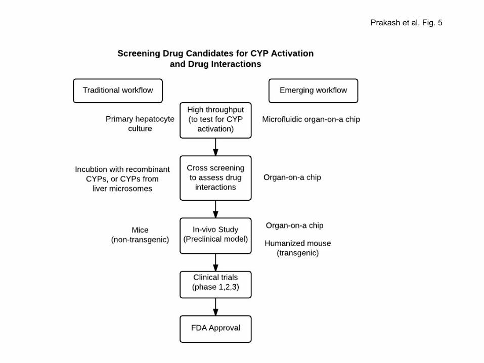
Prakash et al, Fig. 5

Prakash et al, Fig. 6