28 effect of moisturizers on the structure of lipids in the … care... · · 2016-04-04effect of...
TRANSCRIPT

28 Effect of Moisturizers on theStructure of Lipids in the OuterStratum Corneum of Humans
Keith D. Ertel, Ronald R. Warner, and Ying. L. Boissy
CONTENTS
28.1 Introduction. . . . . . . . . . . . . . . . . . . . . . . . . . . . . . . . . . . . . . . . . . . . . . . . . . . . . . . . . . . . . . . . . . . . . . . . . . . . . . . . . . 35128.1.1 Inner Stratum Corneum Lipids . . . . . . . . . . . . . . . . . . . . . . . . . . . . . . . . . . . . . . . . . . . . . . . . . . . 35228.1.2 Outer Stratum Corneum Lipids . . . . . . . . . . . . . . . . . . . . . . . . . . . . . . . . . . . . . . . . . . . . . . . . . . . 352
28.2 Tape Strip Protocol . . . . . . . . . . . . . . . . . . . . . . . . . . . . . . . . . . . . . . . . . . . . . . . . . . . . . . . . . . . . . . . . . . . . . . . . . . 35328.3 Normal Lipid Structure of the Outer Stratum Corneum. . . . . . . . . . . . . . . . . . . . . . . . . . . . . . . . . . . 354
28.3.1 Young Skin . . . . . . . . . . . . . . . . . . . . . . . . . . . . . . . . . . . . . . . . . . . . . . . . . . . . . . . . . . . . . . . . . . . . . . . . 35428.3.2 Old Skin . . . . . . . . . . . . . . . . . . . . . . . . . . . . . . . . . . . . . . . . . . . . . . . . . . . . . . . . . . . . . . . . . . . . . . . . . . . 354
28.4 The Effect of Surfactant-Based Cleansers . . . . . . . . . . . . . . . . . . . . . . . . . . . . . . . . . . . . . . . . . . . . . . . . . 35528.5 The Effect of Moisturizers . . . . . . . . . . . . . . . . . . . . . . . . . . . . . . . . . . . . . . . . . . . . . . . . . . . . . . . . . . . . . . . . . . 356
28.5.1 Mineral Oil . . . . . . . . . . . . . . . . . . . . . . . . . . . . . . . . . . . . . . . . . . . . . . . . . . . . . . . . . . . . . . . . . . . . . . . . 35828.5.2 Petrolatum . . . . . . . . . . . . . . . . . . . . . . . . . . . . . . . . . . . . . . . . . . . . . . . . . . . . . . . . . . . . . . . . . . . . . . . . . 359
28.5.2.1 Neat Petrolatum . . . . . . . . . . . . . . . . . . . . . . . . . . . . . . . . . . . . . . . . . . . . . . . . . . . . . . . 35928.5.2.2 Formulated Petrolatum . . . . . . . . . . . . . . . . . . . . . . . . . . . . . . . . . . . . . . . . . . . . . . . 360
28.5.3 Sucrose Esters of Fatty Acids . . . . . . . . . . . . . . . . . . . . . . . . . . . . . . . . . . . . . . . . . . . . . . . . . . . . 36028.5.3.1 Neat SEFA . . . . . . . . . . . . . . . . . . . . . . . . . . . . . . . . . . . . . . . . . . . . . . . . . . . . . . . . . . . . 36028.5.3.2 Formulated SEFA . . . . . . . . . . . . . . . . . . . . . . . . . . . . . . . . . . . . . . . . . . . . . . . . . . . . . 360
28.5.4 Product Comparisons from Clinical Studies . . . . . . . . . . . . . . . . . . . . . . . . . . . . . . . . . . . . . 36128.5.4.1 Neat Petrolatum versus Neat SEFA versus Glycerin-Based
Moisturizing Lotion. . . . . . . . . . . . . . . . . . . . . . . . . . . . . . . . . . . . . . . . . . . . . . . . . . . 36128.5.4.2 Moisturizing Body Wash versus Synthetic Bar +
Glycerin-Based Moisturizing Lotion. . . . . . . . . . . . . . . . . . . . . . . . . . . . . . . . . 36628.6 Conclusions . . . . . . . . . . . . . . . . . . . . . . . . . . . . . . . . . . . . . . . . . . . . . . . . . . . . . . . . . . . . . . . . . . . . . . . . . . . . . . . . . 366References . . . . . . . . . . . . . . . . . . . . . . . . . . . . . . . . . . . . . . . . . . . . . . . . . . . . . . . . . . . . . . . . . . . . . . . . . . . . . . . . . . . . . . . . . . . 370
28.1 INTRODUCTION
Lipids in the stratum corneum (SC) account for only about 15% of its weight, yet they constitutethe primary barrier of the skin,1–5 forming a protective sheath that shields us from desiccation andenvironmental insults.6 These barrier lipids exist in the SC intercellular space as highly organizedlamellar bilayers that are readily visualized by the marriage of transmission electron microscopy(TEM) with RuO4 staining.7,8 The lamellar organization consists of a unique pattern of alternatingelectron-lucent and electron-dense lamellae forming repeating structures7–10 that are often referredto as Landmann units.10 This lamellar structure appears throughout most of the SC thickness,
351

352 Dry Skin and Moisturizers: Chemistry and Function
FIGURE 28.1 Normal structure of the lipid lamellae in the intercellular space. Shown are three Landmann unitsseparating the darkly staining corneocytes above and below this intercellular space. Bar = 25 nm.
with variability occurring primarily in the number of Landmann units that bridge the intercellu-lar space.8,10–12 However, more diverse structures have been described in the outer SC,11–14 perhapsreflecting environmental impact12,14 or inherent differences in lipid composition.15–18
This chapter focuses on the lipid structure found in the outermost layers of the SC in humans.We present a modified TEM technique to investigate this structure, attempt to systematize andunderstand the variability in lipid structure observed in the outer SC, and explore the effect ofmoisturizers on the outer SC at microscopic and macroscopic levels.
28.1.1 INNER STRATUM CORNEUM LIPIDS
The Landmann-unit structure of intercellular lipid lamellae is illustrated in Figure 28.1. This structureis found throughout nearly all of the normal SC. Swartzendruber et al. proposed a plausible molecularmodel that accounts for the electron-lucent and electron-dense lamellar structure of the Landmannunit.10 These Landmann units are dynamic in nature. At least in the inner and middle SC they arealtered by age,19 disease,8,11,20–22 and hormonal status23,24; by experimental solvent treatment25–27
and topical inhibitor treatment.28,29 They are known to reform spontaneously following solventextraction,5,26 and topical application of certain lipids is also reported to effect lamellar repair andbarrier improvement.29–33
28.1.2 OUTER STRATUM CORNEUM LIPIDS
In contrast to the more extensive studies of the intercellular lipids of the inner and middle SC, thereare few studies of the lipid structure in the outer SC. Evidence suggests that the intercellular lipidcomposition in the uppermost layers of the SC differs from that found in the lower layers.34 Theouter SC lipids also exhibit structural variability compared to the inner and middle stratum corneum,both with regard to lipid ordering and lateral packing16,35 and the number of intercellular lamellae,which increases from the usual two or three to in excess of 100 bilayers.13 For normal skin withlittle or no visible dryness the outer SC intercellular space is filled with an amorphous lipid material,whereas in soap-treated skin with pronounced visible dryness this space is filled with numerousdisorganized lamellae.14 A separate in vitro study using human skin substrate also showed disorderedlipid lamellae in the outer SC following soap treatment, less lipid disruption following treatment witha soap/glycerin/oil bar, and normal lamellae following treatment with an isethionate-based bar.12
To the extent that lipids are involved in corneocyte cohesion,36–39 the lipid structure in the outerSC is presumably very important for proper desquamation. However, because the outer SC interfaceswith the surrounding environment, its lipids are the most susceptible to structural alterations causedby environmental insult or consumer products that often contain surfactants or solvents.5,12,30 While
Dow
nloa
ded
by [
Uni
vers
ity o
f C
inci
nnat
i] a
t 12:
09 0
3 M
arch
201
6
Effect of Moisturizers on the Structure of Lipids in SC of Humans 353
the quantity of SC lipid is apparently not a primary determinant of dryness in normal skin,40 theremay be a functional relationship between the lipid structure of the outer SC and skin dryness.
If consumer products containing soaps or solvents can damage the outer SC lipid structure, thenproducts like moisturizers might also have an impact on this structure. For example, glycerin isreported to increase water binding in the SC and act as a corneodesmolytic,41 inhibit humidity-induced SC lipid crystalline phase transitions,42 and speed barrier recovery.43 Maleated soybeanoil inhibits crystalline phase transitions and reduces water loss in model SC lipid systems.42 Andpetrolatum, which is often viewed as a gold standard for moisturization, can permeate the upperlayers of the SC, affect SC lipid structure, and accelerate barrier repair.29,44 Conversely, there isevidence that single components of physiologic lipid mixtures and some moisturizers interfere withrecovery following experimental barrier disruption.31,45,46
Studies employing mixtures of physiological lipids provide important insights into how topicalapplication of these products can impact SC lipids. However, moisturizers sold in the mass market areoften quite different from these specialty formulations, being based on more common moisturizingingredients. Although commercial moisturizers typically improve skin condition, relatively little isknown about how they effect this improvement. These products appear to provide a continuum ofeffects ranging from the purely cosmetic, such as temporarily camouflaging visible dry flakes, to morefunctional effects such as abetting biological repair processes.47 As noted previously, one mechanismby which the latter might occur is by aiding the digestion of desmosomes that are abnormally retainedin the outer SC, thereby enhancing the desquamation process.41,48 Another mechanism, however,might involve the SC lipids. Moisturizers often contain lipophilic materials, and lipids play a veryimportant role in skin barrier properties,49,50 so it is reasonable to assume that moisturizers in someway interact with the SC lipids to improve the skin barrier and thereby enhance SC hydration bya mechanism other than simple occlusion.44,45,49–51
This chapter investigates alterations in the lipid structure of the outer SC that are induced bymoisturizing ingredients and commercial moisturizing products. As a preface to this investigation,we also examine the normal variability in the lipid structure of the outer SC and how it is affectedby factors such as age, level of visible dryness, and personal cleanser use.
28.2 TAPE STRIP PROTOCOL
The outer SC was sampled by tape stripping (Scotch Magic Tape 810, 3M) using a modificationof a previously reported procedure.14 The tape was applied to the lateral leg surface using gentlepressure and carefully removed after approximately 30 sec. Under stereomicroscope observation,regions of the tape having large clusters of skin flakes were cut out and placed in 0.25% RuO4in a 0.1 M cacodylate buffer for 1 h at 4◦C, rinsed briefly in 0.1 M cacodylate buffer, and thendehydrated through a graded acetone series prior to Epon embedding and overnight polymerizationat 65◦C. Thin sections were cut on an ultramicrotome, counterstained with uranyl acetate and leadcitrate, and analyzed in a Philips CM12 at 100 keV. The lipid structure of the SC improves asa function of depth into the SC; by the third tape strip, lipid structure has normalized to the typicalLandmann pattern.14 To focus on the superficial SC only one tape strip was taken, and wheneverpossible micrographs were obtained only from the outermost 3 to 4 corneocytes, adjacent to the tape.Similarly, to minimize possible artifacts resulting from the mechanical process of tape stripping orfrom previously uplifted scale, whenever possible micrographs were taken from closely apposedintercellular regions, thus minimizing potential problems of physical trauma or interference fromthe tape adhesive or applied materials. Since the assessments of lipid structure were qualitative andsubjective, tape strip samples were blinded until the analysis completed.
Although this tape-stripping approach is a useful procedure, it does have limitations. For example,there are limitations inherent to RuO4 staining due to its poor penetration and high reactivity,as discussed previously.22 These staining limitations are superimposed on the problems of
Dow
nloa
ded
by [
Uni
vers
ity o
f C
inci
nnat
i] a
t 12:
09 0
3 M
arch
201
6

354 Dry Skin and Moisturizers: Chemistry and Function
representative sampling associated with the tape stripping procedure. Only limited areas withina tape strip meet the analysis criteria for TEM inspection, and lipid structure varies even within asingle tape strip. Nevertheless, this normal variation in lipid structure is relatively small compared tothe large structural changes that are encountered in the outer SC, as will be seen. In our experience,the outer SC lipid structure of an individual’s skin is relatively constant over large areas, so that theirouter SC lipid structure is quite consistent over an entire leg and similar between legs. The variationthat does exist, however, limits the ability to detect small changes in lipid structure. In particular,it is difficult to detect improvements in lipid structure due to the use of moisturizing products whenthe skin is already in good condition.
Another important limitation is the labor-intensive nature of TEM investigations; the number ofsamples that can be analyzed in a reasonable time period is small. In the background studies presentedhere, a minimum of three SC samples were analyzed for each treatment except for mineral oil, wherea single sample was analyzed.
28.3 NORMAL LIPID STRUCTURE OF THE OUTERSTRATUM CORNEUM
The objective of this study was to observe the lipid structure of the outer SC in a population ofpeople engaged in their usual personal care practices. Accordingly, in this study of normal lipidstructure healthy female participants were selected at random without advance knowledge of theirusual body skin care practices and without any preconditioning or product use restrictions. The agesof the selected individuals ranged from 22 to 52. Leg dryness was evaluated by an expert grader priorto tape strip sampling.52
28.3.1 YOUNG SKIN
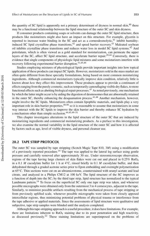
The lipids of young skin (individuals in their early twenties) with little or no visible dryness typicallyhave a good Landmann unit structure even at the surface of the SC, as shown in Figure 28.2(a).Youthful skin in good condition is invariably associated with closely apposed corneocytes, narrowintercellular spaces, and distinct bilayer structures. In contrast, young individuals with dry skin do nothave Landmann units in their outer SC. A variety of intercellular lipid morphologies is observed indifferent individuals with poor skin grade including fibrous, mesh, and amorphous structures. Usuallythe intercellular spaces are considerably widened. An example of the latter is shown in Figure 28.2(b),in which the intercellular spaces are filled with an amorphous material having a variety of textures.
28.3.2 OLD SKIN
Focal domains that are depleted or devoid of lipid bilayers are reported in aged (>80 years) skin.19
The oldest subject who participated in the present work was considerably younger than this, butwe typically did not observe intercellular lipids with a Landmann unit structure in the outer SC inindividuals over 40 years of age, regardless of skin condition. It thus appears that loss of SC lipidstructure begins much earlier in life than was previously reported, and on this basis we define “oldskin” to be skin from a person greater than age 40. An example of lipid structure from an “old” personwith good skin condition is shown in Figure 28.3(a). It is common to find lamellae, but these lamellaeare seldom present as fully formed Landmann units. Often lamellae are present at the periphery ofcorneocytes separated by a central band of nonlamellar amorphous/fibrous material as shown inFigure 28.3(a). Other intercellular spaces are simply filled with nonlamellar material (not shown).As with more youthful skin, the corneocytes are nevertheless typically closely apposed. In olderindividuals with dry skin the intercellular spaces can become spectacularly abnormal. Very widenedintercellular spaces are common, usually filled with amorphous material that can contain a great
Dow
nloa
ded
by [
Uni
vers
ity o
f C
inci
nnat
i] a
t 12:
09 0
3 M
arch
201
6
Effect of Moisturizers on the Structure of Lipids in SC of Humans 355
FIGURE 28.2 (a) Landmann units from the outer SC of a person 24-years old, skin grade 0.5. (b) A mixtureof amorphous materials with different textures in the intercellular space of the outer SC from a person 28 yearsold, skin grade 5.0. Bar = 100 nm.
diversity of lipid structures. An example is shown in Figure 28.3(b); the outermost intercellularspace appears to consist of a two-phase system, the noncontinuous phase being membrane-bound.Vesicles are apparent. The intercellular spaces are generally widened, many apparently filled withan amorphous material. There is no organized lamellar structure.
28.4 THE EFFECT OF SURFACTANT-BASED CLEANSERS
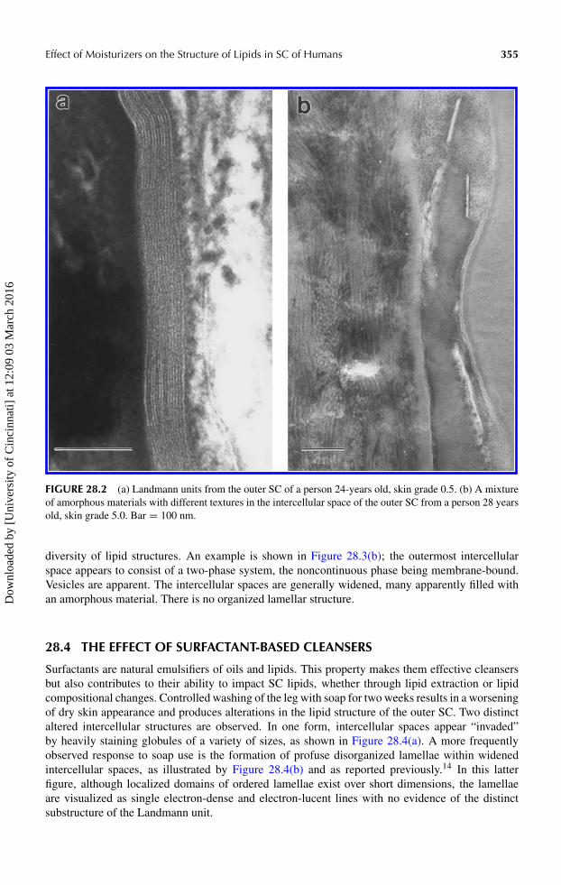
Surfactants are natural emulsifiers of oils and lipids. This property makes them effective cleansersbut also contributes to their ability to impact SC lipids, whether through lipid extraction or lipidcompositional changes. Controlled washing of the leg with soap for two weeks results in a worseningof dry skin appearance and produces alterations in the lipid structure of the outer SC. Two distinctaltered intercellular structures are observed. In one form, intercellular spaces appear “invaded”by heavily staining globules of a variety of sizes, as shown in Figure 28.4(a). A more frequentlyobserved response to soap use is the formation of profuse disorganized lamellae within widenedintercellular spaces, as illustrated by Figure 28.4(b) and as reported previously.14 In this latterfigure, although localized domains of ordered lamellae exist over short dimensions, the lamellaeare visualized as single electron-dense and electron-lucent lines with no evidence of the distinctsubstructure of the Landmann unit.
Dow
nloa
ded
by [
Uni
vers
ity o
f C
inci
nnat
i] a
t 12:
09 0
3 M
arch
201
6

356 Dry Skin and Moisturizers: Chemistry and Function
FIGURE 28.3 (a) Lipid structure in the intercellular space from a person 45-years old, skin grade 1.0. Thecorneocytes are closely apposed and lamellae are frequent, but these lamellae appear somewhat disorganizedand do not form Landmann units. The core of the intercellular space is filled with nonlamellar material thatis amorphous and fibrous with interspersed granular deposits. (b) Lipid structure from a person 49 years old,skin grade 3.5. The outermost intercellular space contains vesicular structures and membrane-bounded phases.Inner intercellular spaces appear to contain largely amorphous material. Bar = 100 nm.
Synthetic, that is, nonsoap surfactants often exhibit better skin compatibility than soap and arefound in a range of personal care products. Cleansers based on these surfactants generally produceless visible irritation and dryness than soap; however, they can still remove significant quantitiesof lipid from the skin during washing.53 Effects on SC lipid bilayer structure consistent with thosewe observed following soap washing were recently reported following controlled washing withcleansers based on “mild” synthetic surfactant systems.54 Thus, it appears that surfactant-inducedchanges in the lipid structure of the outer SC are possible with a wide range of cleanser types,not just with soap.
28.5 THE EFFECT OF MOISTURIZERS
The results presented thus far show that the lipid structure of the outer SC varies with age anddry skin condition, and that cleansing products can degrade this lipid structure. We now returnto questions raised earlier: do moisturizing ingredients enter the SC, and if so, can they alter theouter SC lipid structure? To address these questions we investigated the effect of neat moistur-izing ingredients, reduced-concentration (i.e., “formulated”) moisturizing ingredients, and fullyformulated commercial products on the lipid structure of the outer SC of the leg following two orthree weeks of product use.
Dow
nloa
ded
by [
Uni
vers
ity o
f C
inci
nnat
i] a
t 12:
09 0
3 M
arch
201
6

Effect of Moisturizers on the Structure of Lipids in SC of Humans 357
FIGURE 28.4 (a) Soap treatment frequently results in the formation of many darkly staining globular bodiesin an amorphous matrix. (b) The signature pattern of soap use is the presence of widened intercellular spacesthat are filled with numerous disorganized lamellae without a Landmann pattern. Bar = 100 nm.
Dow
nloa
ded
by [
Uni
vers
ity o
f C
inci
nnat
i] a
t 12:
09 0
3 M
arch
201
6

358 Dry Skin and Moisturizers: Chemistry and Function
For maximum comparative value the focus of this discussion is on results generated in matchedstudies conducted on a 42-year old male, though similar results were obtained from other subjects.Mineral oil, petrolatum formulated at 10% in an oil-in-water emulsion vehicle containing highlevels of humectants, and sucrose esters of fatty acids (SEFA, The Procter & Gamble Company,Cincinnati, OH) formulated at 2 and 10% in the same vehicle, were applied at 3 mg/cm2 on the lowerleg twice a day for two weeks. Neat petrolatum and neat SEFA were applied ad lib twice a day fortwo weeks. In all cases the final product application was 12 h before tape stripping. All subjects useda syndet-based bar for daily personal cleansing, avoiding direct application of the bar or its lather tothe treatment areas.
28.5.1 MINERAL OIL
The control, nontreated site is shown in Figure 28.5(a), and the mineral oil-treated site inFigure 28.5(b) (same magnification). In the control skin, the outermost layers contain amorphousmaterial and darkly staining globules. Lamellar structures are found in lower layers but the lamellaedo not appear to form Landmann units. Following use of mineral oil the intercellular space is uni-formly filled with a smooth-appearing amorphous material, presumably the mineral oil. Intercellularspaces were occasionally focally dilated. There seemed to be little effect of the mineral oil other thanas a “spacer” separating corneocytes.
FIGURE 28.5 (a) Control, nontreated site from a 42-year-old male. The outermost (right) layers contain darklystaining globules in an amorphous matrix. Lamellae are present in deeper corneocyte layers, but Landmannunits are rare. (b) Treatment with mineral oil results in the formation of large amorphous phases containingsome darkly staining material. Bar = 200 nm.
Dow
nloa
ded
by [
Uni
vers
ity o
f C
inci
nnat
i] a
t 12:
09 0
3 M
arch
201
6

Effect of Moisturizers on the Structure of Lipids in SC of Humans 359
28.5.2 PETROLATUM
28.5.2.1 Neat Petrolatum
Petrolatum comprises a complex hydrocarbon mixture that is about 60 to 70% mineral oil, theremainder consisting primarily of paraffin and microcrystalline wax. Despite this composition,the effect of petrolatum on outer SC lipids is distinct from that of mineral oil. As shown inFigure 28.6(a), neat petrolatum forms lamellar-like “streamers” in the intercellular space, as seenpreviously.44 The streamers appear to be suspended in a nonstaining or empty intercellular medium,
FIGURE 28.6 (a) Neat petrolatum site from a 42-year-old male. Flocculent/fibrous material existing as“streamers” or bands is present within an otherwise empty-appearing intercellular space. (b) Formulated (10%)petrolatum site. Lamellae, occasionally forming Landmann units, are sometimes separated by a thin layer ofmore darkly staining amorphous material. Bar = 200 nm.
Dow
nloa
ded
by [
Uni
vers
ity o
f C
inci
nnat
i] a
t 12:
09 0
3 M
arch
201
6

360 Dry Skin and Moisturizers: Chemistry and Function
or possibly water. In other areas, petrolatum forms a more continuous amorphous phase, also reportedpreviously.45 Intercellular structures intermediate between these two appearances are also formed(data not shown).
In other studies, similar streamer and amorphous structures were observed in a young female withdry skin following the above treatment protocol, although the amorphous phase was less prominent.In contrast, the streamer phase was less obvious in older individuals treated with 2 mg/cm2 twicea day for three weeks.
28.5.2.2 Formulated Petrolatum
The “streamer” phase observed with neat petrolatum (Figure 28.6[a]) was not observed, but amor-phous material was common (data not shown). Reasonable lamellae were occasionally encountered,as shown in Figure 28.6(b). Often these lamellae were separated by thin expanses of amorphousmaterial, as shown in the center of Figure 28.6(b). In general, treatment with “formulated” petro-latum resulted in an appearance of the intercellular lipids that was much improved over that of neatpetrolatum or mineral oil. The corneocytes were more closely apposed, and Landmann units weremore common.
28.5.3 SUCROSE ESTERS OF FATTY ACIDS
28.5.3.1 Neat SEFA
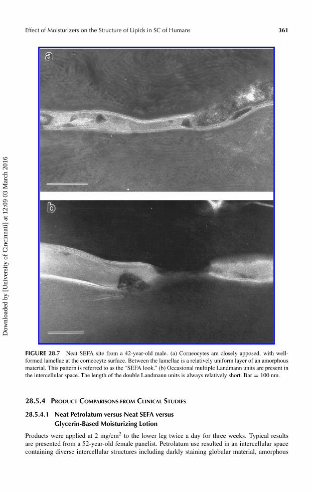
Treatment with SEFA resulted in a very characteristic appearance of the intercellular space, shownin Figure 28.7(a), which we describe as the “SEFA look.” The corneocytes are relatively closelyapposed, single Landmann units are present at corneocyte margins, and the slightly expanded inter-vening space is “plugged” with an amorphous material, presumably SEFA. Unlike the other productsabove, multiple Landmann units are occasionally present, although the multiple units are usuallypresent in short regions within the SEFA “plug,” as shown in Figure 28.7(b). Very similar resultswere obtained in a young female with dry skin.
28.5.3.2 Formulated SEFA
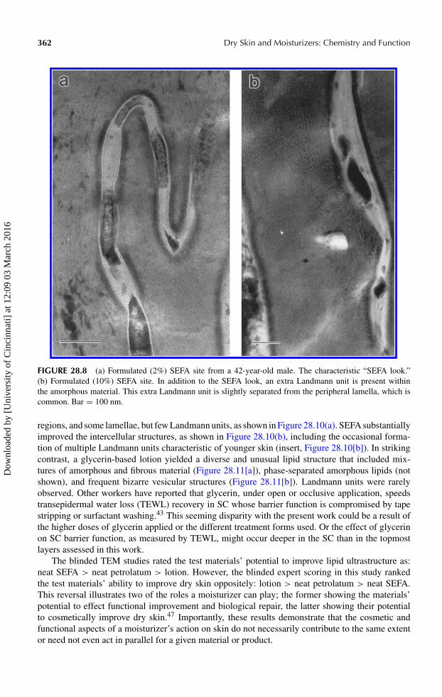
The structure of the lipids in the intercellular space is overwhelmingly the “SEFA look” for both the2 and 10% concentrations, as shown in Figure 28.8(a). With the 10% concentration, extra Landmannunits within the SEFA phase were occasionally seen, as shown in Figure 28.8(b).
In a separate study, 2 mg/cm2 of 2 or 10% SEFA in a humectant vehicle were applied to the lowerleg twice a day for three weeks. The control nontreated site of a 52-year-old female subject, shownin Figure 28.9(a), is characterized by numerous disorganized lamellae characteristic of soap use.Numerous darkly staining globular deposits were also common (data not shown). The humectantvehicle alone resulted in substantial improvement in the outer SC lipid structure of this subject, butLandmann units were not common and many intercellular spaces contained indistinct or amorphousmaterial (data not shown). Notably, the vehicle did not produce the “SEFA look.” Following treatmentwith the 2% SEFA preparation, the “SEFA look” was commonly observed (Figure 28.9[b]), but sotoo were Landmann units, which is an unusual finding for a person of this age. Following treatmentwith the 10% SEFA preparation, the “SEFA look” was less common, and Landmann units morecommon (Figure 28.9[c]).
To conclude this section, previous reports on the beneficial effects of topically applied mois-turizing preparations have often focused on optimizing their physiological lipid composition. Theresults found for the “formulated” SEFA and petrolatum in this work, when viewed relative to theneat materials, suggest that proper formulation of even nonphysiological moisturizing agents willenhance the beneficial effect these materials have on outer SC lipid structure.
Dow
nloa
ded
by [
Uni
vers
ity o
f C
inci
nnat
i] a
t 12:
09 0
3 M
arch
201
6
Effect of Moisturizers on the Structure of Lipids in SC of Humans 361
FIGURE 28.7 Neat SEFA site from a 42-year-old male. (a) Corneocytes are closely apposed, with well-formed lamellae at the corneocyte surface. Between the lamellae is a relatively uniform layer of an amorphousmaterial. This pattern is referred to as the “SEFA look.” (b) Occasional multiple Landmann units are present inthe intercellular space. The length of the double Landmann units is always relatively short. Bar = 100 nm.
28.5.4 PRODUCT COMPARISONS FROM CLINICAL STUDIES
28.5.4.1 Neat Petrolatum versus Neat SEFA versusGlycerin-Based Moisturizing Lotion
Products were applied at 2 mg/cm2 to the lower leg twice a day for three weeks. Typical resultsare presented from a 52-year-old female panelist. Petrolatum use resulted in an intercellular spacecontaining diverse intercellular structures including darkly staining globular material, amorphous
Dow
nloa
ded
by [
Uni
vers
ity o
f C
inci
nnat
i] a
t 12:
09 0
3 M
arch
201
6
362 Dry Skin and Moisturizers: Chemistry and Function
FIGURE 28.8 (a) Formulated (2%) SEFA site from a 42-year-old male. The characteristic “SEFA look.”(b) Formulated (10%) SEFA site. In addition to the SEFA look, an extra Landmann unit is present withinthe amorphous material. This extra Landmann unit is slightly separated from the peripheral lamella, which iscommon. Bar = 100 nm.
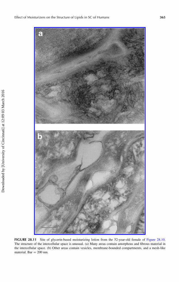
regions, and some lamellae, but few Landmann units, as shown in Figure 28.10(a). SEFA substantiallyimproved the intercellular structures, as shown in Figure 28.10(b), including the occasional forma-tion of multiple Landmann units characteristic of younger skin (insert, Figure 28.10[b]). In strikingcontrast, a glycerin-based lotion yielded a diverse and unusual lipid structure that included mix-tures of amorphous and fibrous material (Figure 28.11[a]), phase-separated amorphous lipids (notshown), and frequent bizarre vesicular structures (Figure 28.11[b]). Landmann units were rarelyobserved. Other workers have reported that glycerin, under open or occlusive application, speedstransepidermal water loss (TEWL) recovery in SC whose barrier function is compromised by tapestripping or surfactant washing.43 This seeming disparity with the present work could be a result ofthe higher doses of glycerin applied or the different treatment forms used. Or the effect of glycerinon SC barrier function, as measured by TEWL, might occur deeper in the SC than in the topmostlayers assessed in this work.
The blinded TEM studies rated the test materials’ potential to improve lipid ultrastructure as:neat SEFA > neat petrolatum > lotion. However, the blinded expert scoring in this study rankedthe test materials’ ability to improve dry skin oppositely: lotion > neat petrolatum > neat SEFA.This reversal illustrates two of the roles a moisturizer can play; the former showing the materials’potential to effect functional improvement and biological repair, the latter showing their potentialto cosmetically improve dry skin.47 Importantly, these results demonstrate that the cosmetic andfunctional aspects of a moisturizer’s action on skin do not necessarily contribute to the same extentor need not even act in parallel for a given material or product.
Dow
nloa
ded
by [
Uni
vers
ity o
f C
inci
nnat
i] a
t 12:
09 0
3 M
arch
201
6

Effect of Moisturizers on the Structure of Lipids in SC of Humans 363
FIGURE 28.9 (a) Control nontreated site from a 52-year-old female. The characteristic lipid structure resultingfrom soap use (winter xerosis14) is evident — compare with Figure 28.4(b). (b) Use of formulated (2%) SEFAresults in the SEFA look, as well as Landmann units. (c) With use of formulated (10%) SEFA, Landmann unitsare commonly observed. Bar = 100 nm.
Dow
nloa
ded
by [
Uni
vers
ity o
f C
inci
nnat
i] a
t 12:
09 0
3 M
arch
201
6

364 Dry Skin and Moisturizers: Chemistry and Function
FIGURE 28.10 Neat-petrolatum-treated site from a (different) 52-year-old female. (a) A great variety ofintercellular structures are present, but the “streamer” phase typical of petrolatum (Figure 28.6[a]) was notseen. Amorphous regions and expanded intercellular regions containing many darkly staining globular regionsare very common, as are lamellae without a Landmann pattern. Landmann units were rare. (b) Neat-SEFA-treated site. The SEFA look is evident. The dark spindle-shaped structures near the center of the micrograph arepresumably desmosomes undergoing degradation. In many areas with the SEFA look, multiple, short-lengthLandmann units are common in the intercellular space, as shown. Normal well-formed Landmann units arerelatively common, as shown in the insert. Bar = 100 nm.
Dow
nloa
ded
by [
Uni
vers
ity o
f C
inci
nnat
i] a
t 12:
09 0
3 M
arch
201
6
Effect of Moisturizers on the Structure of Lipids in SC of Humans 365
FIGURE 28.11 Site of glycerin-based moisturizing lotion from the 52-year-old female of Figure 28.10.The structure of the intercellular space is unusual. (a) Many areas contain amorphous and fibrous material inthe intercellular space. (b) Other areas contain vesicles, membrane-bounded compartments, and a mesh-likematerial. Bar = 200 nm.
Dow
nloa
ded
by [
Uni
vers
ity o
f C
inci
nnat
i] a
t 12:
09 0
3 M
arch
201
6
366 Dry Skin and Moisturizers: Chemistry and Function
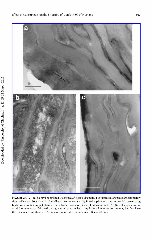
28.5.4.2 Moisturizing Body Wash versus Synthetic Bar +Glycerin-Based Moisturizing Lotion
In a clinical study, a body wash treatment at 10 µl/cm2 (rinse-off application) and a glycerin-basedlotion treatment at 1 µl/cm2 (leave-on application) were applied to the medial aspect of the legsof female panelists once daily for 25 days. Good repair of the lipids in the intercellular space wasroutinely obtained with the body wash, which contains 17.5% petrolatum as a skin benefit agent, asillustrated by a particularly dramatic improvement shown in Figure 28.12. The control (water only)treatment site of a 28-year-old panelist, shown in Figure 28.12(a), is characterized by intercellularspaces filled with amorphous material. The effect of the body wash on this subject’s outer SClipids is shown in Figure 28.12(b). The majority of the intercellular space is filled with Landmannunits, although amorphous material was occasionally found in some regions. The effect of a syndetbar followed by application of a glycerin-based moisturizing lotion is shown in Figure 28.12(c).This bar/lotion regimen resulted in a clear improvement relative to the control but many intercellularregions are still dilated with amorphous material. Although lamellae are present, Landmann units arerelatively rare.
A more typical response produced by these treatments is shown in Figure 28.13, which is froma separate clinical study that used the same treatments applied for only 14 days. The 48-year-old panelist had a moderate amount of skin dryness and the SC at the control site exhibited anintercellular lipid structure similar to that of Figure 28.3(a), that is, a good lipid structure for thatage. In this case the body wash resulted in no dramatic change in intercellular lipid structure, shownin Figure 28.13(a), although there was significant improvement in the visual skin grade. The limitedimprovement in outer SC lipid structure might reflect the shorter treatment period, the decreaseddosing compared to the moisturizer study, or possibly the impact of age. Because the aged stratumcorneum barrier exhibits a reduced resistance to insult and slower repair than in young skin due todiminished lamellar body secretion,19 and because the SC turnover rate also typically slows withage, the functional benefits of topical moisturizers might require more time to manifest in “old” skinthan in younger skin. Regardless, these results show that while lipid structure was not improved,treatment with the body wash preserved existing lipid structure. In contrast, use of the syndet barfollowed by the glycerin-based lotion degraded lipid ultrastructure in the outer SC, as shown inFigure 28.13(b). The intercellular spaces contain amorphous and “fuzzy” material, and prominentdisorganized, undulating lamellae. Nevertheless, the visual skin grade was dramatically improvedwith this latter regimen, again illustrating the distinction between a moisturizers’ cosmetic andfunctional effects.47
28.6 CONCLUSIONS
An improved understanding of the structure of the SC barrier is of interest for many reasons suchas enhancing percutaneous penetration and, as discussed in this chapter, optimizing topical therapyfor the treatment of dry or damaged skin. The results of this TEM work show that the lipid structureof the outer SC is quite variable. Typically, the intercellular spaces in the outer SC are considerablywidened and filled with nonlamellar material. These data are consistent with earlier TEM studies13,14
and with an infrared spectroscopic study that found less structured lipids in the outer SC16 comparedto the middle and inner regions.
Contrary to an earlier report that lipids uniformly have an amorphous structure in the outer SCof normal skin with little or no visible dryness,14 we instead found considerable variation in thislipid structure among individuals. Intercellular lipids in young skin with little dryness typically hada good Landmann unit structure, even at the surface of the SC. This ideal Landmann unit structurewas generally absent in young individuals with dry skin or in individuals over the age of 40 regardlessof their dry skin level. In attempting to make sense of this variation, we believe we can generalizeand conclude that the outer SC lipid structure is related to an individual’s age and dry skin condition.
Dow
nloa
ded
by [
Uni
vers
ity o
f C
inci
nnat
i] a
t 12:
09 0
3 M
arch
201
6
Effect of Moisturizers on the Structure of Lipids in SC of Humans 367
FIGURE 28.12 (a) Control nontreated site from a 28-year-old female. The intercellular spaces are completelyfilled with amorphous material. Lamellar structures are rare. (b) Site of application of a commercial moisturizingbody wash containing petrolatum. Lamellae are common, as are Landmann units. (c) Site of application ofa mild synthetic bar followed by a glycerin-based moisturizing lotion. Lamellae are present, but few havethe Landmann unit structure. Amorphous material is still common. Bar = 200 nm.
Dow
nloa
ded
by [
Uni
vers
ity o
f C
inci
nnat
i] a
t 12:
09 0
3 M
arch
201
6

368 Dry Skin and Moisturizers: Chemistry and Function
FIGURE 28.13 Tape strips from a 48-year-old female. (a) Site of application of a commercial moisturizingbody wash containing petrolatum. The appearance is typical of a person over age 40 with good skin condition.The corneocytes are closely apposed and lamellae are frequent, but the lamellae appear somewhat disorganizedand amorphous/fibrous material is present between lamellae. Landmann units are nevertheless easy to find,as shown in the left intercellular space. (b) Site of application of a mild synthetic bar followed by a glycerin-based moisturizing lotion. The intercellular spaces contain amorphous and fibrous material as well as prominentwavy lamellae without a Landmann pattern. Bar = 100 nm.
Dow
nloa
ded
by [
Uni
vers
ity o
f C
inci
nnat
i] a
t 12:
09 0
3 M
arch
201
6

Effect of Moisturizers on the Structure of Lipids in SC of Humans 369
The outer SC lipid structure of older individuals is altered compared to that found in young skin,the older SC having fewer lipid lamellae and more open intercellular clefts.19 This paucity of lamellaemay be due to a decrease in SC lipid synthesis or lamellar body secretion, resulting in a decreased SClipid content.19,51 These results are from observations made on individuals of advanced age, 80 yearsold. However, we observed changes in the outer SC lipid structure of individuals as young as 40 yearsold. This is not entirely unexpected since literature data support age-associated changes in SC lipids inrelatively young individuals. For example, there is a sharp decrease in SC lipid content by age 45and a nearly constant SC lipid profile afterwards.55 Total SC ceramides undergo a sharp drop inconcentration around age 40,40 and levels of individual ceramide species also change with age andfemale hormonal status.23,56 A challenge for skin moisturizers is to arrest, and ideally reverse, theage-related decline in the SC lipid barrier that accompanies these changes.
The blinded assessment of changes in SC lipid structure generally corresponded well with theindependent, expert assessments of dry skin appearance. However, a number of striking outliersshow that lipid structure is not the major determinant of dry skin appearance, which is not surprisinggiven the complexity of SC homeostasis. A good example was the neat petrolatum/SEFA/lotioncomparison mentioned earlier, in which neat SEFA improved lipid structure but produced onlya marginal reduction in dry skin appearance, whereas the glycerin-based lotion apparently degradedlipid structure but yielded skin with minimal visible flaking. This suggests that another mechanism,such as desmosomal breakdown, is a more important determinant of the skin’s dry appearance.However, a healthy, nondry SC may ultimately rely more on the integrity of the lipid barrier than onthe state of desmosome degradation in the outer SC layers. Such effects may appear over a longer timeframe, for example, during the regression period that is used in some clinical protocols, or requirethe evaluation of endpoints other than dry appearance.
The discrepancy between visual appearance and lipid structure may be a consequence of com-mercial products being formulated to achieve visual improvement rather than functional change,that is, improved skin health. This is not unexpected since dry skin is readily observed by consumersand is an important signal of the need to apply moisturizer.47 However, as this work has shown,a reduction in dry appearance does not necessarily mean that there is an improvement in the func-tional characteristics of the skin, only that it looks better. Thus, relying on a visual endpoint forevaluating moisturizer efficacy can yield commercially successful products that provide a marginalskin health benefit. An example are reports that some commercial moisturizing lotions may actuallyimpede barrier recovery after experimental barrier perturbation.45,57 A moisturizer should ideallyaddress not only visual skin problems but also address the underlying biological causes to achievehealthy skin; there is a clear need for evaluation tools and endpoints for skin health beyond visualinspection.
We observed distinctive changes in the outer SC lipid structure with use of different treatmentsfrom soap to oil. Some of these lipid structures were sufficiently unique to provide unequivocalidentification of product treatment, and to a lesser degree panelist age and skin condition. Basedon TEM results, we believe that moisturizing materials enter the intercellular space of the SC andbecome a part of the SC, as was previously shown for petrolatum.29,44 The mechanism by whicha nonphysiological moisturizing material improves skin barrier lipids is uncertain, and multipleprocesses are likely involved. Given the chemical nature of the materials studied, we consider itunlikely that any of the treatments participated directly or physically in the formation of Landmannunits. Of some note is the observation that petrolatum and SEFA were not as effective in reformingLandmann units when applied neat as when they were applied as reduced concentration or fullyformulated products. The moisturizing body wash only contains petrolatum and polymers as mois-turizing ingredients, which suggests that the quantity of petrolatum or its delivery form is importantfor promoting the conditions necessary for SC lipid repair. Likewise, for SEFA applied as a for-mulated product, the combination of humectants with some degree of occlusion may promote theinternal conditions needed for the intrinsic formation of Landmann units. A semi-occluded environ-ment is reported to accelerate TEWL barrier recovery following experimental insult58,59; this type
Dow
nloa
ded
by [
Uni
vers
ity o
f C
inci
nnat
i] a
t 12:
09 0
3 M
arch
201
6

370 Dry Skin and Moisturizers: Chemistry and Function
of environment might similarly favor lipid bilayer reformation in the outer SC. However, apparentlack of Landmann unit reformation following treatment with the glycerin-based moisturizer suggeststhat the choice of nonphysiologic ingredient or the manner in which it is formulated is also impor-tant to provide conditions that promote this reformation.60,61 Beyond ingredient and delivery issuesthere remains the issue of product aesthetics and convenience; a product will benefit skin only ifit is used. Of 651 dermatologist respondents in a recent survey, over 60% believed that less thanhalf their adult female patients apply lotion as recommended.62 Lack of convenience was cited asa factor contributing to this poor compliance by over 83% of the dermatologist respondents. Thedevelopment of nontraditional product forms to deliver moisturizing benefits, such as moisturizingcleansers54,63 and moisturizers intended for use in the shower,62 can provide increased convenienceand could improve moisturizer usage compliance.
The SC is a highly complex system and we do not claim to fully understand the lipid structureof the outer SC or its implications on the basis of this investigation. The conclusions reached aretherefore predicated on certain key observations and assumptions. We observed the ideal Landmannunit lipid structure in young individuals with little or no skin dryness, the absence of this structurein individuals with a high level of dryness, and the reappearance of Landmann units with treatmentby moisturizing products. We therefore assume that this Landmann unit structure is the ideal lipidstructure for the outer SC, as it is throughout its lower regions. In a system undergoing desquamationthat may involve lipids,36–39 this is an important assumption. We further assume that this idealLandmann unit structure in the outer SC is important to skin health and a parameter by whichmoisturizers’ potential to impact skin health should be judged. Both of these hypotheses warrantfurther testing.
In summary our microscopy study shows that topical moisturizers enter into the SC and canaffect lipid structure. The lipid structure is related to visible skin dryness but is not the primary factordetermining the level of dryness. For SEFA and petrolatum, formulated products showed a greaterrestorative effect on ideal Landmann unit lipid structure than did the neat materials. In our experiencemost of the moisturizing materials and products that we investigated to date are effective at reducingvisible dry skin, but far fewer materials are able to substantially reform Landmann units, particularlyin individuals over age 40. Is there hope that moisturizers might restore the ideal Landmann unitlipid structure common in the healthy skin of youth? With ongoing work looking at new moisturizingagents, new delivery systems, and alternative product forms, we believe the promise is there, as shownfor older individuals in Figure 28.9 and Figure 28.10.
REFERENCES
1. Berenson, G.S. and Burch, G.E., Studies of diffusion of water through dead human skin: the effect ofdifferent environmental states and of chemical alterations of the epidermis, Am. J. Trop. Med. Hyg., 31,842, 1995.
2. Onken, H.D. and Moyer, C.A., The water barrier in human epidermis, Arch. Dermatol., 87, 584,1963.
3. Elias, P.M., Lipids and the epidermal permeability barrier, Arch. Dermatol. Res., 270, 95, 1981.4. Elias, P.M., Cooper, E.R., Korc, A., and Brown, B.E., Percutaneous transport in relation to stratum
corneum structure and lipid composition, J. Invest. Dermatol., 76, 297, 1981.5. Grubauer, G., Feingold, K.R., Harris, R.M., and Elias, P.M., Lipid content and lipid type as determinants
of the epidermal permeability barrier, J. Lipid Res., 30, 89, 1989.6. Elias, P.M., Epidermal lipids, membranes, and keratinization, Int. J. Dermatol., 20, 1, 1981.7. Madison, K.C., Swartzendruber, D.C., Wertz, P.W., and Downing, D.T., Presence of intact intercellular
lipid lamellae in the upper layers of the stratum corneum, J. Invest. Dermatol., 88, 714, 1987.8. Hou, S.Y.E., Mitra, A.K., White, S.H., Menon, G.K., Ghadially, R., and Elias, P.M., Membrane
structures in normal and essential fatty acid-deficient stratum corneum: characterization by rutheniumtetroxide staining and x-ray diffraction, J. Invest. Dermatol., 96, 215, 1991.
Dow
nloa
ded
by [
Uni
vers
ity o
f C
inci
nnat
i] a
t 12:
09 0
3 M
arch
201
6

Effect of Moisturizers on the Structure of Lipids in SC of Humans 371
9. Landmann, L., Epidermal permeability barrier: transformation of lamellar granule disks into intercellularsheets by a membrane fusion process, J. Invest. Dermatol., 87, 202, 1986.
10. Swartzendruber, D.C., Wertz, P.W., Kitko, D.J., Madison, K.C., and Downing, D.T., Molecular modelsof the intercellular lipid lamellae in mammalian stratum corneum, J. Invest. Dermatol., 92, 251,1989.
11. Fartasch, M., Epidermal barrier in disorders of the skin, Microsc. Res. Tech., 38, 361, 1997.12. Misra, M., Ananthapadmanabhan, K.P., Hoyberg, K., Gursky, R.P., Prowell, S., and Aronson, M.,
Correlation between surfactant-induced ultrastructural changes in epidermis and transepidermal waterloss, J. Soc. Cosmet. Chem., 48, 219, 1997.
13. Fartasch, M., Bassukas, I.D., and Diepgen, T.L., Structural relationship between epidermal lipid lamel-lae, lamellar bodies and desmosomes in human epidermis: an ultrastructural study, Br. J. Dermatol.,128, 1, 1993.
14. Rawlings, A.V., Watkinson, A., Rogers, J., Mayo, H.J., and Scott, I.R., Abnormalities in stratumcorneum structure, lipid composition, and desmosome degradation in soap-induced winter xerosis,J. Soc. Cosmet. Chem., 45, 203, 1994.
15. Elias, P.M., Menon, G.K., Grayson, S., and Brown, B.E., Membrane structural alterations in murinestratum corneum: relationship to the localization of polar lipids and phospholipases, J. Invest. Dermatol.,91, 3, 1988.
16. Bommannan, D., Potts, R.O., and Guy, R.H., Examination of stratum corneum barrier function in vivoby infrared spectroscopy, J. Invest. Dermatol., 95, 403, 1990.
17. Bonté, F., Saunois, A., Pinguet, P., and Meybeck, A., Existence of a lipid gradient in the upper stratumcorneum and its possible biological significance, Arch. Dermatol. Res., 289, 78, 1997.
18. Long, S.A., Wertz, P.W., Strauss, J.S., and Downing, D.T., Human stratum corneum polar lipids anddesquamation, Arch. Dermatol. Res., 277, 284, 1985.
19. Ghadially, R., Brown, B.E., Sequeira-Martin, S.M., Feingold, K.R., and Elias, P.M., The aged epi-dermal permeability barrier. Structural, functional, and lipid biochemical abnormalities in humans anda senescent murine model, J. Clin. Invest., 95, 2281, 1995.
20. Ghadially, R., Williams, M.L., Hou, S.Y., and Elias, P.M., Membrane structural abnormalities in thestratum corneum of the autosomal recessive ichthyoses, J. Invest. Dermatol., 99, 755, 1992.
21. Ghadially, R., Reed, J.T., and Elias, P.M., Stratum corneum structure and function correlates withphenotype in psoriasis, J. Invest. Dermatol., 107, 558, 1996.
22. Menon, G. and Ghadially, R., Morphology of lipid alterations in the epidermis: a review, Microsc. Res.Tech., 37, 180, 1997.
23. Denda, M., Koyama, J., Hori, J., Horii, I., Takahashi, M., Hara, M., and Tagami, H., Age- andsex-dependent changes in stratum corneum sphingolipids, Arch. Dermatol. Res., 285, 415, 1993.
24. Misra, M., Feinberg, C., Matzke, M., and Pocalyko, D., Hormone replacement therapy (HRT) maintainsskin’s lipid barrier, in the Proceedings of 62nd annual meeting of the American Academy of Dermatology,February 6–11, 2004, Washington, DC.
25. Imokawa, G., Kuno, H., and Kawai, M., Stratum corneum lipids serve as a bound-water modulator,J. Invest. Dermatol., 96, 845, 1991.
26. Menon, G.K., Feingold, K.R., and Elias, P.M., Lamellar body secretory response to barrier disruption,J. Invest. Dermatol., 98, 279, 1992.
27. Fartasch, M., Ultrastructure of the epidermal barrier after irritation, Microsc. Res. Tech., 37, 193,1997.
28. Menon, G.K., Feingold, K.R., Mao-Qiang, M., Schaude, M., and Elias, P.M., Structural basis forthe barrier abnormality following inhibition of HMG CoA reductase in murine epidermis, J. Invest.Dermatol., 98, 209, 1992.
29. Mao-Qiang, M., Brown, B.E., Wu-Pong, S., Feingold, K.R., and Elias, P.M., Exogenous nonphysiologicvs physiologic lipids. Divergent mechanisms for correction of permeability barrier dysfunction, Arch.Dermatol., 131, 809, 1995.
30. Imokawa, G., Akasaki, S., Minematsu, Y., and Kawai, M., Importance of intercellular lipids in water-retention properties of the stratum corneum: induction and recovery study of surfactant dry skin, Arch.Dermatol. Res., 281, 45, 1989.
31. Man, M.Q., Feingold, K.R., and Elias, P.M., Exogenous lipids influence permeability barrier recoveryin acetone-treated murine skin, Arch. Dermatol., 129, 728, 1993.
Dow
nloa
ded
by [
Uni
vers
ity o
f C
inci
nnat
i] a
t 12:
09 0
3 M
arch
201
6

372 Dry Skin and Moisturizers: Chemistry and Function
32. Zettersten, E.M., Ghadially, R., Feingold, K.R., Crumrine, D., and Elias, P.M., Optimal ratios of topicalstratum corneum lipids improve barrier recovery in chronologically aged skin, J. Am. Acad. Dermatol.,37, 403, 1997.
33. De Paepe, K., Roseeuw, D., and Rogiers, V., Repair of acetone- and sodium lauryl sulphate-damagedhuman skin barrier function using topically applied emulsions containing barrier lipids, J. Eur. Acad.Dermatol. Venereol., 16, 587, 2002.
34. Weerheim, A. and Ponce, M., Determination of stratum corneum lipid profile by tape strippingin combination with high-performance thin-layer chromatography, Arch. Dermatol. Res., 293, 191,2001.
35. Pilgram, G.S.K., Engelsma-van Pelt, A.M., Bouwstra, J.A., and Koerten, H.K., Electron diffractionprovides new information on human stratum corneum lipid organization studied in relation to depth andtemperature, J. Soc. Invest. Dermatol., 113, 403, 1999.
36. Elias, P.M., Epidermal lipids, barrier function, and desquamation, J. Invest. Dermatol., 80, 44s, 1983.37. Chapman, S.J., Walsh, A., Jackson, S.M., and Friedmann, P.S., Lipids, proteins and corneocyte adhesion,
Arch. Dermatol. Res., 283, 167, 1991.38. Rawlings, A.V., Scott, I.R., Harding, C.R., and Bowser, P.A., Stratum corneum moisturization at the
molecular level, J. Invest. Dermatol., 103, 731, 1994.39. Sato, J., Denda, M., Nakanishi, J., Nomura, J., and Koyama, J., Cholesterol sulfate inhibits proteases
that are involved in desquamation of stratum corneum, J. Invest. Dermatol., 111, 189, 1998.40. Saint Léger, D., François, A.M., Lévêque, J.L., Stoudemayer, T.J., Grove, G.L., and Kligman, A.M.,
Age-associated changes in stratum corneum lipids and their relation to dryness, Dermatologica, 177,159, 1988.
41. Rawlings, A., Harding, C., Watkinson, A., Banks, J., Ackerman, C., and Sabin, R., The effect ofglycerol and humidity on desmosome degradation in stratum corneum, Arch. Dermatol. Res., 287, 457,1995.
42. Mattai, J., Froebe, C.L., Rhein, L.D., Simion, A.F., Ohlmeyer, H., Su, D.T., and Fribert, S.E., Preventionof model stratum corneum lipid phase transitions in vitro by cosmetic additives — differential scanningcalorimetry, optical microscopy, and water evaporation studies, J. Soc. Cosmet. Chem., 44, 89, 1993.
43. Fluhr, J.W., Gloor, M., Lehmann, L., Lazzerini, S., Distante, F., and Berardesca, E., Glycerol acceleratesrecovery of barrier in vivo, Acta Derm. Venereol., 79, 418, 1999.
44. Ghadially, R., Halkier-Sorensen, L., and Elias, P.M., Effects of petrolatum on stratum corneum structureand function, J. Am. Acad. Dermatol., 26, 387, 1992.
45. Halkier-Sorensen, L., Occupational skin diseases, Contact Derm., 35 (Suppl. 1), 1, 1996.46. Mao-Qiang, M., Elias, P.M., and Feingold, K.R., Fatty acids are required for epidermal permeability
barrier function, J. Clin. Invest., 92, 791, 1993.47. Prall, J.K., Theiler, R.F., Bowser, P.A., and Walsh, M., The effectiveness of cosmetic products in
alleviating a range of skin dryness conditions as determined by clinical and instrumental techniques,Int. J. Cosmet. Sci., 8, 159, 1986.
48. Fartasch, M., Teal, J., and Menon, G.K., Mode of action of glycolic acid on human stratum corneum:ultrastructural and functional evaluation of the epidermal barrier, Arch. Dermatol. Res., 289, 404,1997.
49. Elias, P.M. and Menon, G.K., Structural and lipid biochemical correlates of the epidermal permeabilitybarrier, in Advances in Lipid Research: Skin Lipids, Elias, P.M., Havel, R.J., and Small, D.M., Eds.,Academic Press, New York, 1991, p. 1.
50. Jass, H.E. and Elias, P.M., The living stratum corneum: implications for cosmetic formulation, Cosmet.Toilet., 106, 47, 1991.
51. Ghadially, R., Brown, B.E., Hanley, K., Reed, J.T., Feingold, K.R., and Elias, P.M., Decreased epidermallipid synthesis accounts for altered barrier function in aged mice, J. Invest. Dermatol., 106, 1064, 1996.
52. Lukacovic, M.F., Dunlap, F.E., Michaels, S.E., Visscher, M.O., and Watson, D.D., Forearm wash testto evaluate the clinical mildness of cleansing products, J. Soc. Cosmet. Chem., 39, 355, 1988.
53. Morganti, P., Natural soap and syndet bars, Cosmet. Toilet., 110, 89, 1995.54. Coffindaffer, T.W., Kinderdine, S., Schnicker, M., Li, J., Boissy, Y., Lindberg, S., and Domaschko, D.,
Assessment of leading facial skin cleansers by microscopic evaluation of the stratum corneum, in theProceedings of 61st annual meeting of the American Academy of Dermatology, March 21–26, 2003,San Francisco, CA.
Dow
nloa
ded
by [
Uni
vers
ity o
f C
inci
nnat
i] a
t 12:
09 0
3 M
arch
201
6
Effect of Moisturizers on the Structure of Lipids in SC of Humans 373
55. Imokawa, G., Abe, A., Jin, K., Higaki, Y., Kawashima, M., and Hidano, A., Decreased level of ceramidesin stratum corneum of atopic dermatitis: an etiologic factor in atopic dry skin, J. Invest. Dermatol., 96,523, 1991.
56. Rogers, J., Harding, C., Mayo, A., Banks, J., and Rawlings, A., Stratum corneum lipids: the effect ofageing and the seasons, Arch. Dermatol. Res., 288, 765, 1996.
57. Mortz, C.G., Andersen, K.E., and Halkier-Sorensen, L., The efficacy of different moisturizers on barrierrecovery in hairless mice evaluated by non-invasive bioengineering methods. A model to select thepotentially most effective product, Contact Derm., 36, 297, 1997.
58. Welzel, J., Wilhelm, K.P., and Wolff, H.H., Skin permeability barrier and occlusion: no delay of repairin irritated human skin, Contact Derm., 35, 163, 1996.
59. Visscher, M., Hoath, S.B., Conroy, E., and Wickett, R.R., Effect of semipermeable membranes on skinrepair following tape stripping, Arch. Dermatol. Res., 293, 491, 2001.
60. Man, M.Q., Feingold, K.R., and Elias, P.M., Exogenous lipids influence permeability barrier recoveryin acetone-treated murine skin, Arch. Dermatol., 129, 728, 1993.
61. Summers, R.S., Summers, B., Chandar, P., Feinberg, C., Gursky, R., and Rawlings, A.V., The effect oflipids, with and without humectant, on skin xerosis, J. Soc. Cosmet. Chem., 47, 27, 1996.
62. Roberts, W.E., Ertel, K.D., Hartwig, P.M., Bacon, R., Rodriguez, V., and Farris, R., Breaking the cycleof dry body skin through effective product design, in the Proceedings of 62nd annual meeting of theAmerican Academy of Dermatology, February 6–11, 2004, Washington, DC.
63. Ertel, K., Brackett, W., Robisson, M., and Hunt, J., Alternative personal cleanser forms for improved skinbenefits and patient satisfaction, in the Proceedings of 61st annual meeting of the American Academyof Dermatology, March 21–26, 2003, San Francisco, CA.
Dow
nloa
ded
by [
Uni
vers
ity o
f C
inci
nnat
i] a
t 12:
09 0
3 M
arch
201
6