28-1 chapter 28 lecture outline see powerpoint image slides for all figures and tables pre-inserted...
TRANSCRIPT

28-1
Chapter 28
Lecture Outline
See PowerPoint Image Slides
for all figures and tables pre-inserted into
PowerPoint without notes.
Copyright (c) The McGraw-Hill Companies, Inc. Permission required for reproduction or display.

28-2
Female Reproductive System
• Reproductive Anatomy
• Puberty and Menopause
• Oogenesis and the Sexual Cycle
• Female Sexual Response
• Pregnancy and Childbirth
• Lactation
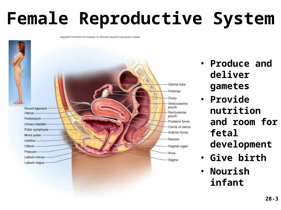
28-3
• Produce and deliver gametes
• Provide nutrition and room for fetal development
• Give birth• Nourish infant
Female Reproductive System
28-4
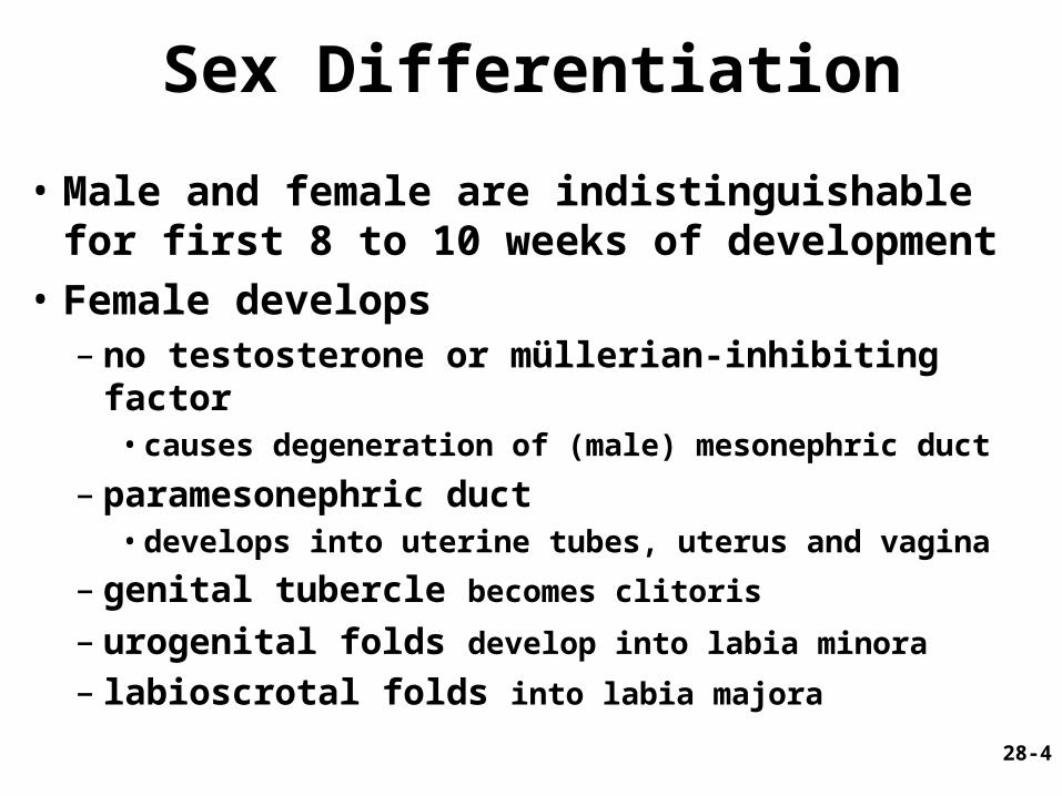
Sex Differentiation
• Male and female are indistinguishable for first 8 to 10 weeks of development
• Female develops– no testosterone or müllerian-inhibiting factor
• causes degeneration of (male) mesonephric duct
– paramesonephric duct • develops into uterine tubes, uterus and vagina
– genital tubercle becomes clitoris
– urogenital folds develop into labia minora – labioscrotal folds into labia majora

28-5
Ovary
• Produces eggs and hormones– almond-shaped, 3 cm x 1.5 cm x 1 cm– tunica albuginea capsule like on testes– cortex produces gametes; medulla holds vessels
• Each egg develops in its own fluid-filled follicle and is released by ovulation
• Ligaments– attached to uterus by ovarian ligament– attached to pelvic wall by suspensory ligament
• contains ovarian artery, vein and nerves
– anchored to broad ligament by mesovarium

28-6
Anatomy of Ovary
28-7
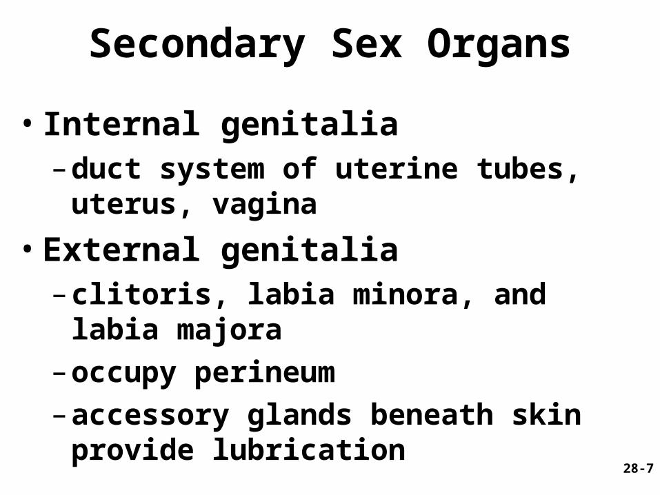
Secondary Sex Organs
• Internal genitalia– duct system of uterine tubes, uterus,
vagina
• External genitalia– clitoris, labia minora, and labia majora
– occupy perineum
– accessory glands beneath skin provide lubrication

28-8
• 10 cm long, muscular tube lined with ciliated cells
• Major portions– narrow isthmus near uterus– body (ampulla): middle
portion– flares distally into
infundibulum with fimbriae
• Enclosed in superior margin of broad ligament (mesosalpinx)
Uterine (Fallopian) Tubes

28-9
Epithelial Lining of Uterine Tube

28-10
Uterus
• Thick-walled, pear-shaped muscular chamber that opens into vagina and tilts forward over urinary bladder– internal and external os of cervical canal– openings into uterine tubes in two upper corners
• Domed fundus above body of organ
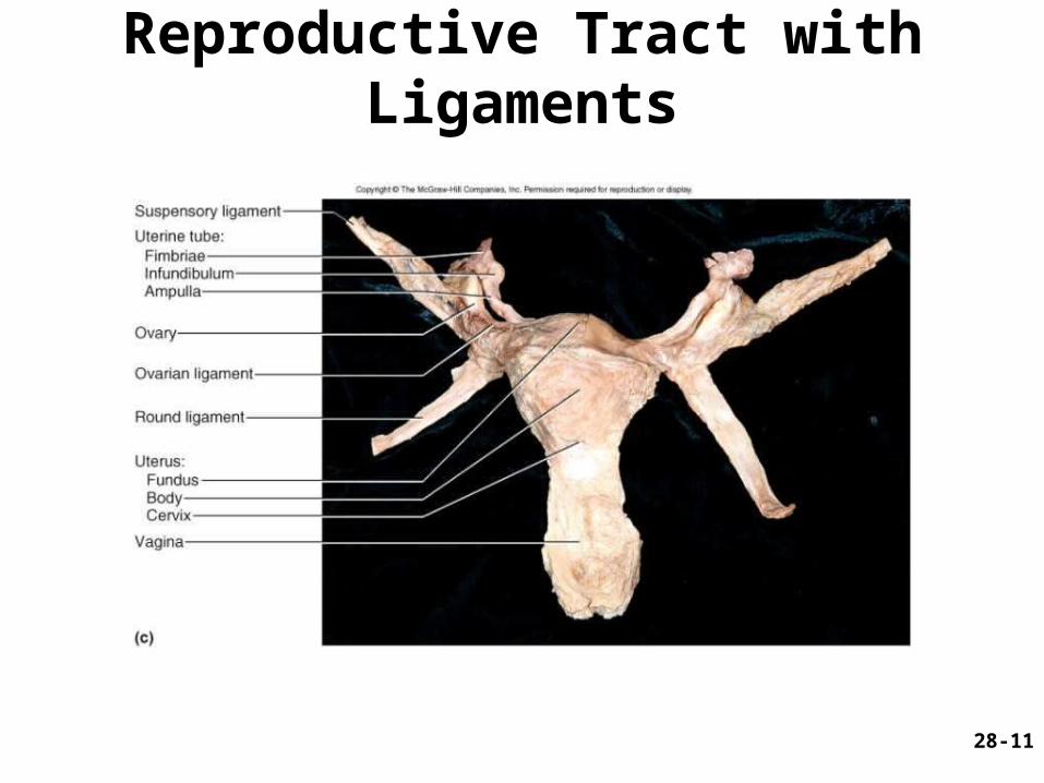
28-11
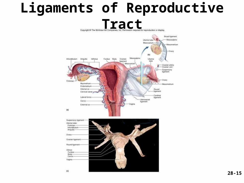
Reproductive Tract with Ligaments

28-12
Histology of Uterine Wall
• Perimetrium - external serosa layer
• Myometrium - middle muscular layer – 1.25 cm thick in nonpregnant uterus– smooth muscle
• produces labor contractions, expels fetus
• Endometrium– simple columnar epithelium with thick layer
compound tubular glands• stratum functionalis – superficial, shed each period• stratum basalis - deep layer, regenerates a new
stratum functionalis with each menstrual cycle
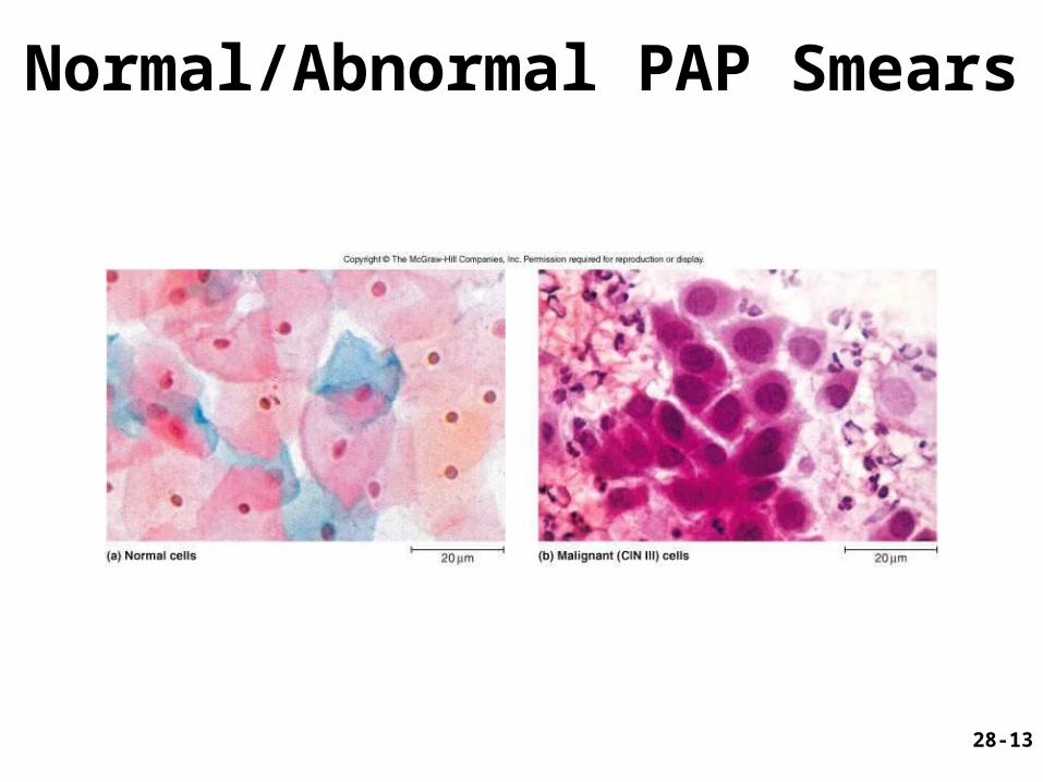
28-13
Normal/Abnormal PAP Smears

28-14
Vessels of Reproductive Tract
Hormonal changes cause spiral artery vasoconstriction, necrosis of stratum functionalis and menstrual flow
28-15
Ligaments of Reproductive Tract

28-16
Vagina
• 8-10 cm distensible muscular tube– allows for discharge of menstrual fluid, receipt of
penis, semen and birth of baby
• Outer adventitia, middle muscularis and inner mucosa
• Epithelium– child - simple cuboidal– puberty - estrogens transform to stratified squamous
• bacteria ferment glycogen rich cells producing acidic pH
• Tilted posteriorly between rectum and urethra– urethra embedded in its anterior wall

28-17
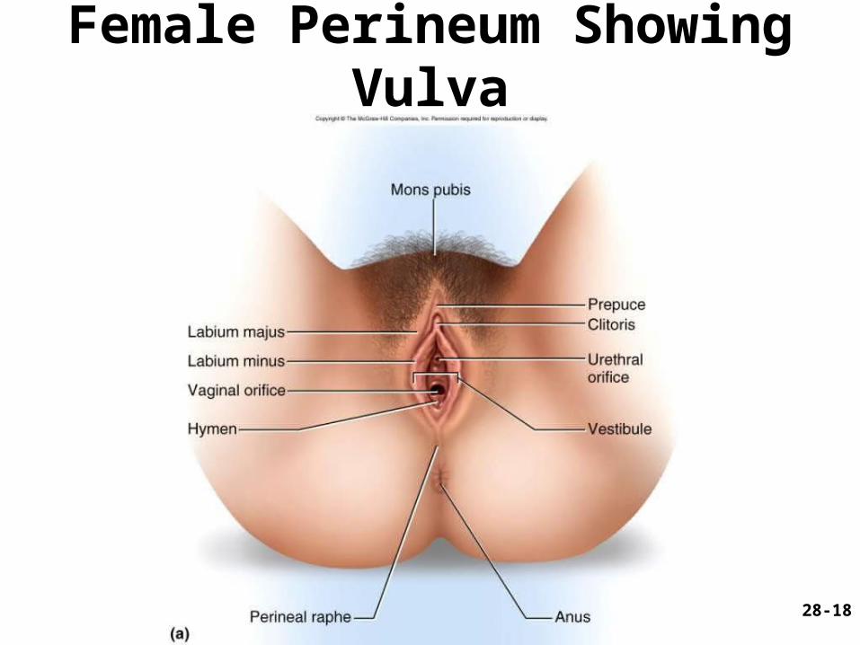
Vulva (Pudendum)
• Mons pubis - mound of fat over pubic symphysis; covered by pubic hair
• Labia majora - thick folds of skin• Labia minora - medial, thin hairless folds
– form vestibule contains urethral and vaginal openings– form hoodlike prepuce over clitoris
• Clitoris - erectile, sensory organ• Vestibular bulbs - erectile tissue around vagina• Greater and lesser vestibular and paraurethral
glands open into vestibule for lubrication
28-18
Female Perineum Showing Vulva

28-19
Components of Female Perineum
28-20
Breasts
• Overlies pectoralis major– conical body, nipple at apex– axillary tail contains many lymphatic vessels
• Nipple surrounded by areola– dermal blood vessels closer to surface– melanocytes darken during pregnancy– smooth muscle contracts wrinkling skin and erecting
nipple in response to cold, touch and arousal
• Suspensory ligaments from skin, muscle• Nonlactating breast has little glandular tissue

28-21
Anatomy of Lactating Breast
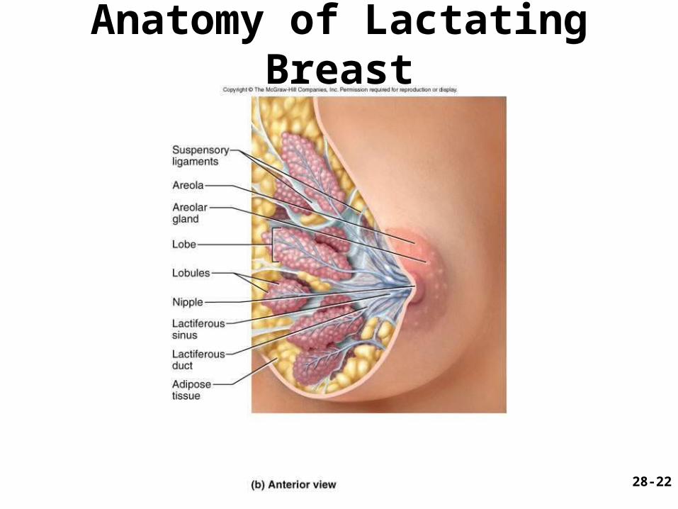
28-22
Anatomy of Lactating Breast
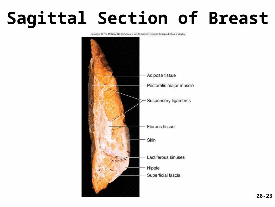
28-23
Sagittal Section of Breast
28-24
Breast Cancer
• 1 out of 8 American women• Tumors begin with cells from mammary ducts
– may metastasize by lymphatics
• Symptoms may include palpable lump, skin puckering, skin texture and drainage from nipple
• Most breast cancer is nonhereditary– some stimulated by estrogen
• Risk factors include – aging, ionizing radiation, carcinogenic chemicals,
alcohol, smoking and fat intake – 70% lack risk factors

28-25
Cancer Screening and Treatment
28-26
Puberty
• Begins at age 9-10 (US)
• Triggered by rising levels of GnRH – stimulates anterior lobe of pituitary to produce
• follicle-stimulating hormone (FSH) • luteinizing hormone (LH)
• Follicles develop and begin to secrete estrogen and progesterone

28-27
Puberty
• Thelarche - development of breasts
• Pubarche - growth of pubic and axillary hair; apocrine and sebaceous glands
• Menarche - first menstrual period– requires at least 17% body fat in teenager,
22% in adult • leptin stimulates gonadotropin secretion• improved nutrition ( body fat) has lowered avg.
age of onset to 12
• Female hormones secreted cyclically and in sequence

28-28
Climacteric• Midlife change in hormone secretion
– due to age related depletion of follicles– occurs with menopause (cessation of
menstruation); average age of 52
• Results– atrophy of uterus, vagina and breasts– skin becomes thinner, bone mass declines,
and risks of cardiovascular disease increase
– hot flashes (sudden dilation of cutaneous arteries) occur several times a day
• HRT = hormone replacement therapy

28-29
Oogensis and Sexual Cycle
• Reproductive cycle - events occurring between fertilization and birth
• Sexual cycle - events recurring every month when pregnancy does not occur– ovarian cycle = events in ovaries– menstrual cycle = parallel changes in uterus
28-30
Oogenesis
• Monthly event produces haploid egg by meiosis• Embryonic development of ovary
– female germ cells arise from yolk sac– differentiate into oogonia, multiply– transform into primary oocytes - early meiosis I– most degenerate (atresia) by childhood– by puberty 400,000 oocytes remain
• FSH stimulates completion of meiosis I, produces secondary oocyte and 1st polar body– proceeds to meiosis II and ceases until fertilization– after fertilization , releases 2nd polar body

28-31
Oogenesis and Follicle Development
28-32
Sexual Cycle
• Averages 28 days, ranges from 20 to 45• Hormone cycle: hierarchy of control
– hypothalamus pituitary ovaries uterus
• Follicular phase (2 weeks) – menstruation occurs during first 3 to 5 days of cycle– uterus replaces lost endometrium and follicles grow
• Luteal phase (2 weeks)– corpus luteum stimulates endometrial thickening– endometrium lost without pregnancy
28-33
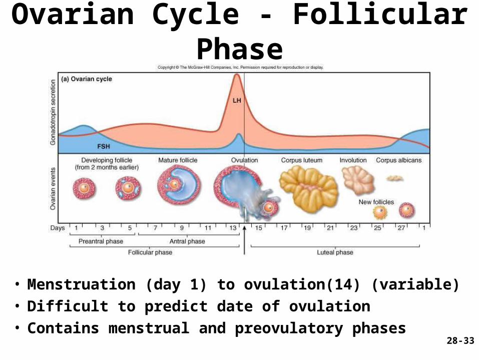
Ovarian Cycle - Follicular Phase
• Menstruation (day 1) to ovulation(14) (variable)• Difficult to predict date of ovulation• Contains menstrual and preovulatory phases
28-34
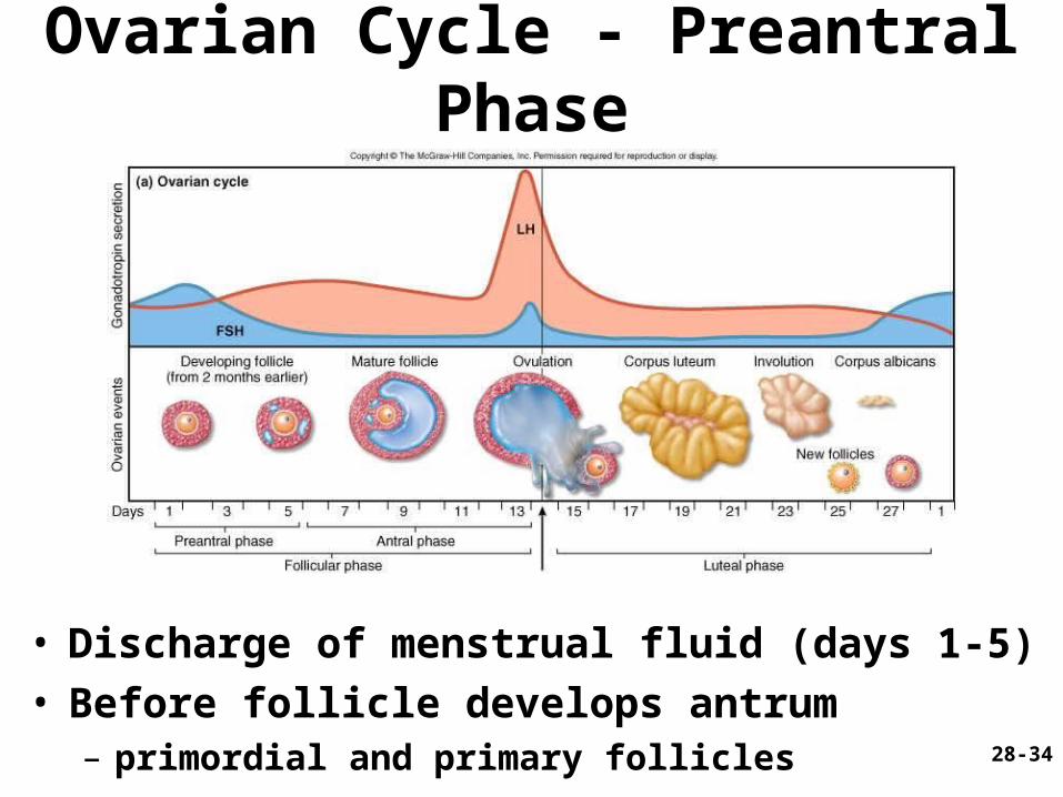
Ovarian Cycle - Preantral Phase
• Discharge of menstrual fluid (days 1-5)• Before follicle develops antrum
– primordial and primary follicles
28-35
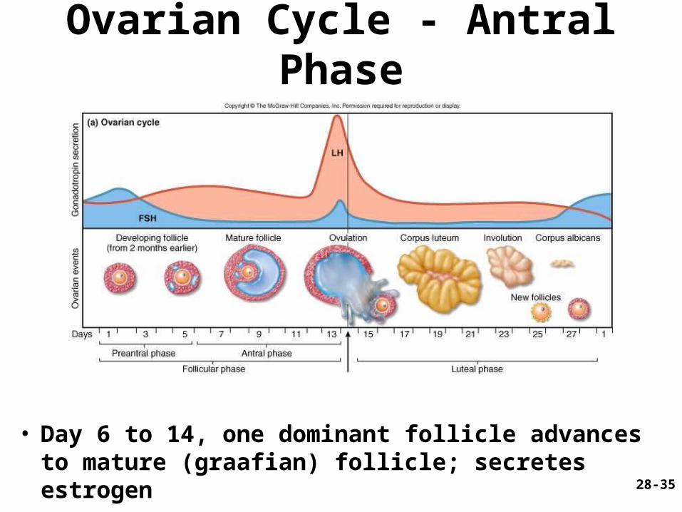
Ovarian Cycle - Antral Phase
• Day 6 to 14, one dominant follicle advances to mature (graafian) follicle; secretes estrogen
28-36
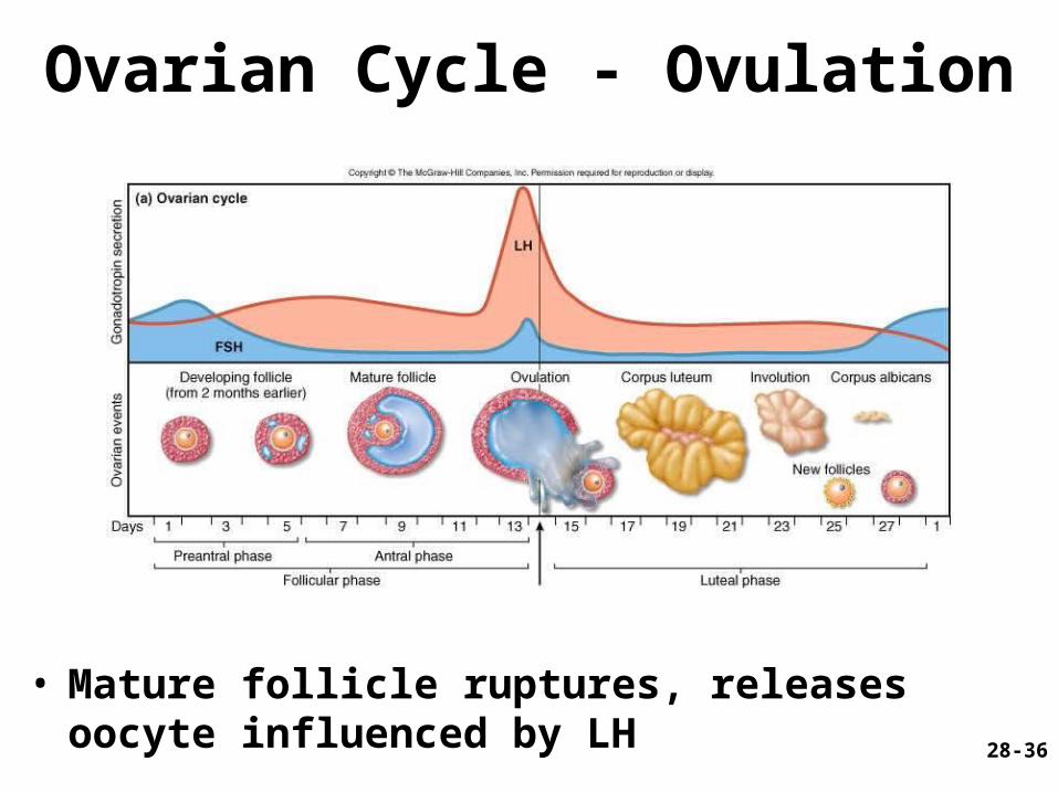
Ovarian Cycle - Ovulation
• Mature follicle ruptures, releases oocyte influenced by LH

28-37
Histology of Ovarian Follicles

28-38
Pituitary-Ovarian Axis

28-39
Ovarian Cycle - Luteal Phase
• Corpus luteum - forms from ruptured follicle, under influence of LH; secretes progesterone
28-40
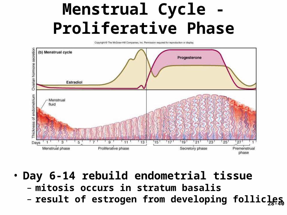
Menstrual Cycle - Proliferative Phase
• Day 6-14 rebuild endometrial tissue – mitosis occurs in stratum basalis– result of estrogen from developing follicles
28-41
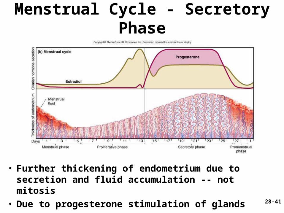
• Further thickening of endometrium due to secretion and fluid accumulation -- not mitosis
• Due to progesterone stimulation of glands
Menstrual Cycle - Secretory Phase

28-42
Menstrual Cycle Premenstrual Phase
• Involution of corpus luteum, progesterone falls– spiral arteries constrict causes endometrial ischemia– stratum functionalis sloughs

28-43
Menstrual Cycle - Menstrual Phase
• Blood, serous fluid and endometrial tissue are discharged
28-44
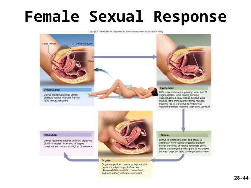
Female Sexual Response

28-45
Pregnancy and Childbirth
• Gestation (pregnancy) – lasts an average of 266 days from conception
to childbirth– gestational calendar measured from first day
of the woman’s last menstrual period (LMP)
• Birth predicted 280 days from LMP– 3 three month intervals called trimesters
28-46
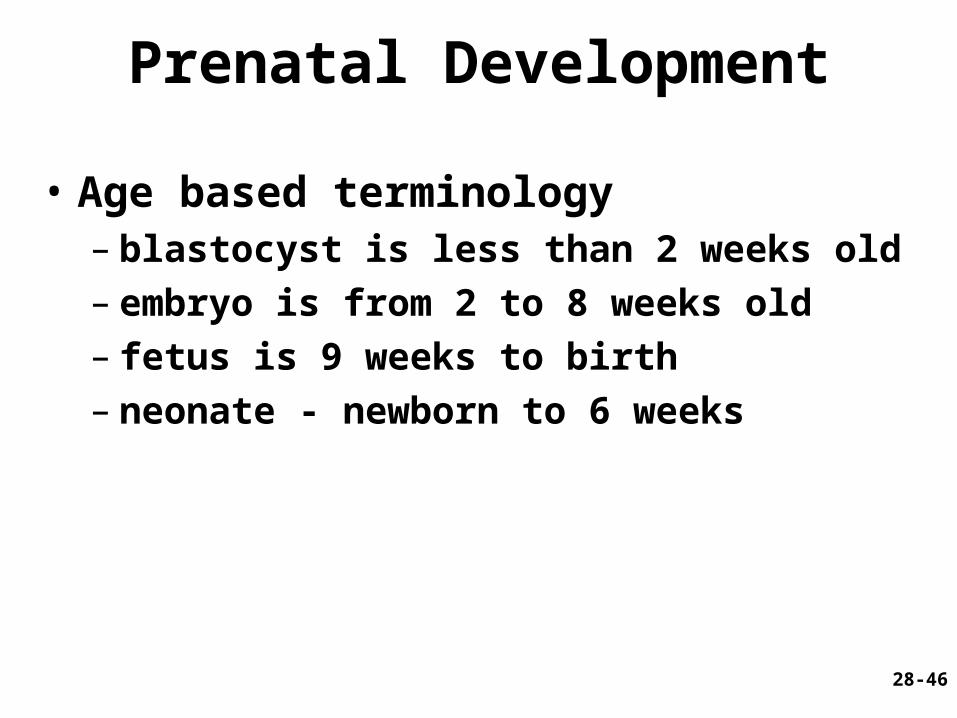
Prenatal Development
• Age based terminology– blastocyst is less than 2 weeks old– embryo is from 2 to 8 weeks old– fetus is 9 weeks to birth– neonate - newborn to 6 weeks

28-47
Hormones of Pregnancy
• HCG (human chorionic gonadotropin)– secreted by trophoblast within 9 days of
conception– prevents involution of corpus luteum
• Estrogens – increases to 30 times normal before birth– corpus luteum is source for first 12 weeks until
placenta takes over– causes uterine, mammary duct and breast
enlargement
28-48
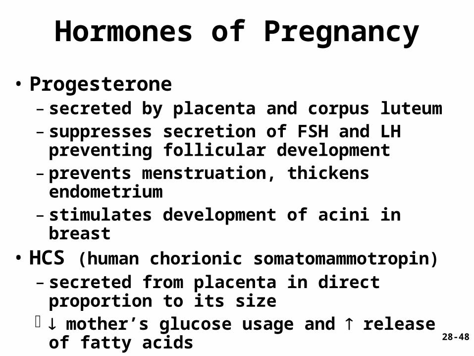
Hormones of Pregnancy
• Progesterone – secreted by placenta and corpus luteum– suppresses secretion of FSH and LH
preventing follicular development– prevents menstruation, thickens endometrium– stimulates development of acini in breast
• HCS (human chorionic somatomammotropin)– secreted from placenta in direct proportion to
its size mother’s glucose usage and release of fatty
acids
28-49
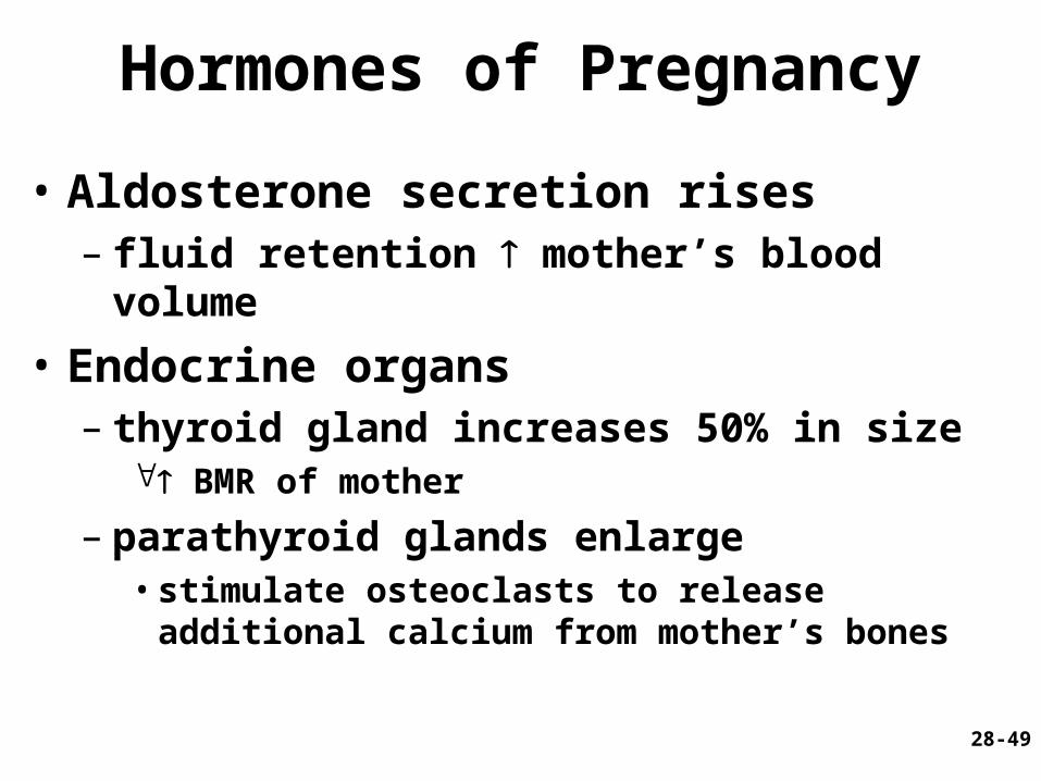
Hormones of Pregnancy
• Aldosterone secretion rises – fluid retention mother’s blood volume
• Endocrine organs– thyroid gland increases 50% in size
BMR of mother
– parathyroid glands enlarge • stimulate osteoclasts to release additional calcium
from mother’s bones
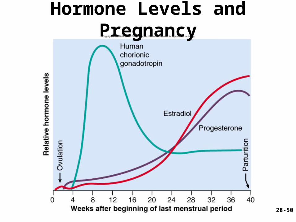
28-50
Hormone Levels and Pregnancy

28-51
Adjustments to Pregnancy

28-52
Adjustments to Pregnancy
• Digestive System – nausea
• first few months
– constipation and heartburn due to intestinal motility • pressure on stomach
• Metabolism– BMR may stimulate appetite
• healthy weight gain - 24 lb.

28-53
Adjustments to Pregnancy
• Nutrition– placenta stores nutrients for 3rd trimester
• protein, iron, calcium, phosphates
– vitamin K• reduces risk of hemorrhages in neonatal brain
– folic acid • prevent neurological disorders
– spina bifida, anencephaly • supplements must be started before pregnancy
28-54
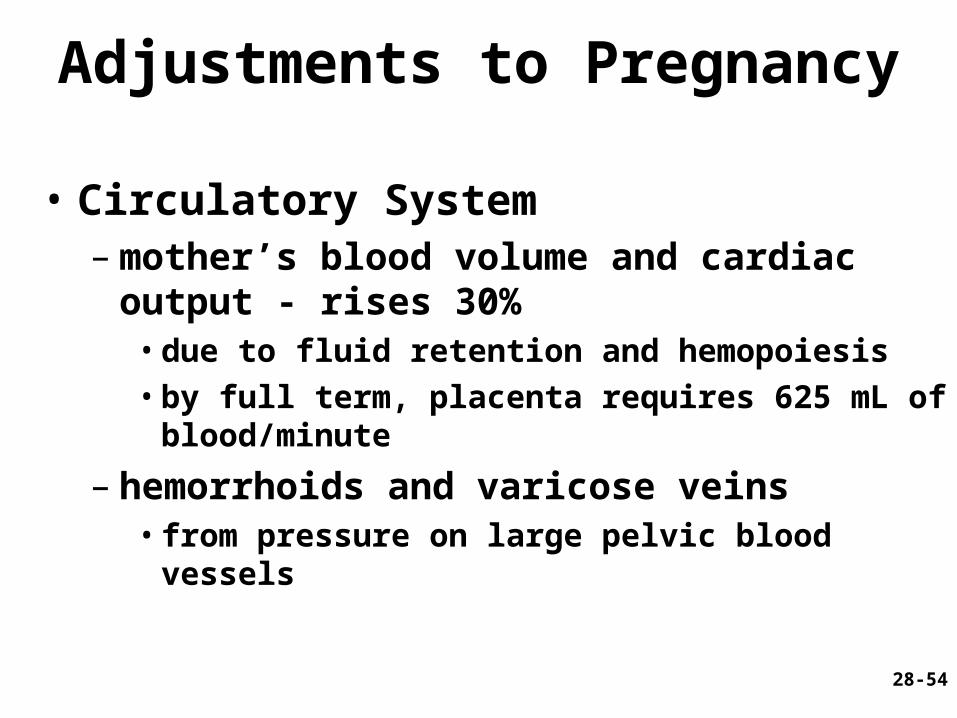
Adjustments to Pregnancy
• Circulatory System– mother’s blood volume and cardiac output -
rises 30% • due to fluid retention and hemopoiesis• by full term, placenta requires 625 mL of
blood/minute
– hemorrhoids and varicose veins • from pressure on large pelvic blood vessels
28-55
Adjustments to Pregnancy
• Respiratory System– minute ventilation about 50%
• demands of fetus, higher maternal metabolic rate
• ventilation adjusted to keep PCO2 lower than normal
– respiratory rate • difficult to breathe deeply
28-56
Adjustments to Pregnancy
• Urinary System– salt and water retention
• due to aldosterone and steroids
– GFR by 50% and output is slightly elevated• mother disposes additional metabolic wastes
frequency of urination • due to bladder compression
28-57
Adjustments to Pregnancy
• Integumentary Systems– stretch marks
• due to dermal stretching
– linea alba may become dark (linea nigra)– temporary chloasma or “mask of pregnancy”
• blotchy darkening of skin over nose and cheeks
28-58
Childbirth - Uterine Contractility
• Parturition – process of giving birth
• by contraction of uterine and abdominal muscles
• Braxton Hicks contractions – throughout gestation– strengthen late in pregnancy - false labor
28-59
Childbirth - Uterine Contractility
• Progesterone inhibits contractions • Estrogen stimulates contractions• Near full term - posterior pituitary releases
more oxytocin, uterus produces more receptors– directly stimulates myometrial contractions– stimulates fetal membranes to produce
prostaglandins - synergists of oxytocin
• Stretching – increases contractility of smooth muscle– role in initiating labor
28-60
Labor Contractions
• Contractions begin 30 minutes apart and eventually occur every 1-3 minutes– periodically relax to blood flow to placenta
and fetus– contractions strongest in fundus and body of
uterus, pushes fetus into cervix
28-61
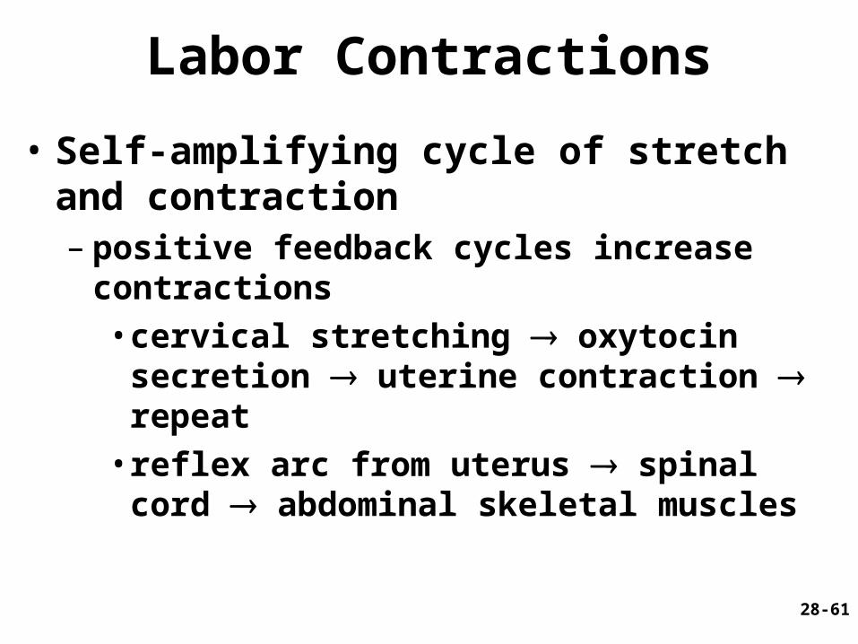
Labor Contractions
• Self-amplifying cycle of stretch and contraction– positive feedback cycles increase contractions
• cervical stretching oxytocin secretion uterine contraction repeat
• reflex arc from uterus spinal cord abdominal skeletal muscles
28-62
Pain of Labor
• Ischemia of myometrium
• Stretching of cervix, vagina and perineum – episiotomy prevents tearing
• Large fetal head in a narrow pelvic outlet

28-63
Stages of Labor -- Early Dilation
• Widening of cervical canal by effacement (thinning) of cervix to reach 10 cm -- diameter of fetal head
• Rupture of fetal membranes and loss of amniotic fluid
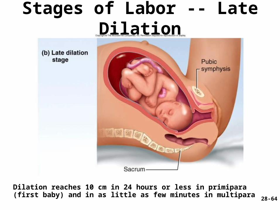
28-64
Stages of Labor -- Late Dilation
Dilation reaches 10 cm in 24 hours or less in primipara (first baby) and in as little as few minutes in multipara

28-65
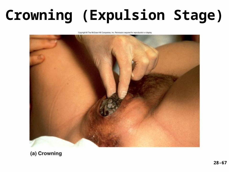
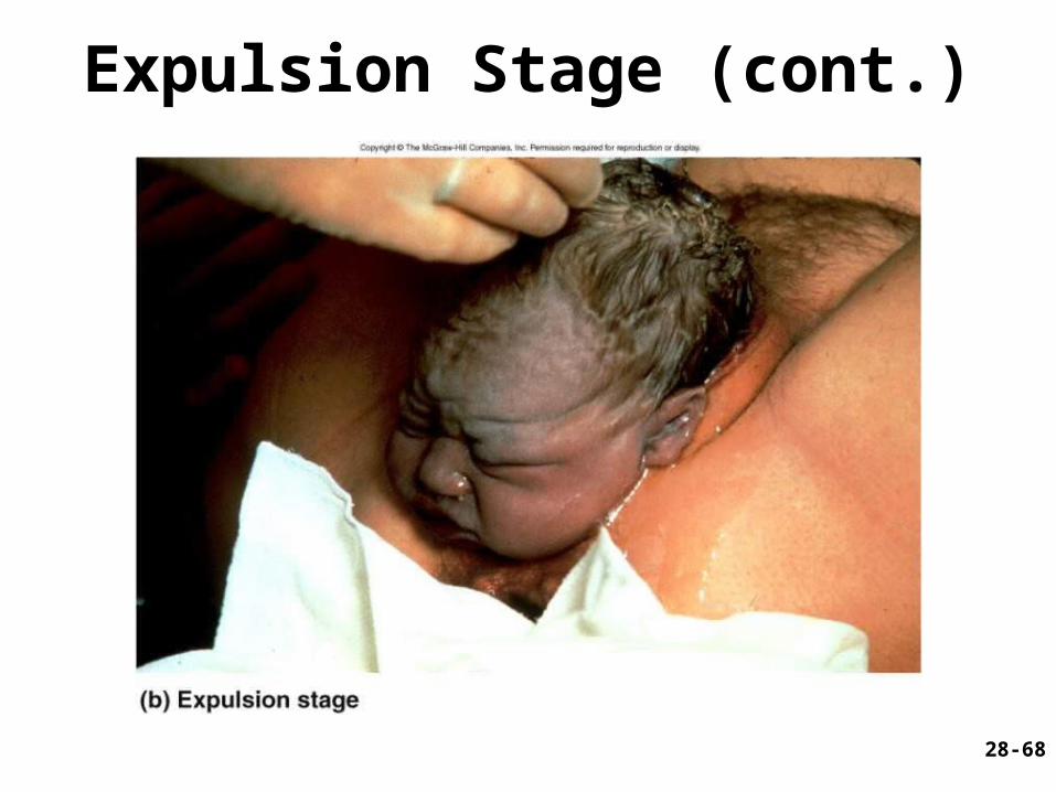
Stages of Labor -- Expulsion
• Time baby’s head enters vagina until delivery– up to 30 minutes
• Valsalva maneuver helps to expel fetus

28-66
Stages of Labor -- Placental
• Uterine contractions continue causing placental separation
28-67
Crowning (Expulsion Stage)
28-68
Expulsion Stage (cont.)

28-69
Placental Stage
28-70
Puerperium
• First 6 weeks after delivery
• Anatomy and physiology return to normal– involution of uterus
• to pre-gravid weight in 4 weeks• accomplished by autolysis by lysosomal enzymes
– vaginal discharge called lochia
– breastfeeding promotes involution• suppresses estrogen secretion• stimulates oxytocin which causes myometrial
contraction
28-71
Mammary Gland Development
• Lactation – synthesis and ejection of milk from mammary
glands in breast
• Ducts grow and branch – due to high estrogen levels in pregnancy
• Followed by budding and development of acini at the ends of the ducts – due to progesterone
28-72
Colostrum and Milk Synthesis
• Colostrum forms in late pregnancy– similar to breast milk; contains 1/3 less fat,
thinner– first 1 to 3 days after birth– contains IgA protection from gastroenteritis
• Synthesis is promoted by prolactin (from pituitary)– synthesis of hormone begins 5 weeks into
pregnancy, by full term it is 20x normal level– steroid hormones from placenta oppose it until
birth

28-73
Colostrum and Milk Synthesis
• At birth, prolactin secretion drops, but 20 times after nursing– without nursing, milk production stops in 1
week
• 5-10% of women become pregnant while nursing– inhibition of GnRH and reduced ovarian
cycling

28-74
Prolactin and Lactation

28-75
Milk Ejection
• Controlled by a neuroendocrine reflex– infant’s suckling stimulates sensory
receptors in nipple, signaling hypothalamus and posterior pituitary to release oxytocin
– oxytocin stimulates myoepithelial cells
• Myoepithelial cells surround secretory cells in acinus– contract to squeeze milk into duct
• milk flow within 30-60 seconds after suckling begins
28-76
Breast Milk
• Supplies antibodies and colonizes intestine with beneficial bacteria
• Colostrum and milk have a laxative effect that clears intestine of meconium (green, bile-filled fecal material in newborn)
• Nursing woman can produce 1.5L per day
• Cow’s milk not a good substitute– 1/3 less lactose but 3 times as much protein– harder to digest and more nitrogenous waste
(diaper rash)
28-77
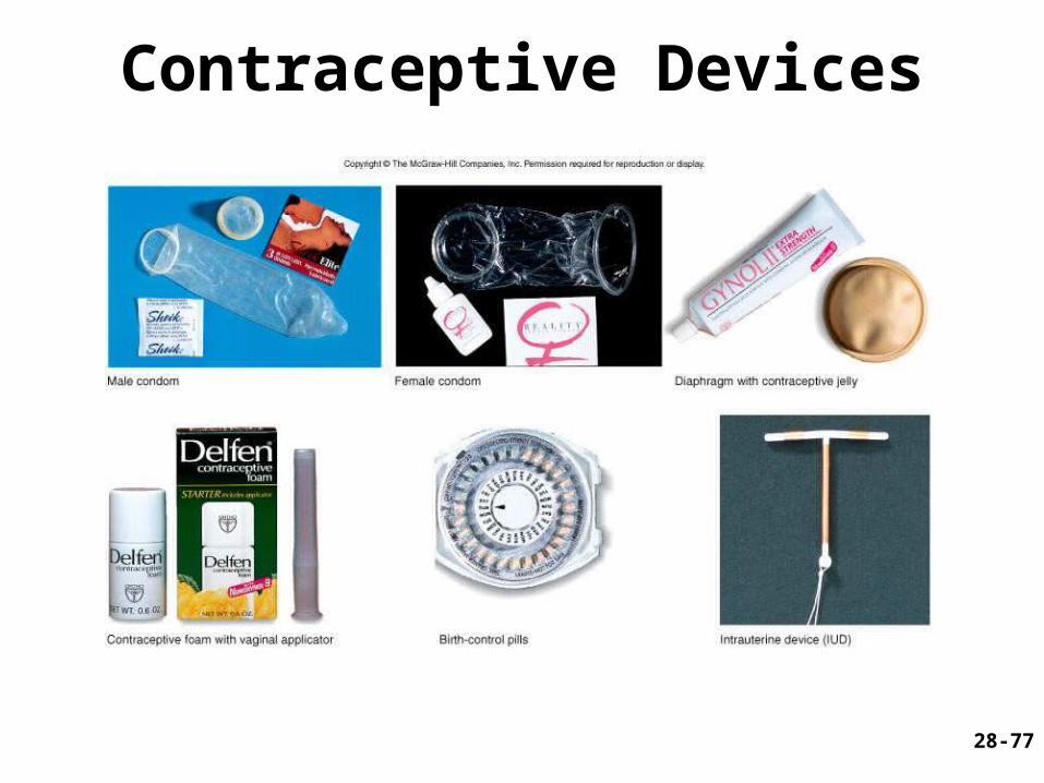
Contraceptive Devices