25.1 pharmaceutical products and drug action
TRANSCRIPT
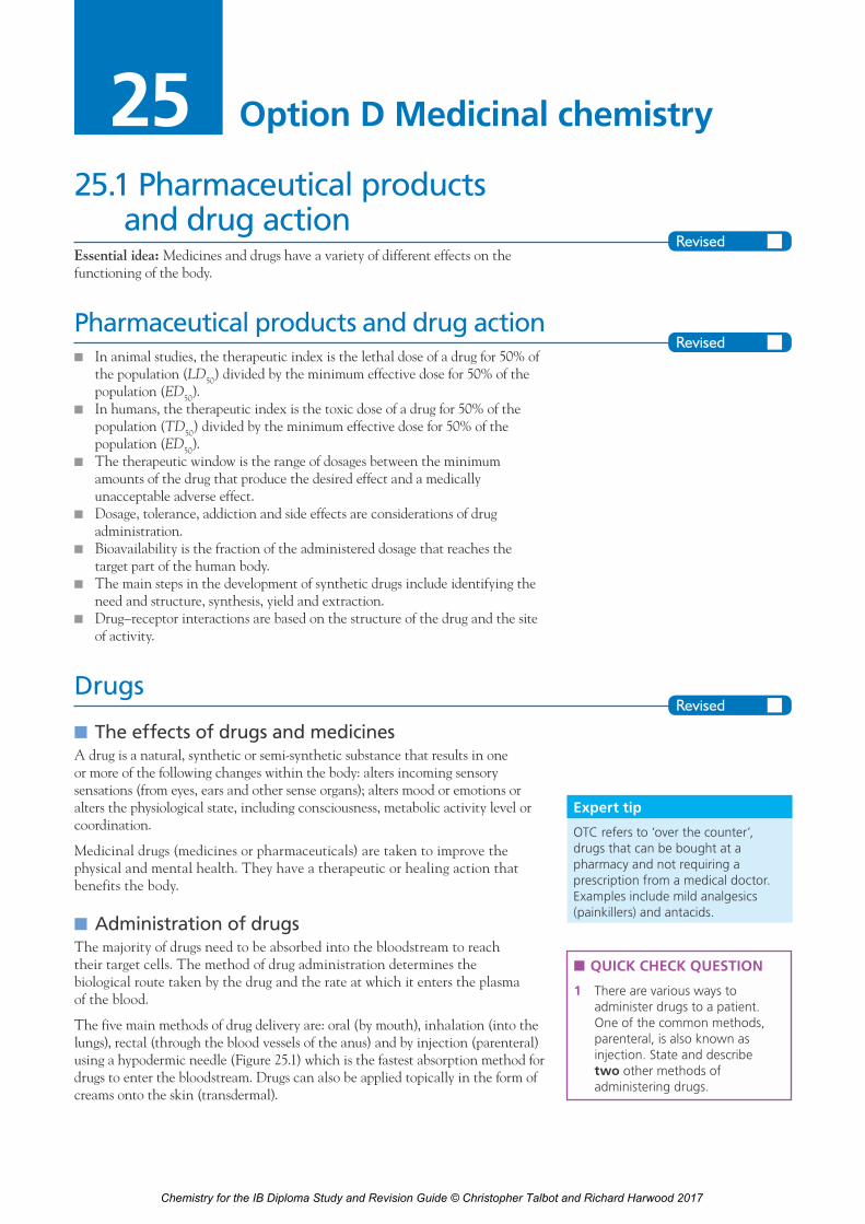
25.1 Pharmaceutical products and drug action
Essential idea: Medicines and drugs have a variety of different effects on the functioning of the body.
Pharmaceutical products and drug actionn In animal studies, the therapeutic index is the lethal dose of a drug for 50% of
the population (LD50) divided by the minimum effective dose for 50% of the population (ED50).
n In humans, the therapeutic index is the toxic dose of a drug for 50% of the population (TD50) divided by the minimum effective dose for 50% of the population (ED50).
n The therapeutic window is the range of dosages between the minimum amounts of the drug that produce the desired effect and a medically unacceptable adverse effect.
n Dosage, tolerance, addiction and side effects are considerations of drug administration.
n Bioavailability is the fraction of the administered dosage that reaches the target part of the human body.
n The main steps in the development of synthetic drugs include identifying the need and structure, synthesis, yield and extraction.
n Drug–receptor interactions are based on the structure of the drug and the site of activity.
Drugs
■n The effects of drugs and medicinesA drug is a natural, synthetic or semi-synthetic substance that results in one or more of the following changes within the body: alters incoming sensory sensations (from eyes, ears and other sense organs); alters mood or emotions or alters the physiological state, including consciousness, metabolic activity level or coordination.
Medicinal drugs (medicines or pharmaceuticals) are taken to improve the physical and mental health. They have a therapeutic or healing action that benefits the body.
■n Administration of drugsThe majority of drugs need to be absorbed into the bloodstream to reach their target cells. The method of drug administration determines the biological route taken by the drug and the rate at which it enters the plasma of the blood.
The five main methods of drug delivery are: oral (by mouth), inhalation (into the lungs), rectal (through the blood vessels of the anus) and by injection (parenteral) using a hypodermic needle (Figure 25.1) which is the fastest absorption method for drugs to enter the bloodstream. Drugs can also be applied topically in the form of creams onto the skin (transdermal).
Option D Medicinal chemistry25
Expert tip
OTC refers to ‘over the counter’, drugs that can be bought at a pharmacy and not requiring a prescription from a medical doctor. Examples include mild analgesics (painkillers) and antacids.
n QUICK CHECK QUESTION
1 There are various ways to administer drugs to a patient. One of the common methods, parenteral, is also known as injection. State and describe two other methods of administering drugs.
Chemistry for the IB Diploma Study and Revision Guide © Christopher Talbot and Richard Harwood 2017

25 Medicinal chemistry172
Development and testing of new medicinal drugsThe research and development of new drugs is a long and expensive process. A new lead compound (with biological activity) may be isolated and purified from an existing species, often a plant, fungus or marine organism. However, medicinal drug development often starts by finding an active compound from ‘chemical libraries’ containing thousands of synthetic molecules.
A lead compound is often chemically modified to improve its physical properties and biological effects. Some new medicinal drugs are developed by a process of rational design. They are designed (often via computer modelling) and synthesized knowing the structure and shape of the protein receptor the drug molecule interacts with.
■n Computer-aided drug designComputers are an important part of the process of the design of synthetic drugs and have a large number of uses, which include structure analysis, structure comparisons, lead compound design, identification of active conformations (interconvertible three-dimensional shapes) and pharmacophores (a set of features common to a group of active molecules), protein and binding site structure and ligand binding. A ligand is a small molecule (that can be a drug) that binds to a receptor.
A potential drug candidate needs to undergo extensive testing in vitro testing with bacteria, cells or biological molecules and later with animals and humans (in vivo testing). Anti-cancer drugs are tested with cancer cells grown in culture. The results of large-scale animal testing will establish the lethal dose required to kill 50% of the animal test population. This is known as the LD50 value (Figure 25.2), but it is also important to carry out tests that identify chronic, long-term toxicity that is non-lethal.
intramuscular(usually injectedinto arm, leg or
buttock muscles)
subcutaneous(injected directly
under the skin)
intravenous (this has the most
rapid effect as the drug entersthe bloodstream directly)
skin
fatty tissue
vein
muscle
Figure 25.1 Summary of common methods of drug delivery by injection
n QUICK CHECK QUESTION
2 Creating a new pharmaceutical compound is a long, expensive and complex process. Outline the main stages of this process in the correct order.
Expert tip
Medicinal chemists aim to have the minimum number of synthetic steps, to increase the overall yield.
Chemistry for the IB Diploma Study and Revision Guide © Christopher Talbot and Richard Harwood 2017

25.1 Pharmaceutical products and drug action 173
The effective dose required to bring about a measurable effect in 50% of the test animal population will also be determined. This is known as the ED50 value. The therapeutic index can be calculated: LD50/ED50.
Drugs will then be subjected to a variety of clinical testing phases with volunteers (first phase) and at later stages with human patients subject to approval by a drug regulatory authority (Figure 25.3). In humans the therapeutic index is defined as TD50/ED50, where TD50 represent the toxic dose for 50% of the human test population.
Expert tip
A large LD50 value means that the substance is relatively non-toxic and that a large quantity of the substance is required to cause a toxic response. A small LD50 value means that the substance is relatively toxic and that only a small quantity of the substance is needed to cause a toxic response.
10 000 possible projects initiated
App
roxi
mat
e tim
e/ye
ars
final drug administration approval for one new drug
0
5
10
laboratory and animal studies
1st phase of clinical studies (initial trials on humans)
2nd phase of clinical studiestesting for efficacy and
side effects
3rd phase of clinical studies(extensive trialling)
new drug applicationlogged with drug
administration
review by drugadmin.
Figure 25.3 Summary of the steps in the development of a new drug
Dos
e/pp
m ×
exp
osur
e tim
e300000
250000
200000
150000
100000
50000
0methanol ethanol petrol
(gasoline)diesel
ratsmice
Key:
Figure 25.2 Inhalation LD50 values of common fuels
Human clinical trials will indicate whether there are any short-term side effects and allow the therapeutic window (Figure 25.4) to be established. This is the range of dosages between the minimal dose required to produce a therapeutic effect and a harmful effect due to toxicity at high dosage.
Expert tip
All substances are potentially poisonous: it is only the dose that determines whether a substance is poisonous. The concepts of toxicology imply that no drug is 100 per cent safe.
Pat
ient
s af
fect
ed o
r re
spon
ding
/%
adverseeffect
therapeuticeffect
therapeutic index
therapeutic window
Dose administered/mg kg–1
toxicside effect
ED50 TD50
0
50
100
Figure 25.4 The therapeutic index and therapeutic window
Chemistry for the IB Diploma Study and Revision Guide © Christopher Talbot and Richard Harwood 2017

25 Medicinal chemistry174
Side effects of a drug are unwanted, or unintended and sometimes harmful or unpleasant effects that appear with a dose within the therapeutic window. A risk to benefit ratio will determine whether the drug’s side effects are critical. For example, side effects may be considered less of an issue if the drug is shown to be highly effective at treating a particular condition or infectious disease. They usually increase with dosage.
n QUICK CHECK QUESTION
3 a The results for the therapeutic and adverse effects of a new potential drug for treating high cholesterol levels are shown below.
Pat
ient
s re
spon
ding
therapeuticeffect
Dose/mg kg–1
toxiceffect
20 80
0
50
100
Calculate the therapeutic index of the drug. Label on the graph the therapeutic window of the drug.
b Another drug that has the same effect was also studied and the therapeutic index of the second drug is 110. State and explain which of the two drugs is more likely to be approved for over the counter (OTC) sales.
NATURE OF SCIENCEAbuse potential refers to a drug that is used in non-medical situations for the positive psychoactive effects it produces. These drugs are characterized by their central nervous system (CNS) activity. Examples of the psychoactive effects they produced include sedation, euphoria, distortions of perception, hallucinations, and mood changes. Drugs with abuse potential often produce psychic or physical dependence and may lead to the disorder of addiction.
Expert tip
One side effect of aspirin is irritation of the stomach lining.
Drug testing on human patients is usually carried out using a double blind approach with a placebo. Half of the clinical patients are administered with the drug and the other half are given an inert chemical placebo, which resembles the drug in appearance and taste, but has no bioactive chemicals. The term double blind (Figure 25.5) means that neither the patients nor the medical staff administering the drug know which patients are receiving the drug.
Expert tip
The placebo effect occurs when a placebo promotes the ‘natural healing’ effects of the human immune and endocrine system. Any medicinal drug needs to be more effective than a placebo during clinical trials. It is not clear why the placebo works, but a person’s hope about a treatment can trigger a biochemical effect presumably via the immune system and endocrine (hormone) system.
volunteers
group toreceive drug
group toreceive placebo
evaluationperiod
random placement intotreatment and placebo groups
double blind – neithervolunteers nor doctorsknow who has placebo
compare resultsfrom treatment
and placebo groups
Figure 25.5 Double blind testing
Chemistry for the IB Diploma Study and Revision Guide © Christopher Talbot and Richard Harwood 2017
25.1 Pharmaceutical products and drug action 175
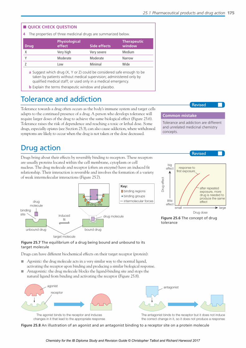
Tolerance and addictionTolerance towards a drug often occurs as the body’s immune system and target cells adapts to the continued presence of a drug. A person who develops tolerance will require larger doses of the drug to achieve the same biological effect (Figure 25.6). Tolerance raises the risk of dependence and reaching a toxic or lethal dose. Some drugs, especially opiates (see Section 25.3), can also cause addiction, where withdrawal symptoms are likely to occur when the drug is not taken or the dose decreased.
Drug actionDrugs bring about their effects by reversibly binding to receptors. These receptors are usually proteins located within the cell membrane, cytoplasm or cell nucleus. The drug molecule and receptor (often an enzyme) have an induced fit relationship. Their interaction is reversible and involves the formation of a variety of weak intermolecular interactions (Figure 25.7).
small large
littleeffect
bigeffect
Drug dose
Dru
g ef
fect
after repeatedexposure, moredrug is needed toproduce the sameeffect
response tofirst exposure
Figure 25.6 The concept of drug tolerance
Common mistake
Tolerance and addiction are different and unrelated medicinal chemistry concepts.
n QUICK CHECK QUESTION
4 The properties of three medicinal drugs are summarized below.
DrugPhysiological effect Side effects
Therapeutic window
X Very high Very severe Medium
Y Moderate Moderate Narrow
Z Low Minimal Wide
a Suggest which drug (X, Y or Z) could be considered safe enough to be taken by patients without medical supervision; administered only by qualifi ed medical staff; or used only in a medical emergency.
b Explain the terms therapeutic window and placebo.
Drugs can have different biochemical effects on their target receptor (protein):
n Agonistic: the drug molecule acts in a very similar way to the normal ligand, activating the receptor upon binding and producing a similar biological response.
n Antagonistic: the drug molecule blocks the ligand-binding site and stops the natural ligand from binding and activating the receptor (Figure 25.8).
drugmolecule
drug molecule
binding site
bindingsite
target molecule
inducedfit
unbound drug bound drug
binding regions
binding groups
intermolecular forces
Key:
Figure 25.7 The equilibrium of a drug being bound and unbound to its target molecule
Figure 25.8 An illustration of an agonist and an antagonist binding to a receptor site on a protein molecule
agonist
receptor
The agonist binds to the receptor and induceschanges in it that lead to the appropriate response
The antagonist binds to the receptor but it does not inducethe correct change in it, so it does not produce a response
antagonist
Chemistry for the IB Diploma Study and Revision Guide © Christopher Talbot and Richard Harwood 2017
25 Medicinal chemistry176
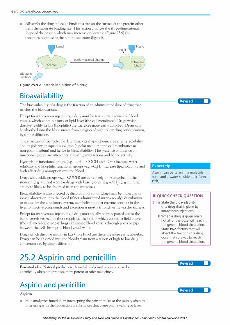
n Allosteric: the drug molecule binds to a site on the surface of the protein other than the substrate binding site. This action changes the three-dimensional shape of the protein which may increase or decrease (Figure 25.9) the receptor’s response to the natural substrate (ligand).
allostericinhibitor
active site(shut)
ligand ligandno fit
conformational change
receptor
Figure 25.9 Allosteric inhibition of a drug
BioavailabilityThe bioavailability of a drug is the fraction of an administered dose of drug that reaches the bloodstream.
Except for intravenous injections, a drug must be transported across the blood vessels, which contain a fatty or lipid layer (the cell membrane). Drugs which dissolve readily in fats (lipophilic) are therefore more easily absorbed. Drugs can be absorbed into the bloodstream from a region of high to low drug concentration, by simple diffusion.
The structure of the molecule determines its shape, chemical reactivity, solubility and its polarity, in aqueous solution (a polar medium) and cell membranes (a non-polar medium) and hence its bioavailability. The presence or absence of functional groups are often critical to drug interactions and hence activity.
Hydrophilic functional groups (e.g. –NH2, –COOH and –OH) increase water solubility and lipophilic functional groups (e.g. –C6H5) increase lipid solubility and both affect drug absorption into the blood.
Drugs with acidic groups (e.g. –COOH) are more likely to be absorbed in the stomach (e.g. aspirin) whereas drugs with basic groups (e.g. –NH2) (e.g. quinine) are more likely to be absorbed from the intestines.
Bioavailability is also affected by dissolution of solids (drugs may be molecular or ionic), absorption into the blood (if not administered intravenously), distribution to tissues, by the circulatory system, metabolism (under enzyme control) in the liver to inactive compounds and excretion is mostly through urine via the kidneys.
Except for intravenous injections, a drug must usually be transported across the blood vessels (especially those supplying the brain), which contain a lipid bilayer (the cell membrane. Most drugs can escape blood vessels through pores or gaps between the cells lining the blood vessel walls.
Drugs which dissolve readily in fats (lipophilic) are therefore more easily absorbed. Drugs can be absorbed into the bloodstream from a region of high to low drug concentration, by simple diffusion.
25.2 Aspirin and penicillinEssential idea: Natural products with useful medicinal properties can be chemically altered to produce more potent or safer medicines.
Aspirin and penicillinAspirin
n Mild analgesics function by intercepting the pain stimulus at the source, often by interfering with the production of substances that cause pain, swelling or fever.
Expert tip
Aspirin can be taken in a molecular form and a water-soluble ionic form (salt).
n QUICK CHECK QUESTION
5 a State the bioavailability of a drug that is given by intravenous injections.
b When a drug is given orally, not all of the dose will reach the general blood circulation. State two factors that will affect the fraction of a drug dose that survives to reach the general blood circulation.
Chemistry for the IB Diploma Study and Revision Guide © Christopher Talbot and Richard Harwood 2017

25.2 Aspirin and penicillin 177
n Aspirin is prepared from salicylic acid.n Aspirin can be used as an anticoagulant, in prevention of the recurrence of
heart attacks and strokes and as a prophylactic.
Penicillin
n Penicillins are antibiotics produced by fungi.n A beta-lactam ring is a part of the core structure of penicillins.n Some antibiotics work by preventing cross-linking of the bacterial cell walls.n Modifying the side-chain results in penicillins that are more resistant to the
penicillinase enzyme.
AspirinAspirin is a mild analgesic (painkiller) and can be prepared in the laboratory by reacting 2-hydroxybenzoic acid (salicylic acid) with ethanoic anhydride (in the presence of concentrated sulfuric acid, which acts as a catalyst) (Figure 25.10). Aspirin (2-ethanoyl-2-hydroxybenzoic acid)) is the ethanoate ester of 2-hydroxybenzoic acid. (Ethanoyl chloride can also be used as an acetylating agent.)
+
salicylic acid (2-hydroxybenzoic acid)
mp 159 °C
O
COHO
H +
aspirin(acetylsalicylic acid)
mp 128–137 °C
O
C
COHO
CH3O
ethanoic acid
CH3COOH
ethanoic anhydridebp 140 °C
C
OO
COH3C CH3
H+
Figure 25.10 Reaction between salicylic acid and ethanoic anhydride to form aspirin (acetylsalicylic acid)
The crude (impure aspirin) crystals can be removed by suction filtration, washed with cold water and then purified by recrystallization. The product can be recrystallized from hot water to obtain pure aspirin. Soluble impurities are left in solution.
The process of recrystallization involves dissolution of the solid in an appropriate solvent at an elevated temperature and the subsequent re-formation of the crystals upon cooling, so that any impurities remain in solution.
The experimental yield can be measured from the mass of pure aspirin obtained and the percentage yield can be calculated using the stoichiometric equation and molar masses of reactant and products.
The purity of the aspirin can be determined from the melting point of the crystals. The presence of impurities will lower the melting point and cause it to melt over a wider range of temperatures.
■n Infrared spectrum of aspirinThe purity of aspirin can also be investigated by recording the infrared spectrum (Figure 25.11). It shows two peaks at 1750 cm−1 and 1680 cm−1 due to the presence of two carbonyl groups, >C=O and a very broad absorption peak between wavenumbers 2500 and 3500 cm−1. This is due to the carboxylic acid group, –COOH (which engages in hydrogen bonding).
n QUICK CHECK QUESTION
6 Identify the type of reaction used to convert 2-hydroxybenzoic acid to aspirin.
Expert tip
There is often a smell of ‘vinegar’ in old aspirin bottles because aspirin tends to hydrolyse spontaneously to some extent.
Expert tip
The melting point is lowered by impurities because the regular packing in the lattice is disrupted and the intermolecular forces weakened.
Wave number/cm–1
Tran
smitt
ance
/%
350030
40
60
80
100
3000 2500 2000 1500 1000 500
Figure 25.11 The infrared spectrum of aspirin
Chemistry for the IB Diploma Study and Revision Guide © Christopher Talbot and Richard Harwood 2017

25 Medicinal chemistry178
■n Therapeutic properties of aspirinAspirin can be used as an antipyretic (to reduce fever) and an anti-inflammatory. It blocks the function of platelets (to make the blood thinner and be more easily pumped), to help prevent the recurrence of heart attacks and strokes and is also used as a prophylactic.
Salicylic acid (2-hydoxybenzoic acid) is also an analgesic but irritates and damages the mouth, oesophagus and stomach lining, so has been replaced by aspirin. It can be extracted from the bark of a willow tree.
■n Mode of actionThe mechanism of aspirin’s analgesic properties involves inhibiting the enzyme cyclooxygenase at the site of an injury. The enzyme is involved in catalysing the formation of substances known as prostaglandins. They accumulate at the site of injury and are involved in the transmission of nerve impulses to the brain (which are interpreted as pain).
The most common side effect of aspirin is that it can cause bleeding in the lining of the stomach. This effect is increased when taking aspirin tablets with alcohol (ethanol) which has a synergistic effect.
Young children are not advised to take large dosages of aspirin during a viral infection since it is linked to Reye’s syndrome, a potentially fatal liver and brain disorder. Aspirin causes convulsions if injected into the brain. This is a toxic effect caused by the release of 2-hydroxybenzoic acid by hydrolysis. Very large dosages of aspirin orally can be fatal due to acidosis, a lowering of the pH of the blood.
■n Soluble aspirinThe molecular or free acid form of aspirin has limited solubility in water due to the presence of a non-polar benzene ring. It is in reversible equilibrium with its carboxylate anion (Figure 25.12) and its bioavailability in blood is limited.
Expert tip
A prophylactic is a medicine used to treat or prevent the occurrence of a disease or condition.
Expert tip
A synergistic effect occurs when the combination of the two drugs is greater than either individual effect. This can be beneficial but can also be very harmful.
n QUICK CHECK QUESTIONS
7 Outline how aspirin functions as an analgesic.
8 State one important use for aspirin other than the relief of pain and fever.
The carboxylic acid group of aspirin can be ionized via a neutralization reaction with a strong base or calcium hydroxide to convert onto a more soluble ionic form. This is known as ‘soluble aspirin’ (Figure 25.13).
n QUICK CHECK QUESTION
9 A soluble ionic version of aspirin can be made by reacting it with a strong base, such as sodium hydroxide. Explain why this process increases the bioavailability of the drug.
However, once the aspirin anion reaches the acidic gastric juice of the stomach it is converted back to its molecular (free acid) or un-ionized form.
O
OHC
O
C
O
CH3
O
O–C
O
C
O
CH3
+ H+
Figure 25.12 Partial dissociation of aspirin in water
+
O
C
COHO
CH3O NaOH + H2O
O
C
CO–O
CH3O
Na+
Figure 25.13 Formation of the soluble sodium salt of aspirin
Chemistry for the IB Diploma Study and Revision Guide © Christopher Talbot and Richard Harwood 2017
25.2 Aspirin and penicillin 179
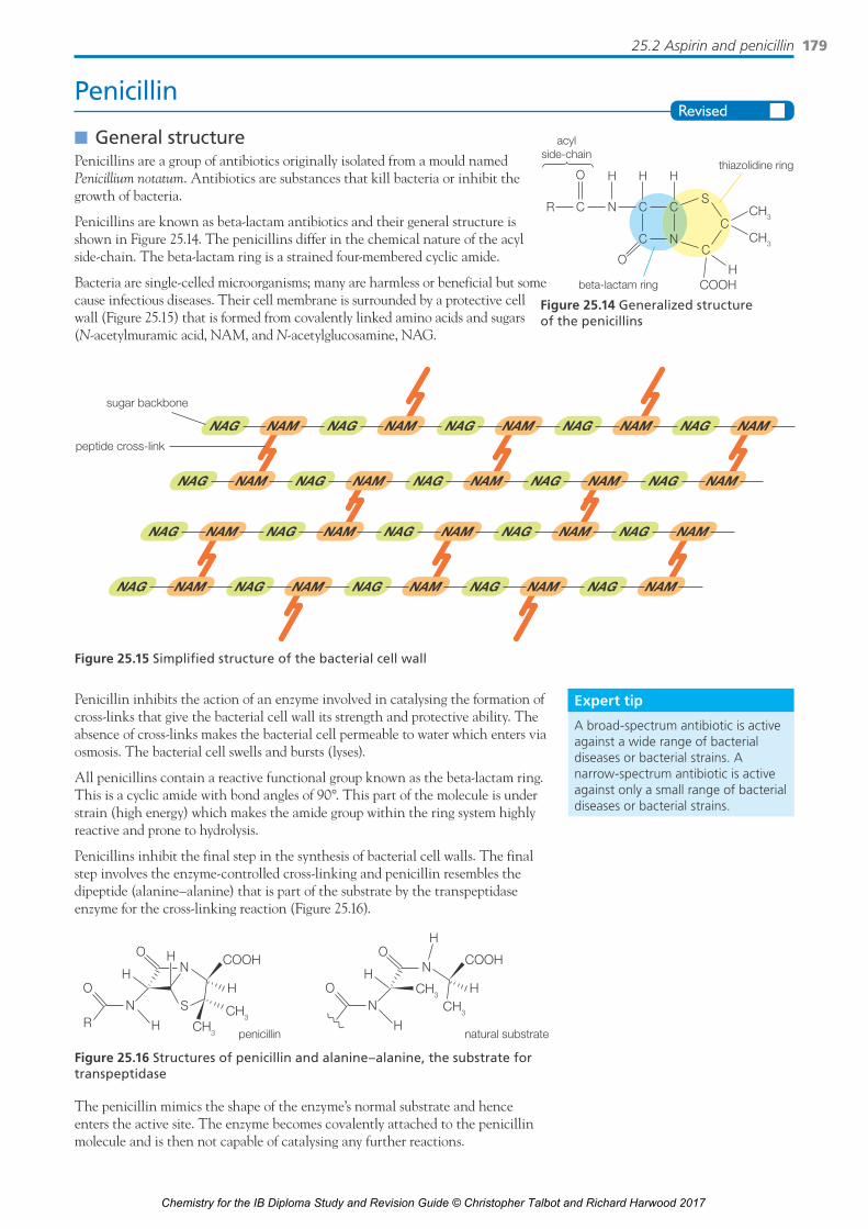
Penicillin
■n General structurePenicillins are a group of antibiotics originally isolated from a mould named Penicillium notatum. Antibiotics are substances that kill bacteria or inhibit the growth of bacteria.
Penicillins are known as beta-lactam antibiotics and their general structure is shown in Figure 25.14. The penicillins differ in the chemical nature of the acyl side-chain. The beta-lactam ring is a strained four-membered cyclic amide.
Bacteria are single-celled microorganisms; many are harmless or beneficial but some cause infectious diseases. Their cell membrane is surrounded by a protective cell wall (Figure 25.15) that is formed from covalently linked amino acids and sugars (N-acetylmuramic acid, NAM, and N-acetylglucosamine, NAG.
CH3
CH3
COOHH
acylside-chain
beta-lactam ring
thiazolidine ring
C N
H
R
O
C
H
CS
C
C
H
C
O
N
Figure 25.14 Generalized structure of the penicillins
Expert tip
A broad-spectrum antibiotic is active against a wide range of bacterial diseases or bacterial strains. A narrow-spectrum antibiotic is active against only a small range of bacterial diseases or bacterial strains.
Penicillin inhibits the action of an enzyme involved in catalysing the formation of cross-links that give the bacterial cell wall its strength and protective ability. The absence of cross-links makes the bacterial cell permeable to water which enters via osmosis. The bacterial cell swells and bursts (lyses).
All penicillins contain a reactive functional group known as the beta-lactam ring. This is a cyclic amide with bond angles of 90°. This part of the molecule is under strain (high energy) which makes the amide group within the ring system highly reactive and prone to hydrolysis.
Penicillins inhibit the final step in the synthesis of bacterial cell walls. The final step involves the enzyme-controlled cross-linking and penicillin resembles the dipeptide (alanine–alanine) that is part of the substrate by the transpeptidase enzyme for the cross-linking reaction (Figure 25.16).
COOH
HN
R H
HH
S
O
O
N
CH3
CH3
COOH
HN
H
HH
O
O
N
CH3
CH3
penicillin natural substrate
COOH
HN
R H
HH
S
O
O
N
CH3
CH3
COOH
HN
H
HH
O
O
N
CH3
CH3
penicillin natural substrate
Figure 25.16 Structures of penicillin and alanine–alanine, the substrate for transpeptidase
The penicillin mimics the shape of the enzyme’s normal substrate and hence enters the active site. The enzyme becomes covalently attached to the penicillin molecule and is then not capable of catalysing any further reactions.
Figure 25.15 Simplified structure of the bacterial cell wall
NAMNAGNAMNAGNAMNAGNAMNAGNAMNAG
NAMNAGNAMNAGNAMNAGNAMNAGNAMNAG
NAM
sugar backbone
peptide cross-link
NAGNAMNAGNAMNAGNAMNAGNAMNAG
NAMNAGNAMNAGNAMNAGNAMNAGNAMNAG
Chemistry for the IB Diploma Study and Revision Guide © Christopher Talbot and Richard Harwood 2017

25 Medicinal chemistry180
■n Antibiotic resistanceSome bacteria in a population of a strain of bacteria may be naturally resistant to the effect of an antibiotic. For example, some strains of bacteria secrete an enzyme known as penicillinase (β-lactamase) (Figure 25.17) which hydrolyses penicillin G (where R is the benzyl group, –CH2–C6H5). Penicillin G was the first penicillin to be developed but had to be injected intravenously because it was hydrolysed by the acid in the stomach. The activity of penicillinase can be potentiated by the co-administration of β-lactase inhibitors such as clavulanic acid.
HN
N
COO−
R
penicillin
S
O
O
HN
HNOH
COO−
R
penicilloic acid
S
O
O
β-lactamase
β-lactam ring
β-Lactamase breaks the peptidebond in the β-lactam ring ofpenicillin to disable the molecule.Bacteria with this enzyme canresist the effects of penicillin andother β-lactam antibiotics.
Figure 25.17 Action of pencillinase (beta-lactamase)
Medicinal chemists have synthesized a large number of other penicillins which have a chemically modified side-chain, for example, penicillin V where R = C6H5OCH2–). This molecule has a different structure and shape from penicillin G and does not act as a substrate for pencillinase. It is resistant to gastric juice.
Patients not completing the full prescribed course of antibiotics and the use of antibiotics in animal feeds have contributed to the development of antibiotic resistance.
Bacteria either have pre-existing resistance to drugs, or they develop resistance. Often resistance to a certain drug from a particular class leads to resistance to all other drugs in that class. Mechanisms involve enzymes that break down antibiotics or pump them out from the cytoplasm. Over-prescription of antibiotics also increases the chance of antibiotic-resistant strains developing or being selected for. The antibiotics can also enter humans via eating meat and dairy products.
Expert tip
Compliance with a course of antibiotics means that the full course of antibiotics is taken and administered at the correct times.
n QUICK CHECK QUESTIONS
10 Identify the functional group present in the β-lactam ring and explain why the ring is important in the functioning of penicillin as an antibacterial.
11 Modern penicillins have a similar structure to penicillin G but a different side-chain. State two advantages of modifying the side-chain.
12 The efficiency of certain drugs is strongly dependent on the frequency and regularity of their administration. Explain the importance of patient compliance when the patient is treated with antibacterials.
NATURE OF SCIENCEScientific creativity takes various forms. One of these is serendipity – discovering phenomena while diverting from intended research. In 1928 Alexander Fleming returned to his laboratory in a London hospital, he picked up a culture plate of the Staphylococcus bacteria that he had left on the bench for some weeks. A contaminating mould had grown on the dish and around it for some distance the bacterial colonies were absent or dead. Subsequent research by Fleming revealed that the ‘mould juice’ was effective against a wide range of bacterial strains including many that are highly pathogenic to humans.
25.3 OpiatesEssential idea: Potent medical drugs prepared by chemical modification of natural products can be addictive and become substances of abuse.
Opiatesn The ability of a drug to cross the blood–brain barrier depends on its chemical
structure and solubility in water and lipids.n Opiates are natural narcotic analgesics that are derived from the opium poppy.
Chemistry for the IB Diploma Study and Revision Guide © Christopher Talbot and Richard Harwood 2017

25.3 Opiates 181
n Morphine and codeine are used as strong analgesics. Strong analgesics work by temporarily bonding to receptor sites in the brain, preventing the transmission of pain impulses without depressing the central nervous system.
n Medical use and addictive properties of opiate compounds are related to the presence of opioid receptors in the brain.
OpiatesOpiates are natural narcotic analgesics that are derived from the opium poppy (Papaver somniferum). The unripe seed pods contain opium and are the source of morphine and codeine. Opiates are natural alkaloids found in opium. Alkaloids are nitrogen-containing bases extracted from plants (Figure 25.18).
Morphine is a powerful painkiller (strong analgesic) with sleep-inducing properties (narcotic). Morphine is available only on prescription and is given to relieve the pain caused by severe injury, surgery and cancer, although patients often develop tolerance and dependence. It can be taken orally or injected. Codeine is used in cough mixtures and is a less powerful analgesic. Codeine is a pro-drug and in the body about 10% of codeine is converted to morphine.
Diamorphine (heroin) is easily synthesized from morphine via a simple single-step synthesis. Morphine can also be converted to codeine, by replacing an alcohol group, –OH, by an ether group, –OCH3 (methoxy). This can be achieved by reacting morphine with CH3I in KOH(aq).
The structure of morphine consists of five rings forming a T-shaped molecule. The important binding groups on morphine are the phenol, the benzene ring, and the ionized amine. All the opiates have a common structural feature known as the phenylamine chemical moiety which gives the molecule rigidity and allows it to interact strongly with the opioid receptors.
The opiates work as powerful analgesics by binding and interacting with opioid protein receptors (Figure 25.19) on the surfaces of brain cells (neurons). They prevent nerve impulses (pain signals) from causing changes inside brain cells without depressing the central nervous system (brain and spinal cord).
Opioids is a more general term used to describe all compounds with structures similar to morphine. The term narcotic originally referred medically to any compound with any sleep-inducing properties. It has since become associated with opiates and opioids.
■n Diamorphine (heroin)Diamorphine (heroin) is a semi-synthetic opiate. It can be prepared from morphine by reaction with ethanoic anhydride in an acetylation reaction (–O–CO–R is an acetyl group) (Figure 25.20).
O
O
HO
H
H H
15 16+
NHCH3
bindinggroups
London(dispersion forces)
hydrogen bonding
ionic
Figure 25.19 Important functional groups for analgesic activity in protonated morphine
Figure 25.18 Structures of morphine, codeine and heroin (diamorphine)
c
O
NCH
2
CH2
CH3
heroin
OCCH3
O
OCCH3
O
b
O
N
OH
CH2
CH2
CH3
codeine
OCH3
a
OH
O
N
OH
CH2
CH2
CH3
morphine
Chemistry for the IB Diploma Study and Revision Guide © Christopher Talbot and Richard Harwood 2017

25 Medicinal chemistry182
During the acetylation of morphine both hydroxyl groups are substituted with ester groups which significantly reduces the polarity of the molecule. This increases its lipophilicity (ability to dissolve in lipids) and hence its ability to cross the blood–brain barrier (Figure 25.21).
+ +O
OH
N CH2
CH2
H3C
OH
2CH3COOH
CH2
O
N CH2H3C
OC
O
O
CO
CH3
CH3
C
O O
CO CH32H3C
Figure 25.20 The formation of diamorphine (heroin)
In the brain, diamorphine is rapidly metabolized into morphine, which binds to the opioid receptor. This makes diamorphine about five times more effective an analgesic than morphine when injected into the blood because it crosses the blood–brain barrier in greater quantities. The morphine and diamorphine molecules both contain a tertiary amine group and can both be converted into ionic salts by reacting with hydrochloric acid to form a soluble tertiary ammonium salt. Morphine is often injected in the form of morphine hydrochloride to increase its bioavailability. It reverts back to the undissociated or free base form to cross the blood–brain barrier.
Effects of opiatesAll of the classical opiates can cause addiction and lead to the development of tolerance to dependence. Withdrawal symptoms occur within one day for addicts if the drug usage is stopped. These include hot and cold sweats, diarrhoea, anxiety, weakness and muscle cramps that can last for months.
The short-term and long-term effects of strong opiates are summarized in Table 25.1.
Table 25.1 Short-term and long-term effect of opiates
Short-term effects Long-term effects
Induces a sense of euphoria (great happiness) Constipation (anti-diarrhoeal)
Dulling of pain (analgesic) Loss of sex drive
Depress nervous system (sedation); vasodilation (blood vessels widen) Disrupts the menstrual cycle
Slow breathing rate and heart rate Reduced appetite
Cough reflex inhibited (antitussive) Risk of HIV, hepatitis infection, etc. through the use of shared needles
High dosages can lead to coma or death via suffocation Social problems, such as theft and prostitution
n QUICK CHECK QUESTIONS
13 Identify the reagent and by-product when diamorphine (heroin) is synthesized from morphine.
14 Discuss how the differences in structure between morphine and diamorphine (heroin) affect their absorption across the brain–blood barrier.
Figure 25.21 The blood–brain barrier: many large drug molecules cannot pass through the capillary wall and glial cells
glial cell
blood
capillary wall
drug molecule
Chemistry for the IB Diploma Study and Revision Guide © Christopher Talbot and Richard Harwood 2017
25.4 pH regulation of the stomach 183
Some side effects can be advantageous. For example, the observation that morphine causes constipation has led to the design of opioids which are used in the treatment of diarrhoea. Euphoria can be a useful side effect when treating pain in terminally ill patients. However, the effect is not observed in patients suffering severe pain.
Methadone (Figure 25.22) is a synthetic opioid which is frequently used to treat heroin addicts. Although chemically different from morphine and heroin, it acts on the same opioid receptors in the brain and produces many of the same effects, with the exception of the euphoria.
NO
Figure 25.22 Structure of methadone
Methadone’s usefulness in treating heroin addicts is due to its long duration of effect and its ability to block the heroin withdrawal symptoms. At high concentrations it can block the euphoric effects of heroin and morphine.
25.4 pH regulation of the stomachEssential idea: Excess stomach acid is a common problem that can be alleviated by compounds that increase the stomach pH by neutralizing or reducing its secretion.
pH regulation of the stomachn Non-specific reactions, such as the use of antacids, are those that work to
reduce the excess stomach acid.n Active metabolites are the active forms of a drug after it has been processed by
the body.
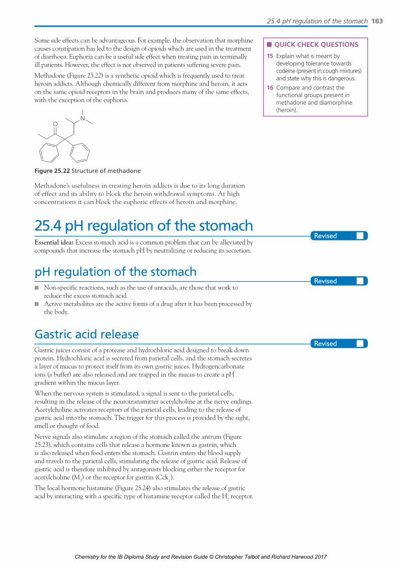
Gastric acid releaseGastric juices consist of a protease and hydrochloric acid designed to break down protein. Hydrochloric acid is secreted from parietal cells, and the stomach secretes a layer of mucus to protect itself from its own gastric juices. Hydrogencarbonate ions (a buffer) are also released and are trapped in the mucus to create a pH gradient within the mucus layer.
When the nervous system is stimulated, a signal is sent to the parietal cells, resulting in the release of the neurotransmitter acetylcholine at the nerve endings. Acetylcholine activates receptors of the parietal cells, leading to the release of gastric acid into the stomach. The trigger for this process is provided by the sight, smell or thought of food.
Nerve signals also stimulate a region of the stomach called the antrum (Figure 25.23), which contains cells that release a hormone known as gastrin, which is also released when food enters the stomach. Gastrin enters the blood supply and travels to the parietal cells, stimulating the release of gastric acid. Release of gastric acid is therefore inhibited by antagonists blocking either the receptor for acetylcholine (M3) or the receptor for gastrin (Cck2).
The local hormone histamine (Figure 25.24) also stimulates the release of gastric acid by interacting with a specific type of histamine receptor called the H2 receptor.
n QUICK CHECK QUESTIONS
15 Explain what is meant by developing tolerance towards codeine (present in cough mixtures) and state why this is dangerous.
16 Compare and contrast the functional groups present in methadone and diamorphine (heroin).
Chemistry for the IB Diploma Study and Revision Guide © Christopher Talbot and Richard Harwood 2017

25 Medicinal chemistry184
AntacidsDyspesia, or indigestion, occurs when excess gastric juice (hydrochloric acid) is secreted by the stomach and there is reflux into the oesphagus. Antacids are medicines to reduce excess hydrochloric acid in the stomach. They are weak bases and remove the excess hydrogen ions via neutralization. Suitable compounds to act as antacids are metal hydroxides (unless alkalis), metal carbonates, metal hydrogencarbonates and some metal oxides.
Expert tip
Alkalis are not suitable for use as antacids. They are highly corrosive and their neutralization is more exothermic than an insoluble weak base. Typical neutralization reactions are:
NaHCO3(s) + HCl(aq) → NaCl(aq) + H2O(l) + CO2(g)
CaCO3(s) + 2HCl(aq) → CaCl2(aq) + H2O(l) + CO2(g)
MgO(s) + 2HCl(aq) → MgCl2(aq) + H2O(l)
Al(OH)3(s) + 3HCl(aq) → AlCl3(aq) + 3H2O(l)
Ca(OH)2(s) + 2HCl(aq) → CaCl2(aq) + 2H2O(l)
A calculation based on the mole concept and a balanced equation allows the most effective antacid by mass to be determined.
Worked example
Rolaids is a commercial antacid used to treat dyspepsia caused by excess acidity in the stomach. Its active ingredients are calcium carbonate (500 mg) and magnesium hydroxide (100 mg).
Write balanced equations for the reactions of magnesium hydroxide and calcium carbonate with the acid present in gastric juice
Mg(OH)2 + 2HCl → MgCl2 + 2H2O
CaCO3 + 2HCl → CaCl2 + H2O + CO2
State one reason why potassium hydroxide is not used in antacids.
It is an alkali/strong base; corrosive with a high enthalpy of neutralization.
State one problem when using large amounts of calcium carbonate compared with magnesium hydroxide in Rolaids.
Bloating/flatulence/production of carbon dioxide gas, CO2.
Expert tip
Note that aluminium hydroxide neutralizes three moles of acid (H+) per mole of base compared to two moles of acid (H+) neutralized per mole of calcium hydroxide.
n QUICK CHECK QUESTION
17 A 1.20 g antacid tablet contains 80.0% by mass of magnesium hydroxide as the active ingredient. Deduce what volume of 0.1500 mol dm−3 HCl the antacid tablet can neutralize.
duodenum(small intestine)
pyloric sphincter
antrum
stomach
oesophagushistamine
acetylcholine(neurotransmitter)
gastrin (hormone)
cAMP
parietalcells
protompump
stomach
++
+
++
HCl(aq)
H+(aq) Cl–(aq)
H2
M3
CcK2
receptors
ion channel
key:
Figure 25.23 Factors influencing the release of gastric acid
HN
NH2N
Figure 25.24 Structure of histamine
Chemistry for the IB Diploma Study and Revision Guide © Christopher Talbot and Richard Harwood 2017

25.4 pH regulation of the stomach 185
Table 25.2 is a comparison of compounds used in antacid preparations.
Table 25.2 A comparison of compounds used in antacid preparation.
Antacid compound Action Side effect
Calcium hydroxide, Ca(OH)2
Although is a strong base, Ca(OH)2 is only slightly soluble in water, and the solution is thus weakly basic. A saturated solution of calcium hydroxide (limewater) rapidly neutralizes stomach acid.
A strong base is rarely used as an antacid as extended use can lead to tissue damage.
Magnesium hydroxide, Mg(OH)2
Mg(OH)2 also rapidly neutralizes stomach acid but has a laxative effect and can cause diarrhoea.
Laxative properties; excess quantities can lead to toxicity; effects include severe allergic reactions, nausea and black faeces.
Aluminium hydroxide, Al(OH)3
Aluminium hydroxide has a very low solubility in water and dissolves slowly in the stomach to relieve indigestion. It may cause constipation.
The aluminium ion, Al3+, has a high charge density due to its high charge to size ratio and can effectively bind drugs, calcium or phosphate ions thus inhibiting its absorption; depletion of calcium ions, Ca2+, leads to a risk of weaker bones.
Calcium carbonate, CaCO3
Calcium carbonate, CaCO3, is a strong fast-acting antacid and the same mass works longer than that of sodium hydrogencarbonate, NaHCO3.
Repeated use as an antacid may lead to excessive amounts of calcium ions being absorbed into the body and may result in kidney stones; calcium carbonate, CaCO3, can produce the acid rebound effect resulting in the stomach quickly having excess gastric juice within a short time.
Sodium hydrogencarbonate, NaHCO3, as a solid mixture with citric acid
Reacts in water to release carbon dioxide, CO2 gas; induces belching and flatulence, thus relieving discomfort. NaHCO3 is soluble in water, works quickly and provides short-term relief.
Fluid retention (bloating) and alkalosis (low pH) due to high HCO3
−(aq) concentration. High Na+(aq) concentration leads to high blood pressure, heart failure or kidney problems and may lead to hypertension (high blood pressure).
n QUICK CHECK QUESTIONS
18 An antacid called magnesium trisilicate may be recommended for recurring heart burn or indigestion. Its use may lead to high a magnesium level and muscle weakness.
Mg2+
Mg2+
SiO O
Si = OO = Si
O−−O
O− −O
a State the types of bonding present in magnesium silicate.
b State one function of the hydrochloric acid present in gastric juice.
c Magnesium trisilicate reacts with hydrochloric acid to form silicon dioxide, water and magnesium chloride. Write a balanced equation for this reaction. State a possible effect of the silicon dioxide.
19 A laboratory test to determine how much hydrochloric acid is neutralized by a brand of antacid does not give a complete picture of its effectiveness in the stomach. State and explain what other factors might be important when assessing antacids.
20 Some antacids fizz when dissolved in a glass of water. They contain sodium hydrogen carbonate and citric acid. Write an ionic equation for the reaction.
21 Explain how heartburn is caused.
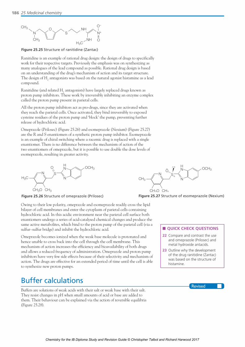
Specific inhibition of hydrochloric acid production in the stomachExcessive production of hydrochloric acid can be inhibited using drugs, such as ranitidine (Zantac) (Figure 25.25), which act as an antagonist of the histamine-H2 protein receptor on parietal cells, which is responsible for promoting hydrochloric acid production.
Chemistry for the IB Diploma Study and Revision Guide © Christopher Talbot and Richard Harwood 2017
25 Medicinal chemistry186
Ranitidine is an example of rational drug design: the design of drugs to specifically work for their respective targets. Previously the emphasis was on synthesizing as many analogues of the lead compound as possible. Rational drug design is based on an understanding of the drug’s mechanism of action and its target structure. The design of H2 antagonists was based on the natural agonist histamine as a lead compound.
Ranitidine (and related H2 antagonists) have largely replaced drugs known as proton pump inhibitors. These work by irreversibly inhibiting an enzyme complex called the proton pump present in parietal cells.
All the proton pump inhibitors act as pro-drugs, since they are activated when they reach the parietal cells. Once activated, they bind irreversibly to exposed cysteine residues of the proton pump and ‘block’ the pump, preventing further release of hydrochloric acid.
Omeprazole (Prilosec) (Figure 25.26) and esomeprazole (Nexium) (Figure 25.27) are the R and S enantiomers of a synthetic proton pump inhibitor. Esomeprazole is an example of chiral switching where a racemic drug is replaced with a single enantiomer. There is no difference between the mechanism of action of the two enantiomers of omeprazole, but it is possible to use double the dose levels of esomeprazole, resulting in greater activity.
OSN
NHCH3
N+
CH3 NHH3C
O−
O
Figure 25.25 Structure of ranitidine (Zantac)
Owing to their low polarity, omeprazole and esomeprazole readily cross the lipid bilayer of cell membranes and enter the cytoplasm of parietal cells containing hydrochloric acid. In this acidic environment near the parietal cell surface both enantiomers undergo a series of acid-catalysed chemical changes and produce the same active metabolites, which bind to the proton pump of the parietal cell (via a sulfur–sulfur bridge) and inhibit the hydrochloric acid.
Omeprazole becomes ionized when the weak base molecule is protonated and hence unable to cross back into the cell through the cell membrane. This mechanism of action increases the efficiency and bioavailability of both drugs and allows a reduced frequency of administration. Omeprazole and proton pump inhibitors have very few side effects because of their selectivity and mechanism of action. The drugs are effective for an extended period of time until the cell is able to synthesize new proton pumps.
Buffer calculationsBuffers are solutions of weak acids with their salt or weak base with their salt. They resist changes in pH when small amounts of acid or base are added to them. Their behaviour can be explained via the action of reversible equilibria (Figure 25.28).
n QUICK CHECK QUESTIONS
22 Compare and contrast the use and omeprazole (Prilosec) and metal hydroxide antacids.
23 Outline why the development of the drug ranitidine (Zantac) was based on the structure of histamine.
H3C
CH3
NS
OCH3OHN
CH3O
N
Figure 25.26 Structure of omeprazole (Prilosec)
CH3
CH3
N
OCH3HN
CH3O
N S+
O−
Figure 25.27 Structure of esomeprazole (Nexium)
Chemistry for the IB Diploma Study and Revision Guide © Christopher Talbot and Richard Harwood 2017

25.4 pH regulation of the stomach 187
A buffer consists of a mixture of a weak conjugate acid–base pair, here represented as HX and X−. When a small portion of OH− is added to the buffer (left), it reacts with HX, decreasing [HX] and increasing [X−] in the buffer. When a small portion of H+ is added to the buffer (right), it reacts with X−, decreasing [X−] and increasing [HX] in the buffer.
The pH of a buffer solution can be calculated with the logarithmic form of the equilibrium law applied to the dissociation of a weak acid:
pH = pKa + log10([salt]/[acid])
This expression is known as the Henderson–Hasselbalch equation. The buffer will be most effective when the concentration of the weak acid is equal to the concentration of the salt of the weak acid.
Worked example
Calculate the pH of a buffer containing 0.100 mol dm−3 phenylamine, C6H5NH2, and 0.250 mol dm−3 phenylammonium chloride, C6H5NH3
+. Ka of C6H5NH3+ =
2.63 × 10⁻⁵.
pH = pKa + log10 ([A−(aq)]/[HA(aq)]
pH = 4.58 + log10 (0.1/0.25) = 4.58 + (−0.398) = 4.18
Expert tip
Diluting a buffer solution with water does not change the ratio of the concentrations of the salt and acid so the pH does not change (unless the dilution is so great that the assumptions made when deriving the equation no longer apply).
buffer afteraddition of HO−
HXOH− H+
HXX− X− HX X−
buffer afteraddition of H+
buffer with equalconcentrations ofweak acid and itsconjugate base
OH− + HX HXH+ + X−H2O + X−
Figure 25.28 Action of an acidic buffer
Calculations with the Henderson–Hasselbalch equation may require conversions involving H+ and pH; Ka and pKa and calculations of concentrations from masses of pure substances and volumes of solutions.
How buffer solutions limit changes in pHIt is possible to show quantitatively the ability of buffer solutions to limit changes in pH. For example, a buffer made from 0.60 mol dm−3 HClO (Ka = 3.7 × 10⁻⁸) and 0.20 mol dm−3 NaClO has a pH of 7.0.
If 0.01 moles of HCl are added to 100 cm3 of the buffer:
ClO−(aq) + H3O+(aq) → HClO(aq) + H2O(l)
Initial amounts: 0.02 0.01 0.06
Final amounts: 0.01 – 0.07
So the [base]/[acid] ratio is 0.01/0.07; pH = pKa + log10([base]/[acid]) = 6.58 (a change of 0.4 units). If 0.01 moles of HCl were added to 100 cm3 of pure water, the pH of the resulting solution would be 1.0 (a change of 6 pH units).
Chemistry for the IB Diploma Study and Revision Guide © Christopher Talbot and Richard Harwood 2017

25 Medicinal chemistry188
If 0.01 moles of NaOH are added to the buffer:
HClO(aq) +OH–(aq) → ClO−(aq) + H2O(l)
Initial amounts: 0.06 0.01 0.02
Final amounts: 0.05 – 0.03
So the [base]/[acid] ratio is 0.03/0.05; pH = pKa + log10([base]/[acid]) = 7.21 (a change of 0.2 pH units). If 0.01 moles of NaOH were added to 100 cm3 of pure water, the pH of the resulting solution would be 13.0.
Hence buffer solutions do not significantly change their pH on adding small quantities of acid or alkali.
However, it is possible to exceed the buffering capacity of a buffer solution by adding too much acid or alkali; in such cases the buffer will be unable to maintain the pH and the pH will change dramatically. This would be the case of more than 0.02 moles of HCl or more than 0.06 moles of NaOH were added to the above mixture.
Worked example
Determine the pH of a buffer solution formed by adding 50.00 cm3 of 0.0100 mol dm−3 sulfuric acid to 50.00 cm3 of 0.0400 mol dm−3 methylamine.
n(H2SO4) = (50.00/1000) dm−3 × 0.0100 mol dm−3 = 0.5n(CH3NH2)reacted = 0.5n(CH3NH3
+)buffer; n(CH3NH2)buffer = (50.00/1000) dm3 × 0.0400 mol dm−3 − 2 × (50.00/1000) dm3 × 0.0100 mol dm−3; n(CH3NH2) = n(CH3NH3
+); [CH3NH2] = [CH3NH3
+] in buffer; pOH = pKb = 3.34, pH = 10.66.
Worked example
Calculate the ratio of methanoate ions/methanoic acid to give a buffer of pH 4.00. The pK of methanoic acid is 3.75.
pH = pKa + log10[base]/[acid]; 4.00 = 3.75 + log10 [HCO2–]/[HCOOH]; so [HCO2
−]/[HCOOH] = 100.25 = 1.78
Worked example
Calculate the amount (in mol) of sodium hydroxide that must be added to 100.0 cm3 of 0.20 mol dm−3 HCOOH to prepare a solution buffered at pH 4.00.
If the concentration of OH− which is added is x mol dm−3 then this will react with HCOOH to produce HCOO− so that: [HCOOH] = (0.20 − x) mol dm−3 and [HCOO− = x mol dm−3. If the pH = 4.00, then [HCOO−]/[HCOOH] = 1.78. Hence: x/(0.20 − x) = 1.78, x = 0.13. To achieve [OH − (aq)] = 1.13 mol dm−3 in 100 cm3, the amount of NaOH that must be added is 0.13 mol dm−3 × 0.1000 dm3 = 0.013 mol.
n QUICK CHECK QUESTIONS
24 The pH of blood is 7.4 and is regulated by the bicarbonate buffering system composed of carbonic acid, H2CO3, and hydrogencarbonate ion, HCO3
−.
a Using relevant chemical equations, show how this buffering system keeps the pH of blood constant when a small amount of acid or alkali is added.
b The pKa of carbonic acid is 6.1. Calculate the ratio of the concentrations of carbonic acid and hydrogencarbonate in blood and comment on the buffering capacity of blood.
25 HEPES is a zwitterionic buffer that can be made by dissolving solid sodium hydroxide in a HEPES solution.
Calculate the pH of the buffer solution formed when 20.00 g of sodium hydroxide is added to 1.00 dm3 of a 1.00 mol dm−3 solution of HEPES (pKa = 7.50). Assume that there is no change in volume when the sodium hydroxide is added.
S
N
N
O O
O
HOHO
S
N
N
O O
O−
HO
H
HEPES
+ OH− + H2O
Chemistry for the IB Diploma Study and Revision Guide © Christopher Talbot and Richard Harwood 2017

25.5 Anti-viral medications 189
25.5 Anti-viral medicationsEssential idea: Anti-viral medications have recently been developed for some viral infections while others are still being researched.
Anti-viral medicationsn Viruses lack a cell structure and so are more difficult to target with drugs than
bacteria.n Anti-viral drugs may work by altering the cell’s genetic material so that the
virus cannot use it to multiply. Alternatively, they may prevent the viruses from multiplying by blocking enzyme activity within the host cell.
BacteriaBacteria (Figure 25.29) are single-celled (unicellular) microorganisms that reproduce outside cells by binary division (a form of asexual reproduction). Bacterial cells are too large to enter human cells.
Bacteria can exchange genetic material and acquire genes for antibiotic resistance by a process known as conjugation (involving the pili). There is no nuclear membrane surrounding their DNA and no membrane bound structures within their cytoplasm. They have a protective cell wall and some species have a rotating flagellum for movement.
VirusesViruses are much smaller and simpler in structure than bacteria. Viruses (Figure 25.30) contain DNA or RNA surrounded by a capsid composed of regularly packed capsomeres, each containing a number of protein molecules. Some viruses, such as HIV, have a cell membrane formed from their host cell. Viruses are non-cellular (acellular): there is no nucleus or cytoplasm. They cannot self-multiply and can only replicate inside a living cell (host cell). Viruses are typically 100× smaller than bacteria.
pili – enable attachment tosurfaces and to other bacteriafor exchange of genetic material
ribosomes* – sitesof protein synthesis
nucleoid* – genetic material:a single circular chromosomeof about 4000 genes
cell wall* – protects cell from rupturecaused by osmosis, and possible harmfrom other organisms
cytoplasm* – site of thechemical reactions of life (metabolism)
flagella – bring aboutmovement of the bacterium
lipid globules/glycogen granules
mesosome
plasmid – double stranded circular DNA,often carries genes for antibiotic resistance
plasma membrane* – a barrier across which all nutrientsand waste products must pass
* structures thatoccur in all bacteria
1µm
Figure 25.29 The structure of Escherichia coli – a ‘typical’ bacterium
Chemistry for the IB Diploma Study and Revision Guide © Christopher Talbot and Richard Harwood 2017
25 Medicinal chemistry190
Once a virus or its nucleic acid enters a cell the host cell’s enzymes and ribosomes are used to make new viral proteins and enzymes that self-assemble into viruses. The viruses will then exit from the cell through the cell membrane (lysis), leaving behind a dead or damaged cell.
■n RetrovirusesIn many RNA and DNA viruses, such as flu and herpes, nucleic acid replication occurs entirely in the cytoplasm.
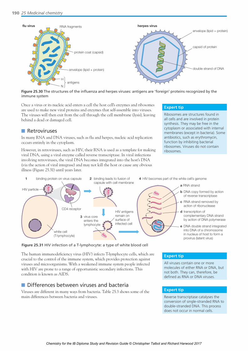
However, in retroviruses, such as HIV, their RNA is used as a template for making viral DNA, using a viral enzyme called reverse transcriptase. In viral infections involving retroviruses, the viral DNA becomes integrated into the host’s DNA (via the action of viral integrase) and may not kill the host or cause any obvious illness (Figure 25.31) until years later.
Expert tip
Ribosomes are structures found in all cells and are involved in protein synthesis. They may be free in the cytoplasm or associated with internal membranes (except in bacteria). Some antibiotics, such as erythromycin, function by inhibiting bacterial ribosomes. Viruses do not contain ribosomes.
Expert tip
All viruses contain one or more molecules of either RNA or DNA, but not both. They can, therefore, be defined as RNA or DNA viruses.
flu virus herpes virusRNA fragments
protein coat (capsid)
envelope (lipid + protein)
Hantigens
N
capsid of protein
envelope (lipid + protein)
double strand of DNA
Figure 25.30 The structures of the influenza and herpes viruses: antigens are ‘foreign’ proteins recognized by the immune system
HIV particle
1 4binding protein on virus capsule binding leads to fusion ofcapsule with cell membrane
HIV becomes part of the white cell’s genome
a RNA strand
b DNA copy formed by action of reverse transcriptase
c RNA strand removed by action of ribonuclease
d transcription of complementary DNA strand by action of DNA polymerase
e DNA double strand integrated into DNA of a chromosome in nucleus of host to form a provirus (latent virus)
virus coreenters thelymphocyte
HIV antigensremain onsurface ofinfected cell
white cell(T-lymphocyte)
CD4 receptor
2
3
Figure 25.31 HIV infection of a T-lymphocyte: a type of white blood cell
The human immunodeficiency virus (HIV) infects T-lymphocyte cells, which are crucial to the control of the immune system, which provides protection against viruses and microorganisms. With a weakened immune system people infected with HIV are prone to a range of opportunistic secondary infections. This condition is known as AIDS.
■n Differences between viruses and bacteriaViruses are different in many ways from bacteria. Table 25.3 shows some of the main differences between bacteria and viruses.
Expert tip
Reverse transcriptase catalyses the conversion of single-stranded RNA to double-stranded DNA. This process does not occur in normal cells.
Chemistry for the IB Diploma Study and Revision Guide © Christopher Talbot and Richard Harwood 2017

25.5 Anti-viral medications 191
Table 25.3 The main differences between bacteria and viruses
Bacteria Viruses
Ribosomes Present Absent
Number of cells Unicellular; one cell (but can form cooperating colonies) No cells (acellular)
Internal structure DNA floating freely in cytoplasm; has cell wall and cell membrane
DNA or RNA enclosed inside a coat (capsid) of protein or glycoproteins
Cell wall composition Peptidoglycan/lipopolysaccharide No cell wall; protein coat (capsid) present instead
Treatment Antibiotics Vaccines prevent the spread of infection and anti-viral drugs help to slow replication but cannot stop it completely
Enzymes Yes Yes in some, for example reverse transcriptase in retroviruses
Nucleus No – the nuclear material (nucleoid) is not surrounded by a nuclear membrane
No
Virulence Yes Yes
Infection Localized Systemic
Reproduction Binary fission – a form of asexual reproduction Invades a host cell and takes over the cell, causing it to make copies of the viral genome (DNA or RNA); destroys or damages the host cell, releasing new viruses
Size Larger (1000 nm Smaller (20–400 nm)
Anti-viral drugsAnti-virals are medicinal drugs (medications) useful in treating or controlling viral infections. Viruses are a serious health threat and there is a need for new anti-viral drugs. Vaccination is effective against many viruses, but is less effective against viruses that readily mutate. Anti-viral drug research has been helped by advantages in molecular biology, genetic engineering, computer modelling and X-ray crystallography.
In general, there are four types of actions for anti-viral drugs:
n Preventing the genetic material from being injected through the cell membrane (Figure 25.32); viruses have to interact and bind with specific receptors (proteins or glycoproteins) on the cell membrane of a cell and release its genetic material.
n To block entry, anti-viral molecules can be synthesized that are structurally similar to the virus-associated protein so they bind strongly to the receptor or even bind to the viral capsid (protein coat). They can also inhibit the uncoating process: the release of viral nucleic acids from the capsid that covers them.
n Inhibiting the replication of the virus: the drug may mimic nucleotides, the monomers of DNA or RNA, so that they are incorporated into the enzyme-controlled synthesis of DNA or RNA, which is then terminated (Figure 25.33). These anti-viral agents are known as nucleoside analogues.
n Inhibiting the action of reverse transcriptase present in retroviruses, for example HIV (Figure 25.34).
NH2
HC
NH2H3C
Figure 25.32 Structures of amantadine and rimantadine: uncoating inhibitors
n QUICK CHECK QUESTIONS
26 State the function of reverse transcriptase in the HIV virus and other retroviruses.
27 Outline why viruses are often more difficult to target with drugs than bacteria.
HO
N3
CH2
H3C
CC
C
C
O
OO
N
N
H
H
HH
H
Figure 25.33 Structure of azidothymidine (AZT) or Zidovudine, used in the clinical treatment of AIDS
O
NN N
NH
Figure 25.34 Nevirapine, a non-nucleotide reverse transcriptase inhibitor for the treatment of HIV infection
Chemistry for the IB Diploma Study and Revision Guide © Christopher Talbot and Richard Harwood 2017

25 Medicinal chemistry192
Preventing new viruses from leaving the cell: new DNA and viral proteins self-assemble into new viruses (viral particles). Following rupture of host cell membranes (or budding), these viruses leave the host cell; this then results in new infections in other cells of the body. Drugs may be developed that prevent the exit of the mature viruses.
Designing safe and effective anti-viral drugs is difficult, because viruses use the host’s cells to replicate. This makes it difficult to find targets for the drug that would interfere with the virus without also harming the host organism’s cells. The major problem in developing vaccines and anti-viral drugs is due to viral variation and rapid evolutionary change leading to drug resistance. Many anti-virals can only slow down or sometimes stop the replication of the virus.
■n HIV infectionHIV is spread from contact with infected blood, semen or vaginal fluids. Most people get the virus by having unprotected sex with someone who has HIV. Another common way is through sharing drug needles with someone who is infected with HIV. The virus can also be passed from a mother to her baby during pregnancy (via the placenta), birth or breast-feeding.
■n HIV therapyThere is no effective vaccine; current anti-viral drugs for treating HIV just slow down the replication rate of the HIV virus. A mixture (‘cocktail’) of anti-virals is most effective in managing HIV infection. Retroviruses, such as HIV, have a higher mutation rate than DNA viruses, which leads to drug resistance and the need for new anti-virals. Preventing infection by the use of condoms is the most effective method for reducing HIV infection rates. However, HIV is also transmitted by blood and can pass from an infected mother to her baby.
■n Social and economic issues related to HIVThere are also social and economic issues related to the issue of AIDS and HIV infection. Countries with the highest rates of infection including new infections are often poor developing countries and unable to afford retroviral medication. Education related to the treatment and prevention of HIV infection may not be present in certain areas. The use of condoms may not be acceptable to certain cultures or religious groups.
HIV/AIDS affects economic growth by reducing the availability of human capital (workers). Without proper prevention programmes, nutrition, health care and medicine that is available in developing countries, large numbers of people are dying of HIV in some developing countries, especially Africa (where the virus originated). People with HIV/AIDS (‘HIV positive’) will not only be unable to work, but will also require significant medical care. In some heavily infected areas, the epidemic has left behind many orphans.
■n Oseltamivir and zanamivirOseltamivir (Tamiflu) and zanamivir (Relenza) are two anti-viral drugs specifically designed to treat influenza (flu), caused by the influenza A and B viruses. Oseltamivir is taken orally and zanamivir is inhaled as a dry powder. The prodrug oseltamivir is itself not virally effective; however, once in the liver it is hydrolysed to its active metabolite – the free oseltamivir carboxylate.
Oseltamivir and zanamivir are neuraminidase inhibitors (Figure 25.35), acting as a competitive inhibitor (by reversibly binding to the active site) of the activity of the viral neuraminidase enzyme upon sialic acid, found on glycoproteins on the surface of the host cells. By blocking the activity of the enzyme, oseltamivir and zanamivir prevent new virus particles from departing the host cell.
n QUICK CHECK QUESTIONS
28 Anti-viral drugs work in a number of ways to inhibit viral activity. State two general ways by which an anti-viral drug may work.
29 Describe the differences in the ways that bacteria and viruses multiply.
30 Explain why effective treatment of AIDS with anti-viral drugs is difficult.
Chemistry for the IB Diploma Study and Revision Guide © Christopher Talbot and Richard Harwood 2017

25.6 Environmental impact of some medications 193
Both molecules contain a six-membered ring with three chiral carbon atoms (marked with asterisks in Figure 25.36). Both drugs engage in a variety of favourable interactions, including hydrogen bonding and ionic interactions with the active sites of neuraminidases.
The action of neuraminidase in replication of virions in influenza infection
The replication is blocked by neuraminidase inhibitors
neuraminidase cleavesreceptor
virion
new virions released
new virions released
hemagglutinin
neuraminidase inhibitors
neuraminidase
sialic acid-containingreceptor
nucleus
virion(mature virus
particle)
virion
Figure 25.35 Oseltamivir and zanamivir are neuraminidase inhibitors
Zanamivir contains a number of polar hydroxyl and amine groups together with an ionizable carboxylic acid group which make it more soluble in water.
25.6 Environmental impact of some medications
Essential idea: The synthesis, isolation and administration of medications can have an effect on the environment.
Environmental impact of some medicationsn High-level waste (HLW) is waste that gives off large amounts of ionizing
radiation for a long time.n Low-level waste (LLW) is waste that gives off small amounts of ionizing
radiation for a short time.n Antibiotic resistance occurs when microorganisms become resistant to
antibacterials.
OC
CH3CH2O
CH
CH2
CH2
HN
CH3C
H3C
CH3C O
NH2
O
*
**
**
*
**
OCCH
OHHN
CH2HO
CH3C O
OH
HNC
NH2
NH
OHC
O
Figure 25.36 The structures of oseltamivir and zanamivir (the chiral carbon atoms are marked with *; common structural features are shown in green)
Chemistry for the IB Diploma Study and Revision Guide © Christopher Talbot and Richard Harwood 2017

25 Medicinal chemistry194
Antibiotic wasteBacteria in fresh water or in the water within soil can absorb antibiotics that enter the sewage from the urine or being disposed of in water. The bacterial population will be subjected to natural evolution for antibiotic resistance (Figure 25.37). This often occurs via production of enzymes that degrade the antibiotic or catalyse the formation of modifications to the cell wall.
If these bacteria enter drinking water they can cause an infectious disease that will be resistant to one more antibiotics. Antibiotics also enter fresh water from farms where animals have been given antibiotics. The antibiotic enters the water from the faeces and urine.
Solvent wasteMany different solvents may be used in the synthesis of medicinal drugs (pharmaceuticals). Solvents are usually organic liquids that are used to extract or dissolve substances. Most industrial solvents used in industry are organic and, due to their weak intermolecular forces, have low boiling points and hence high volatility.
Many organic solvents are flammable and their vapours may contribute to the greenhouse effect and hence global warming. Chlorinated solvents contribute to ozone depletion. The carbon–chlorine bonds are broken in the presence of ultraviolet radiation (of the appropriate energy), releasing reactive chlorine atoms. They are also involved in the formation of hydrogen chloride in photochemical smog.
Chlorinated waste cannot be incinerated with common organic waste because their incomplete combustion could produce highly toxic phosgene (COCl2) and dioxins. To minimize the formation of such by-products, chlorinated solvents must be incinerated separately at very high temperatures or recycled by distillation.
Some of the organic solvents, such as benzene, carbon tetrachloride and chloroform (trichloromethane) are carcinogenic; others are toxic. Health issues from exposure to organic solvents include damage to the skin, eye injury, damage to the kidneys, the liver and reproductive organs, and cancer, including leukemia.
Environmental problems include pollution of the air, soil and water leading to harmful effects to plants, animals and aquatic organisms. Chlorinated compounds tend to be non-biodegradable and become concentrated as they move up food chains.
Radioactive wasteRadioactive waste can be divided into high-level waste (HLW) and low-level waste (LLW). Low-level waste include items such as rubber gloves, syringes, vials, paper towels and protective clothing, such as gloves, that have been used in areas where radioactive materials are handled.
Sources of radiation that expose patients to radiation in hospitals include radiotherapy using gamma radiation (from cobalt-60), diagnostic medical nuclear procedures, X-rays, PET (positron emission tomography) scans, bone scans, thyroid scans and radioisotope therapy where a radioisotope is attached to another molecule or antibody, which then guides it to the target tissue after being injected or taken orally. The level of activity is low and the half-lives of the radioactive isotopes (radioisotopes) are generally short – minutes or a few days.
HLW has high activity and generally the isotopes have long half-lives so the waste will remain active (and often hot) for many years. It is generated in nuclear power stations.
n QUICK CHECK QUESTIONS
31 State two problems with the overuse and over-prescription of antibiotics.
32 Dichloromethane can undergo combustion to form carbon dioxide, water and chlorine, or carbon dioxide and hydrogen chloride.
a Write balanced equations.
b State two environmental problems associated with chlorinated solvents.
before natural selection
after natural selection
final population of bacteria
antibiotic resistance level
low high
Figure 25.37 Outline of process of antibiotic resistance
Chemistry for the IB Diploma Study and Revision Guide © Christopher Talbot and Richard Harwood 2017

25.6 Environmental impact of some medications 195
Any type of radioactive waste needs to be kept separate from other types of waste. LLW is usually disposed of in a landfill or diluted in the sea; HLW is vitrified (turned into an inert glass-like material) and stored underground in concrete bunkers.
LLW will have low-energy alpha and low-energy beta particles from radioisotopes of short half-lives, whereas HLW will release radiation from high-energy beta and gamma emitters.
Nuclear waste can be present in the solid, liquid or gaseous form. If present in fresh water it can enter into food chains and radioactive substances can then be passed up the food chain (and sometimes concentrated) via plants to animals and humans.
Green chemistryGreen chemistry (also known as sustainable chemistry) aims to minimize the production and use of hazardous chemical substances. This reduces the pollution at its source and conserves natural (chemical and biological) resources including energy. This is especially important in the pharmaceutical industry where often the research and development of a new drug involves many steps, each involving many potentially polluting substances, including organic solvents, acids and alkalis.
Important factors when designing and producing new pharmaceuticals include:
n Aiming for a high atom economy and low environmental factor. The atom economy (Figure 25.38) is the ratio of the total mass of the desired product(s) to the total mass of all the products. Essentially this is a measure of how much of the reactants remain in the final product.
n QUICK CHECK QUESTION
33 High-level nuclear waste is generated by nuclear fission power stations. An example of low-level nuclear waste is a smoke detector. Compare the activities and half-lives of low-level and high-level nuclear waste.
+H+
+
OH
OHC
O
salicylic acidmolar mass
138.1g mol−1
CH3COOH
acetic acidacetylsalicylic acidmolar mass
180.2g mol−1
OC
OHC
O
CH3
O
acetic anhydridemolar mass
102.1g mol−1
CH3 OC
CH3C
OO
180.2138.1 + 102.1
% atom economy = × 100% = 75.0%
Figure 25.38 Atom economy for the synthesis of aspirin
n The environment factor (E-factor) is defined as the mass of the total waste product divided by the mass of the desired product.
E-factor =total waste (kg)
mass of desired product (kg)n Pharmaceutical chemistry, where almost all the products contain carbon, also
considers the concept of carbon efficiency.
Carbon efficiency =amount of carbon in product
total amount of carbon present in reactantsThe number of steps in a multi-step synthesis should be kept to a minimum. Generally the more separate steps required to reach the desired product the lower the percentage yield and the higher the amount of waste reactant and products and the more energy used.
Medicinal chemists, where possible, will use greener and safer solvents, such as liquid (supercritical) carbon dioxide, and reactants. Solvents, especially organic solvents, play an important role in many of the separate steps in an organic synthesis. The energy and materials needed to manufacture the solvent as well as the problems caused by the disposal of the solvents (if they cannot be recycled) all need to be considered.
In addition, green synthetic chemists will also consider using renewable feed-stocks, using suitable catalysts to reduce energy demands by lowering operating temperatures and to consider the potential drug regarding its breakdown product and disposal after use.
Chemistry for the IB Diploma Study and Revision Guide © Christopher Talbot and Richard Harwood 2017
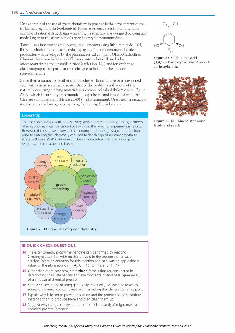
25 Medicinal chemistry196
One example of the use of green chemistry in practice is the development of the influenza drug Tamiflu (oseltamivir). It acts as an enzyme inhibitor and is an example of rational drug design – meaning its structure was designed by computer modelling to fit the active site of a specific enzyme neuraminidase.
Tamiflu was first synthesized in very small amounts using lithium nitride, LiN3 [Li+N3
–], which acts as a strong reducing agent. The first commercial scale production was developed by the pharmaceutical company GlaxoSmithKline. Chemists here avoided the use of lithium nitride but still used other azides (containing the unstable nitride (azide) ion, N3
−) and ion exchange chromatography as a purification technique rather than the greener recrystallization.
Since then a number of synthetic approaches to Tamiflu have been developed, each with a more sustainable route. One of the problems is that one of the naturally occurring starting materials is a compound called shikimic acid (Figure 25.39) which is currently uneconomical to synthesize and is isolated from the Chinese star anise plant (Figure 25.40) (Illicium anisatum). One green approach is its production by bioengineering using fermenting E. coli bacteria.
OH
OHHO
O OH
Figure 25.39 Shikimic acid (3,4,5-trihydroxycyclohex-1-ene-1-carboxylic acid)
Figure 25.40 Chinese star anise fruits and seeds
Expert tip
The atom economy calculation is a very simple representation of the ‘greenness’ of a reaction as it can be carried out without the need for experimental results. However, it is useful as a low atom economy at the design stage of a reaction prior to entering the laboratory can lead to the design of a cleaner synthetic strategy (Figure 25.41). However, it does ignore solvents and any inorganic reagents, such as acids and bases.
wastereduction
benign bydesign
reducedtoxicity
fewerancillaries
energyefficiency
renewablefeedstocks
catalystefficiency
qualitycontrol
atomeconomy
greenchemistry
safetycontrol
Figure 25.41 Principles of green chemistry
n QUICK CHECK QUESTIONS
34 The ester 2-methylpropyl methanoate can be formed by reacting 2-methylpropan-1-ol with methanoic acid in the presence of an acid catalyst. Write an equation for this reaction and calculate an approximate value for the atom economy. (Ar: O = 16, C = 12 and H = 1)
35 Other than atom economy, state three factors that are considered in determining the sustainability and environmental friendliness (‘greenness’) of an industrial chemical process.
36 State one advantage of using genetically modifi ed (GM) bacteria to act as source of shikimic acid compared with harvesting the Chinese star anise plant.
37 Explain why it better to prevent pollution and the production of hazardous materials than to produce them and then clean them up.
38 Suggest why using a catalyst (or a more efficient catalyst) might make a chemical process ‘greener’.
Chemistry for the IB Diploma Study and Revision Guide © Christopher Talbot and Richard Harwood 2017

25.7 Taxol – a chiral auxiliary case study 197
NH
OHOH
OH
CH3
H3CO
O
O
O
O
O
OO
OH
OO
Figure 25.42 Structure of Taxol (paclitaxel)
OHO OH
H
O
HO
HO
O
O
H
AcO
10-Deacetylbaccatin III
Figure 25.43 Structure of 10-deacetylbaccatin III (Ac = ethanoyl (‘acetyl’))
Expert tip
Natural products, often from plants, are a rich source of biologically active compounds. Many of today’s medicines are either obtained directly from a natural source, or were developed from a lead compound originally obtained from a natural source.
n QUICK CHECK QUESTIONS
39 a State what cancers Taxol is used to treat.
b Outline what is meant by the term ‘semi-synthetic’ drug.
40 The intraperitoneal LD50 for Taxol (rat): 32.53 mg kg−1. Explain the meaning of this term.
25.7 Taxol – a chiral auxiliary case study
Essential idea: Chiral auxiliaries allow the production of individual enantiomers of chiral molecules.
Taxol – a chiral auxiliary case studyn Taxol is a drug that is commonly used to treat several different forms of cancer.n Taxol naturally occurs in yew trees but is now commonly synthetically
produced.n A chiral auxiliary is an optically active substance that is temporarily
incorporated into an organic synthesis so that it can be carried out asymmetrically with the selective formation of a single enantiomer.
TaxolTaxol (paclitaxel) (Figure 25.42) is a chemotherapeutic agent used to treat cancers of the ovary, brain, breasts and lungs. It is administered intravenously as an emulsion in water. It works by preventing cancer cells from undergoing cell division. It binds to and stops microtubules extending during cell division (mitosis) and so does not allow the chromosomes to separate.
Taxol was originally isolated from the bark of the Pacific yew tree (Taxus brevifolia) but the yield by repeated liquid–liquid solvent extraction was extremely low. The removal of the bark kills the trees. Several multi-step total syntheses of Taxol have been developed, but the overall yield is low and not commercially viable for Taxol production. A semi-synthetic and renewable route was developed from the compound 10-deactylbaccatin III (Figure 25.43) found in the needles of the European yew tree, Taxus baccata. As the needles regrow, this is a renewable source of a useful precursor to Taxol.
Chiral auxiliariesTaxol has 11 chiral carbon atoms (Figure 25.44) and hence a large number (211) of stereoisomers, only one of which is Taxol. The stereochemistries of the intermediates have to be carefully controlled during the total synthesis of Taxol.
Chemistry for the IB Diploma Study and Revision Guide © Christopher Talbot and Richard Harwood 2017

25 Medicinal chemistry198
The ability to synthesize single enantiomers of chiral molecules is important as different enantiomers can interact with biological receptors very differently (Figure 25.45).
Figure 25.44 The structure of Taxol showing the absolute stereochemistry at each chiral carbon
R enantiomer S enantiomer
binding site of theprotein receptor
binding site of theprotein receptor
Figure 25.45 The importance of chirality in drug action – only one enantiomer can bind to the receptor strongly
Expert tip
A molecule is chiral if it is non-superimposable on its mirror image. Any molecule that has a single stereogenic centre can exist as two enantiomers.
A chiral auxiliary (Figure 25.46) is a chiral control element temporarily incorporated into the structure of the substrate in order to direct the stereochemistry at new stereogenic centre(s) formed in a reaction. The chiral auxiliary is removed (either immediately during work-up or in a separate subsequent step) and may be recovered for re-use. Prochiral molecules are those that can be converted from achiral to chiral in a single step. A diastereoselective reaction is one in which one diastereomer is formed in preference to another.
n QUICK CHECK QUESTIONS
41 State the importance of chirality in drug action.
42 The semi-synthesis of Taxol often uses a chiral auxiliary. Describe the use of chiral auxiliaries in asymmetric synthesis.
43 Suggest the properties of a good chiral auxiliary.
44 Suggest two disadvantages of the use of a chiral auxiliary.
Figure 25.46 The use of a chiral auxiliary
chiral productdiastereoselectivetransformation
prochiralsubstrate
auxiliaryrecycle
chiralauxiliary
O
HOR
O
XcR
E
O
Xc
H–Xc
R*
E
*
O
HOR
The chiral auxiliary used in one synthesis of Taxol is a single enantiomer of trans-2-phenylcyclohexanol (Figure 25.47) that has two stereogenic centres. These favour the formation of specific diasteroisomers in the subsequent steps of the synthesis.
HO
Figure 25.47 The structure of the chiral auxiliary trans-2- phenylcyclohexanol
OH
Ph O
ONH
Ph
O
RSS S S
R
S
OAc O
OH
OHS
RRS
HO
O
O
O
AcPh
Chemistry for the IB Diploma Study and Revision Guide © Christopher Talbot and Richard Harwood 2017

25.8 Nuclear medicine 199
PolarimetryTwo enantiomers have the same properties (NMR, solubility, melting point, etc.), except in the presence of other chiral molecules or in their interaction with plane polarized light (Table 25.4).
Pure enantiomers can be distinguished by the use of a polarimeter because they rotate the plane of plane polarized light by the same angle but in opposite directions to each other (clockwise versus anti-clockwise).
The direction of rotation allows the identification of which enantiomer is present. The angle of rotation can be used to provide an indication of the composition of a mixture of two enantiomers.
If the sample does not rotate the plane polarized light then it either contains achiral (optically inactive) compounds or a pair of enantiomers in equimolar quantities – a racemic mixture, or racemate. The effect of equal amounts of each enantiomer on the plane polarized light cancels each other out.
Table 25.4 The physical properties of enantiomers A and B compared
Property A alone B alone Racemic A and B
Melting point Identical to B Identical to A May be different from A and B
Boiling point Identical to B Identical to A May be different from A and B
Optical rotation Equal in magnitude but opposite in sign to B
Equal in magnitude but opposite in sign to B
Zero degrees
The optical purity of a mixture of enantiomers can be calculated from the following expression:
observed specific rotationspecific rotation of pure enantiomer
× 100
For example, a sample of (+) limonene contaminated with (−) limonene has a specific rotation of +106° but the literature value is +123°. Hence the optical purity = (+106/+123) × 100 = 86%.
n QUICK CHECK QUESTIONS
45 Describe the composition of a racemic mixture.
46 Tabtoxin is a natural toxin produced by bacteria found in lilac trees. Its skeletal structure is shown below.
NH2OHOH
HO O
O
O
NH
NH
a State two techniques that could be used to extract impure tabtoxin from the tree.
b Identify the chiral centres present in a molecule of tabtoxin and state the number of diastereoisomers.
c Identify an apparatus that can be used to distinguish between a pair of enantiomers of tabtoxin.
d Explain how the differentiation between the pair of enantiomers is obtained using this apparatus.
25.8 Nuclear medicineEssential idea: Nuclear radiation, while dangerous owing to its ability to damage cells and cause mutations, can also be used to both diagnose and cure diseases.
Chemistry for the IB Diploma Study and Revision Guide © Christopher Talbot and Richard Harwood 2017

25 Medicinal chemistry200
Nuclear medicinen Alpha, beta, gamma, proton, neutron and positron emissions are all used for
medical treatment.n Magnetic resonance imaging (MRI) is an application of NMR technology.n Radiotherapy can be internal and/or external.n Targeted alpha therapy (TAT) and boron neutron capture therapy (BNCT)
are two methods which are used in cancer treatment.
Nuclear medicine and radiotherapyNuclear medicine is the use of radioactive materials (usually in the form of soluble complexes or salts) in medicine for diagnostic imaging and therapy (usually cancer treatment). Diagnostic imaging is mainly performed using two different types of equipment; a gamma camera and a positron emission tomography (PET) scanner.
In radiotherapy, the aim is to localize the radioactive material in cancerous (malignant) cells so they are killed by the ionizing radiation. Targeted radionuclide therapy involves introducing a radioisotope by injection or ingestion. For example, radioactive compounds containing iodine-131 are used in the treatment of thyroid tumours. External beam radiotherapy involves the use of a beam of ionizing radiation directed at the tumour (most commonly gamma radiation). For example, gamma radiation from a linear accelerator or traditionally cobalt-60 (Figure 25.48).
Alpha, beta, gamma, proton, neutron and positron emissions are all used for medical treatment and/or diagnosis. The properties of alpha particles and beta particles are summarized below in Table 25.5.
Table 25.5 The properties of alpha, beta (minus) and gamma radiations
Alpha particle Beta (minus) particle Gamma ray
Relative charge and relative mass +2 and 4 −1 and 0 0 and 0
Nature Helium nucleus or helium ion High-speed electron Electromagnetic wave with very short wavelength <10−10 m
Representation 42He 0
−1e00γ
Action of magnetic field Deflected towards cathode (negative)
Deflected towards anode (positive)
Not deflected
Velocity 1/10 speed of light Up to 9/10 speed of light Speed of light (3.00 × 10⁹ m s−1)
Ionizing power Very high; nearly 100× that of beta particles
100× times less than that of gamma rays
Low
Penetrating power Low; easily stopped by air 100× that of alpha particles 10× that of beta particles
Kinetic energy High Considerably less than alpha particles
Zero
Nature of product Product obtained by the loss of one alpha particle has an atomic number less 2 units and a mass number less 4 units
Product obtained by the loss of one beta particle has an atomic number greater by 1 unit, without any change in mass number
There is no change in atomic number or mass number
Table 25.6 summarizes the nuclear symbols for the particles (emissions) used in medicine. An unstable nucleus emits radiation (alpha or beta particles or gamma rays) which carry energy away from the nucleus, thus making the nucleus more stable.
A positron is a particle that has the same mass as an electron, but an opposite charge. It is the anti-particle of the electron and produced during beta (positive) decay. The positron is represented as 01e. The isotope carbon-11 undergoes decay by positron emission: 11
6C → 115B + 01e. The positron has a very short life because it is
annihilated and converted to energy (gamma rays): 01e + 0−1e → 200γ.
rotationaxis
leadcontainer
cobalt-60source
exit channel forgamma radiation
lead collimatorplates
gammabeam
Figure 25.48 Gamma therapy
Table 25.6 Nuclear symbols
Particle Symbol
Neutron 10n
Proton 11H or 11p
Electron 0−1e
Alpha particle 42He or 4
2α
Beta particle 0−1e, 0
−1β or β−
Positron 0+1e, 01β or β+
Chemistry for the IB Diploma Study and Revision Guide © Christopher Talbot and Richard Harwood 2017

25.8 Nuclear medicine 201
Nuclear equations and radioactive decay calculationsIn nuclear equations, reactant and product nuclides are represented by their atomic numbers, mass numbers and chemical symbols. The totals of the mass numbers and atomic numbers on both sides are equal.
Radioactive decay (Figure 25.49) is governed by an exponential decay of the numbers of radioactive nuclei N, Nt = N0e
–kt, where N0 is the number of such nuclei at time zero and k is the decay rate for the process. The half-life of the reaction is the time for half of the nuclei to decay and is related to the decay rate by t1
2 = ln2
k or t12 = 0.693/k.
Worked example4921Sc produces a beta (minus) particle and a neutron. Write a nuclear equation describing this process.4921Sc → 48
22Ti + 0−1e + 1
0n: (mass (nucleon) number: 49 = (48) + (0) + (1); atomic number (proton number): 21 = 22 → Ti + (−1) + (0).Worked example
Iron-59 is used to study iron metabolism and has a half-life of 45.1 days. Determine how many days it would take for 32 mg to decay to 1 mg.
32 → 16 → 8 → 4 → 2 → 1, 5 half-life, 5 × 45.1 days = 225.5 days.
n QUICK CHECK QUESTION
47 The radionuclide cobalt-60 (t12
= 5.3 years) is used in external radiotherapy. It emits a beta particle and a gamma ray, producing a stable isotope of another element.
a Deduce the nuclear equation for the decay of cobalt-60.
b Outline the technique of external radiotherapy.
Worked example
Technetium-99m has a half-life of 6 hours. Draw a graph showing how 100 mg of 99
43Tc decays over time.
Calculate the mass of 9943Tc remaining after 2.00 days.
k = ln 2t½
; k = 0.693 / 6 = 0.1155 hours
N = N0 e−kt; N = 100e(0.1155 x 48) = 0.391 mg.
Time/h0
120
Am
ount
N/m
g
6 18t½ = 6h
At t½, N = 50 mg(half of 100 mg)
12 24 30 36 4842
100
80
60
20
40
0
Radioactive decay processes
Neu
tron
num
ber
N
Proton number Z
β− decayβ−
β+
ν
ν
γ
α
2 fewer protons2 fewer neutrons
1 more proton1 less neutron
1 less proton1 more neutron
same protonsand neutrons
ZN
Z + 1N − 1
Neu
tron
num
ber
N
Proton number Z
β+ decay
ZN
Z − 1N + 1
Neu
tron
num
ber
N
Proton number Z
γ decay
ZN
Neu
tron
num
ber
N
Proton number Z
α decay
ZN
Z − 2N − 2
24He + energyZ
AX Z−2X +A−4
A
A
−10β + energyZ
AX Z+1X +
+10β + energyZ
AX Z−1X +
energy (gamma ray
photons)ZAX* Z
AX +
Figure 25.49 A summary of radioactive decay processes
Chemistry for the IB Diploma Study and Revision Guide © Christopher Talbot and Richard Harwood 2017

25 Medicinal chemistry202
Magnetic resonance imaging (MRI)Proton nuclear magnetic resonance (NMR) detects the presence of hydrogen atoms (protons) by placing them in a very powerful magnetic field, then exciting the hydrogen atoms (protons) with a pulse of radio frequency radiation. This corresponding to the resonance energy required to cause the hydrogen atoms (protons) to move to the high energy state where the hydrogen atom’s (proton’s) internal magnetic field is aligned anti-parallel to the external magnetic field.
The NMR instrument then detects the weak radio frequency radiation from the protons as they ‘relax’ back to their original energy states. The frequency of this signal from the proton is proportional to the magnetic field to which they are subjected during this relaxation process.
In magnetic resonance imaging (MRI) (Figure 25.50), an image of a cross-section of a body can be made by producing a magnetic field gradient across the tissue so that a certain value of magnetic field can be associated with a given location in the tissue.pole-pieces of magnetproducing non-uniform
field
RF coil
patient
pole-piece of magnet producinglarge uniform field RF
generator
RFreceiver
processor
display
Figure 25.50 Schematic diagram of a magnetic resonance scanner. (The magnetic field is usually generated by a.c. currents in wires rather than using permanent magnets.)
Since the hydrogen atom (proton) signal frequency is proportional to that magnetic field, a given proton signal frequency is associated to a location in the tissue (Figure 25.51). This provides the information to map the tissue in terms of the protons present there. Since the proton density varies with the type of tissue, a contrast agent is present to image the organs and other tissue variations in the body. The three-dimensional information then comes from varying the gradient in three orthogonal directions (x, y and z) of the magnetic field.
varying magnetic field,magnetic field ‘gradient’
frequency f2
frequency f3
Figure 25.51 Protons undergoing relaxation and releasing radio waves of different energy (frequency) that depends on the position (strength of the magnetic field) in the magnetic field gradient
Expert tip
MRI is not an example of nuclear medicine: compared with nuclear medicine which uses ionizing radiation, MRI uses non-ionizing radiation in the form of radio frequency waves.
Chemistry for the IB Diploma Study and Revision Guide © Christopher Talbot and Richard Harwood 2017

25.8 Nuclear medicine 203
Positron emission tomography (PET)A beta-positive (i.e. positron) emitting radioactive substance (usually fluorine-18) is introduced into the body (through an intravenous injection). The positrons emerge into the surrounding tissues, meet electrons, and both electron and positron cease to exist. This is called mutual annihilation (Figure 25.52), and involves conversion of the masses of the particles into energy that is carried away by two gamma ray photons which can be detected from outside the body (by a PET scanner; Figure 25.53). Scintillators convert gamma photons into visible light; photomultipliers multiply the current produced by incident light.
Worked example
A freshly prepared sample of the radioisotope fluorine-18 has a mass of 0.010 g. The decay constant of fluorine-18 is 1.05 × 10⁻⁴ s−1. Determine the initial activity of the sample, the activity after 2 hours and the half-life of the isotope.
Initial number of 18F nuclei in sample = (0.0118 ) × 6.02 × 1023 = 3.344 × 10²⁰
Initial activity, A0 = λN0 = 1.05 × 10−4 × 3.344 × 1020 = 3.512 × 1016 = 3.5 × 10¹⁶ disintegrations per second
Activity after 2 hours, A = A0e−λt= 3.512 × 10¹⁶ × e−1.05 × 10−4 × 2 × 360 = 1.6 × 10¹⁶
disintegrations per second
Half-life = ln 2λ = ln 2
(1.05 × 10⁻⁴) = 6.6 × 10³ s = 110 minutes
positron-emittingnucleus
positronpath
positron–electronannihilation
annihilationphotons
~0.5 mm
Figure 25.52 General principle of PET imaging
photomultipliersscintillators
gamma photonsemitted in pairs
head
Figure 25.53 Arrangement of PET scanners for a PET scan
Targeted radionuclide therapy
■n Targeted alpha therapy (TAT)TAT is used to direct alpha radiation directly at cancerous cells. The range of alpha particles in human tissues is typically 50–100 μm, the diameter of about ten cells. The energy from the alpha radiation is largely contained within the tumour cells and there is little damage to the surrounding normal cells.
Lead-212 atoms are typically used in targeted alpha therapy:21282Pb → 208
80Hg + 42He
The specific ‘targeting’ of cancer cells is achieved by using tumour-selective carrier molecules, such as monoclonal antibodies (Figure 25.54) or peptides (small proteins). Often a chelating or complexing agent is used. TAT is used to treat cancers that have spread and formed secondary tumours within the body.
alpha particlesemitted kill the
cancer cellchelator
(chelating agent)binding
antibodytargetcancer cell
antigen
cellnucleus cancer cell
Figure 25.54 Target alpha therapy
n QUICK CHECK QUESTIONS
48 Define targeted alpha therapy. Write a balanced nuclear equation showing the loss of an alpha particle from actinium-225.
49 Explain how targeted alpha therapy works well for cancers that have spread around the body.
Chemistry for the IB Diploma Study and Revision Guide © Christopher Talbot and Richard Harwood 2017

25 Medicinal chemistry204
■n Boron neutron capture therapy (BNCT)High-intensity boron neutron beams are used in BNCT, which uses the ability of non-radioactive boron-10 nuclei to absorb neutrons and undergo a transmutation reaction.
After capturing a low-energy neutron a lithium-7 nuclide and a high-energy alpha particle are formed:
105B + 10n → 73Li + 42He
The cancer patient is injected with a boron compound which is selectively absorbed by cancer cells. The patient is then irradiated with neutron radiation which causes production of high-energy alpha particles inside the tumour cells (Figure 25.55).
cancer cell1. boron compound (B) selectively absorbed by cancer cell
2. neutron beam
3. boron atoms absorb neutrons
4. boron atoms decay, emitting cell-killing radiation
N+B N+BB
B
BB
Figure 25.55 Boron neutron capture therapy
■n Other targeted therapyOther examples of radioisotopes used in targeted radionuclide therapy are lutetium-177 (beta and gamma emitter) and yttrium-90 (beta emitter). Lutetium-177 has a shorter penetration range than yttrium-90 making it suitable for treating smaller tumours.
Lutetium-177 is often used in conjunction with a peptide (small protein) to selectively target neuro-endocrine tumours. Yttrium-90 therapy is often used in the treatment of liver cancer. Large numbers of small plastic or glass beads containing yttrium-90 are injected directly into the arteries that supply blood to the liver.
Proton beam therapyProton beam therapy (Figure 25.56) or proton therapy is a recent development of nuclear medicine which uses protons to irradiate cancerous tissue. A linear accelerator imparts high kinetic energies to protons in a narrow beam, which is then directed at a tumour. The protons damage the DNA of cells, causing them to die or be unable to divide. Cancer cells are particularly vulnerable to DNA damage because of their high rate of cell division.
protonpencilbeam
scanning magnets
scanning magnets
high-densitystructure
targetvolume
criticalstructure
body surface
Figure 25.56 Proton beam therapy
In contrast to other types of nuclear or ionizing radiation the absorption of protons reaches a maximum within a narrow range, deep inside the patient’s body. This phenomenon is known as the Bragg peak effect (Figure 25.57) and allows the proton beam to be focused on the tumour with minimal damage to surrounding healthy tissue. The Bragg peak effect occurs because all the protons in the beam have the same speed and hence the same kinetic energy.
n QUICK CHECK QUESTIONS
50 Explain how boron neutron capture therapy can be used to kill cancerous cells.
51 a Write a nuclear equation for the beta decay of lutetium-177.
b A patient is given a 20.0 mg dose of a soluble lutetium-177 compound. Given that the half-life of lutetium-177 is 6.71 days, calculate how much of the lutetium-177 is present in the patient’s body after 4 weeks.
52 90Sr is a radioisotope and is a long-term health problem because it substitutes for calcium in bones. Suggest why strontium can readily substitute for calcium.
100
50
0
Rad
iatio
n do
se/%
Bragg’s peak
Depth in tissue/arbitrary units
Figure 25.57 Absorption of protons (with high kinetic energy) by cells in body tissue
n QUICK CHECK QUESTION
53 Explain why proton beam therapy is more effective in treating cancers than traditional methods of external radiotherapy, such as cobalt-60.
Expert tip
A transmutation is a nuclear reaction involving the formation of a new daughter nuclide.
Chemistry for the IB Diploma Study and Revision Guide © Christopher Talbot and Richard Harwood 2017

25.9 Drug detection and analysis 205
Use of technetium-99mTechnetium-99m is used in nuclear medicine for gamma camera imaging and imaging and SPECT (single photon emission computed tomography). It is the most commonly used radioisotope in nuclear medicine. It decays to form a stable daughter product and emits gamma rays which readily pass out and through the body and can be easily detected. Its half-life of six hours minimizes the patient’s exposure to gamma radiation. Technetium is a transition metal and exists in several stable oxidation states, e.g. technetate(VII), TcO4
−, and forms complex ions with a variety of ligands.
Diagnostic radiographyOwing to its relatively short half-life, Tc-99 is generated in hospitals by the beta decay of molybdenum-99. It can be used diagnostically, for example, to determine the flow of blood through the brain’s hemispheres. Its progress through the body can be followed by detecting the gamma radiation it emits.
The decay product, Tc-99 (in the ground state) is a beta emitter, but the energy released within the body is relatively low and the amount of Tc-99m in the body rapidly decreases over time.
99m43Tc ææÆ 99
43Tc ææÆ 9944Ru + 0−1e
−
Side effects from radiotherapyThe side effects of ionizing radiation depend upon the type of emission, the intensity and duration of the ionizing energy. External radiotherapy tends to cause more side effects than targeted therapy, as more healthy tissue and organs are irradiated in the process. Common side effects include hair loss, nausea, fatigue, loss of appetite and sterility.
The cells of the body may undergo physical and chemical changes because of exposure to ionizing radiation. Physical changes involve burning; chemical changes may involve damage to DNA molecules (mutation), which can lead to cancer.
NATURE OF SCIENCEExposure to ionizing radiation can have many benefits for individuals and society. Medical imaging examinations that involve radiation determine whether organs are functioning properly or bones are broken and in treating cancer. Low levels of radiation exposure are used for most medical examinations involving radiation, although when we are using radiation to treat a disease (e.g. cancer), very high doses are needed to kill the cancer cells. Ionizing radiation has benefits, but there are also risks and these have to be evaluated. The small radiation doses used to conduct medical examinations carry little or no risk, while exposure to high levels may cause observable health effects.
25.9 Drug detection and analysisEssential idea: A variety of analytical techniques is used for detection, identification, isolation and analysis of medicines and drugs.
Drug detection and analysisn Organic structures can be analysed and identified through the use of infrared
spectroscopy, mass spectroscopy and proton NMR.n The presence of alcohol in a sample of breath can be detected through the use
of either a redox reaction or a fuel cell type of breathalyser.
n QUICK CHECK QUESTION
54 Write a nuclear equation describing the formation of technetium-99m from molybdenum-99 by beta decay.
6 h
γ
21100y
n QUICK CHECK QUESTIONS
55 State two reasons why Tc-99m is suitable for medical imaging.
56 State two common short-term side effects of radiotherapy.
57 Outline one ethical implication of using radioisotopes in nuclear medicine.
Chemistry for the IB Diploma Study and Revision Guide © Christopher Talbot and Richard Harwood 2017

25 Medicinal chemistry206
Identification of drugs using spectroscopic and spectrometric analysisMass spectrometry can be used to determine the molecular mass of molecules and give insight into the structure of molecules from a consideration of fragment ions.
Infrared spectroscopy can identify specific functional groups as the bonds connecting different types of atom vibrate at different frequencies and so absorb in different regions of the infrared spectrum.
A 1H NMR spectrum consists of a series of resonances that correspond to protons in different chemical environments. The position of each resonance (chemical shift) and area under each resonance gives information about the relative number of hydrogen atoms (protons) and the particular chemical environment in which each different hydrogen atom (proton) exists.
The shape of the resonance (multiplicity) gives information about how many protons are attached to the atoms adjacent to the proton causing the resonance. If there n adjacent protons, then the resonance will be split into n + 1 peaks.
These techniques are routinely applied to assigning structures of drugs or their precursors.
■n Infrared spectrumGBL (γ-butyrolactone) (Figure 25.58) can be used as a recreational drug and has effects similar to alcohol. Figure 25.59 shows the infrared spectrum of GBL.
3625
2992
2917
1770
1170
1377
1461
1038
992
931
870 80
1
676
637
492
gamma-butyrolactone100
50
10
Figure 25.59 Infrared spectrum of GBL
Table 25.7 shows an analysis of the absorption bands in the infrared spectrum of GBL.
Table 25.7 An analysis of the absorption bands in the infrared spectrum of gamma-butyrolactone (GBL)
Vibrational mode Wavenumber/ cm–1 Strength of peak
Carbonyl stretching mode 1770 Very strong
C–H bond 2992 Relatively weak
In-plane deformation of the carboxyl hydrogen and stretching of the carbon–oxygen bond in the carboxyl group
1450–1150 Strong
Stretching C–O bond of the terminal hydroxyl group
1038 Strong
■n Nuclear magnetic resonanceEthyl 3-oxobutanoate (Figure 25.60) is a chemical intermediate for the synthesis of antibiotics, analgesics and antimalarial agents.
OO
Figure 25.58 Structure of gamma-butyrolactone (dihydrofuran-2(3H)-one)
Expert tip
The weak –OH stretch observed in this spectrum is from contaminating water.
Chemistry for the IB Diploma Study and Revision Guide © Christopher Talbot and Richard Harwood 2017

25.9 Drug detection and analysis 207
7
2
2
3
3
6 5 4 3ppm, δ
2 1 0
Figure 25.61 1H NMR spectrum of ethyl 3-oxobutanoate
The proton or 1H NMR spectrum (Figure 25.61) has a quartet coupled to a triplet indicative of an ethyl group. The methylene group, –CH2–, must be adjacent to an electron withdrawing group since it is shifted to 4.1d. The two singlets at 2.2d and 3.2d suggest isolated –CH2– and –CH3 groups and the –CH2– group must be adjacent to one or more electronegative groups.
■n Mass spectrometryThe molecular ion, M+, of ibuprofen (a mild analgesic) is expected at 206 (Figure 25.62).
m/z
m/z
206
i.e.
i.e.
164
163
145
107
M+
(M–C3H6)+
i.e.(M–C3H7)+
(M–C3H7–H2O)+
C7H7O+
ion
100
%
50
41 57
5165
91
107
119
129 145
161
206
ibuprofen
77
150100 200 250 300
50
0
CH–COOH
CH2
CH
CH3
CH3CH3
CH–COOH
CH3
CH3
+
CC O
CH2
CH3
+CH
CH3
CH3
+CH2
+
+
CH3
m/z
207
161
119
91
65
M+
(M–COOH)+
(M–COOH–C3H6)+
(M–COOH–C3H6–C2H4)+
(M–COOH–C3H6–C2H4–C2H2)+
ion
i.e.
Figure 25.62 The mass spectrum of ibuprofen
O
H3C CH3
O
OFigure 25.60 Structure of ethyl 3-oxobutanoate
Chemistry for the IB Diploma Study and Revision Guide © Christopher Talbot and Richard Harwood 2017

25 Medicinal chemistry208
The fragment ions indicate that fragmentation occurs from both side-chains. Losing the carboxylic acid group is the most common first fragmentation.
Purification of organic productsMass spectrometry and a variety of spectroscopic techniques are used to identify pure substances. There are a number of physical techniques that can be used to purify mixtures and prepare pure substances.
For small amounts, chromatography can be used to separate and identify mixtures, often containing a large number of components, both qualitatively and quantitatively. It can also be used to determine the purity of a substance.
In all forms of chromatography there is a solid stationary phase and a liquid mobile phase that flows across the stationary phase. Chromatographic techniques depend on the components in the mixture having different tendencies to adsorb onto a surface or partition (distribute) with a solvent.
Fractional distillation (Figure 25.63) is a method for separating mixtures of miscible liquids with different boiling points. On a laboratory scale, the process takes place using distillation apparatus with a glass fractionating column between the flask and the still-head. The column is hotter at the bottom and cooler at the top.
coolingwater
fractionating columnpacked withglass beads
thermometer
liquidmixture
�
If the flask contains a mixture of two liquids, the boiling liquid produces a vapour that is richer in the more volatile of the two liquids (the liquid with the lower boiling point).
As the vapour condenses in the fractionating column it runs back into the flask. As it does, it meets more of the rising vapour, some of which condenses as it transfers its heat energy to the descending liquid. That, in turn, may vaporize again. In this way the mixture repeatedly boils and condenses as it rises up the fractionating column. It is in effect like carrying out a whole series of simple distillations. This can be represented as a series of steps in a boiling-point composition diagram (Figure 25.64).
Expert tip
The IB will assume that H2O and CO2 are not lost from the molecules (due to the low pressure) and knowledge of rearrangements will not be tested.
Figure 25.63 Fractional distillation
Chemistry for the IB Diploma Study and Revision Guide © Christopher Talbot and Richard Harwood 2017

25.9 Drug detection and analysis 209
Boi
ling
poin
t at
cons
tant
pre
ssur
econdensing
boiling
liquid
composition
vapour
composition
TM
TB
TA
V1
L2L
3
M
L4
V2
V3
Mole fraction A
Mole fraction B
TB is the boiling point of pure B
where TA is the boiling point of pure A
1.0 0.5 0.0
0.0 0.5 1.0
L1
Figure 25.64 The boiling-point composition diagram for the a mixture of two ideal liquids with different boiling points
The ‘mole fraction’ is a way of expressing the concentration of a solution or mixture. It is equal to the amount (in mol) of one component divided by the total amount (in mol) in the solution or mixture.
Another form of distillation is steam distillation (Figure 25.65), which is useful for separating an organic material with a high boiling point and poor solubility in water. The technique is used when organic compounds decompose below their normal boiling point.
steam
thermometer
mixture of organicliquid and water
gentleheat
cooling water out
cooling water in
condenser
water and the immiscible organicliquid separate on standing
Figure 25.65 Steam distillation
Solvent extraction can be used for compounds that distribute between two solvents, one of which is usually aqueous. This depends upon the relative solubility of a substance in the two solvents. An organic substance is extracted into the layer in which it is more soluble and the layers separated using a separating funnel (Figure 25.66). A drying agent (usually an anhydrous salt) is added to remove traces of water in the organic layer and then filtered off. The solvent is then evaporated to leave the required compound.
If the solute is in the same molecular state in both liquid phases, the following simple partition equilibrium expression will apply:
Pc = [X(organic)][X(aq)]
Pc is called the partition/distribution coefficient. It has no units and is temperature dependent.
If more of the substance X is added to the system, the solute will distribute itself between the immiscible liquids so that the ratio of the solute concentrations remains the same at constant temperature independently of the total quantity of X in the same molecular state (known as the partition equilibrium law).
less dense layer
more dense layer
Figure 25.66 Use of a separating funnel
Expert tip
Normally, the aqueous layer is the bottom layer as water usually has higher density than the organic solvent.
Chemistry for the IB Diploma Study and Revision Guide © Christopher Talbot and Richard Harwood 2017

25 Medicinal chemistry210
The hydrophobicity of a molecule of an organic molecule is measured by its log Pc value where Pc can be measured experimentally by measuring the relative solubility of a compound in an octano-1-ol/water mixture.
Pc = [X(octan-1-ol)][X(aq)]
The more hydrophobic the compound is, the greater the proportion of it will dissolve in the organic layer, and the higher the value of Pc or log10Pc.
■n Raoult’s lawIn a system consisting of a mixture of two miscible liquids at equilibrium, Raoult’s law states that each component will exert a vapour pressure proportional to its mole fraction.
Expert tip
Raoult’s law describes an ideal mixture of liquids in which the intermolecular forces in both liquids are almost identical (Figure 25.67). This occurs with similar compounds, such as isomers, with identical molar masses.
In an ideal solution, theforces between thesolvent molecules…
This means that it takes the sameamount of kinetic energy for solventmolecules to break away from thesurface in either case.
…are exactly the sameas those between solventand solute.
Figure 25.67 Concept of an ideal solution
Raoult’s law can be expressed mathematically:
p(A) = p°(A) × x(A)
where p(A) is the vapour pressure of A over the mixture of the two liquids (at a given temperature); p°(A) is the vapour pressure over a pure sample of A at the same temperature and x(A) is the mole fraction of A, which is the ratio of the amount of A to the sum of all the amounts of the component liquids in the mixture.
This means that a graph of mole fraction against vapour pressure for each of the liquids will be of the type shown in Figure 25.68, that is, a straight line passing through the origin.
The total vapour pressure of the liquid mixture is found by adding the vapour pressures of the two components (two liquids) and it will vary with composition as shown in Figure 25.69.
1.0 0.5Mole fraction AMole fraction B
where PA is the vapour pressure of pure APB is the vapour pressure of pure B
component A
component B
total vapour pressure
0.00.0 0.5 1.0
Vapo
ur p
ress
ure
atco
nsta
nt te
mpe
ratu
re
PA
PB
Figure 25.69 The relationship between mole fractions and partial and total vapour pressures for an ideal mixture of two liquids
1.0 0.5 0.0
Vapo
ur p
ress
ure pure component
component in a solution
Mole fraction of component in solution
Figure 25.68 The relationship between vapour pressure and mole fraction for components in an ideal mixture of liquids
Chemistry for the IB Diploma Study and Revision Guide © Christopher Talbot and Richard Harwood 2017

25.9 Drug detection and analysis 211
Worked example
At 80 °C, the vapour pressure of benzene is 1.0 × 10⁵ Pa and the vapour pressure of methylbenzene is 4.0 × 10⁵ Pa. An ideal solution contains 7.812 g of benzene and 36.86 g of methylbenzene. These are two miscible liquids.
Calculate the amounts (mol) and mole fractions of benzene and methylbenzene. Apply Raoult’s law to calculate the partial pressures of benzene and methylbenzene and the total vapour pressure.
Amount of benzene = 7.812 g/78.12 g mol−1 = 0.1 mol
Amount of methylbenzene = 36.86 g/92.15 g mol−1 = 0.4 mol
Mole fraction of benzene = 0.1/(04 + 0.1) = 0.2
Mole fraction of methylbenzene = 1.0 − 0.2 = 0.8 mol
Partial pressure of benzene = 1.0 × 105 Pa × 0.2 = 2 × 10⁴ Pa
Partial pressure of methylbenzene = 4.0 × 104 Pa × 0.8 = 3.2 × 10⁴ Pa
Total pressure = 2 × 10⁴ Pa + 3.2 × 10⁴ Pa = 5.2 × 10⁴ Pa
Worked example
The vapour pressure-composition diagram for a mixture of benzene and methylbenzene is shown below.
Vapo
ur p
ress
ure
0 1.0Mole fraction of C6H6
Sketch and explain the boiling-point composition diagram for the mixture.
Use your graph to explain what happens in the distillation column when a mixture containing 50% by mass of benzene is fractionally distilled.
Tem
pera
ture
/°C
liquid
vapourTR
T1
TQ L1
0 1.0Mole fraction of C6H6
W2
W1
When a mixture containing 50% by mass of benzene is fractionally distilled it boils at T1 and gives a vapour (W1) which is richer in benzene. When this vapour comes into contact with the cold surfaces of the glass beads in the fractionating column, it condenses into a liquid (L1). The condensed liquid (L1) boils again when it comes into contact with the ascending hot vapour to produce another vapour (W2), which is even richer in benzene. The process of vaporization and condensation repeats in the fractionating column. Finally, pure benzene is distilled over at a constant temperature of TQ.
n QUICK CHECK QUESTIONS
58 a Define the term vapour pressure of a liquid.
b State two factors that affect the value of the vapour pressure of a liquid mixture.
59 Two liquids X and Y boil at 110 °C and 140 °C. Which of them has the higher vapour pressure at 50 °C?
60 The vapour pressure of a pure a liquid is 30 mmHg at 320 K. The vapour pressure of the liquid in the solution with liquid B is 28 mmHg at the same temperature. Deduce the mole fraction of B in the solution if it obeys Raoult’s law.
61 State and explain which of the following pairs of liquids is/are expected to obey Raoult’s law: hexane and heptane; chloromethane and propanone; cyclohexanol and water
Chemistry for the IB Diploma Study and Revision Guide © Christopher Talbot and Richard Harwood 2017
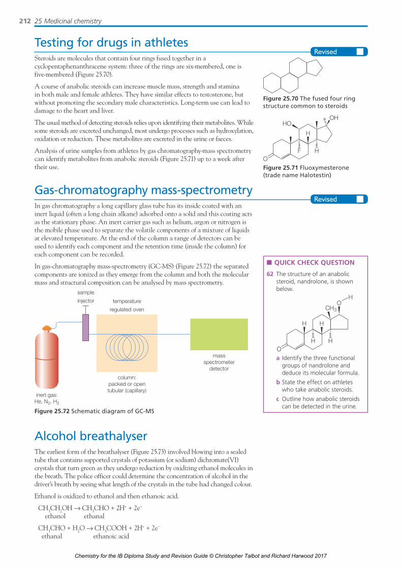
25 Medicinal chemistry212
Testing for drugs in athletesSteroids are molecules that contain four rings fused together in a cyclopentaphenanthracene system: three of the rings are six-membered, one is five-membered (Figure 25.70).
A course of anabolic steroids can increase muscle mass, strength and stamina in both male and female athletes. They have similar effects to testosterone, but without promoting the secondary male characteristics. Long-term use can lead to damage to the heart and liver.
The usual method of detecting steroids relies upon identifying their metabolites. While some steroids are excreted unchanged, most undergo processes such as hydroxylation, oxidation or reduction. These metabolites are excreted in the urine or faeces.
Analysis of urine samples from athletes by gas chromatography-mass spectrometry can identify metabolites from anabolic steroids (Figure 25.71) up to a week after their use.
Gas-chromatography mass-spectrometryIn gas chromatography a long capillary glass tube has its inside coated with an inert liquid (often a long chain alkane) adsorbed onto a solid and this coating acts as the stationary phase. An inert carrier gas such as helium, argon or nitrogen is the mobile phase used to separate the volatile components of a mixture of liquids at elevated temperature. At the end of the column a range of detectors can be used to identify each component and the retention time (inside the column) for each component can be recorded.
In gas-chromatography mass-spectrometry (GC-MS) (Figure 25.72) the separated components are ionized as they emerge from the column and both the molecular mass and structural composition can be analysed by mass spectrometry.
column:packed or opentubular (capillary)
massspectrometer
detector
sample
injector temperature
regulated oven
inert gas:He, N2, H2
Figure 25.72 Schematic diagram of GC-MS
Alcohol breathalyserThe earliest form of the breathalyser (Figure 25.73) involved blowing into a sealed tube that contains supported crystals of potassium (or sodium) dichromate(VI) crystals that turn green as they undergo reduction by oxidizing ethanol molecules in the breath. The police officer could determine the concentration of alcohol in the driver’s breath by seeing what length of the crystals in the tube had changed colour.
Ethanol is oxidized to ethanol and then ethanoic acid.
CH3CH2OH → CH3CHO + 2H+ + 2e−
ethanol ethanal
CH3CHO + H2O → CH3COOH + 2H+ + 2e−
ethanal ethanoic acid
n QUICK CHECK QUESTION
62 The structure of an anabolic steroid, nandrolone, is shown below.
O
H
H
OCH3
H
H H
a Identify the three functional groups of nandrolone and deduce its molecular formula.
b State the effect on athletes who take anabolic steroids.
c Outline how anabolic steroids can be detected in the urine.
Figure 25.70 The fused four ring structure common to steroids
OF
HO
H
OH
H
Figure 25.71 Fluoxymesterone (trade name Halotestin)
Chemistry for the IB Diploma Study and Revision Guide © Christopher Talbot and Richard Harwood 2017

25.9 Drug detection and analysis 213
Green chromium(III) ions are formed as the ethanol molecules are oxidized.
Cr2O72−(aq) + 14H+(aq) + 6e− → 2Cr3+(aq) + 7H2O(l)
person breathes airwith alcohol intomouthpiece
glass tube containing sodiumdichromate(VI)– sulfuric acidcoated on silica gel particles
as person blows into the tube,the plastic bag becomes inflated
Figure 25.73 A simple breathalyser
Modern hand-intoximeters may be based on a redox reaction based on fuel cell technology. It takes the form of a porous disc with catalytic platinum, silver or gold electrodes saturated with an electrolyte of phosphorus(V) acid or sodium hydroxide.
When the driver breathes into the intoximeter the ethanol in the breath is catalytically oxidized to ethanoic acid at the metal electrode on one side of the membrane:
CH3CH2OH + H2O → CH3COOH + 4H+ + 4e−
Oxygen is kept in contact with the metal on the other side to act as a reference electrode (cf. the standard hydrogen electrode). The oxygen is reduced to water:
O2 + 4H+ + 4e− → 2H2O
The instrument is calibrated by passing air with known ethanol concentrations through and measuring the resulting cell voltage.
n QUICK CHECK QUESTION
63 Ethanol is relatively volatile and at high concentrations can be detected in the breath using a breathalyser that contains potassium dichromate(VI). Deduce the oxidation and reduction half-equations that occur in the breathalyser.
Chemistry for the IB Diploma Study and Revision Guide © Christopher Talbot and Richard Harwood 2017

214 Acknowledgements
AcknowledgementsPhoto credits
p.26 © Chris Talbot; p.33 © Phil Degginger/Alamy; p.124 all © David Talbot
Chemistry for the IB Diploma Study and Revision Guide © Christopher Talbot and Richard Harwood 2017