2018 acc/aha/hrs guideline on the ... - ern-ithaca.eu
TRANSCRIPT

Circulation. 2019;140:e333–e381. DOI: 10.1161/CIR.0000000000000627 August 20, 2019 e333
*Writing committee members are required to recuse themselves from voting on sections to which their specific relationships with industry may apply; see Appendix 1 for detailed information. †ACC/AHA Representative. ‡STS Representative. §HRS Representative. ‖PACES Representative. ¶ACC/AHA Task Force on Clinical Practice Guidelines Liaison. #AATS Representative. **Dr. Pellegrini contributed to this article in her personal capacity. The views expressed are her own and do not necessarily represent the views of the US Department of Veterans Affairs or the US government. ††ACC/AHA Performance Measures Representative.
WRITING COMMITTEE MEMBERS*Fred M. Kusumoto, MD, FACC, FAHA, FHRS, ChairMark H. Schoenfeld, MD, FACC, FAHA, FHRS, Vice ChairColetta Barrett, RN, FAHA†James R. Edgerton, MD, FACC, FHRS‡Kenneth A. Ellenbogen, MD, FACC, FAHA, FHRS*†Michael R. Gold, MD, PhD, FACC*§Nora F. Goldschlager, MD, FACC, FAHA, FHRS†Robert M. Hamilton, MD‖
José A. Joglar, MD, FACC, FAHA, FHRS¶Robert J. Kim, MD†Richard Lee, MD, MBA#Joseph E. Marine, MD, MBA, FACC, FHRS§Christopher J. McLeod, MB, ChB, PhD, FACC, FAHA, FHRS†Keith R. Oken, MD, FACC†Kristen K. Patton, MD, FACC, FAHA, FHRS†Cara N. Pellegrini, MD, FHRS*§**Kimberly A. Selzman, MD, MPH, FACC, FHRS†Annemarie Thompson, MD†Paul D. Varosy, MD, FACC, FAHA, FHRS††
© 2018 by the American College of Cardiology Foundation, the American Heart Association, Inc., and the Heart Rhythm Society.
ACC/AHA/HRS GUIDELINE
2018 ACC/AHA/HRS Guideline on the Evaluation and Management of Patients With Bradycardia and Cardiac Conduction Delay: Executive SummaryA Report of the American College of Cardiology/American Heart Association Task Force on Clinical Practice Guidelines, and the Heart Rhythm Society
Circulation
ACC/AHA Task Force Members, see page e362
https://www.ahajournals.org/journal/circ
Key Words: AHA Scientific Statements ◼ ablation ◼ ambulatory electrocardiography ◼ aminophylline ◼ atrioventricular block ◼ atropine ◼ AV block ◼ beta-adrenergic agonist ◼ bradyarrhythmia ◼ bradycardia ◼ bundle branch block ◼ cardiac pacing ◼ cardiac resynchronization therapy ◼ cardiac sinus pause ◼ cardiac surgery ◼ congenital heart disease ◼ digoxin antibodies Fab fragments ◼ electrocardiogram ◼ glucagon ◼ heart block ◼ Holter monitoring ◼ intraoperative ◼ isoproterenol ◼ lamin A-C ◼ left bundle branch block ◼ muscular dystrophies ◼ myocardial infarction ◼ myotonic dystrophy ◼ pacemaker ◼ pacing ◼ preoperative ◼ quality of life ◼ right bundle branch block ◼ sarcoidosis ◼ shared decision-making ◼ sick sinus syndrome ◼ sinus arrest ◼ sinus bradycardia syndrome ◼ sinus node dysfunction ◼ spinal cord injuries ◼ syncope ◼ theophylline ◼ transcatheter aortic valve replacement
The American Heart Association requests that this document be cited as follows: Kusumoto FM, Schoenfeld MH, Barrett C, Edgerton JR, Ellenbogen KA, Gold MR, Goldschlager NF, Hamilton RM, Joglar JA, Kim RJ, Lee R, Marine JE, McLeod CJ, Oken KR, Patton KK, Pellegrini C, Selzman KA, Thompson A, Varosy PD. 2018 ACC/AHA/HRS guideline on the evaluation and management of patients with bradycardia and cardiac conduction delay: executive summary: a report of the American College of Cardiology/American Heart Association Task Force on Clinical Practice Guidelines and the Heart Rhythm Society. Circulation. 2019;140:e333–e381. DOI: 10.1161/CIR.0000000000000627
Developed in Collaboration With
the American Association for
Thoracic Surgery, the Pediatric
& Congenital Electrophysiology
Society, and the Society of Thoracic
Surgeons
Endorsed by the American
Association for Thoracic Surgery,
the Pediatric & Congenital Electro-
physiology Society, and the Society
of Thoracic Surgeons
Dow
nlo
aded
from
http
://ahajo
urn
als.org
by o
n S
eptem
ber 2
0, 2
020

Kusumoto et al 2018 Bradycardia Clinical Practice Guidelines: Executive Summary
August 20, 2019 Circulation. 2019;140:e333–e381. DOI: 10.1161/CIR.0000000000000627e334
CLIN
ICA
L S
TA
TE
ME
NT
S
AN
D G
UID
ELIN
ES
TABLE OF CONTENTSTop 10 Take-Home Messages . . . . . . . . . . . . . . . . . . . . e335Preamble . . . . . . . . . . . . . . . . . . . . . . . . . . . . . . . . . . . e3351. Introduction . . . . . . . . . . . . . . . . . . . . . . . . . . . . . . e336
1.1. Methodology and Evidence Review . . . . . . . . . e3361.2. Organization of the Writing Committee. . . . . . e3361.3. Document Review and Approval . . . . . . . . . . . e3371.4. Scope of the Guideline . . . . . . . . . . . . . . . . . . e3371.5. Class of Recommendation and Level
of Evidence . . . . . . . . . . . . . . . . . . . . . . . . . . . e3371.6. Abbreviations . . . . . . . . . . . . . . . . . . . . . . . . . e337
2. Epidemiology and Definitions . . . . . . . . . . . . . . . . . e3402.1. Definitions. . . . . . . . . . . . . . . . . . . . . . . . . . . . e340
3. General Evaluation of Patients With Documented or Suspected Bradycardia or Conduction Disorders . . . . . . . . . . . . . . . . . . . . . e3413.1. History and Physical Examination
of Patients With Documented or Suspected Bradycardia or Conduction Disorders . . . . . . . . e341
3.2. Noninvasive Evaluation . . . . . . . . . . . . . . . . . . e3413.2.1. Resting ECG in Patients With
Documented or Suspected Bradycardia or Conduction Disorders . . . . e341
3.2.2. Exercise Electrocardiographic Testing in Patients With Documented or Suspected Bradycardia or Conduction Disorders. . . . . . . . . . . . . . . . . . . . . . . . e346
3.2.3. Ambulatory Electrocardiography in Patients With Documented or Suspected Bradycardia or Conduction Disorders. . . . . . . . . . . . . . . . . . . . . . . . e346
3.2.4. Imaging in Patients With Documented or Suspected Bradycardia or Conduction Disorders. . . . . . . . . . . . . . . . . . . . . . . . e347
3.2.5. Laboratory Testing in Patients With Documented or Suspected Bradycardia or Conduction Disorders. . . e347
3.2.6. Genetic Testing in Patients With Documented or Suspected Bradycardia or Conduction Disorders . . . . . . . . . . . . e347
3.2.7. Sleep Apnea Evaluation and Treatment in Patients With Documented or Suspected Bradycardia or Conduction Disorders. . . e347
3.3. Invasive Testing . . . . . . . . . . . . . . . . . . . . . . . . e3473.3.1. Implantable Cardiac Monitor in
Patients With Documented or Suspected Bradycardia or Conduction Disorders. . . e347
3.3.2. Electrophysiology Study in Patients With Documented or Suspected Bradycardia or Conduction Disorders. . . e347
4. Bradycardia Attributable to Sinus Node Dysfunction. . . . . . . . . . . . . . . . . . . . . . . . . . . . . . . e3484.1. Acute Management of Sinus Node
Dysfunction . . . . . . . . . . . . . . . . . . . . . . . . . . . e3484.1.1. Acute Management of Reversible
Causes of Sinus Node Dysfunction . . . . e3484.1.2. Acute Medical Therapy for
Bradycardia . . . . . . . . . . . . . . . . . . . . . . e3494.1.3. Temporary Pacing for Bradycardia
Attributable to SND. . . . . . . . . . . . . . . . e350
4.2. Chronic Therapy/Management of Bradycardia Attributable to SND . . . . . . . . . e3514.2.1. General Principles of Chronic
Therapy/Management of Bradycardia Attributable to SND. . . . . . . . . . . . . . . . e351
4.2.2. Transient/Reversible Causes (Including Medications) of Bradycardia Attributable to SND . . . . . . e351
4.2.3. Additional Testing of Bradycardia Attributable to SND. . . . . . . . . . . . . . . . e351
4.3.4. Permanent Pacing for Chronic Therapy/Management of Bradycardia Attributable to SND. . . . . . . . . . . . . . . . e351
5. Bradycardia Attributable to Atrioventricular Block . . . . . . . . . . . . . . . . . . . . . . . . . . . . . . . . . . . . e3535.1. Pathophysiology, Etiology, and
Classification of Bradycardia Attributable to Atrioventricular Block . . . . . . . . . . . . . . . . . e353
5.2. Acute Management. . . . . . . . . . . . . . . . . . . . . e3535.2.1. Acute Management of Reversible
Causes of Bradycardia Attributable to Atrioventricular Block . . . . . . . . . . . . e353
5.2.2. Acute Medical Therapy for Bradycardia Attributable to Atrioventricular Block . . . . . . . . . . . . . . e354
5.2.3. Temporary Pacing for Atrioventricular Block. . . . . . . . . . . . . . . . . . . . . . . . . . . e354
5.3. Chronic Therapy/Management of Bradycardia Attributable to Atrioventricular Block . . . . . . . . . . . . . . . . . . . . . . . . . . . . . . . . e3555.3.1. General Principles of Chronic
Therapy/Management of Bradycardia Attributable to Atrioventricular Block . . . e356
5.3.2. Transient/Potentially Reversible Causes of Atrioventricular Block . . . . . . e356
5.3.3. Additional Testing for Chronic Therapy/Management of Bradycardia Attributable to Atrioventricular Block . . . . . . . . . . . . e356
5.3.4. Permanent Pacing . . . . . . . . . . . . . . . . . e3566. Conduction Disorders (With 1:1 Atrioventricular
Conduction) . . . . . . . . . . . . . . . . . . . . . . . . . . . . . . e3576.1. Evaluation of Conduction Disorders . . . . . . . . . e3576.2. Management of Conduction Disorders
(With 1:1 Atrioventricular Conduction) . . . . . . e3587. Special Populations . . . . . . . . . . . . . . . . . . . . . . . . . e359
7.1. Perioperative Management . . . . . . . . . . . . . . . e3597.1.1. Patients at Risk for Bradycardia During
Noncardiac Surgery or Procedures . . . . . e3597.1.2. Postoperative Bradycardia and
Conduction Disorders After Cardiac Surgery . . . . . . . . . . . . . . . . . . . . . . . . . e359
7.2. Bradycardia Management for Adult Congenital Heart Disease. . . . . . . . . . . . . . . . . e361
7.3. Management of Bradycardia in Patients With an Acute MI . . . . . . . . . . . . . . . . . . . . . . e361
7.4. Neurologic Disorders . . . . . . . . . . . . . . . . . . . . e3617.4.1. Epilepsy. . . . . . . . . . . . . . . . . . . . . . . . . e361
8. Evaluation of the Risks for Ventricular Arrhythmias in Patients Who Require Permanent Pacing . . . . . . . . . . . . . . . . . . . . . . . . . . e362
9. Shared Decision-Making . . . . . . . . . . . . . . . . . . . . . e362
Dow
nlo
aded
from
http
://ahajo
urn
als.org
by o
n S
eptem
ber 2
0, 2
020

Kusumoto et al 2018 Bradycardia Clinical Practice Guidelines: Executive Summary
Circulation. 2019;140:e333–e381. DOI: 10.1161/CIR.0000000000000627 August 20, 2019 e335
CLIN
ICA
L S
TA
TE
ME
NT
S
AN
D G
UID
ELIN
ES
10. Discontinuation of Pacemaker Therapy . . . . . . . . . . e362Presidents and Staff . . . . . . . . . . . . . . . . . . . . . . . . . . . e362References . . . . . . . . . . . . . . . . . . . . . . . . . . . . . . . . . . e363Appendix 1 Author Relationships With Industry
and Other Entities (Relevant). . . . . . . . . . . e378Appendix 2 Abbreviated Reviewer Relationships
With Industry and Other Entities . . . . . . . . e380
TOP 10 TAKE-HOME MESSAGES
1. Sinus node dysfunction is most often related to age-dependent progressive fibrosis of the sinus nodal tissue and surrounding atrial myocardium leading to abnormalities of sinus node and atrial impulse formation and propagation and will therefore result in various bradycardic or pause-related syndromes.
2. Both sleep disorders of breathing and nocturnal bradycardias are relatively common, and treat-ment of sleep apnea not only reduces the fre-quency of these arrhythmias but also may offer cardiovascular benefits. The presence of noctur-nal bradycardias should prompt consideration for screening for sleep apnea, beginning with solici-tation of suspicious symptoms. However, noctur-nal bradycardia is not in itself an indication for permanent pacing.
3. The presence of left bundle branch block on electrocardiogram markedly increases the likeli-hood of underlying structural heart disease and of diagnosing left ventricular systolic dysfunction. Echocardiography is usually the most appropriate initial screening test for structural heart disease, including left ventricular systolic dysfunction.
4. In sinus node dysfunction, there is no established minimum heart rate or pause duration where permanent pacing is recommended. Establishing temporal correlation between symptoms and bra-dycardia is important when determining whether permanent pacing is needed.
5. In patients with acquired second-degree Mobitz type II atrioventricular block, high-grade atrio-ventricular block, or third-degree atrioventricu-lar block not caused by reversible or physiologic causes, permanent pacing is recommended regardless of symptoms. For all other types of atrioventricular block, in the absence of condi-tions associated with progressive atrioventricu-lar conduction abnormalities, permanent pacing should generally be considered only in the pres-ence of symptoms that correlate with atrioven-tricular block.
6. In patients with a left ventricular ejection fraction between 36% to 50% and atrioventricular block, who have an indication for permanent pacing and are expected to require ventricular pacing
>40% of the time, techniques that provide more physiologic ventricular activation (eg, cardiac resynchronization therapy, His bundle pacing) are preferred to right ventricular pacing to prevent heart failure.
7. Because conduction system abnormalities are common after transcatheter aortic valve replace-ment, recommendations on postprocedure sur-veillance and pacemaker implantation are made in this guideline.
8. In patients with bradycardia who have indi-cations for pacemaker implantation, shared decision-making and patient-centered care are endorsed and emphasized in this guideline. Treatment decisions are based on the best avail-able evidence and on the patient’s goals of care and preferences.
9. Using the principles of shared decision-making and informed consent/refusal, patients with decision- making capacity or his/her legally defined sur-rogate has the right to refuse or request with-drawal of pacemaker therapy, even if the patient is pacemaker dependent, which should be considered palliative, end-of-life care, and not physician-assisted suicide. However, any decision is complex, should involve all stakeholders, and will always be patient specific.
10. Identifying patient populations that will benefit the most from emerging pacing technologies (eg, His bundle pacing, transcatheter leadless pacing systems) will require further investigation as these modalities are incorporated into clinical practice.
PREAMBLE
Since 1980, the American College of Cardiology (ACC) and American Heart Association (AHA) have translated scientific evidence into clinical practice guidelines with recommendations to improve cardiovascular health. These guidelines, which are based on systematic meth-ods to evaluate and classify evidence, provide a founda-tion for the delivery of quality cardiovascular care. The ACC and AHA sponsor the development and publica-tion of clinical practice guidelines without commercial support, and members volunteer their time to the writ-ing and review efforts.
Clinical practice guidelines provide recommenda-tions applicable to patients with or at risk of developing cardiovascular disease. The focus is on medical practice in the United States, but these guidelines are relevant to patients throughout the world. Although guidelines may be used to inform regulatory or payer decisions, the intent is to improve quality of care and align with patients’ interests. Guidelines are intended to define practices meeting the needs of patients in most, but
Dow
nlo
aded
from
http
://ahajo
urn
als.org
by o
n S
eptem
ber 2
0, 2
020

Kusumoto et al 2018 Bradycardia Clinical Practice Guidelines: Executive Summary
August 20, 2019 Circulation. 2019;140:e333–e381. DOI: 10.1161/CIR.0000000000000627e336
CLIN
ICA
L S
TA
TE
ME
NT
S
AN
D G
UID
ELIN
ES
not all, circumstances, and should not replace clinical judgment.
Recommendations for guideline-directed manage-ment and therapy, which encompasses clinical evalu-ation, diagnostic testing, and both pharmacological and procedural treatments, are effective only when fol-lowed by both practitioners and patients. Adherence to recommendations can be enhanced by shared decision-making between clinicians and patients, with patient engagement in selecting interventions on the basis of individual values, preferences, and associated condi-tions and comorbidities.
The ACC/AHA Task Force on Clinical Practice Guide-lines strives to ensure that the guideline writing com-mittee both contains requisite expertise and is represen-tative of the broader medical community by selecting experts from a broad array of backgrounds represent-ing different geographic regions, sexes, races, eth-nicities, intellectual perspectives/biases, and scopes of clinical practice, and by inviting organizations and pro-fessional societies with related interests and expertise to participate as partners or collaborators. The ACC and AHA have rigorous policies and methods to ensure that documents are developed without bias or improper in-fluence. The complete policy on relationships with in-dustry and other entities (RWI) can be found online.
Beginning in 2017, numerous modifications to the guidelines have been and continue to be implemented to make guidelines shorter and enhance “user friendli-ness.” Guidelines are written and presented in a modu-lar knowledge chunk format, in which each chunk in-cludes a table of recommendations, a brief synopsis, recommendation-specific supportive text and, when appropriate, flow diagrams or additional tables. Hyper-linked references are provided for each modular knowl-edge chunk to facilitate quick access and review. More structured guidelines—including word limits (“tar-gets”) and a web guideline supplement for useful but noncritical tables and figures—are 2 such changes. This Preamble is an abbreviated version, with the detailed version available online.
The reader is encouraged to consult the full-text guidelineP-1 for additional guidance and details about bradycardia and cardiac conduction delay, because the executive summary contains mainly the recom-mendations.
Glenn N. Levine, MD, FACC, FAHAChair, ACC/AHA Task Force on Clinical
Practice Guidelines
1. INTRODUCTION
1.1. Methodology and Evidence Review
The recommendations listed in this guideline are, whenever possible, evidence based. An initial extensive
evidence review, which included literature derived from research involving human subjects, published in Eng-lish, and indexed in MEDLINE (through PubMed), EM-BASE, the Cochrane Library, the Agency for Healthcare Research and Quality, and other selected databases rel-evant to this guideline, was conducted from January 2017 to September 2017. Key search words included but were not limited to the following: AV block, brady-cardia, bundle branch block, conduction disturbance, left bundle branch block, loop recorder, pauses, per-manent pacemaker, sick sinus syndrome, sinus node dysfunction, and temporary pacemaker. Additional relevant studies published through January 2018, dur-ing the guideline writing process, were also considered by the writing committee and added to the evidence tables when appropriate. The final evidence tables are included in the Online Data Supplement and summa-rize the evidence used by the writing committee to formulate recommendations. References selected and published in the present document are representative and not all-inclusive.
As noted in the detailed version of the Preamble, an independent evidence review committee was com-missioned to perform a formal systematic review of 1 critical clinical question related to bradycardia, the re-sults of which were considered by the writing commit-tee for incorporation into this guideline. Concurrent with this process, writing committee members evalu-ated study data relevant to the rest of the guideline. The findings of the evidence review committee and the writing committee members were formally presented and discussed, and then recommendations were devel-oped. The systematic review, titled “Impact of Physi-ologic Versus Right Ventricular Pacing Among Patients With Left Ventricular Ejection Fraction Greater Than 35%: A Systematic Review for the 2018 ACC/AHA/HRS Guideline on the Evaluation and Management of Pa-tients With Bradycardia and Cardiac Conduction Delay” is published in conjunction with this guidelineS1-1 and its respective data supplements are available online. The evidence review committee report informed recom-mendations in Section 6.4.4.1.
1.2. Organization of the Writing Committee
The writing committee consisted of cardiac electro-physiologists, clinicians, cardiologists, surgeons, an anesthesiologist, and a lay/patient representative. The writing committee included representatives from the ACC, AHA, Heart Rhythm Society (HRS), American Association for Thoracic Surgery (AATS), Pediatric & Congenital Electrophysiology Society (PACES), and the Society of Thoracic Surgeons (STS). Appendix 1 of the present document lists writing committee members’ relevant RWI. For the purposes of full transparency, the
Dow
nlo
aded
from
http
://ahajo
urn
als.org
by o
n S
eptem
ber 2
0, 2
020

Kusumoto et al 2018 Bradycardia Clinical Practice Guidelines: Executive Summary
Circulation. 2019;140:e333–e381. DOI: 10.1161/CIR.0000000000000627 August 20, 2019 e337
CLIN
ICA
L S
TA
TE
ME
NT
S
AN
D G
UID
ELIN
ES
writing committee members’ comprehensive disclosure information is available online.
1.3. Document Review and Approval
This document was reviewed by 2 official reviewers each nominated by the ACC, AHA, and HRS; 1 official lay reviewer nominated by the AHA; 1 organizational reviewer each from the AATS, PACES, and STS; and 31 individual content reviewers. Reviewers’ RWI informa-tion was distributed to the writing committee and is published as an abbreviated table in this document (Ap-pendix 2). The reviewers’ detailed RWI information is available online.
This document was approved for publication by the governing bodies of the ACC, the AHA, and the HRS; and was endorsed by the American Association for Tho-racic Surgery, the Pediatric & Congenital Electrophysiol-ogy Society, and the Society of Thoracic Surgeons.
1.4. Scope of the Guideline
The purpose of this ACC/AHA/HRS guideline is to pro-vide guidance to clinicians for the management of pa-tients with bradycardia, or symptoms thought to be associated with bradycardia or cardiac conduction sys-tem disorders. This guideline supersedes the pacemaker recommendations made in the “ACC/AHA/HRS 2008 Guidelines for Device-Based Therapy of Cardiac Rhythm Abnormalities”S1.4-1,S1.4-2 and “2012 ACCF/AHA/HRS Fo-cused Update Incorporated Into the ACCG/AHA/HRS 2008 Guidelines for Device-Based Therapy of Cardiac Rhythm Abnormalities.”S1.4-2 The guideline will be use-ful to general internists, family physicians, emergency physicians, anesthesiologists, surgeons, cardiologists, and arrhythmia specialists. This document is aimed at the adult population (>18 years of age) and offers no specific recommendations in pediatric patients, al-though some of the evidence review included pediatric patients. Although background on the pathophysiology and epidemiology of bradycardia and cardiac conduc-tion disorders is summarized, this guideline is not in-tended to be an exhaustive review. Rather, it focuses on practical clinical evaluation and management. Specific objectives and goals include:
• Describe the clinical significance of bradycardia with respect to mortality, symptoms (eg, syncope, impaired functional capacity), and exacerbations of associated disorders (eg, ischemia, heart failure, provoked tachyarrhythmias).
• Address inherited and acquired disorders of the sinus node, atrioventricular node, His-Purkinje fibers, and intramyocardial conducting tissue, including the effects of medications, aging, meta-bolic derangements, trauma, radiation, infiltrative,
ischemic, and inflammatory disorders, infectious and toxic agents and iatrogenic factors.
• Delineate the clinical presentation and general approach to clinical evaluation of patients with overt or suspected bradycardias or conduction diseases.
• Comprehensively evaluate the evidence support-ing recommendations for the selection and tim-ing of available diagnostic testing modalities, including monitoring devices and electrophysi-ologic testing.
• Define the evidence base supporting recommenda-tions for the use of available treatment modalities, including lifestyle interventions, pharmacotherapy and external and implanted device-based thera-pies, with particular attention to indications for temporary and permanent pacing.
• Address special considerations that may be appli-cable to distinct populations based on age (>18 years of age), comorbidities or other relevant factors.
• Identify knowledge gaps, pertinent trials in prog-ress and directions for future research.
Table 1 lists other guidelines and pertinent docu-ments that the writing committee considered for this guideline. The listed documents contain relevant infor-mation for the management of patients with bradycar-dia or cardiac conduction system disorder.
1.5. Class of Recommendation and Level of Evidence
Recommendations are designated with both a class of recommendation (COR) and a level of evidence (LOE). The class of recommendation indicates the strength of recommendation, encompassing the estimated magni-tude and certainty of benefit in proportion to risk. The level of evidence rates the quality of scientific evidence supporting the intervention on the basis of the type, quantity, and consistency of data from clinical trials and other sources (Table 2).S1.5-1
1.6. Abbreviations
Abbreviation Meaning/Phrase
ACHD adult congenital heart disease
AF atrial fibrillation
CRT cardiac resynchronization therapy
ECG electrocardiogram
EPS electrophysiology study
LBBB left bundle branch block
MI myocardial infarction
SND sinus node dysfunction
Dow
nlo
aded
from
http
://ahajo
urn
als.org
by o
n S
eptem
ber 2
0, 2
020

August 20, 2019 Circulation. 2019;140:e333–e381. DOI: 10.1161/CIR.0000000000000627e338
CLIN
ICA
L S
TA
TE
ME
NT
S
AN
D G
UID
ELIN
ES
Kusumoto et al 2018 Bradycardia Clinical Practice Guidelines: Executive Summary
Table 1. Associated Guidelines and Related References
Title Organization
Publication Year
(Reference)
Guidelines
Ventricular arrhythmias and sudden cardiac death ACC/AHA/HRS 2017S1.4-3
Syncope ACC/AHA/HRS 2017S1.4-4
Stable ischemic heart disease ACC/AHA/AATS/PCNA/SCAI/STS 2014*S1.4-5
2012S1.4-6
Atrial fibrillation AHA/ACC/HRS 2014S1.4-7
Perioperative cardiovascular evaluation and management of patients undergoing non-cardiac surgery
ACC/AHA 2014S1.4-8
Non–ST-elevation acute coronary syndromes AHA/ACC 2014S1.4-9
Heart failure ACC/AHA 2013S1.4-10
ST-elevation myocardial infarction ACC/AHA 2013S1.4-11
Device-based therapy for cardiac rhythm abnormalities ACC/AHA/HRS 2013S1.4-2
Coronary artery bypass graft surgery ACC/AHA 2011S1.4-12
Hypertrophic cardiomyopathy ACC/AHA 2011S1.4-13
Percutaneous coronary intervention ACC/AHA/SCAI 2011S1.4-14
Guidelines for cardiopulmonary resuscitation and emergency cardiovascular care—Part 9: post-cardiac arrest care
AHA 2010S1.4-15
Other related references
Expert consensus statement on cardiovascular implantable electronic device lead management and extraction
HRS 2017S1.4-16
Management of cardiac involvement associated with neuromuscular diseases AHA 2017S1.4-17
Expert consensus statement on magnetic resonance imaging HRS 2017S1.4-18
Eligibility and disqualification recommendations for competitive athletes with cardiovascular abnormalities: Task Force 9: arrhythmias and conduction defects
ACC/AHA 2015S1.4-19
Expert consensus statement on the diagnosis and treatment of postural tachycardia syndrome, inappropriate sinus tachycardia, and vasovagal syncope
HRS 2015S1.4-20
Expert consensus statement on the recognition and management of arrhythmias in adult congenital heart disease
PACES/HRS 2014S1.4-21
Expert consensus statement on the use of implantable cardioverter-defibrillator therapy in patients who are not included or not well represented in clinical trials
HRS/ACC/AHA 2014S1.4-22
Expert consensus statement on the diagnosis and management of arrhythmias associated with cardiac sarcoidosis
HRS 2014S1.4-23
Cardiac pacing and cardiac resynchronization therapy ESC 2013S1.4-24
Expert consensus statement on pacemaker device and mode selection HRS/ACCF 2012S1.4-25
Expert consensus statement on the state of genetic testing for the channelopathies and cardiomyopathies
HRS/EHRA 2011S1.4-26
Expert consensus statement on the management of cardiovascular implantable electronic devices (CIEDs) in patients nearing end of life or requesting withdrawal of therapy
HRS 2010S1.4-27
Recommendations for the standardization and interpretation of the electrocardiogram: part III: intraventricular conduction disturbances: a scientific statement
AHA/ACCF/HRS 2009S1.4-28
Recommendations for the standardization and interpretation of the electrocardiogram: part V: electrocardiogram changes associated with cardiac chamber hypertrophy: a scientific statement
AHA/ACCF/HRS 2009S1.4-29
AATS indicates American Association for Thoracic Surgery; ACC, American College of Cardiology; ACCF, American College of Cardiology Foundation; AHA, American Heart Association; EHRA, European Heart Rhythm Association; ESC, European Society of Cardiology; HRS, Heart Rhythm Society; PACES, Pediatric & Congenital Electrophysiology Society; PCNA, Preventive Cardiovascular Nurses Association; SCAI, Society for Cardiovascular Angiography and Interventions; and STS, Society of Thoracic Surgeons.
*Focused Update.
Dow
nlo
aded
from
http
://ahajo
urn
als.org
by o
n S
eptem
ber 2
0, 2
020

Kusumoto et al 2018 Bradycardia Clinical Practice Guidelines: Executive Summary
Circulation. 2019;140:e333–e381. DOI: 10.1161/CIR.0000000000000627 August 20, 2019 e339
CLIN
ICA
L S
TA
TE
ME
NT
S
AN
D G
UID
ELIN
ES
Table 2. Applying Class of Recommendation and Level of Evidence to Clinical Strategies, Interventions, Treatments, or Diagnostic Testing in Patient
Care* (Updated August 2015)
Dow
nlo
aded
from
http
://ahajo
urn
als.org
by o
n S
eptem
ber 2
0, 2
020

Kusumoto et al 2018 Bradycardia Clinical Practice Guidelines: Executive Summary
August 20, 2019 Circulation. 2019;140:e333–e381. DOI: 10.1161/CIR.0000000000000627e340
CLIN
ICA
L S
TA
TE
ME
NT
S
AN
D G
UID
ELIN
ES
2. EPIDEMIOLOGY AND DEFINITIONS
2.1. Definitions
See Table 3.
Table 3. Table of Definitions
Term Definition or Description
Sinus node dysfunction (with accompanying symptoms)
Sinus bradycardia: Sinus rate <50 bpm
Ectopic atrial bradycardia: Atrial depolarization attributable to an atrial pacemaker other than the sinus node with a rate <50 bpm
Sinoatrial exit block: Evidence that blocked conduction between the sinus node and adjacent atrial tissue is present. Multiple electrocardiographic manifestations including “group beating” of atrial depolarization and sinus pauses.
Sinus pause: Sinus node depolarizes >3 s after the last atrial depolarization
Sinus node arrest: No evidence of sinus node depolarization
Tachycardia-bradycardia (“tachy-brady”) syndrome: Sinus bradycardia, ectopic atrial bradycardia, or sinus pause alternating with periods of abnormal atrial tachycardia, atrial flutter, or AF.S2.1-1 The tachycardia may be associated with suppression of sinus node automaticity and a sinus pause of variable duration when the tachycardia terminates.
Chronotropic incompetence: Broadly defined as the inability of the heart to increase its rate commensurate with increased activity or demand, in many studies translates to failure to attain 80% of expected heart rate reserve during exercise.
Isorhythmic dissociation: Atrial depolarization (from either the sinus node or ectopic atrial site) is slower than ventricular depolarization (from an atrioventricular nodal, His bundle, or ventricular site).
Atrioventricular blockS2.1-2 First-degree atrioventricular block: P waves associated with 1:1 atrioventricular conduction and a PR interval >200 ms (this is more accurately defined as atrioventricular delay because no P waves are blocked)
Second-degree atrioventricular block: P waves with a constant rate (<100 bpm) where atrioventricular conduction is present but not 1:1
Mobitz type I: P waves with a constant rate (<100 bpm) with a periodic single nonconducted P wave associated with P waves before and after the nonconducted P wave with inconstant PR intervals
Mobitz type II: P waves with a constant rate (< 100 bpm) with a periodic single nonconducted P wave associated with other P waves before and after the nonconducted P wave with constant PR intervals (excluding 2:1 atrioventricular block)
2:1 atrioventricular block: P waves with a constant rate (or near constant rate because of ventriculophasic sinus arrhythmia) rate (<100 bpm) where every other P wave conducts to the ventricles
Advanced, high-grade or high-degree atrioventricular block: ≥2 consecutive P waves at a constant physiologic rate that do not conduct to the ventricles with evidence for some atrioventricular conduction
Third-degree atrioventricular block (complete heart block): No evidence of atrioventricular conduction
Vagally mediated atrioventricular block: Any type of atrioventricular block mediated by heightened parasympathetic tone
Infranodal block: atrioventricular conduction block where clinical evidence or electrophysiologic evidence suggests that the conduction block occurs distal to the atrioventricular node
Conduction tissue diseaseS2.1-2 RBBB (as defined in adults):
Complete RBBB
1. QRS duration ≥120 ms
2. rsr′, rsR′, rSR′, or rarely a qR in leads V1 or V
2. The R′ or r′ deflection is usually wider than the initial R wave. In a
minority of patients, a wide and often notched R wave pattern may be seen in lead V1 and/or V
2.
3. S wave of greater duration than R wave or >40 ms in leads I and V6 in adults
4. Normal R peak time in leads V5 and V
6 but >50 ms in lead V
1
Incomplete RBBB: Same QRS morphology criteria as complete RBBB but with a QRS duration between 110 and 119 ms
LBBB (as defined in adults):
Complete LBBB:
1. QRS duration ≥120 ms in adults
2. Broad notched or slurred R wave in leads I, aVL, V5, and V
6 and an occasional RS pattern in V
5 and V
6 attributed to
displaced transition of QRS complex
3. Absent Q waves in leads I, V5, and V
6, but in the lead aVL, a narrow Q wave may be present in the absence of
myocardial pathology
4. R peak time >60 ms in leads V5 and V
6 but normal in leads V
1, V
2, and V
3, when small initial R waves can be discerned
in the precordial leads
5. ST and T waves usually opposite in direction to QRS
(Continued )
Dow
nlo
aded
from
http
://ahajo
urn
als.org
by o
n S
eptem
ber 2
0, 2
020

Kusumoto et al 2018 Bradycardia Clinical Practice Guidelines: Executive Summary
Circulation. 2019;140:e333–e381. DOI: 10.1161/CIR.0000000000000627 August 20, 2019 e341
CLIN
ICA
L S
TA
TE
ME
NT
S
AN
D G
UID
ELIN
ES
3. GENERAL EVALUATION OF PATIENTS WITH DOCUMENTED OR SUSPECTED BRADYCARDIA OR CONDUCTION DISORDERS
3.1. History and Physical Examination of Patients With Documented or Suspected Bradycardia or Conduction Disorders
Recommendation for History and Physical Examination in Patients
With Documented or Suspected Bradycardia or Conduction
Disorders
COR LOE Recommendation
I C-EO
1. In patients with suspected bradycardia or conduction disorders a comprehensive history and physical examination should be performed.
See Figure 1 for the evaluation of bradycardia and conduction disease algorithm, Figure 2 for the initial evaluation of suspected or documented sinus node
dysfunction algorithm, Figure 3 for the initial evalu-ation of suspected atrioventricular block algorithm, a list of medications that can induce/exacerbate brady-cardia or conduction disorders in Table 4, and condi-tions associated with bradycardia and conduction dis-orders in Table 5.
3.2. Noninvasive Evaluation
3.2.1. Resting ECG in Patients With Documented or Suspected Bradycardia or Conduction Disorders
Recommendation for Electrocardiogram (ECG) in Patients With
Documented or Suspected Bradycardia or Conduction Disorders
Referenced studies that support the recommendation are
summarized in Online Data Supplement 1.
COR LOE Recommendation
I B-NR
1. In patients with suspected bradycardia or conduction disorder, a 12-lead ECG is recommended to document rhythm, rate, and conduction, and to screen for structural heart disease or systemic illness.S3.2.1-1–S3.2.1-4
Incomplete LBBB:
1. QRS duration between 110 and 119 ms in adults
2. Presence of left ventricular hypertrophy pattern
3. R peak time >60 ms in leads V4, V
5, and V
6
4. Absence of Q wave in leads I, V5, and V
6
Nonspecific intraventricular conduction delay (as defined in adults): QRS duration >110 ms where morphology criteria for RBBB or LBBB are not present
Left anterior fascicular block:
QRS duration <120 ms
Frontal plane axis between −45° and −90°
qR (small r, tall R) pattern in lead aVL
R-peak time in lead aVL of ≥45 ms
rS pattern (small r, deep S) in leads II, III, and aVF
Left posterior fascicular block:
QRS duration <120 ms
Frontal plane axis between 90° and 180° in adults. Because of the more rightward axis in children up to 16 years of age, this criterion should only be applied to them when a distinct rightward change in axis is documented.
rS (small r, deep S) pattern in leads I and aVL
qR (small q, tall R) pattern in leads III and aVF
Maximum predicted heart rate for age calculated as 220 – age (y).AF indicates atrial fibrillation; bpm, beats per minute; LBBB, left bundle branch block; and RBBB, right bundle branch block.
Table 3. Continued
Term Definition or Description
Dow
nlo
aded
from
http
://ahajo
urn
als.org
by o
n S
eptem
ber 2
0, 2
020

August 20, 2019 Circulation. 2019;140:e333–e381. DOI: 10.1161/CIR.0000000000000627e342
CLIN
ICA
L S
TA
TE
ME
NT
S
AN
D G
UID
ELIN
ES
Kusumoto et al 2018 Bradycardia Clinical Practice Guidelines: Executive Summary
Figure 1. Evaluation of bradycardia and conduction disease algorithm. Colors correspond to Class of Recommendation in Table 2. See Section 4 in the full-text guideline for discussion. Dashed lines indicate possible optional strategies based on the specific clinical situation. *Sinus bradycardia, ectopic atrial rhythm, junctional rhythm, sinus pause. †Refer to Section 3.3.2. Figure 2. ‡Refer to Sec-tion 3.3.2. Figure 3. §Refer to Section 6.1. Figure 8. ‖Monitor choice based on the frequency of symptoms. AV indicates atrioventricular; and ECG, electrocardio-gram/electrocardiographic.
Dow
nlo
aded
from
http
://ahajo
urn
als.org
by o
n S
eptem
ber 2
0, 2
020

Kusumoto et al 2018 Bradycardia Clinical Practice Guidelines: Executive Summary
Circulation. 2019;140:e333–e381. DOI: 10.1161/CIR.0000000000000627 August 20, 2019 e343
CLIN
ICA
L S
TA
TE
ME
NT
S
AN
D G
UID
ELIN
ES
Figure 2. Initial evaluation of suspected or documented sinus node dysfunction algorithm. Colors correspond to Class of Recommendation in Table 2. See Section 4 in the full-text guideline for discussion. *Sinus pauses, sinus bradycardia, junctional rhythm, ectopic atrial rhythm (all with heart rates <50 bpm) while awake. †The electrophysiology test should not be done primarily for sinus node dysfunction. If electrophysiology testing is being performed for another reason (eg risk stratification for sudden cardiac death), evaluation of sinus node function may be useful to help inform whether an atrial lead for atrial pacing would have potential benefits. ‡Refer to Section 4.3.4.1., Figure 6. ACHD indicates adult congenital heart disease; CM, cardiomyopathy; and ECG, electrocardiogram/electrocardiographic.
Dow
nlo
aded
from
http
://ahajo
urn
als.org
by o
n S
eptem
ber 2
0, 2
020

August 20, 2019 Circulation. 2019;140:e333–e381. DOI: 10.1161/CIR.0000000000000627e344
CLIN
ICA
L S
TA
TE
ME
NT
S
AN
D G
UID
ELIN
ES
Kusumoto et al 2018 Bradycardia Clinical Practice Guidelines: Executive Summary
Figure 3. Initial evaluation of suspected atrioventricular block algorithm. Colors correspond to Class of Recommendation in Table 2. *Targeted Advanced Imaging—Magnetic Resonance Imaging (MRI): Amyloidosis, myocarditis, hemochromatosis, sarcoidosis, CHD, sinus of Valsalva aneurysm, aortic dissection, arrhythmogenic right ventricular cardiomyopathy; fluoro-deoxy-glucose (fludeoxyglucose)-positron emission tomography (FDG PET): sarcoidosis; 99m technetium pyrophosphate (Tc PYP) or 99m technetium 3,3-diphosphono-1,2-pro-panodicarboxylic acid (TC-DPD): Transthyretin (TTR) amyloidosis; cardiac computed tomography (CT): CHD, sinus of Valsalva aneurysm, aortic dissection, arrhyth-mogenic right ventricular cardiomyopathy; echo longitudinal strain: Amyloidosis; transesophageal echocardiogram (TEE): Endocarditis, sinus of Valsalva aneurysm, aortic dissection, CHD. †Refer to Section 5.3., Figure 7. ‡The atrioventricular node is more likely the site of block with second-degree Mobitz type I atrioventricular block and a narrow QRS complex or severe first-degree atrioventricular block (>0.30 s) with a narrow QRS complex. AV indicates atrioventricular; ACHD, adult congenital heart disease; CHD, congenital heart disease; and CM, cardiomyopathy.
Dow
nlo
aded
from
http
://ahajo
urn
als.org
by o
n S
eptem
ber 2
0, 2
020

Kusumoto et al 2018 Bradycardia Clinical Practice Guidelines: Executive Summary
Circulation. 2019;140:e333–e381. DOI: 10.1161/CIR.0000000000000627 August 20, 2019 e345
CLIN
ICA
L S
TA
TE
ME
NT
S
AN
D G
UID
ELIN
ES
Table 4. Medications That Can Induce/Exacerbate Bradycardia or Conduction Disorders
Antihypertensive Antiarrhythmic Psychoactive Other
Beta adrenergic receptor blockers (including beta adrenergic blocking eye drops used for glaucoma)
Adenosine Donepezil Anesthetic drugs (propofol)
Clonidine Amiodarone Lithium Cannabis
Methyldopa Dronedarone Opioid analgesics Digoxin
Non-dihydropyridine calcium channel blockers Flecainide Phenothiazine antiemetics and antipsychotics Ivabradine
Reserpine Procainamide Phenytoin Muscle relaxants (eg, succinylcholine)
Propafenone Selective serotonin reuptake inhibitors
Quinidine Tricyclic antidepressants
Sotalol
Congenital heart disease surgery
Septal myomectomy for hypertrophic obstructive cardiomyopathy
Valve surgery (including percutaneous valve replacement)
Extrinsic
Autonomic perturbation
Carotid sinus hypersensitivity
Neurally-mediated syncope/presyncope
Physical conditioning
Situational syncope
Cough
Defecation
Glottic stimulation
Medical procedures
Micturition
Vomiting
Sleep (with or without sleep apnea)
Metabolic
Acidosis
Hyperkalemia
Hypokalemia
Hypothermia
Hypothyroidism
Hypoxia
Adapted with permission from Mangrum and DiMarcoS3.1-1 and Vogler et al.S3.1-2
Table 5. ContinuedTable 5. Conditions Associated With Bradycardia and Conduction
DisordersAdapted with permission from Mangrum and DiMarcoS3.1-1
and Vogler et alS3.1-2
Intrinsic
Cardiomyopathy (ischemic or nonischemic)
Congenital heart disease
Degenerative fibrosis
Infection/inflammation
Chagas disease
Diphtheria
Infectious endocarditis
Lyme disease
Myocarditis
Sarcoidosis
Toxoplasmosis
Infiltrative disorders
Amyloidosis
Hemochromatosis
Lymphoma
Ischemia/infarction
Rheumatological conditions
Rheumatoid arthritis
Scleroderma
Systemic lupus erythematosus
Surgical or procedural trauma
Cardiac procedures such as ablation or cardiac catheterization
(Continued )
Dow
nlo
aded
from
http
://ahajo
urn
als.org
by o
n S
eptem
ber 2
0, 2
020

Kusumoto et al 2018 Bradycardia Clinical Practice Guidelines: Executive Summary
August 20, 2019 Circulation. 2019;140:e333–e381. DOI: 10.1161/CIR.0000000000000627e346
CLIN
ICA
L S
TA
TE
ME
NT
S
AN
D G
UID
ELIN
ES
3.2.2. Exercise Electrocardiographic Testing in Patients With Documented or Suspected Bradycardia or Conduction Disorders
Recommendations for Exercise Electrocardiographic Testing in
Patients With Documented or Suspected Bradycardia or Conduction
Disorders
Referenced studies that support recommendations are summarized
in Online Data Supplement 2.
COR LOE Recommendations
IIa B-NR
1. In patients with suspected chronotropic incompetence, exercise electrocardiographic testing is reasonable to ascertain the diagnosis and provide information on prognosis.S3.2.2-1,S3.2.2-2
IIa C-LD
2. In patients with exercise-related symptoms suspicious for bradycardia or conduction disorders, or in patients with 2:1 atrioventricular block of unknown level, exercise electrocardiographic testing is reasonable.S3.2.2-3,S3.2.2-4
3.2.3. Ambulatory Electrocardiography in Patients With Documented or Suspected Bradycardia or Conduction Disorders
Recommendation for Ambulatory Electrocardiography in Patients
With Documented or Suspected Bradycardia or Conduction
Disorders
Referenced studies that support the recommendation are
summarized in Online Data Supplement 3.
COR LOE Recommendation
I B-NR
1. In the evaluation of patients with documented or suspected bradycardia or conduction disorders, cardiac rhythm monitoring is useful to establish correlation between heart rate or conduction abnormalities with symptoms, with the specific type of cardiac monitor chosen based on the frequency and nature of symptoms, as well as patient preferences.S3.2.3-1–S3.2.3-12
See Table 6, cardiac rhythm monitors, for monitor types, descriptions, and patient selection.
Table 6. Cardiac Rhythm Monitors
Types of Monitor Device Description Patient Selection
Nonphysician prescribed smartphone-based systems
Commercially available smartphone–based systems
Can record a rhythm strip when the patient has symptoms or continuously depending on the technology
Patient access to the technology
Holter monitor Continuous recording for 24–72 h; up to 2 wk with newer models
Symptom rhythm correlation can be achieved through a patient event diary and patient-activated annotations
Symptoms frequent enough to be detected within a short period (24–72 h) of monitoring
Patient-activated, transtelephonic monitor (event monitor)
A recording device that transmits patient-activated data (live or stored) via an analog telephone line to a central remote monitoring station (eg, physician office)
Frequent, spontaneous symptoms likely to recur within 2–6 wk
Limited use in patients with incapacitating symptoms
External loop recorder (patient or auto triggered)*
A device that continuously records and stores rhythm data over weeks to months
Patient activated, or auto triggered (eg, to record asymptomatic arrhythmias) to provide a recording of events antecedent to (3–14 min), during, and after (1–4 min) the triggered event
Newer models are equipped with a cellular telephone, which transmits triggered data automatically over a wireless network to a remote monitoring system
Frequent, spontaneous symptoms potentially related to bradycardia or conduction disorder, likely to recur within 2–6 wk
External patch recorders Patch device that continuously records and stores rhythm data, with patient-trigger capability to allow for symptom-rhythm correlation
No leads or wires, and adhesive to chest wall/sternum
Various models record from 2–14 d
Offers accurate means of assessing burden of AF
Patient activated, or auto triggered (eg, to record asymptomatic arrhythmias) to provide a recording of events antecedent to, during, and after the triggered event
Can be considered as an alternative to external loop recorder
Given that it is leadless, can be accurately self-applied, and is largely water resistant, it may be more comfortable and less cumbersome than an external loop recorder, potentially improving compliance
Unlike Holter monitors and other external monitors, it offers only 1-lead recording
Mobile cardiac outpatient telemetry
Device that records and transmits data (up to 30 d) from preprogrammed arrhythmias or patient activation to a communication hub at the patient’s home
Significant arrhythmias are detected; the monitor automatically transmits the patient’s electrocardiographic data through a wireless network to the central monitoring station, which is attended by trained technicians 24 h/d
Spontaneous symptoms, potentially related to bradycardia or conduction disorder, that are too brief, too subtle, or too infrequent to be readily documented with patient activated monitors
In high-risk patients whose rhythm requires real-time monitoring
Implantable cardiac monitor
Subcutaneously implanted device, with a battery life of 2–3 y
Triggered by the patient (or often family member witness) to store the event
Models allow for transtelephonic transmission, as well as automatic detection of significant arrhythmias with remote monitoring
Recurrent, infrequent, unexplained symptoms, potentially related to bradycardia or conduction disorder after a nondiagnostic initial workup, with or without structural heart disease
*Higher yield in patients who are able to record a diary to correlate with possible arrhythmia. Adapted with permission from Shen et alS3.2.3-13
AF indicates atrial fibrillation.
Dow
nlo
aded
from
http
://ahajo
urn
als.org
by o
n S
eptem
ber 2
0, 2
020

Kusumoto et al 2018 Bradycardia Clinical Practice Guidelines: Executive Summary
Circulation. 2019;140:e333–e381. DOI: 10.1161/CIR.0000000000000627 August 20, 2019 e347
CLIN
ICA
L S
TA
TE
ME
NT
S
AN
D G
UID
ELIN
ES
3.2.4. Imaging in Patients With Documented or Suspected Bradycardia or Conduction Disorders
Recommendations for Cardiac Imaging in Bradycardia or
Conduction Disorders
Referenced studies that support recommendations are summarized
in Online Data Supplements 3 and 4.
COR LOE Recommendations
I B-NR
1. In patients with newly identified left bundle branch block (LBBB), second-degree Mobitz type II atrioventricular block, high-grade atrioventricular block, or third-degree atrioventricular block with or without apparent structural heart disease or coronary artery disease, transthoracic echocardiography is recommended.S3.2.4-1–S3.2.4-10
IIa B-NR
2. In selected patients presenting with bradycardia or conduction disorders other than LBBB, second-degree Mobitz type II atrioventricular block, high-grade atrioventricular block, or third-degree atrioventricular block, transthoracic echocardiography is reasonable if structural heart disease is suspected.S3.2.4-3,S3.2.4-11–S3.2.4-13
IIa C-LD
3. In selected patients with bradycardia or bundle branch block, disease-specific advanced imaging (eg, transesophageal echocardiography, computed tomography, cardiac magnetic resonance imaging, or nuclear imaging) is reasonable if structural heart disease is suspected yet not confirmed by other diagnostic modalities.S3.2.4-14–S3.2.4-22
III: No
BenefitB-NR
4. In the evaluation of patients with asymptomatic sinus bradycardia or first-degree atrioventricular block and no clinical evidence of structural heart disease, routine cardiac imaging is not indicated.S3.2.4-22–S3.2.4-24
3.2.5. Laboratory Testing in Patients With Documented or Suspected Bradycardia or Conduction Disorders
Recommendation for Laboratory Testing in Patients With
Documented or Suspected Bradycardia or Conduction Disorders
COR LOE Recommendation
IIa C-LD
1. In patients with bradycardia, laboratory tests (eg, thyroid function tests, Lyme titer, potassium, pH) based on clinical suspicion for a potential underlying cause are reasonable.S3.2.5-1–S3.2.5-4
3.2.6. Genetic Testing in Patients With Documented or Suspected Bradycardia or Conduction Disorders
Recommendations for Genetic Testing in Documented or Suspected
Bradycardia or Conduction Disorders
COR LOE Recommendations
I C-EO
1. In patients in whom a conduction disorder-causative mutation has been identified, genetic counseling and mutation-specific genetic testing of first-degree relatives is recommended to identify similarly affected individuals.
IIb C-EO
2. In patients with inherited conduction disease, genetic counseling and targeted testing may be considered to facilitate cascade screening of relatives as part of the diagnostic evaluation.
3.2.7. Sleep Apnea Evaluation and Treatment in Patients With Documented or Suspected Bradycardia or Conduction Disorders
Recommendation for Sleep Apnea Evaluation and Treatment in
Patients With Documented or Suspected Bradycardia or Conduction
Disorders
Referenced studies that support recommendations are summarized
in Online Data Supplement 5.
COR LOE Recommendations
I B-NR
1. In patients with documented or suspected bradycardia or conduction disorder during sleep, screening for symptoms of sleep apnea syndrome is recommended with subsequent confirmatory testing directed by clinical suspicion.S3.2.7-1–S3.2.7-11
I B-NR
2. In patients with sleep-related bradycardia or conduction disorder and documented obstructive sleep apnea, treatment directed specifically at the sleep apnea (eg continuous positive airway pressure and weight loss) is recommended.S3.2.7-12–S3.2.7-16
IIa B-NR
3. In patients who have previously received or are being considered for a permanent pacemaker for bradycardia or conduction disorder, screening for sleep apnea syndrome is reasonable.S3.2.7-10,S3.2.7-11
3.3. Invasive Testing
3.3.1. Implantable Cardiac Monitor in Patients With Documented or Suspected Bradycardia or Conduction Disorders
Recommendation for Implantable Cardiac Monitor in Patients With
Documented or Suspected Bradycardia or Conduction Disorders
Referenced studies that support the recommendation are
summarized in Online Data Supplement 6.
COR LOE Recommendation
IIa C-LD
1. In patients with infrequent symptoms (>30 days between symptoms) suspected to be caused by bradycardia, long-term ambulatory monitoring with an implantable cardiac monitor is reasonable if initial noninvasive evaluation is nondiagnostic.S3.3.1-1–S3.3.1-3
3.3.2. Electrophysiology Study in Patients With Documented or Suspected Bradycardia or Conduction Disorders
Recommendation for Electrophysiology Testing in Patients With
Documented or Suspected Bradycardia or Conduction Disorders
Referenced studies that support recommendations are summarized
in Online Data Supplement 7.
COR LOE Recommendation
IIb C-LD
1. In patients with symptoms suspected to be attributable to bradycardia, an electrophysiology study (EPS) may be considered in selected patients for diagnosis of, and elucidation of bradycardia mechanism, if initial non-invasive evaluation is nondiagnostic.S3.3.2-1–S3.3.2-5
Dow
nlo
aded
from
http
://ahajo
urn
als.org
by o
n S
eptem
ber 2
0, 2
020

Kusumoto et al 2018 Bradycardia Clinical Practice Guidelines: Executive Summary
August 20, 2019 Circulation. 2019;140:e333–e381. DOI: 10.1161/CIR.0000000000000627e348
CLIN
ICA
L S
TA
TE
ME
NT
S
AN
D G
UID
ELIN
ES
4. BRADYCARDIA ATTRIBUTABLE TO SINUS NODE DYSFUNCTION
4.1. Acute Management of Sinus Node Dysfunction
See Figure 4 for an acute bradycardia algorithm.
Figure 4. Acute bradycardia algorithm. Colors correspond to Class of Recommendation in Table 2. See Sections 5.3. and 6.3. in the full-text guideline for discussion. *Atropine should not be given in patients after heart transplant. †In patients with drug toxicity and severe symptoms, preparation for pacing should proceed simultaneously with pharmacologic treatment of drug toxicity. ‡Refer to Section 4.1.3., Figure 5. AADs indicates anti-arrhythmic drugs; AV, atrioventricular; BB, beta blocker; CCB, calcium channel blocker; COR, Class of Recommenda-tion; ECG, electrocardiographic; H+P, history and physical examination; IMI, inferior myocardial infarction; IV, intravenous; PM, pacemaker; S/P, status post; and VS, vital signs.
4.1.1. Acute Management of Reversible Causes of Sinus Node DysfunctionSee Table 7 for common potentially reversible or treat-able causes of SND.
Recommendation for Acute Management of Reversible Causes for
Bradycardia Attributable to Sinus Node Dysfunction
COR LOE Recommendation
I C-EO
1. In symptomatic patients presenting with sinus node dysfunction (SND), evaluation and treatment of reversible causes is recommended.
Table 7. Common Potentially Reversible or Treatable Causes of
SNDS4.1.1-1
Acute myocardial ischemia or infarctionS4.1.1-2–S4.1.1-4
Athletic trainingS4.1.1-5
Atrial fibrillationS4.1.1-6
Cardiac surgery
Valve replacement,S4.1.1-7,S4.1.1-8 maze procedure,S4.1.1-7 coronary artery bypass graftS4.1.1-9,S4.1.1-10
Drugs or toxins*
Toluene, organophosphates, tetrodotoxin, cocaineS4.1.1-11
Electrolyte abnormality
Hyperkalemia,S4.1.1-12 hypokalemia,S4.1.1-13 hypoglycemiaS4.1.1-14
Heart transplant:S4.1.1-15 Acute rejection, chronic rejection, remodelingS4.1.1-16,S4.1.1-17
HypervagotoniaS4.1.1-18,S4.1.1-19
Hypothermia
Therapeutic (post-cardiac arrest coolingS4.1.1-20) or environmental exposureS4.1.1-21
HypothyroidismS4.1.1-22
Hypovolemic shockS4.1.1-23
Hypoxemia, hypercarbia, acidosisS4.1.1-24
Sleep apnea, respiratory insufficiency (suffocation, drowning,S4.1.1-25 stroke,S4.1.1-26 drug overdose)
InfectionS4.1.1-27
Lyme disease,S4.1.1-28 legionella, psittacosis, typhoid fever, typhus, listeria,S4.1.1-29 malaria, leptospirosis, Dengue fever, viral hemorrhagic fevers, Guillain-BarreS4.1.1-30
Medications*
Beta blockers, non-dihydropyridine calcium channel blockers, digoxin,S4.1.1-31 antiarrhythmic drugs, lithium,S4.1.1-32 methyldopa, risperidone, cisplatin, interferon
SND indicates sinus node dysfunction.*Partial list.
Dow
nlo
aded
from
http
://ahajo
urn
als.org
by o
n S
eptem
ber 2
0, 2
020

Kusumoto et al 2018 Bradycardia Clinical Practice Guidelines: Executive Summary
Circulation. 2019;140:e333–e381. DOI: 10.1161/CIR.0000000000000627 August 20, 2019 e349
CLIN
ICA
L S
TA
TE
ME
NT
S
AN
D G
UID
ELIN
ES
4.1.2. Acute Medical Therapy for Bradycardia
4.1.2.1. Atropine and Beta-Agonists for Bradycardia Attributable to SNDSee Table 8 for acute medical management of bradycar-dia attributable to SND or atrioventricular block.
Recommendations for Atropine and Beta-Agonists for Bradycardia
Attributable to SND
Referenced studies that support recommendations are summarized
in Online Data Supplements 8, 9, 10, and 11.
COR LOE Recommendations
IIa C-LD
1. In patients with SND associated with symptoms or hemodynamic compromise, atropine is reasonable to increase sinus rate.S4.1.2.1-1–S4.1.2.1-4
IIb C-LD
2. In patients with SND associated with symptoms or hemodynamic compromise who are at low likelihood of coronary ischemia, isoproterenol, dopamine, dobutamine, or epinephrine may be considered to increase heart rate and improve symptoms.S4.1.2.1-5–S4.1.2.1-11
III: Harm C-LD
3. In patients who have undergone heart transplant without evidence for autonomic reinnervation, atropine should not be used to treat sinus bradycardia.S4.1.2.1-12,S4.1.2.1-13
Table 8. Acute Medical Management of Bradycardia Attributable to SND or Atrioventricular Block
Medication Dosage Comments
Symptomatic sinus bradycardia or atrioventricular block
Atropine 0.5–1 mg IV (may be repeated every 3–5 min to a maximum dose of 3 mg)S4.1.2.4-8–S4.1.2.4-12
Dopamine 5 to 20 mcg/kg/min IV. starting at 5 mcg/kg/min and increasing by 5 mcg/kg/min every 2 minS4.1.2.4-13
Dosages of >20 mcg/kg/min may result in vasoconstriction or arrhythmias
Isoproterenol 20–60 mcg IV bolus followed doses of 10–20 mcg, or infusion of 1–20 mcg/min based on heart rate responseS4.1.2.4-14–S4.1.2.4-20
Monitor for potential development of ischemic chest pain
Epinephrine 2–10 mcg/min IV or 0.1–0.5 mcg/kg/min IV titrated to desired effectS4.1.2.4-19,S4.1.2.4-21
Second- or third-degree atrioventricular block associated with acute inferior MI
Aminophylline 250-mg IV bolus
Calcium channel blocker overdose
10% calcium chloride 1–2 g IV every 10–20 min or an infusion of 0.2–0.4 mL/kg/hS4.1.2.4-22–S4.1.2.4-24
10% calcium gluconate 3–6 g IV every 10–20 min or an infusion at 0.6–1.2 mL/kg/hS4.1.2.4-22–S4.1.2.4-24
Beta-blocker or calcium channel blocker overdose
Glucagon 3–10 mg IV with infusion of 3–5 mg/hS4.1.2.4-25,S4.1.2.4-26
High dose insulin therapy IV bolus of 1 unit/kg followed by an infusion of 0.5 units/kg/h.S4.1.2.4-24,S4.1.2.4-27,S4.1.2.4-28
Follow glucose and potassium levels
Digoxin overdose
Digoxin antibody fragment Dosage is dependent on amount ingested or known digoxin concentrationS4.1.2.4-29–S4.1.2.4-36
One vial binds approximately 0.5 mg of digoxin.
Administer over at least 30 min
May be repeated
Post-heart transplant
Aminophylline 6 mg/kg in 100–200 mL of IV fluid over 20–30 min
Theophylline 300 mg IV, followed by oral dose of 5–10 mg/kg/d titrated to effect
Therapeutic serum levels range from 10–20 mcg/mL
Usual posttransplant dosages average 450 mg±100 mg/d
Spinal cord injury
Aminophylline 6 mg/kg in 100–200 mL of IV fluid over 20–30 minS4.1.2.4-7
Theophylline Oral dose of 5–10 mg/kg/d titrated to effectS4.1.2.4-6 Effective dosages often result in serum levels below the usual effective range of 10–20 mcg/mL
IV indicates intravenous; MI, myocardial infarction; and SND, sinus node dysfunction.
Recommendations for Atropine and Beta-Agonists for Bradycardia
Attributable to SND (Continued)
COR LOE Recommendations
Dow
nlo
aded
from
http
://ahajo
urn
als.org
by o
n S
eptem
ber 2
0, 2
020

Kusumoto et al 2018 Bradycardia Clinical Practice Guidelines: Executive Summary
August 20, 2019 Circulation. 2019;140:e333–e381. DOI: 10.1161/CIR.0000000000000627e350
CLIN
ICA
L S
TA
TE
ME
NT
S
AN
D G
UID
ELIN
ES
4.1.2.2. Therapy of Beta Blocker and Calcium Channel Blocker Mediated Bradycardia Attributable to SND or Atrioventricular Block
Recommendations for Therapy of Beta Blocker and Calcium Channel
Blocker Mediated Bradycardia
Referenced studies that support recommendations are summarized
in Online Data Supplement 12.
COR LOE Recommendations
IIa C-LD
1. In patients with bradycardia associated with symptoms or hemodynamic compromise because of calcium channel blocker overdose, intravenous calcium is reasonable to increase heart rate and improve symptoms.S4.1.2.2-1–S4.1.2.2-3
IIa C-LD
2. In patients with bradycardia associated with symptoms or hemodynamic compromise because of beta-blocker or calcium channel blocker overdose, glucagon is reasonable to increase heart rate and improve symptoms.S4.1.2.2-4,S4.1.2.2-5
IIa C-LD
3. In patients with bradycardia associated with symptoms or hemodynamic compromise because of beta-blocker or calcium channel blocker overdose, high dose insulin therapy is reasonable to increase heart rate and improve symptoms.S4.1.2.2-6,S4.1.2.2-7
4.1.2.3. Therapy of Digoxin Mediated Bradycardia Attributable to Either SND or Atrioventricular Block
Recommendations for Therapy of Digoxin Mediated Bradycardia
Attributable to SND or Atrioventricular Block
Referenced studies that support recommendations are summarized
in Online Data Supplements 13, 14, and 15.
COR LOE Recommendations
IIa C-LD
1. In patients with bradycardia associated with symptoms or hemodynamic compromise in the setting of digoxin toxicity, digoxin Fab antibody fragment is reasonable to increase heart rate and improve symptoms.S4.1.2.3-1–S4.1.2.3-8
III: No
BenefitC-LD
2. In patients with bradycardia associated with symptoms or hemodynamic compromise attributable to digoxin toxicity, dialysis is not recommended for removal of digoxin.S4.1.2.3-9
4.1.2.4. Aminophylline or Theophylline for Bradycardia Attributable to SND
Recommendations for Theophylline/Aminophylline for Bradycardia
Attributable to SND
Referenced studies that support recommendations are summarized
in Online Data Supplements 16 and 17.
COR LOE Recommendations
IIa C-LD
1. In post-heart transplant patients, aminophylline or theophylline is reasonable to increase heart rate if clinically indicated.S4.1.2.4-1–S4.1.2.4-4
IIa C-LD
2. In patients with SND associated with symptoms or hemodynamic compromise in the setting of acute spinal cord injury, aminophylline or theophylline is reasonable to increase heart rate and improve symptoms.S4.1.2.4-5–S4.1.2.4-7
4.1.3. Temporary Pacing for Bradycardia Attributable to SNDSee Figure 5 for an acute pacing algorithm.
Recommendations for Temporary Pacing for Bradycardia
Attributable to SND
Referenced studies that support recommendations are summarized
in Online Data Supplements 18, 19, 20, and 21.
COR LOE Recommendations
IIa C-LD
1. In patients with persistent hemodynamically unstable SND refractory to medical therapy, temporary transvenous pacing is reasonable to increase heart rate and improve symptoms until a permanent pacemaker is placed or the bradycardia resolves.S4.1.3-1–S4.1.3-15
IIb C-LD
2. In patients with SND with severe symptoms or hemodynamic compromise, temporary transcutaneous pacing may be considered to increase heart rate and improve symptoms until a temporary transvenous or permanent pacemaker is placed or the bradycardia resolves.S4.1.3-16–S4.1.3-21
III: Harm C-LD
3. In patients with SND with minimal and/or infrequent symptoms without hemodynamic compromise, temporary transcutaneous or transvenous pacing should not be performed.S4.1.3-1,S4.1.3-2,
S4.1.3-8,S4.1.3-9,S4.1.3-11,S4.1.3-12,S4.1.3-14,S4.1.3-22
Dow
nlo
aded
from
http
://ahajo
urn
als.org
by o
n S
eptem
ber 2
0, 2
020

Kusumoto et al 2018 Bradycardia Clinical Practice Guidelines: Executive Summary
Circulation. 2019;140:e333–e381. DOI: 10.1161/CIR.0000000000000627 August 20, 2019 e351
CLIN
ICA
L S
TA
TE
ME
NT
S
AN
D G
UID
ELIN
ES
4.2. Chronic Therapy/Management of Bradycardia Attributable to SND
4.2.1. General Principles of Chronic Therapy/Management of Bradycardia Attributable to SND
Recommendations for General Principles of Chronic Therapy/
Management of Bradycardia Attributable to SND
Referenced studies that support recommendations are summarized
in Online Data Supplements 22 and 23.
COR LOE Recommendations
III: Harm C-LD
1. In asymptomatic individuals with sinus bradycardia or sinus pauses that are secondary to physiologically elevated parasympathetic tone, permanent pacing should not be performed.S4.2.1-1–S4.2.1-7
III: Harm C-LD
2. In patients with sleep-related sinus bradycardia or transient sinus pauses occurring during sleep, permanent pacing should not be performed unless other indications for pacing are present.S4.2.1-1–S4.2.1-7
III: Harm C-LD
3. In patients with asymptomatic SND, or in those in whom the symptoms have been documented to occur in the absence of bradycardia or chronotropic incompetence, permanent pacing should not be performed.S4.2.1-5–S4.2.1-7
4.2.2. Transient/Reversible Causes (Including Medications) of Bradycardia Attributable to SND
Recommendation for Transient/Reversible Causes of Sinus Bradycardia
COR LOE Recommendation
I C-EO
1. Patients presenting with symptomatic SND secondary to a reversible cause should first be managed by directing the therapy at eliminating or mitigating the offending condition.
4.2.3. Additional Testing of Bradycardia Attributable to SND
Recommendations for Additional Testing of Bradycardia
Attributable to SND
COR LOE Recommendations
IIb C-EO
1. In patients with symptoms suggestive of bradycardia (eg, syncope, lightheadedness) who are also undergoing an EPS for another indication, evaluation of sinus node function as part of the EPS may be considered.
IIb C-EO
2. In symptomatic patients with suspected SND, EPS for the assessment of sinus node function may be considered when the diagnosis remains uncertain after initial noninvasive evaluations.S4.2.3-1–S4.2.3-5
III: No
BenefitC-LD
3. In patients with asymptomatic sinus bradycardia, an EPS should not be performed unless other indications for electrophysiological testing exist.S4.2.3-6,S4.2.3-7
4.3.4. Permanent Pacing for Chronic Therapy/Management of Bradycardia Attributable to SND
Recommendations for Permanent Pacing for Chronic Therapy/
Management of Bradycardia Attributable to SND
Referenced studies that support recommendations are summarized
in Online Data Supplements 24 and 25.
COR LOE Recommendations
I C-LD
1. In patients with symptoms that are directly attributable to SND, permanent pacing is indicated to increase heart rate and improve symptoms.S4.3.4-1,S4.3.4-2
I C-EO
2. In patients who develop symptomatic sinus bradycardia as a consequence of guideline-directed management and therapy for which there is no alternative treatment and continued treatment is clinically necessary, permanent pacing is recommended to increase heart rate and improve symptoms.
IIa C-EO
3. For patients with tachy-brady syndrome and symptoms attributable to bradycardia, permanent pacing is reasonable to increase heart rate and reduce symptoms attributable to hypoperfusion.
IIa C-EO
4. In patients with symptomatic chronotropic incompetence, permanent pacing with rate-responsive programming is reasonable to increase exertional heart rates and improve symptoms.
IIb C-LD
5. In patients with symptoms that are likely attributable to SND, a trial of oral theophylline may be considered to increase heart rate, improve symptoms, and help determine the potential effects of permanent pacing.S4.3.4-3,S4.3.4-4
Figure 5. Acute pacing algorithm. Colors correspond to Class of Recommendation in Table 2. See Sections 5.4. and 6.3. in the full-text guideline for discussion. *Refer to Section 4.3.4.1., Figure 6 for chronic SND and Section 5.3., Figure 7 for chronic atrioventricular block. †Careful management of anesthesia to avoid or mini-mize the use of drugs associated with bradycardia is required.
Dow
nlo
aded
from
http
://ahajo
urn
als.org
by o
n S
eptem
ber 2
0, 2
020

Kusumoto et al 2018 Bradycardia Clinical Practice Guidelines: Executive Summary
August 20, 2019 Circulation. 2019;140:e333–e381. DOI: 10.1161/CIR.0000000000000627e352
CLIN
ICA
L S
TA
TE
ME
NT
S
AN
D G
UID
ELIN
ES
4.3.4.1. Permanent Pacing Techniques and Methods for Chronic Therapy/Management of Bradycardia Attributable to SNDSee Figure 6 for the chronic SND management algorithm.
Recommendations for Permanent Pacing Techniques and Methods for
Chronic Therapy/Management of Bradycardia Attributable to SND
Referenced studies that support recommendations are summarized
in Online Data Supplement 25.
COR LOE Recommendations
I B-R
1. In symptomatic patients with SND, atrial-based pacing is recommended over single chamber ventricular pacing.S4.3.4.1-1–S4.3.4.1-4
I B-R
2. In symptomatic patients with SND and intact atrioventricular conduction without evidence of conduction abnormalities, dual chamber or single chamber atrial pacing is recommended.S4.3.4.1-5
IIa B-R
3. In symptomatic patients with SND who have dual chamber pacemakers and intact atrioventricular conduction, it is reasonable to program the dual chamber pacemaker to minimize ventricular pacing.S4.3.4.1-6
IIa C-EO
4. In symptomatic patients with SND in which frequent ventricular pacing is not expected or the patient has significant comorbidities that are otherwise likely to determine the survival and clinical outcomes, single chamber ventricular pacing is reasonable.
Figure 6. Chronic SND management algorithm. Colors correspond to Class of Recommendation in Table 2. See Sections 4.3. and 5.5. in the full text guideline for discussion. Dashed lines indicate possible optional strategies based on the specific clinical situation. *Symptomatic patients with very infrequent need for pacing for rate support or patients with significant comorbidities. AV indicates atrioventricular; GDMT, guideline-directed management and therapy; PPM, permanent pacemaker; and RV, right ventricular.
Recommendations for Permanent Pacing Techniques and Methods
for Chronic Therapy/Management of Bradycardia Attributable to
SND (Continued)
COR LOE Recommendations
Dow
nlo
aded
from
http
://ahajo
urn
als.org
by o
n S
eptem
ber 2
0, 2
020

Kusumoto et al 2018 Bradycardia Clinical Practice Guidelines: Executive Summary
Circulation. 2019;140:e333–e381. DOI: 10.1161/CIR.0000000000000627 August 20, 2019 e353
CLIN
ICA
L S
TA
TE
ME
NT
S
AN
D G
UID
ELIN
ES
5. BRADYCARDIA ATTRIBUTABLE TO ATRIOVENTRICULAR BLOCK
5.1. Pathophysiology, Etiology, and Classification of Bradycardia Attributable to Atrioventricular Block
See Table 9 for the etiology of atrioventricular block.
5.2. Acute Management
5.2.1. Acute Management of Reversible Causes of Bradycardia Attributable to Atrioventricular Block
Recommendations for Acute Management of Reversible Causes of
Bradycardia Attributable to Atrioventricular Block
Referenced studies that support recommendations are summarized
in Online Data Supplement 26.
COR LOE Recommendations
I B-NR
1. Patients with transient or reversible causes of atrioventricular block, such as Lyme carditis or drug toxicity, should have medical therapy and supportive care, including temporary transvenous pacing if necessary, before determination of need for permanent pacing.S5.2.1-1–S5.2.1-5
IIa B-NR
2. In selected patients with symptomatic second-degree or third-degree atrioventricular block who are on chronic stable doses of medically necessary antiarrhythmic or beta-blocker therapy, it is reasonable to proceed to permanent pacing without further observation for drug washout or reversibility.S5.2.1-6–S5.2.1-9
IIa B-NR
3. In patients with second-degree or third-degree atrioventricular block associated with cardiac sarcoidosis, permanent pacing, with additional defibrillator capability if needed and meaningful survival of greater than 1 year is expected, without further observation for reversibility is reasonable.S5.2.1-10,S5.2.1-11
Adrenal disease (eg, pheochromocytoma, hypoaldosteronism)
Other diseases
Neuromuscular diseases (eg, myotonic dystrophy, Kearns-Sayre syndrome, Erb’s dystrophy)
Lymphoma
Iatrogenic
Medication related
Beta blockers, verapamil, diltiazem, digoxin
Antiarrhythmic drugs
Neutraceuticals
Catheter ablation
Cardiac surgery, especially valve surgery
TAVR, alcohol septal ablation
RA indicates rheumatoid arthritis; MI, myocardial infarction; SLE, systemic lupus erythematosus; and TAVR, transcatheter aortic valve replacement.
Table 9. Continued
Table 9. Etiology of Atrioventricular Block
Congenital/genetic
Congenital AV block (associated with maternal systemic lupus erythematosus)
Congenital heart defects (eg, L-TGA)
Genetic (eg, SCN5A mutations)
Infectious
Lyme carditis
Bacterial endocarditis with perivalvar abscess
Acute rheumatic fever
Chagas disease
Toxoplasmosis
Inflammatory/infiltrative
Myocarditis
Amyloidosis
Cardiac sarcoidosis
Rheumatologic disease: Systemic sclerosis, SLE, RA, reactive arthritis (Reiter’s syndrome)
Other cardiomyopathy—idiopathic, valvular
Ischemic
Acute MI
Coronary ischemia without infarction—unstable angina, variant angina
Chronic ischemic cardiomyopathy
Degenerative
Lev’s and Lenegre’s diseases
Vagotonic-associated with increased vagal tone
Sleep, obstructive sleep apnea
High-level athletic conditioning
Neurocardiogenic
Metabolic/endocrine
Acid-base disorders
Poisoning/overdose (eg, mercury, cyanide, carbon monoxide, mad honey)
Thyroid disease (both hypothyroidism and hyperthyroidism)
(Continued )
Dow
nlo
aded
from
http
://ahajo
urn
als.org
by o
n S
eptem
ber 2
0, 2
020

Kusumoto et al 2018 Bradycardia Clinical Practice Guidelines: Executive Summary
August 20, 2019 Circulation. 2019;140:e333–e381. DOI: 10.1161/CIR.0000000000000627e354
CLIN
ICA
L S
TA
TE
ME
NT
S
AN
D G
UID
ELIN
ES
IIb C-LD
4. In patients with symptomatic second-degree or third-degree atrioventricular block associated with thyroid function abnormalities but without clinical myxedema, permanent pacing without further observation for reversibility may be considered.S5.2.1-12
5.2.2. Acute Medical Therapy for Bradycardia Attributable to Atrioventricular Block
Recommendations for Acute Medical Therapy for Bradycardia
Attributable to Atrioventricular Block
Referenced studies that support recommendations are summarized
in Online Data Supplements 27 and 28.
COR LOE Recommendations
IIa C-LD
1. For patients with second-degree or third-degree atrioventricular block believed to be at the atrioventricular nodal level associated with symptoms or hemodynamic compromise, atropine is reasonable to improve atrioventricular conduction, increase ventricular rate, and improve symptoms.S5.2.2-1–S5.2.2-3
IIb B-NR
2. For patients with second-degree or third-degree atrioventricular block associated with symptoms or hemodynamic compromise and who have low likelihood for coronary ischemia, beta-adrenergic agonists, such as isoproterenol, dopamine, dobutamine, or epinephrine, may be considered to improve atrioventricular conduction, increase ventricular rate, and improve symptoms.S5.2.2-3–S5.2.2-7
IIb C-LD
3. For patients with second-degree or third-degree atrioventricular block associated with symptoms or hemodynamic compromise in the setting of acute inferior myocardial infarction (MI), intravenous aminophylline may be considered to improve atrioventricular conduction, increase ventricular rate, and improve symptoms.S5.2.2-8–S5.2.2-11
5.2.3. Temporary Pacing for Atrioventricular Block
Recommendations for Temporary Pacing for Bradycardia
Attributable to Atrioventricular Block
Referenced studies that support recommendations are summarized
in Online Data Supplements 29 and 30.
COR LOE Recommendations
IIa B-NR
1. For patients with second-degree or third-degree atrioventricular block associated with symptoms or hemodynamic compromise that is refractory to medical therapy, temporary transvenous pacing is reasonable to increase heart rate and improve symptoms.S5.2.3-1–S5.2.3-7
IIa B-NR
2. For patients who require prolonged temporary transvenous pacing, it is reasonable to choose an externalized permanent active fixation lead over a standard passive fixation temporary pacing lead.S5.2.3-8–S5.2.3-14
IIb B-R
3. For patients with second-degree or third-degree atrioventricular block and hemodynamic compromise refractory to antibradycardic medical therapy, temporary transcutaneous pacing may be considered until a temporary transvenous or permanent pacemaker is placed or the bradyarrhythmia resolves.S5.2.3-15–S5.2.3-20
Recommendations for Acute Medical Therapy for Bradycardia
Attributable to Atrioventricular Block (Continued)
COR LOE Recommendations
Recommendations for Acute Management of Reversible Causes of
Bradycardia Attributable to Atrioventricular Block (Continued)
COR LOE Recommendations
Dow
nlo
aded
from
http
://ahajo
urn
als.org
by o
n S
eptem
ber 2
0, 2
020

Kusumoto et al 2018 Bradycardia Clinical Practice Guidelines: Executive Summary
Circulation. 2019;140:e333–e381. DOI: 10.1161/CIR.0000000000000627 August 20, 2019 e355
CLIN
ICA
L S
TA
TE
ME
NT
S
AN
D G
UID
ELIN
ES
5.3. Chronic Therapy/Management of Bradycardia Attributable to Atrioventricular Block
See Figure 7 for management of bradycardia or pauses attributable to chronic atrioventricular block algorithm.
Figure 7. Management of bradycardia or pauses attributable to chronic atrioventricular block algorithm. Colors correspond to Class of Recommendation in Table 2. Refer to Section 6.4. in the full-text guideline for discussion. *Symptoms correlate with atrioventricular block. †PR interval >240 ms, LBBB. ‡PR interval >240 ms, QRS >120 ms or fascicular block. §Refer to heart failure guidelines.S5.3-1,S5.3-2 AV indicates atrioventricular; GDMT, guideline directed management and therapy; HF, heart failure; LBBB, left bundle branch block; and LVEF, left ventricular ejection fraction.
Dow
nlo
aded
from
http
://ahajo
urn
als.org
by o
n S
eptem
ber 2
0, 2
020
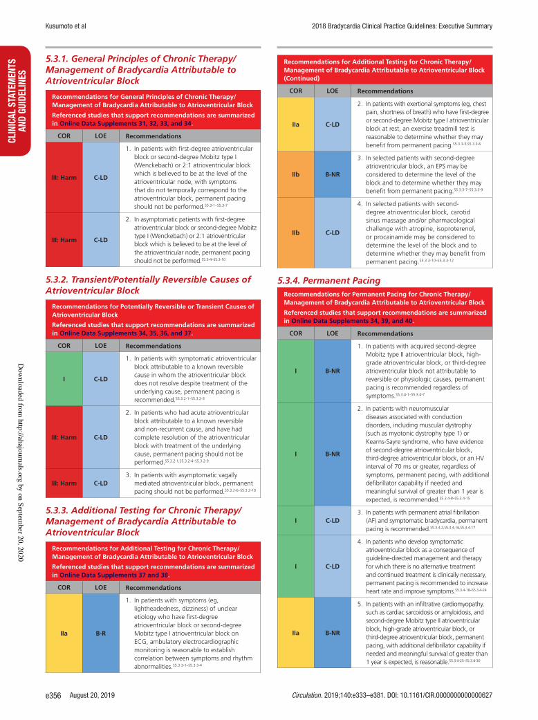
Kusumoto et al 2018 Bradycardia Clinical Practice Guidelines: Executive Summary
August 20, 2019 Circulation. 2019;140:e333–e381. DOI: 10.1161/CIR.0000000000000627e356
CLIN
ICA
L S
TA
TE
ME
NT
S
AN
D G
UID
ELIN
ES
5.3.1. General Principles of Chronic Therapy/Management of Bradycardia Attributable to Atrioventricular Block
Recommendations for General Principles of Chronic Therapy/
Management of Bradycardia Attributable to Atrioventricular Block
Referenced studies that support recommendations are summarized
in Online Data Supplements 31, 32, 33, and 34.
COR LOE Recommendations
III: Harm C-LD
1. In patients with first-degree atrioventricular block or second-degree Mobitz type I (Wenckebach) or 2:1 atrioventricular block which is believed to be at the level of the atrioventricular node, with symptoms that do not temporally correspond to the atrioventricular block, permanent pacing should not be performed.S5.3-1–S5.3-7
III: Harm C-LD
2. In asymptomatic patients with first-degree atrioventricular block or second-degree Mobitz type I (Wenckebach) or 2:1 atrioventricular block which is believed to be at the level of the atrioventricular node, permanent pacing should not be performed.S5.3-4–S5.3-10
5.3.2. Transient/Potentially Reversible Causes of Atrioventricular Block
Recommendations for Potentially Reversible or Transient Causes of
Atrioventricular Block
Referenced studies that support recommendations are summarized
in Online Data Supplements 34, 35, 36, and 37.
COR LOE Recommendations
I C-LD
1. In patients with symptomatic atrioventricular block attributable to a known reversible cause in whom the atrioventricular block does not resolve despite treatment of the underlying cause, permanent pacing is recommended.S5.3.2-1–S5.3.2-3
III: Harm C-LD
2. In patients who had acute atrioventricular block attributable to a known reversible and non-recurrent cause, and have had complete resolution of the atrioventricular block with treatment of the underlying cause, permanent pacing should not be performed.S5.3.2-1,S5.3.2-4–S5.3.2-9
III: Harm C-LD
3. In patients with asymptomatic vagally mediated atrioventricular block, permanent pacing should not be performed.S5.3.2-6–S5.3.2-10
5.3.3. Additional Testing for Chronic Therapy/Management of Bradycardia Attributable to Atrioventricular Block
Recommendations for Additional Testing for Chronic Therapy/
Management of Bradycardia Attributable to Atrioventricular Block
Referenced studies that support recommendations are summarized
in Online Data Supplements 37 and 38.
COR LOE Recommendations
IIa B-R
1. In patients with symptoms (eg, lightheadedness, dizziness) of unclear etiology who have first-degree atrioventricular block or second-degree Mobitz type I atrioventricular block on ECG, ambulatory electrocardiographic monitoring is reasonable to establish correlation between symptoms and rhythm abnormalities.S5.3.3-1–S5.3.3-4
IIa C-LD
2. In patients with exertional symptoms (eg, chest pain, shortness of breath) who have first-degree or second-degree Mobitz type I atrioventricular block at rest, an exercise treadmill test is reasonable to determine whether they may benefit from permanent pacing.S5.3.3-5,S5.3.3-6
IIb B-NR
3. In selected patients with second-degree atrioventricular block, an EPS may be considered to determine the level of the block and to determine whether they may benefit from permanent pacing.S5.3.3-7–S5.3.3-9
IIb C-LD
4. In selected patients with second-degree atrioventricular block, carotid sinus massage and/or pharmacological challenge with atropine, isoproterenol, or procainamide may be considered to determine the level of the block and to determine whether they may benefit from permanent pacing.S5.3.3-10–S5.3.3-12
5.3.4. Permanent Pacing
Recommendations for Permanent Pacing for Chronic Therapy/
Management of Bradycardia Attributable to Atrioventricular Block
Referenced studies that support recommendations are summarized
in Online Data Supplements 34, 39, and 40.
COR LOE Recommendations
I B-NR
1. In patients with acquired second-degree Mobitz type II atrioventricular block, high-grade atrioventricular block, or third-degree atrioventricular block not attributable to reversible or physiologic causes, permanent pacing is recommended regardless of symptoms.S5.3.4-1–S5.3.4-7
I B-NR
2. In patients with neuromuscular diseases associated with conduction disorders, including muscular dystrophy (such as myotonic dystrophy type 1) or Kearns-Sayre syndrome, who have evidence of second-degree atrioventricular block, third-degree atrioventricular block, or an HV interval of 70 ms or greater, regardless of symptoms, permanent pacing, with additional defibrillator capability if needed and meaningful survival of greater than 1 year is expected, is recommended.S5.3.4-8–S5.3.4-15
I C-LD
3. In patients with permanent atrial fibrillation (AF) and symptomatic bradycardia, permanent pacing is recommended.S5.3.4-2,S5.3.4-16,S5.3.4-17
I C-LD
4. In patients who develop symptomatic atrioventricular block as a consequence of guideline-directed management and therapy for which there is no alternative treatment and continued treatment is clinically necessary, permanent pacing is recommended to increase heart rate and improve symptoms.S5.3.4-18–S5.3.4-24
IIa B-NR
5. In patients with an infiltrative cardiomyopathy, such as cardiac sarcoidosis or amyloidosis, and second-degree Mobitz type II atrioventricular block, high-grade atrioventricular block, or third-degree atrioventricular block, permanent pacing, with additional defibrillator capability if needed and meaningful survival of greater than 1 year is expected, is reasonable.S5.3.4-25–S5.3.4-30
Recommendations for Additional Testing for Chronic Therapy/
Management of Bradycardia Attributable to Atrioventricular Block
(Continued)
COR LOE Recommendations
Dow
nlo
aded
from
http
://ahajo
urn
als.org
by o
n S
eptem
ber 2
0, 2
020

Kusumoto et al 2018 Bradycardia Clinical Practice Guidelines: Executive Summary
Circulation. 2019;140:e333–e381. DOI: 10.1161/CIR.0000000000000627 August 20, 2019 e357
CLIN
ICA
L S
TA
TE
ME
NT
S
AN
D G
UID
ELIN
ES
IIa B-NR
6. In patients with lamin A/C gene mutations, including limb-girdle and Emery-Dreifuss muscular dystrophies, with a PR interval greater than 240 ms and LBBB, permanent pacing, with additional defibrillator capability if needed and meaningful survival of greater than 1 year is expected, is reasonable.S5.3.4-31–S5.3.4-33
IIa C-LD
7. In patients with marked first-degree or second-degree Mobitz type I (Wenckebach) atrioventricular block with symptoms that are clearly attributable to the atrioventricular block, permanent pacing is reasonable.S5.3.4-34–S5.3.4-37
IIb C-LD
8. In patients with neuromuscular diseases, such as myotonic dystrophy type 1, with a PR interval greater than 240 ms, a QRS duration greater than 120 ms, or fascicular block, permanent pacing, with additional defibrillator capability if needed and meaningful survival of greater than 1 year is expected, may be considered.S5.3.4-9–S5.3.4-13,S5.3.4-15
5.3.4.1. Permanent Pacing Techniques and Methods for Chronic Therapy/Management of Bradycardia Attributable to Atrioventricular Block
Recommendations for Permanent Pacing Techniques and Methods
for Chronic Therapy/Management of Bradycardia Attributable to
Atrioventricular Block
Referenced studies that support recommendations are summarized
in Online Data Supplements 39 and 40 and the Systematic Review.
COR LOE Recommendations
I A
1. In patients with SND and atrioventricular block who require permanent pacing, dual chamber pacing is recommended over single chamber ventricular pacing.S5.3.4.1-1–S5.3.4.1-7
I A
2. In select patients with atrioventricular block who require permanent pacing in whom frequent ventricular pacing is not expected, or who have significant comorbidities that are likely to determine clinical outcomes and that may limit the benefit of dual chamber pacing, single chamber ventricular pacing is effective.S5.3.4.1-1–S5.3.4.1-6,S5.3.4.1-8–S5.3.4.1-10
I B-R
3. For patients in sinus rhythm with a single chamber ventricular pacemaker who develop pacemaker syndrome, revising to a dual chamber pacemaker is recommended.S5.3.4.1-1,
S5.3.4.1-2,S5.3.4.1-5,S5.3.4.1-8–S5.3.4.1-10
IIa B-RSR
4. In patients with atrioventricular block who have an indication for permanent pacing with a left ventricular ejection fraction between 36% and 50% and are expected to require ventricular pacing more than 40% of the time, it is reasonable to choose pacing methods that maintain physiologic ventricular activation (eg, cardiac resynchronization therapy [CRT] or His bundle pacing) over right ventricular pacing.S5.3.4.1-7,S5.3.4.1-11–S5.3.4.1-19
IIa B-R
5. In patients with atrioventricular block who have an indication for permanent pacing with a left ventricular ejection fraction between 36% and 50% and are expected to require ventricular pacing less than 40% of the time, it is reasonable to choose right ventricular pacing over pacing methods that maintain physiologic ventricular activation (eg, CRT or His bundle pacing).S5.3.4.1-15,S5.3.4.1-16,
S5.3.4.1-20,S5.3.4.1-21
IIb B-RSR
6. In patients with atrioventricular block at the level of the atrioventricular node who have an indication for permanent pacing, His bundle pacing may be considered to maintain physiologic ventricular activation.S5.3.4.1-19,S5.3.4.1-22–S5.3.4.1-25
III: Harm C-LD
7. In patients with permanent or persistent AF in whom a rhythm control strategy is not planned, implantation of an atrial lead should not be performed.S5.3.4.1-26,S5.3.4.1-27
SR indicates systematic review.
6. CONDUCTION DISORDERS (WITH 1:1 ATRIOVENTRICULAR CONDUCTION)
6.1. Evaluation of Conduction Disorders
See Figure 8 for evaluation of conduction disorders algorithm.
Recommendations for Evaluation of Conduction Disorders (With 1:1
Atrioventricular Conduction and Normal PR Interval)
Referenced studies that support recommendations are summarized
in Online Data Supplements 41 and 42.
COR LOE Recommendations
I B-NR
1. In patients with newly detected LBBB, a transthoracic echocardiogram to exclude structural heart disease is recommended.S6.1-1–S6.1-3
I C-LD
2. In symptomatic patients with conduction system disease, in whom atrioventricular block is suspected, ambulatory electrocardiographic monitoring is useful.S6.1-4–S6.1-11
IIa B-NR
3. In selected patients presenting with intraventricular conduction disorders other than LBBB, transthoracic echocardiography is reasonable if structural heart disease is suspected.S6.1-3,S6.1-12,S6.1-13
IIa B-NR
4. In patients with symptoms suggestive of intermittent bradycardia (eg, lightheadedness, syncope), with conduction system disease identified by ECG and no demonstrated atrioventricular block, EPS is reasonable.S6.1-14
IIa C-LD
5. In selected patients with LBBB in whom structural heart disease is suspected and echocardiogram is unrevealing, advanced imaging (eg, cardiac MRI, computed tomography, or nuclear studies) is reasonable.S6.1-15
Recommendations for Permanent Pacing for Chronic Therapy/
Management of Bradycardia Attributable to Atrioventricular Block
(Continued)
COR LOE Recommendations
Recommendations for Permanent Pacing Techniques and Methods
for Chronic Therapy/Management of Bradycardia Attributable to
Atrioventricular Block (Continued)
COR LOE Recommendations
Dow
nlo
aded
from
http
://ahajo
urn
als.org
by o
n S
eptem
ber 2
0, 2
020

Kusumoto et al 2018 Bradycardia Clinical Practice Guidelines: Executive Summary
August 20, 2019 Circulation. 2019;140:e333–e381. DOI: 10.1161/CIR.0000000000000627e358
CLIN
ICA
L S
TA
TE
ME
NT
S
AN
D G
UID
ELIN
ES
IIb C-LD
6. In selected asymptomatic patients with extensive conduction system disease (bifascicular or trifascicular block), ambulatory electrocardiographic recording may be considered to document suspected higher degree of atrioventricular block.S6.1-4,S6.1-6
IIb C-LD
7. In selected asymptomatic patients with LBBB in whom ischemic heart disease is suspected, stress testing with imaging may be considered.S6.1-2
6.2. Management of Conduction Disorders (With 1:1 Atrioventricular Conduction)
See Figure 9 for management of conduction disorders algorithm.
Recommendations for Management of Conduction Disorders (With
1:1 Atrioventricular Conduction and Normal PR Intervals)
Referenced studies that support recommendations are summarized
in Online Data Supplements 41 and 42.
COR LOE Recommendations
I C-LD
1. In patients with syncope and bundle branch block who are found to have an HV interval 70 ms or greater or evidence of infranodal block at EPS, permanent pacing is recommendedS6.2-1,S6.2-2
I C-LD
2. In patients with alternating bundle branch block, permanent pacing is recommended.S6.2-3
IIa C-LD
3. In patients with Kearns-Sayre syndrome and conduction disorders, permanent pacing is reasonable, with additional defibrillator capability if appropriate and meaningful survival of greater than 1 year is expected.S6.2-4,S6.2-5
IIb C-LD
4. In patients with Anderson-Fabry disease and QRS prolongation greater than 110 ms, permanent pacing, with additional defibrillator capability if needed and meaningful survival of greater than 1 year is expected, may be considered.S6.2-6,S6.2-7
IIb C-LD
5. In patients with heart failure, a mildly to moderately reduced left ventricular ejection fraction (36%–50%), and LBBB (QRS ≥150 ms), CRT therapy may be considered.S6.2-8,S6.2-9
III: Harm B-NR
6. In asymptomatic patients with isolated conduction disease and 1:1 atrioventricular conduction, permanent pacing is not indicated (in the absence of other indications for pacing).S6.2-10–S6.2-15
Figure 8. Evaluation of conduction disorders algorithm. Colors correspond to Class of Recommendation in Table 2. See Section 7.4. in the full-text guideline for discussion. *Refer to Section 6.2., Figure 9. †Advanced imaging could include magnetic resonance imaging, computed tomography, or transesophageal echocardiography. ‡Monitor choice based on the frequency of symptoms. §Extensive conduction disease (eg, first degree atrioventricular block combined with LBBB). ACHD indicates adult congenital heart disease; CM, cardiomyopathy; ECG, electrocardiogram/electrocardio-graphic; LBBB, left bundle branch block; and RBBB, right bundle branch block.
Recommendations for Evaluation of Conduction Disorders (With 1:1
Atrioventricular Conduction and Normal PR Interval) (Continued)
COR LOE Recommendations
Dow
nlo
aded
from
http
://ahajo
urn
als.org
by o
n S
eptem
ber 2
0, 2
020

Kusumoto et al 2018 Bradycardia Clinical Practice Guidelines: Executive Summary
Circulation. 2019;140:e333–e381. DOI: 10.1161/CIR.0000000000000627 August 20, 2019 e359
CLIN
ICA
L S
TA
TE
ME
NT
S
AN
D G
UID
ELIN
ES
7. SPECIAL POPULATIONS
7.1. Perioperative Management
7.1.1. Patients at Risk for Bradycardia During Noncardiac Surgery or Procedures
Recommendations for Patients at Risk for Bradycardia During
Noncardiac Surgery or Procedures
Referenced studies that support recommendations are summarized
in Online Data Supplements 42, 44, and 45.
COR LOE Recommendations
IIa B-NR
1. In patients who are thought to be at high risk for the development of intraoperative or periprocedural bradycardia because of patient characteristics or procedure type, placement of transcutaneous pacing pads is reasonable.S7.1.1-1–S7.1.1-3
III: Harm B-NR
2. In patients with LBBB who require pulmonary artery catheterization for intraoperative monitoring, routine prophylactic temporary transvenous pacing should not be performed.S7.1.1-4,S7.1.1-5
7.1.2. Postoperative Bradycardia and Conduction Disorders After Cardiac Surgery
7.1.2.1. Coronary Artery Bypass
Recommendations for Pacing After Isolated Coronary Artery Bypass
Surgery
Referenced studies that support recommendations are summarized
in Online Data Supplement 47.
COR LOE Recommendations
I B-NR
1. In patients who have new postoperative SND or atrioventricular block associated with persistent symptoms or hemodynamic instability that does not resolve after isolated coronary artery bypass surgery, permanent pacing is recommended before discharge.S7.1.2.1-1–S7.1.2.1-9
IIa B-NR
2. In patients undergoing isolated coronary artery bypass surgery, routine placement of temporary epicardial pacing wires is reasonable.S7.1.2.1-5,S7.1.2.1-10,S7.1.2.1-11
IIb C-EO
3. In patients undergoing coronary artery bypass surgery who will likely require future CRT or ventricular pacing, intraoperative placement of a permanent epicardial left ventricular lead may be considered.
7.1.2.2. Surgery for Atrial Fibrillation
Recommendations for Pacing After Surgery for Atrial Fibrillation
Referenced studies that support recommendations are summarized
in Online Data Supplement 48.
COR LOE Recommendations
I B-NR
1. In patients undergoing surgery for AF, routine placement of temporary epicardial pacing wires is recommended.S7.1.2.2-1–S7.1.2.2-4
I B-NR
2. In patients who have new postoperative SND or atrioventricular block associated with symptoms or hemodynamic instability that does not resolve after surgery for AF, permanent pacing is recommended before discharge.S7.1.2.2-1–S7.1.2.2-4
IIb C-EO
3. In patients undergoing surgery for AF who will likely require future CRT or ventricular pacing, intraoperative placement of a permanent epicardial left ventricular lead may be considered.
7.1.2.3. Valvular Surgery
7.1.2.3.1. Surgical Aortic Valve Replacement or Repair
Recommendations for Pacing After Aortic Valve Surgery
Referenced studies that support recommendations are summarized
in Online Data Supplement 48.
COR LOE Recommendations
I C-LD
1. In patients undergoing surgical aortic valve replacement or repair, routine placement of temporary epicardial pacing wires is recommended.S7.1.2.3.1-1–S7.1.2.3.1-3
Figure 9. Management of conduction disorders algorithm. Colors correspond to Class of Recommendation in Table 2. *For severe first-degree atrioventricular block or first-degree atrioventricular block with an accompanying neuromuscular disease, also refer to Section 5.3., Figure 7, the atrioventricular block algorithm. †See Section 3.3.2., Figure 3. AV indicates atrioventricular; BBB, bundle branch block; HF, heart failure; LBBB, left bundle branch block; and LVEF, left ventricular ejection fraction.
Dow
nlo
aded
from
http
://ahajo
urn
als.org
by o
n S
eptem
ber 2
0, 2
020

Kusumoto et al 2018 Bradycardia Clinical Practice Guidelines: Executive Summary
August 20, 2019 Circulation. 2019;140:e333–e381. DOI: 10.1161/CIR.0000000000000627e360
CLIN
ICA
L S
TA
TE
ME
NT
S
AN
D G
UID
ELIN
ES
I B-NR
2. In patients who have new postoperative SND or atrioventricular block associated with persistent symptoms or hemodynamic instability that does not resolve after aortic valve replacement, permanent pacing is recommended before discharge.S7.1.2.3.1-1–S7.1.2.3.1-5
IIb C-EO
3. In patients undergoing aortic valve surgery who will likely require future CRT or ventricular pacing, intraoperative placement of a permanent epicardial left ventricular lead may be considered.
7.1.2.3.2. Mitral Valve Surgery
Recommendations for Pacing After Mitral Valve Surgery
Referenced studies that support recommendations are summarized
in Online Data Supplement 48.
COR LOE Recommendations
I B-NR
1. In patients who have new postoperative SND or atrioventricular block associated with persistent symptoms or hemodynamic instability that does not resolve after mitral valve repair or replacement surgery, permanent pacing is recommended before discharge.S7.1.2.3.2-1,S7.1.2.3.2-2
IIa C-LD
2. In patients undergoing mitral valve surgery, routine placement of temporary epicardial pacing wires is reasonable.S7.1.2.3.2-1–S7.1.2.3.2-3
IIb C-EO
3. In patients undergoing surgical mitral valve repair or replacement who will likely require future CRT or ventricular pacing, intraoperative placement of a permanent epicardial left ventricular lead may be considered.
7.1.2.3.3. Tricuspid Valve Surgery
Recommendations for Pacing After Tricuspid Valve Surgery
Referenced studies that support recommendations are summarized
in Online Data Supplement 48.
COR LOE Recommendations
I C-LD
1. In patients undergoing tricuspid valve surgery, routine placement of temporary epicardial pacing wires is recommended.S7.1.2.3.3-1–S7.1.2.3.3-4
I B-NR
2. In patients who have new postoperative SND or atrioventricular block associated with symptoms or hemodynamic instability that does not resolve after tricuspid valve surgery, permanent pacing is recommended before discharge.S7.1.2.3.3-1–S7.1.2.3.3-4
IIa C-LD
3. In patients who are undergoing tricuspid valve replacement or tricuspid repair with high risk for postoperative atrioventricular block, intraoperative placement of permanent epicardial leads at the time of cardiac surgery is reasonable.S7.1.2.3.3-1–S7.1.2.3.3-5
7.1.2.4. Transcatheter Aortic Valve Replacement
Recommendations for Conduction Disturbances After Transcatheter
Aortic Valve Replacement
Referenced studies that support recommendations are summarized
in Online Data Supplement 49.
COR LOE Recommendations
I B-NR
1. In patients who have new atrioventricular block after transcatheter aortic valve replacement associated with symptoms or hemodynamic instability that does not resolve, permanent pacing is recommended before discharge.S7.1.2.4-1–S7.1.2.4-4
IIa B-NR
2. In patients with new persistent bundle branch block after transcatheter aortic valve replacement, careful surveillance for bradycardia is reasonable.S7.1.2.4-5,S7.1.2.4-6
IIb B-NR
3. In patients with new persistent LBBB after transcatheter aortic valve replacement, implantation of a permanent pacemaker may be considered.S7.1.2.4-4,S7.1.2.4-7–S7.1.2.4-10
7.1.2.5. Heart Transplant, Surgical Myectomy, and Alcohol Septal Ablation
7.1.2.5.1. Surgical Myectomy and Alcohol Septal Ablation for Hypertrophic Cardiomyopathy
Recommendations for Patients Undergoing Surgical Myectomy or
Alcohol Septal Ablation for Hypertrophic Cardiomyopathy
Referenced studies that support recommendations are summarized
in Online Data Supplements 51 and 52.
COR LOE Recommendations
I B-NR
1. In patients with second-degree Mobitz type II atrioventricular block, high-grade atrioventricular block, or persistent complete atrioventricular block after alcohol septal ablation or surgical myectomy, permanent pacing is recommended before discharge.S7.1.2.5.1-1–S7.1.2.5.1-4
IIa B-NR
2. In selected patients with hypertrophic cardiomyopathy who require permanent pacing for rate support after alcohol septal ablation or surgical myectomy and are at high risk for sudden cardiac death and meaningful survival of greater than 1 year is expected, selecting a device with defibrillator capabilities is reasonable.S7.1.2.5.1-5–S7.1.2.5.1-7
IIb C-LD
3. In patients with hypertrophic cardiomyopathy who undergo alcohol septal ablation and who are at risk for developing late atrioventricular block, prolonged ambulatory electrocardiographic monitoring may be considered.S7.1.2.5.1-1,S7.1.2.5.1-2,S7.1.2.5.1-4,S7.1.2.5.1-7,S7.1.2.5.1-8
IIb C-LD
4. In patients with hypertrophic cardiomyopathy, evaluation of ventriculoatrial conduction by EPS at the time of alcohol septal ablation may be considered for identifying future risk of atrioventricular block.S7.1.2.5.1-9
Recommendations for Pacing After Aortic Valve Surgery
(Continued)
COR LOE Recommendations
Dow
nlo
aded
from
http
://ahajo
urn
als.org
by o
n S
eptem
ber 2
0, 2
020

Kusumoto et al 2018 Bradycardia Clinical Practice Guidelines: Executive Summary
Circulation. 2019;140:e333–e381. DOI: 10.1161/CIR.0000000000000627 August 20, 2019 e361
CLIN
ICA
L S
TA
TE
ME
NT
S
AN
D G
UID
ELIN
ES
7.2. Bradycardia Management for Adult Congenital Heart Disease
Recommendations for Management of Bradycardia in Adults With
Adult Congenital Heart Disease
Referenced studies that support recommendations are summarized
in Online Data Supplement 53.
COR LOE Recommendations
I B-NR
1. In adults with adult congenital heart disease (ACHD )and symptomatic SND or chronotropic incompetence, atrial based permanent pacing is recommended.S7.2-1–S7.2-6
I B-NR
2. In adults with ACHD and symptomatic bradycardia related to atrioventricular block, permanent pacing is recommended.S7.2-7–S7.2-9
I B-NR
3. In adults with congenital complete atrioventricular block with any symptomatic bradycardia, a wide QRS escape rhythm, mean daytime heart rate below 50 bpm, complex ventricular ectopy, or ventricular dysfunction, permanent pacing is recommended.S7.2-10,S7.2-11
I B-NR
4. In adults with ACHD and postoperative second-degree Mobitz type II atrioventricular block, high-grade atrioventricular block, or third-degree atrioventricular block that is not expected to resolve, permanent pacing is recommended.S7.2-12,S7.2-13
IIa B-NR
5. In asymptomatic adults with congenital complete atrioventricular block, permanent pacing is reasonable.S7.2-7–S7.2-11
IIa B-NR
6. In adults with repaired ACHD who require permanent pacing for bradycardic indications, a bradycardia device with atrial antitachycardia pacing capabilities is reasonable.S7.2-14,S7.2-15
IIa C-EO
7. In adults with ACHD with preexisting sinus node and/or atrioventricular conduction disease who are undergoing cardiac surgery, intraoperative placement of epicardial permanent pacing leads is reasonable.
IIb B-NR
8. In adults with ACHD and pacemakers, atrial-based permanent pacing for the prevention of atrial arrhythmias may be considered.S7.2-3–S7.2-5,S7.2-16
III: Harm B-NR
9. In selected adults with ACHD and venous to systemic intracardiac shunts, placement of endocardial pacing leads is potentially harmful.S7.2-17,S7.2-18
7.3. Management of Bradycardia in Patients With an Acute MI
Recommendations for Management of Bradycardia in the Context
of Acute MI
Referenced studies that support recommendations are summarized
in Online Data Supplement 54.
COR LOE Recommendations
I B-NR
1. In patients presenting with an acute MI, temporary pacing is indicated for medically refractory symptomatic or hemodynamically significant bradycardia related to SND or atrioventricular block.S7.3-1–S7.3-4
I B-NR
2. Patients who present with SND or atrioventricular block in the setting of an acute MI should undergo a waiting period before determining the need for permanent pacing.S7.3-1,S7.3-4–S7.3-7
I B-NR
3. In patients presenting with an acute MI with second-degree Mobitz type II atrioventricular block, high-grade atrioventricular block, alternating bundle branch block, or third-degree atrioventricular block (persistent or infranodal), permanent pacing is indicated after a waiting period.S7.3-7,S7.3-8
IIa B-NR
4. In patients with an acute MI with symptomatic or hemodynamically significant sinus bradycardia or atrioventricular block at the level of the atrioventricular node, the administration of atropine is reasonable.S7.3-9–S7.3-11
III: Harm B-NR
5. In patients with an acute MI and transient atrioventricular block that resolves, permanent pacing should not be performed.S7.3-1,S7.3-4,S7.3-7,S7.3-12–S7.3-16
III: Harm B-NR
6. In patients with an acute MI and a new bundle branch block or isolated fascicular block in the absence of second-degree or third-degree atrioventricular block, permanent pacing should not be performed.S7.3-17–S7.3-19
7.4. Neurologic Disorders
7.4.1. Epilepsy
Recommendation for Patients With Epilepsy and Symptomatic
Bradycardia
Referenced studies that support the recommendation are
summarized in Online Data Supplement 55.
COR LOE Recommendation
IIa C-LD
1. In patients with epilepsy associated with severe symptomatic bradycardia (ictal bradycardia) where antiepileptic medications are ineffective, permanent pacing is reasonable for reducing the severity of symptoms.S7.4.1-1–S7.4.1-4
Dow
nlo
aded
from
http
://ahajo
urn
als.org
by o
n S
eptem
ber 2
0, 2
020

Kusumoto et al 2018 Bradycardia Clinical Practice Guidelines: Executive Summary
August 20, 2019 Circulation. 2019;140:e333–e381. DOI: 10.1161/CIR.0000000000000627e362
CLIN
ICA
L S
TA
TE
ME
NT
S
AN
D G
UID
ELIN
ES
8. EVALUATION OF THE RISKS FOR VENTRICULAR ARRHYTHMIAS IN PATIENTS WHO REQUIRE PERMANENT PACING
Recommendation for Management of Bradycardia and Conduction
Tissue Disease in Patients Who Require Pacing Therapy and May
Also Be at Risk for Ventricular Arrhythmias
Referenced studies that support the recommendation are
summarized in Online Data Supplement 56.
COR LOE Recommendation
I B-NR
1. In patients who require permanent pacing therapy, before implantation, an assessment of the risk of future ventricular arrhythmias and need for an implantable cardioverter defibrillator should be performed.S8-1–S8-7
9. SHARED DECISION-MAKING
Recommendations for Shared Decision-Making for Pacemaker
Implantation in the Setting of Guideline-Based Indications for
Bradycardia Pacing
COR LOE Recommendations
I C-LD
1. In patients with symptomatic bradycardia or conduction disorder, clinicians and patients should engage in a shared decision-making approach in which treatment decisions are based not only on the best available evidence, but also on the patient’s goals of care, preferences, and values.S9-1–S9-6
I C-LD
2. Patients considering implantation of a pacemaker or with a pacemaker that requires lead revision or generator change should be informed of procedural benefits and risks, including the potential short- and long-term complications and possible alternative therapy, if any, in light of their goals of care, preferences, and values.S9-1–S9-6
III: No
BenefitC-LD
3. In patients with indications for permanent pacing but also with significant comorbidities such that pacing therapy is unlikely to provide meaningful clinical benefit, or if patient goals of care strongly preclude pacemaker therapy, implantation or replacement of a pacemaker should not be performed.S9-1–S9-6
10. DISCONTINUATION OF PACEMAKER THERAPY
Recommendation for Discontinuation of Pacemaker Therapy
COR LOE Recommendation
IIa C-LD
1. In patients who present for pacemaker pulse generator replacement, or for management of pacemaker related complications, in whom the original pacing indication has resolved or is in question, discontinuation of pacemaker therapy is reasonable after evaluation of symptoms during a period of monitoring while pacing therapy is off.S10-1,S10-2
ACC/AHA TASK FORCE MEMBERS
Glenn N. Levine, MD, FACC, FAHA, Chair; Patrick T. O’Gara, MD, MACC, FAHA, Chair-Elect; Jonathan L. Halperin, MD, FACC, FAHA, Immediate Past Chair*; Sana M. Al-Khatib, MD, MHS, FACC, FAHA; Joshua A. Beckman, MD, MS, FAHA; Kim K. Birtcher, PharmD, MS, AACC; Biykem Bozkurt, MD, PhD, FACC, FAHA*; Ralph G. Brindis, MD, MPH, MACC*; Joaquin E. Cigar-roa, MD, FACC; Lesley H. Curtis, PhD, FAHA*; Anita Deswal, MD, MPH, FACC, FAHA; Lee A. Fleisher, MD, FACC, FAHA; Federico Gentile, MD, FACC; Samuel Gid-ding, MD, FAHA*; Zachary D. Goldberger, MD, MSc, FACC, FAHA; Mark A. Hlatky, MD, FACC, FAHA; John Ikonomidis, MD, PhD, FAHA*; José A. Joglar, MD, FACC, FAHA; Laura Mauri, MD, MSc, FAHA*; Mariann R. Piano, RN, PhD, FAHA; Susan J. Pressler, PhD, RN, FAHA*; Barbara Riegel, PhD, RN, FAHA*; Duminda N. Wijeysundera, MD, PhD
PRESIDENTS AND STAFF
American College of Cardiology
C. Michael Valentine, MD, FACC, PresidentTimothy W. Attebery, MBA, FACHE, Chief Executive
OfficerWilliam J. Oetgen, MD, MBA, FACC, Executive Vice
President, Science, Education, Quality, and PublishingMaryAnne Elma, MPH, Senior Director, Science, Education,
Quality, and PublishingAmelia Scholtz, PhD, Publications Manager, Science,
Education, Quality, and Publishing
American College of Cardiology/American Heart Association
Katherine A. Sheehan, PhD, Director, Guideline Strategy and Operations
Abdul R. Abdullah, MD, Science and Medicine AdvisorThomas S. D. Getchius, Manager, Guideline ScienceSam Shahid, MBBS, MPH, Associate Science and Medicine
AdvisorZainab Shipchandler, MPH, Associate Guideline Advisor,
Clinical Practice Guidelines
American Heart Association
Ivor J. Benjamin, MD, FAHA, PresidentNancy Brown, Chief Executive OfficerRose Marie Robertson, MD, FAHA, Chief Science and
Medical OfficerGayle R. Whitman, PhD, RN, FAHA, FAAN, Senior Vice
President, Office of Science Operations
* Former Task Force member; current member during the writing effort.
Dow
nlo
aded
from
http
://ahajo
urn
als.org
by o
n S
eptem
ber 2
0, 2
020

Kusumoto et al 2018 Bradycardia Clinical Practice Guidelines: Executive Summary
Circulation. 2019;140:e333–e381. DOI: 10.1161/CIR.0000000000000627 August 20, 2019 e363
CLIN
ICA
L S
TA
TE
ME
NT
S
AN
D G
UID
ELIN
ES
Paul St. Laurent, DNP, APRN, Science and Medicine Advisor
Jody Hundley, Production and Operations Manager, Scientific Publications, Office of Science Operations
ARTICLE INFORMATION
This document was approved by the American College of Cardiology Clinical Policy Approval Committee, the American Heart Association Science Advisory and Coordinating Committee, and the Heart Rhythm Society in August 2018, and the American Heart Association Executive Committee in October 2018.
Supplemental materials are available with this article at https://www. ahajournals.org/doi/suppl/10.1161/CIR.0000000000000627.
This article has been copublished in the Journal of the American College of
Cardiology and HeartRhythm.Copies: This document is available on the websites of the American College
of Cardiology (www.acc.org), American Heart Association (professional.heart.org), and the Heart Rhythm Society (www.hrsonline.org). A copy of the docu-ment is also available at https://professional.heart.org/statements by selecting the “Guidelines & Statements” button. To purchase additional reprints, call 843-216-2533 or e-mail [email protected].
The expert peer review of AHA-commissioned documents (eg, scientific statements, clinical practice guidelines, systematic reviews) is conducted by the AHA Office of Science Operations. For more on AHA statements and guide-lines development, visit https://professional.heart.org/statements. Select the “Guidelines & Statements” drop-down menu near the top of the webpage, then click “Publication Development.”
Permissions: Multiple copies, modification, alteration, enhancement, and/or distribution of this document are not permitted without the express permis-sion of the American Heart Association. Instructions for obtaining permission are located at https://www.heart.org/permissions. A link to the “Copyright Per-missions Request Form” appears in the second paragraph (https://www.heart.org/en/about-us/statements-and-policies/copyright-request-form).
REFERENCES
PREAMBLE P-1. Kusumoto FM, Schoenfeld MH, Barrett C, et al. 2018 ACC/AHA/HRS
guideline on the evaluation and management of patients with brady-cardia and cardiac conduction delay: a report of the American College of Cardiology/American Heart Association Task Force on Clinical Practice Guidelines and the Heart Rhythm Society. Circulation. 2018. In press.
1. INTRODUCTION S1-1. Slotwiner DJ, Raitt MH, Del-Carpio Munoz F, et al. Impact of physiologic
versus right ventricular pacing among patients with left ventricular ejection fraction greater than 35%: a systematic review for the 2018 ACC/AHA/HRS guideline on the evaluation and management of patients with bra-dycardia and cardiac conduction delay: a report of the American College of Cardiology/American Heart Association Task Force on Clinical Practice Guidelines and the Heart Rhythm Society. Circulation. 2018. In press.
1.4. Scope of the Guideline S1.4-1. Epstein AE, DiMarco JP, Ellenbogen KA, et al. ACC/AHA/HRS 2008
guidelines for device-based therapy of cardiac rhythm abnormali-ties: a report of the American College of Cardiology/American Heart Association Task Force on Practice Guidelines (Writing Committee to Revise the ACC/AHA/NASPE 2002 Guideline Update for Implantation of Cardiac Pacemakers and Antiarrhythmia Devices). Developed in col-laboration with the American Association for Thoracic Surgery and Society of Thoracic Surgeons. Circulation. 2008;117:e350–408.
S1.4-2. Epstein AE, DiMarco JP, Ellenbogen KA, et al. 2012 ACCF/AHA/HRS focused update incorporated into the ACCF/AHA/HRS 2008 guidelines for device-based therapy of cardiac rhythm abnormalities: a report of the American College of Cardiology Foundation/American Heart Association Task Force on Practice Guidelines and the Heart Rhythm Society. Circulation. 2012;127:e283–352.
S1.4-3. Al-Khatib SM, Stevenson WG, Ackerman MJ, et al. 2017 AHA/ACC/HRS guideline for management of patients with ventricular arrhyth-mias and the prevention of sudden cardiac death: a report of the American College of Cardiology/American Heart Association Task Force on Clinical Practice Guidelines and the Heart Rhythm Society. Circulation. 2018;138:e272–391.
S1.4-4. Shen WK, Sheldon RS, Benditt DG, et al. 2017 ACC/AHA/HRS guideline for the evaluation and management of patients with syncope: a report of the American College of Cardiology/American Heart Association Task Force on Clinical Practice Guidelines and the Heart Rhythm Society. Circulation. 2017;136:e60–122.
S1.4-5. Fihn SD, Blankenship JC, Alexander KP, et al. 2014 ACC/AHA/AATS/PCNA/SCAI/STS focused update of the guideline for the diagnosis and management of patients with stable ischemic heart disease: a report of the American College of Cardiology/American Heart Association Task Force on Practice Guidelines, and the American Association for Thoracic Surgery, Preventive Cardiovascular Nurses Association, Society for Cardiovascular Angiography and Interventions, and Society of Thoracic Surgeons. Circulation. 2014;130:1749–67.
S1.4-6. Fihn SD, Gardin JM, Abrams J, et al. 2012 ACCF/AHA/ACP/AATS/PCNA/SCAI/STS guideline for the diagnosis and management of patients with stable ischemic heart disease: a report of the American College of Cardiology Foundation/American Heart Association Task Force on Practice Guidelines, and the American College of Physicians, American Association for Thoracic Surgery, Preventive Cardiovascular Nurses Association, Society for Cardiovascular Angiography and Interventions, and Society of Thoracic Surgeons. Circulation. 2012;126:e354–471.
S1.4-7. January CT, Wann LS, Alpert JS, et al. 2014 AHA/ACC/HRS guide-line for the management of patients with atrial fibrillation: a report of the American College of Cardiology/American Heart Association Task Force on Practice Guidelines and the Heart Rhythm Society. Circulation. 2014;130:e199–267.
S1.4-8. Fleisher LA, Fleischmann KE, Auerbach AD, et al. 2014 ACC/AHA guideline on perioperative cardiovascular evaluation and manage-ment of patients undergoing noncardiac surgery: a report of the American College of Cardiology/American Heart Association Task Force on practice guidelines. Circulation. 2014;130:e278–333.
S1.4-9. Amsterdam EA, Wenger NK, Brindis RG, et al. 2014 AHA/ACC guideline for the management of patients with non-ST-elevation acute coronary syndromes: a report of the American College of Cardiology/American Heart Association Task Force on Practice Guidelines. Circulation. 2014;130:e344–426.
S1.4-10. Yancy CW, Jessup M, Bozkurt B, et al. 2013 ACCF/AHA guideline for the management of heart failure: a report of the American College of Cardiology Foundation/American Heart Association Task Force on Practice Guidelines. Circulation. 2013;128:e240–327.
S1.4-11. O’Gara PT, Kushner FG, Ascheim DD, et al. 2013 ACCF/AHA guide-line for the management of ST-elevation myocardial infarction: a report of the American College of Cardiology Foundation/American Heart Association Task Force on Practice Guidelines. Circulation. 2013;127:e362–425.
S1.4-12. Hillis LD, Smith PK, Anderson JL, et al. 2011 ACCF/AHA guideline for coronary artery bypass graft surgery. a report of the American College of Cardiology Foundation/American Heart Association Task Force on Practice Guidelines. Developed in collaboration with the American Association for Thoracic Surgery, Society of Cardiovascular Anesthesiologists, and Society of Thoracic Surgeons. Circulation. 2011;124:e652–735.
S1.4-13. Gersh BJ, Maron BJ, Bonow RO, et al. 2011 ACCF/AHA guideline for the diagnosis and treatment of hypertrophic cardiomyopathy: a report of the American College of Cardiology Foundation/American Heart Association Task Force on Practice Guidelines. Developed in collaboration with the American Association for Thoracic Surgery, American Society of Echocardiography, American Society of Nuclear Cardiology, Heart Failure Society of America, Heart Rhythm Society, Society for Cardiovascular Angiography and Interventions, and Society of Thoracic Surgeons. Circulation. 2011;124:e783–831.
S1.4-14. Levine GN, Bates ER, Blankenship JC, et al. 2011 ACCF/AHA/SCAI guideline for percutaneous coronary intervention: a report of the American College of Cardiology Foundation/American Heart Association Task Force on Practice Guidelines and the Society for Cardiovascular Angiography and Interventions. Circulation. 2011;124:e574–651.
Dow
nlo
aded
from
http
://ahajo
urn
als.org
by o
n S
eptem
ber 2
0, 2
020

Kusumoto et al 2018 Bradycardia Clinical Practice Guidelines: Executive Summary
August 20, 2019 Circulation. 2019;140:e333–e381. DOI: 10.1161/CIR.0000000000000627e364
CLIN
ICA
L S
TA
TE
ME
NT
S
AN
D G
UID
ELIN
ES
S1.4-15. Peberdy MA, Callaway CW, Neumar RW, et al. Part 9: post-cardiac arrest care: 2010 American Heart Association guidelines for car-diopulmonary resuscitation and emergency cardiovascular care. Circulation. 2010;122:S768–86.
S1.4-16. Kusumoto FM, Schoenfeld MH, Wilkoff BL, et al. 2017 HRS expert consensus statement on cardiovascular implantable electronic device lead management and extraction. Heart Rhythm. 2017;14:e503–51.
S1.4-17. Feingold B, Mahle WT, Auerbach S, et al. Management of cardiac involve-ment associated with neuromuscular diseases: a scientific statement from the American Heart Association. Circulation. 2017;136:e200–31.
S1.4-18. Indik JH, Gimbel JR, Abe H, et al. 2017 HRS expert consensus state-ment on magnetic resonance imaging and radiation exposure in patients with cardiovascular implantable electronic devices. Heart Rhythm. 2017;14:e97–153.
S1.4-19. Zipes DP, Link MS, Ackerman MJ, et al. Eligibility and disqualifica-tion recommendations for competitive athletes with cardiovascular abnormalities: Task Force 9: arrhythmias and conduction defects: a scientific statement from the American Heart Association and American College of Cardiology. Circulation. 2015;132:e315–25.
S1.4-20. Sheldon RS, Grubb BP 2nd, Olshansky B, et al. 2015 Heart Rhythm Society expert consensus statement on the diagnosis and treatment of postural tachycardia syndrome, inappropriate sinus tachycardia, and vasovagal syncope. Heart Rhythm. 2015;12:e41–63.
S1.4-21. Khairy P, Van Hare GF, Balaji S, et al. PACES/HRS expert consensus statement on the recognition and management of arrhythmias in adult congenital heart disease: developed in partnership between the Pediatric and Congenital Electrophysiology Society (PACES) and the Heart Rhythm Society (HRS). Heart Rhythm. 2014;11:e102–65.
S1.4-22. Kusumoto FM, Calkins H, Boehmer J, et al. HRS/ACC/AHA expert consensus statement on the use of implantable cardioverter-defibril-lator therapy in patients who are not included or not well repre-sented in clinical trials. Circulation. 2014;130:94–125.
S1.4-23. Birnie DH, Sauer WH, Bogun F, et al. HRS expert consensus state-ment on the diagnosis and management of arrhythmias associated with cardiac sarcoidosis. Heart Rhythm. 2014;11:1305–23.
S1.4-24. Brignole M, Auricchio A, Baron-Esquivias G, et al. 2013 ESC guide-lines on cardiac pacing and cardiac resynchronization therapy: the Task Force on Cardiac Pacing and Resynchronization Therapy of the European Society of Cardiology (ESC). Developed in collaboration with the European Heart Rhythm Association (EHRA). Eur Heart J. 2013;34:2281–329.
S1.4-25. Gillis AM, Russo AM, Ellenbogen KA, et al. HRS/ACCF expert con-sensus statement on pacemaker device and mode selection. J Am Coll Cardiol. 2012;60:682–703.
S1.4-26. Ackerman MJ, Priori SG, Willems S, et al. HRS/EHRA expert consen-sus statement on the state of genetic testing for the channelopathies and cardiomyopathies this document was developed as a partner-ship between the Heart Rhythm Society (HRS) and the European Heart Rhythm Association (EHRA). Heart Rhythm. 2011;8:1308–39.
S1.4-27. Lampert R, Hayes DL, Annas GJ, et al. HRS expert consensus state-ment on the management of cardiovascular implantable electronic devices (CIEDs) in patients nearing end of life or requesting with-drawal of therapy. Heart Rhythm. 2010;7:1008–26.
S1.4-28. Surawicz B, Childers R, Deal BJ, et al. AHA/ACCF/HRS recommenda-tions for the standardization and interpretation of the electrocardio-gram: part III: intraventricular conduction disturbances: a scientific statement from the American Heart Association Electrocardiography and Arrhythmias Committee, Council on Clinical Cardiology; the American College of Cardiology Foundation; and the Heart Rhythm Society. Circulation. 2009;119:e235–40.
S1.4-29. Hancock EW, Deal BJ, Mirvis DM, et al. AHA/ACCF/HRS recommen-dations for the standardization and interpretation of the electrocar-diogram: part V: electrocardiogram changes associated with cardiac chamber hypertrophy: a scientific statement from the American Heart Association Electrocardiography and Arrhythmias Committee, Council on Clinical Cardiology; the American College of Cardiology Foundation; and the Heart Rhythm Society. Circulation. 2009;119:e251–61.
1.5. Class of Recommendation and Level of Evidence S1.5-1. Halperin JL, Levine GN, Al-Khatib SM, et al. Further evolution of the
ACC/AHA clinical practice guideline recommendation classification
system: a report of the American College of Cardiology/American Heart Association Task Force on Clinical Practice Guidelines. Circulation. 2015;133:1426–8.
2. EPIDEMIOLOGY AND DEFINITIONS
2.1. Definitions S2.1-1. Page RL, Joglar JA, Caldwell MA, et al. 2015 ACC/AHA/HRS guide-
line for the management of adult patients with supraventricu-lar tachycardia: a report of the American College of Cardiology/American Heart Association Task Force on Clinical Practice Guidelines and the Heart Rhythm Society. Circulation. 2016;133: e506–74.
S2.1-2. Surawicz B, Childers R, Deal BJ, et al. AHA/ACCF/HRS recommen-dations for the standardization and interpretation of the elec-trocardiogram: part III: intraventricular conduction disturbances: a scientific statement from the American Heart Association Electrocardiography and Arrhythmias Committee, Council on Clinical Cardiology; the American College of Cardiology Foundation; and the Heart Rhythm Society. Circulation. 2009;119: e235–40.
3. GENERAL EVALUATION OF PATIENTS WITH DOCUMENTED OR SUSPECTED BRADYCARDIA OR CONDUCTION DISORDERS
3.1. History and Physical Examination of Patients With Documented or Suspected Bradycardia or Conduction Disorders S3.1-1. Mangrum JM, DiMarco JP. The evaluation and management of bra-
dycardia. N Engl J Med. 2000;342:703–9. S3.1-2. Vogler J, Breithardt G, Eckardt L. Bradyarrhythmias and conduction
blocks. Rev Esp Cardiol (Engl Ed). 2012;65:656–67.
3.2. NONINVASIVE EVALUATION
3.2.1. Resting ECG in Patients With Documented or Suspected Bradycardia or Conduction Disorders S3.2.1-1. Del Rosso A, Ungar A, Maggi R, et al. Clinical predictors of cardiac
syncope at initial evaluation in patients referred urgently to a general hospital: the EGSYS score. Heart. 2008;94:1620–6.
S3.2.1-2. Linzer M, Yang EH, Estes NA 3rd, et al. Diagnosing syncope. Part 1: value of history, physical examination, and electrocardiography. Clinical Efficacy Assessment Project of the American College of Physicians. Ann Intern Med. 1997;126:989–96.
S3.2.1-3. Perez-Rodon J, Martinez-Alday J, Baron-Esquivias G, et al. Prognostic value of the electrocardiogram in patients with syncope: data from the group for syncope study in the emergency room (GESINUR). Heart Rhythm. 2014;11:2035–44.
S3.2.1-4. Thiruganasambandamoorthy V, Hess EP, Turko E, et al. Defining abnormal electrocardiography in adult emergency department syncope patients: the Ottawa Electrocardiographic Criteria. CJEM. 2012;14:248–58.
3.2.2. Exercise Electrocardiographic Testing in Patients With Documented or Suspected Bradycardia or Conduction Disorders
S3.2.2-1. Lauer MS, Francis GS, Okin PM, et al. Impaired chronotropic response to exercise stress testing as a predictor of mortality. JAMA. 1999;281:524–9.
Dow
nlo
aded
from
http
://ahajo
urn
als.org
by o
n S
eptem
ber 2
0, 2
020

Kusumoto et al 2018 Bradycardia Clinical Practice Guidelines: Executive Summary
Circulation. 2019;140:e333–e381. DOI: 10.1161/CIR.0000000000000627 August 20, 2019 e365
CLIN
ICA
L S
TA
TE
ME
NT
S
AN
D G
UID
ELIN
ES
S3.2.2-2. Savonen KP, Kiviniemi V, Laukkanen JA, et al. Chronotropic incom-petence and mortality in middle-aged men with known or sus-pected coronary heart disease. Eur Heart J. 2008;29:1896–902.
S3.2.2-3. Doi A, Tsuchihashi K, Kyuma M, et al. Diagnostic implications of modified treadmill and head-up tilt tests in exercise-related syncope: comparative studies with situational and/or vasovagal syncope. Can J Cardiol. 2002;18:960–6.
S3.2.2-4. Woelfel AK, Simpson RJ Jr, Gettes LS, et al. Exercise-induced distal atrioventricular block. J Am Coll Cardiol. 1983;2:578–81.
3.2.3. Ambulatory Electrocardiography in Patients With Documented or Suspected Bradycardia or Conduction Disorders
S3.2.3-1. Barrett PM, Komatireddy R, Haaser S, et al. Comparison of 24-hour Holter monitoring with 14-day novel adhesive patch electrocardio-graphic monitoring. Am J Med. 2014;127:95.e11–7.
S3.2.3-2. Brown AP, Dawkins KD, Davies JG. Detection of arrhythmias: use of a patient-activated ambulatory electrocardiogram device with a solid-state memory loop. Br Heart J. 1987;58:251–3.
S3.2.3-3. Cumbee SR, Pryor RE, Linzer M. Cardiac loop ECG recording: a new noninvasive diagnostic test in recurrent syncope. South Med J. 1990;83:39–43.
S3.2.3-4. Gibson TC, Heitzman MR. Diagnostic efficacy of 24-hour electrocar-diographic monitoring for syncope. Am J Cardiol. 1984;53:1013–7.
S3.2.3-5. Joshi AK, Kowey PR, Prystowsky EN, et al. First experience with a mobile cardiac outpatient telemetry (MCOT) system for the diagnosis and management of cardiac arrhythmia. Am J Cardiol. 2005;95:878–81.
S3.2.3-6. Linzer M, Yang EH, Estes NA 3rd, et al. Diagnosing syncope. Part 2: unexplained syncope. Clinical Efficacy Assessment Project of the American College of Physicians. Ann Intern Med. 1997;127:76–86.
S3.2.3-7. Locati ET, Moya A, Oliveira M, et al. External prolonged electro-cardiogram monitoring in unexplained syncope and palpitations: results of the SYNARR-Flash study. Europace. 2016;18:1265–72.
S3.2.3-8. Reiffel JA, Schwarzberg R, Murry M. Comparison of autotriggered memory loop recorders versus standard loop recorders versus 24-hour Holter monitors for arrhythmia detection. Am J Cardiol. 2005;95:1055–9.
S3.2.3-9. Rosenberg MA, Samuel M, Thosani A, et al. Use of a noninva-sive continuous monitoring device in the management of atrial fibrillation: a pilot study. Pacing Clin Electrophysiol. 2013;36: 328–33.
S3.2.3-10. Rothman SA, Laughlin JC, Seltzer J, et al. The diagnosis of cardiac arrhythmias: a prospective multi-center randomized study compar-ing mobile cardiac outpatient telemetry versus standard loop event monitoring. J Cardiovasc Electrophysiol. 2007;18:241–7.
S3.2.3-11. Sivakumaran S, Krahn AD, Klein GJ, et al. A prospective random-ized comparison of loop recorders versus Holter monitors in patients with syncope or presyncope. Am J Med. 2003;115:1–5.
S3.2.3-12. Turakhia MP, Hoang DD, Zimetbaum P, et al. Diagnostic utility of a novel leadless arrhythmia monitoring device. Am J Cardiol. 2013;112:520–4.
S3.2.3-13. Shen WK, Sheldon RS, Benditt DG, et al. 2017 ACC/AHA/HRS guideline for the evaluation and management of patients with syncope: a report of the American College of Cardiology/American Heart Association Task Force on Clinical Practice Guidelines and the Heart Rhythm Society. Circulation. 2017;136:e60–122.
3.2.4. Imaging in Patients With Documented or Suspected Bradycardia or Conduction Disorders
S3.2.4-1. Schneider JF, Thomas HE Jr, Kreger BE, et al. Newly acquired left bundle-branch block: the Framingham study. Ann Intern Med. 1979;90:303–10.
S3.2.4-2. Schneider JF, Thomas HE Jr, Sorlie P, et al. Comparative features of newly acquired left and right bundle branch block in the general population: the Framingham study. Am J Cardiol. 1981;47:931–40.
S3.2.4-3. Talreja D, Gruver C, Sklenar J, et al. Efficient utilization of echocar-diography for the assessment of left ventricular systolic function. Am Heart J. 2000;139:394–8.
S3.2.4-4. Dhingra R, Ho Nam B, Benjamin EJ, et al. Cross-sectional relations of electrocardiographic QRS duration to left ventricular dimensions: the Framingham Heart Study. J Am Coll Cardiol. 2005;45:685–9.
S3.2.4-5. Dhingra R, Pencina MJ, Wang TJ, et al. Electrocardiographic QRS duration and the risk of congestive heart failure: the Framingham Heart Study. Hypertension. 2006;47:861–7.
S3.2.4-6. Badheka AO, Singh V, Patel NJ, et al. QRS duration on electrocardi-ography and cardiovascular mortality (from the National Health and Nutrition Examination Survey-III). Am J Cardiol. 2013;112:671–7.
S3.2.4-7. Bogale N, Orn S, James M, et al. Usefulness of either or both left and right bundle branch block at baseline or during follow-up for predicting death in patients following acute myocardial infarction. Am J Cardiol. 2007;99:647–50.
S3.2.4-8. Eriksson P, Hansson PO, Eriksson H, et al. Bundle-branch block in a general male population: the study of men born 1913. Circulation. 1998;98:2494–500.
S3.2.4-9. Fahy GJ, Pinski SL, Miller DP, et al. Natural history of isolated bundle branch block. Am J Cardiol. 1996;77:1185–90.
S3.2.4-10. Imanishi R, Seto S, Ichimaru S, et al. Prognostic significance of inci-dent complete left bundle branch block observed over a 40-year period. Am J Cardiol. 2006;98:644–8.
S3.2.4-11. Chiu DT, Shapiro NI, Sun BC, et al. Are echocardiography, telemetry, ambulatory electrocardiography monitoring, and cardiac enzymes in emergency department patients presenting with syncope useful tests? A preliminary investigation. J Emerg Med. 2014;47:113–8.
S3.2.4-12. Douglas PS, Garcia MJ, Haines DE, et al. ACCF/ASE/AHA/ASNC/HFSA/HRS/SCAI/SCCM/SCCT/SCMR 2011 appropriate use criteria for echocardiography: a report of the American College of Cardiology Foundation Appropriate Use Criteria Task Force, American Society of Echocardiography, American Heart Association, American Society of Nuclear Cardiology, Heart Failure Society of America, Heart Rhythm Society, Society for Cardiovascular Angiography and Interventions, Society of Critical Care Medicine, Society of Cardiovascular Computed Tomography, Society for Cardiovascular Magnetic Resonance American College of Chest Physicians. J Am Soc Echocardiogr. 2011;24:229–67.
S3.2.4-13. Menozzi C, Brignole M, Alboni P, et al. The natural course of untreated sick sinus syndrome and identification of the variables predictive of unfavorable outcome. Am J Cardiol. 1998;82:1205–9.
S3.2.4-14. Bluemke DA, Achenbach S, Budoff M, et al. Noninvasive coronary artery imaging: magnetic resonance angiography and multidetec-tor computed tomography angiography: a scientific statement from the American Heart Association Committee on Cardiovascular Imaging and Intervention of the Council on Cardiovascular Radiology and Intervention, and the Councils on Clinical Cardiology and Cardiovascular Disease in the Young. Circulation. 2008;118:586–606.
S3.2.4-15. Bokhari S, Castano A, Pozniakoff T, et al. (99m)Tc-pyrophosphate scintigraphy for differentiating light-chain cardiac amyloidosis from the transthyretin-related familial and senile cardiac amyloidoses. Circ Cardiovasc Imaging. 2013;6:195–201.
S3.2.4-16. Cheong B, Huber S, Muthupillai R, et al. Evaluation of myocardial iron overload by T(2)* Cardiovascular Magnetic Resonance Imaging. Tex Heart Inst J. 2005;32:448–9.
S3.2.4-17. Fontana M, Pica S, Reant P, et al. Prognostic value of late gadolinium enhancement cardiovascular magnetic resonance in cardiac amyloi-dosis. Circulation. 2015;132:1570–9.
S3.2.4-18. Franco A, Javidi S, Ruehm SG. Delayed myocardial enhancement in cardiac magnetic resonance imaging. J Radiol Case Rep. 2015;9:6–18.
S3.2.4-19. Greulich S, Deluigi CC, Gloekler S, et al. CMR imaging predicts death and other adverse events in suspected cardiac sarcoidosis. JACC Cardiovasc Imaging. 2013;6:501–11.
S3.2.4-20. Haq M, Pawar S, Berk JL, et al. Can 99mTc-pyrophosphate aid in early detection of cardiac involvement in asymptomatic variant TTR amyloidosis? JACC Cardiovasc Imaging. 2017;10:713–4.
S3.2.4-21. Taylor AJ, Cerqueira M, Hodgson JM, et al. ACCF/SCCT/ACR/AHA/ASE/ASNC/NASCI/SCAI/SCMR 2010 appropriate use criteria for cardiac computed tomography. A report of the American College of Cardiology Foundation Appropriate Use Criteria Task Force, the Society of Cardiovascular Computed Tomography, the American College of Radiology, the American Heart Association, the American Society of Echocardiography, the American Society of Nuclear Cardiology, the North American Society for Cardiovascular Imaging, the Society for Cardiovascular Angiography and Interventions, and the Society for Cardiovascular Magnetic Resonance. Circulation. 2010;122:e525–55.
S3.2.4-22. Mendu ML, McAvay G, Lampert R, et al. Yield of diagnostic tests in evaluating syncopal episodes in older patients. Arch Intern Med. 2009;169:1299–305.
Dow
nlo
aded
from
http
://ahajo
urn
als.org
by o
n S
eptem
ber 2
0, 2
020

Kusumoto et al 2018 Bradycardia Clinical Practice Guidelines: Executive Summary
August 20, 2019 Circulation. 2019;140:e333–e381. DOI: 10.1161/CIR.0000000000000627e366
CLIN
ICA
L S
TA
TE
ME
NT
S
AN
D G
UID
ELIN
ES
S3.2.4-23. Recchia D, Barzilai B. Echocardiography in the evaluation of patients with syncope. J Gen Intern Med. 1995;10:649–55.
S3.2.4-24. Sarasin FP, Junod AF, Carballo D, et al. Role of echocardiogra-phy in the evaluation of syncope: a prospective study. Heart. 2002;88:363–7.
3.2.5. Laboratory Testing in Patients With Documented or Suspected Bradycardia or Conduction Disorders
S3.2.5-1. Chon SB, Kwak YH, Hwang SS, et al. Severe hyperkalemia can be detected immediately by quantitative electrocardiography and clini-cal history in patients with symptomatic or extreme bradycardia: a retrospective cross-sectional study. J Crit Care. 2013;28:1112.e7–13.
S3.2.5-2. Mandell BF. Cardiovascular involvement in systemic lupus erythema-tosus. Semin Arthritis Rheum. 1987;17:126–41.
S3.2.5-3. Nakayama Y, Ohno M, Yonemura S, et al. A case of transient 2:1 atrioventricular block, resolved by thyroxine supplementation for sub-clinical hypothyroidism. Pacing Clin Electrophysiol. 2006;29:106–8.
S3.2.5-4. Noble K, Isles C. Hyperkalaemia causing profound bradycardia. Heart. 2006;92:1063.
3.2.7. Sleep Apnea Evaluation and Treatment in Patients With Documented or Suspected Bradycardia or Conduction Disorders
S3.2.7-1. Tilkian AG, Guilleminault C, Schroeder JS, et al. Sleep-induced apnea syndrome. Prevalence of cardiac arrhythmias and their rever-sal after tracheostomy. Am J Med. 1977;63:348–58.
S3.2.7-2. Guilleminault C, Connolly SJ, Winkle RA. Cardiac arrhythmia and conduction disturbances during sleep in 400 patients with sleep apnea syndrome. Am J Cardiol. 1983;52:490–4.
S3.2.7-3. Shepard JW Jr, Garrison MW, Grither DA, et al. Relationship of ventricular ectopy to oxyhemoglobin desaturation in patients with obstructive sleep apnea. Chest. 1985;88:335–40.
S3.2.7-4. Shepard JW Jr. Gas exchange and hemodynamics during sleep. Med Clin North Am. 1985;69:1243–64.
S3.2.7-5. Hoffstein V, Mateika S. Cardiac arrhythmias, snoring, and sleep apnea. Chest. 1994;106:466–71.
S3.2.7-6. Boudoulas H, Schmidt HS, Clark RW, et al. Anthropometric char-acteristics, cardiac abnormalities and adrenergic activity in patients with primary disorders of sleep. J Med. 1983;14:223–38.
S3.2.7-7. Mehra R, Benjamin EJ, Shahar E, et al. Association of nocturnal arrhythmias with sleep-disordered breathing: the Sleep Heart Health study. Am J Respir Crit Care Med. 2006;173:910–6.
S3.2.7-8. Miller WP. Cardiac arrhythmias and conduction disturbances in the sleep apnea syndrome. Prevalence and significance. Am J Med. 1982;73:317–21.
S3.2.7-9. Flemons WW, Remmers JE, Gillis AM. Sleep apnea and car-diac arrhythmias. Is there a relationship? Am Rev Respir Dis. 1993;148:618–21.
S3.2.7-10. Stegman SS, Burroughs JM, Henthorn RW. Asymptomatic bradyar-rhythmias as a marker for sleep apnea: appropriate recognition and treatment may reduce the need for pacemaker therapy. Pacing Clin Electrophysiol. 1996;19:899–904.
S3.2.7-11. Garrigue S, Pepin JL, Defaye P, et al. High prevalence of sleep apnea syndrome in patients with long-term pacing: the European Multicenter Polysomnographic study. Circulation. 2007;115:1703–9.
S3.2.7-12. Becker H, Brandenburg U, Peter JH, et al. Reversal of sinus arrest and atrioventricular conduction block in patients with sleep apnea during nasal continuous positive airway pressure. Am J Respir Crit Care Med. 1995;151:215–8.
S3.2.7-13. Koehler U, Fus E, Grimm W, et al. Heart block in patients with obstructive sleep apnoea: pathogenetic factors and effects of treat-ment. Eur Respir J. 1998;11:434–9.
S3.2.7-14. Grimm W, Koehler U, Fus E, et al. Outcome of patients with sleep apnea-associated severe bradyarrhythmias after continuous positive airway pressure therapy. Am J Cardiol. 2000;86:688–92. a9.
S3.2.7-15. Harbison J, O’Reilly P, McNicholas WT. Cardiac rhythm disturbances in the obstructive sleep apnea syndrome: effects of nasal continu-ous positive airway pressure therapy. Chest. 2000;118:591–5.
S3.2.7-16. Daccarett M, Segerson NM, Hamdan AL, et al. Relation of daytime bradyarrhythmias with high risk features of sleep apnea. Am J Cardiol. 2008;101:1147–50.
3.3. Invasive Testing3.3.1. Implantable Cardiac Monitor in Patients With Documented or Suspected Bradycardia or Conduction Disorders
S3.3.1-1. Farwell DJ, Freemantle N, Sulke AN. Use of implantable loop record-ers in the diagnosis and management of syncope. Eur Heart J. 2004;25:1257–63.
S3.3.1-2. Krahn AD, Klein GJ, Yee R, et al. Randomized assessment of syn-cope trial: conventional diagnostic testing versus a prolonged moni-toring strategy. Circulation. 2001;104:46–51.
S3.3.1-3. Podoleanu C, DaCosta A, Defaye P, et al. Early use of an implantable loop recorder in syncope evaluation: a randomized study in the con-text of the French healthcare system (FRESH study). Arch Cardiovasc Dis. 2014;107:546–52.
3.3.2. Electrophysiology Study in Patients With Documented or Suspected Bradycardia or Conduction Disorders
S3.3.2-1. DiMarco JP, Garan H, Ruskin JN. Approach to the patient with recur-rent syncope of unknown cause. Mod Concepts Cardiovasc Dis. 1983;52:11–6.
S3.3.2-2. Fisher JD. Role of electrophysiologic testing in the diagnosis and treatment of patients with known and suspected bradycardias and tachycardias. Prog Cardiovasc Dis. 1981;24:25–90.
S3.3.2-3. Gulamhusein S, Naccarelli GV, Ko PT, et al. Value and limitations of clinical electrophysiologic study in assessment of patients with unexplained syncope. Am J Med. 1982;73:A53.
S3.3.2-4. Krol RB, Morady F, Flaker GC, et al. Electrophysiologic testing in patients with unexplained syncope: Clinical and noninvasive predic-tors of outcome. J Am Coll Cardiol. 1987;10:358–63.
S3.3.2-5. Denniss AR, Ross DL, Richards DA, et al. Electrophysiologic studies in patients with unexplained syncope. Int J Cardiol. 1992;35:211–7.
4. BRADYCARDIA ATTRIBUTABLE TO SINUS NODE DYSFUNCTION
4.1. Acute Management of Sinus Node Dysfunction4.1.1. Acute Management of Reversible Causes of Sinus Node Dysfunction
S4.1.1-1. Da Costa D, Brady WJ, Edhouse J. Bradycardias and atrioventricular conduction block. BMJ. 2002;324:535–8.
S4.1.1-2. Alboni P, Baggioni GF, Scarfo S, et al. Role of sinus node artery dis-ease in sick sinus syndrome in inferior wall acute myocardial infarc-tion. Am J Cardiol. 1991;67:1180–4.
S4.1.1-3. Ando G, Gaspardone A, Proietti I. Acute thrombosis of the sinus node artery: arrhythmological implications. Heart. 2003;89:E5.
S4.1.1-4. Rokseth R, Hatle L. Sinus arrest in acute myocardial infarction. Br Heart J. 1971;33:639–42.
S4.1.1-5. Zehender M, Meinertz T, Keul J, et al. ECG variants and cardiac arrhythmias in athletes: clinical relevance and prognostic impor-tance. Am Heart J. 1990;119:1378–91.
S4.1.1-6. Jackson LR 2nd, Rathakrishnan B, Campbell K, et al. Sinus node dysfunction and atrial fibrillation: a reversible phenomenon? Pacing Clin Electrophysiol. 2017;40:442–50.
S4.1.1-7. Pasic M, Musci M, Siniawski H, et al. Transient sinus node dys-function after the Cox-Maze III procedure in patients with organic heart disease and chronic fixed atrial fibrillation. J Am Coll Cardiol. 1998;32:1040–7.
S4.1.1-8. Shin H, Yozu R, Higashi S, et al. Sinus node function after mitral valve surgery using the superior septal approach. Ann Thorac Surg. 2001;71:587–90.
S4.1.1-9. Cleveland RJ, Nelson RJ, Zeilenga DW, et al. Atrial pacing following open-heart surgery. Arch Surg. 1972;105:26–9.
S4.1.1-10. Peretto G, Durante A, Limite LR, et al. Postoperative arrhythmias after cardiac surgery: incidence, risk factors, and therapeutic man-agement. Cardiol Res Pract. 2014;2014:615987.
Dow
nlo
aded
from
http
://ahajo
urn
als.org
by o
n S
eptem
ber 2
0, 2
020

Kusumoto et al 2018 Bradycardia Clinical Practice Guidelines: Executive Summary
Circulation. 2019;140:e333–e381. DOI: 10.1161/CIR.0000000000000627 August 20, 2019 e367
CLIN
ICA
L S
TA
TE
ME
NT
S
AN
D G
UID
ELIN
ES
S4.1.1-11. Franklin SM, Thihalolipavan S, Fontaine JM. Sinus bradycardia in habitual cocaine users. Am J Cardiol. 2017;119:1611–5.
S4.1.1-12. Ettinger PO, Regan TJ, Oldewurtel HA. Hyperkalemia, cardiac conduction, and the electrocardiogram: a review. Am Heart J. 1974;88:360–71.
S4.1.1-13. Lee G, McGavigan AD, Hillock RJ, et al. A grave case of bradycardia. Pacing Clin Electrophysiol. 2006;29:788–90.
S4.1.1-14. Bolognesi R, Tsialtas D, Bolognesi MG, et al. Marked sinus bradycar-dia and QT prolongation in a diabetic patient with severe hypogly-cemia. J Diabetes Complications. 2011;25:349–51.
S4.1.1-15. Luebbert JJ, Lee FA, Rosenfeld LE. Pacemaker therapy for early and late sinus node dysfunction in orthotopic heart transplant recipients: a sin-gle-center experience. Pacing Clin Electrophysiol. 2008;31:1108–12.
S4.1.1-16. Bertolet BD, Eagle DA, Conti JB, et al. Bradycardia after heart transplantation: reversal with theophylline. J Am Coll Cardiol. 1996;28:396–9.
S4.1.1-17. Heinz G, Hirschl M, Buxbaum P, et al. Sinus node dysfunction after orthotopic cardiac transplantation: postoperative incidence and long-term implications. Pacing Clin Electrophysiol. 1992;15:731–7.
S4.1.1-18. Santinelli V, Chiariello M, Clarizia M, et al. Sick sinus syndrome: the role of hypervagotonia. Int J Cardiol. 1984;5:532–5.
S4.1.1-19. Desai JM, Scheinman MM, Strauss HC, et al. Electrophysiologic effects on combined autonomic blockade in patients with sinus node disease. Circulation. 1981;63:953–60.
S4.1.1-20. Thomsen JH, Hassager C, Bro-Jeppesen J, et al. Sinus bradycardia during hypothermia in comatose survivors of out-of-hospital cardiac arrest - a new early marker of favorable outcome? Resuscitation. 2015;89:36–42.
S4.1.1-21. Mattu A, Brady WJ, Perron AD. Electrocardiographic manifestations of hypothermia. Am J Emerg Med. 2002;20:314–26.
S4.1.1-22. Grais IM, Sowers JR. Thyroid and the heart. Am J Med. 2014;127:691–8.
S4.1.1-23. Secher NH, Sander Jensen K, Werner C, et al. Bradycardia dur-ing severe but reversible hypovolemic shock in man. Circ Shock. 1984;14:267–74.
S4.1.1-24. Zwillich C, Devlin T, White D, et al. Bradycardia during sleep apnea. Characteristics and mechanism. J Clin Invest. 1982;69:1286–92.
S4.1.1-25. Grmec S, Strnad M, Podgorsek D. Comparison of the character-istics and outcome among patients suffering from out-of-hospital primary cardiac arrest and drowning victims in cardiac arrest. Int J Emerg Med. 2009;2:7–12.
S4.1.1-26. Myers MG, Norris JW, Hachinski VC, et al. Cardiac sequelae of acute stroke. Stroke. 1982;13:838–42.
S4.1.1-27. Cunha BA. The diagnostic significance of relative bradycardia in infectious disease. Clin Microbiol Infect. 2000;6:633–4.
S4.1.1-28. Woolf PK, Lorsung EM, Edwards KS, et al. Electrocardiographic findings in children with Lyme disease. Pediatr Emerg Care. 1991;7:334–6.
S4.1.1-29. Gee SW, Karsies TJ. Listeria meningitis-associated bradyarrhythmia treated with isoproterenol. Am J Emerg Med. 2015;33:306.e1–2.
S4.1.1-30. Narayan D, Huang MT, Mathew PK. Bradycardia and asystole requir-ing permanent pacemaker in Guillain-Barre syndrome. Am Heart J. 1984;108:426–8.
S4.1.1-31. Ma G, Brady WJ, Pollack M, et al. Electrocardiographic manifesta-tions: digitalis toxicity. J Emerg Med. 2001;20:145–52.
S4.1.1-32. Talati SN, Aslam AF, Vasavada B. Sinus node dysfunction in asso-ciation with chronic lithium therapy: a case report and review of literature. Am J Ther. 2009;16:274–8.
4.1.2. Acute Medical Therapy for Bradycardia
4.1.2.1. Atropine and Beta-Agonists for Bradycardia to SND
S4.1.2.1-1. Brady WJ, Swart G, DeBehnke DJ, et al. The efficacy of atropine in the treatment of hemodynamically unstable bradycardia and atrio-ventricular block: prehospital and emergency department consider-ations. Resuscitation. 1999;41:47–55.
S4.1.2.1-2. Scheinman MM, Thorburn D, Abbott JA. Use of atropine in patients with acute myocardial infarction and sinus bradycardia. Circulation. 1975;52:627–33.
S4.1.2.1-3. Swart G, Brady WJ Jr, DeBehnke DJ, et al. Acute myocardial infarc-tion complicated by hemodynamically unstable bradyarrhythmia: prehospital and ED treatment with atropine. Am J Emerg Med. 1999;17:647–52.
S4.1.2.1-4. Warren JV, Lewis RP. Beneficial effects of atropine in the pre-hos-pital phase of coronary care. Am J Cardiol. 1976;37:68–72.
S4.1.2.1-5. Gee SW, Karsies TJ. Listeria meningitis-associated bradyarrhyth-mia treated with isoproterenol. Am J Emerg Med. 2015;33:306.e1–2.
S4.1.2.1-6. Herman SC, Zhou J. Isoproterenol infusion for treatment of refrac-tory symptomatic bradycardia in parturients with congenital com-plete heart block. Int J Obstet Anesth. 2011;20:361–3; author reply 3.
S4.1.2.1-7. Mandel WJ, Hayakawa H, Allen HN, et al. Assessment of sinus node function in patients with the sick sinus syndrome. Circulation. 1972;46:761–9.
S4.1.2.1-8. Ogawa H, Inoue T, Miwa S, et al. Heart rate responses to auto-nomic drugs in sick sinus syndrome–correlation with syncope and electrophysiologic data. Jpn Circ J. 1991;55:15–23.
S4.1.2.1-9. Sodeck GH, Domanovits H, Meron G, et al. Compromising brady-cardia: management in the emergency department. Resuscitation. 2007;73:96–102.
S4.1.2.1-10. Strauss HC, Bigger JT, Saroff AL, et al. Electrophysiologic evalua-tion of sinus node function in patients with sinus node dysfunc-tion. Circulation. 1976;53:763–76.
S4.1.2.1-11. Morrison LJ, Long J, Vermeulen M, et al. A randomized controlled feasibility trial comparing safety and effectiveness of prehospital pacing versus conventional treatment: `PrePACE.’ Resuscitation. 2008;76:341–9.
S4.1.2.1-12. Bernheim A, Fatio R, Kiowski W, et al. Atropine often results in complete atrioventricular block or sinus arrest after cardiac trans-plantation: an unpredictable and dose-independent phenom-enon. Transplantation. 2004;77:1181–5.
S4.1.2.1-13. Link MS, Berkow LC, Kudenchuk PJ, et al. Part 7: adult advanced cardiovascular life support. 2015 American Heart Association guidelines update for cardiopulmonary resuscita-tion and emergency cardiovascular care. Circulation. 2015;132: S444–64.
4.1.2.2. Therapy of Beta Blocker and Calcium Channel Blocker Mediated Bradycardia Attributable to SND or Atrioventricular Block
S4.1.2.2-1. Howarth DM, Dawson AH, Smith AJ, et al. Calcium channel blocking drug overdose: an Australian series. Hum Exp Toxicol. 1994;13:161–6.
S4.1.2.2-2. Ramoska EA, Spiller HA, Winter M, et al. A one-year evaluation of calcium channel blocker overdoses: toxicity and treatment. Ann Emerg Med. 1993;22:196–200.
S4.1.2.2-3. St-Onge M, Dube PA, Gosselin S, et al. Treatment for calcium channel blocker poisoning: a systematic review. Clin Toxicol (Phila). 2014;52:926–44.
S4.1.2.2-4. Bailey B. Glucagon in beta-blocker and calcium channel blocker overdoses: a systematic review. J Toxicol Clin Toxicol. 2003;41:595–602.
S4.1.2.2-5. Love JN, Sachdeva DK, Bessman ES, et al. A potential role for glucagon in the treatment of drug-induced symptomatic brady-cardia. Chest. 1998;114:323–6.
S4.1.2.2-6. Engebretsen KM, Kaczmarek KM, Morgan J, et al. High-dose insu-lin therapy in beta-blocker and calcium channel-blocker poison-ing. Clin Toxicol (Phila). 2011;49:277–83.
S4.1.2.2-7. Woodward C, Pourmand A, Mazer-Amirshahi M. High dose insu-lin therapy, an evidence based approach to beta blocker/calcium channel blocker toxicity. Daru. 2014;22:36.
4.1.2.3. Therapy of Digoxin Mediated Bradycardia Attributable to Either SND or Atrioventricular Block
S4.1.2.3-1. Antman EM, Wenger TL, Butler VP Jr, et al. Treatment of 150 cases of life-threatening digitalis intoxication with digoxin- specific Fab antibody fragments. Final report of a multicenter study. Circulation. 1990;81:1744–52.
S4.1.2.3-2. Chan BS, Buckley NA. Digoxin-specific antibody fragments in the treatment of digoxin toxicity. Clin Toxicol (Phila). 2014;52:824–36.
S4.1.2.3-3. Hickey AR, Wenger TL, Carpenter VP, et al. Digoxin immune Fab therapy in the management of digitalis intoxication: safety and efficacy results of an observational surveillance study. J Am Coll Cardiol. 1991;17:590–8.
Dow
nlo
aded
from
http
://ahajo
urn
als.org
by o
n S
eptem
ber 2
0, 2
020

Kusumoto et al 2018 Bradycardia Clinical Practice Guidelines: Executive Summary
August 20, 2019 Circulation. 2019;140:e333–e381. DOI: 10.1161/CIR.0000000000000627e368
CLIN
ICA
L S
TA
TE
ME
NT
S
AN
D G
UID
ELIN
ES
S4.1.2.3-4. Lapostolle F, Borron SW, Verdier C, et al. Assessment of digoxin antibody use in patients with elevated serum digoxin following chronic or acute exposure. Intensive Care Med. 2008;34:1448–53.
S4.1.2.3-5. Lapostolle F, Borron SW, Verdier C, et al. Digoxin-specific Fab frag-ments as single first-line therapy in digitalis poisoning. Crit Care Med. 2008;36:3014–8.
S4.1.2.3-6. Smith TW, Butler VP Jr, Haber E, et al. Treatment of life-threaten-ing digitalis intoxication with digoxin-specific Fab antibody frag-ments: experience in 26 cases. N Engl J Med. 1982;307:1357–62.
S4.1.2.3-7. Wenger TL. Experience with digoxin immune Fab (ovine) in patients with renal impairment. Am J Emerg Med. 1991;9:21–3; discussion 33–4.
S4.1.2.3-8. Wenger TL, Butler VP Jr, Haber E, et al. Treatment of 63 severely digitalis-toxic patients with digoxin-specific antibody fragments. J Am Coll Cardiol. 1985;5:118a–1123a.
S4.1.2.3-9. Mowry JB, Burdmann EA, Anseeuw K, et al. Extracorporeal treatment for digoxin poisoning: systematic review and recom-mendations from the EXTRIP Workgroup. Clin Toxicol (Phila). 2016;54:103–14.
4.1.2.4. Aminophylline or Theophylline for Bradycardia Attributable to SND
S4.1.2.4-1. Rothman SA, Jeevanandam V, Seeber CP, et al. Electrophysiologic effects of intravenous aminophylline in heart transplant recipi-ents with sinus node dysfunction. J Heart Lung Transplant. 1995;14:429–35.
S4.1.2.4-2. Redmond JM, Zehr KJ, Gillinov MA, et al. Use of theophylline for treatment of prolonged sinus node dysfunction in human orthotopic heart transplantation. J Heart Lung Transplant. 1993;12:133–8; discussion 8–9.
S4.1.2.4-3. Heinz G, Kratochwill C, Buxbaum P, et al. Immediate normaliza-tion of profound sinus node dysfunction by aminophylline after cardiac transplantation. Am J Cardiol. 1993;71:346–9.
S4.1.2.4-4. Bertolet BD, Eagle DA, Conti JB, et al. Bradycardia after heart transplantation: reversal with theophylline. J Am Coll Cardiol. 1996;28:396–9.
S4.1.2.4-5. Schulz-Stubner S. The use of small-dose theophylline for the treat-ment of bradycardia in patients with spinal cord injury. Anesth Analg. 2005;101:1809–11.
S4.1.2.4-6. Sadaka F, Naydenov SK, Ponzillo JJ. Theophylline for brady-cardia secondary to cervical spinal cord injury. Neurocrit Care. 2010;13:389–92.
S4.1.2.4-7. Pasnoori VR, Leesar MA. Use of aminophylline in the treatment of severe symptomatic bradycardia resistant to atropine. Cardiol Rev. 2004;12:65–8.
S4.1.2.4-8. Brady WJ, Swart G, DeBehnke DJ, et al. The efficacy of atropine in the treatment of hemodynamically unstable bradycardia and atrioventricular block: prehospital and emergency department considerations. Resuscitation. 1999;41:47–55.
S4.1.2.4-9. Scheinman MM, Thorburn D, Abbott JA. Use of atropine in patients with acute myocardial infarction and sinus bradycardia. Circulation. 1975;52:627–33.
S4.1.2.4-10. Swart G, Brady WJ Jr, DeBehnke DJ, et al. Acute myocardial infarc-tion complicated by hemodynamically unstable bradyarrhythmia: prehospital and ED treatment with atropine. Am J Emerg Med. 1999;17:647–52.
S4.1.2.4-11. Warren JV, Lewis RP. Beneficial effects of atropine in the pre-hos-pital phase of coronary care. Am J Cardiol. 1976;37:68–72.
S4.1.2.4-12. Feigl D, Ashkenazy J, Kishon Y. Early and late atrioventricular block in acute inferior myocardial infarction. J Am Coll Cardiol. 1984;4:35–8.
S4.1.2.4-13. Morrison LJ, Long J, Vermeulen M, et al. A randomized controlled feasibility trial comparing safety and effectiveness of prehospital pacing versus conventional treatment: `PrePACE.’ Resuscitation. 2008;76:341–9.
S4.1.2.4-14. Mandel WJ, Hayakawa H, Allen HN, et al. Assessment of sinus node function in patients with the sick sinus syndrome. Circulation. 1972;46:761–9.
S4.1.2.4-15. Ogawa H, Inoue T, Miwa S, et al. Heart rate responses to auto-nomic drugs in sick sinus syndrome–correlation with syncope and electrophysiologic data. Jpn Circ J. 1991;55:15–23.
S4.1.2.4-16. Strauss HC, Bigger JT, Saroff AL, et al. Electrophysiologic evalua-tion of sinus node function in patients with sinus node dysfunc-tion. Circulation. 1976;53:763–76.
S4.1.2.4-17. Gee SW, Karsies TJ. Listeria meningitis-associated bradyarrhyth-mia treated with isoproterenol. Am J Emerg Med. 2015;33:306.e1–2.
S4.1.2.4-18. Herman SC, Zhou J. Isoproterenol infusion for treatment of refrac-tory symptomatic bradycardia in parturients with congenital com-plete heart block. Int J Obstet Anesth. 2011;20:361–3; author reply 3.
S4.1.2.4-19. Sodeck GH, Domanovits H, Meron G, et al. Compromising brady-cardia: management in the emergency department. Resuscitation. 2007;73:96–102.
S4.1.2.4-20. Chihrin SM, Mohamed U, Yee R, et al. Utility of isoproterenol in unmasking latent escape rhythm in pacemaker dependent patients undergoing pacemaker replacement. Am J Cardiol. 2008;101:631–3.
S4.1.2.4-21. Link MS, Berkow LC, Kudenchuk PJ, et al. Part 7: adult advanced cardiovascular life support. 2015 American Heart Association guidelines update for cardiopulmonary resuscitation and emer-gency cardiovascular care. Circulation. 2015;132:S444–64.
S4.1.2.4-22. Howarth DM, Dawson AH, Smith AJ, et al. Calcium channel blocking drug overdose: an Australian series. Hum Exp Toxicol. 1994;13:161–6.
S4.1.2.4-23. Ramoska EA, Spiller HA, Winter M, et al. A one-year evaluation of calcium channel blocker overdoses: toxicity and treatment. Ann Emerg Med. 1993;22:196–200.
S4.1.2.4-24. St-Onge M, Dube PA, Gosselin S, et al. Treatment for calcium channel blocker poisoning: a systematic review. Clin Toxicol (Phila). 2014;52:926–44.
S4.1.2.4-25. Bailey B. Glucagon in beta-blocker and calcium channel blocker overdoses: a systematic review. J Toxicol Clin Toxicol. 2003;41:595–602.
S4.1.2.4-26. Love JN, Sachdeva DK, Bessman ES, et al. A potential role for glucagon in the treatment of drug-induced symptomatic brady-cardia. Chest. 1998;114:323–6.
S4.1.2.4-27. Engebretsen KM, Kaczmarek KM, Morgan J, et al. High-dose insu-lin therapy in beta-blocker and calcium channel-blocker poison-ing. Clin Toxicol (Phila). 2011;49:277–83.
S4.1.2.4-28. Greene SL, Gawarammana I, Wood DM, et al. Relative safety of hyperinsulinaemia/euglycaemia therapy in the management of calcium channel blocker overdose: a prospective observational study. Intensive Care Med. 2007;33:2019–24.
S4.1.2.4-29. Antman EM, Wenger TL, Butler VP Jr, et al. Treatment of 150 cases of life-threatening digitalis intoxication with digoxin-spe-cific Fab antibody fragments. Final report of a multicenter study. Circulation. 1990;81:1744–52.
S4.1.2.4-30. Chan BS, Buckley NA. Digoxin-specific antibody fragments in the treatment of digoxin toxicity. Clin Toxicol (Phila). 2014;52:824–36.
S4.1.2.4-31. Hickey AR, Wenger TL, Carpenter VP, et al. Digoxin immune Fab therapy in the management of digitalis intoxication: safety and efficacy results of an observational surveillance study. J Am Coll Cardiol. 1991;17:590–8.
S4.1.2.4-32. Lapostolle F, Borron SW, Verdier C, et al. Digoxin-specific Fab frag-ments as single first-line therapy in digitalis poisoning. Crit Care Med. 2008;36:3014–8.
S4.1.2.4-33. Lapostolle F, Borron SW, Verdier C, et al. Assessment of digoxin antibody use in patients with elevated serum digoxin follow-ing chronic or acute exposure. Intensive Care Med. 2008;34: 1448–53.
S4.1.2.4-34. Smith TW, Butler VP Jr, Haber E, et al. Treatment of life-threaten-ing digitalis intoxication with digoxin-specific Fab antibody frag-ments: experience in 26 cases. N Engl J Med. 1982;307:1357–62.
S4.1.2.4-35. Wenger TL. Experience with digoxin immune Fab (ovine) in patients with renal impairment. Am J Emerg Med. 1991;9:21–3; discussion 33–4.
S4.1.2.4-36. Wenger TL, Butler VP Jr, Haber E, et al. Treatment of 63 severely digitalis-toxic patients with digoxin-specific antibody fragments. J Am Coll Cardiol. 1985;5:118a–1123a.
4.1.3. Temporary Pacing for Bradycardia Attributable to SND
S4.1.3-1. Austin JL, Preis LK, Crampton RS, et al. Analysis of pacemaker malfunction and complications of temporary pacing in the coro-nary care unit. Am J Cardiol. 1982;49:301–6.
S4.1.3-2. Betts TR. Regional survey of temporary transvenous pacing proce-dures and complications. Postgrad Med J. 2003;79:463–5.
Dow
nlo
aded
from
http
://ahajo
urn
als.org
by o
n S
eptem
ber 2
0, 2
020

Kusumoto et al 2018 Bradycardia Clinical Practice Guidelines: Executive Summary
Circulation. 2019;140:e333–e381. DOI: 10.1161/CIR.0000000000000627 August 20, 2019 e369
CLIN
ICA
L S
TA
TE
ME
NT
S
AN
D G
UID
ELIN
ES
S4.1.3-3. Bjornstad CC, Gjertsen E, Thorup F, et al. Temporary cardiac pace-maker treatment in five Norwegian regional hospitals. Scand Cardiovasc J. 2012;46:137–43.
S4.1.3-4. Ferguson JD, Banning AP, Bashir Y. Randomised trial of temporary cardiac pacing with semirigid and balloon-flotation electrode cath-eters. Lancet. 1997;349:1883.
S4.1.3-5. Garcia Guerrero JJ, Fernandez de la Concha Castaneda J, Lopez Quero D, et al. Lower incidence of venous thrombosis with tempo-rary active-fixation lead implantation in mobile patients. Europace. 2010;12:1604–7.
S4.1.3-6. Hynes JK, Holmes DR Jr, Harrison CE. Five-year experience with temporary pacemaker therapy in the coronary care unit. Mayo Clin Proc. 1983;58:122–6.
S4.1.3-7. Jou YL, Hsu HP, Tuan TC, et al. Trends of temporary pace-maker implant and underlying disease substrate. Pacing Clin Electrophysiol. 2010;33:1475–84.
S4.1.3-8. Jowett NI, Thompson DR, Pohl JE. Temporary transvenous cardiac pacing: 6 years experience in one coronary care unit. Postgrad Med J. 1989;65:211–5.
S4.1.3-9. López Ayerbe J, Villuendas Sabaté R, García García C, et al. Temporary pacemakers: current use and complications. Revista Española de Cardiología (English Edition). 2004;57:1045–52.
S4.1.3-10. McCann P. A review of temporary cardiac pacing wires. Indian Pacing Electrophysiol J. 2007;7:40–9.
S4.1.3-11. Muñoz Bono J, Prieto Palomino MA, Macías Guarasa I, et al. Efficacy and safety of non-permanent transvenous pacemaker implantation in an intensive care unit. Medicina Intensiva (English Edition). 2011;35:410–6.
S4.1.3-12. Nolewajka AJ, Goddard MD, Brown TC. Temporary transvenous pacing and femoral vein thrombosis. Circulation. 1980;62:646–50.
S4.1.3-13. Smith I, Monk TG, White PF. Comparison of transesophageal atrial pacing with anticholinergic drugs for the treatment of intraopera-tive bradycardia. Anesth Analg. 1994;78:245–52.
S4.1.3-14. Sodeck GH, Domanovits H, Meron G, et al. Compromising brady-cardia: management in the emergency department. Resuscitation. 2007;73:96–102.
S4.1.3-15. Weinstein J, Gnoj J, Mazzara JT, et al. Temporary transvenous pac-ing via the percutaneous femoral vein approach. A prospective study of 100 cases. Am Heart J. 1973;85:695–705.
S4.1.3-16. Bektas F, Soyuncu S. The efficacy of transcutaneous cardiac pacing in ED. Am J Emerg Med. 2016;34:2090–3.
S4.1.3-17. Clinton JE, Zoll PM, Zoll R, et al. Emergency noninvasive external cardiac pacing. J Emerg Med. 1985;2:155–62.
S4.1.3-18. Hedges JR, Feero S, Shultz B, et al. Prehospital transcutane-ous cardiac pacing for symptomatic bradycardia. Pacing Clin Electrophysiol. 1991;14:1473–8.
S4.1.3-19. Morrison LJ, Long J, Vermeulen M, et al. A randomized controlled feasibility trial comparing safety and effectiveness of prehospital pacing versus conventional treatment: `PrePACE.’ Resuscitation. 2008;76:341–9.
S4.1.3-20. Sherbino J, Verbeek PR, MacDonald RD, et al. Prehospital trans-cutaneous cardiac pacing for symptomatic bradycardia or bradyasystolic cardiac arrest: a systematic review. Resuscitation. 2006;70:193–200.
S4.1.3-21. Zoll PM, Zoll RH, Falk RH, et al. External noninvasive temporary cardiac pacing: clinical trials. Circulation. 1985;71:937–44.
S4.1.3-22. Murphy JJ. Current practice and complications of temporary trans-venous cardiac pacing. BMJ. 1996;312:1134.
4.2. Chronic Therapy/Management of Bradycardia Attributable to SND
4.2.1. General Principles of Chronic Therapy/Management of Bradycardia Attributable to SND
S4.2.1-1. Brodsky M, Wu D, Denes P, et al. Arrhythmias documented by 24 hour continuous electrocardiographic monitoring in 50 male medical stu-dents without apparent heart disease. Am J Cardiol. 1977;39:390–5.
S4.2.1-2. Meytes I, Kaplinsky E, Yahini JH, et al. Wenckebach A-V block: a frequent feature following heavy physical training. Am Heart J. 1975;90:426–30.
S4.2.1-3. Northcote RJ, Canning GP, Ballantyne D. Electrocardiographic find-ings in male veteran endurance athletes. Br Heart J. 1989;61:155–60.
S4.2.1-4. Viitasalo MT, Kala R, Eisalo A. Ambulatory electrocardiographic recording in endurance athletes. Br Heart J. 1982;47:213–20.
S4.2.1-5. Gadler F, Valzania C, Linde C. Current use of implantable electrical devices in Sweden: data from the Swedish pacemaker and implant-able cardioverter-defibrillator registry. Europace. 2015;17:69–77.
S4.2.1-6. Armaganijan LV, Toff WD, Nielsen JC, et al. Are elderly patients at increased risk of complications following pacemaker implan-tation? A meta-analysis of randomized trials. Pacing Clin Electrophysiol. 2012;35:131–4.
S4.2.1-7. Kusumoto FM, Schoenfeld MH, Wilkoff BL, et al. 2017 HRS expert consensus statement on cardiovascular implantable elec-tronic device lead management and extraction. Heart Rhythm. 2017;14:e503–51.
4.2.3. Additional Testing of Bradycardia Attributable to SND
S4.2.3-1. Alboni P, Filippi L, Pirani R, et al. Reproducibility of electrophysi-ological parameters of sinus node following autonomic blockade. Int J Cardiol. 1983;4:431–42.
S4.2.3-2. Bergfeldt L, Vallin H, Rosenqvist M, et al. Sinus node recov-ery time assessment revisited: role of pharmacologic blockade of the autonomic nervous system. J Cardiovasc Electrophysiol. 1996;7:95–101.
S4.2.3-3. Narula OS, Shantha N, Vasquez M, et al. A new method for measure-ment of sinoatrial conduction time. Circulation. 1978;58:706–14.
S4.2.3-4. Reiffel JA, Kuehnert MJ. Electrophysiological testing of sinus node function: diagnostic and prognostic application-including updated information from sinus node electrograms. Pacing Clin Electrophysiol. 1994;17:349–65.
S4.2.3-5. Strauss HC, Saroff AL, Bigger JT Jr, et al. Premature atrial stimu-lation as a key to the understanding of sinoatrial conduction in man. Presentation of data and critical review of the literature. Circulation. 1973;47:86–93.
S4.2.3-6. Denniss AR, Ross DL, Richards DA, et al. Electrophysiologic studies in patients with unexplained syncope. Int J Cardiol. 1992;35:211–7.
S4.2.3-7. de Marneffe M, Jacobs P, Englert M. Reproducibility of electro-physiologic parameters of extrinsic sinus node function in patients with and without sick sinus syndrome. Pacing Clin Electrophysiol. 1986;9:482–9.
4.3.4. Permanent Pacing for Chronic Therapy/Management of Bradycardia Attributable to SND
S4.3.4-1. Sharma AD, Rizo-Patron C, Hallstrom AP, et al. Percent right ventricular pacing predicts outcomes in the DAVID trial. Heart Rhythm. 2005;2:830–4.
S4.3.4-2. Sweeney MO, Prinzen FW. A new paradigm for physiologic ven-tricular pacing. J Am Coll Cardiol. 2006;47:282–8.
S4.3.4-3. Alboni P, Menozzi C, Brignole M, et al. Effects of permanent pace-maker and oral theophylline in sick sinus syndrome the THEOPACE study: a randomized controlled trial. Circulation. 1997;96:260–6.
S4.3.4-4. Saito D, Matsubara K, Yamanari H, et al. Effects of oral theophyl-line on sick sinus syndrome. J Am Coll Cardiol. 1993;21:1199–204.
4.3.4.1. Permanent Pacing Techniques and Methods for Chronic Therapy/Management of Bradycardia Attributable to SND
S4.3.4.1-1. Andersen HR, Nielsen JC, Thomsen PE, et al. Long-term follow-up of patients from a randomised trial of atrial versus ventricular pac-ing for sick-sinus syndrome. Lancet. 1997;350:1210–6.
S4.3.4.1-2. Connolly SJ, Kerr CR, Gent M, et al. Effects of physiologic pac-ing versus ventricular pacing on the risk of stroke and death due to cardiovascular causes. Canadian Trial of Physiologic Pacing Investigators. N Engl J Med. 2000;342:1385–91.
S4.3.4.1-3. Lamas GA, Lee KL, Sweeney MO, et al. Ventricular pacing or dual-chamber pacing for sinus-node dysfunction. N Engl J Med. 2002;346:1854–62.
S4.3.4.1-4. Lamas GA, Orav EJ, Stambler BS, et al. Quality of life and clinical outcomes in elderly patients treated with ventricular pacing as compared with dual-chamber pacing. Pacemaker Selection in the Elderly Investigators. N Engl J Med. 1998;338:1097–104.
Dow
nlo
aded
from
http
://ahajo
urn
als.org
by o
n S
eptem
ber 2
0, 2
020

Kusumoto et al 2018 Bradycardia Clinical Practice Guidelines: Executive Summary
August 20, 2019 Circulation. 2019;140:e333–e381. DOI: 10.1161/CIR.0000000000000627e370
CLIN
ICA
L S
TA
TE
ME
NT
S
AN
D G
UID
ELIN
ES
S4.3.4.1-5. Brandt NH, Kirkfeldt RE, Nielsen JC, et al. Single lead atrial vs. dual chamber pacing in sick sinus syndrome: extended register-based follow-up in the DANPACE trial. Europace. 2017;19:1981–7.
S4.3.4.1-6. Sweeney MO, Bank AJ, Nsah E, et al. Minimizing ventricular pacing to reduce atrial fibrillation in sinus-node disease. N Engl J Med. 2007;357:1000–8.
5. BRADYCARDIA ATTRIBUTABLE TO ATRIOVENTRICULAR BLOCK
5.2. Acute Management
5.2.1. Acute Management of Reversible Causes of Bradycardia Attributable to Atrioventricular Block
S5.2.1-1. Antman EM, Wenger TL, Butler VP Jr, et al. Treatment of 150 cases of life-threatening digitalis intoxication with digoxin-specific Fab antibody fragments. Final report of a multicenter study. Circulation. 1990;81:1744–52.
S5.2.1-2. Forrester JD, Mead P. Third-degree heart block associated with lyme carditis: review of published cases. Clin Infect Dis. 2014;59:996–1000.
S5.2.1-3. Hickey AR, Wenger TL, Carpenter VP, et al. Digoxin immune Fab therapy in the management of digitalis intoxication: safety and efficacy results of an observational surveillance study. J Am Coll Cardiol. 1991;17:590–8.
S5.2.1-4. van der Linde MR. Lyme carditis: clinical characteristics of 105 cases. Scand J Infect Dis Suppl. 1991;77:81–4.
S5.2.1-5. McAlister HF, Klementowicz PT, Andrews C, et al. Lyme carditis: an important cause of reversible heart block. Ann Intern Med. 1989;110:339–45.
S5.2.1-6. Kenneback G, Tabrizi F, Lindell P, et al. High-degree atrioventricular block during anti-arrhythmic drug treatment: use of a pacemaker with a bradycardia-detection algorithm to study the time course after drug withdrawal. Europace. 2007;9:186–91.
S5.2.1-7. Knudsen MB, Thogersen AM, Hjortshoj SP, et al. The impact of drug discontinuation in patients treated with temporary pace-maker due to atrioventricular block. J Cardiovasc Electrophysiol. 2013;24:1255–8.
S5.2.1-8. Osmonov D, Erdinler I, Ozcan KS, et al. Management of patients with drug-induced atrioventricular block. Pacing Clin Electrophysiol. 2012;35:804–10.
S5.2.1-9. Zeltser D, Justo D, Halkin A, et al. Drug-induced atrioventricular block: prognosis after discontinuation of the culprit drug. J Am Coll Cardiol. 2004;44:105–8.
S5.2.1-10. Kandolin R, Lehtonen J, Kupari M. Cardiac sarcoidosis and giant cell myocarditis as causes of atrioventricular block in young and middle-aged adults. Circ Arrhythm Electrophysiol. 2011;4: 303–9.
S5.2.1-11. Sadek MM, Yung D, Birnie DH, et al. Corticosteroid therapy for cardiac sarcoidosis: a systematic review. Can J Cardiol. 2013;29:1034–41.
S5.2.1-12. Ozcan KS, Osmonov D, Erdinler I, et al. Atrioventricular block in patients with thyroid dysfunction: prognosis after treatment with hormone supplementation or antithyroid medication. J Cardiol. 2012;60:327–32.
5.2.2. Acute Medical Therapy for Bradycardia Attributable to Atrioventricular Block
S5.2.2-1. Brady WJ, Swart G, DeBehnke DJ, et al. The efficacy of atropine in the treatment of hemodynamically unstable bradycardia and atrio-ventricular block: prehospital and emergency department consid-erations. Resuscitation. 1999;41:47–55.
S5.2.2-2. Feigl D, Ashkenazy J, Kishon Y. Early and late atrioventricular block in acute inferior myocardial infarction. J Am Coll Cardiol. 1984;4:35–8.
S5.2.2-3. Sodeck GH, Domanovits H, Meron G, et al. Compromising brady-cardia: management in the emergency department. Resuscitation. 2007;73:96–102.
S5.2.2-4. Chihrin SM, Mohamed U, Yee R, et al. Utility of isoproterenol in unmasking latent escape rhythm in pacemaker dependent patients undergoing pacemaker replacement. Am J Cardiol. 2008;101:631–3.
S5.2.2-5. Dhingra RC, Winslow E, Pouget JM, et al. The effect of isopro-terenol on atrioventricular and intraventricular conduction. Am J Cardiol. 1973;32:629–36.
S5.2.2-6. Hatle L, Rokseth R. Conservative treatment of AV block in acute myocardial infarction. Results in 105 consecutive patients. Br Heart J. 1971;33:595–600.
S5.2.2-7. Morrison LJ, Long J, Vermeulen M, et al. A randomized controlled feasibility trial comparing safety and effectiveness of prehospital pacing versus conventional treatment: `PrePACE.’ Resuscitation. 2008;76:341–9.
S5.2.2-8. Altun A, Kirdar C, Ozbay G. Effect of aminophylline in patients with atropine-resistant late advanced atrioventricular block during acute inferior myocardial infarction. Clin Cardiol. 1998;21:759–62.
S5.2.2-9. Bertolet BD, McMurtrie EB, Hill JA, et al. Theophylline for the treat-ment of atrioventricular block after myocardial infarction. Ann Intern Med. 1995;123:509–11.
S5.2.2-10. Goodfellow J, Walker PR. Reversal of atropine-resistant atrioven-tricular block with intravenous aminophylline in the early phase of inferior wall acute myocardial infarction following treatment with streptokinase. Eur Heart J. 1995;16:862–5.
S5.2.2-11. Hurley KF, Magee K, Green R. Aminophylline for bradyasystolic car-diac arrest in adults. Cochrane Database Syst Rev. 2015:Cd006781.
5.2.3. Temporary Pacing for Atrioventricular Block
S5.2.3-1. Bjornstad CC, Gjertsen E, Thorup F, et al. Temporary cardiac pace-maker treatment in five Norwegian regional hospitals. Scand Cardiovasc J. 2012;46:137–43.
S5.2.3-2. Murphy JJ. Current practice and complications of temporary trans-venous cardiac pacing. BMJ. 1996;312:1134.
S5.2.3-3. Betts TR. Regional survey of temporary transvenous pacing proce-dures and complications. Postgrad Med J. 2003;79:463–5.
S5.2.3-4. Hynes JK, Holmes DR Jr, Harrison CE. Five-year experience with temporary pacemaker therapy in the coronary care unit. Mayo Clin Proc. 1983;58:122–6.
S5.2.3-5. López Ayerbe J, Villuendas Sabaté R, García García C, et al. Temporary pacemakers: current use and complications. Revista Española de Cardiología (English Edition). 2004;57:1045–52.
S5.2.3-6. Ferguson JD, Banning AP, Bashir Y. Randomised trial of temporary cardiac pacing with semirigid and balloon-flotation electrode cath-eters. Lancet. 1997;349:1883.
S5.2.3-7. Lang R, David D, Klein HO, et al. The use of the balloon-tipped floating catheter in temporary transvenous cardiac pacing. Pacing Clin Electrophysiol. 1981;4:491–6.
S5.2.3-8. Braun MU, Rauwolf T, Bock M, et al. Percutaneous lead implan-tation connected to an external device in stimulation-dependent patients with systemic infection–a prospective and controlled study. Pacing Clin Electrophysiol. 2006;29:875–9.
S5.2.3-9. Chihrin SM, Mohammed U, Yee R, et al. Utility and cost effec-tiveness of temporary pacing using active fixation leads and an externally placed reusable permanent pacemaker. Am J Cardiol. 2006;98:1613–5.
S5.2.3-10. de Cock CC, Van Campen CM. In’t Veld JA, et al. Utility and safety of prolonged temporary transvenous pacing using an active-fixation lead: comparison with a conventional lead. Pacing Clin Electrophysiol. 2003;26:1245–8.
S5.2.3-11. Kawata H, Pretorius V, Phan H, et al. Utility and safety of tem-porary pacing using active fixation leads and externalized re-usable permanent pacemakers after lead extraction. Europace. 2013;15:1287–91.
S5.2.3-12. Kornberger A, Schmid E, Kalender G, et al. Bridge to recovery or permanent system implantation: an eight-year single-center experience in transvenous semipermanent pacing. Pacing Clin Electrophysiol. 2013;36:1096–103.
S5.2.3-13. Lever N, Ferguson JD, Bashir Y, et al. Prolonged temporary cardiac pacing using subcutaneous tunnelled active-fixation permanent pacing leads. Heart. 2003;89:209–10.
S5.2.3-14. Zei PC, Eckart RE, Epstein LM. Modified temporary cardiac pacing using transvenous active fixation leads and external re-sterilized pulse generators. J Am Coll Cardiol. 2006;47:1487–9.
Dow
nlo
aded
from
http
://ahajo
urn
als.org
by o
n S
eptem
ber 2
0, 2
020
Kusumoto et al 2018 Bradycardia Clinical Practice Guidelines: Executive Summary
Circulation. 2019;140:e333–e381. DOI: 10.1161/CIR.0000000000000627 August 20, 2019 e371
CLIN
ICA
L S
TA
TE
ME
NT
S
AN
D G
UID
ELIN
ES
S5.2.3-15. Morrison LJ, Long J, Vermeulen M, et al. A randomized controlled feasibility trial comparing safety and effectiveness of prehospital pacing versus conventional treatment: `PrePACE.’ Resuscitation. 2008;76:341–9.
S5.2.3-16. Cummins RO, Graves JR, Larsen MP, et al. Out-of-hospital transcu-taneous pacing by emergency medical technicians in patients with asystolic cardiac arrest. N Engl J Med. 1993;328:1377–82.
S5.2.3-17. Barthell E, Troiano P, Olson D, et al. Prehospital external cardiac pacing: a prospective, controlled clinical trial. Ann Emerg Med. 1988;17:1221–6.
S5.2.3-18. Hedges JR, Feero S, Shultz B, et al. Prehospital transcutane-ous cardiac pacing for symptomatic bradycardia. Pacing Clin Electrophysiol. 1991;14:1473–8.
S5.2.3-19. Sherbino J, Verbeek PR, MacDonald RD, et al. Prehospital transcuta-neous cardiac pacing for symptomatic bradycardia or bradyasystolic cardiac arrest: a systematic review. Resuscitation. 2006;70:193–200.
S5.2.3-20. Zoll PM, Zoll RH, Falk RH, et al. External noninvasive temporary cardiac pacing: clinical trials. Circulation. 1985;71:937–44.
5.3. Chronic Therapy/Management of Bradycardia Attributable to Atrioventricular Block
S5.3-1. Yancy CW, Jessup M, Bozkurt B, et al. 2016 ACC/AHA/HFSA focused update on new pharmacological therapy for heart failure: an update of the 2013 ACCF/AHA Guideline for the management of heart fail-ure: a report of the American College of Cardiology/American Heart Association Task Force on Clinical Practice Guidelines and the Heart Failure Society of America. Circulation. 2016;134:e282–93.
S5.3-2. Yancy CW, Jessup M, Bozkurt B, et al. 2013 ACCF/AHA guideline for the management of heart failure: a report of the American College of Cardiology Foundation/American Heart Association Task Force on Practice Guidelines. Circulation. 2013;128:e240–327.
5.3.1. General Principles of Chronic Therapy/Management of Bradycardia Attributable to Atrioventricular Block
S5.3-1. Dhingra RC, Denes P, Wu D, et al. The significance of second degree atrioventricular block and bundle branch block. Observations regarding site and type of block. Circulation. 1974;49:638–46.
S5.3-2. Dhingra RC, Wyndham C, Bauernfeind R, et al. Significance of block distal to the His bundle induced by atrial pacing in patients with chronic bifascicular block. Circulation. 1979;60:1455–64.
S5.3-3. Strasberg B, Amat YLF, Dhingra RC, et al. Natural history of chronic second-degree atrioventricular nodal block. Circulation. 1981;63:1043–9.
S5.3-4. Gadler F, Valzania C, Linde C. Current use of implantable electrical devices in Sweden: data from the Swedish pacemaker and implant-able cardioverter-defibrillator registry. Europace. 2015;17:69–77.
S5.3-5. Armaganijan LV, Toff WD, Nielsen JC, et al. Are elderly patients at increased risk of complications following pacemaker implantation? A meta-analysis of randomized trials. Pacing Clin Electrophysiol. 2012;35:131–4.
S5.3-6. Kusumoto FM, Schoenfeld MH, Wilkoff BL, et al. 2017 HRS expert consensus statement on cardiovascular implantable elec-tronic device lead management and extraction. Heart Rhythm. 2017;14:e503–51.
S5.3-7. Lee JZ, Ling J, Diehl NN, et al. Mortality and cerebrovascular events after heart rhythm disorder management procedures. Circulation. 2018;137:24–33.
S5.3-8. Barold SS. Indications for permanent cardiac pacing in first-degree AV block: class I, II, or III? Pacing Clin Electrophysiol. 1996;19:747–51.
S5.3-9. Kim YH, O’Nunain S, Trouton T, et al. Pseudo-pacemaker syn-drome following inadvertent fast pathway ablation for atrioven-tricular nodal reentrant tachycardia. J Cardiovasc Electrophysiol. 1993;4:178–82.
S5.3-10. Barold SS, Ilercil A, Leonelli F, et al. First-degree atrioventricular block. Clinical manifestations, indications for pacing, pacemaker management & consequences during cardiac resynchronization. J Interv Card Electrophysiol. 2006;17:139–52.
5.3.2. Transient/Potentially Reversible Causes of Atrioventricular Block
S5.3.2-1. Panic G, Stanulovic V, Popov T. Atrio-ventricular block as the first presentation of disseminated Lyme disease. Int J Cardiol. 2011;150:e104–6.
S5.3.2-2. Zipes DP. Second-degree atrioventricular block. Circulation. 1979;60:465–72.
S5.3.2-3. Robinson ML, Kobayashi T, Higgins Y, et al. Lyme carditis. Infect Dis Clin North Am. 2015;29:255–68.
S5.3.2-4. Carano N, Bo I, Tchana B, et al. Adams-Stokes attack as the first symptom of acute rheumatic fever: report of an adolescent case and review of the literature. Ital J Pediatr. 2012;38:61.
S5.3.2-5. Guerrero-Marquez FJ, Arana-Rueda E, Pedrote A. Idiopathic par-oxysmal atrio-ventricular block. What is the mechanism? J Atr Fibrillation. 2016;9:1449.
S5.3.2-6. Gadler F, Valzania C, Linde C. Current use of implantable electrical devices in Sweden: data from the Swedish pacemaker and implant-able cardioverter-defibrillator registry. Europace. 2015;17:69–77.
S5.3.2-7. Armaganijan LV, Toff WD, Nielsen JC, et al. Are elderly patients at increased risk of complications following pacemaker implantation? A meta-analysis of randomized trials. Pacing Clin Electrophysiol. 2012;35:131–4.
S5.3.2-8. Kusumoto FM, Schoenfeld MH, Wilkoff BL, et al. 2017 HRS expert consensus statement on cardiovascular implantable elec-tronic device lead management and extraction. Heart Rhythm. 2017;14:e503–51.
S5.3.2-9. Lee JZ, Ling J, Diehl NN, et al. Mortality and cerebrovascular events after heart rhythm disorder management Procedures. Circulation. 2018;137:24–33.
S5.3.2-10. Alboni P, Holz A, Brignole M. Vagally mediated atrioventricular block: pathophysiology and diagnosis. Heart. 2013;99:904–8.
5.3.3. Additional Testing for Chronic Therapy/Management of Bradycardia Attributable to Atrioventricular Block
S5.3.3-1. Giada F, Gulizia M, Francese M, et al. Recurrent unexplained pal-pitations (RUP) study comparison of implantable loop recorder versus conventional diagnostic strategy. J Am Coll Cardiol. 2007;49:1951–6.
S5.3.3-2. Kinlay S, Leitch JW, Neil A, et al. Cardiac event recorders yield more diagnoses and are more cost-effective than 48-hour Holter moni-toring in patients with palpitations. A controlled clinical trial. Ann Intern Med. 1996;124:16–20.
S5.3.3-3. Podoleanu C, DaCosta A, Defaye P, et al. Early use of an implant-able loop recorder in syncope evaluation: a randomized study in the context of the French healthcare system (FRESH study). Arch Cardiovasc Dis. 2014;107:546–52.
S5.3.3-4. Sivakumaran S, Krahn AD, Klein GJ, et al. A prospective ran-domized comparison of loop recorders versus Holter monitors in patients with syncope or presyncope. Am J Med. 2003;115:1–5.
S5.3.3-5. Bakst A, Goldberg B, Schamroth L. Significance of exercise-induced second degree atrioventricular block. Br Heart J. 1975;37:984–6.
S5.3.3-6. Shetty RK, Agarwal S, Ganiga Sanjeeva NC, et al. Trifascicular block progressing to complete AV block on exercise: a rare presentation demonstrating the usefulness of exercise testing. BMJ Case Rep. 2015;2015:bcr2014209180.
S5.3.3-7. Fisher JD. Role of electrophysiologic testing in the diagnosis and treatment of patients with known and suspected bradycardias and tachycardias. Prog Cardiovasc Dis. 1981;24:25–90.
S5.3.3-8. Katritsis DG, Josephson ME. Electrophysiological testing for the inves-tigation of bradycardias. Arrhythm Electrophysiol Rev. 2017;6:24–8.
S5.3.3-9. Zipes DP. Second-degree atrioventricular block. Circulation. 1979;60:465–72.
S5.3.3-10. Mangiardi LM, Bonamini R, Conte M, et al. Bedside evaluation of atrioventricular block with narrow QRS complexes: usefulness of carotid sinus massage and atropine administration. Am J Cardiol. 1982;49:1136–45.
S5.3.3-11. Twidale N, Heddle WF, Tonkin AM. Procainamide administration dur-ing electrophysiology study–utility as a provocative test for intermittent atrioventricular block. Pacing Clin Electrophysiol. 1988;11:1388–97.
S5.3.3-12. Brembilla-Perrot B, Muhanna I, Nippert M, et al. Paradoxical effect of isoprenaline infusion. Europace. 2005;7:621–7.
Dow
nlo
aded
from
http
://ahajo
urn
als.org
by o
n S
eptem
ber 2
0, 2
020

Kusumoto et al 2018 Bradycardia Clinical Practice Guidelines: Executive Summary
August 20, 2019 Circulation. 2019;140:e333–e381. DOI: 10.1161/CIR.0000000000000627e372
CLIN
ICA
L S
TA
TE
ME
NT
S
AN
D G
UID
ELIN
ES
5.3.4. Permanent Pacing
S5.3.4-1. Dhingra RC, Denes P, Wu D, et al. The significance of second degree atrioventricular block and bundle branch block. Observations regarding site and type of block. Circulation. 1974;49:638–46.
S5.3.4-2. Ector H, Rolies L, De Geest H. Dynamic electrocardiography and ventricular pauses of 3 seconds and more: etiology and therapeutic implications. Pacing Clin Electrophysiol. 1983;6:548–51.
S5.3.4-3. Edhag O, Swahn A. Prognosis of patients with complete heart block or arrhythmic syncope who were not treated with artificial pacemakers. A long-term follow-up study of 101 patients. Acta Med Scand. 1976;200:457–63.
S5.3.4-4. Shaw DB, Eraut D. Prevalence and morbidity of heart block in Devon. Br Med J. 1970;1:144–7.
S5.3.4-5. Shaw DB, Kekwick CA, Veale D, et al. Survival in second degree atrioventricular block. Br Heart J. 1985;53:587–93.
S5.3.4-6. Simon AB, Zloto AE. Atrioventricular block: natural history after permanent ventricular pacing. Am J Cardiol. 1978;41:500–7.
S5.3.4-7. Strasberg B, Amat YLF, Dhingra RC, et al. Natural history of chronic second-degree atrioventricular nodal block. Circulation. 1981;63:1043–9.
S5.3.4-8. Bhakta D, Shen C, Kron J, et al. Pacemaker and implantable cardio-verter-defibrillator use in a US myotonic dystrophy type 1 popula-tion. J Cardiovasc Electrophysiol. 2011;22:1369–75.
S5.3.4-9. Facenda-Lorenzo M, Hernandez-Afonso J, Rodriguez-Esteban M, et al. Cardiac manifestations in myotonic dystrophy type 1 patients followed using a standard protocol in a specialized unit. Rev Esp Cardiol (Engl Ed). 2013;66:193–7.
S5.3.4-10. Groh WJ. Arrhythmias in the muscular dystrophies. Heart Rhythm. 2012;9:1890–5.
S5.3.4-11. Groh WJ, Groh MR, Saha C, et al. Electrocardiographic abnormali-ties and sudden death in myotonic dystrophy type 1. N Engl J Med. 2008;358:2688–97.
S5.3.4-12. Ha AH, Tarnopolsky MA, Bergstra TG, et al. Predictors of atrio-ventricular conduction disease, long-term outcomes in patients with myotonic dystrophy types I and II. Pacing Clin Electrophysiol. 2012;35:1262–9.
S5.3.4-13. Kabunga P, Lau AK, Phan K, et al. Systematic review of cardiac electrical disease in Kearns-Sayre syndrome and mitochondrial cytopathy. Int J Cardiol. 2015;181:303–10.
S5.3.4-14. Lazarus A, Varin J, Babuty D, et al. Long-term follow-up of arrhythmias in patients with myotonic dystrophy treated by pac-ing: a multicenter diagnostic pacemaker study. J Am Coll Cardiol. 2002;40:1645–52.
S5.3.4-15. Wahbi K, Meune C, Porcher R, et al. Electrophysiological study with prophylactic pacing and survival in adults with myotonic dystrophy and conduction system disease. JAMA. 2012;307:1292–301.
S5.3.4-16. Hilgard J, Ezri MD, Denes P. Significance of ventricular pauses of three seconds or more detected on twenty-four-hour Holter recordings. Am J Cardiol. 1985;55:1005–8.
S5.3.4-17. Saxon LA, Albert BH, Uretz EF, et al. Permanent pacemaker place-ment in chronic atrial fibrillation associated with intermittent AV block and cerebral symptoms. Pacing Clin Electrophysiol. 1990;13:724–9.
S5.3.4-18. O’Gara PT, Kushner FG, Ascheim DD, et al. 2013 ACCF/AHA guideline for the management of ST-elevation myocardial infarc-tion: a report of the American College of Cardiology Foundation/American Heart Association Task Force on Practice Guidelines. Circulation. 2013;127:e362–425.
S5.3.4-19. Amsterdam EA, Wenger NK, Brindis RG, et al. 2014 AHA/ACC guideline for the management of patients with non-ST-elevation acute coronary syndromes: a report of the American College of Cardiology/American Heart Association Task Force on Practice Guidelines. Circulation. 2014;130:e344–426.
S5.3.4-20. Rationale, design and organization of the Second Chinese Cardiac Study (CCS-2): a randomized trial of clopidogrel plus aspirin, and of metoprolol, among patients with suspected acute myocardial infarction. Second Chinese Cardiac Study (CCS-2) Collaborative Group. J Cardiovasc Risk. 2000;7:435–41.
S5.3.4-21. Packer M, Coats AJ, Fowler MB, et al. Effect of carvedilol on survival in severe chronic heart failure. N Engl J Med. 2001;344:1651–8.
S5.3.4-22. Dargie HJ. Design and methodology of the CAPRICORN trial - a randomised double blind placebo controlled study of the impact of carvedilol on morbidity and mortality in patients with left
ventricular dysfunction after myocardial infarction. Eur J Heart Fail. 2000;2:325–32.
S5.3.4-23. Sweeney MO, Hellkamp AS, Ellenbogen KA, et al. Adverse effect of ventricular pacing on heart failure and atrial fibrillation among patients with normal baseline QRS duration in a clinical trial of pacemaker therapy for sinus node dysfunction. Circulation. 2003;107:2932–7.
S5.3.4-24. Kiehl EL, Makki T, Kumar R, et al. Incidence and predictors of right ventricular pacing-induced cardiomyopathy in patients with com-plete atrioventricular block and preserved left ventricular systolic function. Heart Rhythm. 2016;13:2272–8.
S5.3.4-25. Kato Y, Morimoto S, Uemura A, et al. Efficacy of corticosteroids in sarcoidosis presenting with atrioventricular block. Sarcoidosis Vasc Diffuse Lung Dis. 2003;20:133–7.
S5.3.4-26. Reisinger J, Dubrey SW, Lavalley M, et al. Electrophysiologic abnor-malities in AL (primary) amyloidosis with cardiac involvement. J Am Coll Cardiol. 1997;30:1046–51.
S5.3.4-27. Sadek MM, Yung D, Birnie DH, et al. Corticosteroid therapy for cardiac sarcoidosis: a systematic review. Can J Cardiol. 2013;29:1034–41.
S5.3.4-28. Sayed RH, Rogers D, Khan F, et al. A study of implanted cardiac rhythm recorders in advanced cardiac AL amyloidosis. Eur Heart J. 2015;36:1098–105.
S5.3.4-29. Takaya Y, Kusano KF, Nakamura K, et al. Outcomes in patients with high-degree atrioventricular block as the initial manifestation of cardiac sarcoidosis. Am J Cardiol. 2015;115:505–9.
S5.3.4-30. Zhou Y, Lower EE, Li HP, et al. Cardiac sarcoidosis: the impact of age and implanted devices on survival. Chest. 2017;151:139–48.
S5.3.4-31. Arbustini E, Pilotto A, Repetto A, et al. Autosomal dominant dilated cardiomyopathy with atrioventricular block: a lamin A/C defect-related disease. J Am Coll Cardiol. 2002;39:981–90.
S5.3.4-32. Anselme F, Moubarak G, Savoure A, et al. Implantable cardioverter-defibrillators in lamin A/C mutation carriers with cardiac conduc-tion disorders. Heart Rhythm. 2013;10:1492–8.
S5.3.4-33. Hasselberg NE, Edvardsen T, Petri H, et al. Risk prediction of ven-tricular arrhythmias and myocardial function in Lamin A/C muta-tion positive subjects. Europace. 2014;16:563–71.
S5.3.4-34. Barold SS. Indications for permanent cardiac pacing in first-degree AV block: class I, II, or III? Pacing Clin Electrophysiol. 1996;19:747–51.
S5.3.4-35. Birkhahn RH, Gaeta TJ, Tloczkowski J, et al. Emergency medicine-trained physicians are proficient in the insertion of transvenous pacemakers. Ann Emerg Med. 2004;43:469–74.
S5.3.4-36. Ferguson JD, Banning AP, Bashir Y. Randomised trial of temporary cardiac pacing with semirigid and balloon-flotation electrode cath-eters. Lancet. 1997;349:1883.
S5.3.4-37. Kim YH, O’Nunain S, Trouton T, et al. Pseudo-pacemaker syn-drome following inadvertent fast pathway ablation for atrioven-tricular nodal reentrant tachycardia. J Cardiovasc Electrophysiol. 1993;4:178–82.
5.3.4.1. Permanent Pacing Techniques and Methods for Chronic Therapy/Management of Bradycardia Attributable to Atrioventricular Block
S5.3.4.1-1. Lamas GA, Orav EJ, Stambler BS, et al. Quality of life and clinical outcomes in elderly patients treated with ventricular pacing as compared with dual-chamber pacing. Pacemaker Selection in the Elderly Investigators. N Engl J Med. 1998;338:1097–104.
S5.3.4.1-2. Lamas GA, Lee KL, Sweeney MO, et al. Ventricular pacing or dual-chamber pacing for sinus-node dysfunction. N Engl J Med. 2002;346:1854–62.
S5.3.4.1-3. Connolly SJ, Kerr CR, Gent M, et al. Effects of physiologic pac-ing versus ventricular pacing on the risk of stroke and death due to cardiovascular causes. Canadian Trial of Physiologic Pacing Investigators. N Engl J Med. 2000;342:1385–91.
S5.3.4.1-4. Kerr CR, Connolly SJ, Abdollah H, et al. Canadian Trial of Physiological Pacing: effects of physiological pacing during long-term follow-up. Circulation. 2004;109:357–62.
S5.3.4.1-5. Dretzke J, Toff WD, Lip GY, et al. Dual chamber versus single cham-ber ventricular pacemakers for sick sinus syndrome and atrioven-tricular block. Cochrane Database Syst Rev. 2004:CD003710.
S5.3.4.1-6. Sweeney MO, Hellkamp AS, Ellenbogen KA, et al. Adverse effect of ventricular pacing on heart failure and atrial fibrillation among
Dow
nlo
aded
from
http
://ahajo
urn
als.org
by o
n S
eptem
ber 2
0, 2
020

Kusumoto et al 2018 Bradycardia Clinical Practice Guidelines: Executive Summary
Circulation. 2019;140:e333–e381. DOI: 10.1161/CIR.0000000000000627 August 20, 2019 e373
CLIN
ICA
L S
TA
TE
ME
NT
S
AN
D G
UID
ELIN
ES
patients with normal baseline QRS duration in a clinical trial of pacemaker therapy for sinus node dysfunction. Circulation. 2003;107:2932–7.
S5.3.4.1-7. Wilkoff BL, Cook JR, Epstein AE, et al. Dual-chamber pacing or ventricular backup pacing in patients with an implantable defibril-lator: the Dual Chamber and VVI Implantable Defibrillator (DAVID) Trial. JAMA. 2002;288:3115–23.
S5.3.4.1-8. Toff WD, Camm AJ, Skehan JD. Single-chamber versus dual-chamber pacing for high-grade atrioventricular block. N Engl J Med. 2005;353:145–55.
S5.3.4.1-9. Link MS, Hellkamp AS, Estes NA 3rd, et al. High incidence of pace-maker syndrome in patients with sinus node dysfunction treated with ventricular-based pacing in the Mode Selection Trial (MOST). J Am Coll Cardiol. 2004;43:2066–71.
S5.3.4.1-10. Ellenbogen KA, Stambler BS, Orav EJ, et al. Clinical charac-teristics of patients intolerant to VVIR pacing. Am J Cardiol. 2000;86:59–63.
S5.3.4.1-11. Kindermann M, Hennen B, Jung J, et al. Biventricular versus con-ventional right ventricular stimulation for patients with standard pacing indication and left ventricular dysfunction: the Homburg Biventricular Pacing Evaluation (HOBIPACE). J Am Coll Cardiol. 2006;47:1927–37.
S5.3.4.1-12. Vatankulu MA, Goktekin O, Kaya MG, et al. Effect of long-term resynchronization therapy on left ventricular remodeling in pace-maker patients upgraded to biventricular devices. Am J Cardiol. 2009;103:1280–4.
S5.3.4.1-13. Gierula J, Cubbon RM, Jamil HA, et al. Cardiac resynchronization therapy in pacemaker-dependent patients with left ventricular dysfunction. Europace. 2013;15:1609–14.
S5.3.4.1-14. Brignole M, Botto G, Mont L, et al. Cardiac resynchronization therapy in patients undergoing atrioventricular junction ablation for permanent atrial fibrillation: a randomized trial. Eur Heart J. 2011;32:2420–9.
S5.3.4.1-15. Yu CM, Chan JY, Zhang Q, et al. Biventricular pacing in patients with bradycardia and normal ejection fraction. N Engl J Med. 2009;361:2123–34.
S5.3.4.1-16. Albertsen AE, Mortensen PT, Jensen HK, et al. Adverse effect of right ventricular pacing prevented by biventricular pacing during long-term follow-up: a randomized comparison. Eur J Echocardiogr. 2011;12:767–72.
S5.3.4.1-17. Brignole M, Botto GL, Mont L, et al. Predictors of clinical efficacy of `Ablate and Pace’ therapy in patients with permanent atrial fibrillation. Heart. 2012;98:297–302.
S5.3.4.1-18. Curtis AB, Worley SJ, Adamson PB, et al. Biventricular pacing for atrioventricular block and systolic dysfunction. N Engl J Med. 2013;368:1585–93.
S5.3.4.1-19. Slotwiner DJ, Raitt MH, Del-Carpio Munoz F, et al. Impact of physiologic versus right ventricular pacing among patients with left ventricular ejection fraction greater than 35%: a systematic review for the 2018 ACC/AHA/HRS guideline on the evaluation and management of patients with bradycardia and cardiac con-duction delay: a report of the American College of Cardiology/American Heart Association Task Force on Clinical Practice Guidelines and the Heart Rhythm Society. Circulation. 2018. In press.
S5.3.4.1-20. Brignole M, Gammage M, Puggioni E, et al. Comparative assess-ment of right, left, and biventricular pacing in patients with per-manent atrial fibrillation. Eur Heart J. 2005;26:712–22.
S5.3.4.1-21. Stockburger M, Gomez-Doblas JJ, Lamas G, et al. Preventing ventricular dysfunction in pacemaker patients without advanced heart failure: results from a multicentre international randomized trial (PREVENT-HF). Eur J Heart Fail. 2011;13:633–41.
S5.3.4.1-22. Kronborg MB, Mortensen PT, Poulsen SH, et al. His or para-His pacing preserves left ventricular function in atrioventricular block: a double-blind, randomized, crossover study. Europace. 2014;16:1189–96.
S5.3.4.1-23. Lustgarten DL, Crespo EM, Arkhipova-Jenkins I, et al. His-bundle pacing versus biventricular pacing in cardiac resynchronization therapy patients: a crossover design comparison. Heart Rhythm. 2015;12:1548–57.
S5.3.4.1-24. Sharma PS, Dandamudi G, Naperkowski A, et al. Permanent His-bundle pacing is feasible, safe, and superior to right ventricu-lar pacing in routine clinical practice. Heart Rhythm. 2015;12: 305–12.
S5.3.4.1-25. Occhetta E, Bortnik M, Magnani A, et al. Prevention of ventricular desynchronization by permanent para-Hisian pacing after atrio-ventricular node ablation in chronic atrial fibrillation: a crossover, blinded, randomized study versus apical right ventricular pacing. J Am Coll Cardiol. 2006;47:1938–45.
S5.3.4.1-26. Ellenbogen KA, Hellkamp AS, Wilkoff BL, et al. Complications arising after implantation of DDD pacemakers: the MOST experi-ence. Am J Cardiol. 2003;92:740–1.
S5.3.4.1-27. Udo EO, Zuithoff NP, van Hemel NM, et al. Incidence and predic-tors of short- and long-term complications in pacemaker therapy: the FOLLOWPACE study. Heart Rhythm. 2012;9:728–35.
6. CONDUCTION DISORDERS (WITH 1:1 ATRIOVENTRICULAR CONDUCTION)
6.1. Evaluation of Conduction Disorders S6.1-1. Fahy GJ, Pinski SL, Miller DP, et al. Natural history of isolated bun-
dle branch block. Am J Cardiol. 1996;77:1185–90. S6.1-2. Schneider JF, Thomas HE Jr, Kreger BE, et al. Newly acquired left
bundle-branch block: the Framingham study. Ann Intern Med. 1979;90:303–10.
S6.1-3. Talreja D, Gruver C, Sklenar J, et al. Efficient utilization of echocar-diography for the assessment of left ventricular systolic function. Am Heart J. 2000;139:394–8.
S6.1-4. Barrett PM, Komatireddy R, Haaser S, et al. Comparison of 24-hour Holter monitoring with 14-day novel adhesive patch electrocardiographic monitoring. Am J Med. 2014;127:95.e11–7.
S6.1-5. Brown AP, Dawkins KD, Davies JG. Detection of arrhythmias: use of a patient-activated ambulatory electrocardiogram device with a solid-state memory loop. Br Heart J. 1987;58:251–3.
S6.1-6. Joshi AK, Kowey PR, Prystowsky EN, et al. First experience with a mobile cardiac outpatient telemetry (MCOT) system for the diagnosis and management of cardiac arrhythmia. Am J Cardiol. 2005;95:878–81.
S6.1-7. Locati ET, Moya A, Oliveira M, et al. External prolonged electro-cardiogram monitoring in unexplained syncope and palpitations: results of the SYNARR-Flash study. Europace. 2016;18:1265–72.
S6.1-8. Reiffel JA, Schwarzberg R, Murry M. Comparison of autotriggered memory loop recorders versus standard loop recorders versus 24-hour Holter monitors for arrhythmia detection. Am J Cardiol. 2005;95:1055–9.
S6.1-9. Rothman SA, Laughlin JC, Seltzer J, et al. The diagnosis of cardiac arrhythmias: a prospective multi-center randomized study com-paring mobile cardiac outpatient telemetry versus standard loop event monitoring. J Cardiovasc Electrophysiol. 2007;18:241–7.
S6.1-10. Sivakumaran S, Krahn AD, Klein GJ, et al. A prospective ran-domized comparison of loop recorders versus Holter monitors in patients with syncope or presyncope. Am J Med. 2003;115:1–5.
S6.1-11. Turakhia MP, Hoang DD, Zimetbaum P, et al. Diagnostic utility of a novel leadless arrhythmia monitoring device. Am J Cardiol. 2013;112:520–4.
S6.1-12. Chiu DT, Shapiro NI, Sun BC, et al. Are echocardiography, telem-etry, ambulatory electrocardiography monitoring, and cardiac enzymes in emergency department patients presenting with syncope useful tests? A preliminary investigation. J Emerg Med. 2014;47:113–8.
S6.1-13. Douglas PS, Garcia MJ, Haines DE, et al. ACCF/ASE/AHA/ASNC/HFSA/HRS/SCAI/SCCM/SCCT/SCMR 2011 appropriate use criteria for echocardiography: a report of the American College of Cardiology Foundation Appropriate Use Criteria Task Force, American Society of Echocardiography, American Heart Association, American Society of Nuclear Cardiology, Heart Failure Society of America, Heart Rhythm Society, Society for Cardiovascular Angiography and Interventions, Society of Critical Care Medicine, Society of Cardiovascular Computed Tomography, Society for Cardiovascular Magnetic Resonance American College of Chest Physicians. J Am Soc Echocardiogr. 2011;24:229–67.
S6.1-14. McAnulty JH, Rahimtoola SH, Murphy E, et al. Natural history of “high-risk” bundle-branch block: final report of a prospective study. N Engl J Med. 1982;307:137–43.
Dow
nlo
aded
from
http
://ahajo
urn
als.org
by o
n S
eptem
ber 2
0, 2
020
Kusumoto et al 2018 Bradycardia Clinical Practice Guidelines: Executive Summary
August 20, 2019 Circulation. 2019;140:e333–e381. DOI: 10.1161/CIR.0000000000000627e374
CLIN
ICA
L S
TA
TE
ME
NT
S
AN
D G
UID
ELIN
ES
S6.1-15. Mahmod M, Karamitsos TD, Suttie JJ, et al. Prevalence of cardio-myopathy in asymptomatic patients with left bundle branch block referred for cardiovascular magnetic resonance imaging. Int J Cardiovasc Imaging. 2012;28:1133–40.
6.2. Management of Conduction Disorders (With 1:1 Atrioventricular Conduction) S6.2-1. Scheinman MM, Peters RW, Suave MJ, et al. Value of the H-Q inter-
val in patients with bundle branch block and the role of prophylactic permanent pacing. Am J Cardiol. 1982;50:1316–22.
S6.2-2. Morady F, Higgins J, Peters RW, et al. Electrophysiologic testing in bundle branch block and unexplained syncope. Am J Cardiol. 1984;54:587–91.
S6.2-3. Kubis M, Svejda J. Indication of permanent pacing after acute myo-cardial infarction complicated by combined intraventricular block. Cor Vasa. 1982;24:295–301.
S6.2-4. Khambatta S, Nguyen DL, Beckman TJ, et al. Kearns-Sayre syn-drome: a case series of 35 adults and children. Int J Gen Med. 2014;7:325–32.
S6.2-5. Polak PE, Zijlstra F, Roelandt JR. Indications for pacemaker implanta-tion in the Kearns-Sayre syndrome. Eur Heart J. 1989;10:281–2.
S6.2-6. O’Mahony C, Coats C, Cardona M, et al. Incidence and predictors of anti-bradycardia pacing in patients with Anderson-Fabry disease. Europace. 2011;13:1781–8.
S6.2-7. Sene T, Lidove O, Sebbah J, et al. Cardiac device implantation in Fabry disease: A retrospective monocentric study. Medicine (Baltimore). 2016;95:e4996.
S6.2-8. Witt CM, Wu G, Yang D, et al. Outcomes with left bundle branch block and mildly to moderately reduced left ventricular function. JACC Heart Fail. 2016;4:897–903.
S6.2-9. Chung ES, Katra RP, Ghio S, et al. Cardiac resynchronization therapy may benefit patients with left ventricular ejection fraction >35%: a PROSPECT trial substudy. Eur J Heart Fail. 2010;12:581–7.
S6.2-10. Peters RW, Scheinman MM, Modin C, et al. Prophylactic permanent pacemakers for patients with chronic bundle branch block. Am J Med. 1979;66:978–85.
S6.2-11. DePasquale NP, Bruno MS. Natural history of combined right bundle branch block and left anterior hemiblock (bilateral bundle branch block). Am J Med. 1973;54:297–303.
S6.2-12. Gadler F, Valzania C, Linde C. Current use of implantable electrical devices in Sweden: data from the Swedish pacemaker and implant-able cardioverter-defibrillator registry. Europace. 2015;17:69–77.
S6.2-13. Armaganijan LV, Toff WD, Nielsen JC, et al. Are elderly patients at increased risk of complications following pacemaker implantation? A meta-analysis of randomized trials. Pacing Clin Electrophysiol. 2012;35:131–4.
S6.2-14. Kusumoto FM, Schoenfeld MH, Wilkoff BL, et al. 2017 HRS expert consensus statement on cardiovascular implantable elec-tronic device lead management and extraction. Heart Rhythm. 2017;14:e503–51.
S6.2-15. Lee JZ, Ling J, Diehl NN, et al. Mortality and cerebrovascular events after heart rhythm disorder management procedures. Circulation. 2018;137:24–33.
7. SPECIAL POPULATIONS
7.1. Perioperative Management
7.1.1. Patients at Risk for Bradycardia During Noncardiac Surgery or Procedures
S7.1.1-1. Gauss A, Hubner C, Meierhenrich R, et al. Perioperative transcu-taneous pacemaker in patients with chronic bifascicular block or left bundle branch block and additional first-degree atrioventricular block. Acta Anaesthesiol Scand. 1999;43:731–6.
S7.1.1-2. Im SH, Han MH, Kim SH, et al. Transcutaneous temporary car-diac pacing in carotid stenting: noninvasive prevention of angio-plasty-induced bradycardia and hypotension. J Endovasc Ther. 2008;15:110–6.
S7.1.1-3. Marrocco-Trischitta MM, Mazzone P, Vitale R, et al. Temporary transvenous pacemaker implantation during carotid endar-terectomy in patients with trifascicular block. Ann Vasc Surg. 2016;34:206–11.
S7.1.1-4. Morris D, Mulvihill D, Lew WY. Risk of developing complete heart block during bedside pulmonary artery catheterization in patients with left bundle-branch block. Arch Intern Med. 1987;147:2005–10.
S7.1.1-5. Unnikrishnan D, Idris N, Varshneya N. Complete heart block dur-ing central venous catheter placement in a patient with pre-exist-ing left bundle branch block. Br J Anaesth. 2003;91:747–9.
7.1.2. Postoperative Bradycardia and Conduction Disorders After Cardiac Surgery
7.1.2.1. Coronary Artery Bypass
S7.1.2.1-1. Caspi Y, Safadi T, Ammar R, et al. The significance of bundle branch block in the immediate postoperative electrocardio-grams of patients undergoing coronary artery bypass. J Thorac Cardiovasc Surg. 1987;93:442–6.
S7.1.2.1-2. Cook DJ, Bailon JM, Douglas TT, et al. Changing incidence, type, and natural history of conduction defects after coronary artery bypass grafting. Ann Thorac Surg. 2005;80:1732–7.
S7.1.2.1-3. Mackstaller LL, Alpert JS. Atrial fibrillation: a review of mecha-nism, etiology, and therapy. Clin Cardiol. 1997;20:640–50.
S7.1.2.1-4. Ngaage DL, Schaff HV, Mullany CJ, et al. Does preoperative atrial fibrillation influence early and late outcomes of coronary artery bypass grafting? J Thorac Cardiovasc Surg. 2007;133:182–9.
S7.1.2.1-5. Puskas JD, Sharoni E, Williams WH, et al. Is routine use of tem-porary epicardial pacing wires necessary after either OPCAB or conventional CABG/CPB? Heart Surg Forum. 2003;6:E103–6.
S7.1.2.1-6. Satinsky JD, Collins JJ Jr, Dalen JE. Conduction defects after car-diac surgery. Circulation. 1974;50:(suppl 2):II170–4.
S7.1.2.1-7. Tuzcu EM, Emre A, Goormastic M, et al. Incidence and prognos-tic significance of intraventricular conduction abnormalities after coronary bypass surgery. J Am Coll Cardiol. 1990;16:607–10.
S7.1.2.1-8. Yesil M, Bayata S, Arikan E, et al. Should we revascularize before implanting a pacemaker? Clin Cardiol. 2008;31:498–501.
S7.1.2.1-9. Zeldis SM, Morganroth J, Horowitz LN, et al. Fascicular conduc-tion distrubances after coronary bypass surgery. Am J Cardiol. 1978;41:860–4.
S7.1.2.1-10. Bethea BT, Salazar JD, Grega MA, et al. Determining the utility of temporary pacing wires after coronary artery bypass surgery. Ann Thorac Surg. 2005;79:104–7.
S7.1.2.1-11. Bougioukas I, Jebran AF, Grossmann M, et al. Is there a correlation between late re-exploration after cardiac surgery and removal of epicardial pacemaker wires? J Cardiothorac Surg. 2017;12:3.
7.1.2.2. Surgery for AF
S7.1.2.2-1. Gammie JS, Haddad M, Milford-Beland S, et al. Atrial fibrillation correction surgery: lessons from the Society of Thoracic Surgeons National Cardiac Database. Ann Thorac Surg. 2008;85:909–14.
S7.1.2.2-2. Gillinov AM, Gelijns AC, Parides MK, et al. Surgical ablation of atrial fibrillation during mitral-valve surgery. N Engl J Med. 2015;372:1399–409.
S7.1.2.2-3. Phan K, Xie A, La Meir M, et al. Surgical ablation for treatment of atrial fibrillation in cardiac surgery: a cumulative meta-analysis of randomised controlled trials. Heart. 2014;100:722–30.
S7.1.2.2-4. Saint LL, Damiano RJ Jr, Cuculich PS, et al. Incremental risk of the Cox-Maze IV procedure for patients with atrial fibrilla-tion undergoing mitral valve surgery. J Thorac Cardiovasc Surg. 2013;146:1072–7.
7.1.2.3. Valvular Surgery
7.1.2.3.1. Surgical Aortic Valve Replacement or Repair
S7.1.2.3.1-1. Bagur R, Manazzoni JM, Dumont É, et al. Permanent pacemaker implantation following isolated aortic valve replacement in a large cohort of elderly patients with severe aortic stenosis. Heart. 2011;97:1687–94.
S7.1.2.3.1-2. Dawkins S, Hobson AR, Kalra PR, et al. Permanent pacemaker implantation after isolated aortic valve replacement: incidence, indications, and predictors. Ann Thorac Surg. 2008;85:108–12.
Dow
nlo
aded
from
http
://ahajo
urn
als.org
by o
n S
eptem
ber 2
0, 2
020

Kusumoto et al 2018 Bradycardia Clinical Practice Guidelines: Executive Summary
Circulation. 2019;140:e333–e381. DOI: 10.1161/CIR.0000000000000627 August 20, 2019 e375
CLIN
ICA
L S
TA
TE
ME
NT
S
AN
D G
UID
ELIN
ES
S7.1.2.3.1-3. Limongelli G, Ducceschi V, D’Andrea A, et al. Risk factors for pacemaker implantation following aortic valve replacement: a single centre experience. Heart. 2003;89:901–4.
S7.1.2.3.1-4. Baraki H, Al Ahmad A, Jeng-Singh S, et al. Pacemaker depen-dency after isolated aortic valve replacement: do conductance disorders recover over time? Interact Cardiovasc Thorac Surg. 2013;16:476–81.
S7.1.2.3.1-5. Greason KL, Lahr BD, Stulak JM, et al. Long-term mortality effect of early pacemaker implantation after surgical aortic valve replacement. Ann Thorac Surg. 2017;104:1259–64.
7.1.2.3.2. Mitral Valve Surgery
S7.1.2.3.2-1. Berdajs D, Schurr UP, Wagner A, et al. Incidence and pathophysi-ology of atrioventricular block following mitral valve replacement and ring annuloplasty. Eur J Cardiothorac Surg. 2008;34:55–61.
S7.1.2.3.2-2. Goldstein D, Moskowitz AJ, Gelijns AC, et al. Two-year outcomes of surgical treatment of severe ischemic mitral regurgitation. N Engl J Med. 2016;374:344–53.
S7.1.2.3.2-3. Levin R, Leacche M, Petracek MR, et al. Extending the use of the pacing pulmonary artery catheter for safe minimally invasive cardiac surgery. J Cardiothor Vasc Anesthesia. 2010;24:568–73.
7.1.2.3.3. Tricuspid Valve Surgery
S7.1.2.3.3-1. Chikwe J, Itagaki S, Anyanwu A, et al. Impact of concomitant tricuspid annuloplasty on tricuspid regurgitation, right ventricu-lar function, and pulmonary artery hypertension after repair of mitral valve prolapse. J Am Coll Cardiol. 2015;65:1931–8.
S7.1.2.3.3-2. Jokinen JJ, Turpeinen AK, Pitkanen O, et al. Pacemaker therapy after tricuspid valve operations: implications on mortality, mor-bidity, and quality of life. Ann Thorac Surg. 2009;87:1806–14.
S7.1.2.3.3-3. McCarthy PM, Bhudia SK, Rajeswaran J, et al. Tricuspid valve repair: durability and risk factors for failure. J Thorac Cardiovasc Surg. 2004;127:674–85.
S7.1.2.3.3-4. Scully HE, Armstrong CS. Tricuspid valve replacement. Fifteen years of experience with mechanical prostheses and bioprosthe-ses. J Thorac Cardiovasc Surg. 1995;109:1035–41.
S7.1.2.3.3-5. de Cock CC, Vinkers M, Van Campe LC, et al. Long-term out-come of patients with multiple (> or = 3) noninfected transve-nous leads: a clinical and echocardiographic study. Pacing Clin Electrophysiol. 2000;23:423–6.
7.1.2.4. Transcatheter Aortic Valve Replacement
S7.1.2.4-1. Piazza N, Onuma Y, Jesserun E, et al. Early and persistent intra-ventricular conduction abnormalities and requirements for pace-making after percutaneous replacement of the aortic valve. JACC Cardiovasc Interv. 2008;1:310–6.
S7.1.2.4-2. Siontis GC, Juni P, Pilgrim T, et al. Predictors of permanent pace-maker implantation in patients with severe aortic stenosis under-going TAVR: a meta-analysis. J Am Coll Cardiol. 2014;64:129–40.
S7.1.2.4-3. Boerlage-Van Dijk K, Kooiman KM, Yong ZY, et al. Predictors and permanency of cardiac conduction disorders and necessity of pacing after transcatheter aortic valve implantation. Pacing Clin Electrophysiol. 2014;37:1520–9.
S7.1.2.4-4. Regueiro A, Abdul-Jawad Altisent O, Del Trigo M, et al. Impact of new-onset left bundle branch block and periprocedural permanent pacemaker implantation on clinical outcomes in patients undergoing transcatheter aortic valve replacement: a systematic review and meta-analysis. Circ Cardiovasc Interv. 2016;9:e003635.
S7.1.2.4-5. Watanabe Y, Kozuma K, Hioki H, et al. Pre-existing right bundle branch block increases risk for death after transcatheter aor-tic valve replacement with a balloon-expandable valve. JACC Cardiovasc Interv. 2016;9:2210–6.
S7.1.2.4-6. Egger F, Nurnberg M, Rohla M, et al. High-degree atrioventricular block in patients with preexisting bundle branch block or bundle branch block occurring during transcatheter aortic valve implan-tation. Heart Rhythm. 2014;11:2176–82.
S7.1.2.4-7. Roten L, Wenaweser P, Delacretaz E, et al. Incidence and predic-tors of atrioventricular conduction impairment after transcath-eter aortic valve implantation. Am J Cardiol. 2010;106:1473–80.
S7.1.2.4-8. Nazif TM, Dizon JM, Hahn RT, et al. Predictors and clinical out-comes of permanent pacemaker implantation after transcatheter
aortic valve replacement: the PARTNER (Placement of AoRtic TraNscathetER Valves) trial and registry. JACC Cardiovasc Interv. 2015;8:60–9.
S7.1.2.4-9. Urena M, Mok M, Serra V, et al. Predictive factors and long-term clinical consequences of persistent left bundle branch block fol-lowing transcatheter aortic valve implantation with a balloon-expandable valve. J Am Coll Cardiol. 2012;60:1743–52.
S7.1.2.4-10. Testa L, Latib A, De Marco F, et al. Clinical impact of persistent left bundle-branch block after transcatheter aortic valve implantation with CoreValve Revalving System. Circulation. 2013;127:1300–7.
7.1.2.5. Heart Transplant, Surgical Myectomy, and Alcohol Septal Ablation
7.1.2.5.1. Surgical Myectomy and Alcohol Septal Ablation for Hypertrophic Cardiomyopathy
S7.1.2.5.1-1. Axelsson A, Weibring K, Havndrup O, et al. Atrioventricular conduction after alcohol septal ablation for obstructive hyper-trophic cardiomyopathy. J Cardiovasc Med (Hagerstown). 2014;15:214–21.
S7.1.2.5.1-2. El-Jack SS, Nasif M, Blake JW, et al. Predictors of complete heart block after alcohol septal ablation for hypertrophic cardiomyopa-thy and the timing of pacemaker implantation. J Interv Cardiol. 2007;20:73–6.
S7.1.2.5.1-3. Liebregts M, Faber L, Jensen MK, et al. Outcomes of alcohol septal ablation in younger patients with obstructive hypertrophic cardiomyopathy. JACC Cardiovasc Interv. 2017;10:1134–43.
S7.1.2.5.1-4. Veselka J, Lawrenz T, Stellbrink C, et al. Low incidence of pro-cedure-related major adverse cardiac events after alcohol septal ablation for symptomatic hypertrophic obstructive cardiomyopa-thy. Can J Cardiol. 2013;29:1415–21.
S7.1.2.5.1-5. Maron BJ, Spirito P, Shen WK, et al. Implantable cardioverter-defi-brillators and prevention of sudden cardiac death in hypertrophic cardiomyopathy. JAMA. 2007;298:405–12.
S7.1.2.5.1-6. Thavikulwat AC, Tomson TT, Knight BP, et al. Appropriate implantable defibrillator therapy in adults with hypertrophic car-diomyopathy. J Cardiovasc Electrophysiol. 2016;27:953–60.
S7.1.2.5.1-7. Wang W, Lian Z, Rowin EJ, et al. Prognostic implications of nonsustained ventricular tachycardia in high-risk patients with hypertrophic cardiomyopathy. Circ Arrhythm Electrophysiol. 2017;10:e004604.
S7.1.2.5.1-8. Balt JC, Wijffels MC, Boersma LV, et al. Continuous rhythm moni-toring for ventricular arrhythmias after alcohol septal ablation for hypertrophic cardiomyopathy. Heart. 2014;100:1865–70.
S7.1.2.5.1-9. Lawrenz T, Lieder F, Bartelsmeier M, et al. Predictors of complete heart block after transcoronary ablation of septal hypertrophy: results of a prospective electrophysiological investigation in 172 patients with hypertrophic obstructive cardiomyopathy. J Am Coll Cardiol. 2007;49:2356–63.
7.2. Bradycardia Management for Adult Congenital Heart Disease
S7.2-1. Albin G, Hayes DL, Holmes DR Jr. Sinus node dysfunction in pedi-atric and young adult patients: treatment by implantation of a per-manent pacemaker in 39 cases. Mayo Clin Proc. 1985;60:667–72.
S7.2-2. Diller GP, Dimopoulos K, Okonko D, et al. Heart rate response during exercise predicts survival in adults with congenital heart disease. J Am Coll Cardiol. 2006;48:1250–6.
S7.2-3. Fishberger SB, Wernovsky G, Gentles TL, et al. Factors that influ-ence the development of atrial flutter after the Fontan operation. J Thorac Cardiovasc Surg. 1997;113:80–6.
S7.2-4. Gelatt M, Hamilton RM, McCrindle BW, et al. Arrhythmia and mortality after the Mustard procedure: a 30-year single-center experience. J Am Coll Cardiol. 1997;29:194–201.
S7.2-5. Helbing WA, Hansen B, Ottenkamp J, et al. Long-term results of atrial correction for transposition of the great arteries. Comparison of Mustard and Senning operations. J Thorac Cardiovasc Surg. 1994;108:363–72.
S7.2-6. Sanders P, Morton JB, Kistler PM, et al. Electrophysiological and elec-troanatomic characterization of the atria in sinus node disease: evi-dence of diffuse atrial remodeling. Circulation. 2004;109:1514–22.
Dow
nlo
aded
from
http
://ahajo
urn
als.org
by o
n S
eptem
ber 2
0, 2
020

Kusumoto et al 2018 Bradycardia Clinical Practice Guidelines: Executive Summary
August 20, 2019 Circulation. 2019;140:e333–e381. DOI: 10.1161/CIR.0000000000000627e376
CLIN
ICA
L S
TA
TE
ME
NT
S
AN
D G
UID
ELIN
ES
S7.2-7. Connelly MS, Liu PP, Williams WG, et al. Congenitally corrected transposition of the great arteries in the adult: functional status and complications. J Am Coll Cardiol. 1996;27:1238–43.
S7.2-8. Graham TP Jr, Bernard YD, Mellen BG, et al. Long-term outcome in congenitally corrected transposition of the great arteries: a multi-institutional study. J Am Coll Cardiol. 2000;36:255–61.
S7.2-9. Lundstrom U, Bull C, Wyse RK, et al. The natural and “unnatu-ral” history of congenitally corrected transposition. Am J Cardiol. 1990;65:1222–9.
S7.2-10. Dewey RC, Capeless MA, Levy AM. Use of ambulatory electrocar-diographic monitoring to identify high-risk patients with congenital complete heart block. N Engl J Med. 1987;316:835–9.
S7.2-11. Michaelsson M, Jonzon A, Riesenfeld T. Isolated congenital com-plete atrioventricular block in adult life. A prospective study. Circulation. 1995;92:442–9.
S7.2-12. Glikson M, Dearani JA, Hyberger LK, et al. Indications, effective-ness, and long-term dependency in permanent pacing after cardiac surgery. Am J Cardiol. 1997;80:1309–13.
S7.2-13. Kim MH, Deeb GM, Eagle KA, et al. Complete atrioventricular block after valvular heart surgery and the timing of pacemaker implanta-tion. Am J Cardiol. 2001;87:649–51, a10.
S7.2-14. Rhodes LA, Walsh EP, Gamble WJ, et al. Benefits and potential risks of atrial antitachycardia pacing after repair of congenital heart dis-ease. Pacing Clin Electrophysiol. 1995;18:1005–16.
S7.2-15. Stephenson EA, Casavant D, Tuzi J, et al. Efficacy of atrial antitachy-cardia pacing using the Medtronic AT500 pacemaker in patients with congenital heart disease. Am J Cardiol. 2003;92:871–6.
S7.2-16. Janousek J, Paul T, Luhmer I, et al. Atrial baffle procedures for com-plete transposition of the great arteries: natural course of sinus node dysfunction and risk factors for dysrhythmias and sudden death. Z Kardiol. 1994;83:933–8.
S7.2-17. DeSimone CV, Friedman PA, Noheria A, et al. Stroke or transient ischemic attack in patients with transvenous pacemaker or defibril-lator and echocardiographically detected patent foramen ovale. Circulation. 2013;128:1433–41.
S7.2-18. Khairy P, Landzberg MJ, Gatzoulis MA, et al. Transvenous pacing leads and systemic thromboemboli in patients with intracardiac shunts: a multicenter study. Circulation. 2006;113:2391–7.
7.3. Management of Bradycardia in Patients With an Acute MI S7.3-1. Auffret V, Loirat A, Leurent G, et al. High-degree atrioventricular
block complicating ST segment elevation myocardial infarction in the contemporary era. Heart. 2016;102:40–9.
S7.3-2. Hynes JK, Holmes DR Jr, Harrison CE. Five-year experience with tem-porary pacemaker therapy in the coronary care unit. Mayo Clin Proc. 1983;58:122–6.
S7.3-3. Jowett NI, Thompson DR, Pohl JE. Temporary transvenous cardiac pacing: 6 years experience in one coronary care unit. Postgrad Med J. 1989;65:211–5.
S7.3-4. Kim HL, Kim SH, Seo JB, et al. Influence of second- and third-degree heart block on 30-day outcome following acute myocardial infarction in the drug-eluting stent era. Am J Cardiol. 2014;114: 1658–62.
S7.3-5. Ginks WR, Sutton R, Oh W, et al. Long-term prognosis after acute anterior infarction with atrioventricular block. Br Heart J. 1977;39:186–9.
S7.3-6. Singh SM, FitzGerald G, Yan AT, et al. High-grade atrioventricu-lar block in acute coronary syndromes: insights from the Global Registry of Acute Coronary Events. Eur Heart J. 2015;36:976–83.
S7.3-7. Watson RD, Glover DR, Page AJ, et al. The Birmingham Trial of per-manent pacing in patients with intraventricular conduction disorders after acute myocardial infarction. Am Heart J. 1984;108:496–501.
S7.3-8. Ritter WS, Atkins JM, Blomqvist CG, et al. Permanent pacing in patients with transient trifascicular block during acute myocardial infarction. Am J Cardiol. 1976;38:205–8.
S7.3-9. Feigl D, Ashkenazy J, Kishon Y. Early and late atrioventricular block in acute inferior myocardial infarction. J Am Coll Cardiol. 1984;4:35–8.
S7.3-10. Scheinman MM, Thorburn D, Abbott JA. Use of atropine in patients with acute myocardial infarction and sinus bradycardia. Circulation. 1975;52:627–33.
S7.3-11. Swart G, Brady WJ Jr, DeBehnke DJ, et al. Acute myocardial infarc-tion complicated by hemodynamically unstable bradyarrhythmia: prehospital and ED treatment with atropine. Am J Emerg Med. 1999;17:647–52.
S7.3-12. Gang UJ, Hvelplund A, Pedersen S, et al. High-degree atrioventricu-lar block complicating ST-segment elevation myocardial infarction in the era of primary percutaneous coronary intervention. Europace. 2012;14:1639–45.
S7.3-13. Gadler F, Valzania C, Linde C. Current use of implantable electrical devices in Sweden: data from the Swedish pacemaker and implant-able cardioverter-defibrillator registry. Europace. 2015;17:69–77.
S7.3-14. Armaganijan LV, Toff WD, Nielsen JC, et al. Are elderly patients at increased risk of complications following pacemaker implantation? A meta-analysis of randomized trials. Pacing Clin Electrophysiol. 2012;35:131–4.
S7.3-15. Kusumoto FM, Schoenfeld MH, Wilkoff BL, et al. 2017 HRS expert consensus statement on cardiovascular implantable elec-tronic device lead management and extraction. Heart Rhythm. 2017;14:e503–51.
S7.3-16. Lee JZ, Ling J, Diehl NN, et al. Mortality and cerebrovascular events after heart rhythm disorder management procedures. Circulation. 2018;137:24–33.
S7.3-17. Col JJ, Weinberg SL. The incidence and mortality of intraventricu-lar conduction defects in acute myocardial infarction. Am J Cardiol. 1972;29:344–50.
S7.3-18. Hindman MC, Wagner GS, JaRo M, et al. The clinical significance of bundle branch block complicating acute myocardial infarction. 2. Indications for temporary and permanent pacemaker insertion. Circulation. 1978;58:689–99.
S7.3-19. Hindman MC, Wagner GS, JaRo M, et al. The clinical significance of bundle branch block complicating acute myocardial infarction. 1. Clinical characteristics, hospital mortality, and one-year follow-up. Circulation. 1978;58:679–88.
7.4. Neurologic Disorders
7.4.1. Epilepsy
S7.4.1-1. Bestawros M, Darbar D, Arain A, et al. Ictal asystole and ictal syncope: insights into clinical management. Circ Arrhythm Electrophysiol. 2015;8:159–64.
S7.4.1-2. Lanz M, Oehl B, Brandt A, et al. Seizure induced cardiac asystole in epilepsy patients undergoing long term video-EEG monitoring. Seizure. 2011;20:167–72.
S7.4.1-3. Schuele SU, Bermeo AC, Alexopoulos AV, et al. Video-electrographic and clinical features in patients with ictal asystole. Neurology. 2007;69:434–41.
S7.4.1-4. Tenyi D, Gyimesi C, Kupo P, et al. Ictal asystole: a systematic review. Epilepsia. 2017;58:356–62.
8. EVALUATION OF THE RISKS FOR VENTRICULAR ARRHYTHMIAS IN PATIENTS WHO REQUIRE PERMANENT PACING S8-1. Anselme F, Moubarak G, Savoure A, et al. Implantable cardioverter-
defibrillators in lamin A/C mutation carriers with cardiac conduction disorders. Heart Rhythm. 2013;10:1492–8.
S8-2. Gadler F, Valzania C, Linde C. Current use of implantable electrical devices in Sweden: data from the Swedish pacemaker and implant-able cardioverter-defibrillator registry. Europace. 2015;17:69–77.
S8-3. Gupta N, Kiley ML, Anthony F, et al. Multi-Center, Community-Based Cardiac Implantable Electronic Devices Registry: population, device utilization, and outcomes. J Am Heart Assoc. 2016;5:e002798.
S8-4. Kirkfeldt RE, Johansen JB, Nohr EA, et al. Complications after cardiac implantable electronic device implantations: an analy-sis of a complete, nationwide cohort in Denmark. Eur Heart J. 2014;35:1186–94.
S8-5. Maron BJ, Spirito P, Shen WK, et al. Implantable cardioverter-defi-brillators and prevention of sudden cardiac death in hypertrophic cardiomyopathy. JAMA. 2007;298:405–12.
Dow
nlo
aded
from
http
://ahajo
urn
als.org
by o
n S
eptem
ber 2
0, 2
020

Kusumoto et al 2018 Bradycardia Clinical Practice Guidelines: Executive Summary
Circulation. 2019;140:e333–e381. DOI: 10.1161/CIR.0000000000000627 August 20, 2019 e377
CLIN
ICA
L S
TA
TE
ME
NT
S
AN
D G
UID
ELIN
ES
S8-6. Sochala M, Wahbi K, Sorbets E, et al. Risk for complications after pacemaker or cardioverter defibrillator implantations in patients with myotonic dystrophy type 1. J Neuromuscul Dis. 2017;4:175–81.
S8-7. Takaya Y, Kusano KF, Nakamura K, et al. Outcomes in patients with high-degree atrioventricular block as the initial manifestation of car-diac sarcoidosis. Am J Cardiol. 2015;115:505–9.
9. SHARED DECISION-MAKING S9-1. Ottenberg AL, Mueller PS, Topazian RJ, et al. “It’s not broke, so let’s
not try to fix it”: why patients decline a cardiovascular implantable electronic device. Pacing Clin Electrophysiol. 2014;37:1306–14.
S9-2. Lewis KB, Stacey D, Matlock DD. Making decisions about implantable cardioverter-defibrillators from implantation to end of life: an integrative review of patients’ perspectives. Patient. 2014;7:243–60.
S9-3. Stewart GC, Weintraub JR, Pratibhu PP, et al. Patient expectations from implantable defibrillators to prevent death in heart failure. J Card Fail. 2010;16:106–13.
S9-4. Hauptman PJ, Chibnall JT, Guild C, et al. Patient perceptions, physi-cian communication, and the implantable cardioverter-defibrillator. JAMA Intern Med. 2013;173:571–7.
S9-5. Yuhas J, Mattocks K, Gravelin L, et al. Patients’ attitudes and per-ceptions of implantable cardioverter-defibrillators: potential barri-ers to appropriate primary prophylaxis. Pacing Clin Electrophysiol. 2012;35:1179–87.
S9-6. Elwyn G, Edwards A, Kinnersley P, et al. Shared decision making and the concept of equipoise: the competences of involving patients in healthcare choices. Br J Gen Pract. 2000;50:892–9.
10. DISCONTINUATION OF PACEMAKER THERAPY S10-1. Iskos D, Lurie KG, Sakaguchi S, et al. Termination of implantable
pacemaker therapy: experience in five patients. Ann Intern Med. 1997;126:787–90.
S10-2. Martinelli M, Costa R, Nishioka S, et al. Criteria for pacemaker explant in patients without a precise indication for pacemaker implantation. Pacing Clin Electrophysiol. 2002;25:272–7.
Dow
nlo
aded
from
http
://ahajo
urn
als.org
by o
n S
eptem
ber 2
0, 2
020

August 20, 2019 Circulation. 2019;140:e333–e381. DOI: 10.1161/CIR.0000000000000627e378
CLIN
ICA
L S
TA
TE
ME
NT
S
AN
D G
UID
ELIN
ES
Kusumoto et al 2018 Bradycardia Clinical Practice Guidelines: Executive Summary
Appendix 1. Author Relationships With Industry and Other Entities (Relevant)—2018 ACC/AHA/HRS Guideline on the Evaluation and Management
of Patients With Bradycardia and Cardiac Conduction Delay (July 2018)
Committee
Member Employment Consultant Speakers Bureau
Ownership/
Partnership/
Principal
Personal
Research
Institutional,
Organizational,
or Other
Financial
Benefit
Expert
Witness
Voting
Recusals by
Section*
Fred M. Kusumoto, Chair
Mayo Clinic—Professor of
Medicine
None None None None None None None
Mark H. Schoenfeld, Vice Chair
Yale University School of Medicine—Clinical Professor of Medicine
None None None None None None None
Coletta C. Barrett
American Heart Association—
Chairman of the Board
None None None None None None None
James R. Edgerton
The Heart Hospital Baylor—Director of
Education
None None None None None None None
Kenneth A. Ellenbogen
VCU Medical Center—
Director, Clinical Electrophysiology
• Biotronik†• Boston Scientific†• Janssen
Pharmaceuticals• Medtronic†• Pfizer†• St. Jude Medical†
• Biosense Webster†• Biotronik†• Boston Scientific†• Medtronic†• St. Jude Medical†
None • Biosense Webster†
• Boston Scientific†
• Medtronic†• Sanofi-
Aventis†• Medtronic
(DSMB)†
• Biosense Webster†
• Boston Scientific†
• Medtronic†• Sanofi-Aventis
None 4.3.1, 4.3.2, 5, 6, 7, 8, 9,
11, 13
Michael R. Gold
Medical University of South Carolina—
Director, Division of Cardiology and
Professor of Medicine
• Boston Scientific†• Medtronic• St. Jude Medical
None None • Boston Scientific‡
• St. Jude Medical‡
None None 4.3.1, 5, 6, 7, 8, 9, 11,
13
Nora F. Goldschlager
University of California San Francisco—
Professor of Clinical Medicine
None None None None None None None
Robert M. Hamilton
University of Toronto—Professor
of Pediatrics
None None None None None None None
José A. Joglar
UT Southwestern Medical Center
University—Associate Professor of Internal Medicine
None None None None None None None
Robert J. Kim
University of Florida College of
Medicine—Assistant Professor
None None None None None None None
Richard Lee St. Louis University Hospital—Co–
Director, Center for Comprehensive Cardiovascular Care
None None None None None None None
Joseph E. Marine
Johns Hopkins University—
Associate Professor of Medicine
None None None None None None None
Christopher J. McLeod
Mayo Clinic—Co–Director, Division of Cardiovascular
Diseases
None None None None None None None
(Continued )
Dow
nlo
aded
from
http
://ahajo
urn
als.org
by o
n S
eptem
ber 2
0, 2
020

Kusumoto et al 2018 Bradycardia Clinical Practice Guidelines: Executive Summary
Circulation. 2019;140:e333–e381. DOI: 10.1161/CIR.0000000000000627 August 20, 2019 e379
CLIN
ICA
L S
TA
TE
ME
NT
S
AN
D G
UID
ELIN
ES
Keith R. Oken
Mayo Clinic—Program Director,
Cardiovascular Diseases Fellowship
and Assistant Professor of
Medicine
None None None None None None None
Kristen K. Patton
University of Washington—Professor of
Medicine
None None None None None None None
Cara Pellegrini
University of California San
Francisco School of Medicine—
Associate Professor
• Abbott• Medtronic
None None None None None 4.3.1, 5, 6, 7, 8, 9, 11,
13
Kimberly A. Selzman
University of Utah School
of Medicine—Associate Professor
of Medicine
None None None None None None None
Annemarie Thompson
Duke University School of
Medicine—Professor of
Anesthesiology and Medicine
None None None None None None None
Paul D. Varosy
VA Eastern Colorado Health Care System—
Director, Cardiac Electrophysiology;
University of Colorado—
Associate Professor of Medicine
None None None None None None None
This table represents the relationships of committee members with industry and other entities that were determined to be relevant to this document. These relationships were reviewed and updated in conjunction with all meetings and/or conference calls of the writing committee during the document development process. The table does not necessarily reflect relationships with industry at the time of publication. A person is deemed to have a significant interest in a business if the interest represents ownership of ≥5% of the voting stock or share of the business entity, or ownership of ≥$5 000 of the fair market value of the business entity; or if funds received by the person from the business entity exceed 5% of the person’s gross income for the previous year. Relationships that exist with no financial benefit are also included for the purpose of transparency. Relationships in this table are modest unless otherwise noted.
According to the ACC/AHA, a person has a relevant relationship IF: a) the relationship or interest relates to the same or similar subject matter, intellectual property or asset, topic, or issue addressed in the document; or b) the company/entity (with whom the relationship exists) makes a drug, drug class, or device addressed in the document or makes a competing drug or device addressed in the document; or c) the person or a member of the person’s household, has a reasonable potential for financial, professional or other personal gain or loss as a result of the issues/content addressed in the document.
CMS reported payments from Cardiofocus to Dr. Kusumoto in 2016 and 2017. Dr. Kusumoto has established that the study he participated in ended in 2014 and was published in 2015.
CMS reported consulting payments to Dr. Lee from Abbott, Cryolife and Maquet in 2016. Dr. Lee has established that his participation with the companies ended in January 2015.
CMS reported research payments from Medtronic to Dr. McLeod in 2016 and 2017. Dr. McLeod is disputing the payments.*Writing committee members are required to recuse themselves from voting on sections to which their specific relationships with industry and other entities
may apply. Section numbers pertain to those in the full-text guideline.†Significant relationship.‡No financial benefit.ACC indicates American College of Cardiology; AHA, American Heart Association; DSMB, data safety monitoring board; HRS, Heart Rhythm Society; UT,
University of Texas; VA, Veterans Affairs; and VCU, Virginia Commonwealth University.
Appendix 1. Continued
Committee
Member Employment Consultant Speakers Bureau
Ownership/
Partnership/
Principal
Personal
Research
Institutional,
Organizational,
or Other
Financial
Benefit
Expert
Witness
Voting
Recusals by
Section*
Dow
nlo
aded
from
http
://ahajo
urn
als.org
by o
n S
eptem
ber 2
0, 2
020

August 20, 2019 Circulation. 2019;140:e333–e381. DOI: 10.1161/CIR.0000000000000627e380
CLIN
ICA
L S
TA
TE
ME
NT
S
AN
D G
UID
ELIN
ES
Kusumoto et al 2018 Bradycardia Clinical Practice Guidelines: Executive Summary
Appendix 2. Abbreviated Reviewer Relationships With Industry and Other Entities—2018 ACC/AHA/HRS Guideline on the Evaluation and
Management of Patients With Bradycardia and Cardiac Conduction Delay (August 2018)*
Reviewer Representation Employment Comprehensive RWI?
Yong-Mei Cha Official Reviewer—AHA Mayo Clinic, Division of Cardiovascular Diseases No
Zachary D. Goldberger Official Reviewer—ACC/AHA Task Force on Clinical Practice Guidelines
University of Washington School of Medicine—Assistant Professor of Medicine; Division of Cardiology, Harborview Medical Center
No
Richard J. Kovacs Official Reviewer—ACC Science and Quality Committee
Indiana University School of Medicine, Krannert Institute of Cardiology—Professor of Clinical Medicine
Yes
Kevin F. Kwaku Official Reviewer—AHA Dartmouth-Hitchcock Medical No
Daniel M. Philbin Jr Official Reviewer—ACC Board of Governors
New England Heart Institute Yes
Peter A. Brady Organizational Reviewer—HRS Mayo Clinic, Mayo Foundation No
Ratika Parkash Organizational Reviewer—HRS Dalhousie University and Nova Scotia Health Authority—Professor of Medicine, Division of Cardiology (Arrhythmia); Director of Research, Division of Cardiology
Yes
Jonathan Philpott Organizational Reviewer—STS Mid-Atlantic Cardiothoracic Surgeons Yes
Kevin Shannon Organizational Reviewer—PACES Mattel Children's Hospital at UCLA—Clinical Professor, Division of Pediatric Cardiology
Yes
Gus J. Vlahakes Organizational Reviewer—AATS Harvard Medical School and Massachusetts General Hospital—Professor of Surgery
No
Nazem Akoum Content Reviewer University of Washington No
Sana M. Al-Khatib Content Reviewer—ACC/AHA Task Force on Clinical Practice Guidelines
Duke Clinical Research Institute— Professor of Medicine Yes
Joshua A. Beckman Content Reviewer—ACC/AHA Task Force on Clinical Practice Guidelines
Vanderbilt University Medical Center— Director, Section of Vascular Medicine
Yes
Kim K. Birtcher Content Reviewer—ACC/AHA Task Force on Clinical Practice Guidelines
University of Houston College of Pharmacy—Clinical Professor
Yes
Mitchell I. Cohen Content Reviewer—PACES Pediatric Cardiology Associates No
Freddy Del-Carpio Munoz Content Reviewer—ERC Member Mayo Clinic No
Bernard Dennis Content Reviewer—ACC/AHA Lay Reviewer
Dennis Associates, LLC No
Anita Deswal Content Reviewer—ACC/AHA Task Force on Clinical Practice Guidelines
Michael E. DeBakey VA Medical Center—Chief, Cardiology; Baylor College of Medicine—Professor of Medicine
Yes
Andrew E. Epstein Content Reviewer The Hospital of the University of Pennsylvania—Professor of Medicine
Yes
Michael E. Field Content Reviewer University of Wisconsin School of Medicine and Public Health—Director, Clinical Electrophysiology and Cardiac Arrhythmia Service
No
Michael S. Firstenberg Content Reviewer—ACC Surgeons Member Section and Leadership Council
The Medical Center of Aurora—Chair, Cardiovascular and Cardiothoracic Surgery
Yes
John D. Fisher Content Reviewer—ACC Electrophysiology Member Section Chair
Montefiore Medical Center— Program Director CCEP Yes
Federico Gentile Content Reviewer—ACC/AHA Task Force on Clinical Practice Guidelines
Centro Cardiologico Gentile No
Anne M. Gillis Content Reviewer University of Calgary Yes
Bulent Gorenek Content Reviewer Eskisehir Osmangazi University Cardiology Department— Professor of Cardiology
Yes
Mohamed H. Hamdan Content Reviewer University of Wisconsin-Madison— Chief of Cardiovascular Medicine
Yes
Glenn N. Levine Content Reviewer—ACC/AHA Task Force on Clinical Practice Guidelines
Baylor College of Medicine— Professor of Medicine; Michael E. DeBakey Medical Center—Director, Cardiac Care Unit
Yes
Mark S. Link Content Reviewer UT Southwestern Medical Center Yes
Daniel L. Lustgarten Content Reviewer University of Vermont School of Medicine Yes
(Continued )
Dow
nlo
aded
from
http
://ahajo
urn
als.org
by o
n S
eptem
ber 2
0, 2
020

Kusumoto et al 2018 Bradycardia Clinical Practice Guidelines: Executive Summary
Circulation. 2019;140:e333–e381. DOI: 10.1161/CIR.0000000000000627 August 20, 2019 e381
CLIN
ICA
L S
TA
TE
ME
NT
S
AN
D G
UID
ELIN
ES
Siva K. Mulpuru Content Reviewer—ERC Member Mayo Clinic—Associate Professor Yes
Patrick T. O’Gara Content Reviewer—ACC/AHA Task Force on Clinical Practice Guidelines
Harvard Medical School—Professor of Medicine; Brigham and Women’s Hospital—Director, Strategic Planning
Yes
Brian Olshansky Content Reviewer Professor of Medicine Yes
David S. Park Content Reviewer—AHA NYU Langone Health—Assistant Professor, Department of Medicine
No
Mariann Piano Content Reviewer—ACC/AHA Task Force on Clinical Practice Guidelines
Vanderbilt University School of Nursing—Nancy and Hilliard Travis Professor of Nursing; Senior Associate Dean for Research
Yes
Merritt H. Raitt Content Reviewer—ERC Vice Chair Oregon Heath & Science University; VA Portland Health Care System—Cardiologist
No
Satish R. Raj Content Reviewer University of Calgary Yes
Win-Kuang Shen Content Reviewer Mayo Clinic Arizona, Phoenix Campus—Professor of Medicine; Chair, Department of Cardiovascular Diseases
No
David J. Slotwiner Content Reviewer—ERC Chair Weill Cornell Medical College—Chief, Division of Cardiology; Assistant Professor of Clinical Medicine
No
Cynthia M. Tracy Content Reviewer The George Washington University School of Medicine & Health Sciences—Professor of Medicine; Associate Director of Cardiology
Yes
Richard G. Trohman Content Reviewer Rush University Medical Center Yes
Gaurav A. Upadhyay Content Reviewer—ACC Electrophysiology Member Section
The University of Chicago Medicine—Assistant Professor of Medicine; Director, Heart Station
Yes
This table represents all relationships of reviewers with industry and other entities that were reported at the time of peer review, including those not deemed to be relevant to this document, at the time this document was under review. The table does not necessarily reflect relationships with industry at the time of publication. A person is deemed to have a significant interest in a business if the interest represents ownership of ≥5% of the voting stock or share of the business entity, or ownership of ≥$5 000 of the fair market value of the business entity; or if funds received by the person from the business entity exceed 5% of the person’s gross income for the previous year. Relationships that exist with no financial benefit are also included for the purpose of transparency. Relationships in this table are modest unless otherwise noted. Names are listed in alphabetical order within each category of review. Please refer to http://www.acc.org/guidelines/about-guidelines-and-clinical-documents/relationships-with-industry-policy for definitions of disclosure categories or additional information about the ACC/AHA Disclosure Policy for Writing Committees.
*Detailed reviewer disclosures can be found online.AATS indicates American Association for Thoracic Surgery; ACC, American College of Cardiology; AHA, American Heart Association; CCEP, Clinical Cardiac
Electrophysiology; ERC, evidence review committee; HRS, Heart Rhythm Society; LLC, limited liability company, NYU, New York University; PACES, Pediatric & Congenital Electrophysiology Society; STS, the Society of Thoracic Surgeons; UCLA, University of California, Los Angeles; UT, University of Texas; and VA, Veterans Affairs.
Appendix 2. Continued
Reviewer Representation Employment Comprehensive RWI?
Dow
nlo
aded
from
http
://ahajo
urn
als.org
by o
n S
eptem
ber 2
0, 2
020