2017 liver imaging reporting and data system (li-rads) · 2017 liver imaging reporting and data...
TRANSCRIPT
2017 Liver Imaging Reporting and Data System (LI-RADS)
Using Ultrasound Screening for the High Risk Patient
Nicole E. Curci, MD
Michigan Sonographers Society Spring Symposium
April 28, 2018

• None
Financial Disclosures

• Hepatocellular carcinoma (HCC) is a deadly epidemic
• Second-most common cause of cancer related death worldwide
• Significant risk factors:
– Cirrhosis (except from vascular etiology, i.e. Budd Chiari)
– Chronic hepatitis B infection
• Screening can detect HCC at an earlier potentially curable stage
– Local therapy
– Liver transplantation
– Improved survival
Introduction

• Ultrasound screening has many advantages:
– Widespread availability
– Non-invasive
– No ionizing radiation
– Lower cost
• Sensitivity: 58-89%
• Specificity: >90%
Introduction

Evidence for US Screening
• 19,200 subjects with hepatitis B or chronic hepatitis
– 9757 randomized to screening (US and AFP)
– 9443 randomized to control (no screening)
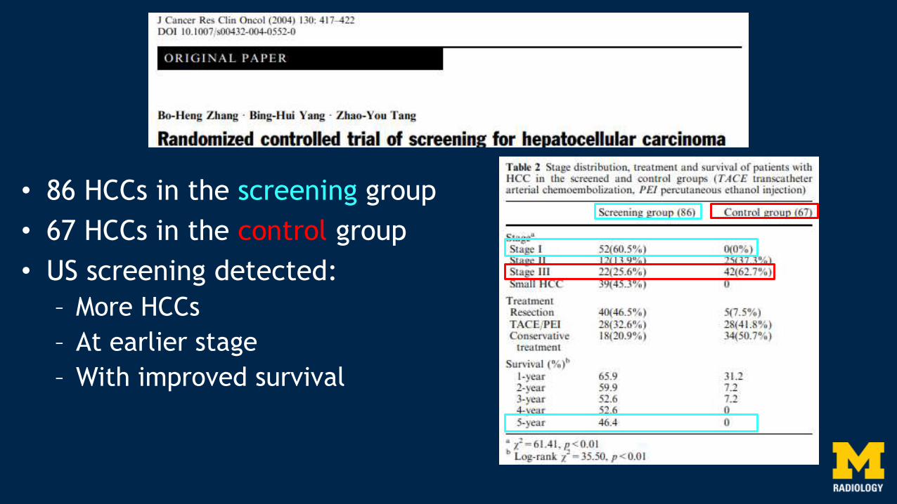
Evidence for US Screening
• 86 HCCs in the screening group
• 67 HCCs in the control group
• US screening detected:
– More HCCs
– At earlier stage
– With improved survival

• Unanimous support for screening ultrasound for HCC in high risk patients :
– American Association for the Study of Liver Diseases (AASLD)
– European Association for the Study of the Liver (EASL)
– Korean Liver Cancer Study Group and the National Cancer Center (KLCSG-NCC)
– Japanese Society of Hepatology (JSH)
– Asian Pacific Association for the Study of the Liver (APASL)
• Initial screening at 6 month intervals
– With tumor markers (JSH and APASL)
Societal Guidelines

Technical Considerations
• NPO for 4-6 hours prior
– Decrease bowel gas and improve visualization
• Position both supine and left lateral decubitus
– Subcostal and intercostal acoustic windows may be used
• Use highest frequency that allows imaging entire depth of liver and diaphragm

Recommended Images
• Longitudinal Images:
– Left lobe:
• Left of midline
• Midline (include aorta)
• With IVC
• With left portal vein
– Right lobe:
• With gallbladder
• With right kidney
• Include right hemidiaphragm
• Far lateral
• Main portal vein (grayscale and color)
• Common duct at porta hepatis(including diameter measurement)

Recommended Images
• Transverse Images:
– Left lobe:
• Dome with hepatic veins
• With left portal vein (check for umbilical vein)
• Main portal bifurcation
– Right lobe:
• Dome with hepatic veins
• With right portal vein
• With main portal vein
• With gallbladder
• With right kidney
• Near liver tip

Characteristics of Observations
• “Observation” preferred term for focal findings
– Nonjudgmental – does not imply a level of suspicion
• Characterize size and echogenicity of observations not definitely benign
• Classically, HCC considered hypoechoic to background liver
– Not true – HCC also may be iso- or hyperechoic
– Therefore, echogenicity does not impact US LI-RADS categorization

Size is Everything
• Size is critical for both screening/surveillance and definitive diagnosis
• Definitive diagnosis of HCC can be made noninvasively using either of two similar systems:
– Organ Procurement and Transplantation Network/United Network for Organ Sharing (OPTN/UNOS)
– American College of Radiology (ACR)
– 1 cm size threshold for both systems
Observations below 1 cm cannot meet diagnostic criteria for HCC

US LI-RADS Algorithm
• System only applies to patients at high risk for HCC
– Cirrhosis
– Hepatitis B
– Prior HCC
• 3 imaging categories, based on:
– Size
– Definitely benign finding
– New thrombus

US LI-RADS Algorithm
• US-1:– No observation
– Definitely benign observation
• US-2:
– Not definitely benign observation
– Less than 1 cm size (not aggregate)
• US-3:
– Not definitely benign observation
– Greater than 1 cm size
– New thrombus

US-1 Negative
• Observation with features that are definitely benign:
– Cyst
– Previously characterized finding
• Resume routine (q 6 months) ultrasound screening

US-1 Negative
Resume routine (q 6 months) ultrasound screening

US-2 Subthreshold
• Observation with features that are not definitely benign
• Observation(s) are less than 1 cm
• Short term (3-6 months) follow-up ultrasound

US-3 Positive
• Observation with features that are not definitely benign
• Observation(s) are 1 cm or larger
• New thrombus
• Further characterization with multiphase CT or MRI

US-3 Positive
Further characterization with multiphase CT or MRI

Other Considerations
• Both extrinsic and intrinsic factors can impact US sensitivity
• Extrinsic:
– Large body habitus
– Rib shadows or bowel gas
– Inability to suspend respiration
– Overlying bandages
• Intrinsic:
– Increased sound attenuation due to steatosis or fibrosis

Visualization Score
• Adequacy of liver visualization may affect sensitivity
• Three visualization categories included in US LI-RADS:
– Visualization A: no or minimal limitations
– Visualization B: moderate limitations
– Visualization C: severe limitations

Visualization A
• No limitations
• Unlikely to impact sensitivity
• Entire liver visualized

Visualization A
• Minimal limitations
• Unlikely to impact sensitivity
• Liver visualized in near entirety

Visualization B
• Moderate limitations
• May decrease sensitivity
• Heterogeneous liver
• Modest sound attenuation
• Small portions of liver not visualized

Visualization B
• Moderate limitations
• May decrease sensitivity
• Heterogeneous liver
• Modest sound attenuation
• Small portions of liver not visualized

Visualization C
• Severe limitations
• Significantly lower sensitivity
• Marked heterogeneity
• Substantial sound attenuation
• Large (>50%) portions of liver not visualized

Visualization C
• Severe limitations
• Significantly lower sensitivity
• Marked heterogeneity
• Substantial sound attenuation
• Large (>50%) portions of liver not visualized

Visualization C
• Severe limitations
• Significantly lower sensitivity
• Marked heterogeneity
• Substantial sound attenuation
• Large (>50%) portions of liver not visualized

Future Work
• Visualization score is subjective
• No management recommendations made based on visualization score
• How should we screen Visualization C?
– CT?
– MR?
– CE US?
• Standardized structured reports will help us research answers

Structured Reporting

Structured Reporting

Structured Reporting

• Proven survival benefit from US screening in high risk patients
• Standardization in US utilization, reporting, and management has multiple advantages:
– Improve communication with patients and referring physicians
– Unify screening and surveillance algorithms
– Improve patient outcomes
– Supply quantitative data for future research
• 2017 US LI-RADS is version 1.0
• Much more to come in the future!
Conclusion

Thank [email protected]