2017 htrc irvine tendon transfers mimm - asht · courtesy of michael s. bednar, md wartenberg’s...
TRANSCRIPT

Tendon TransfersJoanne Mimm, MPT, CHT
Restore balance Indications◦ Nerve injury◦ paralyzed muscle◦ damaged tendon/muscle◦ CNS lesion
Consider◦ action ◦ Functional gain
■ Strength-ability to generate tension–
–
number of muscle fiberscross sectional diameter of all its fibersMuscle will lose a grade following transfer
■ Variabi l i ty■ muscle 2x stronger than another■ Greatest force at resting length
–
–
Leverage : “The power of a lever moving about a point”
Torque/movement : Force X moment arm Moment arm: perpendicular distance form the
axis = the lever
Drag: ◦ internal resistance◦ friction
Comprehensive Review Course in Hand Surgery CD-ROM
Actual strength of a muscle does not change Direction of force changes Blood and nerve supply –are not affected by
transfer Muscles adapt tension to demand; ◦ strong muscle will atrophy◦ weak muscle will hypertrophy

Potential excursion –◦ Free from restrictions◦ Based on number of sarcomeres◦ Intact limb – very few muscles
achieve full potential
Excursion permitted by the surrounding connecting tissue◦ Usually assessed during surgery◦ Varies◦ Can be decreased by too much dissection Scar
Purpose: identify assets limitations determine goals for pre-op treatmentpatient education for post op
precautionsprotocolsexpectations
Observation – watch functional use or limits of affected limb
History/Physical
Grip/Pinch – watch for substitution or motor signs (ie. Froments); watch for abnormal grasp patters
Sensibility
Froments Stretch opposing muscles
to prevent contracture◦ FDS –stretch with wrist ext
Splinting◦ Goals To temporarily restore
balance externally for an imbalance internally
To prevent deformity or to correct existing joint contractures

To prevent◦ over-stretching◦ adaptive shortening ◦ compensatory patterns
Increase function
Joint and Soft tissue mobilization to maintain ROM
Muscle training and strengthening◦ isolate muscle that will be
transferred biofeedback and/or FES can be
used to isolate the muscle and give feedback to the patient
◦ minimize loss of strengthduring immobilization
Communicating with the Surgeon/ Op Report◦ Specific muscle/tendon units ◦ Anatomical route◦ Site of tendon suture or anastomosis◦ How did the wound bed look? ◦ What is the potential for tendon scarring?◦ Was a tendon graft used? From where?◦ Ideal to go to operating room
Stages of Postoperativetreatment General guidelines■ Protective Stage/Early Stage
–Protect transfer●Generally, immobilized in protective post-op
cast for 1-4 weeks; usually 3 weeks.Transfer is immobilized so tension on the juncture site is minimized●
–
–
Control edemaPrevent stiffness in uninvolved joints
■ Protect transfer between exercises with thermoplastic splints
■ Activate transfer being careful not to over-stress tissue
Active motion will tear some adhesions and prevent others from forming.
■Monitor early motion making sure desired position is maintained

� Short frequent exercise sessions– lim it fatigue of transferred tendon.
� Always be aware of avoid overstretching.SometimesLimited arc /isolated joint motiondynamic splint with stop blocks
�
–
� Can use biofeedback and/or electrical stimulation (at sub-tetany contraction) at 4-5 weeks post-operatively..
– Mobilize surrounding soft tissue to increase available excursion
– Add scar management– Gradually incorporate transfer motion
into functional activities
■ Resistive/Late stage 8-12 weeks
–
Add resistance to transfer
●
Strengthen gradually and avoid substitution patternsAdd putty
–Restore passive motion
●
Gentle passive stretches watching effect on transfer (be careful not to over stretch!)
Focus on hand function
–
Try light functional tasks- opposition Facilitate by incorporating motion of
transferred muscle in activity Use biofeedback Use training splints
Taping used for facilitation
Re-ed + blocks full flexion
Use of Extensor indicisto the EPL
Easy to activate transfer
Commonly used

Extrinsic muscles Affect: Pronation- Pronator Teres and Quadratus Wrist flexion- Flexor Carpi radialis Wrist radial deviation Finger flexion- FDS all; FDP index and middle Thumb Flexion-Flexor Pollicis longusIntrinsics affected: MP flexion Index and Middle- lumbricales 1&2 Thumb flexion- FPB Thumb opposition- opponens pollicis
Less common With high median nerve injury – fewer motors
to choose fromNeeded Function Preferred Transfer Other Option
Opposition EIP to APB + EPL EDM or PL
FPL Thumb Flexion BR to FPL ECRL
Finger Flexion FDP of index, middle to ring,small
ECRL to FDP index and small
Forearm Pronation Zancolli biceps rerouting Radius derotational osteotomy (rare)
Sensibility Flag flap Neurovascular cutaneous island pedicle from ring
Loss of index DIP and thumb IP flexion, PT
Direct trauma to the AIN- Fractue/ compartment syndrome
Originates off the MN 5-8 cm distal to the medial epicondyle
Pseudo AIN◦ Parsonage-Turner
syndrome/ Brachial plexopathy
◦ Affects the fascicles more proximally

Loss of thumb opposition (APB, OP, FPB)
Loss of palmar sensation thumb, index, middle and radial border of ring fingers
Goal: restore thumb opposition◦ Common motors chosen for this transfer: FDS of II or IV EIP palmaris longus (Camitz) abductor digiti minimi extensor digiti minimi
Ideal line of pull for transfer is toward the pisiform
Transfers which pass distal increase thumb flexion
Transfers which pass palmar and radial increase thumb abduction
Regional Review Course 1998
EIP transfer◦ Doesn’t require a pulley ◦ Can be used with scarring
of palmar forearm muscles and/or tendons◦ Need to extend tendon
length by including 1 cm of extensor hood with EIP tendon
Comprehensive Review Course in Hand Surgery CD-ROM
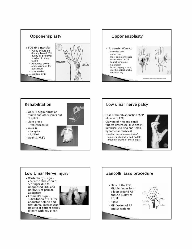
FDS ring transfer◦ Pulley should be
distally based FCU pulley or proximal border of palmar fascia
◦ Adequate power and excursion for abduction
◦ May weaken residual gripRegional Review Course 1998
PL transfer (Camitz)◦ Provides best
abduction◦ Most commonly used
with severe carpal tunnel syndrome◦ Significant
bowstringing occurs, may be objectionable cosmetically
Comprehensive Review Course in Hand Surgery CD-ROM
Week 4 begin AROM of thumb and other joints out of splint
Light grasp◦ Prehension tasks
Week 6 ◦ d/c splint◦ A/PROM
Week 8: PRE’s
Loss of thumb adduction (AdP, ulnar ½ of FPB)
Clawing of ring and small fingers (Interossei muscles (4), lumbricals to ring and small, hypothenar muscles)◦ Median nerve innervation of
lumbricals to index and middle prevent clawing of these digits
Courtesy of Michael S. Bednar, MD
Wartenberg’s sign –eccentric abduction of 5th finger due to unopposed EDQ and paralysis of palmar adductors
Froment’s sign –substitution of FPL for adductor pollicis and first dorsal interosseus; positive if patient flexes IP joint with key pinch
Slips of the FDS Middle finger form a loop around A1 and A2 pulley of RF, SF
“lasso” MP flexion of RF
and SF with MF

Brand ECRB transferGoal:restore intrinsic function of MP flexion; control clawdeform ity– uses ECRB as amotor prolonged withpalmaris longus free graft
�
●
ECRB is passed radially around radialside of forearm, extended by graft into4 slips, passed through the carpaltunnel and volar to the deep transversemetacarpal ligament into the lateralband of the dorsal apparatus (Green’sOperative Hand Surgery)
Splint intrinsic plus with wristfor 3-4 weeks
�
Increase amount of extension at the MP gradually
No unsupervised full ROM Easier to stretch transfer much later Avoid making a fist in the first few weeks –
too much stress on transfer- flexors are strong
4-6 weeks- add blocked PIP, DIP extension 8 weeks begin gentle PRE’s
• use lumbrical bar as a splint assist when progressing to light functional activities
• protect transfer from heavy use for up to 3 months post surgery- do not allow hyperextension at MP’s
Most transfers provide improved stability and improved pinch strength of 25-50%◦ Common motors include EIP, brachioradialis
Boyes uses Brachioradialis extended with free graft; lengthened tendon end is passed through interspace between 3rd and 4th metacarpals to insert on abductor tubercle of thumb.
RADIAL NERVE
Triceps. long headTriceps, lateral head Triceps,
medial headBrachioradialisExtensor carpi radialis longus _
Extensor carpi radial.is brevis
AnconeusSupinator
Extensor digitorum copununis _
Extensor digiti minimi ---
Extensor carpi ulnarisAbductor pollicis JongusExtensor pollicis longusExtensor pollicis brevisExtensor indicis proprius .,,_.
Elbow extension Wrist extension MP finger extension Thumb extension
Supination◦ Biceps still intact

Loss of ECU but will see extension in radial direction (ECRB, ECRL intact)
Decreased strength in supination (supinator) Loss of EDC and thumb extension
Pronator Teresto ECRB/L to restore wrist extension
Palmaris Longus or FDS to EPL to restore thumb extension
FCR to EDC◦ Less strength than FCU
FCU to EDC◦ Contraindicated in
Posterior Interosseous Nerve Palsy (removes remaining ulnar wrist deviator)◦ Weakens power of
finger flexionComprehensive Review Course in Hand Surgery CD-ROM
Thumb separate but all 4 fingers extend simultaneously

Separation of thumb/index extension and M/R/S F extension
FDS (m) to EPL/ED (1) IF
FDS (r) to ED (M/R), EDQ
PT to ECRB
Splint◦ Elbow at 90 degrees◦ Forearm pronated maximally◦ Wrist extended (30-45) ◦ MPs extended to neutral with IPs free◦ Thumb is held in radial abduction
Mobilization exercises start at 3-4 weeks Protective splinting is continued for 3-4 more
weeks for a total of approximately 8 weeks
◦ performed with support to the wrist in extension◦ Designed to protect the tendon from composite flexion
stretch MP flexion/extension with IPs straight IP flexion/extension with MP extension Wrist flexion to neutral (from protected position) Thumb IP flexion/extension with thumb in slight radial
abduction Elbow flexion/extension with forearm pronated Forearm rotation with elbow flexed and wrist/fingers
maintained in extension
Avoid simultaneous wrist flexion/finger flexion until approximately 8 weeks post-op
Resistance initiated around 8 weeks; ex. Velcro dowel board
Preoperative preparation pays off Functional and familiar tasks Use gravity eliminated plane Biofeedback / FES Ask the patient to perform the original motion
of the transferred muscle◦ ex. FDS of ring for thumb opposition: block other
fingers in extension and ask pt to bend ring finger at PIP
Use training splints – lumbrical bar following intrinsic transfer as an assist