2014 library award -- qilei chen
DESCRIPTION
jnnTRANSCRIPT

2014 Hong Kong Baptist University Library Award for Excellence in Undergraduate Research
Comparative Authentication of Three “Snow Lotus” Herbs by Macroscopic and Microscopic Features Qilei Chen
with
Tao Yi, Yina Tang, Lai Lai Wong, Xiaoxuan Huang, Zhongzhen Zhao, and Hubiao Chen
This is the pre-peer reviewed version of the published article: Chen, Q.,
Yi, T., Tang, Y., Wong, L. L., Huang, X., Zhao, Z. and Chen, H. (2014),
Comparative authentication of three “snow lotus” herbs by
macroscopic and microscopic features. Microsc. Res. Tech., 77: 631–
641. doi: 10.1002/jemt.22381, which has been published in final form
at http://onlinelibrary.wiley.com/doi/10.1002/jemt.22381/abstract.
1
Comparative Authentication of Three “Snow Lotus” Herbs
by Macroscopic and Microscopic Features
QILEI CHEN, TAO YI,* YINA TANG, LAI LAI WONG, XIAOXUAN HUANG,
ZHONGZHEN ZHAO, AND HUBIAO CHEN*
School of Chinese Medicine, Hong Kong Baptist University, Kowloon Tong, Hong Kong Special
Administrative Region, People’s Republic of China
*Correspondence to: Dr. Tao Yi or Dr. Hubiao Chen, School of Chinese Medicine, Hong Kong
Baptist University, Kowloon Tong, Hong Kong Special Administrative Region, People’s
Republic of China. E-mail: [email protected]; [email protected]
2
ABSTRACT
“Snow lotus” is a famous Chinese Materia Medica derived from species of the genus Saussurea
(Compositae). To differentiate three representative easily-confused snow lotus herbs, namely, S.
involucrata (Kar. et Kir.) Sch.-Bip, S. laniceps Hand.-Mazz. and S. medusa Maxim., macroscopic
features of the three herbs were systemically observed, and microscopic features were compared
under the ordinary light microscopy, polarized light microscopy and scanning electron
microscopy. The results indicate that, as for macroscopical identification, capitula situation and
arrangement, and as for microscopical identification, pollen grains, non-glandular hairs,
glandular hairs, cells of inner surface of the microdiodange, and epidermal cells of the corolla
can be used to authenticate the three snow lotus herbs. Comprehensive table comparing the
characteristics were presented in this study. Scanning electron microscopy has been found to
provide a number of unique characteristics of pollen grains. Based on the observation of pollen
grains, evolution sequence of the three species was speculated. The authentication method was
proven to be efficient, convenient, simple and reliable. It can be widely applied to other herbal
materials.
KEY WORDS
Snow lotus; light microscopy; scanning electron microscopy; microscopic features; pollen grain

3
INTRODUCTION
“Snow lotus”, a famous Chinese Materia Medica (CMM) derived from the dried aerial parts
of species of the genus Saussurea (Compositae), has been widely used in Tibetan, Mongolian,
Uighur and Chinese folk medicines (Health Bureau of Autonomous Region of Xizang, 1973;
Meyer 1983; Xie, 1994; Xiao, 2007). One of the botanical origins of “Snow lotus” is S.
involucrata (Kar. et Kir.) Sch.-Bip. (Fig. 1), a plant growing in the mountains at heights of
4,000–4,300 m in the Tianshan and A’er Tai areas of China (Chen et al., 1999; Yi et al., 2010);
this species has long been used for diminishing inflammation and facilitating blood circulation,
e.g. treating rheumatoid arthritis, stomachache and dysmenorrhea in Uighur folk medicine (Liu
and Shawuti, 1999; Jiang and Li, 1999; Xiao, 2007; Chinese Pharmacopoeia Commitee, 2010).
Two other species of the same genus, S. laniceps Hand.-Mazz. and S. medusa Maxim. (Fig. 1),
mainly distributed in the Qinghai-Tibet plateau at heights of 3500–5300 m, have also been used
under the name “Snow Lotus”. These latter two species are prescribed for the treatment of pain
and inflammatory conditions in Tibetan folk medicine (Commission of Chinese Ethnomedicine,
1984; Jiang and Li, 1999; Xiao, 2007).
The three species have been confused because of their shared folk names, morphological
appearances and medicinal uses (Yang, 1991; Chen et al., 1999; Xiao, 2007). Moreover,
Saussurea plants, once dried or processed for herbal use, can be easily damaged or broken apart
during commercial distribution, thus morphological characters are usually not sufficient for their
identification. For some patent formulations, snow lotus herbs are powdered. Powdered herbs are
not only very difficult to identify by sight but also easily replaced by other materials. For these
reasons, a variety of identification methods and characters are needed to accurately authenticate
the three species of snow lotus herbs. Therefore, it is necessary to distinguish the three species
clearly.
In our previous studies (Yi et al., 2009 a, b; Yi et al., 2010; Yi et al., 2012), the chemical
composition and pharmacological activities of these three herbs have been investigated. However,
the chemical and pharmacological approaches, as used in that study, while accurate, involve
much time and effort, as well as expensive experimental equipment and animals. Since the three
herbs have similar clinical uses, their chemical profiles and pharmacological effects cannot be
distinguished effectively. In comparison, observing microscopic features is an efficient, simple,
economical, reliable, and effective means for identifying CMM plant material powders (Liang et

4
al., 2006; An et al., 2009). With the use of ordinary light microscope and polarized light
microscope, CMM can be successfully identified. The application and advantages of polarized
light microscopy in observing CMM plant tissues are described in our previous publication
(Tang et al., 2013).
Apart from using optical microscopes, comparing pollen grain characters under scanning
electron microscope is also an effective and reliable approach for herbal identification
(Wodehouse, 1928). Since pollen grains are single cells and spores, the pollen grain characters
are indicative of relationships among plants (Wodehouse, 1935). The size, shape and color of the
grains, the number and arrangement of the germinal apertures, and the sculpturing of the exines,
are of high diagnostic value (Erdtman, 1943). It has been reported that scanning electron
microscopy has been widely used to observe pollen grains of plants to distinguish different
species (Ceter et al., 2013). The present study represents the first attempt to systemically observe
and compare the macroscopical and microscopical features of powdered materials and pollen
grains of the three medicinal species of Saussurea. The method used is convenient and reliable,
and can be used to authenticate the three representative snow lotus herbs. This method can be
widely applied to other Saussurea herbs and even to other confused CMM.
5
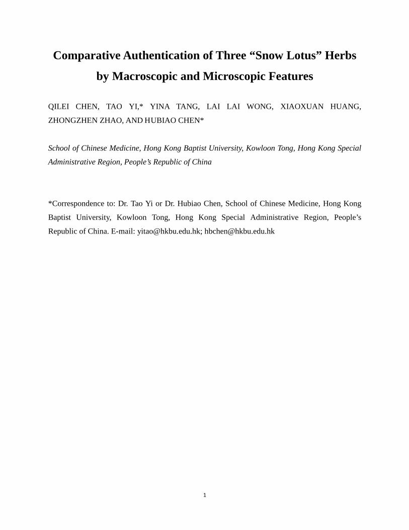
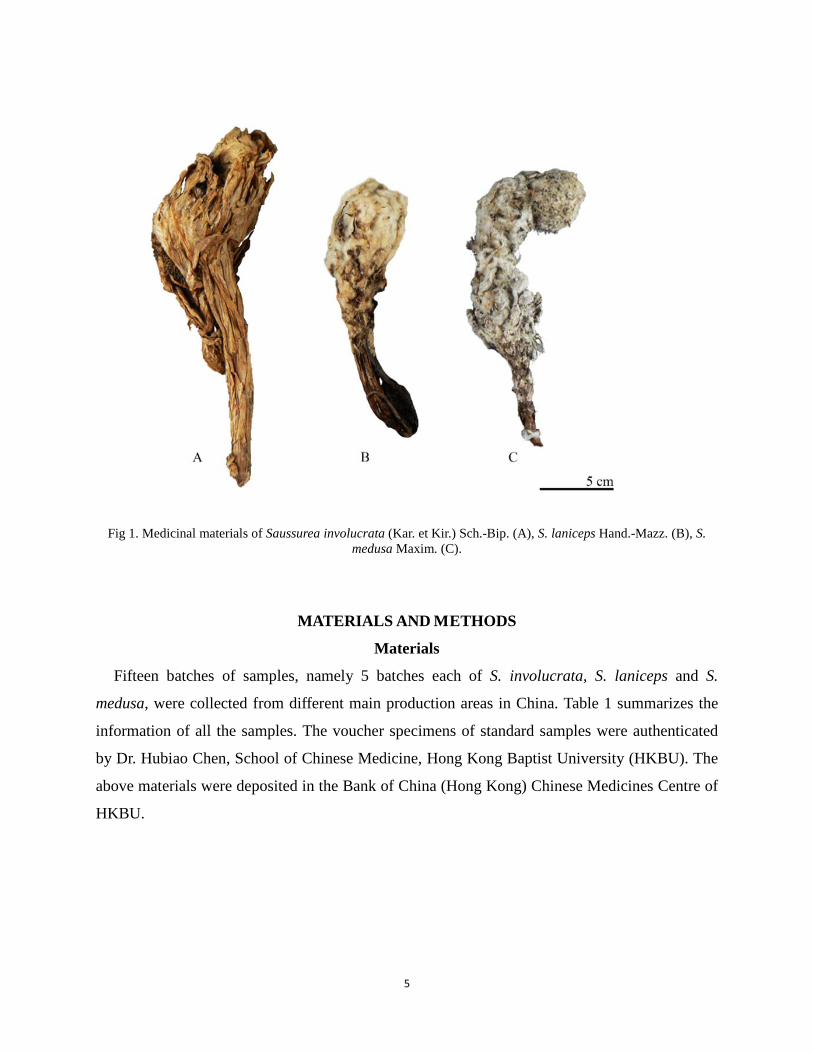
Fig 1. Medicinal materials of Saussurea involucrata (Kar. et Kir.) Sch.-Bip. (A), S. laniceps Hand.-Mazz. (B), S.
medusa Maxim. (C).
MATERIALS AND METHODS
Materials
Fifteen batches of samples, namely 5 batches each of S. involucrata, S. laniceps and S.
medusa, were collected from different main production areas in China. Table 1 summarizes the
information of all the samples. The voucher specimens of standard samples were authenticated
by Dr. Hubiao Chen, School of Chinese Medicine, Hong Kong Baptist University (HKBU). The
above materials were deposited in the Bank of China (Hong Kong) Chinese Medicines Centre of
HKBU.

6
TABLE 1. Collection data of three “snow lotus” herbs
Reagents
Chloral hydrate and glycerin (Uni-chem, England) were used for mounting microscope slides.
Chloral hydrate solution and dilute glycerin were prepared according to procedures described in
Appendix XV B of the Pharmacopoeia of the People’s Republic of China (Chinese
Pharmacopoeia Commitee, 2010). Purified water was provided by a Millipore water purification
system (Millipore, Bedford, MA)
Apparatus
Axioplan 2 and Axiophot 2 universal microscopes with reflector Axiophot Photo Module
(Zeiss Group) were used for observing powdered samples; images were recorded with a Leica
direct current (DC) camera. LEO1530 Field Emission Scanning Electron Microscope (Oxford
Instrument) was used for observing pollen grains.
Software
Matrox inspector (Matrox Electronic Systems), Leica IM50 and SmartSEM® were used.
Methods
Macroscopic Identification of Crude Drugs. Each sample was measured and examined
with regard to appearance, surface texture, odor and taste. The color photographs were taken
with a digital camera (Panasonic DMC-FZ7, Panasonic, Japan).
Microscopic Identification of Powdered Crude Drugs. All herbal samples were powdered,
passed through a 250 μm sieve, and treated with chloral hydrate solution for observation. At least
Latin name Batch No. (Collection year) Collection area
S. involucrata
SI01 (2007) Tianshan, Xinjiang, China SI02 (2007) Tianshan, Xinjiang, China SI03 (2007) Tianshan, Xinjiang, China SI04 (2006) Xinyuan, Xinjiang, China SI05 (2005) Lhasa, Tibet, China
S. laniceps
SL01 (2008) Lhasa, Tibet SL02 (2007) Lhasa, Tibet SL03 (2007) Lhasa, Tibet SL04 (2007) Lhasa, Tibet SL05 (2007) Lhasa, Tibet
S. medusa
SM01 (2007) Aba, Sichuan, China SM02 (2007) Qinghai, China SM03 (2007) Aba, Sichuan, China SM04 (2007) Lhasa, Tibet SM05 (2007) Lhasa, Tibet
7
10 different slides of each powder sample were examined under ordinary light microscope and
polarized light microscope. Distinctive microscopic features observed were digitally recorded.
Microscopic Identification of Pollen Grains. Pollen grains were removed from anthers of
the herbarium materials, suspended in distilled water on coverslips glued to 12 mm specimen
holders, and air-dried (Andersen and Bertelsen, 1972). The specimens were coated with gold and
observed with a field emission scanning electron microscope (SEM) operated at 20 kV.
Palynological characters examined included equatorial and polar diameters, shapes and
ornamentation.
RESULTS
S. involucrata
Macroscopic Characteristics. Leaves oblong or wide-lanceolate, yellowish-green, tips blunt
or slightly acuminate, bases decurrent, margins serrate and ciliate. Bracteal leaves ovate or oval,
margins sharply toothed, yellowish-white, texture membranous, 55-70 mm long and 20-70 mm
wide. Involucral bracts hemispherical, surrounding capitula, 10 mm in diameter, lanceolate, in
three to four layers, bracts of the outer layer oblong, 11 mm long and 5 mm wide,
brownish-yellow and pubescent throughout; bracts of the middle and inner layers lanceolate,
15-18 mm long and 2 mm wide, yellowish-white and pubescent only on top. Capitula crowded at
top of stem. All flowers tubular, corolla purplish-red, limb 9 mm long, tube 7 mm long, anther
purple, stigma 2-lobed. Achenes ovate, longitudinally ribbed, 3 mm long, grayish-white. Pappi
grayish-white, in two layers, outer layer coarse and 3 mm long, inner layer feathery and 15 mm
long. Odor specially fragrant, taste slightly bitter (Fig. 1).
Microscopic Characteristics.
Optical Microscopy (Table 3): Powder yellowish-green, with light fragrance.
1. Pollen grains: Subrounded, yellow, 23-52 μm in diameter, 3 germination ditches, warts on the
outer surface.
2. Non-glandular hairs: Of three kinds (numbers in accordance with respective pictures in Table
3, same as below). (1) Unicellular; linear and bulgy; cell walls smooth. (2) Unicellular;
twisted and flat; cell walls uneven. (3) Multicellular, comprised of 5-6 cells as a single stalk;
cell walls smooth.
8
3. Glandular hairs: Rare. Mostly broken. Head with 5-6 single- or double-celled flat layers,
colorless or pale yellow; stalk colorless, with interlocked cells in each layer.
4. Pappi: Filamentous structure with longitudinally aligned fiber cells. Fiber width 9-18 μm.
5. Cells of inner surface of microdiodange: Rectangular, arranged orderly; sometimes pale
yellow; cell walls thickened in longitudinal stripes and appearing bright blue under polarized
light; outer peripheral walls irregularly prominent in lateral view.
6. Stigmas: With short scale-like protrusions. Tips blunt; other cells brown.
7. Epidermal cells of corolla: (1a) Tip of limb yellow, with papillary protrusions; (1b) other cells
of limb tightly arranged in lines, pale yellow, anticlinal walls undulate; (2a) tube cells
colorless, rectangular, arranged tightly in lines; (2b) cells below the tube epidermis containing
crystals that appear bright blue under polarized light.
8. Epidermal cells of involucral bract: Elongate spindle-shaped; colorless; cell walls appearing
bright blue under polarizing light; pit canals dense.
9. Epidermal cells of bracteal leaf: (1) Upper epidermis colorless, irregular in shape; anticlinal
walls sinuous; (2) lower epidermis similar to upper epidermis, but with deeply sinuous
anticlinal walls; non-glandular hairs present on both upper and lower epidermal cells; stomata
anomocytic.
10. Epidermal cells of leaf: Polygonal; pale brown; anticlinal walls slightly curved.
Scanning Electron Microscopy: Pollen grains subrounded, yellow, 23-52 μm in diameter, 3
germinating ditches, warts coarse and widely spaced, with reticular sculpturing on the surface
(Fig. 2).
9
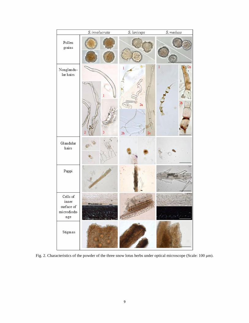
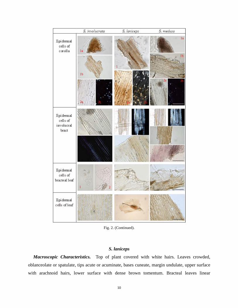
Fig. 2. Characteristics of the powder of the three snow lotus herbs under optical microscope (Scale: 100 μm).
10
Fig. 2. (Continued).
S. laniceps
Macroscopic Characteristics. Top of plant covered with white hairs. Leaves crowded,
oblanceolate or spatulate, tips acute or acuminate, bases cuneate, margin undulate, upper surface
with arachnoid hairs, lower surface with dense brown tomentum. Bracteal leaves linear
11
lanceolate, both surfaces covered with white tomentum. Involucral bracts broadly campanulate,
15 mm in diameter, in 3-4 layers, outer layer lanceolate or linear lanceolate, 6 mm long and 1
mm wide, covered with white or brown hairs, inner layer lanceolate, tip acuminate, 9 mm long
and 4 mm wide, covered densely with long, dark brown hairs. Capitula arranged in paniculate
spikes along upper stem. All flowers tubular, white, 8-12 mm long, limbs 3 times longer than
tubes, anthers light purple, stigma 2-lobed. Achenes cylindrical, 3 mm long. Pappi gray, 2 layers,
outer layer coarse and short, inner layer feathery and long. Odor light, taste slightly bitter (Fig.
1).
Microscopic Characteristics.
Optical Microscopy (Table 3): Powder yellowish-green, with light fragrance.
1. Pollen grains: Subrounded, pale yellow, 24-49 μm in diameter, 3 germinating ditches; densely
covered with perforate and verrucate warts, surrounded by reticulate sculpturing on the
surface.
2. Non-glandular hairs: Abundant. Multicellular. Of two kinds. (1) Comprised of 4-7 short cells
with a single slender cell on top; cell connections shrunken and yellow; cell walls smooth. (2)
Mostly broken, comprised of more than 3 long cells, which can be either curly (2a, 2b) or
straight (2c); cell connections smooth and colorless; cell walls smooth.
3. Glandular hairs: Rare. Head yellow, with 5-6 single-celled flat layers; stalk usually unicellular
and colorless.
4. Pappi: Filamentous structure with longitudinally aligned fiber cells. Fiber width 9-17 μm.
5. Cells of inner surface of microdiodange: Rectangular, arranged orderly; colorless or pale
yellow; cell walls smooth, with no specific view under polarized light.
6. Stigmas: With short scale-like protrusions. Tips blunt; other cells dark brown.
7. Epidermal cells of corolla: (1) Limb brownish-yellow; tip of limb with papillary protrusions;
other cells of limb thin rectangular, anticlinal walls slightly curved; (2a) tube cells pale yellow
or brownish-yellow, rectangular, arranged tightly in lines; (2b) cells below the tube epidermis
containing dense prismatic crystals that appeared bright blue or yellow under polarized light.
8. Epidermal cells of involucral bract: Elongate spindle-shaped; colorless or pale yellow; both
cell lumina and wall appeared bright blue under polarized light.
9. Epidermal cells of bracteal leaf: Irregular in shape; colorless or pale brown; anticlinal walls
thin and undulate; stomata anomocytic.
12
10. Epidermal cells of leaf: Rectangular, arranged tightly in lines; pale brown; anticlinal walls
straight.
Scanning Electron Microscopy: Pollen grains subrounded, pale yellow, 24-50 μm in
diameter, 3 germination ditches, warts more crowded than those of S. involucrata, with reticular
sculpturing on the surface smoother than that of S. involucrata (Fig. 2).
S. medusa
Macroscopic Characteristics. Whole plant cylindrical, covered with thick white or
grayish-white long tomentum. Leaves ovate or linear speculate, tips blunt or rounded, bases
cuneate, margins whole or with laciniate, both surfaces covered with long white tomentum.
Bracteal leaves linear lanceolate, both surfaces with long white tomentum. Involucral bracts
narrow cylindrical, 5-7 mm in diameter, in 3 layers; bracts of outer layer ovate, acuminate,
purple, covered with white or brown tomentum, 11 mm long and 2 mm wide; bracts of middle
layer oblanceolate, 10 mm long and 4 mm wide; bracts of inner layer oblanceolate, 11 mm long
and 2 mm wide. Capitula sometimes densely crowded at top of stem, arranged in racemes. All
flowers tubular, 10 mm long, limb as long as tube, tube purple or purplish-blue, anther purple,
stigma 2-lobed. Achenes rare, spindle-shaped, 8-9 mm long and 4-8 mm in diameter. Pappi white
or grayish-white, in 2 layers, outer layer coarse, inner layer feathery. Odor light, taste slightly
bitter (Fig. 1).
Microscopic Characteristics.
Optical Microscopy (Table 3): Powder yellowish-green, with light fragrance.
1. Pollen grains: Subrounded, pale yellow or colorless, 30-57 μm in diameter, 3 germinating
ditches, outer surface sculptured with dense spinules.
2. Non-glandular hairs: Abundant. Of two kinds. (1) Comprised of 4-8 or more short cells in a
single row, with a single slender cell on top; cell connections shrunken and yellow; cell walls
smooth. (2) Of 3-5 cells with two subcategories based on color of cell contents, (2a)
yellowish-brown and (2b) pale red; cell connections slightly shrunken; cell walls with
longitudinal and corrugated striations.
3. Glandular hairs: Rare. Head with 2-4 layers, each layer with 1-2 rounded cells; stalk
unicellular; whole hair brown.

13
4. Pappi: Filamentous structure with longitudinally aligned fiber cells. Fiber width 10-19 μm.
5. Cells of inner surface of microdiodange: Rectangular, arranged orderly; colorless; cell walls
thickened in longitudinal stripes and appearing bright blue under polarized light; outer
peripheral walls irregularly prominent in lateral view.
6. Stigmas: With short scale-like protrusions. Tips blunt; other cells brown.
7. Epidermal cells of corolla: (1a) Tip of limb brown or pale purple, with papillary protrusions;
(1b) other cells of limb thin rectangular, brownish-yellow, with smooth anticlinal walls; (2a)
tube cells colorless, slender spindle-shaped; (2b) cells below the tube epidermis containing a
few prismatic crystals that appear bright blue under polarized light.
8. Epidermal cells of involucral bract: Spindle-shaped or rectangular; colorless, yellowish-brown
or pale purple; both cell lumina and wall appearing bright blue under polarized light.
9. Epidermal cells of bracteal leaf: Irregular-shaped to rectangular; pale brown to brown;
anticlinal walls smooth; stomata anomocytic.
10. Epidermal cells of leaf: Polygonal to rectangular; cell walls and lumina brown.
Scanning Electron Microscopy: Pollen grains subrounded, pale yellow or colorless, 30-57
μm in diameter, 3 germinating ditches, spinules dense and with blunt tips, outer surface with
granular sculptures (Fig. 2).

14
TABLE 2. Comparison of macroscopic characteristics of the three snow lotus herbs S. involucrata S. laniceps S. medusa
Whole plant Sparsely covered with hairs
Usually with hairs, only on top
Entirely covered with hairs
Leaves
Shape Tip Base Margin Surface
Oblong or wide lanceolate Blunt or slightly acuminate Decurrent Serrate and ciliate Sparsely covered with hairs
Oblanceolate or spatulate Acute or acuminate Cuneate Undulate Both surfaces with hairs
Long oval or linear spatulate Blunt or rounded Cuneate Whole or with laciniate; Both surfaces with hairs
Bracteal leaves
Shape Margin Texture Surface
Ovate or oval Sharply toothed Membranous Sparsely covered with hairs
Linear lanceolate Not observable Not observable Both surfaces with hairs
Linear lanceolate Not observable Not observable Both surfaces with hairs
Involucral bracts
No. of layers Bract shape Bract size
3-4, surrounding capitula Lanceolate 11-18 mm × 2-5 mm
3-4 Broadly campanulate 6-9 mm × 1-4 mm
3 Slender cylindrical 10-11 mm × 2-4 mm
Capitula Clump at top of stem Some clump at top of stem; paniculate spike inflorescence
Some clump at top of stem; hemispherical total inflorescence
Flowers
Limb Tube Anther Stigma
9 mm long 7 mm long Purple 2-lobed
6-9 mm long 2-3 mm long Light purple 2-lobed
5 mm long 5 mm long Purple 2-lobed
Achenes Shape Size
Ovate; longitudinally ribbed 3 mm long
Cylindrical; 3 mm long
Spindle; 8-9 mm long
Pappi No. of layers Color
2 Grayish-white
2 Gray
2 Grayish-white
Odor Fragrant Light Light Taste Slightly bitter Slightly bitter Slightly bitter
15
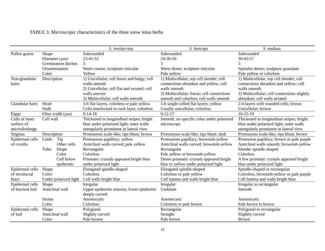
TABLE 3. Microscopic characteristics of the three snow lotus herbs
S. involucrata S. laniceps S. medusa Pollen grains Shape
Diameter (μm) Germination ditches Ornamentation Color
Subrounded 23-41-52 3 Warts coarse; sculpture reticular Yellow
Subrounded 24-36-50 3 Warts dense; sculpture reticular Pale yellow
Subrounded 30-43-57 3 Spinules dense; sculpture granulate Pale yellow or colorless
Non-glandular hairs
Description 1) Unicellular; cell linear and bulgy; cell walls smooth 2) Unicellular; cell flat and twisted; cell walls uneven 3) Multicellular; cell walls smooth
1) Multicellular; top cell slender; cell connections shrunken and yellow; cell walls smooth 2) Multicellular; linear; cell connections smooth and colorless; cell walls smooth
1) Multicellular; top cell slender; cell connections shrunken and yellow; cell walls smooth 2) Multicellular; cell connections slightly shrunken; cell walls striated
Glandular hairs Head Stalk
5-6 flat layers, colorless or pale yellow Cells interlocked in each layer, colorless
5-6 single-celled flat layers; yellow Usually unicellular, colorless
2-4 layers with rounded cells; brown Unicellular; brown
Pappi Fiber width (μm) 9-14-18 9-12-17 10-15-19 Cells of inner surface of microdiodange
Cell wall Thickened in longitudinal stripes; bright blue under polarized light; outer walls unregularly prominent in lateral view
Smooth; no specific color under polarized microscope
Thickened in longitudinal stripes; bright blue under polarized light; outer walls unregularly prominent in lateral view
Stigmas Description Protrusions scale-like; tips blunt; brown Protrusions scale-like; tips blunt; dark Protrusions scale-like; tips blunt; brown Epidermal cells of corolla
Limb: Tip Other cells
Tube: Shape Color Cell below epidermis
Protrusions papillary; yellow Anticlinal walls curved; pale yellow Rectangular Colorless Prismatic crystals appeared bright blue under polarized light
Protrusions papillary; brownish-yellow Anticlinal walls curved; brownish-yellow Rectangular Pale yellow or brownish-yellow Dense prismatic crystals appeared bright blue or yellow under polarized light
Protrusion papillary; brown or pale purple Anticlinal walls smooth; brownish-yellow Slender spindle-shaped Colorless A few prismatic crystals appeared bright blue under polarized light
Epidermal cells of involucral bract
Shape Color Under polarized light
Elongated spindle-shaped Colorless Cell walls bright blue
Elongated spindle-shaped Colorless or pale yellow Cell lumina and walls bright blue
Spindle-shaped or rectangular Colorless, brownish-yellow or pale purple Cell lumina and walls bright blue
Epidermal cells of bracteal leaf
Shape Anticlinal wall
Stoma Color
Irregular Upper epidermis sinuous; lower epidermis deeply curved Anomocytic Colorless
Irregular Undulate Anomocytic Colorless or pale brown
Irregular to rectangular Smooth Anomocytic Pale brown to brown
Epidermal cells of leaf
Shape Anticlinal wall Color
Polygonal Slightly curved Pale brown
Rectangular Straight Pale brown
Polygonal to rectangular Slightly curved Brown

16
Fig 3. Pollen grain from the three snow lotus herbs under scanning electron microscope (A1, A2: S.
involucrata; B1, B2: S. laniceps; C1, C2: S. medusa; Scale: 5 μm).
DISCUSSION
TABLE 4. Key based on macroscopic characteristics of the three snow lotus herbs 1. Capitula crowded at top of stem, supported or surrounded by broad,
membranous, and colored involucral bracts —Subgen. Amphilaena (Stschegl.) Lipsch.
—S. involucrata 1. Capitula not supported or surrounded by broad, membranous and colored
involucral bracts; some capitula crowded at top of stem, usually supported or half-surrounded by involucral bracts covered with a thick layer of soft, woolly hairs
—Subgen. Eriocoryne (DC.) Hook. f. 2. Capitula abundant, arranged in paniculate spikes along upper stem
—S. laniceps 2. Capitula arrange in hemispherical racemes
—S. medusa

17
TABLE 5. Key based on pollen grains of the three snow lotus herbs under scanning electron microscope 1. Spinules dense and with blunt tips; no evident warts; outer surface with granular
sculptures —S. medusa
1. Spinules not evident; warts coarse and prominent; outer surface with reticular sculptures 2. Warts widely-spaced; reticular sculptures relatively rough
—S. involucrata 2. Warts densely-arranged; reticular sculptures relatively smooth
—S. laniceps
18
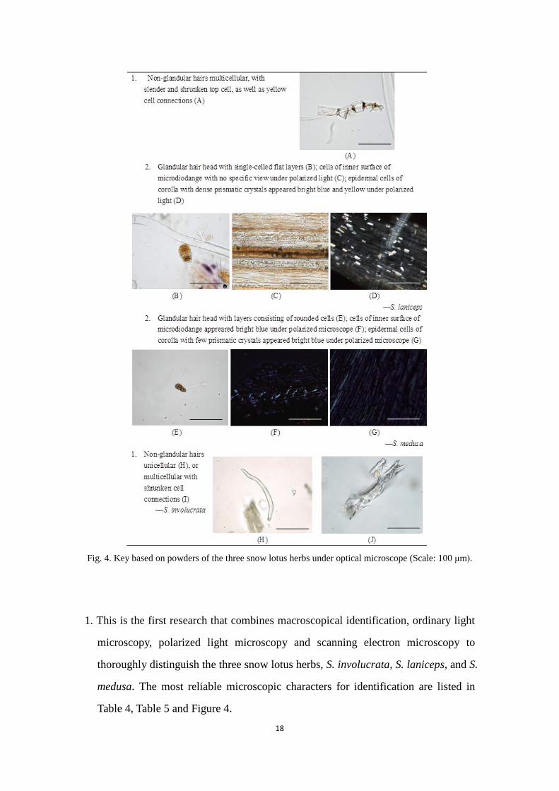
Fig. 4. Key based on powders of the three snow lotus herbs under optical microscope (Scale: 100 μm).
1. This is the first research that combines macroscopical identification, ordinary light
microscopy, polarized light microscopy and scanning electron microscopy to
thoroughly distinguish the three snow lotus herbs, S. involucrata, S. laniceps, and S.
medusa. The most reliable microscopic characters for identification are listed in
Table 4, Table 5 and Figure 4.

19
2. For macroscopic features, the three herbs are very similar, as can be expected since
they are all members of the genus Saussurea DC. However, they belong to two
subgenera: S. involucrata in Subgen. Amphilaena (Stschegl.) Lipsch. while both S.
laniceps and S. medusa are in Subgen. Eriocoryne (DC.) Hook. f.. Species from the
two subgenera can be distinguished according to their capitula arrangement; this
also stands for S. involucrata, S. laniceps and S. medusa.
3. For the three types of microscopy, they contribute in different ways in
authenticating herbal powders. Ordinary light microscopy shows basic
microscopical features, such as cell morphological characters, sizes, and colors
(Table 3, Fig. 2 and Fig. 4). Polarized light microscopy reveals more information
when samples are nearly identical under ordinary light, including 1) the cuticle
properties of plant tissue cells, and 2) the distribution of crystals (Table 3, Fig. 2
and Fig. 4). SEM gives detailed information about surfaces. Because pollen grains
are noteworthy for the specific ornamentation of their exine layers, SEM can give
detailed specific information about pollen. (Fig. 3 and Table 5). Therefore, with the
combined use of ordinary light microscopy, polarized light microscopy and
scanning electron microscopy, many independent characteristics of the powders
can be observed, making authentication efficient and accurate.
For optical microscopy, in choosing characters by which to authenticate
species, we chose characters that are least affected by preparation processes and are
most stable in different populations. For example, cell color can vary with the
degree of permeabilization during mounting, and cell size can vary according to
plant age, sample origin, etc. Thus, for keys to differentiate these three snow lotus
herbs, we chose characteristics of non-glandular hairs, glandular hairs, cells of
inner surface of microdiodange and crystal distribution underneath epidermal cells
of corolla.
Non-glandular hairs are frequently observed under the microscope, and they
differ according to the two subgenera: S. involucrata have unique unicellular or
multicellular non-glandular hairs, while S. laniceps and S. medusa share a same
multicellular type, with top cell slender, and cell connections yellow and shrunken.
20
As for glandular hairs, S. involucrata and S. laniceps share a similar type with a
flat and layered head, while S. medusa have a unique type, each with a head
consisting of rounded cells. Since glandular hairs are rare in S. involucrata and S.
laniceps, the two species can be further differentiated according to the walls of
cells of the inner surface of the microdiodange. These cells of the latter species
appear bright blue under polarized light, while the former do not. The three species
can also be distinguished based on the amount of crystals underneath the epidermal
cells of corolla, that is, in descending order, S. laniceps, S. involucrata, and S.
medusa.
For scanning electron microscopy, interspecific differences of pollen grains,
including exine ornamentation, colpus width, spinulate size, distribution density
and size, were studied. As shown in Figure 3 and Table 5, the three kinds of pollen
grains can be distinguished according to spinulate density, wart density and
sculptures on the outer surface. Pollen grains from S. medusa can be identified
from the three species by their dense spinules, lack of evident warts, and granular
sculpturing on the surface. Those from S. involucrata and S. laniceps, in contrast,
are with little spinules, prominent warts and reticular sculpturing on the surface. To
further differentiate, pollen grains from S. involucrata have sparser warts and
rougher reticular sculpturing than those from S. laniceps have.
SEM observation of exine ornamentations, shapes, and sizes of pollen grains
can reflect the evolution traits of the specific species (Wang et al., 2008). As for
exine ornamentation, smoothness is considered most primitive, then negative
ornamentation, protrusions (e.g. granules), and spinules developed, gradually
evolving into reticular, undulate or striated sculptures (Walker, 1976). According to
this hypothesis, S. medusa (with granular sculpturing) is more primitive than S.
involucrata and S. laniceps (both with reticular sculpturing). As for shape, oval is
considered to be more advanced than spherical (Wodehouse, 1935; Mullaer, 1979).
The pollen grains of the three species are all subrounded, indicating that they are
relatively primitive in Saussurea genus. As for size, pollen grains are believed to
have evolved from larger to smaller (Covas et Schnack, 1944). In this regard, the

21
evolutionary sequence, from most primitive to most developed, should be S.
involucrata (average diameter 47 μm), S. medusa (43 μm), and S. laniceps (36 μm).
Therefore, the evolutionary trait above do not support the one in Flora Reipublicae
popularis sinicae (Jiang et Li, 1999), which is, from most primitive to most
developed, S. laniceps, S. medusa, and S. involucrata. As for the relationship
between pollen grain characters and evolutionary sequence, further studies should
be conducted in the future.
CONCLUSIONS
Our study has proved that the combining characters derived from observation
through macroscopical identification, ordinary light microscopy, polarized light
microscopy and scanning electron microscopy can be successfully applied in the
authentication of the three Sausaurea species used as the medicinal herb “snow lotus”.
The method has proven to be simple, convenient, and reliable. The macroscopic and
microscopic characteristics presented in this article are proposed as a reference to
establish authenticity of the drugs and may be used to differentiate the drugs from
adulterants. We believe that this method can be widely applied to identify other
CMM.
ACKNOWLEDGMENT
This research was funded by the Faculty Research Grant of Hong Kong Baptist
University (FRG/08-09/II-52) and General Research Fund of Hong Kong
(HKBU-260111).
REFERENCES An J, Li J, Wang JG, Zhang ZF, Chen C, Zhang H. 2009. Authenticating and
distinguishing the eight species of traditional Tibetan medicine “Meiduoluomi” by microscopic technique. Microsc Res Tech 72: 727–736.
Andersen TS, Bertelsen F. 1972. Scanning electron microscope studies of pollen of cereals and other grasses. Grana 12: 79–86.

22
Ceter T, Pinar NM, İnceer H, Hayirlioğlu-Ayaz S, Yaprak AE. 2013. The comparative pollen morphology of genera Matricaria L. and Tripleurospemum Sch. Bip. (Asteraceae) in Turkey. Plant Syst Evol 299: 959–977.
Chen FJ, Yang YG, Zhao DX, Gui YL, Guo ZC. 1999. Advances in studies of species habitats distribution and chemical composition of snow lotuses (Saussurea) in China. Chin Bull Bot 16: 561–566.
Chinese Pharmacopoeia Committee. 2010. Pharmacopoeia of the People’s Republic of China, Vol. 1. Beijing: China Medical Science and Technology Press. pp. 50–51.
Commission of Chinese Ethnomedicine. 1984. Ethnomedicine of China, Vol. 1. Beijing: The Ethnic Publishing House. pp. 261–263.
Covas G, Schnack B. 1944. Tres nuevas especies de Glandularia de la flora Argentina. Rev Argent Argron 11: 89–97.
Erdtman G. 1943. An introduction to pollen analysis. Waltham: Chromica Botanica Company. pp. 49–55.
Health Bureau of Autonomous Region of Xizang. 1973. The useful material medica of Tibet. Lhasa: Xizang People’s Publishing House. pp. 156, 351, 537, 778.
Jiang Y, Li BT. 1999. Flora of China, Vol. 78. Beijing: Science Press. pp. 1–36. Liang ZT, Jiang ZH, Leung KSY, Peng Y, Zhao ZZ. 2006. Distinguishing the
medicinal herb Oldenlandia diffusa from similar species of the same genus using fluorescence microscopy. Microsc Res Tech 69: 277–282.
Liu YM, Shawuti Y. 1999. Pharmacography of Uighur. Xinjiang: Science and Technology Health Publisher. pp. 385–389.
Meyer F. 1983. Gso-ba-rig-pa, le systeme medical tibetain (French edition). Paris: Centre National de la Recherche Scientifique. pp. 176–179.
Muller J. 1979. Form and function in angiosperm pollen. Ann Mo Bot Gard 66: 593–632.
Tang YN, He XC, Chen QL, Fan LL, Zhang JY, Zhao ZZ, Dong LS, Liang ZT, Yi T, Chen HB. 2013. A mixed microscopic method for differentiating seven species of “Bixie”‐related Chinese Materia Medica. Microsc Res Tech 77: 57–70.
Walker JW. 1974. Evolution of exine structure in the pollen of primitive angiosperms. Amer J Bot 61: 891–902.
Wodehouse RP. 1928. The phylogenetic value of pollen-grain characters. Ann Bot 42: 891–934.
Wodehouse RP. 1935. Pollen grains: Their structure, identification and significance in science and medicine. New York: McGraw-Hil Book Co. Inc.. pp. 323–340
Xiao PG. 2007. Modern Chinese materia medica, Vol. 5. Beijing: Chemical Industry Press. pp. 43–57.
Xie ZW. 1994. The statement for species of Chinese material medica, Vol. 2. Shanghai: Shanghai Scientific and Technological Publishing House. pp. 340.
Yang YC. 1991. Tibetan Medicines. Xining: Qinghai People’s Publishing House. pp. 222–223.
Yi T, Chen HB, Zhao ZZ, Jiang ZH, Cai SQ, Wang TM. 2009a. Identification and determination of the major constituents in the traditional Uighur medicinal plant Saussurea involucrata by LC–DAD–MS. Chromatographia 69: 537– 542.
23
Yi T, Chen HB, Zhao ZZ, Jiang ZH, Cai SQ, Wang TM. 2009b. Comparative analysis of the major constituents in the traditional Tibetan medicinal plants Saussurea laniceps and S. medusa by LC–DAD–MS. Chromatographia 70: 957–962.
Yi T, Lo HW, Zhao ZZ, Yu ZL, Yang ZJ, Chen HB. 2012. Comparison of the chemical composition and pharmacological effects of the aqueous and ethanolic extracts from a Tibetan “Snow Lotus” (Saussurea laniceps) herb. Molecules 17: 7183–7194.
Yi T, Zhao ZZ, Yu ZL, Chen HB. 2010. Comparison of the anti-inflammatory and anti-nociceptive effects of three medicinal plants known as “Snow Lotus” herb in traditional Uighur and Tibetan medicines. J Ethnopharmacol 128: 405–411.