©2014 delmar, cengage learning. all rights reserved. may not be scanned, copied, duplicated, or...
TRANSCRIPT

©2014 Delmar, Cengage Learning. All Rights Reserved. May not be scanned, copied, duplicated, or posted to a publicly accessible website, in whole or in part. 1©2014 Delmar, Cengage Learning. All Rights Reserved. May not be scanned, copied,
duplicated, or posted to a publicly accessible website, in whole or in part.
©2014 Delmar, Cengage Learning. All Rights Reserved. May not be scanned, copied, duplicated, or posted to a publicly accessible website, in whole or in part. 2©2014 Delmar, Cengage Learning. All Rights Reserved. May not be scanned, copied, duplicated, or posted to a publicly accessible website, in whole or in part.
CHAPTER 16
Ophthalmic Surgery

©2014 Delmar, Cengage Learning. All Rights Reserved. May not be scanned, copied, duplicated, or posted to a publicly accessible website, in whole or in part. 3
Objectives
• After studying this chapter, you will be able to:– Recognize the anatomy of the eye– Summarize the pathology that prompts
surgical intervention of the eye and related terminology
– Determine any special preoperative preparation procedures

©2014 Delmar, Cengage Learning. All Rights Reserved. May not be scanned, copied, duplicated, or posted to a publicly accessible website, in whole or in part. 4
Objectives (cont’d.)
– Indicate the names and uses of ophthalmic instruments, supplies, and drugs
– Indicate the names and uses of special equipment
– Determine the intraoperative preparation of the patient undergoing an ophthalmic procedure
– Summarize the surgical steps of ophthalmic procedures

©2014 Delmar, Cengage Learning. All Rights Reserved. May not be scanned, copied, duplicated, or posted to a publicly accessible website, in whole or in part. 5
Objectives (cont’d.)
– Interpret the purpose and expected outcomes of the ophthalmic procedure
– Recognize the immediate postoperative care and possible complications of the ophthalmic procedure
– Assess any specific variations related to the preoperative, intraoperative, and postoperative care of the ophthalmic patient

©2014 Delmar, Cengage Learning. All Rights Reserved. May not be scanned, copied, duplicated, or posted to a publicly accessible website, in whole or in part. 6
Introduction to Ophthalmic Surgery
• Ophthalmic surgery – Specialty of microsurgery since – Involves use of the microscope on the
majority of procedures and microinstruments
• Surgical technologist – Must have knowledge of how to set up and
run the microscope, and be familiar with other specialty equipment (e.g., phaco machine)
©2014 Delmar, Cengage Learning. All Rights Reserved. May not be scanned, copied, duplicated, or posted to a publicly accessible website, in whole or in part. 7
Introduction to Ophthalmic Surgery (cont’d.)
• Surgical technologist must also have fine motor skills – Properly handle small instruments without
damaging them – Properly pass small instruments to the
surgeon in position of use so he/she does not have to look up from the microscope
©2014 Delmar, Cengage Learning. All Rights Reserved. May not be scanned, copied, duplicated, or posted to a publicly accessible website, in whole or in part. 8
Introduction to Ophthalmic Surgery (cont’d.)
• Surgical technologist must become used to looking through the microscope – Assist the surgeon during procedure (e.g.,
irrigating the eye, cutting a small-diameter suture, etc.)
• Purpose of this chapter– Familiarize the student with the ophthalmic
procedures commonly performed as preparation for surgical rotation
©2014 Delmar, Cengage Learning. All Rights Reserved. May not be scanned, copied, duplicated, or posted to a publicly accessible website, in whole or in part. 9
Instruments, Routine Equipment, and Supplies
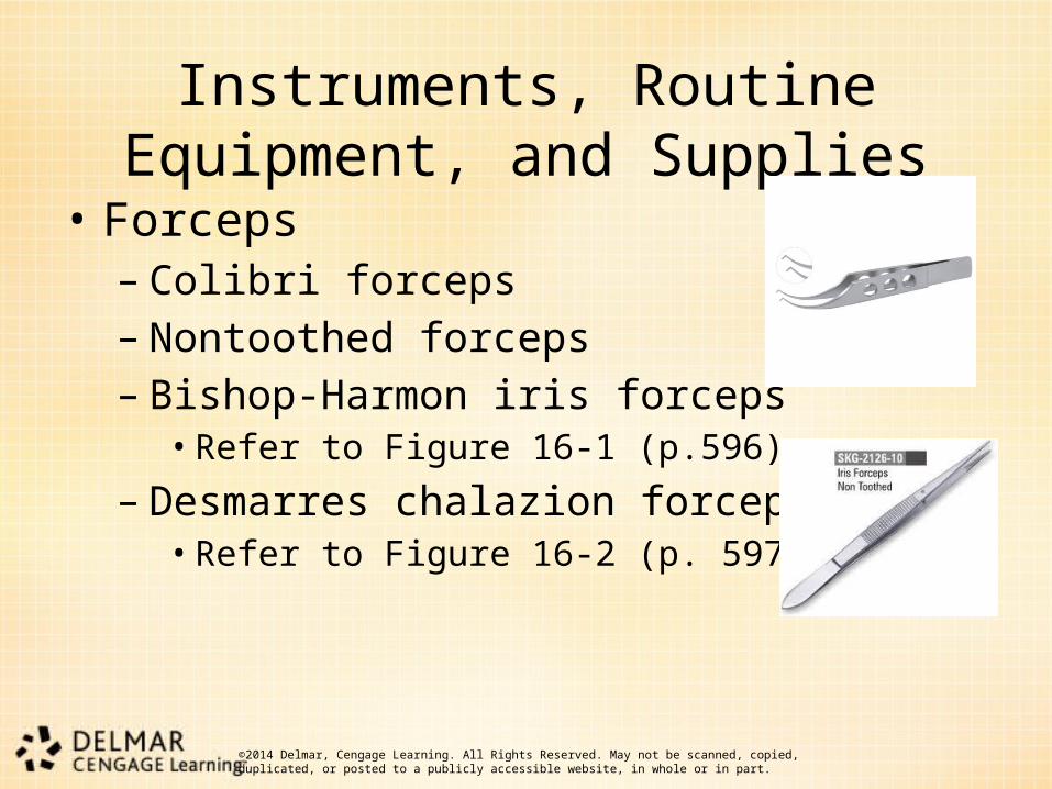
• Forceps– Colibri forceps– Nontoothed forceps– Bishop-Harmon iris forceps
• Refer to Figure 16-1 (p.596)
– Desmarres chalazion forceps• Refer to Figure 16-2 (p. 597)

©2014 Delmar, Cengage Learning. All Rights Reserved. May not be scanned, copied, duplicated, or posted to a publicly accessible website, in whole or in part. 10
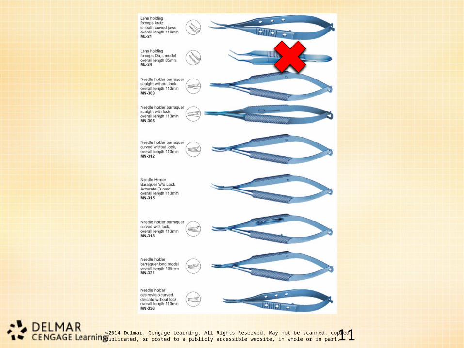
Instruments, Routine Equipment, and Supplies (cont’d.)
• Needle holders– Castroviejo: locking or nonlocking
• Refer to Figure 16-3 (p. 597)
– Microsurgical needle holder
©2014 Delmar, Cengage Learning. All Rights Reserved. May not be scanned, copied, duplicated, or posted to a publicly accessible website, in whole or in part. 1111
©2014 Delmar, Cengage Learning. All Rights Reserved. May not be scanned, copied, duplicated, or posted to a publicly accessible website, in whole or in part. 12
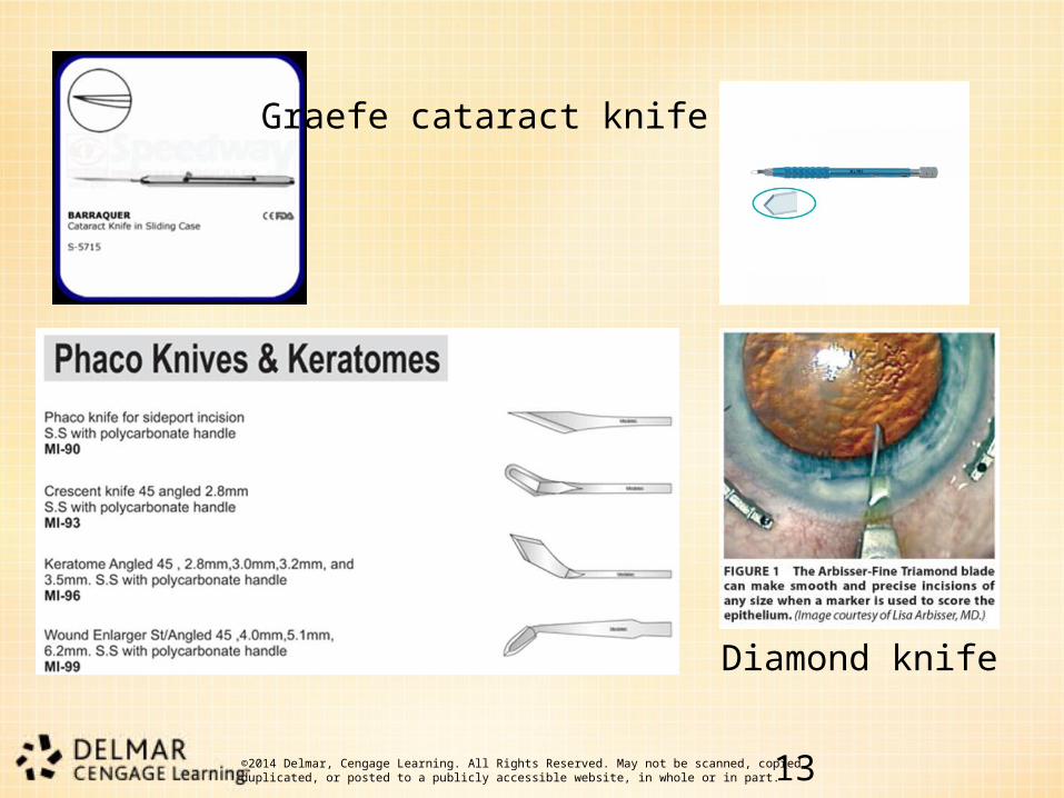
Instruments, Routine Equipment, and Supplies (cont’d.)
• Scalpels– Graefe cataract knife– Keratomes – Razor fragments– Diamond knife– Oscillating knife– Beaver blades– No. 15 knife blade
©2014 Delmar, Cengage Learning. All Rights Reserved. May not be scanned, copied, duplicated, or posted to a publicly accessible website, in whole or in part. 1313
Graefe cataract knife
Diamond knife

©2014 Delmar, Cengage Learning. All Rights Reserved. May not be scanned, copied, duplicated, or posted to a publicly accessible website, in whole or in part. 1414
Oscillating kni
Beaver blades
©2014 Delmar, Cengage Learning. All Rights Reserved. May not be scanned, copied, duplicated, or posted to a publicly accessible website, in whole or in part. 15
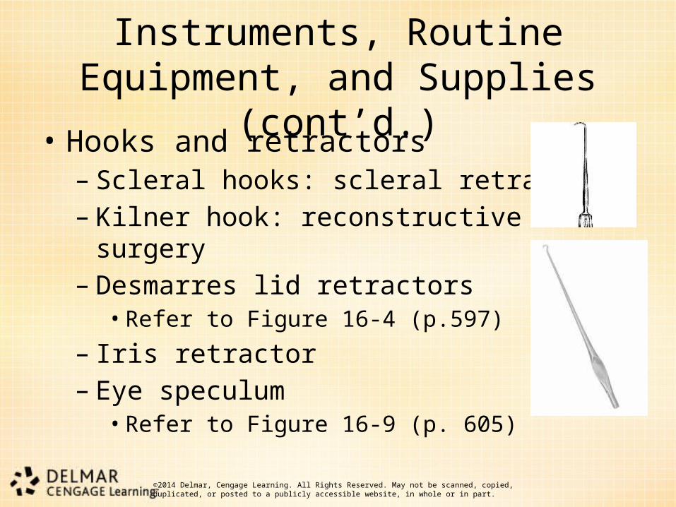
Instruments, Routine Equipment, and Supplies (cont’d.)
• Hooks and retractors– Scleral hooks: scleral retraction– Kilner hook: reconstructive surgery– Desmarres lid retractors
• Refer to Figure 16-4 (p.597)
– Iris retractor – Eye speculum
• Refer to Figure 16-9 (p. 605)
©2014 Delmar, Cengage Learning. All Rights Reserved. May not be scanned, copied, duplicated, or posted to a publicly accessible website, in whole or in part. 16
Instruments, Routine Equipment, and Supplies (cont’d.)
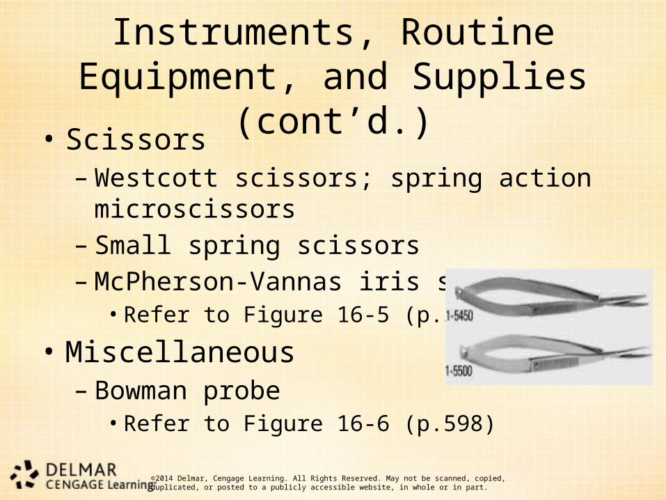
• Scissors– Westcott scissors; spring action microscissors– Small spring scissors– McPherson-Vannas iris scissors
• Refer to Figure 16-5 (p.598)
• Miscellaneous – Bowman probe
• Refer to Figure 16-6 (p.598)
©2014 Delmar, Cengage Learning. All Rights Reserved. May not be scanned, copied, duplicated, or posted to a publicly accessible website, in whole or in part. 17
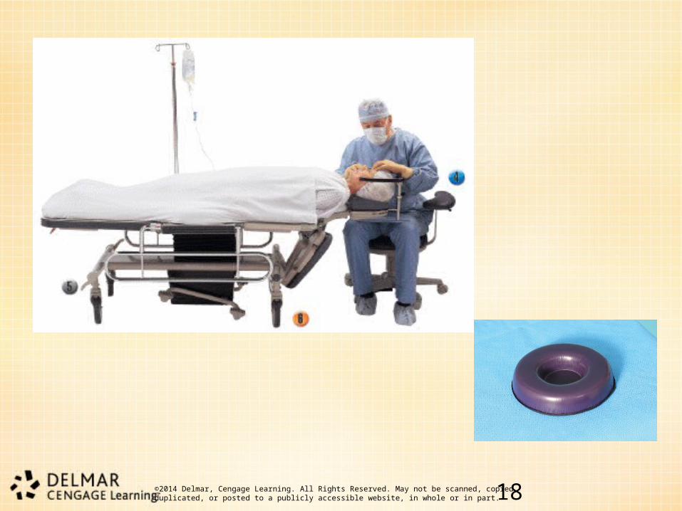
Instruments, Routine Equipment, and Supplies (cont’d.)
• Routine equipment – Ophthalmic stretcher with attachable wrist rest
for the surgeon– Donut for positioning the head– Electrosurgical unit (ESU)
©2014 Delmar, Cengage Learning. All Rights Reserved. May not be scanned, copied, duplicated, or posted to a publicly accessible website, in whole or in part. 1818

©2014 Delmar, Cengage Learning. All Rights Reserved. May not be scanned, copied, duplicated, or posted to a publicly accessible website, in whole or in part. 19
Instruments, Routine Equipment, and Supplies (cont’d.)
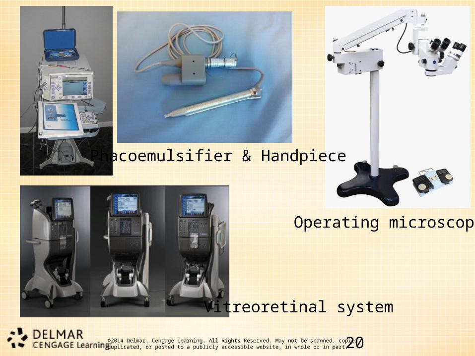
• Special equipment– Phacoemulsifier: uses ultrasonic energy to
fragment the hard lens material, which then can be aspirated from the eye
– Operating microscope: magnifies surgical site through use of lenses
– Vitreoretinal system: provides light, suction, diathermy, and intraoperative access to the surgeon
©2014 Delmar, Cengage Learning. All Rights Reserved. May not be scanned, copied, duplicated, or posted to a publicly accessible website, in whole or in part. 2020
Vitreoretinal system
Operating microscope
Phacoemulsifier & Handpiece
©2014 Delmar, Cengage Learning. All Rights Reserved. May not be scanned, copied, duplicated, or posted to a publicly accessible website, in whole or in part. 21
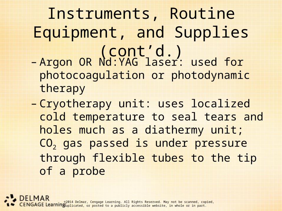
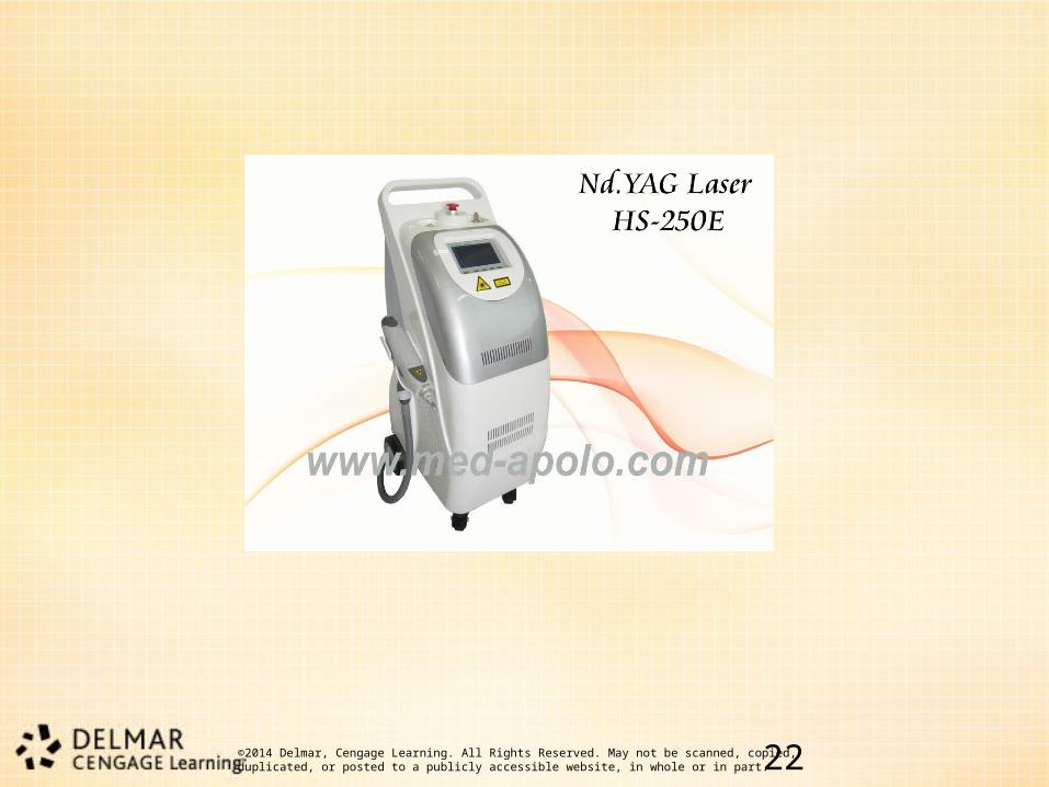
Instruments, Routine Equipment, and Supplies (cont’d.)
– Argon OR Nd:YAG laser: used for photocoagulation or photodynamic therapy
– Cryotherapy unit: uses localized cold temperature to seal tears and holes much as a diathermy unit; CO2 gas passed is under pressure through flexible tubes to the tip of a probe
©2014 Delmar, Cengage Learning. All Rights Reserved. May not be scanned, copied, duplicated, or posted to a publicly accessible website, in whole or in part. 2222

©2014 Delmar, Cengage Learning. All Rights Reserved. May not be scanned, copied, duplicated, or posted to a publicly accessible website, in whole or in part. 23
Instruments, Routine Equipment, and Supplies (cont’d.)
• Routine supplies– Prep set– Eye or head and neck back table pack
(depending on procedure) – Wexcel sponges– Sterile gloves– Sterile cotton swabs– Suture material (surgeon’s preference)

©2014 Delmar, Cengage Learning. All Rights Reserved. May not be scanned, copied, duplicated, or posted to a publicly accessible website, in whole or in part. 24
Instruments, Routine Equipment, and Supplies (cont’d.)
– BSS– 27- and 30-gauge disposable needles,
straight and angled– Needles and sutures
• Refer to Tables 16-1 and 16-2

©2014 Delmar, Cengage Learning. All Rights Reserved. May not be scanned, copied, duplicated, or posted to a publicly accessible website, in whole or in part. 25
Surgical Intervention
• A variety of surgical procedures and approaches are available– There are elements of consistency in every
ophthalmic procedure • Practical considerations • Procedural considerations

©2014 Delmar, Cengage Learning. All Rights Reserved. May not be scanned, copied, duplicated, or posted to a publicly accessible website, in whole or in part. 26
Surgical Intervention (cont’d.)
• Procedures– Laparotomy – Surgical repair of chalzion– Transconjunctival approach– Surgical repair of entropion– Iridectomy – Strabismus correction: recession/resection– Adjustable suture surgery
©2014 Delmar, Cengage Learning. All Rights Reserved. May not be scanned, copied, duplicated, or posted to a publicly accessible website, in whole or in part. 27
Surgical Intervention (cont’d.)
– Scleral buckle– Dacryocystorhinostomy– Enucleation – Keratoplasty (corneal transplant)– Cataract extraction
• Extracapsular • Intracapsular
– Vitrectomy– Repair of traumatic eyelid laceration

©2014 Delmar, Cengage Learning. All Rights Reserved. May not be scanned, copied, duplicated, or posted to a publicly accessible website, in whole or in part. 28
Summary
• This chapter reviewed:– The pathology that prompts surgical
intervention of the eye – Ophthalmic instruments– Special equipment– Surgical steps of ophthalmic procedures– Preoperative, intraoperative, and
postoperative preparation and care