2010 harry m. zweig memorial fund for equine research summary report
TRANSCRIPT

2010 Harry M. Zweig Memorial Fund for Equine Research
Summary Report The 2010 Annual Report covering the period of January 1, 2010 through December 31, 2010 is enclosed. For this reporting period, The Harry M. Zweig Memorial Fund for Equine Research Committee granted approval of 9 of 14 submitted projects. Five were new studies, one was a supplemental, and three were continuation awards. The total amount allocated for 2010 awards was $443,705. Copies of the investigators’ reports are provided in Appendix-A. Additionally, Cornell hosted its second annual poster session celebrating the collaboration between the Harry M. Zweig Memorial Fund for Equine Research and Cornell University. Members of the faculty, students, and staff show-cased their research to the research community and the Harry M. Zweig Memorial Fund for Equine Research Committee at the annual poster session on Thursday November 18, 2010. 2010 Harry M. Zweig Memorial Fund for Equine Research Awards
CONTINUATION AWARDS
AWARD
Douglas Antczak Expression Microarrays and Equine Placental Development
$35,000
Sylvia Bedford-Guaus Bettina Wagner NEW AWARDS
Further Characterization of the Specific Activity and Ultrastructural Localization of Phospholipase C Zeta in Fertile and Subfertile Stallions Analysis of the Innate Immune Response to EHV-1 Infection
$35,774 $50,887
Norm Ducharme Tissue Engineered Cartilage in the Equine Airway (1 Year Award)
$97,729
Lisa Fortier Optimization of Platelet Rich Plasma Components for the Treatment of Tendonitis (2 Year Award)
$71,563
Robert Gilbert Controlled Postponement of Ovulation by Progestagen Treatment (1 Year Award)
$56,884
Vicki Meyers-Wallen Generation of a Molecular Resource to Identify Gene Mutations Causing Inherited Equine Sterility and Infertility (1 Year Award)
$48,000
Alan Nixon Targeted Delivery of Stem Cells for Pro-Inflammatory Cytokine Suppression in Arthritic Joints (2 Year Award)
$47,068
SUPPLEMENTAL Susan Fubini
Indices of Intra-Abdominal Fibrinolysis in Colic Foals: Pathogenic and Prognostic Markers
$800
TOTAL: $443,705 Interim & Completed 2009 Awards
Dr. Ainsworth’s project entitled “Deciphering the Mechanism of Equine Inflammatory Airway Disease in vitro” received a no cost extension through June 30, 2011. An interim report is included in this report (Appendix B). Dr. Bedford-Guaus’ project entitled “Characterization of the Specific Activity and Ultrastructural Localization of Phosphoilpase C zeta PLC in Fertile & Subfertile Stallions” received a no cost extension through June 30, 2011. An interim report is included in this report (Appendix B). Dr. Ducharme’s project entitled “Factors Affecting Airway Stability at Exercise: A combined Neuroanatomical, Clinical and Engineering Methodology” received a no cost extension through June 30, 2010. A Final report is included in this report (Appendix B). Dr. Nixon’s project entitled, “Genomic Profiling of Osteochondritis Dissecans Using an Equine Whole Transcript Exon Array” received a no cost extension through June 30, 2010. A final report will be included in next year’s report. Dr. Gillian Perkins’ project entitled “Immunization Against Strangles Using a Vectored Equine Herpesvirus Vaccine” received a no cost extension through June 30, 2011. An annual report is included in this report (Appendix B).
FURTHER SECURED FUNDING FROM RESEARCH AWARDS IN 2010 In November 2009, the Zweig Committee voted to increase the amount of support provided by the Incentive Program from $10,000 to $20,000. The Incentive Program enables the Fund to leverage its investment in Zweig-sponsored research by encouraging Veterinary College faculty to seek either additional or supplementary monies from external sponsors that base their award decisions on a process that involves informed scientific review. The external grant must be for a closely related project. Eligible sponsors include, but are not limited to, the Grayson Foundation, the NIH, the NSF, and the USDA’s National Research Initiative. Recipients provide an annual report on the use of these funds. The following external grant awards resulted from Zweig funding:
Principal Investigator
External Award
Sponsor
Project Period
Awarded Amount
Incentive Award
Dr. Lisa Fortier Clinical Administration of Doxycycline for Arthritis
Grayson-Jockey
04/01/10- 03/31/12
$63,073 $5,000
Dr. Douglas. Antczak
Immune Tolerance to Serial Trophoblast Transplants
NIH-Mentored Research Scientists Development Award (K08) to Dr. Margaret Brosnahan
08/01/10- 07/31/15
$503,217 $5,000
PUBLICATIONS Publications resulting from awards from the Harry M. Zweig Memorial Fund for Equine Research during 2010 were: 1. Whole-Genome SNP Association in the Horse: Identification of a Deletion in Myosin Va Responsible for Lavender Foal Syndrome S. Brooks,1* N. Gabreski,1 D. Miller,2 A. Brisbin,3 H. Brown,2¤ C.Streeter,1 J. Mezey,3 D. Cook,4 and D. Antczak2 http://www.ncbi.nlm.nih.gov/pmc/articles/PMC2855325/ 2. Genome Sequence Comparative Analysis and Population Genetics of the Domestic Horse (Equus caballus) D. Antczak D, Wade et al. http://www.sciencemag.org/content/326/5954/865.full 3. Split Immunological Tolerance to trophoblast. De Mestre AM, Wagner B, Antczak D. http://www.ijdb.ehu.es/web/paper.php?doi=082795ad 4. Equine clinical genomics: A clinician's primer. Brosnahan MM, Brooks SA, Antczak D. http://onlinelibrary.wiley.com/doi/10.1111/j.2042-3306.2010.00166.x/full
5. Foals and Adult Horses with Colic Similar Alterations in Plasma and Peritoneal Indices of Fibrinolysis. Watts A, Fubini S, Todhunter, R, Brooks M. 2010 6. Characterization of stallion PLC as a model for evaluation of male fertility. Bedford-Guaus S, McPartlin L, Twomey S, Xie J, Roberson M. 2010 7. Role of the Hypoglossal Nerve in Equine Nasopharyngeal Stability Cheetham J, Pigott J, Hermanson J, Campoy L, Soderholm L, Thorson L, Ducharme NG http://jap.physiology.org/content/107/2/471.full 8. Incidence of Swallowing During Exercise n Horses with Dorsal Displacement of the Soft Palate. Pigott J, Ducharme N, Mitchell L, Soderholm, L, Cheetham J. http://onlinelibrary.wiley.com/doi/10.1111/j.2042-3306.2010.00116.x/abstract 9. A Transducer for Measuring Force on Surgical Sutures. Witte T, Cheetham J, Rawlinson J, Soderholm L, Ducharme N. http://www.ncbi.nlm.nih.gov/pubmed/21197230
10. Molecular Cloning and Characterization of Phospholipase C Zeta in Equine Sperm and Testis Reveals Species-Specific Differences in Expression of Catalytically Active Protein. Bedford-Guaus S, McPartlin L, Xie J, Westmiller S, Buffone M, Roberson M. http://www.ncbi.nlm.nih.gov/pubmed/21389344 11. Equine Herpesvirus Type 1 Modulates CCL2, CCL3, CCL5, CXCL9, and CXCL10 Wimer C, Damiani A, Osterrieder N, Wagner B http://www.unboundmedicine.com/medline/ebm/record/21349590/full_citation/Equine_herpesvirus_type_1_modulates_CCL2_CCL3_CCL5_CXCL9_and_CXCL10_chemokine_expression_
12. Experimental Leptospira interrogans serovar Kennewicki infection of horses. Yan W, Faisal S, Divers T, McDonough S, Akey B, Chang YF. http://www.ncbi.nlm.nih.gov/pubmed/20649749
13. Temporal Analysis of Equine Bone Marrow Aspirate During Establishment of Putative Mesenchymal Progenitor Cell Populations. Radcliffe C, Flaminio M, Fortier L.http://www.ncbi.nlm.nih.gov/pubmed/19604071 14. Maternal immune responses to trophoblast: the contribution of the horse to pregnancy immunology. Noronha L, Antczak D. http://www.ncbi.nlm.nih.gov/pubmed/20618178

Bettina Wagner, DVM, PhD Harry M. Zweig Assistant Professor in Equine Health
At the November 20, 2010 Annual meeting, Dr. Bettina Wagner was appointed to her 3rd year as the Harry M. Zweig Assistant Professor in Equine Health. Her three-year appointment covers the period January 1, 2009 through December 31, 2011. A Research progress report is included in this report (Appendix A).
CORNELL CLINICAL FELLOW IN EQUINE HEALTH At the 2007 Annual meeting, the Harry M. Zweig Committee approved the allocation of funds to support a Cornell Clinical Fellow in Equine Health. Cornell’s College of Veterinary Medicine’s two-year Clinical Fellows Program was the first in the country to address a growing shortage of academic veterinarians who conduct research on animal diseases and basic biology. The two-year program, available to veterinarians who have completed a three-year residency, offers an annual salary of $60,000 plus benefits and an additional $15,000 per year to fund a research project. Dr. Sophy Jesty was selected as Cornell’s 1st Clinical Fellow, supported in part by Zweig funds. Dr. Jesty’s comments regarding her experience are included below: “As a clinical fellow, I worked in a molecular genetics basic science lab at Cornell. I became integrally involved in an ongoing project in regenerative medicine aimed at assessing the cellular response to cardiac injury such as infarction. My responsibilities included molecular techniques, cellular techniques, and whole animal techniques. During the two years, I found that it was a perfect combination of me learning and me teaching. Our research has led to a new understanding of the comparative response to cardiac injury in neonates vs. adults, especially in regards to stem cell capabilities. This knowledge will be incorporated in experiments in the future as we try to augment the body’s natural capabilities in enhancing cardiac repair after injury.” Sarah Pownder, DVM, Department of Clinical Sciences, Section of Large Animal Surgery was selected as the second Cornell Clinical Fellow in Equine Research, and is currently in her first year of the program, beginning August 2010.
OUTREACH 2010
Patent updates for 2011 As previously reported, Patent No. 7,036,460 entitled “Throat Support Device and Methods of Using Same” was issued to Dr. Norm Ducharme, etc. al. During 2007 he has been concentrating on obtaining the foreign applications for approval of this device. At the present time this application is still pending. During 2007, Dr. Chang applied for another provisional patent entitled “Immunologenic Proteins for Genome-Derived Outer Membrant of Leptospira and Compositions and Methods Based Thereon.” At the present time this application is still pending. Zweig News Capsules There were two issues of the Zweig News Capsule published in 2010. Copies of these issues can be found in Appendix (F). All Zweig News Capsules (#49 & #50) and can be found at: http://web.vet.cornell.edu/public/research/zweig/Newsletter/index.html. The College’s Veterinary Medicine Information Technology Department in collaboration with the Communications Department, is currently developing a new Zweig website.

SUMMARY OF EXPENDITURES The 2010 Summary of Allocations was presented and approved at the Zweig Committee Annual Meeting in November 2009. (Appendix C).
2011 ZWEIG PROGRAM
Seven projects were approved for funding, from a roster of 12 applications, at the Harry M. Zweig Memorial Fund annual November (2010) meeting. The list for projects funded for 2011 are shown in (Appendix D).

APPENDIX A
Progress & Final Reports Resulting from 2010 Funding
Douglas Antczak Sylvia Bedford-Guaus
Expression Microarrays and Equine Placental Development Further Characterization of the Specific Activity and Ultrastructural Localization of Phospholipase C zeta in fertile and Subfertile Stallions
Norm Ducharme
Tissue Engineered Cartilage in the Equine Airway
Lisa Fortier
Optimization of Platelet Rich Plasma Components for the Treatment of Tendonitis
Susan Fubini Robert Gilbert Vicki Meyers-Wallen
Indices of Intra-Abdominal Fibrinolysis in Colic Foals: Pathogenic and Prognostic Markers Controlled Postponement of Ovulation by Progestagen Treatment Generation of a Molecular Resource to Identify Gene Mutations Causing Inherited Equine Sterility and Infertility
Alan Nixon
Targeted Delivery of Stem Cells for Pro-Inflammatory Cytokine Suppression in Arthritic Joints
Bettina Wagner Analysis of the Innate Immune Response to EHV-1 Infection

Harry M. Zweig Memorial Fund for Equine Research
2010 Final Report
P.I.: Dr. Douglas Antczak
Title: Expression Microarrays and Equine Placental Development
Project Period: 1/1/09-12/31/10 Reporting Period: 1/1/10-12/31/10

2010 HARRY M. ZWEIG MEMORIAL FUND FINAL REPORT PROJECT TITLE: Expression Microarrays and Equine Placental Development PRINCIPAL INVESTIGATOR(S): Douglas F. Antczak Summary: The goal of this project was to use the genome-level technology of expression microarrays to explore how the cells of the placenta interact with the mother’s immune system cells and with other cells in the uterus during the period of implantation. Microarrays allow scientists to determine which genes are activated in a particular cell type, and how strongly those genes are expressed. Prior technology allowed this type of evaluation for only a few genes at a time, while the microarrays allow assessment of virtually all of the 20,000+ known mammalian genes in a single experiment. For this study the Antczak laboratory designed a microarray specifically to test horse genes. This new microarray device was used to compare gene expression in two related cell types of the early horse placenta. We discovered that the invasive chorionic girdle trophoblast cells switch on several genes normally associated with the immune system as these cells invade into the endometrium of the mare to form the endometrial cups. A particularly novel finding was the expression of a recently described cytokine known as Interleukin 22. We presented these findings at the International Symposium on Equine Reproduction in summer 2010, and we are now writing up the results for publication. During the two year time frame of this grant the Antczak laboratory published a number of research papers, reviews articles, and book chapters that were based in part on research conducted over the past several years with Zweig grants. Those papers are cited at the end of this report. Of particular significance were two publications. The first, published in the journal Science, described the equine genome sequence that has been the focus of our Zweig-funded research for the past decade (Wade et al. 2009). The second paper describes the discovery of the genetic mutation that causes the lethal inherited disease of Arabians known as Lavender Foal Syndrome (Brooks et al., 2010). As part of this research a diagnostic test for the mutation was developed. A provisional patent was filed on this invention through the Cornell Intellectual Property office. This is the first genetic disease in the horse to be discovered and patented at Cornell. The diagnostic test, which can be used by breeders to avoid carrier to carrier matings and thus prevent the occurrence of the disease, is offered through Cornell’s Animal Health Diagnostic Center (AHDC). This is the first test for a genetic disease to be offered through the AHDC. Support from the Zweig Fund was critical to the success of the research projects that lead to these important papers. Refereed research publications Wade, C.M., Giulotto, E., Sigurdsson, S., Zoli, M., Gnerre, S., Imsland, F., Lear, T.L., Adelson, D.L., Bailey, E., Bellone, R.R., Blöcker, H., Distl, O., Edgar, R.C., Garber, M., Leeb, T., Mauceli, E., MacLeod, J.N., Penedo, M.C.T., Raison, J.M., Sharpe, T., Vogel, J., Andersson, L., Antczak, D.F., Biagi, T., Binns, M.M., Chowdhary, B.P., Coleman,

S.J., Della Valle, G., Fryc, S., Guérin, G., Hasegawa, T., Hill, E.W., Jurka, J., Kiialainen, A., Lindgren, G., Liu, J., Magnani, E., Mickelson, J.R., Murray, J., Nergadze, S.G., Onofrio, R., Pedroni, S., Piras, M.F., Raudsepp, T., Rocchi, M., Røed, K.H., Ryder, O.A., Searle, S., Skow, L., Swinburne, J.E., Syvänen, A.C., Tozaki, T., Valberg, S.J., Vaudin, M., White, J.R., Zody, M.C., Broad Institute Genome Sequencing Platform, Broad Institute Whole Genome Assembly Team, Lander, E.S., and Lindblad-Toh, K. (2009) Genome sequence, comparative analysis and population genetics of the domestic horse (Equus caballus). Science 326:865-867. de Mestre, A.M., Noronha, L., Wagner, B. and Antczak, D.F. (2010) Split immunological tolerance to trophoblast. Int. J. Dev. Biol. 54:445-455. Tallmadge, R.L., Campbell, J.A., Miller, D.C., and Antczak, D.F. Analysis of MHC class I genes across horse MHC haplotypes. (2010) Immunogenetics 62:159-172. Brooks, S. A., Gabreski, N., Miller, D., Brisbin, A., Brown, H. E., Streeter, C., Mezey, J., Cook, D., and Antczak, D. F. (2010) Whole Genome SNP Association in the Horse: Identification of a Deletion in Myosin Va Responsible for Lavender Foal Syndrome. PLoS Genetics 6(4):e1000909. Tseng, C. T., Miller, D., Cassano, J., Bailey, E., and Antczak, D. F. (2010) Molecular Identification of Equine Major Histocompatibility Complex Haplotypes using Polymorphic Microsatellites. Animal Genetics 41 Suppl 2:150-153.
Robbin, M. G., Wagner, B., Noronha, L. E., Antczak, D. F., and de Mestre, A. M. (2011) Subpopulations of equine blood lymphocytes expressing regulatory T cell markers. Vet. Immunol. Immunopathol. 140:90-101. de Mestre, A.M., Hanlon, D., Adams, A.P., Runcan, E., Leadbeater, J.C., Erb, H.N., Costa, C.C., Miller, D., Allen, W.R., and Antczak, D.F. (2011) Functions of ectopically transplanted invasive horse trophoblast. Reproduction 141:1-9. Review articles Noronha, L. E. and Antczak, D. F. (2010) Maternal Immune Responses to Trophoblast: The Contribution of the Horse to Pregnancy Immunology. Am. J. Reprod. Immun. 64:231-244. Brosnahan, M. M., Brooks, S. A., and Antczak, D. F. (2010) Equine Clinical Genomics: A Clinician’s Primer. Eq. Vet. J. 42:658-670. Book Chapters Allen, W.R., Kydd, J., Short, R.V. and Antczak, D.F. (2011) Interspecies and extraspecies equine pregnancies. In: Equine Reproduction, 2nd Edn. Eds. A.O. McKinnon, E. Squires, W. Vaala, D. Varner and J.L. Voss. Wiley Blackwell (in press). de Mestre, A.M., Antczak, D.F. and Allen, W.R. (2011) Equine Chorionic Gonadotropins (eCG). In: Equine Reproduction, 2nd Edn. Eds. A.O. McKinnon, E. Squires, W. Vaala , D. Varner and J.L. Voss. Wiley Blackwell (in press).

Harry M. Zweig Memorial Fund for Equine Research
2010 Interim Report
P.I.: Dr. Sylvia Bedford-Guaus
Title: Further Characterization of the Specific Activity and Ultrastructural Localization of Phospholipase C zeta in Fertile and Subfertile Stallions
Project Period: 1/1/09-06/30/11 Reporting Period: 1/1/10-12/31/10 Dr. Bedford-Guaus was granted a no cost extension through June 30, 2011. An annual report is provided.

HARRY M. ZWEIG MEMORIAL FUND
FOR EQUINE RESEARCH PROGRAM PROGRESS REPORT
Further Characterization of the Specific Activity and Ultrastructural Localization of Phospholipase C zeta PLC in Fertile and Subfertile Stallions Principal Investigators PI: Sylvia Bedford-Guaus; CoPI: Mark Roberson
Summary: Oocyte activation at fertilization is brought about by the testis-specific
phospholipase C zeta (PLCZ), owing to its ability to induce intracellular Ca2+ [Ca2+]i oscillations. While this is a highly conserved mechanism amongst mammals, important species-specific differences in PLCZ sequence, activity and expression have been reported. Thus the objectives of this research were to clone and characterize the [Ca2+]i-releasing activity and expression of equine PLCZ in sperm and testis. Molecular cloning of equine PLCZ yielded a 1914 bp sequence that translated into a protein of the appropriate size (~73kD), as detected with an anti-PLCZ specific antibody. Microinjection of 1 µg/µl equine PLCZ cRNA supported [Ca2+]i oscillations in murine oocytes that were of a higher relative frequency than those generated by an equivalent concentration of murine Plcz cRNA. Immunofluorescence revealed expression of PLCZ over the acrosome, equatorial segment and head-midpiece junction; unexpectedly, PLCZ also localized to the principal piece of the flagellum, in all epididymal, uncapacitated and capacitated sperm. Immunostaining over the acrosome was abrogated after induction of acrosomal exocytosis. Moreover, injection of either sperm heads or tails into mouse oocytes showed that PLCZ in both fractions is catalytically active. Immunohistochemistry on equine testis revealed expression as early as the round spermatid stage and injection of these cells supported [Ca2+]i oscillations in oocytes. In summary, we report that equine PLCZ displays higher intrinsic [Ca2+]i-releasing activity than murine PLCZ, and that catalytically active protein is expressed in round spermatids as well as the sperm flagellum, emphasizing important species-specific differences. Moreover, some of these results may suggest potential novel roles for PLCZ in sperm physiology. Specific Aims and Findings: 1. To characterize the specific [Ca2+]i-releasing activity of immature testicular
sperm cells, of different regions of mature sperm, and of equine PLCZ in oocytes. We have thus far ascertained that round spermatids and both the head and flagellum of mature sperm from stallions show [Ca2+]i-releasing activity when injected into mouse oocytes. Moreover, when 1 µg/µl of either equine PLCZ or murine Plcz cRNA was injected into mouse oocytes, the equine construct elicited a 3-fold higher frequency of [Ca2+]i oscillations, thus suggesting that the equine construct has an intrinsic higher [Ca2+]i -releasing activity. We are presently still characterizing the concentration of equine PLCZ cRNA that is able to elicit [Ca2+]i responses similar to those elicited by a single stallion sperm.
2. To investigate the ultrastructural localization of PLCZ in equine sperm. Our studies revealed that PLCZ does not fractionate into lipid rafts.

3. To determine PLCZ levels and regional expression in equine sperm from stallions of variable fertility. These experiments are also on-going. Thus far we have identified a handful of stallions with a history of subfertility in which PLCZ showed abnormal localization or was expressed in decreased amounts relative to that in stallions with normal fertility levels.
Significance: This research will allow us to better understand oocyte activation, a process required for normal embryo development, in the horse, as well as identify a potential cause for subfertility in stallions. Publications:
Bedford-Guaus SJ, McPartlin LA, Xie J, Westmiller SL, Buffone MG, Roberson MS. Molecular cloning and characterization of phospholipase C zeta in equine sperm and testis reveals species-specific differences in expression of catalytically active protein. Biol Reprod 2011 Mar 9 [Epub ahead of print] PMID: 21389344 http://www.biolreprod.org/content/early/2011/03/08/biolreprod.110.089466.long)

Harry M. Zweig Memorial Fund for Equine Research
2010 Interim Report
P.I.: Dr. Norm Ducharme
Title: Tissue Engineered Cartilage in the Equine
Airway
Project Period: 1/1/10-07/31/11 Reporting Period: 1/1/10-12/31/10 Dr. Ducharme was granted a no cost extension through July 31, 2011. An annual report is provided.

2010 HARRY M. ZWEIG MEMORIAL FUND INTERIM REPORT PROJECT TITLE: Tissue Engineered Cartilage in the Equine Airway PRINCIPAL INVESTIGATOR: Norm Ducharme and Jeremy Rawlinson CO-INVESTIGATORS: Lawrence Bonassar, Jon Cheetham, John Hermanson, Bryan Brown Summary: The ongoing Zweig 2010 project, entitled “Tissue Engineered Cartilage in the Equine Airway,” seeks to characterize the structure, content, and function of the cartilaginous structures of the equine airway and to develop methods which will lead to the production of living surgical implants for the reconstruction or replacement of these same structures. The aims of the currently funded project and the progress made towards the completion of each of the aims are described in more detail below. Specific Aims and Findings: Aim 1: The characterization of the biomechanical and biochemical properties of the dominant support structures within the airway: arytenoid cartilage, cricoid cartilage, and epiglottic cartilage. We have performed in vitro studies which have characterized the geometry, biomechanical properties, biochemical content, and structure of the equine arytenoid and cricoid cartilages. Similar studies are ongoing to characterize the geometry, biomechanical properties, biochemical content and structure of the equine epiglottis and trachea. We expect to complete these studies within the next 2-3 months, and the data generated from this research is currently being used to provide baseline targets for the tissue engineering efforts described in further detail below. The results of the biomechanical studies described as part of Aim 1 have been accepted for publication in the Equine Veterinary Journal (Samantha Passman, Jonathan Cheetham, Lawrence Bonassar, Norm Ducharme, Jeremy J. Rawlinson. Biomechanical Characterization of Equine Laryngeal Cartilage. Equine Veterinary Journal. 2011. In press), presented at the most recent meeting of the American College of Veterinary Surgeons, and are discussed more thoroughly below. Additionally, we are currently preparing two manuscripts which focus upon the diverse histologic architecture and biochemical content of the cartilaginous structures of the upper airway. A summary of our findings to date is provided below. Biomechanical Testing(Abstract from publication) Upper airway obstruction is a common problem in the equine athlete as the soft-tissues of the larynx collapse into the airway, yet there is a paucity of biomechanical properties for the structural cartilage components. The purpose of this study was to measure the geometry and compressive mechanical properties of the hyaline cartilage to better understand laryngeal function and morphology. A total of eleven larynges were harvested from Thoroughbred and Standardbred racehorses. During gross dissection, linear dimensions of the cricoid were obtained. From both the cricoid and arytenoid, specimens were cored to obtain 6 mm disc samples from three sites within the dorsal cricoid (caudal, middle, and rostral) and two central sites in the arytenoids (inner, outer). The specimens were mechanically tested using radial confined compression to calculate the aggregate modulus and permeability of the tissue. The biomechanical data were analyzed using a nested mixed effects model. Geometrically,
the cricoid has relatively straight walls compared to the morphology of human, ovine, and canine larynges. There were significant observations of higher modulus with increasing age (0.13 MPa per year; p=0.007) and stiffer cricoid cartilage (2.29 MPa) than the arytenoid cartilage (0.42 MPa; p<0.001), but no difference was observed between the left and right sides. Linear contrasts showed that the rostral aspect (2.51 MPa) of the cricoid was 20% stiffer than the caudal aspect (2.09 MPa; p=0.025), with no difference between the arytenoid sites. The equine larynx is a well-supported structure due to both the geometry and material properties of the cricoid cartilage. The hyaline structure is an order of magnitude higher in compressive modulus compared to the arytenoids and other hyaline-composed tissues. These characterizations are important to understand the biomechanics of laryngeal function and the mechanisms involved with surgical interventions. Histologic Architecture Histologic analysis of the cartilages of the equine larynx revealed regional differences in content and architecture within the arytenoid and trachea with less impressive differences within the cricoid and epiglottis. The latter cartilage has been shown to be composed of primarily elastic cartilage in other species, but has not been studied in horses. The equine arytenoid was reported to contain hyaline cartilage by Passman et al. (2011, see abstract above) who studied lateral (superficial) and medial (deep) portions of the cartilage near its mid-point. These authors characterized the arytenoid as less stiff than the adjacent cricoid cartilage, which was studied along its dorsal body (including samples from the rostral, middle and caudal length of this region). We have found that the arytenoid is composed predominantly of hyaline cartilage in the region adjacent to the cricoarytenoid joint, as well as in a region spanning from the rostral border of the arytenoid to the same central region as sampled by Passman, et al. (2011). However the corniculate process is well invested by elastic cartilage, possibly explaining the mobility of the structure during swallowing and breathing. The trachea was also found to contain mostly hyaline cartilage, however in several specimens we noted an increase in the density of elastic fibers towards the laterodorsal ends of the tracheal rings as compared to the ventral midline. We are in the process of quantifying this observation. The epiglottis was entirely composed of elastic cartilage, reflecting the mobility seen by this structure during swallowing and breathing activities. We sampled four regions of the epiglottis, the rostral apex, and three additional regions spanning from side to side along the epiglottis’ caudal base. Direct visual counts of chondrocyte numbers were obtained and indicated intra- as well as inter-cartilage differences based on a sample of 4 larynges assessed thus far. Briefly, the arytenoid, the rostral, mid-arytenoid sample contained the highest numbers of chondrocytes, 340 cells/mm2. In contrast, the hyaline cartilage immediately rostral to the cricoarytenoid joint had 293 cells/mm2, which was similar to the cell density of the corniculate cartilage region with 267 cells/mm2. There were no significant differences in cell counts between the rostral and caudal portions of the cricoid cartilage, or between the cricoid and the caudal region of the arytenoid. It was interesting that the epiglottis had uniformly high numbers of chondrocytes, with all regions having from 384 to 518 cells/mm2. These numbers were similar to those noted in all regions of the 2nd tracheal ring, which had chondrocyte densities ranging from 458 cells/mm2 at the ventral midline to 505 cells/mm2 at the dorsolateral ends of the tracheal ring. Together, these results demonstrate the diversity of the cartilaginous structures of the equine upper airway. Additionally, during the course of our histologic investigation, we have found that the equine epiglottis contains a large number of previously unrecognized/undescribed glandular structures. This is a unique finding as cartilaginous tissues are generally described as being avascular, aneural, and aglandular. Our research has shown that these epiglottic gland structures may be unique to horses, as they are not reported in

studies of other species. Further investigation of the presence and function of these structures in the equine epiglottis may have important implications for the treatment of epiglottic abnormalities and is ongoing in our laboratories. Biochemical Content We assessed the biochemical content of the same tissues as those described above. The tissues were subjected to a Hoechst DNA assay to assess cellular content, a hydroxyproline assay to assess collagen content, and a 1,9-dimethylmethylene blue assay to assess glycosaminoglycan (GAG) content. The results of the study showed no significant differences in the cellular content of the arytenoid or the cricoid cartilages, and no significant differences between the sites examined within the arytenoid or cricoid cartilages. Statistically significant differences in collagen content were observed between the cricoid and arytenoid cartilages; however, no significant differences were observed between the sites examined within each tissue. Finally, no significant differences were observed for GAG content between the arytenoid and cricoid cartilages or between the sites examined within each tissue. Testing of the elastin content of each tissue type is currently underway with significant differences expected for epiglottic cartilage based upon our histologic observations above. Aim 2: The creation and culture of tissue engineered constructs. We have developed methods which utilize 3D data generated through medical imaging modalities (MRI and CT) towards the creation of anatomically shaped 3D molds of equine airway components. We have also developed the methods to isolate cells from clinically relevant equine cartilage tissues and to seed them into the 3D molds, creating viable living constructs which approximate the shape and size of equine airway tissues. Currently, we have designed and created molds for the equine epiglottis and arytenoids and are working to produce molds which approximate other equine airway tissues. We are also working to culture these constructs for use in Aim 3 of the currently funded project, which is described below. Work resulting from Aim 2 is expected to be included as part of at least 1 publication and was recently presented at the 2010 Annual Meeting and Exposition of the Tissue Engineering and Regenerative Medicne International Society. Additional presentations of the ongoing work are expected at annual meetings of the Biomedical Engineering Society and the newly formed North American Veterinary Regenerative Medicine Association. Details of the methods used and the results to date are described in the abstract below. Three Dimensional Tissue Engineering of the Equine Airway (Abstract from presentation) Upper airway obstruction is a common cause of poor performance in horses. In general, airway obstructions arise from a reduction in neuromuscular function or from a decrease in the mechanical stiffness of the structures of the upper airway. These reductions in muscular control and stiffness eventually decrease the ability of the airway to resist inspiratory or expiratory pressures, causing laryngeal collapse. We propose to restore airway patency through methods that replace the damaged tissue and improve the stiffness of airway structures. Tissue engineering methods have shown promise for the replacment of a wide variety of damaged, diseased, or missing tissue structures. The objective of the present study was to establish a tissue engineering approach to the creation of viable constructs which approximate the shape and size of equine airway structures. Briefly, computed tomography (CT) imaging was performed on intact equine larynges. CT images were then used to create three-dimensional computer models of the cartilaginous structures of the larynx. Anatomically shaped injection molds were then created from the three-dimensional models using rapid prototyping. These molds were then seeded with chondrocytes resuspended within alginate prior to static tissue culture. A bovine source was selected for the present, preliminary study due to
increased availability of bovine as compared to equine tissues. Cells were harvested from either auricular cartilage (elastic cartilage – epiglottis construct) or from the cartilage of the articulating surfaces of the femoro-patella groove and condylar head (hyaline cartilage – arytenoid construct) using 0.2% collagenase digestion. Isolated cells were then resuspended in 2.2% alginate in PBS at a concentration of 25 million cells/mL. The cell/alginate mixture was then combined with 1% calcium sulfate cross-linking solution prior to injection into the mold. The injected mold was placed into a calcium chloride post-cross-linking solution for 1 hour at room temperature prior to de-molding of the construct. Constructs were then cultured in Dulbecco’s Modified Eagle Medium supplemented with 10% fetal bovine serum and a 1% antibiotic/antimycotic solution for up to 4 weeks post seeding and then evaluated for biochemical content (DNA, collagen, GAG, elastin), biomechanical properties (compression testing), and histologic architecture (hematoxylin and eosin, Safranin O, Verhoeff’s elastic stain). Three-dimensional computer reconstructions of individual airway structures were utilized in the creation of molded constructs which were found to approximate the size and shape of equine tissue structures. It was shown that it is possible create constructs consisting of chondrocytes from both elastic and hyaline cartilage sources, and that it is possible to seed such constructs while maintaining 75%+ cell viability. Extracellular matrix content was observed to increase with time in culture and was accompanied by an increase in mechanical stiffness. We have shown that it is possible to create viable constructs which approximate both the shape and size of cartilaginous structures of the equine airway. Additional investigation is required to determine the optimal culture time prior to implantation as well as to evaluate the ability of constructs created in this manner to integrate with native tissues following implantation. If successful, such an approach would represent a significant improvement upon the currently available treatments for damaged airway cartilage in horses and may provide clinical options for replacement of damaged tissues during treatment of obstructive airway diseases. Aim 3: Mechanically test the constructs in an in vitro airflow model. We have developed an in vitro airflow model which will be useful for determining the mechanical function of our cultured tissue engineered constructs. We have utilized and validated this model for the testing of equine airway tissues as part of previously completed Zweig funded research (Cheetham J, Witte TH, Soderholm LV, Hermanson JW, Ducharme NG. In vitro model for testing novel implants for equine laryngoplasty.Vet Surg. 2008 Aug;37(6):588-93). As we complete Aim 2 as described above, we will test the cultured implants in the model as previously described. We expect to complete Aim 3 over the next 3 months as we concurrently complete the work described in Aim 2. It is expected that the results generated will be included in a manuscript which also describes the work performed as part of Aim 2 or as a separate manuscript. Significance: There were several significant findings as well as areas of improvement in our experimental approaches. Our characterization of the cartilaginous structures of the equine airway have led to a number of previously unrecognized intra- and inter-tissue differences in histologic architecture, biochemical content, and biomechanical properties. These differences are likely related to the diverse functional roles played by each of these tissue structures. A full characterization, such as that provided by the ongoing work, will lead to a better understanding of the structure and function of the cartilaginous structures of the upper airway. Further, these characteristics provide a baseline for our tissue engineering efforts as described in Aim 2. In the studies of Aim 2 we have developed three dimensional tissue engineering modalities, which we have shown to be effective in recreating geometrically accurate, viable constructs, which we seek to utilize as transplants for repair of the equine upper airway. If successful, these methods have the potential to significantly improve the standard of care for equine
upper airway disease. We are now also applying the methods developed during the completion of the above described work to tissue engineering of patient specific shaped tracheal replacements. Conference Presentations: Passman SN, Cheetham J, Bonassar LJ, Ducharme NG, Rawlinson JJ. Biomechanical Stiffness Of Equine Laryngeal Cartilage. 2009 Symposium of the American College of Veterinary Surgeons, Washington, D.C. (Oct 2009). Passman SN, Cheetham J, Bonassar LJ, Ducharme NG, Rawlinson JJ. Biomechanical Stiffness of Equine Laryngeal Hyaline Cartilage. 2009 Annual Meeting of the Biomedical Engineering Society, Pittsburgh, PA (Oct 2009). B.N. Brown, N.J. Siebenlist, J. Cheetham, J.W. Hermanson, N.G. Ducharme, L.J. Bonassar. Image Guided Tissue Engineering of Equine Airway Cartilage. Tissue Engineering and Regenerative Medicine International Society North American Chapter, 2010 Annual Meeting, Orlando, FL. December 5-8, 2010. Publications: Passman SN, Cheetham J, Bonassar LJ, Ducharme NG, Rawlinson JJ. Biomechanical Stiffness of Equine Laryngeal Hyaline Cartilage. Equine Veterinary Journal. In Press.). Listing of grant applications and their status resulting from Zweig funding: Pending: 1R21DC012144-01 Ducharme (PI) 12/01/2011 – 11/30/2013 NIDCD Regenerative medicine Approach to Functional Reconstruction of Laryngeal Muscles The goal of this project is to use a scaffold based regenerative medicine approach that has been shown to promote the formation of functionally innervated muscle tissue to restore motion and function to the laryngeal muscles in a clinically relevant large animal model of laryngeal paralysis. 1R21HL111950-01 Cheetham (PI) 12/01/2011 – 11/30/2013 NHLBI Patient Specific Tissue Engineering Approach to Tracheal Reconstruction The goal of this project is to addresses the two most crucial aspects of generating a tracheal replacement: (1) matching the mechanical properties of the implant to those of native tracheal tissue and (2) providing a surface along which adjacent epithelial cells can migrate. Not Funded: N/A Ducharme (PI) 01/01/2010 – 12/31/2010 Solving incorporation issues of Tissue Engineered Cartilage in the Equine Airway Harry M. Zweig Memorial Fund for Equine Research The goal of this project is to develop methods for assessing and improving the incorporation of tissue engineered cartilages within the structures of the equine airway. N/A Ducharme (PI) 04/01/2011 – 03/31/2011 Grayson Jockey Club Research Foundation Integration of Engineered Cartilage in the Equine Airway The goal of this project is to develop methods for enhancing the integration of engineered cartilage constructs following surgical implantation in cartilaginous structures of the equine airway.

Harry M. Zweig Memorial Fund for Equine Research
2010 Annual
P.I.: Dr. Lisa Fortier
Title: Optimization of Platelet Rich Plasma Components for the Treatment of Tendonitis
Project Period: 1/1/10-12/31/11 Reporting Period: 1/1/10-12/31/11
1
2011 HARRY M. ZWEIG MEMORIAL FUND FOR EQUINE RESEARCH PROGRAM
CONTINUATION APPLICATION
PROJECT TITLE: Optimization of platelet rich plasma components for the treatment of tendonitis. PRINCIPAL INVESTIGATOR: Lisa A. Fortier
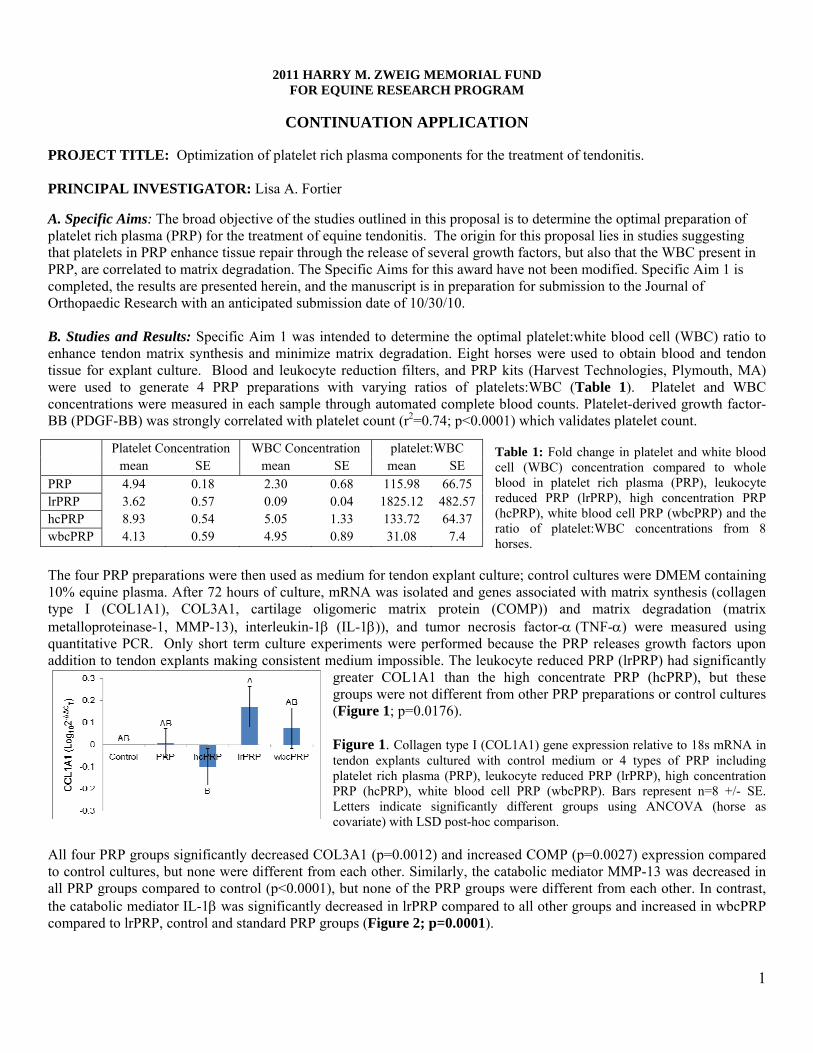
A. Specific Aims: The broad objective of the studies outlined in this proposal is to determine the optimal preparation of platelet rich plasma (PRP) for the treatment of equine tendonitis. The origin for this proposal lies in studies suggesting that platelets in PRP enhance tissue repair through the release of several growth factors, but also that the WBC present in PRP, are correlated to matrix degradation. The Specific Aims for this award have not been modified. Specific Aim 1 is completed, the results are presented herein, and the manuscript is in preparation for submission to the Journal of Orthopaedic Research with an anticipated submission date of 10/30/10. B. Studies and Results: Specific Aim 1 was intended to determine the optimal platelet:white blood cell (WBC) ratio to enhance tendon matrix synthesis and minimize matrix degradation. Eight horses were used to obtain blood and tendon tissue for explant culture. Blood and leukocyte reduction filters, and PRP kits (Harvest Technologies, Plymouth, MA) were used to generate 4 PRP preparations with varying ratios of platelets:WBC (Table 1). Platelet and WBC concentrations were measured in each sample through automated complete blood counts. Platelet-derived growth factor-BB (PDGF-BB) was strongly correlated with platelet count (r2=0.74; p<0.0001) which validates platelet count.
Table 1: Fold change in platelet and white blood cell (WBC) concentration compared to whole blood in platelet rich plasma (PRP), leukocyte reduced PRP (lrPRP), high concentration PRP (hcPRP), white blood cell PRP (wbcPRP) and the ratio of platelet:WBC concentrations from 8 horses.
The four PRP preparations were then used as medium for tendon explant culture; control cultures were DMEM containing 10% equine plasma. After 72 hours of culture, mRNA was isolated and genes associated with matrix synthesis (collagen type I (COL1A1), COL3A1, cartilage oligomeric matrix protein (COMP)) and matrix degradation (matrix metalloproteinase-1, MMP-13), interleukin-1β (IL-1β)), and tumor necrosis factor-α (TNF-α) were measured using quantitative PCR. Only short term culture experiments were performed because the PRP releases growth factors upon addition to tendon explants making consistent medium impossible. The leukocyte reduced PRP (lrPRP) had significantly
greater COL1A1 than the high concentrate PRP (hcPRP), but these groups were not different from other PRP preparations or control cultures (Figure 1; p=0.0176). Figure 1. Collagen type I (COL1A1) gene expression relative to 18s mRNA in tendon explants cultured with control medium or 4 types of PRP including platelet rich plasma (PRP), leukocyte reduced PRP (lrPRP), high concentration PRP (hcPRP), white blood cell PRP (wbcPRP). Bars represent n=8 +/- SE. Letters indicate significantly different groups using ANCOVA (horse as covariate) with LSD post-hoc comparison.
All four PRP groups significantly decreased COL3A1 (p=0.0012) and increased COMP (p=0.0027) expression compared to control cultures, but none were different from each other. Similarly, the catabolic mediator MMP-13 was decreased in all PRP groups compared to control (p<0.0001), but none of the PRP groups were different from each other. In contrast, the catabolic mediator IL-1β was significantly decreased in lrPRP compared to all other groups and increased in wbcPRP compared to lrPRP, control and standard PRP groups (Figure 2; p=0.0001).
Platelet Concentration WBC Concentration platelet:WBC mean SE mean SE mean SE
PRP 4.94 0.18 2.30 0.68 115.98 66.75 lrPRP 3.62 0.57 0.09 0.04 1825.12 482.57 hcPRP 8.93 0.54 5.05 1.33 133.72 64.37 wbcPRP 4.13 0.59 4.95 0.89 31.08 7.4

2
Figure 2. Interleukin-1β (IL-1β) expression in tendon explants cultured in control medium, platelet rich plasma (PRP), leukocyte reduced PRP (lrPRP), high concentration PRP (hcPRP), or white blood cell PRP (wbcPRP). Bars represent n=8 +/- SE. Letters indicate significantly different groups using ANCOVA (horse as covariate) and LSD post-hoc comparison. Similarly, tumor necrosis factor-α (TNF-α) expression was lowest in
tendons cultured in 1rPRP compared to the high WBC PRP preparations (hcPRP and wbcPRP), and not different from control cultures or regular PRP cultures (Figure 3; p=0.0224).
Figure 3. Tumor necrosis factor-α (TNF-α) expression in tendon explants cultured in control medium, platelet rich plasma (PRP), leukocyte reduced PRP (lrPRP), high concentration PRP (hcPRP), or white blood cell PRP (wbcPRP). Bars represent n=8 +/- SE. Letters indicate significantly different groups using ANCOVA (horse as covariate) and LSD post-hoc comparison.
Together, these results suggest that the ratio of platelets to WBC is important towards tendon matrix homeostasis, particularly in the catabolic pathways. Leukocyte reduced PRP has the lowest expression of IL-1β and TNF-α, while wbcPRP and hcPRP, which have the highest WBC concentration, have the highest expression of these inflammatory mediators. The other catabolic mediator, MMP-13 was decreased in all PRP groups compared to control, but no one PRP group was better than then others. It is known that MMP-13 is downstream of IL-1β and TNF-α, and therefore differences between the various PRP groups might not be noticed in our short term cultures of 72 hours duration. Although COL3A1 is present in normal tendons, increased COL3A1 is also associated with scar formation while COL1A1 and COMP are widely accepted as representative of normal tendon matrix. Here again, we see that leukocyte reduced PRP is the only PRP group that increases COL1A1 while also increasing COMP and decreasing COL3A1. C. Significance: Presently, our results suggest that an optimal PRP product would have a high platelet:WBC ratio due to a low WBC concentration which results in increased matrix synthesis and minimal inflammation.
D. Plans: Specific Aim 2 (Year 2). To determine the optimal absolute number of platelets that result in tendon matrix synthesis. In this aim, we will generate PRP preparations with platelet:WBC ratio of 2000:1 but varying numbers of platelets and WBC (Table 2). The WBC concentration of all experimental groups will fall in the category of lrPRP. This aim is presently in progress.
Table 2. Three types of leukocyte reduced PRP (lrPRP) that will be generated in Year 2 to treat tendon explant cultures and determine the effects on tendon matrix metabolism. The lrPRP preparations will all have the same ratio of platelets:WBC, but the absolute number of each will vary.
The knowledge gained though the completion of the studies outlined in this proposal should provide immediately relevant, clinically applicable information regarding the prudent and practical use of platelet rich plasma for treatment of equine superficial digital flexor tendonitis. The expectation is that the results of these studies will provide fundamental information for the design of in vivo investigations into the use of platelet rich plasma for treatment of tendonitis and for more basic scientific investigations into the mechanism of action of PRP.
lrPRP “type”
Platelets (*103/µl)
WBCs (*103/µl)
Platelet:WBC ratio
lrPRP-1 500 0.25 2000:1 lrPRP-2 1500 0.75 2000:1 lrPRP-3 200 0.1 2000:1 control NA NA NA
Harry M. Zweig Memorial Fund
for Equine Research
2010 Annual Report
P.I.: Dr. Susan Fubini
Title: Indices of Intra-Abdominal Fibrinolysis in Colic Foals: Pathogenic and Prognostic Markers
Project Period: Reporting Period:
1/1/08-07/31/11 1/1/10-12/31/10
Dr. Fubini was awarded a no cost extension through July 31, 2011. An annual report will be provided next year.

Harry M. Zweig Memorial Fund for Equine Research
2010 Interim Report
P.I.: Dr. Robert Gilbert
Title: Controlled Postponement of Ovulation by
Progestagen Treatment
Project Period: Reporting Period:
1/1/10-06/30/11 1/1/10-12/31/10
Dr. Gilbert received a no cost extension through June 30, 2011. An interim report is provided.

2010 HARRY M. ZWEIG MEMORIAL FUND INTERIM REPORT PROJECT TITLE: Controlled Postponement of Ovulation by Progestagen Treatment PRINCIPAL INVESTIGATOR: Robert Gilbert Summary: The animal component of this work is completed. Some laboratory assays remain to be done, after which the final analysis and publication will be completed. However, some conclusions are clear:
1. Progestagen treatment, as performed, was ineffective in delaying ovulation. 2. Altrenogest treatment impaired fertility. 3. Both progestagens, but especially CIDR, promptly diminished uterine edema and
virtually eliminated signs of estrus. Specific Aims and Findings: The specific aims of this project were to measure the effect of each of two exogenous progestagens (oral altrenogest or intravaginal application of a CIDR) on:
1. Estrus behavior 2. Follicular growth rate 3. Time of ovulation 4. Postovulatory luteal function 5. Pregnancy rate.
As mentioned above, findings were that:
1. Estrous behavior was promptly eliminated by both treatments – more completely with CIDR. (Concomitantly, uterine edema was eliminated by treatment.)
2. Follicular growth rate was not altered by either treatment. 3. Consistent with (2) time of ovulation was not altered. 4. Postovulatory luteal function is still being evaluated as progesterone assays are
not yet complete. 5. Pregnancy rate was depressed after use of altrenogest.
Significance: Progesterone, and particularly altrenogest, are already being used by some practitioners for postponement of estrus despite lack of evidence of efficacy or safety (for pregnancy). This trial shows lack of efficacy and depression of pregnancy rate. The practice of using altrenogest (or CIDR) for postponement of ovulation should be discontinued. Publications: In preparation. To be submitted once results are complete. Listing of grant applications and their status resulting from Zweig funding: Controlled postponement of ovulation by use of an aromatase inhibitor Submitted to the Zweig Memorial Fund, 2010. Denied.
Harry M. Zweig Memorial Fund for Equine Research
2010 Final Report
P.I.: Dr. Vicki Meyers-Wallen
Title: Generation of a Molecular Resource to Identify Gene Mutations Causing Inherited Equine Sterility and Infertility
Project Period: Reporting Period:
1/1/10-12/31/10 1/1/10-12/31/10
2010 HARRY M. ZWEIG MEMORIAL FUND FINAL REPORT
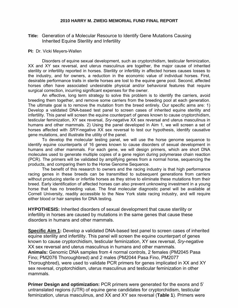
Title: Generation of a Molecular Resource to Identify Gene Mutations Causing Inherited Equine Sterility and Infertility PI: Dr. Vicki Meyers-Wallen
Disorders of equine sexual development, such as cryptorchidism, testicular feminization, XX and XY sex reversal, and uterus masculinus are together, the major cause of inherited sterility or infertility reported in horses. Sterility or infertility in affected horses causes losses to the industry, and for owners, a reduction in the economic value of individual horses. First, desirable performance traits in sterile horses are lost to the equine gene pool. Second, affected horses often have associated undesirable physical and/or behavioral features that require surgical correction, incurring significant expenses for the owner.
An effective, long term strategy to solve this problem is to identify the carriers, avoid breeding them together, and remove some carriers from the breeding pool at each generation. The ultimate goal is to remove the mutation from the breed entirely. Our specific aims are: 1) Develop a validated DNA-based test panel to screen cases of inherited equine sterility and infertility. This panel will screen the equine counterpart of genes known to cause cryptorchidism, testicular feminization, XY sex reversal, Sry-negative XX sex reversal and uterus masculinus in humans and other mammals. 2) Using the panel developed in Aim 1, we will screen a set of horses affected with SRY-negative XX sex reversal to test our hypothesis, identify causative gene mutations, and illustrate the utility of the panel.
To develop the molecular testing panel, we will use the horse genome sequence to identify equine counterparts of 16 genes known to cause disorders of sexual development in humans and other mammals. For each gene, we will design primers, which are short DNA molecules used to generate multiple copies of a gene region during polymerase chain reaction (PCR). The primers will be validated by amplifying genes from a normal horse, sequencing the products, and comparing them to the Horse Genome Sequence.
The benefit of this research to owners and the racing industry is that high performance racing genes in these breeds can be transmitted to subsequent generations from carriers without producing sterile or infertile horses as they strive to eliminate these mutations from their breed. Early identification of affected horses can also prevent unknowing investment in a young horse that has no breeding value. The final molecular diagnostic panel will be available at Cornell University, readily accessible to the New York state racing industry, and will require either blood or hair samples for DNA testing. HYPOTHESIS: Inherited disorders of sexual development that cause sterility or infertility in horses are caused by mutations in the same genes that cause these disorders in humans and other mammals. Specific Aim 1: Develop a validated DNA-based test panel to screen cases of inherited equine sterility and infertility. This panel will screen the equine counterpart of genes known to cause cryptorchidism, testicular feminization, XY sex reversal, Sry-negative XX sex reversal and uterus masculinus in humans and other mammals. Animals: Genomic DNA samples from 4 normal controls, 2 females (PM2045 Pasa Fino; PM2076 Thoroughbred) and 2 males (PM2044 Pasa Fino, PM2077 Thoroughbred), were used to validate PCR primers for genes implicated in XX and XY sex reversal, cryptorchidism, uterus masculinus and testicular feminization in other mammals. Primer Design and optimization: PCR primers were generated for the exons and 5’ untranslated regions (UTR) of equine gene candidates for cryptorchidism, testicular feminization, uterus masculinus, and XX and XY sex reversal (Table 1). Primers were
designed using Primer 3, with an optimum primer length and annealing temperature of 20 and 60°C, respectively. A primer design score was obtained using the program FastPCR (http://primerdigital.com/fastpcr.html), where a score of 80 or above implies that the primer is most likely to lead to successful PCR. FastPCR also identified primers that could potentially form dimers or hairpin structures that can cause PCR failure. Such primers were rejected and replacement primers were designed.
Primers were purchased from IDT technologies (Coralville IA), dissolved in MilliQ water and diluted to 1mM working concentration. Reactions were optimized in an initial “four-step anneal” PCR protocol including initial denaturation at 95°C for 5 minutes, followed by an annealing step divided into four cycles at 68°C for 1 minute, four cycles at 65°C for 1 minute, four cycles at 62°C for 1 minute and 26 cycles at 60°C for 1 minute. In each cycle, the annealing step was followed by elongation at 72 °C for 1 minute. The final elongation step was at 72°C for 5 minutes. For primers that did not produce a single amplicon by this protocol, PCR was performed at varying annealing temperatures, to identify reaction conditions that produced a single amplicon. Details of primer sequences and optimal PCR conditions are included in the Appendix. This information will be furnished to the Animal Health Diagnostic Center (AHDC) at the College of Veterinary Medicine, Cornell University for use in screening clinical cases.
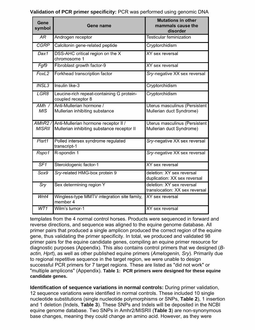
Validation of PCR primer specificity: PCR was performed using genomic DNA
templates from the 4 normal control horses. Products were sequenced in forward and reverse directions, and sequence was aligned to the equine genome database. All primer pairs that produced a single amplicon produced the correct region of the equine gene, thus validating the primer specificity. In total, we produced and validated 98 primer pairs for the equine candidate genes, compiling an equine primer resource for diagnostic purposes (Appendix). This also contains control primers that we designed (B-actin, Hprt), as well as other published equine primers (Amelogenin, Sry). Primarily due to regional repetitive sequence in the target region, we were unable to design successful PCR primers for 7 target regions. These are listed as "did not work" or "multiple amplicons" (Appendix). Table 1: PCR primers were designed for these equine candidate genes. Identification of sequence variations in normal controls: During primer validation, 12 sequence variations were identified in normal controls. These included 10 single nucleotide substitutions (single nucleotide polymorphisms or SNPs, Table 2), 1 insertion and 1 deletion (Indels, Table 3). These SNPs and Indels will be deposited in the NCBI equine genome database. Two SNPs in Amhr2/MISRII (Table 3) are non-synonymous base changes, meaning they could change an amino acid. However, as they were

identified in normal horses, it is unlikely that these are deleterious. Nevertheless, SNPs and Indels are important for researchers to recognize as normal genomic variations, and must be differentiated from causative mutations by further research, such as screening controls within different breeds and performing gene function studies. The SNPs will also be valuable as markers for equine GWAS.

Table 2: SNP genotypes identified in control horses.
*Nonsynonymous changes affect the amino acid code. Table 3: Insertion and deletion (indel) genotypes identified in control horses.
*Frameshift means that the insertion will affect the amino acid code.

Specific Aim 2: Using the panel of primers developed in Aim 1, we will screen a set of horses affected with Sry-negative XX sex reversal to test our hypothesis that mutations in those genes could cause the equine disorder, identify candidate causative mutations, and illustrate the utility of the panel. Confirmation of Sry-negative status of horses affected with XXSR: Our lab had blood samples from 6 XX sex-reversed horses donated from practicing veterinarians and their owners. With the exception of one Pasa Fino horse (Meyers-Wallen et al, 1997), there is no published record of their Sry status (presence or absence of Sry gene). Therefore, PCR for equine Sry (GenBank Acc. # AB004572) was performed using genomic DNA templates from affected and control horses. All 6 affected and 2 females were Sry-negative; the 2 males were Sry-positive (Figure 1, Table 4).
Figure 1: PCR of equine genomic DNA templates with (top) equine primers SRYP2-577 and SRYP2-1171 (618bp product), or (bottom) positive control primers for an X-linked gene (Hprt, 418bp product). Lanes: M: 100bp DNA ladder, 1&2: normal females (PM2045, PM2076), 3-8: XX sex reversed horses (PM2036, PM2050, PM4098, PM4217, PM4350, PM4563), 9&10: normal males (PM2044, PM2077), 11: water template control. All affected and female
horses were Sry-negative; all males were Sry-positive. PCR products were generated from all templates with the X-linked positive control primers (Hprt). As expected, Hprt amplicon density is similar for affected and female horses, which have two X chromosomes (64,XX), and greater density than that of males, which have one X chromosome (64,XY). Table 4: Horses affected with Sry-negative XX sex reversal in which candidate genes were screened by PCR primers produced in this project

Screening 10 candidate genes in Sry-negative XXSR horses: Exons and 5’ UTR of candidate genes were PCR amplified from genomic DNA of 6 Sry-negative XX sex reversed horses and 4 normal controls (Figure 1). A total of 27 sequence variations, including 22 SNPs (Table 5) and 5 indels (Table 6) were identified. The majority of these were distributed among both normal and affected horses or were present in some, but not all, affected horses. Therefore, none of these variations is likely to be the only cause of this disorder in all horse breeds.
Notably, 5 novel single base pair variations in Rspo1 (Rspondin-1) were identified in affected horses, but none was the same as mutations reported in humans with Sry-negative XXSR. Two of these were in the coding region (c390T>C at chr2:20,023,842 and c412G>A at chr2:20,023,864 in Rspo1 Exon 3,Table 5). A third was identified in the UTR (at chr2:20,011,124, Table 5). However, each of these novel single base pair substitutions is likely to be a SNP, and not a causative mutation: The CT genotype at chr2:20,023,842 was also observed in control horses. The novel alleles at chr2:20,023,864 and chr2:20,011,124 were heterozygous in one affected Pasa Fino horse (PM2036), but the other affected Pasa Fino (PM4098) had the same genotype as normal controls at this locus.
Five single base pair variations in other genes (heterozygous genotypes) were found only in affected horses. Most were in genes that have not been associated with Sry-negative XXSR in other mammals (Dax 1 exon 1, Fgf9 intron 1). For Wnt4 exon 4, the nonsynonymous substitution was present in only one affected horse (Table 5). Therefore, it is most likely that these are normal variations in the horse genome (i.e. SNPs). Further research, such as screening more affected horses of these breeds, is necessary to confirm whether they consistently segregate with any disease phenotype.

Table 5: Sequence variations in candidate genes identified in Sry-negative XXSR horses and controls*
*Shaded genotypes indicate variations from the equine genome sequence. Nonsynonymous changes affect the amino acid code, but synonymous changes do not.
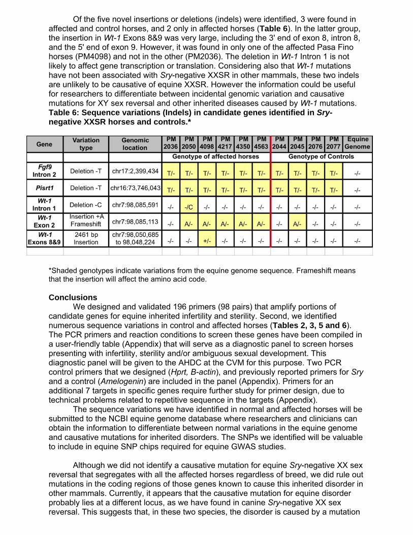
Of the five novel insertions or deletions (indels) were identified, 3 were found in affected and control horses, and 2 only in affected horses (Table 6). In the latter group, the insertion in Wt-1 Exons 8&9 was very large, including the 3' end of exon 8, intron 8, and the 5' end of exon 9. However, it was found in only one of the affected Pasa Fino horses (PM4098) and not in the other (PM2036). The deletion in Wt-1 Intron 1 is not likely to affect gene transcription or translation. Considering also that Wt-1 mutations have not been associated with Sry-negative XXSR in other mammals, these two indels are unlikely to be causative of equine XXSR. However the information could be useful for researchers to differentiate between incidental genomic variation and causative mutations for XY sex reversal and other inherited diseases caused by Wt-1 mutations. Table 6: Sequence variations (Indels) in candidate genes identified in Sry-negative XXSR horses and controls.*
*Shaded genotypes indicate variations from the equine genome sequence. Frameshift means that the insertion will affect the amino acid code. Conclusions We designed and validated 196 primers (98 pairs) that amplify portions of candidate genes for equine inherited infertility and sterility. Second, we identified numerous sequence variations in control and affected horses (Tables 2, 3, 5 and 6). The PCR primers and reaction conditions to screen these genes have been compiled in a user-friendly table (Appendix) that will serve as a diagnostic panel to screen horses presenting with infertility, sterility and/or ambiguous sexual development. This diagnostic panel will be given to the AHDC at the CVM for this purpose. Two PCR control primers that we designed (Hprt, B-actin), and previously reported primers for Sry and a control (Amelogenin) are included in the panel (Appendix). Primers for an additional 7 targets in specific genes require further study for primer design, due to technical problems related to repetitive sequence in the targets (Appendix). The sequence variations we have identified in normal and affected horses will be submitted to the NCBI equine genome database where researchers and clinicians can obtain the information to differentiate between normal variations in the equine genome and causative mutations for inherited disorders. The SNPs we identified will be valuable to include in equine SNP chips required for equine GWAS studies. Although we did not identify a causative mutation for equine Sry-negative XX sex reversal that segregates with all the affected horses regardless of breed, we did rule out mutations in the coding regions of those genes known to cause this inherited disorder in other mammals. Currently, it appears that the causative mutation for equine disorder probably lies at a different locus, as we have found in canine Sry-negative XX sex reversal. This suggests that, in these two species, the disorder is caused by a mutation

in a novel gene, or perhaps a mutation in the regulatory region of a known gene. The latter mutation type can be difficult to find, requiring many SNPs and several affected and control animals for GWAS, or linkage analyses in large pedigrees. Acquisition of carefully phenotyped horses from several breeds and pedigree lines for such studies could be achieved by accumulating cases that are screened with our panel at the AHDC and similar laboratories in other states. Publication of results The results obtained in this project are being compiled in a manuscript, which will be submitted to the journal Reproduction in Domestic Animals. References Bugno M, Klukowska J, Słota E, Tischner M, Switoński M. 2003. A sporadic case of the sex-reversed mare (64,XY; SRY-negative): molecular and cytogenetic studies of the Y chromosome. Theriogenology. 2003 59:1597-1603. Meyers-Wallen VN, Hurtgen J, Schlafer D, Tulleners E, Cleland WR, Ruth GR, Acland GM. 1997. Sry-negative XX true hermaphroditism in a Pasa Fino horse. Equine Vet J 29: 404–408. Hasegawa T, Sato F, Ishida N, Fukushima Y, Mukoyama H. 2000. Sex determination by simultaneous amplification of equine SRY and amelogenin genes. J Vet Med Sci. 62:1109-1110.

Harry M. Zweig Memorial Fund for Equine Research
2010 Annual Report
P.I.: Dr. Alan Nixon
Title: Targeted Delivery of Stem Cells for Pro-Inflammatory Cytokine Suppression in Arthritic Joints
Project Period: Reporting Period:
1/1/10-12/31/11 1/1/10-12/31/10

2011 HARRY M. ZWEIG MEMORIAL FUND FOR EQUINE RESEARCH PROGRAM
CONTINUATION APPLICATION
PROJECT TITLE: Targeted Delivery of Stem Cells for Pro-inflammatory Cytokine Suppression in Arthritic Joints PRINCIPAL INVESTIGATOR(S): Alan Nixon Progress Report -- Sections A-D (Not to exceed 2 pages) A. Specific Aims Aim 1. Examine cultured stem cell adherence in naturally arthritic cartilage using injected cells bearing fluorescent nanoparticles.
Aim 2. Develop, characterize, and evaluate a transplantable stem cell line pre-programmed using transposon-transposase (Sleeping Beauty) based integration of specific Sox transcription factors and TGF-β1, -β2 and -β3 genes.
Aim 3. Develop and evaluate cytokine “resistance” in Sox-TGF pre-programmed stem cells through integrated cytokine RNAi silencing motifs.
Aim 4. Evaluate the long-term improvement in osteoarthritis after direct injection of Sox-TGF-β transduced MSCs intrinsically expressing IL-1 and TNF-α RNA-silencing motifs. Review of the grant found particular concern with Aim 1, which was an essential study to answer the delivery question for the remaining aims. Given this, additional osteoarthritic and normal horses were analyzed using animals included in two other studies (Histogenics and Genzyme trials). Otherwise progress has been extensive on Aim 2, and modest on Aim 3.
B. Studies & Results Aim 1. Examine cultured stem cell adherence in naturally arthritic cartilage using injected cells bearing fluorescent nanoparticles. Analysis of 3 normal and 4 OA joints has shown fluorescently tagged cells were predominantly localized in the synovial membrane 72 hours after injection into OA joints (Fig 1). No cells adhered to normal cartilage, and small numbers of cells adhered to fibrillated cartilage.
Fig 1. (Left) Synovial membrane harvested from a stifle joint 72 hrs after injection showing 625nm labeled MSCs populating the synovial villus. (Right) QDot labeled MSC (arrow) adhering to cartilage surface. Only 1 case of the 4 OA joints assessed showed significant MSC incorporation to the cartilage surface (Fig 2). Fixed tissue processing resulted in loss of labeled cells and frozen sectioning was clearly required to allow accurate verification of soft tissue cell distribution.
Aim 2. Develop, characterize, and evaluate a transplantable stem cell line pre-programmed using transposon-transposase (Sleeping Beauty) based integration of specific Sox transcription factors and TGF-β1, -β2 and -β3 genes. Methods Sox and TGF-β constructs. Full length coding regions of equine Sox 5,6 and 9, were cloned and sequenced, and subcloned into adenovirus and the Sleeping Beauty (SB) pT2HB- SB transposon plasmid upstream of a bicistronic expression/selection cassette derived from pIRESpuro3 (Clontech). Replication deficient, E1- and E3-, adenoviral (Ad) vectors containing an eGFP marker gene were used (AdEasy). Similarly, TGF-β1, β2, and β3 were subcloned into adenovirus and SB vectors.
Fig 2. (Left) Gross appearance of metacarpophalangeal joint cartilage with mild osteoarthritis. (Mid) Fibrillated cartilage shows extensive numbers of fluorescent nanosphere labeled MSCs within the fibrillated cartilage. (Right) The majority of injected MSCs have populated the synovial membrane villi

Cell Transduction Cultured HeLA and 293 cell lines and equine MSCs were transduced with Ad-Sox or AdTGF to evaluate transgene expression and induction of chondrogenesis in MSC monolayer, pellet cultures, and transwell 3D culture inserts. For longer term study of integrating gene effects, similar cell types were electroporated with an equimolar mix of SB-Sox or SB-TGF and the SB transposase-carrying pCMV-SB plasmid. For SB transduced cells after 10 days post selection (puromycin), genomic integration was confirmed by PCR of genomic DNA for the SB-Sox or TGF construct. Expression was assessed in SB-Sox and wild-type cells. Examination of transwell inserts and pellet cultures was done at 21 days. Gene Expression Analysis. RNA was isolated from monolayers at 12 days and from pellet and transwell cultures at 14 or 21 days. Sox TFs and TGF-B gene expression were assessed by qPCR depending on transgene construct, and markers of chondrogenesis including collagen type II and type X, aggrecan and link gene expression was determined by qPCR. Histology. MSCs from 3D culture at 2 and 3 wks were sectioned for immunofluorescence studies of collagen type II. Results Ad-Sox and AdTGFB3 transduced 293 and MSCs at >95% efficiency. Combination of AdSox5+9, Sox6+9, Sox
5+6, Sox5/6/9 and Sox 5/6/9+TGF-B3 all gave high GFP efficiencies, without sign of toxicity. Culture of MSCs in 3D transwell inserts indicated persisting high transduction efficiency (Fig 3). GFP expression slowly declined over 14 days in all cultures.
Fig 3. Ad infected MSC on day 2 after infection and layering in high density transwell insert suspension culture. Efficiency of GFP marker gene expression shows high cell transduction with Sox and TGF-B3 genes.
Combination of Sox and TGFB genes in 14 day pellet culture showed induction of collagen type II, with the most profound formation of type II in Sox5/6/9+TGFB3 3D cultures (Fig 4). SB transduced cells and longer term cultures are being further characterized by qPCR.
Fig 4. Immuno of Ad infected MSC on day 14 after infection and formation of pellet cultures. Sections show collagen type II (green) predominates in TGFB3 and Sox 5/6/9+TGFB3 pellets, and Collagen X formation (Blue) or combination of Type II and X (purple) in Sox containing pellets. Cell nuclei are PI labeled (red).
Based on work so far, Sox TFs induced significant collagen type II and type X formation. Addition of TGF-B3 further increased deposition of Collagen type II and ultimately may be the ideal combination for MSC chondrogenesis. However, the preponderance of collagen type II in AdTGFB3 infected cultures, with little collagen type X, also suggests Sox TFs may not be appropriate for MSC pre-implantation articular cartilage treatment, where type X is not appropriate.
Studies of gene expression by microarray should characterize this better. C. Significance These data indicate significant progress toward forming chondrocytes from pluripotent stem cells. However, Sox transcription factors may be better confined to a brief period of expression, followed by longer (i.e. with integrating vectors) TGF-B3 conditioning of MSCs. D. Plans Specific aim 3 should be completed by years end. Some delay in completing SB assessment for delivery has developed due to a delay in securing an enhanced (codon optimized) transposase known as SB100. We started an MTA in June 2010, and are still to receive the plasmid. However, since this is only the plasmid coding the transposase, experiments using all the existing SB-Sox and SB-TGF transposons can follow, including adding IL-1 silencing. The in vivo study (Aim 4) is expected to start Jan 1, 2011.

Harry M. Zweig Memorial Fund for Equine Research
2010 Final Report
P.I.: Dr. Bettina Wagner, Harry M. Zweig Assistant Professor
Title: Analysis of the Innate Immune Response to EHV-I Infection
Project Period: Reporting Period:
1/1/09-12/31/10 1/1/10-12/31/10

2010 HARRY M. ZWEIG MEMORIAL FUND FINAL REPORT
PROJECT TITLE: Analysis of the Innate Immune Response to EHV-1 Infection
PRINCIPAL INVESTIGATOR(S): Dr. Bettina Wagner
The application preceding this proposal is entitled ‘Analysis of the innate immune response to EHV-1 infection’
(01/01/09 – 12/31/10). The major goal of the project was to investigate the innate immune response to EHV-1
in horses and to identify differences in responses to abortogenic and neurogenic EHV-1 strains.
Publications and presentations resulting from this project:
Wimer CL, Damiani A, Osterrieder N, Wagner B. Equine herpesvirus type-1 modulates CCL2, CCL3, CCL5,
CXCL9, and CXCL10 chemokine expression. Vet. Res., submitted.
Wagner B, Wimer C, Freer H, Erb HN. 2010. Infection of PBMC with neurogenic equine herpesvirus type 1
Ab4 strain induces interferon-alpha and down-regulates IL-10 production. Oral presentation, 9th
International Veterinary Immunology Symposium, August 16th
-20th
2010, Tokyo, Japan.
In addition, two abstracts on (1) chemokine expression and (2) type I interferon and IL-10 production were
submitted to the CRWAD meeting held in December in Chicago. Another manuscript on interferon and IL-
10 production after infections with different EHV-1 strains is in preparation.
Aims and results/currently funded application:
Aim 1 (as actually funded): We will determine whether EHV-1 induces IFN and chemokines by directly
activating TLR2 and TLR9. Equine TLR2 and TLR9 were cloned and transfected into an equine fibroblast cell
line. Stimulation by EHV-1 will be tested using NF-B and IFN reporter assays established in Dr. Leifer’s lab.
We will also determine whether neurogenic EHV-1 strains differ from non-neurogenic EHV-1 strains in their
potential to stimulate the innate immune response through TLRs.
Results: The equine TLR2 and TLR9 genes were amplified from PBMC, cloned into a mammalian expression
vector and used to transfect equine fibroblast cells (NBL-6) and also CHO cells. While NBL-6 cells expressed
the TLR proteins very weakly, both proteins could be expressed successfully in CHO cells. During the
remaining time of the project, CHO cells transfected with the TLR constructs will be infected with different
EHV-1 strains (Ab4, RacL11 and NY03) and NF-B and IFN reporter assays will be performed in Dr. Leifer’s
group to determine the TLR-dependent signaling and stimulation by the different EHV-1 strains.
Aim 2 (as actually funded): We will identify the major chemokines that are produced in response to EHV-1
infection after TLR stimulation. Blood cells lacking expression of T, B and monocyte cells will be enriched
from equine peripheral blood. These cells typically represent dendritic cells that are high producers of IFN and
play an important role in host-defense against viral infection. The cells will be infected with different EHV-1
strains and assayed by PCR for chemokine gene expression. Supernatents will be tested for cytokine and
chemokine production in Aim 3.
Results: The experiment was completed. We infected PBMC from 10 horses with different EHV-1 strains (Ab4,
RacL11 and NY03). The cells were collected for RNA isolation to perform the PCR for different chemokines
(CCL2, CCL3, CCL5, CXCL9, CXCL10). Supernatants were obtained for cytokine detection (IFN-, IL-10,
IL-4) and measured by a multiplex assay [32]. Enrichment of cells was not required.
The quantitative PCR analysis showed that EHV-1 down-regulated CCL2 (Fig. 2A) and CCL3 expression (Fig.
2B). EHV-1 infection increased expression of CCL5 (not shown), CXCL9 (Fig.2C) and CXCL10 (Fig. 2D)

compared to non-infected PBMC. The comparison of the three EHV-1 strains showed that RacL11 induced the
smallest changes compared to non-infected cells, while Ab4 almost always induced the largest changes (Fig. 2).
The induction of IFN-, IL-10 and IL-4 was measured after infection of PBMC with the three EHV-1 strains.
While IFN- induction increased with the viral dose and similarly for all EHV-1 strains (Fig. 3B), the
production of anti-inflammatory IL-10 was decreased after Ab4 infection compared to RacL11 and NY03
strains at medium to high viral infection doses (Fig. 3A). This suggests that the host’s potential to produce anti-
viral IFN- is not affected by EHV-1. It also suggests that EHV-1 developed a mechanism that interferes with
the host’s ability to down-regulate inflammation. Because the latter effect is significantly stronger after
infection with Ab4, the mechanism might contribute to the ability of Ab4 to induce neurological disease. A B
In summary, the comparison of the RacL11, NY03 and Ab4 strains showed that the potential to regulate
chemokine gene expression and IL-10 production is higher in neurogenic than in abortogenic EHV-1 strains.
The ability of Ab4 to down-regulate chemokines and IL-10 more effectively is likely to contribute to the
increased pathogenicity of the Ab4 strain and its ability to induce severe clinical signs and the neurological
manifestation of the disease.
Aim 3 (as actually funded): We will generate monoclonal antibodies to two of the most important chemokines
in the TLR-mediated innate immune response to EHV-1 infection. Dr. Wagner’s lab will use an established
expression system to produce these monoclonal antibodies.
Results: We developed and characterized monoclonal antibodies to CCL3. Antibodies to CCL2 were developed
by Dr. Wagner’s USDA project. The antibodies are currently used to measure CCL2 and CCL3 secretion after
EHV-1 infection. The fusion using CXCL9 is still ongoing and will be finished by the end of this year.
Figure 2: Real-time PCR amplification of different chemokine genes after infection of equine PBMC with EHV-1 strains RacL11, NY03 or Ab4. The gene expression was compared to non-infected PBMC (straight lines at 1). The cells were incubated for 6 hours (no differences to the control/ data not shown), 10 hours (C and D) and 24 hours (A-D) with or without the EHV-1 strains. * P<0.005; ** P<0.01; *** P<0.001; bars and * indicate comparisons between EHV-1 strains; * only indicate comparisons of the respective strain to non-infected cells.
Figure 3: IL-10 (A) and IFN- (B) secretion of equine PBMC after infection with EHV-1 strains RacL11, NY03 or Ab4. IL-10 production was compared from PBMC of adult mares (n=7), pregnant mares (n=9), and mares and foals at day 5 after birth (n=10). The cells were incubated for 48 hours. Then, the supernatants were obtained and cytokines were measured in an equine cytokine multiplex assay. IL-4 was not detected in the samples. * P<0.005; ** P<0.01; *** P<0.001; green * indicate comparisons between RacL11 and Ab4; blue * show NY03 versus Ab4 comparisons. Note
the 10-fold reduced y-axis for foals and IL-10 but not for IFN-.
APPENDIX B
Final/Interim Reports Resulting from 2009 Funding
Harry M. Zweig Memorial Fund for Equine Research
2010 Interim Report
P.I.: Dr. Dorothy Ainsworth
Title: Deciphering the Mechanism of Equine Inflammatory Airway Disease in vitro
Project Period: 1/1/09-6/30/11 Reporting Period: 1/1/10-12/31/10 Dr. Ainsworth was granted a no cost extension through June 30, 2011. A Final report will be submitted next year.
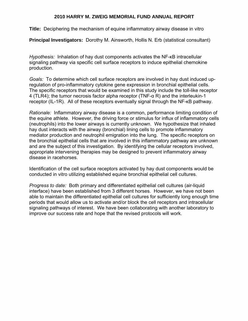
2010 HARRY M. ZWEIG MEMORIAL FUND ANNUAL REPORT
Title: Deciphering the mechanism of equine inflammatory airway disease in vitro Principal Investigators: Dorothy M. Ainsworth, Hollis N. Erb (statistical consultant)
Hypothesis: Inhalation of hay dust components activates the NF-κB intracellular signaling pathway via specific cell surface receptors to induce epithelial chemokine production. Goals: To determine which cell surface receptors are involved in hay dust induced up-regulation of pro-inflammatory cytokine gene expression in bronchial epithelial cells. The specific receptors that would be examined in this study include the toll-like receptor 4 (TLR4); the tumor necrosis factor alpha receptor (TNF-α R) and the interleukin-1 receptor (IL-1R). All of these receptors eventually signal through the NF-κB pathway. Rationale: Inflammatory airway disease is a common, performance limiting condition of the equine athlete. However, the driving force or stimulus for influx of inflammatory cells (neutrophils) into the lower airways is currently unknown. We hypothesize that inhaled hay dust interacts with the airway (bronchial) lining cells to promote inflammatory mediator production and neutrophil emigration into the lung. The specific receptors on the bronchial epithelial cells that are involved in this inflammatory pathway are unknown and are the subject of this investigation. By identifying the cellular receptors involved, appropriate intervening therapies may be designed to prevent inflammatory airway disease in racehorses. Identification of the cell surface receptors activated by hay dust components would be conducted in vitro utilizing established equine bronchial epithelial cell cultures. Progress to date: Both primary and differentiated epithelial cell cultures (air-liquid interface) have been established from 3 different horses. However, we have not been able to maintain the differentiated epithelial cell cultures for sufficiently long enough time periods that would allow us to activate and/or block the cell receptors and intracellular signaling pathways of interest. We have been collaborating with another laboratory to improve our success rate and hope that the revised protocols will work.
Harry M. Zweig Memorial Fund for Equine Research
2010 Final Report
P.I.: Dr. Norm Ducharme
Title: Factors Affecting Airway Stability of Horses
At Exercise: A Combined Neuroanatomical Clinical and Engineering Methodology
Project Period: 1/1/08-06/30/10 Reporting Period: 1/1/10-6/30/10 Dr. Ducharme was granted a no cost extension through June 30, 2010. A final report is provided.
2009 HARRY M. ZWEIG MEMORIAL FUND FINAL REPORT PROJECT TITLE: Factors Affecting Airway Stability of Horses at Exercise: A Combined Neuroanatomical, Clinical and Engineering Methodology PRINCIPAL INVESTIGATOR: Norm G Ducharme and Jeremy Rawlinson Summary: The overall goal of this proposal was to couple bioengineering methods with clinical research to improve our understanding and develop better treatments to correct instability of the upper airway produced by dorsal displacement of the soft palate (DDSP) and recurrent laryngeal neuropathy (RLN). The experiments we performed determined the effect of laryngohyoid position, soft palate stiffness, and laryngeal biomechanics on upper airway stability. We used a combined approach of in vivo, in vitro, and in silico (computational) methods to tackle of the complexity of these problems. Specifically, through in vivo and in vitro experiments, we identified the intrinsic and extrinsic tongue muscles controlling laryngohyoid conformation and nasopharyngeal stability. Using in vitro mechanical testing and with computational constitutive analysis, we calculated the biomechanical properties of the larynx and soft palate. Coupled with medical imaging reconstructions, these data allowed us to generate computer models of these soft tissue structures and investigate implant properties and implantation technique to improve airway stability. These models were also compared with in vitro experiments of palate and laryngeal function as a validation step. Specific Aims and Findings: Aim 1. Determine the effects of selected muscles (hyoglossus, genioglossus, styloglossus, and geniohyoideus) activity on laryngohyoid conformation and nasopharyngeal stability by in-vivo studies using imaging (radiography and CT) and a treadmill stress test to collect airway pressures EMG, and video endoscopic images.
Abstract from (Cheetham et al., J Appl Physiol. 2009) The equine upper airway is highly adapted to provide the extremely high oxygen demand associated with strenuous aerobic exercise in this species. The tongue musculature, innervated by the hypoglossal nerve, plays an important role in airway stability in humans who also have a highly adapted upper airway to allow speech. The role of the hypoglossal nerve in stabilizing the equine upper airway has not been established. Isolated tongues from eight mature horses were dissected to determine the distal anatomy and branching of the equine hypoglossal nerve. Using this information, a peripheral nerve location technique was used to perform bilateral block of the common trunk of the hypoglossal nerve in 10 horses. Each horse was subjected to two trials with bilateral hypoglossal nerve block and two control trials (unblocked). Upper airway stability at exercise was determined using videoendoscopy and measurement of tracheal and pharyngeal pressure. Three main nerve branches were identified, medial and lateral branches and a discrete branch that innervated the geniohyoid muscle alone. Bilateral hypoglossal block induced nasopharyngeal instability in 10/19 trials, and none of the control trials (0/18) resulted in instability (P<0.001). Mean treadmill speed (+/-SD) at the onset of instability was 10.8+/-2.5 m/s. Following its onset, nasopharyngeal instability persisted until the end of the treadmill test. This instability, induced by hypoglossal nerve block, produced an expiratory obstruction similar to that seen in a naturally occurring equine disease (dorsal

displacement of the soft palate, DDSP) with reduced inspiratory and expiratory pharyngeal pressure and increased expiratory tracheal pressure. These data suggest that stability of the equine upper airway at exercise may be mediated through the hypoglossal nerve. Naturally occurring DDSP in the horse shares a number of anatomic similarities with obstructive sleep apnea. Study of species with extreme respiratory adaptation, such as the horse, may provide insight into respiratory functioning in humans.
Also: Abstract (Zantingh et al., Vet Surg 2010) The etiology of Dorsal Displacement of the Soft Palate (DDSP) in horses is not completely understood. DDSP has been experimentally induced by 3 methods. Horses that had DDSP during exercise have been shown to have a more ventral basihyoid bone at rest and a more dorsal laryngeal position following laryngeal ‘tie forward’ surgery is associated with improved post-operative racing performance. An association has also been shown between swallowing and DDSP. The hypoglossal nerve plays an important role in swallowing and we hypothesized that local bilateral blockade of the hypoglossal nerve at the level of the ceratohyoid bones would change laryngohyoid position in horses at rest. Bilateral distal hypoglossal nerve blockade was performed on 10 healthy horses. Lateral radiographs of the larynx were taken before and 30 minutes after bilateral hypoglossal block, and a radiographic reference system was used to determine the position of the caudal aspect of the basihyoid bone, the thyrohyoid-thyroid articulation, and the ossification of the base of the thyroid cartilage. Hypoglossal block produced ventral movement of the larynx. The ossification of the thyroid cartilage was positioned 6.2mm (95% CI 0.7-11.7) more ventral following bilateral hypoglossal block (p=0.03). It is possible that mild neurologic dysfunction of the hypoglossal nerve or muscular dysfunction of the muscles innervated by this nerve predispose a horse to DDSP due to a more ventral larynx position.
Aim 2. Determine cricoarytenoid joint movement using MRI studies and determining the biomechanical properties of the laryngeal cartilages and soft palate by testing fresh laryngeal cadavers.
In the first part of this aim, we used reconstruction software to segment the cartilage tissue in medical images from a magnetic resonance imaging (MRI) dataset. The MRI data were obtained in an in vitro experiment using prosthetic laryngoplasty where the arytenoid was sequentially placed in greater abduction between imaging sessions. Thus, we produced several models to relate the full 3D position of the arytenoid with respect to the cricoid during abduction under increasing suture forces. This serves as the basis for selection of sites for the biomechanical property of the laryngeal cartilages. We completed the studies to characterize the biomechanical properties or equine airway cartilages as described below:
Abstract (Passman et al.,Eq Vet J 2011) Upper airway obstruction is a common problem in the equine athlete as the soft-tissues of the larynx collapse into the airway, yet there is a paucity of biomechanical properties for the structural cartilage components. The purpose of this study was to measure the geometry and compressive mechanical properties of the hyaline cartilage to better understand laryngeal function and morphology. A total of eleven larynges were harvested from Thoroughbred and Standardbred racehorses. During gross dissection, linear dimensions of the cricoid were obtained. From both the cricoid and arytenoid, specimens were cored to obtain 6 mm disc samples from three sites within the dorsal cricoid (caudal, middle, and
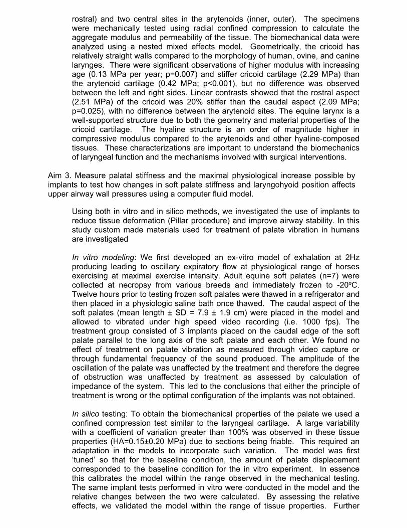
rostral) and two central sites in the arytenoids (inner, outer). The specimens were mechanically tested using radial confined compression to calculate the aggregate modulus and permeability of the tissue. The biomechanical data were analyzed using a nested mixed effects model. Geometrically, the cricoid has relatively straight walls compared to the morphology of human, ovine, and canine larynges. There were significant observations of higher modulus with increasing age (0.13 MPa per year; p=0.007) and stiffer cricoid cartilage (2.29 MPa) than the arytenoid cartilage (0.42 MPa; p<0.001), but no difference was observed between the left and right sides. Linear contrasts showed that the rostral aspect (2.51 MPa) of the cricoid was 20% stiffer than the caudal aspect (2.09 MPa; p=0.025), with no difference between the arytenoid sites. The equine larynx is a well-supported structure due to both the geometry and material properties of the cricoid cartilage. The hyaline structure is an order of magnitude higher in compressive modulus compared to the arytenoids and other hyaline-composed tissues. These characterizations are important to understand the biomechanics of laryngeal function and the mechanisms involved with surgical interventions.
Aim 3. Measure palatal stiffness and the maximal physiological increase possible by implants to test how changes in soft palate stiffness and laryngohyoid position affects upper airway wall pressures using a computer fluid model.
Using both in vitro and in silico methods, we investigated the use of implants to reduce tissue deformation (Pillar procedure) and improve airway stability. In this study custom made materials used for treatment of palate vibration in humans are investigated In vitro modeling: We first developed an ex-vitro model of exhalation at 2Hz producing leading to oscillary expiratory flow at physiological range of horses exercising at maximal exercise intensity. Adult equine soft palates (n=7) were collected at necropsy from various breeds and immediately frozen to -20ºC. Twelve hours prior to testing frozen soft palates were thawed in a refrigerator and then placed in a physiologic saline bath once thawed. The caudal aspect of the soft palates (mean length ± SD = 7.9 ± 1.9 cm) were placed in the model and allowed to vibrated under high speed video recording (i.e. 1000 fps). The treatment group consisted of 3 implants placed on the caudal edge of the soft palate parallel to the long axis of the soft palate and each other. We found no effect of treatment on palate vibration as measured through video capture or through fundamental frequency of the sound produced. The amplitude of the oscillation of the palate was unaffected by the treatment and therefore the degree of obstruction was unaffected by treatment as assessed by calculation of impedance of the system. This led to the conclusions that either the principle of treatment is wrong or the optimal configuration of the implants was not obtained. In silico testing: To obtain the biomechanical properties of the palate we used a confined compression test similar to the laryngeal cartilage. A large variability with a coefficient of variation greater than 100% was observed in these tissue properties (HA=0.15±0.20 MPa) due to sections being friable. This required an adaptation in the models to incorporate such variation. The model was first ‘tuned’ so that for the baseline condition, the amount of palate displacement corresponded to the baseline condition for the in vitro experiment. In essence this calibrates the model within the range observed in the mechanical testing. The same implant tests performed in vitro were conducted in the model and the relative changes between the two were calculated. By assessing the relative effects, we validated the model within the range of tissue properties. Further

variations to the implant technique were incorporated in the model to rapidly assess new positions and implant dimensions. We determined that while rostral-caudal stiffeners implanted at the midline do reduce palate displacement, the relative change is modest and may not be clinically significant. The models indicated that rotating the implants in more lateral positions (30-45-90 degrees to the midline) had a greater reduction in palate displacement due to the bowed shape of the palate during DDSP.
Aim 4. Determine the ideal implant for stabilizing the larynx using biomechanical modeling to quantify the direction and magnitude of the forces acting on the muscular process.
In this aim, we developed a 3D model of arytenoid and cricoid deformation based upon the tissue properties calculated and the images reconstructed in Aim 2. One limitation that arose in developing these model concerns the cricoarytenoid joint – the saddle shaped surface that guides the motion of the arytenoid with respect to the cricoid. This obstacle arose due to the resolution of the MR images. From our biomechanical assessment, the elliptical contours of the joint surface were approximately 15 x 8 mm and oblique to the plane of the MR images. This geometry means that with the MRI spacing at 3mm, only 1-2 slices of MR data were obtained in the region of the joint. This was not adequate for properly reconstructing the joint surface and, therefore, not adequate for modeling the relative motion of the arytenoid with respect to the cricoid. Mechanically, the load path we were analyzing includes the pressure acting to deform the arytenoid. This generates a force on the muscular process, induces motion at the cricoarytenoid joint, stretches the prosthetic suture, and compresses the caudal cricoid. In this load path, the deformation at the muscular process was a limiting step; its tissue structure varies from both the arytenoid and cricoid with possibly higher ratio of elastic and fibrocartilage compared to the hyaline cartilage of the other two. Thus, the deformation characteristics are potentially very different. We are addressing this with additional mechanical testing to identify the stiffness of the tissue in the region of the muscular process. Thus, the model requires some ongoing additions to ensure physiological validity before we can assess the relative deformations along this chain of support.
Significance: There were several significant findings as well as areas of improvement in our experimental approaches. Understanding the neuroanatomical basis has generated new hypotheses for treatment - stability of the equine upper airway at exercise may be mediated through the hypoglossal nerve. This led to a new model of the disease and we are now investigating whether a new treatment targeting the action of the hypoglossal nerve can be designed using neuroprosthesis. Quantifying the mechanical properties of the laryngeal tissue has identified the regions of concern in the cartilaginous support and provided new thoughts for research in tissue engineering. These elements have led to the first reports of properties for these tissues in horses; this was the basis of this year’s new grant proposal to Zweig. Lastly, we have quantified the effects of different surgical techniques on airway stability to improve surgical correction of the soft palate. Although the technique used in humans does not appear valuable to horses, the modeling suggests the length and exact position of similar devices might be useful in horses to reduce airway obstruction or noise. Conference Presentations: Passman SN, Cheetham J, Bonassar LJ, Ducharme NG, Rawlinson JJ. Biomechanical Stiffness Of Equine Laryngeal Cartilage. 2009 Symposium of the American College of Veterinary Surgeons, Washington, D.C. (Oct 2009).

Passman SN, Cheetham J, Bonassar LJ, Ducharme NG, Rawlinson JJ. Biomechanical Stiffness of Equine Laryngeal Hyaline Cartilage. 2009 Annual Meeting of the Biomedical Engineering Society, Pittsburgh, PA (Oct 2009). Passman SN, Cheetham J, Bonassar LJ, Ducharme NG, Rawlinson JJ. Biomechanical Characterization of Equine Laryngeal Cartilage. Eq Vet J in press expected publication time (April 2011) Cheetham J. Neuroanatomy of the Equine Upper Airway. Invited presentation to the British Equine Veterinary Surgeons Meeting. 9/2009 Cheetham J. Neuroanatomy of the Extrinsic Laryngeal Structures. Invited presentation to the American College of Veterinary Surgeons Meeting. 10/2008. Cheetham J. Nasopharyngeal Stability: What do we know? Invited presentation at the World Equine Veterinary Association Congress. Moscow, Russia. January 2008 Ducharme NG. Treatment of DDSP. World Equine Airway symposium, Bern Switzerland August 2009. Ducharme NG. Update on the treatment of DDSP. World Equine Veterinary Congress, Guaruja, Brazil September 2009. Zantingh A.J., Ducharme N.G., Mitchell L.M., Cheetham J. Effect Of Bilateral Hypoglossal Nerve Blockade at the Level of the Ceratohyoid Bones on Radiographic Laryngeal Position in the resting Horse. American College of Veterinary Surgeons Meeting. Seattle. 10/2010. Publications: Cheetham J, Pigott JH, Hermanson JW, Campoy L, Soderholm LV, Thorson LM, Ducharme NG. Role of the hypoglossal nerve in equine nasopharyngeal stability. J Appl Physiol. 2009 Aug;107(2):471-7. Pigott JH, Cheetham J, Mitchell L.M., Soderholm LV, Ducharme NG. Incidence of Swallowing during Exercise in Horses with Dorsal Displacement of the Soft Palate. Equine Vet J. 2010 Nov;42(8):732-7. Passman SN, Cheetham J, Bonassar LJ, Ducharme NG, Rawlinson JJ. Biomechanical Stiffness of Equine Laryngeal Hyaline Cartilage. Equine Veterinary Journal EVJ 2011, in press. Listing of grant applications and their status resulting from Zweig funding: Approved: Med-el corporation- “Laryngeal pacemaker in horses”. NG Ducharme. Co- investigators: Cheetham JC, Rawlinson J and Hermanson J. Zweig 2010 submission – “Tissue Engineered Cartilage in the Equine Airway” Norm Ducharme and Jeremy Rawlinson. Co-Investigators: Lawrence Bonassar, Jon Cheetham, John Hermanson

Harry M. Zweig Memorial Fund for Equine Research
2010 Interim Report
P.I.: Dr. Gillian Perkins
Title: Immunization Against Strangles Using a Vectored Equine Herpesvirus Vaccine
Project Period: Reporting Period:
1/1/09-06/30/11 1/1/10-12/31/10
Dr. Perkins was awarded a no cost extension through June 30, 2011. An interim report is provided.

HARRY M. ZWEIG MEMORIAL FUND FOR EQUINE RESEARCH PROGRAM INTERIM REPORT
Title: Immunization against strangles using a vectored equine herpesvirus vaccine Investigators: Gillian Perkins and Nikolaus Osterrieder
The overall goal of this work is to 1) gain an understanding of what is required for an efficacious, long-lasting immune response to S. equi and to 2) establish equine herpesvirus type 1 (EHV-1) as a platform to deliver immunogens from S. equi to horses to avoid the many complications of S. equi-induced disease. Our hypothesis is that a modified live EHV-1 vaccine derived from a non-pathogenic EHV-1 strain (RacH) can safely and efficiently deliver S. equi antigens and induce a protective immune response against both S. equi and EHV-1 infection. Specific aim 1: To engineer EHV-1 strain RacH recombinants expressing three S. equi antigens (SeM, Se18.9 and IdeE). Specific aim 2: To test the generated EHV-1 virus expressing the S. equi proteins in mice and horses. PROGRESS OF THE PROJECT – 1st YEAR Specific aim 1: Construction of EHV-1 strain RacH recombinants expressing S. equi antigens SeM, Se18.9 and IdeE
1. Description of the work The nucleotIdeE sequences of SeM and Se18.9 separated byan internal ribosomal binding site (named SeM-IRES-Se18.9) were synthesized de novo by a commercial supplier (GenScript). IdeE sequence was separately synthesized by the same company. The sequences were codon-optimized versions of the genes to enable efficient expression in mammalian cells.
Two shuttle plasmids were constructed by cloning separately the SeM-IRES-Se18.9 and IdeE sequences into pEP_CMV plasmid, resulting in the control of the genes by the human cytomegalovirus immediate early promoter and the bovine growth hormone as a transcriptional terminator sequence. The pEP_CMV plasmid also contains a unique I-SceI site and an aphAI gene conferring kanamycin resistance. These elements allow the cloning strategy into the RacH genome. Transfer constructs were generated by PCR and two-step Red recombination was used to insert the SeM-IRES-Se18.9 and IdeE genes into the infectious bacterial artificial chromosome (BAC) clone of the EHV-1 RacH (pRacH). The recombinants pRacH_SeM-IRES-Se18.9 and pRacH_IdeE DNAs were isolated and the structure of the BAC recombinants was verified by restriction enzyme and PCR analysis. Recombinant RacH_SeM-IRES-Se18.9 and RacH_IdeE viruses were achieved by transfection of the respective BAC plasmids into RK13 cells. Expression of the S. equi proteins by the recombinant viruses was verified by western blotting using antibodies raised in rabbits (against SeM and Se18.9 proteins - ceded by Dr Timoney) and mice (against IdeE protein). Expression of S. equi SeM, Se18.9 and IdeE proteins in Escherichia coli
1. Description of the work The SeM, Se18.9 and IdeE genes were PCR amplified and cloned separately into
an arabinose-inducible expression vector pBAD24. In order to facilitate purification from E. coli, a carboxy-terminal tag of six histidine residues (6-His) was added to each recombinant protein. BL21-Rosseta 2 E coli cells were transformed with the expression

plasmids. Protein expression was monitored by SDS-PAGE and Western Blotting using Penta-His antibodies. Bands of approximately 60 kDa, 40kDa and 19 kDa corresponding to SeM, Se18.9 and IdeE proteins respectively, were detected. Protein expression and further purification was optimized testing different growth temperatures, induction time and inducer (arabinose) concentration. Purified proteins are intended to be used in an ELISA system designed to recognize the anti-SeM, anti-Se18.9 and anti-IdeE antibodies in mice and horses experimentally infected with the recombinant viruses.
Antiserum against the purified IdeE protein was produced in mice to verify protein expression by the recombinant RacH_IdeE virus. Specific aim 2: Testing the generated EHV-1 virus expressing the S. equi proteins in mice and horses
1. Description of the work The recombinant EHV-1 RacH-based S. equi vaccines have been generated and optimized. The vaccines have been tested in a mouse model of S. equi infection. Briefly, young female Balb/c mice were vaccinated twice at three week intervals and then challenged with S. equi (1 x 10⁵ PFU intranasally). The vaccines used in the first experiment were: intranasal or subcutaneous EHV-1_SeM/Se18.9; intranasal or subcutaneous EHV-1_IdeE; intranasal or subcutaneous EHV-1_SeM/Se18.9 combined with EHV-1_IdeE; intranasal EHV-1 virus alone, and no vaccination. To detect differences between groups, mice were weighed daily for two weeks and nasal cultures for S. equi performed every other day for the first 6 days post-infection. At the end of two weeks any remaining mice were euthanized and histopathological analysis of the mandibular lymph nodes, lungs, tympanic bulla, bone marrow and brain were performed. When both vaccines were given together (groups 5 and 6) the mice loss less weight and had increased survival than either vaccine given alone. Interestingly, some protection was seen in mice that received the EHV-1 virus without any S. equi antigens suggesting some non-specific immune response generated from the EHV-1 vaccine alone. Mandibular lymph node abscessation was not observed in mice given the combination of EHV-1 and SeM/Se18.9 and IdeE. None of the mice vaccinated with the EHV-1 and S. equi recombinants developed pulmonary abscesses while 40% of those given the negative controls (EHV-1 vaccine alone or unvaccinated mice) did. Other features of the infection were tympanic bulla abscesses that sometimes extended into the meninges. A second study was undertaken to increase group size and investigate the protective role of EHV-1 vaccine alone in prevention of S. equi. The study had a similar design with vaccines given twice 3 weeks apart followed by challenge intranasally with S. equi 3 weeks after the last vaccination and similar testing post-infection to detect differences between groups. The groups included EHV-1_SeM/Se18.9 and EHV-1_IdeE combination vaccine given intranasal or subcutaneous, EHV-1 vaccine alone given intranasal or subcutaneous and negative control (minimum essential media) given intranasal or subcutaneous. This study was completed in March of 2011 and the data is being currently being analyzed. 2. Future actions Statistical analyses will be soon performed on the above described vaccine trial in the mouse model of strangles. The collected data will be presented in a scientific congress and a manuscript will be prepared for submission. Should the vaccines show promise in the mouse model, testing the vaccines in the host-species, the horse would be undertaken.

APPENDIX C
SUMMARY OF 2010 ALLOCATIONS
2010 Research Awards $443,705 2011 Public Relations and $26,500 Administrative Budget 2010 Incentive Awards $10,000 Total Expenditures: $480,205

APPENDIX D
AWARDS FOR 2011

2011 Harry M. Zweig Memorial Fund for Equine Research Awards
CONTINUATION AWARDS
ANNUAL AWARD
Lisa Fortier Optimization of Platelet Rich Plasma Components for the Treatment of Tendonitis
$57,213
Alan Nixon Targeted Delivery of Stem Cells for Pro-Inflammatory Cytokines Suppression in Arthritic Joints
$101,065
Sub-Total: $158,278 NEW
ANNUAL AWARD
Dorothy Ainsworth The Genetic Basis of Recurrent Laryngeal Neuropathy (RLN) in Thoroughbreds (2 Year Award)
$82,250
Jonathan Cheetham Diagnosis of Poor Performance in Racehorses (2 Year Award)
$43,914
Thomas Divers Targeting Platelets as a New Treatment Strategy for Endotoxemia (1 Year Award)
$30,000
Lisa Fortier Determining Anti-Nociceptive and Matrix Restorative Mechanisms of Platelet Rich Plasma in Osteoarthritis (1 Year Award)
$44,646
Bettina Wagner Innate Immune Mechanisms and
T-Cell Responses to Equine Herpesvirus Type 1 in Latently Infected and Naïve Horses” (2 Year Award)
$63,271
Sub-Total: $264,081 TOTAL: $422,359

APPENDIX E
POSTER SESSION PRESENTATIONS
November 18, 2010
Cornell University College of Veterinary Medicine

Corn
Injuries” Cheethamand Treaended wiIn the wicommitte
The Zweand avid amendmegarnered State HorDevelopmfurther th
The comRacing aCornell oState HorState Thoowners, tthe commsupport f
nell Univ
Poste
E
and “Epigenm of the Equatment of Upith her lecturine and cheeee members
eig Fund begsupporter ofent to a Newfrom the Sta
rse Breedingment Fund. The health and
mittee adminand Wageringor his designrse Breedingoroughbred Btrainers, or vmittee, reprefor research p
versity &
er Sessio
Equine rese
Faculty anequine-relaNovembercommitteeResearch aand to find
Speakers inJulia Feliprespective
netics: A Nouine Performpper Airway re, “A New Rse receptionand each oth
gan as a memf thoroughbr
w York Stateate’s tracks g DevelopmeThe Zweig Fd performanc
nistering theg Board or h
nee, a membeg DevelopmeBreeding an
veterinarianssenting the Cprojects adv
Harry MRe
on & Sem
archers pre
nd graduate sated researchr 18, 2010, ate administeriattended the d possible ca
ncluded assope from the lectures on “
ovel Approacmance Testin
Disease in HRoad to Disc
n that followeher.
morial gift frored horse rac pari-mutueland off-trackent Fund andFund gives bce of racing
e fund alwayhis designee,er or the exeent Fund, a m
nd Developms in equine pCollege and vancing equin
. Zweig esearchminar - N
esent to Zwe
student reseah projects int the Veterinng the Harryevent to see
andidates for
ociate profesDepartment“Platelet Ricch in Primarg Center fol
Horses.” Gracovery: Equed, students
om the familcing in New l law grantedk betting, thrd the New Yback to the inhorses.
ys includes th, the dean ofecutive direcmember or t
ment Fund, anractice. Deaits many resne health an
Memoria
Novembe
eig Fund co
archers presen a series of pnary Educatiy M. Zweig e the success r future supp
ssors Dr. List of Clinical ch Plasma Try Immunodelowed with h
aduate studenuine Research
and faculty
ly of Dr. HaYork State.
d the Fund twrough the Ag
York State Thndustry by su
he chairmanf the Collegetor of the Aghe executivend at least fi
an Michael Ksearchers whd athleticism
al Fund f
er 18, 20
mmittee
ented on the posters and lon Center. MMemorial Fuof currently
port.
sa Ann FortiSciences, wreatment of eficiencies.”his talk, “Eant Dr. Margah in the Postdiscussed th
arry M. ZweiThe Fund g
wo percent ogriculture anhoroughbredupporting re
n of the New e of Veterinagriculture ane director of ive New YorKotlikoff curho have recem.
for Equin
010
College's mlectures on Members of und for Equ
y funded proj
er and Dr. Mith their Sports Medi
” Dr. Jonathaarly Diagnosaret Brosnaht-Genome Erheir research
ig, a veterinagrew after anof all moneynd New Yorkd Breeding aesearch mean
York State ary Medicinend New Yorkf the New York State breerrently servesived the Fun
ne
many
the ine jects
Maria
icine an is
han ra.” with
arian n y k
and nt to
e at k ork eders, s on nd’s


Cornell University
and the
Harry M. Zweig
Memorial Fund for
Equine Research
2ND Annual Poster Session
Thursday, November 18, 2010
4:00p.m.– 5:30pm.
Atrium‐Veterinary Education Center
2
Welcome to the 2nd Annual Poster Session of the Harry M. Zweig Memorial Fund for Equine Research, highlighting equine research supported by the Fund.
The posters have been created by faculty, graduate students and residents, most of whom have been a recipient of an award from the Fund.
We would like you to enjoy looking at these posters and feel free to ask questions. We would also like to thank the presenters for taking the time to help us celebrate 31 years of support from the Harry M. Zweig Memorial Fund for Equine Research.
Thank you for your attendance.
Dr. Robert Gilmour, Associate Dean for Research and Graduate Education
Laura Mathews, Zweig Secretary

3
1. Dorothy Ainsworth
Autologous Blood Up-Regulates Chemokine Gene Expression in Equine
Bronchial Epithelial Cell Cultures
Authors: Cladia L. Reyner, Mary Beth Matychak, Dorothy Ainsworth
Department of Animal Science, Cornell University, Ithaca NY USA ,Department of Clinical Sciences, Cornell University, Ithaca NY USA.
Abstract: The etiology of inflammatory airway disease (IAD) in performance horses is likely multi-factorial with potential contributions by infectious agents as well as organic dust (hay), air pollutants or blood (EIPH). The objective of this study was to determine if chemokine expression in airway epithelial cells was up-regulated in response to exposure to blood or plasma and could thus contribute to the development of IAD.
Primary equine bronchial epithelial cell cultures (BECC) were established from 6 healthy horses that previously had been on pasture for 2 months. Five-day old BECC were incubated for 24 h with solutions of PBS, hay dust, LPS, autologous serum and autologous blood. The gene expression of IL-8, CXCL2, IL-1β and TNF-α were measured by kinetic PCR and normalized to GAPDH expression. Treatment effects were analyzed using a Kruskal-Wallis test with post-hoc comparisons performed using Wilcoxon signed rank tests. A Bonferroni correction was applied such that α = 0.05/3.
With the exception of TNF-α, all target genes were expressed in BECCs following PBS treatment. With the exception of CXCL2 gene expression, treatment with autologous serum did not up-regulate chemokine expression compared to PBS treatment. Treatment with autologous blood significantly up-regulated all chemokine gene expression relative to PBS treatment and was no different than treatment with hay dust solution.
We concluded that the airway epithelium responds to blood by enhancing pro-inflammatory chemokine expression and that this result may ultimately contribute to inflammatory cell influx into the airways of performance horses that bleed.
(Funded by Zweig Memorial Equine Fund).

4
2. Mark S. Kraus
Pharmacokinetics of Intravenously Administered Equine Cardiac
Troponin I in Healthy Ponies
Authors: Mark S. Kraus, DVM, Benedikt B. Kaufer, BSc, PhD, Armando Damiani, DVM, PhD, Nickolaus Osterrieer, Dr. Med. Vet., Dr. Dr. med. vet. habil., Mark Rishniw, BVSc, MS, PhD, Wayne Schwark, DVM, PhD, Anna R.
Gelzer, Dr. med. vet., Thomas J. Divers, DVM
Abstract: To determine the elimination half-life (T1/2) equine cardiac troponin I (cTnI) determined by use of a point-of-care analyzer. In order to determine the T1/2 of equine cTnI in horses, cTnI was recombinantly expressed in E.coli. Two healthy ponies received intravenous (IV) injections of recombinant equine cTnI. Plasma cTnI concentrations were measured with a point-of-care cTnI analyzer at multiple time points after injection. Standard pharmacokinetic analysis was performed to establish the elimination half-life of cTnI. For comparative purposes, data were subjected to pharmacokinetic models describing a single versus biphasic elimination profile. Elimination of recombinant equine cTnI following intravenous administration exhibits a short half-life. Establishing the T1/2 of troponin provides critical information in understanding the clinical application of this cardiac biomarker in clinical practice.
5
3. Heidi Reesink
Anatomic and Neuromuscular Characterization of the Equine Cricothyroid Muscle
Authors: Heidi Reesink1, John Hermanson2, Jonathan Cheetham1, Lisa Mitchell1, Vince Soderholm1, Lianciau Mu3, Norm Ducharme1
Abstract: The equine cricothyroid (CT) muscle is described as a paired muscle which tenses the vocal cords. We hypothesized that the equine CT is comprised of >1 muscle compartment with distinct but synergistic functions. Our objective was to precisely describe the anatomy, innervation and function of the CT, and its contribution to laryngeal diameter. Eleven larynges were grossly dissected: seven (five grade 1, two grade 4) were prepared for immunohistochemistry and four underwent Sihler’s (n = 2) and acetylcholinesterase staining of motor end-plates (n =2). Two larynges were stimulated ex vivo, and stimulating electrodes were implanted into left CT compartments in five experimental horses. Gross dissection revealed three distinct compartments: rectus, oblique and horizontal, with oblique comprising the largest volume. The rectus had a smaller mean fiber diameter and greater percentage of type II fibers (~70%) than other compartments (~58%). Ex vivo stimulation caused ventral displacement of the thyroid cartilage relative to the cricoid cartilage, with subsequent vocal cord tensing and increase in the dorsoventral height of the rima glottis. Similar vocal cord elongation was observed in vivo, while combined stimulation of the CT and cricoarytenoideus dorsalis (CAD) caused cross-sectional area to increase over CAD alone at partial abduction. Disparities in anatomy, fiber size and fiber distribution indicate that the rectus may serve a distinct role from the oblique and horizontal compartments. Functional electrical stimulation of the CT and CAD may synergistically enhance laryngeal cross sectional area.

6
4. Bettina Wagner
Infection of PBMC with neuropathogenic equine herpesvirus type 1 Ab4 strain induces interferon-alpha and modulates interleukin-10 production.
Authors: Bettina Wagner1, Christine Wimer1, Heather Freer1, Armando Damiani2, Nikolaus Osterrieder2 and Hollis Erb1
1 Department of Population Medicine and Diagnostic Sciences, College of Veterinary Medicine, Cornell University, Ithaca, NY 14850, USA; 2 Institut für Virologie, Freie Universität Berlin, Philippstrasse 13, 10115 Berlin, Germany
Abstract: Purpose: Equine herpesvirus 1 (EHV-1) is highly prevalent in most horse populations. Horses are routinely vaccinated against EHV-1 and neutralizing antibodies have helped to prevent disease. However, EHV-1 myeloencephalopathy (EHV-1 - EHM) has recently been classified as an emerging disease by the USDA due to the apparent increase in incidence, morbidity, and mortality of neurological disease suggesting a change in virulence of the virus. It was reported that cellular immune mechanisms, in particular cytotoxic T-cells (CTLs), are important to control EHV-1 viremia. Interferon-alpha (IFN-α) has a key function in innate immune regulation by inducing the differentiation and maturation of CTLs. Here, we investigated the influence of abortogenic (RacL11, NY03) and neuropathogenic (Ab4) EHV-1 virus strains on IFN-α, IL-4 and IL-10 secretion in equine PBMC.
Methods: Equine PBMC were infected with RacL11, NY03 or Ab4 EHV-1 strains or kept in medium for 24 hours. IFN-α, IL-10 and IL-4 secretion was detected in the supernatants by a fluorescent bead-based cytokine assay.
Results: The production of IFN-α increased with increasing viral doses and similarly for all three EHV-1 strains. The production of the anti-inflammatory cytokine IL-10 was significantly decreased after Ab4 infection compared to RacL11 and NY03 strains at viral infection doses of MOI 0.3-1. At high doses (MOI 3), IL-10 production was suppressed by all three EHV-1 strains.
Conclusion: The results suggested that abortogenic and neuropathogenic EHV-1 strains equally induce anti-viral IFN-α production in equine PBMC. They also illustrated the differences in the ability of EHV-1 strains to modulate anti-inflammatory IL-10: Neuropathogenic Ab4 strain had an increased potential to down-regulate IL-10 production suggesting to specific viral mechanisms that interfere with the control of inflammation in the host. The variations in innate IL-10 secretion might influence the development of protective immunity and might offer an explanation why neuropathogenic Ab4 induces more severe disease, including myeloencephalopathy, than abortogenic EHV-1 strains.
This work was supported by the Harry M. Zweig Fund for Equine Research.

7
5. Christine Wimer
Title: Equine Herpesvirus Type-1 Modulates CCL2, CCL3, CCL5, CXCL9, and
CXCL10 Chemokine Expression
Authors: Christine L. Wimer1*, Armando Damiani2, Nikolaus Osterrieder2, and Bettina Wagner1 1.
Department of Population Medicine and Diagnostic Sciences, College of Veterinary Medicine, Cornell University Ithaca, New York 14853
2. Institut für Virologie, Freie Universität Berlin, Philippstrasse 13, 10115 Berlin, Germany
Abstract: Equine herpesvirus type 1 (EHV-1) is highly prevalent in horses and causes rhinopneumonitis, abortion, and encephalopathy. Studies on the related human herpes simplex virus and from murine models of EHV-1 suggest that chemokines play important roles in coordinating of innate and adaptive immune responses, and thus effective control of herpesvirus infection and prevention of severe clinical disease.
Methods: Equine peripheral blood mononuclear cells (PBMC) were infected with one of three EHV-1 strains, which differ in pathogenicity (RacL11, NY03 = abortogenic, Ab4 = neurogenic). Changes in CCL2, CCL3, CCL5, CXCL9 and CXCL10 chemokine gene expression relative to non-infected PBMC were measured by real-time PCR.
Results: CXCL9 and CXCL10 gene expression was up-regulated 10 hours post infection and decreased to the level of non-infected cells after 24 hours. CCL2 and CCL3 were significantly down-regulated 24 hours post infection with NY03 and Ab4. CCL5 was up-regulated 24 hours after infection with RacL11. Ab4 infected PBMC had significantly lower expression of all chemokines except CCL2 24 hours post infection then RacL11 infected cells. While there was not a significant difference between NY03 and the other strains, there was a trend with each chemokine toward NY03 inducing less expression then RacL11 but more than Ab4.
Conclusions: The data suggested that EHV-1 infection of PBMC induced up-regulation of inflammatory chemokines CCL5, CXCL9 and CXCL10, and down-regulation of chemotactic CCL2 and CCL3. The data also implies that different EHV-1 strains have varying effects on all five chemokines, with the nuropathogenic strain, Ab4, having the greatest suppressive potential.
8
6. Julia Felippe
Developmental Progression of Equine Immunoglobulin Heavy Chain Variable Region Diversity
Authors: Rebecca L. Tallmadge, Rebecca A. King, M. Julia B. Felippe
Abstract: Humoral immunity is a critical component of the host immune system. It is established during gestation and upon exposure to environmental pathogens. The equine placenta does not transfer maternal antibodies to the fetus during gestation; hence, the horse is a useful model to study developmental humoral diversity because any immunoglobulins detected in the fetuses or pre-suckle neonates are produced in utero. The exquisite specificity and extensive repertoire of humoral immune responses is generated through receptor diversity, in particular the immunoglobulin heavy chain variable region (VDJ). We explored the extent of VDJ diversity in a developmental context, and found usage of multiple IGHV, IGHD, and IGHJ genes and CDR3H length variation already during equine fetal life. By 2 months of age, the number of IGHV, IGHD, and IGHJ genes used was increased and somatic hypermutation was evident, similar to adult levels of diversity.

9
7. Derek Cavatorta
The Equine Autologous Mixed Leukocyte Reaction
Authors: Cavatorta, D.J., Felippe, M.J.B.
Abstract: We are attempting to measure an antigen-specific immune response in the horse using the autologous mixed leukocyte reaction. In this assay, monocyte-derived dendritic cells are incubated with antigen and used to stimulate autologous T cells ex vivo. The magnitude and nature of the T cell response can be analyzed by measuring proliferation, cytokine production, and gene expression of individual T cell subsets. Obtaining relatively pure cell populations is essential for the development of this assay, and we report a method of generating >90% pure monocytes and >95% pure T cells from peripheral blood. Preliminary experiments indicate a response to KLH antigen can be measured with cells from a naïve individual. This response is enhanced by activating the DCs with UV-inactivated E. coli. This assay will provide a useful tool to characterize the equine immune response to vaccination and in various states of disease.
10
8. Shashikant Pujar
Generation of a Molecular Resource and Sequencing of Candidate Genes for Equine Sterility and Infertility
Authors: Shashikant Pujar and Vicki N Meyers-Wallen
Abstract: Inherited disorders of equine sexual development, such as cryptorchidism, testicular feminization, sex reversal and uterus masculinus, cause sterility and infertility in various horse breeds including thoroughbreds. Sterility or infertility causes significant economic losses to the owners, equine industry and loss of traits to the equine gene pool. An effective, long term strategy to tackle this problem is to identify carriers of these traits and avoid breeding them together, while also removing some carriers from the breeding pool at each generation. This is possible only with a better understanding of the molecular basis of these disorders.
Various disorders of sexual development show strikingly similar phenotypes among mammals, including humans. This implies that genes implicated in the human disorders are also likely to play a role in other mammals, including horses. In the first part of this study, we developed a primer resource for equine homologs of 16 such genes. The primers were validated by PCR (Polymerase chain reaction) and are expected to serve as a ready-to-use molecular test panel to identify and eliminate mutations causing sterility and infertility in horses. Several sequence variations, including SNPs (single nucleotide polymorphisms) and indels (insertion or deletion polymorphisms), were identified by sequencing the PCR products and comparing the sequences to the equine genome. These variations will be deposited in the equine genome database as an added resource for the equine genomics community.
In the second part of this study, six animals affected by Sry-negative XX sex reversal were screened for mutations in eight candidate genes. While no definitive causative mutation was found, sequence variations (22 SNPs and 6 indels) were discovered, which can be useful for future studies such as genome scans and candidate gene analyses aimed at finding disease-causing mutations. In addition, screening of Rspo1, a gene implicated in the human disease, suggested that horses, like dogs, have a different etiology for Sry-negative XX sex reversal than humans.
Funded by the Harry M. Zweig Memorial Fund for Equine Research

11
9. Yung-Fu Chang
Development of a Therapeutic Antibody Against Equine Clostridium
difficile Infection
Authors: Weiwei Yan, Tavan, joy Scaria, Thomas Divers, and Yung-Fu Chang Abstract: Clostridium difficile (CD) is the bacterial pathogen identified as the cause of pseudomembranous colitis and is principally responsible for nosocomial antibiotic-associated diarrhea and colitis in both human and horses. The pathologic findings associated with CD infection are generally believed to be caused by two large (approximately 300-kDa) exotoxins, toxins A and B. Therefore, we generated the antibody against these two toxins in horses. We have also established a mouse model to determine whether the antibodies against CD toxin A and B could induce a protection against CD challenge. Using the mouse model of CD infection, we examined the protective efficacy of the antibodies generated in horses. Passive transfer of house antitoxin antibodies protected mice in a dose-dependent manner, demonstrating the principal role of circulating antitoxin antibodies in immunity from this toxin-mediated mucosal disease. These results suggest that therapeutic antibodies may provide protection from CD disease in horses.

12
10. Yung-Fu Chang
Recombinant Leptospira Immunoglobulin-Like Protein B ( rLigB ) Protect Hamsters Against Lethal Infection of Leptospira interrogans Serovar Pomona
Authors: Weiwei Yan, Syed Faisal, Sean McDonough, Thomas Divers, Yung-Fu Chang Abstract: Leptospirosis is a serious worldwide zoonosis caused by infection with pathogenic Leptospira spp. In this study, Leptospiral immunoglobulin-like protein (LigB) was truncated into conserved (LigBcon) and variable (varB1, varB2) fragments and expressed as GST/His-tag fusion proteins. Four-week-old hamsters were immunized with equal amounts of each fragment individually or combined in alum adjuvant at days 0 and 21 and subsequently challenged three weeks after the booster with 2.5 LD(50) live virulent Leptospira interrogans serovar Pomona. Our results demonstrate that immunization with LigB produced strong humoral immune responses as revealed by high titers against each fragment and significant enhancement in Th2 cytokines (IL-4, IL-10). A significant activation of CMI is revealed by enhanced proliferation of lymphocytes and up regulation of Th1 cytokines (IL-12p40, IFN-gamma) was also noted. Of the peptides studied, rLigBcon was able to impart maximum protection (71%), followed by rVarB1 (54%), whereas rVarB2 was not able to impart a significant level of protection (33%) against lethal infection as revealed by enhanced survival and reduced severity of histopathological lesions in vital organs (viz. kidney, liver, spleen) of the immunized animals. Moreover, concurrent administration of all three fragments significantly enhanced the protective efficacy of the vaccine (83%). Overall, our results clearly demonstrate that LigB has emerged as novel protective antigen that can be used in future subunit vaccines against leptospirosis.
13
11. Yung-Fu Chang
Manganese binds to Clostrdium difficile Fbp68 and is essential for fibronectin binding
Authors: Yi-Pin Lin1, Chih-Jung Kuo1, Xhelil Koleci1, Sean P. McDonough2, and Yung-Fu Chang1*
Abstract: Clostridium difficile is an etiological agent of pseudomembranous colitis and antibiotic-associated diarrhea. Adhesion is the crucial first step in bacterial infection. Thus, in addition to toxins, the importance of colonization factors in C. diffcile-associated disease is recognized. In this study, we identified Fbp68, one of the colonization factors that bind to Fn, as a manganese binding protein (KD = 52.70 ±1.97nM). Further, the conformation of Fbp68 changed dramatically upon manganese binding. Manganese binding can also stabilize the structure of Fbp68 as evidenced by the increased Tm measured by thermo-denatured circular dichroism and differential scanning calorimetry (CD, Tm = 58oC to 65oC; DSC, Tm = 59oC to 66oC). In addition, enhanced tolerance to protease K also suggests greatly improved stability of Fbp68 through manganese binding. Fn binding activity was found to be dependent on manganese due to the lack of binding by manganese free Fbp68 to Fn. The C-terminal 194 amino acid residues of Fbp68 (Fbp68C) were discovered to bind to the N-terminal domain (NTD) of Fn (Fbp68C-NTD, KD = 233 ±10 nM, obtained from ITC). Moreover, adhesion of C. difficile to Caco-2 cells can be partially blocked if cells are pretreated with Fbp68C, and the binding of Fbp68C on Fn siRNA transfected cells was significantly reduced. These results raise the possibility that Fbp68 plays a key role in C. difficile adherence on host cells to initiate infectionAuthors: Department of Population Medicine and Diagnostic Sciences, 2Department of Biomedical Sciences, College of Veterinary Medicine, Cornell University, Ithaca, New York

14
12. Yung-Fu Chang
Clostridium difficile transcriptome analysis using pig ligated loop model reveals modulation of pathways not modulated in vitro
Authors: Joy Scaria†1, Tavan Janvilisri†1,, Susan Fubini2, Robin D. Gleed2 , Sean P. McDonough3, and Yung-Fu Chang1*
1Department of Population Medicine and Diagnostic Sciences, 2Department of Clinical Sciences, and 3Department of Biomedical Sciences, College of Veterinary Medicine,
Cornell University, Ithaca, NY USA 14853
Abstract: A pig ligated loop model was used to analyze the in vivo transcriptome response of Clostridium difficile. Bacterial RNA from the loops was retrieved at different time points and was used for microarray analysis. Several virulence associated genes and genes involved in sporulation cascade were differentially expressed (DE). In concordance with observed upregulation of toxin genes in microarray, ELISA estimation of total toxin showed high amount of toxin in the loops. Several genes that were absent in primary annotation of C. difficile 630, but annotated in a secondary annotation were found to be DE. Pathway comparison of DE genes in vitro and in vivo showed that when several pathways expressed in all conditions, several of C. difficile pathways were uniquely expressed only in vivo. The pathways observed to be modulated only in this study could be targets of new therapeutic agents against C. difficile infection.

15
13. L.E. Noronha
Evidence for NK cells in equine endometrial cups
Authors: E. Noronha, K.E. Huggler, R.M. Harman, A.M. de Mestre, and D.F. Antczak
Abstract: Placentation during equine pregnancy is distinguished by a unique subset of trophoblast cells that invade the endometrium and form discrete structures termed the endometrial cups. Shortly after the trophoblasts invade, there is a dramatic infiltration of maternal leukocytes which accumulate around the cups. Our lab has previously characterized the infiltrating leukocytes as primarily CD3+ cells with substantial CD4+ and CD8+ subpopulations. Here, we identify expression of the NK cell marker NKp46 in peripheral blood mononuclear cells (PBMC) and endometrial cup lymphocytes (ECL). PCR primers were developed to detect the equine homolog of NKp46 and expression was detected in resting and stimulated lymphocytes using standard PCR. Quantitative real-time PCR primers and standards were used to quantitate expression in CD3+ and CD3- sorted lymphocytes. NKp46 gene expression in CD3- lymphocytes was 10-fold higher than in CD3+ cells. Levels comparable to CD3-depleted lymphocytes were detected in frozen tissue sections of endometrial cups. The highest levels of expression were detected in leukocytes isolated from the endometrium surrounding the cups of five mares during early pregnancy (days 43-46). NKp46 expression levels were 7-43 fold higher in ECL compared to paired PBMC (p=0.04). Coincident CD3g expression was lower in all ECL compared to paired PBMC (p=0.03). These data provide the first evidence for the presence of NK cells in the equine endometrium during pregnancy.
16
14. Ashlee E. Watts
Coagulation and Fibrinolysis in The Abdomen of Foals and Adult Horses
Authors: Ashlee E. Watts, Susie L. Fubini, Rory J. Todhunter, Marjory Brooks
Abstract: Intra-abdominal adhesions can form following colic or surgical trauma as a generalized peritoneal response to trauma and inflammation. Anecdotal evidence and long-standing veterinary dogma suggests that postoperative adhesion formation is much more common in foals versus adults. We hypothesized that foals have decreased fibrinolytic capacity within the abdomen, predisposing them to adhesion formation following peritoneal trauma. Abdominal fluid and blood were collected from non-colic foals (group A; n=25), colic foals (group B; n=16), non-colic adult horses (group C; n=20), and colic adult horses (group D; n=19). Samples were analyzed for fibrinogen, plasminogen, D-dimer, and antiplasmin. Within the abdominal fluid, all analytes were significantly lower in the non-colic versus colic groups (status effect), supporting the notion that the coagulation and fibrinolytic systems are upregulated in the abdomen of colics. Within the abdominal fluid, there were no differences in the foal versus adult groups for both colic and non-colic (age effect). Within the plasma, several analytes were different due to the effects of both status and age. Foals do not have a predilection for increased intra-abdominal adhesion formation secondary to differences in the intra-abdominal hemostatic balance prior to colic and at the time of presentation for colic. While our study reveals no difference in initial fibrinolytic capacity between adults and foals, it is possible that age-related differences in wound repair and remodeling develop later in the post-operative period. Nevertheless, strategies for the initial management of colic in adult horses may be directly applicable to foals with similar disorders.
Manuscript in press with Am J Vet Res
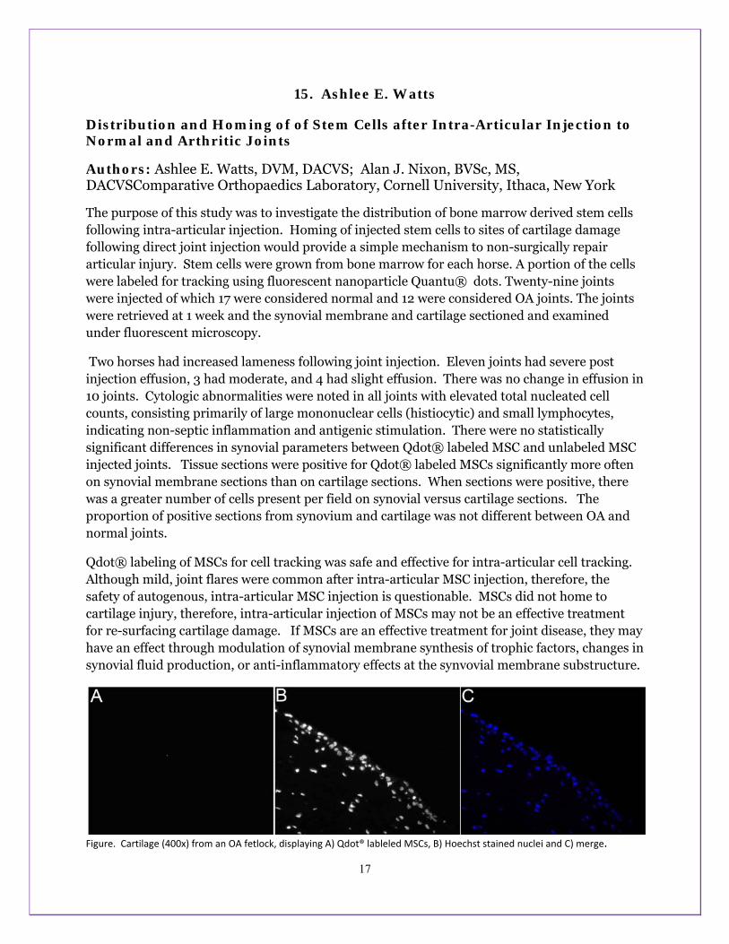
17
15. Ashlee E. Watts
Distribution and Homing of of Stem Cells after Intra-Articular Injection to Normal and Arthritic Joints
Authors: Ashlee E. Watts, DVM, DACVS; Alan J. Nixon, BVSc, MS, DACVSComparative Orthopaedics Laboratory, Cornell University, Ithaca, New York
The purpose of this study was to investigate the distribution of bone marrow derived stem cells following intra-articular injection. Homing of injected stem cells to sites of cartilage damage following direct joint injection would provide a simple mechanism to non-surgically repair articular injury. Stem cells were grown from bone marrow for each horse. A portion of the cells were labeled for tracking using fluorescent nanoparticle Quantu® dots. Twenty-nine joints were injected of which 17 were considered normal and 12 were considered OA joints. The joints were retrieved at 1 week and the synovial membrane and cartilage sectioned and examined under fluorescent microscopy.
Two horses had increased lameness following joint injection. Eleven joints had severe post injection effusion, 3 had moderate, and 4 had slight effusion. There was no change in effusion in 10 joints. Cytologic abnormalities were noted in all joints with elevated total nucleated cell counts, consisting primarily of large mononuclear cells (histiocytic) and small lymphocytes, indicating non-septic inflammation and antigenic stimulation. There were no statistically significant differences in synovial parameters between Qdot® labeled MSC and unlabeled MSC injected joints. Tissue sections were positive for Qdot® labeled MSCs significantly more often on synovial membrane sections than on cartilage sections. When sections were positive, there was a greater number of cells present per field on synovial versus cartilage sections. The proportion of positive sections from synovium and cartilage was not different between OA and normal joints.
Qdot® labeling of MSCs for cell tracking was safe and effective for intra-articular cell tracking. Although mild, joint flares were common after intra-articular MSC injection, therefore, the safety of autogenous, intra-articular MSC injection is questionable. MSCs did not home to cartilage injury, therefore, intra-articular injection of MSCs may not be an effective treatment for re-surfacing cartilage damage. If MSCs are an effective treatment for joint disease, they may have an effect through modulation of synovial membrane synthesis of trophic factors, changes in synovial fluid production, or anti-inflammatory effects at the synvovial membrane substructure.
Figure. Cartilage (400x) from an OA fetlock, displaying A) Qdot® lableled MSCs, B) Hoechst stained nuclei and C) merge.

16. INTERLEUKIN-1 SILENCING IN OSTEOARTHRITIC MODELS USING PLASMID AND INTEGRATING TRANSPOSON BASED RNA INTERFERENCE
+*Nixon, AJ; *Scimeca, MS; *Ortved, K; **Hackett, PB; ***Rossi, JJ. *Comparative Orthopaedics Laboratory, Cornell University College of Veterinary Medicine, Ithaca, NY.
**University of Minnesota, Minneapolis, MN; ***Beckman Research Institute, City of Hope, Duarte, CA +Corresponding author: [email protected]
INTRODUCTION Joint injury can result in a profound imbalance in cartilage catabolic and anabolic processes culminating in degenerative osteoarthritis. Interleukin-1 is regarded as the principal cartilage-degradative cytokine in OA, and has an important role in other joint diseases including rheumatoid arthritis. Reducing the impact of IL-1 has been possible using IL-1 receptor antagonist. However, the transitory nature of IL-1Ra requires daily re-dosing, leading to development of gene therapy approaches to extend the pro-inflammatory cytokine control.1,2 A direct IL-1 transcript silencing approach rather than receptor antagonism has been evaluated using small interfering RNA (siRNA) ribo-oligonucleotides targeting IL-1 mRNA.3 To be effective in vivo, plasmid based IL-1 silencing is necessary to overcome the transitory nature of oligo based knockdown. The hypothesis for this study was that IL-1 expression could be better controlled using short hairpin plasmid and ultimately integrating transposon plasmid based gene silencing motifs targeting IL-1. METHODS Five small interfering RNA’s (siRNA) for IL-1β were chemically generated (Ambion). Chondrocytes cultured in medium containing lipopolysaccharide (LPS) at 25 μg/mL, to induce IL-1β gene expression, were electroporated with siRNA using a BTX ECM 830 electroporator (Harvard Instruments). After electroporation, cells were plated with 25 μg/mL LPS for another 24 hours. RNA was isolated and real time PCR gene expression analysis performed. The most effective siRNA ribo-oligonucleotide was used as the template to form pSH.IL-1β, a plasmid based short hair-pin coding construct based on the pSIREN shuttle plasmid (Clontech). Following LPS treatment, cells were electroporated with 4ug pSH.IL-1β. IL-1b expression was assessed at 24 hrs post-electroporation. Subsequently, the pSH.IL-1β expression cassette was recombined into the Sleeping Beauty (SB) transposon plasmid upstream of a bicistronic expression/selection cassette derived from pIRESpuro3 (Clontech). Cells were electroporated with an equimolar mix of SB-sh.IL1b and the SB transposase-carrying pCMV-SB plasmid. Ten days post selection (puromycin), genomic integration was confirmed by PCR of genomic DNA for the SB-sh.IL1b construct. IL1b expression was assessed in SB-sh.IL1b cells and wild-type cells following 48 hr LPS treatment as described above. Data were analyzed using ANOVA with post-hoc Tukeys classifications. RESULTS Six small interfering RNA’s (siRNA) targeting IL-1β were screened by electroporation into chondrocyte monolayers exposed to LPS. Gene expression for IL-1β and MMP-13 were returned to baseline by several IL-1 silencing ribo-oligonucleotides. Effective siRNA ribo-oligo’s were constructed as plasmid based short hair-pin coding constructs to prolong IL-1 knockdown (pSIREN-IL-1shRNA). IL-1 shRNA loops, with or without IGF-I gene transduction (bicistronic plasmid expressing IGF-I and IL-1β shRNA), prolonged matrix restorative effects. Chondrocyte monolayers expressing IL-1 shRNA had >60% reduction in IL-1
abundance (Fig 1).
Medium from IL-1 shRNA knockdown trials showed consistent IL-1 reduction after shRNA transduction.
Effective IL-1 shRNA and IGF-1 coding motifs were developed into Sleeping Beauty plasmid based vectors for transduction of chondrocyte and synoviocyte cultures. Both transgenes were expressed and IL-1 shRNA suppressed IL-1 fluxes in LPS conditioned cultures (Fig 3).
DISCUSSION: These data show novel IL-1 knockdown using a plasmid coding a short hairpin loop (shRNA) form of an IL-1 interference motif. This plasmid allowed stable expression of siRNA following intracellular Dicer activity. Moreover, development of this plasmid by recombination into the Sleeping Beauty (SB) transposon, provided potential for chromosomal integration and selection of chondrocytes resistant to IL-1 effects. Target gene knockdown provides a non-competitive path to control IL-1 action, compared to previous IL-1 receptor antagonism, where the need for molar excess of IL-1Ra over IL-1 was paramount. Similarly, post-transcriptional gene silencing has the advantage of persistence, particularly when combined with integrating vectors such as SB. Implementation of integrating mechanisms may be useful for substantial control of degradation and improved function in OA joints. Adding IGF-I to the combinatorial approach to drive further repair in the face of diminished catabolic erosion may further improve the possibility effective OA control. REFERENCES [1] Gouze et al. Arth Res Ther 5:301, 2003; [2] Nixon et al Gene Ther 12:177, 2005. [3] Nixon et al Ann NY Acad Sci 1113:1-18,2007. ACKNOWLEDGMENT Supported by Harry M Zweig Foundation and NIH R01 AR055373 +www.vet.cornell.edu/labs/comporth.
Fig 1. IL-1β expression wassuppressed byplasmid basedshRNA interference.
Fig 3. Sleeping Beauty construct; A) Plasmid map for bi-cistronic vector expressing IL-1shRNA and IGF-I. Puromycin resistance conveys cell selection; B) PCR confirmation after SB transduction, confirming integration of SB plasmid containingIL-1shRNA and IGF-I coding regions; C) IL-1 knockdown in cultures transduced with SB-shIL1 and IGF-1.
Fig 2. Aggrecan expression is markedly suppressed by IL-1 beta induction, and partly restored in chondrocytes treated with pIL-1 shRNA for IL-1 knockdown (LPS + pIL-1sh).

17. INTERLEUKIN-1b AND TUMOR NECROSIS FACTOR-a KNOCKDOWN IN CHONDROCYTES BY SHORT INTERFERING RNA USING A DUAL EXPRESSION LONG HAIRPIN CONSTRUCT
*Scimeca, MS; *Nixon, AJ. *Comparative Orthopaedics Laboratory, Cornell University College of Veterinary Medicine, Ithaca, NY.
INTRODUCTION Osteoarthritis is characterized by an imbalance in cartilage catabolic and anabolic processes. Combinatorial therapies may enhance pain relief and improve cartilage function through knockdown of IL-1 and supplementation of growth factors such as IGF-I. Small interfering RNA (siRNA) molecules bind to mRNA to silence specific target genes post-transcriptionally. The hypothesis for this study was that cognate IL-1 and TNF gene expression could be diminished by RNA interference, which would quell degradatory cycles in the osteoarthritic joint and allow IGF-I gene enhanced repair. The purpose of this study was to design and produce a long hairpin (LHp) expressing plasmid for IL-1b and TNFa knockdown, and to verify simultaneous IL-1b and TNFa knockdown by the LHp construct in lipopolysaccharide (LPS) stimulated chondrocytes. METHODS Two DNA fragments based on previously verified siRNA sequences were synthesized by Integrated DNA Technologies (Coralville, IA) and ligated into the U6 promoter driven vector pSIREN shuttle (Clontech [Mountain View, CA]) Ligation products were cloned into E. coli. Positive colonies were identified by PCR and sequenced for further testing in chondrocytes. Plasmids were transfected into equine articular chondrocytes in 24 well plates using FuGene HD Transfection Reagent (Roche [Indianapolis, IN]). At 48 hours post-transfection, chondrocytes were stimulated with 20ug/mL LPS. After 36 hours of LPS stimulation, cells were lysed and total RNA purified. Expression of IL-1b and TNFa were determined by quantitative real time PCR and normalized to 18S rRNA. RESULTS
Sequencing yielded four long hairpin plasmids for testing in chondrocytes. There was no significant difference in IL-1b or TNFa knockdown effect among the four plasmids.
DISCUSSION: Synthetic DNA fragments carrying previously validated IL-1b and TNFa siRNA were ligated successfully into a hairpin RNA expression vector. Four clonally isolated long hairpin plasmids were identified and tested in lipopolysaccharide stimulated chondrocytes. All four long hairpin plasmids knocked down IL-1b expression ~40% and TNFa expression. These experiments confirm that both siRNA motifs can be synthesized using a single RNA promoter to drive DNA coding regions. This plasmid construct can be enabled for clinical use by incorporation into AAV viral vectors and Sleeping Beauty integrating non-viral vectors for injection to the joint. RNA interference provides a novel mechanism for silencing degradatory processes in arthritis. Plasmid based RNA interference to quell IL-1 and TNF may be a major improvement in arthritis control. Integrating plasmid vectors would provide a more robust RNA expression, and the Sleeping Beauty™ transposon-transposase system has promise in this objective.
ACKNOWLEDGMENT
Supported by Harry M Zweig Foundation and NIH R01 AR055373 +www.vet.cornell.edu/labs/comporth.
Fig 1. Overall, LHp plasmids had 40% lower IL-1b expressionthan positive control and all LPS treated cells had higher IL-1bexpression than untreated controls. Different letters indicatesignificant differences at p<0.05.
Fig 2. Overall, LHp plasmids had 62% lower TNFa expression than positive control. There were no significant differences between LHp-LPS groups and untreated controls. Different letters indicate significant differences at p<0.05.
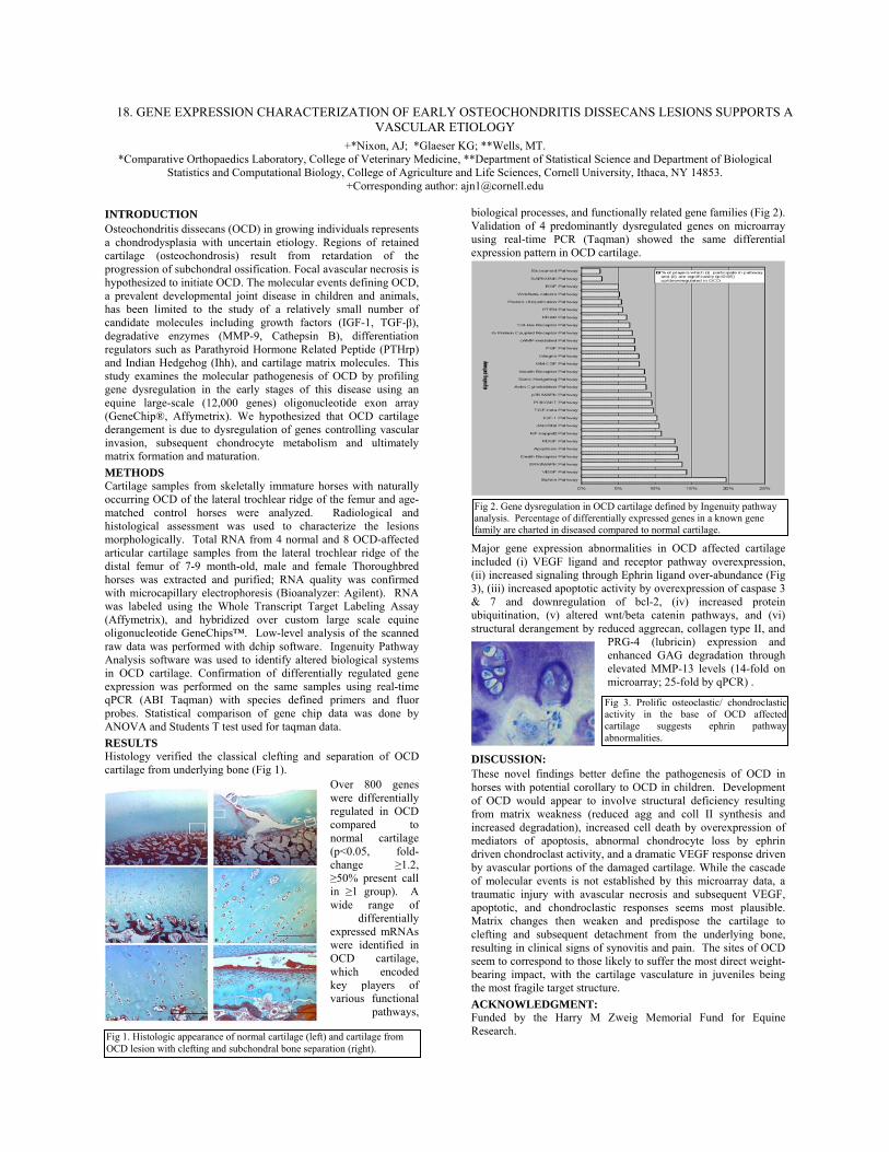
18. GENE EXPRESSION CHARACTERIZATION OF EARLY OSTEOCHONDRITIS DISSECANS LESIONS SUPPORTS A VASCULAR ETIOLOGY
+*Nixon, AJ; *Glaeser KG; **Wells, MT. *Comparative Orthopaedics Laboratory, College of Veterinary Medicine, **Department of Statistical Science and Department of Biological
Statistics and Computational Biology, College of Agriculture and Life Sciences, Cornell University, Ithaca, NY 14853. +Corresponding author: [email protected]
INTRODUCTION Osteochondritis dissecans (OCD) in growing individuals represents a chondrodysplasia with uncertain etiology. Regions of retained cartilage (osteochondrosis) result from retardation of the progression of subchondral ossification. Focal avascular necrosis is hypothesized to initiate OCD. The molecular events defining OCD, a prevalent developmental joint disease in children and animals, has been limited to the study of a relatively small number of candidate molecules including growth factors (IGF-1, TGF-β), degradative enzymes (MMP-9, Cathepsin B), differentiation regulators such as Parathyroid Hormone Related Peptide (PTHrp) and Indian Hedgehog (Ihh), and cartilage matrix molecules. This study examines the molecular pathogenesis of OCD by profiling gene dysregulation in the early stages of this disease using an equine large-scale (12,000 genes) oligonucleotide exon array (GeneChip®, Affymetrix). We hypothesized that OCD cartilage derangement is due to dysregulation of genes controlling vascular invasion, subsequent chondrocyte metabolism and ultimately matrix formation and maturation. METHODS Cartilage samples from skeletally immature horses with naturally occurring OCD of the lateral trochlear ridge of the femur and age-matched control horses were analyzed. Radiological and histological assessment was used to characterize the lesions morphologically. Total RNA from 4 normal and 8 OCD-affected articular cartilage samples from the lateral trochlear ridge of the distal femur of 7-9 month-old, male and female Thoroughbred horses was extracted and purified; RNA quality was confirmed with microcapillary electrophoresis (Bioanalyzer: Agilent). RNA was labeled using the Whole Transcript Target Labeling Assay (Affymetrix), and hybridized over custom large scale equine oligonucleotide GeneChips™. Low-level analysis of the scanned raw data was performed with dchip software. Ingenuity Pathway Analysis software was used to identify altered biological systems in OCD cartilage. Confirmation of differentially regulated gene expression was performed on the same samples using real-time qPCR (ABI Taqman) with species defined primers and fluor probes. Statistical comparison of gene chip data was done by ANOVA and Students T test used for taqman data. RESULTS Histology verified the classical clefting and separation of OCD cartilage from underlying bone (Fig 1).
Over 800 genes were differentially regulated in OCD compared to normal cartilage (p<0.05, fold-change ≥1.2, ≥50% present call in ≥1 group). A wide range of
differentially expressed mRNAs were identified in OCD cartilage, which encoded key players of various functional
pathways,
biological processes, and functionally related gene families (Fig 2). Validation of 4 predominantly dysregulated genes on microarray using real-time PCR (Taqman) showed the same differential expression pattern in OCD cartilage.
Major gene expression abnormalities in OCD affected cartilage included (i) VEGF ligand and receptor pathway overexpression, (ii) increased signaling through Ephrin ligand over-abundance (Fig 3), (iii) increased apoptotic activity by overexpression of caspase 3 & 7 and downregulation of bcl-2, (iv) increased protein ubiquitination, (v) altered wnt/beta catenin pathways, and (vi) structural derangement by reduced aggrecan, collagen type II, and
PRG-4 (lubricin) expression and enhanced GAG degradation through elevated MMP-13 levels (14-fold on microarray; 25-fold by qPCR) .
DISCUSSION: These novel findings better define the pathogenesis of OCD in horses with potential corollary to OCD in children. Development of OCD would appear to involve structural deficiency resulting from matrix weakness (reduced agg and coll II synthesis and increased degradation), increased cell death by overexpression of mediators of apoptosis, abnormal chondrocyte loss by ephrin driven chondroclast activity, and a dramatic VEGF response driven by avascular portions of the damaged cartilage. While the cascade of molecular events is not established by this microarray data, a traumatic injury with avascular necrosis and subsequent VEGF, apoptotic, and chondroclastic responses seems most plausible. Matrix changes then weaken and predispose the cartilage to clefting and subsequent detachment from the underlying bone, resulting in clinical signs of synovitis and pain. The sites of OCD seem to correspond to those likely to suffer the most direct weight-bearing impact, with the cartilage vasculature in juveniles being the most fragile target structure. ACKNOWLEDGMENT: Funded by the Harry M Zweig Memorial Fund for Equine Research. Fig 1. Histologic appearance of normal cartilage (left) and cartilage from
OCD lesion with clefting and subchondral bone separation (right).
Fig 2. Gene dysregulation in OCD cartilage defined by Ingenuity pathway analysis. Percentage of differentially expressed genes in a known gene family are charted in diseased compared to normal cartilage.
Fig 3. Prolific osteoclastic/ chondroclastic activity in the base of OCD affected cartilage suggests ephrin pathway abnormalities.

19. TNF-α GENE SILENCING CONTROLS CATABOLIC ACTIVITY IN OSTEOARTHRITIS *+Nixon, AJ; Scimeca, MS; Witte, TH; Kamm, JL; Khadem, N; **Rossi, JJ.
*Comparative Orthopaedics Laboratory, Cornell University, College of Veterinary Medicine, Ithaca, NY **Beckman Research Institute, City of Hope, Duarte, CA
INTRODUCTION: Catabolic erosion of cartilage and underlying subchondral bone in rheumatoid and osteoarthritis is broadly initiated by cytokines such as interleukin-1 (IL-1) and tumor necrosis factor-alpha (TNF-α). Secondary agents then perpetuate the joint reaction, with continued painful debility and advancing dysfunction. Several mechanisms to control pro-inflammatory cytokine action have evolved, including peptide and gene therapy approaches to block IL-1 and TNF activity.3 However, control of target gene receptor function with IL-1 receptor antagonist and TNF soluble receptor decoys have limited potential to reconstitute matrix components and cartilage function. The transitory nature of these agents leads to frequent re-dosing.1,2 RNA interference may provide a more robust method to control gene function by silencing specific target genes post-transcriptionally. Previous studies show IL-1 gene knockdown alone does not adequately salvage cartilage matrix synthesis.4 The hypothesis for this study was that cartilage matrix reformation could be re-established by silencing additional pro-inflammatory cytokines, using ribo-oligonucleotide and short hairpin plasmid based gene silencing motifs targeting TNF-α. METHODS: Small interfering RNA’s (siRNA) targeting TNF-α were synthesized and screened by electroporation into chondrocyte monolayers exposed to LPS to stimulate an inflammatory state. Gene expression for catabolic cytokines and cartilage matrix gene expression was determined by real-time PCR. The most efficient siRNA ribo-oligo’s were used to design a short-hairpin coding region to code for sense and antisense RNA interference motifs in a plasmid, to prolong TNF-α knockdown (pSH-TNF2). Plasmid based knockdown motifs were introduced by electroporation. Proinflammatory and cartilage matrix gene expression changes were determined by real-time PCR. Results were compared using ANOVA with post-hoc Tukeys classification. RESULTS: Gene expression for the catabolic cytokine TNF-α was abundantly stimulated by LPS. Conversely, matrix aggrecan and collagen type II were suppressed. TNF-α expression was substantially reduced by 2 of seven siRNA targeting TNF (Fig 1). LPS treated chondrocyte cultures showed a small rebound in matrix collagen type II expression when transduced with TNF-α siRNA #2 alone and in combination with IL1b siRNA (Fig 2.)
Chondrocytes transduced with pSH-TNF2 demonstrated a >50% knockdown of TNF-α gene expression. (Fig 3.) and had a small rebound in aggrecan expression (Fig 4.)
0
20
40
60
80
100
120
140
160
180
LPS only pshTNFa + LPS Electroporation only pshTNFa only
TNFa
cop
y# /
ng R
NA
A
B
B
C
0.0E+00
5.0E+04
1.0E+05
1.5E+05
2.0E+05
2.5E+05
3.0E+05
3.5E+05
LPS only pshTNFa +LPS Electroporation only pshTNFa
Agg
reca
n Co
py #
/ ng
RNA
A
B
C
D
DISCUSSION: This study shows the potential of TNF-α silencing using plasmid based shRNA expression using screening to identify the most efficient TNF silencing motif for development to a plasmid (pSH-TNF2). These data show novel TNF knockdown using a plasmid coding a short hairpin loop (shRNA) form of a TNF interference motif. This plasmid allows stable expression of siRNA following intracellular Dicer activity. Moreover, development of this plasmid by recombination into the Sleeping Beauty (SB) transposon, provides the potential for chromosomal integration and selection of chondrocytes resistant to TNF effects. Target gene knockdown provides a non-competitive path to control of TNF action, compared to previous receptor decoy and soluble antibody approaches. Similarly, post-transcriptional gene silencing has the advantage of persistence, particularly when combined with simple plasmid or integrating vectors such as SB. In the present study, prolonged knockdown of TNF-α mRNA led to marginal recovery of matrix molecule expression. However, controlling one pro-inflammatory cytokine was inadequate to allow matrix synthesis to fully rebound. Combinatorial therapy, targeting both IL-1 and TNF-α, has been developed to achieve global knockdown of the two major catabolic agents and improve anabolic recovery.4 Implementation of both mechanisms may be useful for substantial control of degradation and improved function in OA joints. Preliminary data on longer term combination silencing of IL-1b and TNF-α in chondrocyte cultures indicate that aggrecan and collagen type II expression can be returned to levels closer to normal. Adding IGF-I to the combinatorial approach to drive further repair in the face of diminished catabolic erosion further improves the possibility of effective OA control. REFERENCES: [1] Gouze et al. Arth Res Ther 5:301, 2003; [2] Kobayashi et al Arth Rheum 52:128, 2005; [3] Nixon et al Gene Ther 12:177, 2005. [4] Nixon et al Ann NY Acad Sci 1113:1-18,2007. ACKNOWLEDGEMENTS ; Supported by Harry M Zweig Foundation, and NIH R01 AR055373 +www.vet.cornell.edu/labs/comporth. Email: [email protected].
Fig 4. Aggrecangene expression in chondrocyte cultures transduced with pSH-TNF2 and exposed to LPS. N=6. Mean +/-SE.
Fig 1. TNF expression after LPS induction followed bytransduction with TNF silencing ribo-oligonucleotides. TNF siRNA#2 reduces TNFa abundance to normal. N=3. Mean +/- SE.
Fig 3. TNFa gene expression in chondrocytes transduced with pSH-TNF2 and exposed to LPS.N=6. Mean +/-SE.
Fig 2. Collagen type II rebound in chondrocyte cultures transduced with siRNA to TNF, IL-1, or combination of both and exposed to LPS. N=6. mean +/- SE.

22
20. Gilian Perkins
Immunization against Strangles using a vectored Equine Herpesvirus Vaccine
Authors: Gillian Perkins, Sarah Hooper, Armando Damiani, Silke Feineis, Craig Altier, Nikolaus Osterrieder
Abstract: Streptococcus equi subspecies equi is a highly contagious bacterium that causes Strangles in horses a disease characterized by abscessation of lymph nodes. Acute infections and chronic complications, such as abdominal abscesses, guttural pouch empyema, and airway obstruction occur in about 20% of the cases, lead to sustained loss of performance and animal health and can even be fatal. Current strangles vaccines are not without risks and efficacy data are limited. The goal of this work is to induce a long-lasting immune response to EHV-1 and Streptococcus equi by using EHV-1 as a vehicle to deliver S. equi antigens. Two S. equi antigens (SeM and IdeE) were incorporated into the EHV-1 genome using red recombination and tested in vitro. These vaccines were administered (SeM/EHV-1, IdeE/EHV-1 or a combination of both ) to young female Balb/c mice (n=58) intranasally or subcutaneously twice (3weeks apart) and then challenged 3 weeks later with 106 CFU of S.equi subspecies equi strain CF32 intranasally. Weight was monitored daily post-infection and nose-print cultures for Streptococcus equi were performed. Histopathology was done on the tissues of mice that died or were euthanized at the end of the study. No adverse effects of the vaccines were noted. When mice were given both SeM and IdeE regardless of route, less weight loss and mortality was documented. Further data analyses are being performed. Should these results seem promising, further testing regarding efficacy of the vaccine against Strangles will be tested in the target host, the horse.

23
21. Gillian Perkins
RNA Interference does not protect against experimental Equine Herpes Virus-1 Infection
Authors: GA Perkins, N Pusterla, HN Erb, Nikolaus Osterrieder
Abstract: Small interfering (si) RNAs targeted against essential genes of EHV-1 glycoprotein B (sigB3) and origin binding protein (siOri2) reduce EHV-1 replication in vitro and in mice. An EHV-1 challenge infection showed a decrease in severity of neurological symptoms in horses treated with sigB3/ori2 12-h before and 12-h after EHV-1 infection but no difference in the nasal viral load or viremia. We tested whether metaphylactic treatment with EHV-1-specific siRNAs at a higher dose and frequency would reduce viral load and clinical symptoms in EHV-1-infected horses.
Horses were infected intranasally (IN) with 1x 10⁷ PFU of EHV1 virus strain Ab4 (day 0). Seven horses were given 5-nmols each of sigB3 and siOri2 IN 24-h pre-and 12, 24, 36 and 48-h post-EHV-1 infection. The other 6 horses were given 10-nmols of an irrelevant siRNA against firefly luciferase (siLuc). Horses were monitored daily and blood and nasal swabs taken on days -1, 1 to 10, 12, 14 and 21 post-infection (p.i.) for virus isolation and qPCR. Wilcoxon's rank-sum tests were performed.
All horses developed serous nasal discharge and a biphasic fever p.i. Three sigB3/Ori2 and 2 siLuc horses had neurologic signs beginning day 6 p.i. There was no significant difference between groups in clinical signs, nasal viral shedding or viremia. The sigB3/siOri2 regimen did not protect horses against EHV-1 experimental infection.

22. Joy (Chia) T. Tseng
Identification of Serologically Defined Equine Major Histocompatibility Complex Haplotypes using an Intra-MHC Microsatellite Panel
Authors: Joy( Chia) T. Tseng, Donald Miller, Douglas F. Antczak
Abstract: Five microsatellites spanning the Major Histocompatibility Complex (MHC) class I and class II regions were tested on a panel of 277 horses that were either unrelated or members of family trios. A total of 40 haplotypes were identified and 15 of these were linked to 9 serologically identified Equine Lymphocyte Antigen (ELA) types, while the remainder lacked corresponding serological data. The molecular signature of each MHC haplotype was determined based on a particular constellation of microsatellite alleles. Subhaplotypes of serologically defined ELA-A3, -A5, -A9, and -A10 were detected, where more than one microsatellite haplotype was associated with a serological haplotype. The majority of horses examined in this study were Thoroughbreds and Standardbreds, and microsatellite haplotypes associated with these two breeds were identified from the data set. When comparing frequency of MHC microsatellite haplotypes in Thoroughbreds to the serological patterns determined previously, 90% of the chromosomes were comprised of the well-defined ELA haplotypes, and the majority of these chromosomes were accountable by ELA-A2 and ELA-A3. This molecular analysis of the equine MHC complemented the outcome of a historical assay and furthered our knowledge of MHC polymorphisms. We expect that future investigations based on single nucleotide polymorphisms will offer additional insight on genetics and evolution of the ELA system.

25
23. Joy (Chia) T. Tseng
Effect on Equine Pregnancy Rates of Major Histocompatibility Complex Matching between Sires and Dams
Authors: Joy (Chia) T. Tseng, Donald Miller, Emily Silvela, James Hardy, Scott Hoffay, Douglas F. Antczak
Abstract: Several theories have been proposed to explain the high degree of polymorphism of genes in the Major Histocompatibility Complex (MHC) and the resulting high percentage of heterozygous individuals in most natural populations. The MHC has been studied extensively in the context of reproductive success in investigations ranging from mate choice to postnatal survival and fitness. In this retrospective study of pregnancy rates in an experimental horse herd selected for MHC types, we investigated the MHC influence on reproduction from fertilization to Day 34 of pregnancy among five MHC homozygous stallions of two unrelated haplotypes and 105 MHC-typed mares. We tested the hypothesis that pregnancy rates would be higher in MHC incompatible matings than in MHC compatible matings to favor more heterozygous offspring. Historical records of 970 mated estrus cycles and 686 conceptions over 23 years were analyzed. The average pregnancy rates were not significantly different between compatible (70.45%) and incompatible (70.85%) matings (p> 0.5). The compatible mating group was further subdivided into two categories based on the MHC genotypes of the mare and of the probable embryo. We hypothesized that matings producing only MHC homozygous embryos (A x A) would be less successful than matings that had an equal probability of producing MHC homozygous or heterozygous embryos (A x A/B). Again, we found no difference in the pregnancy rates between these two groups. Under the conditions of this study, the MHC compatibility relationship between sire and dam showed no influence on reproductive success in early equine pregnancy in the period from fertilization to day 34 of gestation.
26
24. Alanna Zantingh
Effect Of Bilateral Hypoglossal Nerve Blockade At the Level Of the Ceratohyoid Bones On Radiographic Laryngeal Position In the Resting Horse Authors: Alanna J. Zantingh, Norm G. Ducharme, Lisa M. Mitchell, Jonathan Cheetham Poster presenter: Alanna J. Zantingh, Jonathan Cheetham (depends on who is busier in the hospital at the time of the presentations) Abstract: The etiology of Dorsal Displacement of the Soft Palate (DDSP) in horses is not completely understood. DDSP has been experimentally induced by 3 methods. Horses that had DDSP during exercise have been shown to have a more ventral basihyoid bone at rest and a more dorsal laryngeal position following laryngeal ‘tie forward’ surgery is associated with improved post-operative racing performance. An association has also been shown between swallowing and DDSP. The hypoglossal nerve plays an important role in swallowing and we hypothesized that local bilateral blockade of the hypoglossal nerve at the level of the ceratohyoid bones would change laryngohyoid position in horses at rest. Bilateral distal hypoglossal nerve blockade was performed on 10 healthy horses. Lateral radiographs of the larynx were taken before and 30 minutes after bilateral hypoglossal block, and a radiographic reference system was used to determine the position of the caudal aspect of the basihyoid bone, the thyrohyoid-thyroid articulation, and the ossification of the base of the thyroid cartilage. Hypoglossal block produced ventral movement of the larynx. The ossification of the thyroid cartilage was positioned 6.2mm (95% CI 0.7-11.7) more ventral following bilateral hypoglossal block (p=0.03). It is possible that mild neurologic dysfunction of the hypoglossal nerve or muscular dysfunction of the muscles innervated by this nerve predispose a horse to DDSP due to a more ventral larynx position.

27
25. Margaret Brosnahan
IL-22 and its Receptors Show a Unique Pattern of Expression in Tissues and Immune Cells Associated with Endometrial Cups in Early Equine Pregnancy
Authors: Margaret M. Brosnahan, Mackenzie Adams, Donald Miller, Douglas F. Antczak
Abstract:The invasive chorionic girdle (CG) cells of the equine trophoblast form endometrial cups, structures that produce equine chorionic gonadotropin (eCG) from approximately days 40 to 120 of a normal equine pregnancy. The mechanisms that enable CG cells expressing paternal MHC-I molecules to survive as partial allografts within maternal endometrium are ill-defined at this time, but possibly include immunoregulatory and cell programming events. A gene expression microarray analysis performed by our laboratory showed a greater than 500-fold upregulation of the recently described immunomodulatory cytokine interleukin-22 in CG relative to non-invasive chorion on day 34 of pregnancy. Expression of IL-22 was also greater than 900-fold upregulated in stimulated equine lymphocytes compared to resting lymphocytes. Supportive results were obtained on qRT-PCR assays. Microarray analysis showed consistent expression of membrane receptor IL-22RA1 in these same tissues, no expression of the soluble binding protein IL-22BP in CG or non-invasive chorion, and greater than 250-fold expression of IL-22BP in stimulated as compared to resting lymphocytes. Further investigation using qRT-PCR analysis supports our hypothesis that IL-22 and its membrane receptor may be a mechanism of communication between the invading CG cells and the maternal endometrium. The upregulation of IL-22 in chorionic girdle cells of the equine trophoblast is a novel finding, as this cytokine to date has been reported only in a limited number of immune cells including Th-17 and Th-22 cells along with some natural killer and dendritic cells.

28
26. Sally Ness
An Outbreak of Besnoitiosis: An Emerging and Potentially Zoonotic Disease of Donkeys
Authors: SallyAnne L. Ness, Thomas J. Divers, Jeanine Peters-Kennedy, Dwight B. Bowman, Julia B. Felippe, Linda D. Mittel, Hussni O. Mohammed, Susan E. Wade, Nicole Shultz
Abstract: Besnoitia spp. are cyst-forming coccidian parasites that infect multiple host species worldwide. Recently, multiple donkeys across several Northeastern states have been diagnosed with Besnoitiosis and it appears that the incidence of disease may be increasing. To date, the mode of disease transmission remains unknown and there are no reported effective treatments. A treatment trial with Marquis (ponazuril) of two donkeys with Besnoitiosis failed to show efficacy against the parasite. An epidemiological investigation of an outbreak in a Pennsylvania miniature donkey herd is currently underway.
APPENDIX F
ZWEIG NEWS CAPSULES
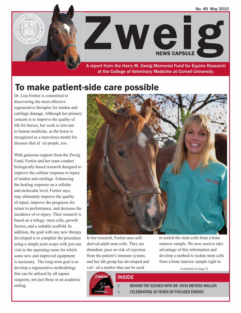
No. 49 May 2010
INSIDE
Zweig
30 years
Dr. Lisa Fortier is committed to discovering the most effective regenerative therapies for tendon and cartilage damage. Although her primary concern is to improve the quality of life for horses, her work is relevant to human medicine, as the horse is recognized as a marvelous model for diseases that af ict people, too.
With generous support from the Zweig Fund, Fortier and her team conduct biologically-based research designed to improve the cellular response to injury of tendon and cartilage. Enhancing the healing response on a cellular and molecular level, Fortier says, may ultimately improve the quality of repair, improve the prognosis for return to performance, and decrease the incidence of re-injury. Their research is based on a trilogy: stem cells, growth factors, and a suitable scaffold. In addition, the goal with any new therapy developed is to complete the procedure using a simple joint scope with just one visit to the operating room for which some new and improved equipment is necessary. The long-term goal is to develop a regenerative methodology that can be utilized by all equine surgeons, not just those in an academic setting.
In her research, Fortier uses self-derived adult stem cells. They are abundant, pose no risk of rejection from the patient’s immune system, and her lab group has developed and veri ed a marker that can be used
3 BEHIND THE SCENES WITH DR. VICKI MEYERS-WALLEN 4 CELEBRATING 30 YEARS OF FOCUSED ENERGY
To make patient-side care possible
(continued on page 2)
A report from the Harry M. Zweig Memorial Fund for Equine Research at the College of Veterinary Medicine at Cornell University.
NEWS CAPSULE
to enrich the stem cells from a bone marrow sample. We now need to take advantage of this information and develop a method to isolate stem cells from a bone marrow sample right in
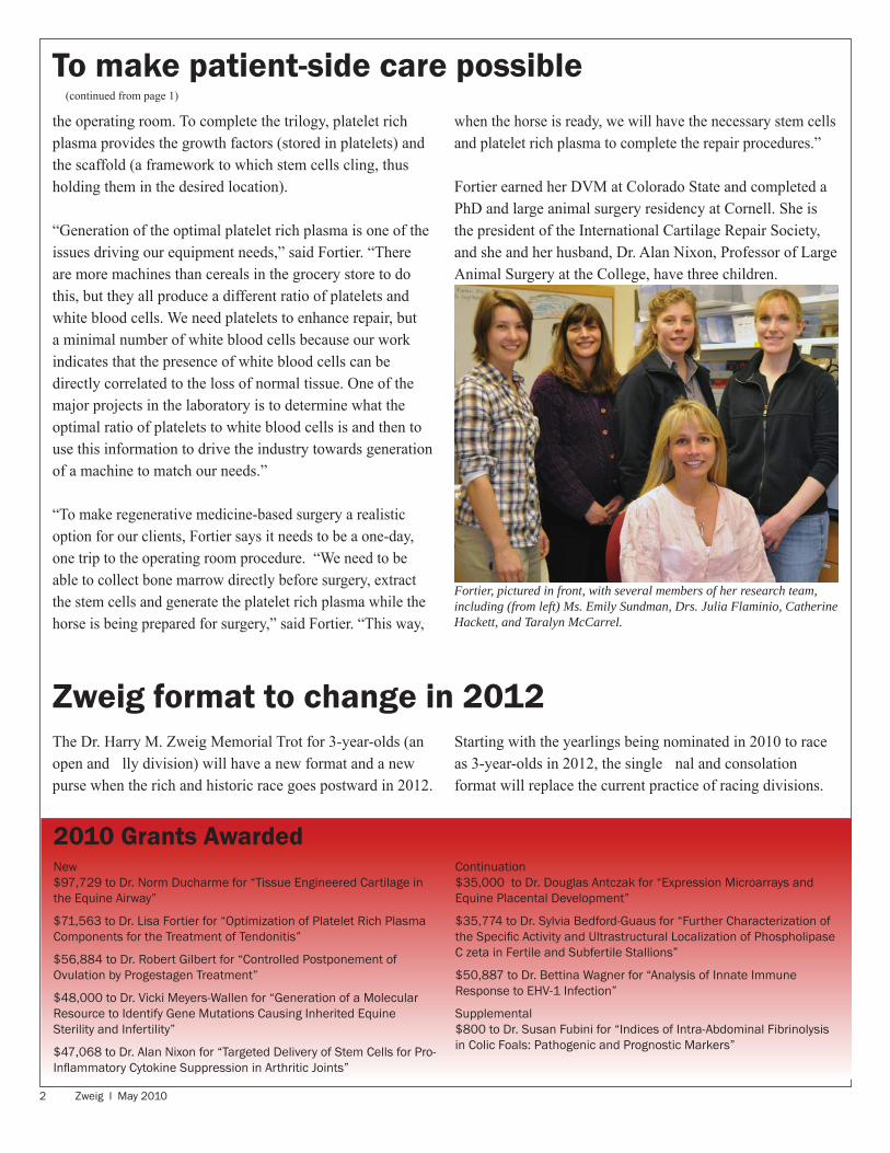
2 Zweig l May 2010
The Dr. Harry M. Zweig Memorial Trot for 3-year-olds (an open and lly division) will have a new format and a new purse when the rich and historic race goes postward in 2012.
Zweig format to change in 2012Starting with the yearlings being nominated in 2010 to race as 3-year-olds in 2012, the single nal and consolation format will replace the current practice of racing divisions.
New$97,729 to Dr. Norm Ducharme for “Tissue Engineered Cartilage in the Equine Airway”
$71,563 to Dr. Lisa Fortier for “Optimization of Platelet Rich Plasma Components for the Treatment of Tendonitis”
$56,884 to Dr. Robert Gilbert for “Controlled Postponement of Ovulation by Progestagen Treatment”
$48,000 to Dr. Vicki Meyers-Wallen for “Generation of a Molecular Resource to Identify Gene Mutations Causing Inherited Equine Sterility and Infertility”
$47,068 to Dr. Alan Nixon for “Targeted Delivery of Stem Cells for Pro-Infl ammatory Cytokine Suppression in Arthritic Joints”
Continuation$35,000 to Dr. Douglas Antczak for “Expression Microarrays and Equine Placental Development”
$35,774 to Dr. Sylvia Bedford-Guaus for “Further Characterization of the Specifi c Activity and Ultrastructural Localization of Phospholipase C zeta in Fertile and Subfertile Stallions”
$50,887 to Dr. Bettina Wagner for “Analysis of Innate Immune Response to EHV-1 Infection”
Supplemental$800 to Dr. Susan Fubini for “Indices of Intra-Abdominal Fibrinolysis in Colic Foals: Pathogenic and Prognostic Markers”
2010 Grants Awarded
(continued from page 1)
the operating room. To complete the trilogy, platelet rich plasma provides the growth factors (stored in platelets) and the scaffold (a framework to which stem cells cling, thus holding them in the desired location).
“Generation of the optimal platelet rich plasma is one of the issues driving our equipment needs,” said Fortier. “There are more machines than cereals in the grocery store to do this, but they all produce a different ratio of platelets and white blood cells. We need platelets to enhance repair, but a minimal number of white blood cells because our work indicates that the presence of white blood cells can be directly correlated to the loss of normal tissue. One of the major projects in the laboratory is to determine what the optimal ratio of platelets to white blood cells is and then to use this information to drive the industry towards generation of a machine to match our needs.” “To make regenerative medicine-based surgery a realistic option for our clients, Fortier says it needs to be a one-day, one trip to the operating room procedure. “We need to be able to collect bone marrow directly before surgery, extract the stem cells and generate the platelet rich plasma while the horse is being prepared for surgery,” said Fortier. “This way,
when the horse is ready, we will have the necessary stem cells and platelet rich plasma to complete the repair procedures.”
Fortier earned her DVM at Colorado State and completed a PhD and large animal surgery residency at Cornell. She is the president of the International Cartilage Repair Society, and she and her husband, Dr. Alan Nixon, Professor of Large Animal Surgery at the College, have three children.
Fortier, pictured in front, with several members of her research team, including (from left) Ms. Emily Sundman, Drs. Julia Flaminio, Catherine Hackett, and Taralyn McCarrel.
To make patient-side care possible
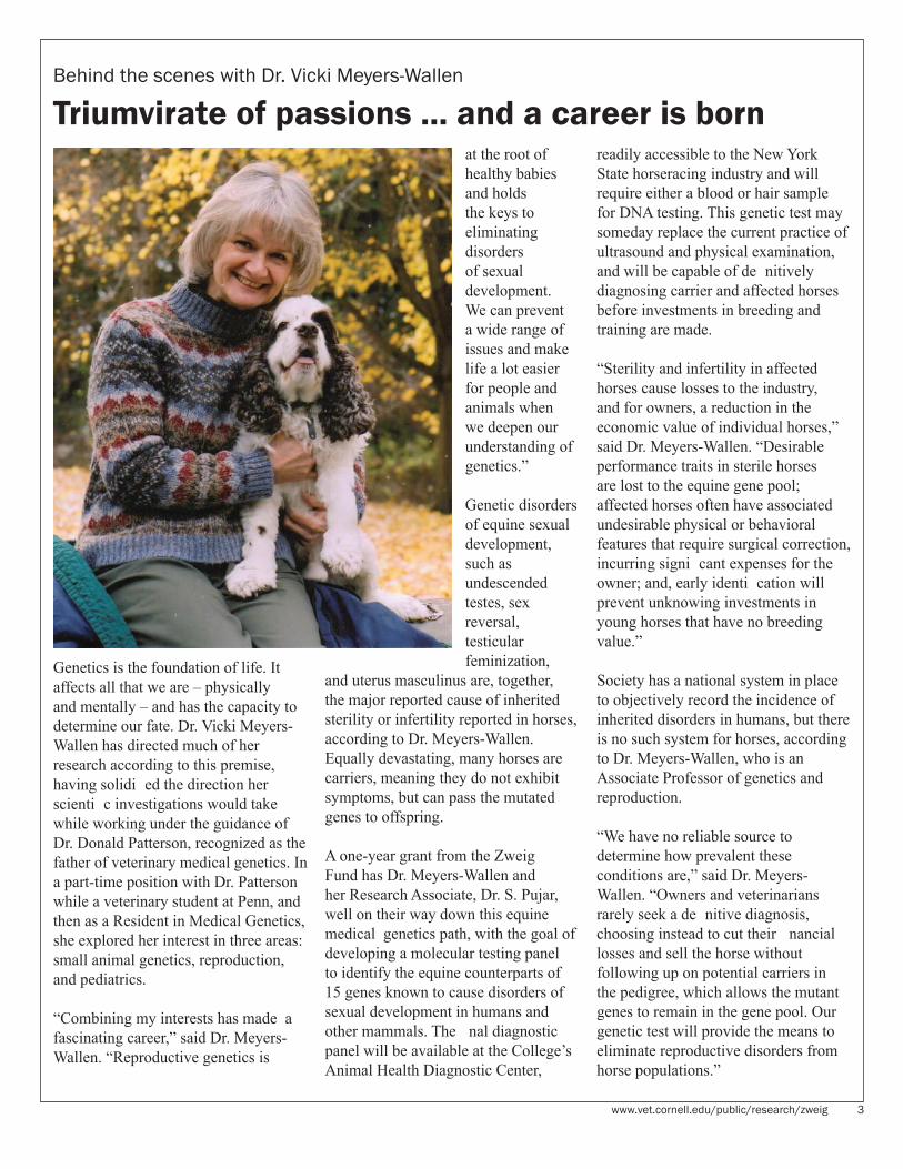
www.vet.cornell.edu/public/research/zweig 3
Behind the scenes with Dr. Vicki Meyers-Wallen
Triumvirate of passions ... and a career is born
Genetics is the foundation of life. It affects all that we are – physically and mentally – and has the capacity to determine our fate. Dr. Vicki Meyers-Wallen has directed much of her research according to this premise, having solidi ed the direction her scienti c investigations would take while working under the guidance of Dr. Donald Patterson, recognized as the father of veterinary medical genetics. In a part-time position with Dr. Patterson while a veterinary student at Penn, and then as a Resident in Medical Genetics, she explored her interest in three areas: small animal genetics, reproduction, and pediatrics.
“Combining my interests has made a fascinating career,” said Dr. Meyers-Wallen. “Reproductive genetics is
at the root of healthy babies and holds the keys to eliminating disorders of sexual development. We can prevent a wide range of issues and make life a lot easier for people and animals when we deepen our understanding of genetics.”
Genetic disorders of equine sexual development, such as undescended testes, sex reversal, testicular feminization,
and uterus masculinus are, together, the major reported cause of inherited sterility or infertility reported in horses, according to Dr. Meyers-Wallen. Equally devastating, many horses are carriers, meaning they do not exhibit symptoms, but can pass the mutated genes to offspring.
A one-year grant from the Zweig Fund has Dr. Meyers-Wallen and her Research Associate, Dr. S. Pujar, well on their way down this equine medical genetics path, with the goal of developing a molecular testing panel to identify the equine counterparts of 15 genes known to cause disorders of sexual development in humans and other mammals. The nal diagnostic panel will be available at the College’s Animal Health Diagnostic Center,
readily accessible to the New York State horseracing industry and will require either a blood or hair sample for DNA testing. This genetic test may someday replace the current practice of ultrasound and physical examination, and will be capable of de nitively diagnosing carrier and affected horses before investments in breeding and training are made.
“Sterility and infertility in affected horses cause losses to the industry, and for owners, a reduction in the economic value of individual horses,” said Dr. Meyers-Wallen. “Desirable performance traits in sterile horses are lost to the equine gene pool; affected horses often have associated undesirable physical or behavioral features that require surgical correction, incurring signi cant expenses for the owner; and, early identi cation will prevent unknowing investments in young horses that have no breeding value.”
Society has a national system in place to objectively record the incidence of inherited disorders in humans, but there is no such system for horses, according to Dr. Meyers-Wallen, who is an Associate Professor of genetics and reproduction.
“We have no reliable source to determine how prevalent these conditions are,” said Dr. Meyers-Wallen. “Owners and veterinarians rarely seek a de nitive diagnosis, choosing instead to cut their nancial losses and sell the horse without following up on potential carriers in the pedigree, which allows the mutant genes to remain in the gene pool. Our genetic test will provide the means to eliminate reproductive disorders from horse populations.”

www.vet.cornell.edu/public/research/zweig 4
Zweig enthusiasts celebrate 30 years of focused energy
Members of the Harry M. Zweig Memorial Fund for Equine Research committee celebrated 30 years of focused energy at the College of Veterinary Medicine in November 2009 with presentations and a wine and cheese reception. Featured speakers at the event included the following:
Sophy A. Jesty, Harry M. Zweig Clinical Fellow“Researching Cardiac Disease, From Mice to Men to Horses”
Bettina Wagner, Harry M. Zweig Assistant Professor (2009-2011)
“The Equine Immune System”
Julia Flaminio, Harry M. Zweig Assistant Professor (2006-2008)
“Immunity Against Rhodococcus equi: The Horse’s Point of View”
Alexander Travis, Associate Professor“Nanoscale Energy Production for Implantable Medical Devices: Moving from Science Fiction to Reality”
Sylvia Bedford-Guaus, Assistant Professor“Molecular Characterization of Equine Phospholipase C Zeta (PLC): A Protein Required for Stallion Sperm Fertility”
Norm Ducharme, Professor and Medical Director, Equine and Farm Animal Hospitals at the Cornell University Hospital for Animals
“Zweig Impact on Upper Airway Surgery”
The Harry M. Zweig Memorial Fund for Equine Research committee. Jean Brown is not pictured. Joseph Lynch (standing, second from left) attended as a delegate for John Sabini.
Dr. Norm Ducharme
Dr. Julia Flaminio
The celebration featured a poster session where nearly 30 researchers shared insights from Zweig-funded research. The posters were created by faculty, graduate students, and residents. Pictured: Dr. Leela Noronha

5 Zweig l May 2010
Th e Harry M. Zweig Memorial Fund for Equine Research honors the late Dr. Harry M. Zweig, a distinguished veterinarian, and his numerous contributions to the state’s equine industry. In 1979, by amendment to the pari-mutuel revenue laws, the New York State legislature created the Harry M. Zweig Memorial Fund to promote equine research at the College of Veterinary Medicine, Cornell University. Th e Harry M. Zweig Committee is established for the purpose of administering the fund and is composed of individuals in specifi ed state agencies and equine industry positions and others who represent equine breeders, owners, trainers, and veterinarians.
CORNELL UNIVERSITYCOLLEGE OF VETERINARY MEDICINE
2010 HARRY M. ZWEIG MEMORIAL FUNDCOMMITTEEJean BrownSr. Vice President, OperationsBlue Chip Farms, Inc.Wallkill, NY
Peter GooldExecutive DirectorAgriculture and NYS Horse Breeding Development FundAlbany, NY
John D. SabiniChairman, NYS Racing and Wagering BoardSchenectady, NY
John E. Jagar, DVMMillbrook, NY
Paul KelleyKelley Racing Stable, LLCGansevoort, NY
Michael I. Kotlikoff, VMD, PhDAustin O. Hooey Dean of Veterinary MedicineCollege of Veterinary Medicine, Cornell UniversityIthaca, NY
F. Richard Lesser, DVMRavena, NY
Paul C. Mountan, DVMRhinebeck, NY
Robert M. Tugel, DVMAvon, NY
Patricia WehleFairport, NY
William Wilmot, DVMNYS Thoroughbred Breeding & Development Fund Corp. Saratoga Springs, NY
Anna ZweigMiddlebrook Farm
Brian ZweigRensselaer, NY
Produced by the College of Veterinary Medicine, Cornell University. Cornell University is an equal-opportunity, affi rmative-action educator and employer.
A horse enthusiast for nearly half of a century, Dr. Robert Tugel is the newest committee member of the Harry M. Zweig Memorial Fund for Equine Research. As such, he is looking forward to his role in continuing the Committee’s long-standing commitment to quality-of-life-changing research.
“I’ve been following the work of this fund–the results that come from the projects supported–for almost 20 years,” said Dr. Tugel, who is the owner of Farmington Equine Associates. “The new information that is learned, the new procedures that are safely and successfully tried, the improved quality of life for horses, is amazing. I am looking forward to my responsibilities to ensure that the funding continues to be strategically channeled for the bene t of horses and the profession.”
Tugel’s practice currently supports three veterinarians and is located at the Finger Lakes Race Track. Given the track’s relatively long season (from mid-April through early December), the practice does not have to follow its clients south in the winter. This, Tugel says, is both an unusual situation for veterinarians who serve race horses and personally gives him the opportunity to enjoy one of Cornell’s favorite winter sports: ice hockey.
After earning his DVM at the University of Missouri in 1980, Tugel worked at a general equine practice in upstate New York for three years before joining Dr. Robert Carr ’63 at the Finger Lakes Race Track veterinary practice. Carr introduced Tugel to the clinicians at the Cornell University Equine Hospital.
“I’ve had many successful interactions with the clinicians at the Cornell Equine Hospital and am continually impressed with their ability to improve the performance capacity and quality of life for Thoroughbreds,” said Tugel, adding that there is nothing more exciting than watching a Thoroughbred in the nal stretch. “I’m looking forward to continuing these interactions in a way that will bene t the whole profession.”
Have you visited our web site lately?www.vet.cornell.edu/public/research/zweig/index.htm provides information on the projects and publications that have resulted from funding by the Zweig Memorial Fund. It also demonstrates the objectives of the Zweig Memorial Fund in promoting equine health with regard to the racing industry. Please encourage equine enthusiasts to visit the site.
Dr. Robert Tugel joins committee

Zweig News CapsuleS3020 Schurman HallIthaca, NY 14853-6401
Join College faculty and racing enthusiasts at the 2010 Harry M. Zweig Memorial Troton Sunday, August 29, at Tioga Downs. This marks the fi fth year the Zweig Trot has been run at the track.
NON-PROFIT ORG.U.S. POSTAGE
PAIDCORNELL
UNIVERSITY

Dr. Maria Julia Bevilaqua Felippe, associate professor of medicine at the College of Veterinary Medicine, has received an NIH Director’s New Innovator Award. The $1.5 million grant is presented over ve years to stimulate highly innovative research and support promising new investigators who are studying biomedical or behavioral research conditions. A diplomate of the American College of Veterinary Internal Medicine, Felippe will use the funds to challenge current thinking on a disease that renders people and horses highly susceptible to recurrent bacterial infections: Common Variable Immunode ciency (CVID).
CVID is the most frequent clinically relevant primary immunode ciency in humans and is a mixed group of heterogeneous conditions linked by a lack of ability to produce antibodies that ght pathogens. CVID in human patients has challenged the eld of clinical immunology in regards to etiology and, consequently, therapeutic intervention. Although traditional thinking presumes CVID is a genetic disorder, data suggest that only a minor percentage of the affected patients are known to have genetic mutations and the disease manifests later in life, leading Felippe to pursue a different line of thinking.
“We hypothesize that CVID in the horse is an epigenetic disease,” Felippe said, explaining that epigenetic conditions alter the activity, or the expression, of genes without changing their structure.
Felippe became interested in the disease as a graduate student in Dr. Douglas Antczak’s laboratory in 2001, where she was presented with a 12-year-old horse that contracted recurrent bacterial infections and meningitis. When the referring veterinarian checked the horse’s antibodies, none
No. 50 November 2010
INSIDE: AN IN-DEPTH LOOK AT IMMUNOLOGY RESEARCH
Zweig
were detected. With Felippe’s further investigation of the immune system status, the horse was found to be lacking a class of cells called B cells, necessary for antibody production.
“In 2001, this condition had not been characterized in equine,” said Felippe. “We reviewed the literature for
2 A TRIO OF AWARDS 3 BEHIND THE SCENES WITH BECKY TALLMADGE
Challenging the Status Quo
(continued on page 2)
A report from the Harry M. Zweig Memorial Fund for Equine Research at the College of Veterinary Medicine at Cornell University.
NEWS CAPSULE

2 Zweig l November 2010
Dr. Douglas Antczak, the Dorothy Havemeyer McConville Professor of Equine Medicine at the James A. Baker Institute for Animal Health, was recently named the Distinguished Veterinary Immunologist of 2010 by the Veterinary Immunology Committee of the International Union of Immunological Societies. Offered once every three years, the award was presented in Tokyo at the International Veterinary Immunology Symposium, where Antczak, who is only the sixth person and the rst Cornellian to receive the award, presented a lecture. The award recognizes his work in the area of veterinary immunology, and speci cally in horse pregnancy immunology, as well as his dedication to training future scientists and his ability to attract NIH funding by demonstrating the relevance of his work to human medicine. This recognition follows on the heels of two other awards: Antczak (along with Cornell colleague Dr. Alan Nixon, professor of orthopedic surgery) was inducted into the University of Kentucky Equine
Research Hall of Fame in October 2009 and was more recently selected for Cornell University’s Polo Club Wall of Honor. A Connecticut native, Antczak joined the faculty at the Baker Institute in 1979. In 1994, he was appointed director of the Baker Institute for Animal Health, a post he held for 15 years. For the past 30 years, Antczak has conducted research in equine immunology, genetics, and reproduction, much of which with support from the Harry M. Zweig Memorial Fund for Equine Research. For more than 15 years, Antczak was also a major participant in the Horse Genome Project. As a Cornell student in the late 1960s, Antczak was a member of the Polo team, serving as captain his senior year. Antczak, who is the father of two Cornell polo team alumnae, has been a strong supporter of the Cornell polo teams for many years. “Polo is a very challenging sport. It requires excellent horsemanship, good hand-to-eye coordination, and the teamwork
A Trio of Awards
of soccer or ice hockey. Very few players can bring together all those skills. Cornell polo is strongly linked to the Veterinary College, through the legendary contributions of the former coach and professor of veterinary obstetrics, the late Dr. Stephen J. Roberts. Coach Roberts established an egalitarian polo culture at Cornell that has introduced generations of students from all backgrounds to this most wonderful game.”
(continued from page 1)
Challenging the Status Quohuman diseases and found the link to CVID, a condition that has been studied in people for more than 40 years. The horse is an excellent natural model for the human condition. Our ndings will help us improve the diagnosis of the disease and the quality of life for horses and people.”
Since 2001, Felippe’s research team, including Research Associate Dr. Rebecca Tallmadge Ingram and technician Mary Beth Matychak, has diagnosed 17 cases of equine CVID, testing from samples that have been submitted from equine referral practices across the country.
Felippe will use the award to conduct RNA-Seq, quantitative RT-PCR, and DNA methylation analyses.Felippe expects that the results from the molecular screening will allow the team to reproduce and manipulate the abnormal B cell development in controlled experimental systems, knowledge that will also enable scientists to classify individual cases into categories based on the factors that cause the disease.

www.vet.cornell.edu/public/research/zweig 3
Behind the scenes with Rebecca Tallmadge
To Keep an Open Mind
Watch for the spring 2011 issue of the Zweig News Capsule for coverage of the second annual Harry M. Zweig Memorial Fund for Equine Research Poster Session. The event will highlight equine research supported by the Fund and is scheduled for Thursday, November 18, 2010, from 3:00pm to 6:00pm in the Atrium in the Veterinary Education Center. The poster sessions follows presentations by four speakers: Dr. Lisa Fortier, Dr. Julia Felippe, Dr. Jonathan Cheetham, and Dr. Margaret Brosnahan. In addition, Dr. Yung-Fu Chang’s research will be highlighted at the poster session.
Poster session highlights research
“I didn’t imagine my professional life in this way when I used to think about it,” said Tallmadge. “But, now, I couldn’t imagine it any other way. At the end of the day, what we do will hopefully positively impact horse health. When we make headway with CVID and foal immunology, we have made life better for another organism. That’s very ful lling.”
sic research. Every experiment might not represent a scienti c breakthrough, but in the aggregate, the body of work could have signi cant implications for the quality of life.”
Tallmadge has had a life-long interest in horses and veterinary medicine. It was her good fortune, she says, to be connected to Dr. Douglas Antczak, as it was under his tutelage that her career plans took shape.
Becky Tallmadge has fallen in love with the job she never knew she wanted. After completing graduate work in Dr. Doug Antczak’s lab, and nishing post doctoral work at Wash-ington State University, she accepted a position as a research associate at Cornell’s College of Veterinary Medi-cine. Working in Dr. Julia Felippe’s laboratory, Tallmadge is committed to developing a deeper understanding of the horse’s immune system, particu-larly of neonate foals.
“I was drawn back to Cornell by the dynamics of Dr. Felippe’s lab,” said Tallmadge. “Her lab is supportive, balances basic research with clini-cal application, promotes a positive atmosphere, and has access to all of Cornell’s amazing facilities, which are a considerable strength.”
In her position, Tallmadge conducts molecular tests in equine develop-mental immunology, writes papers and grants, works with students, and helps keep the lab running smoothly. Her work complements other research projects underway in Felippe’s lab, including the work that was recently recognized with an NIH Director’s In-novator Award.
“It was a boost for all of us in the lab when Dr. Felippe won the Innovator Award for her work with CVID [Com-mon Variable Immunode ciency],” said Tallmadge. “It validated all of the long-term efforts of the lab and rein-forced the idea that we must keep an open mind about the importance of ba-

www.vet.cornell.edu/public/research/zweig 4
Lavender Foal BluesCornell researchers have identi ed the mutated gene that causes Lavender Foal Syndrome (LFS) and developed a simple molecular assay to test for either carriers of LFS or foals af icted with it. This test, which is available at the New York State Veterinary Diagnostic Laboratory (located at the College), will enable breeders to avoid carrier-to-carrier matings that can produce LFS foals.
LFS is an inherited disease of Egyptian Arabian horses and their descendants and is named for the unusual coat color
of affected foals. In addition to other symptoms, they are unable to stand or lie on their stomachs and often suffer seizures.
Drs. Douglas Antczak and Samantha Brooks, assistant professor in the Department of Animal Science, and their staff collaborated on this project. Their work, funded in part by the Morris Animal Foundation, was reported in the journal PLoS Genetics in April 2010.
The Ins and Outs of ImmunityThe immune system is one of nature’s wonders, defending the body from a constant assault by foreign invaders. So how does a fetus, composed of foreign tissue, survive and grow inside its mother’s body without being attacked by her immune defenses?
Dr. Margaret Brosnahan, a post-DVM PhD candidate at the James A. Baker Institute for Animal Health, recently received a Career Development Award from the National Institutes of Health (NIH) to support her research training in immunology. Brosnahan’s research uses the pregnant mare as a model for investigating the mammalian maternal immune system. The NIH’s career awards facilitate advanced research training for veterinarians, physicians
and other highly trained clinicians. As a recipient of the “Mentored Clinical Scientist Award,” Brosnahan will conduct her research under the guidance of designated mentor Dr. Douglas Antczak, whose laboratory has a long-standing history of NIH funding and research in the areas of equine immunology and genetics.
Brosnahan’s research model contributes to basic scienti c knowledge in the elds of reproductive biology and equine immunology and genetics, with the potential to bring advances in the areas of human fertility, contraception and transplantation medicine, as well as speci c equine reproductive and infectious disorders.
Brosnahan earned her B.A. from Bates College and her DVM from Tufts University, where she was awarded two summer NIH training grants to study the demographics and health issues of geriatric horses. Her work resulted in some of the earliest work published in this area. She then completed an internship in large animal medicine and surgery at the University of Minnesota followed by two years in an equine ambulatory practice in Connecticut. Brosnahan subsequently entered an equine internal medicine residency program at Oklahoma State University, and concurrently earned an M.S. in Veterinary Biomedical Sciences for work examining the emergence of antimicrobial resistance in equine populations in Oklahoma. She then became a
Diplomate of the American College of Veterinary Internal Medicine in the specialty of Large Animal Medicine. Since joining the Antczak laboratory in 2009, she has focused her research in the areas of equine immunology and genomics, already authoring a major review paper on equine clinical genomics and presenting a poster on her thesis research at the 2010 International Symposium on Equine Reproduction in Lexington, Kentucky.
5 Zweig l November 2010
Th e Harry M. Zweig Memorial Fund for Equine Research honors the late Dr. Harry M. Zweig, a distinguished veterinarian, and his numerous contributions to the state’s equine industry. In 1979, by amendment to the pari-mutuel revenue laws, the New York State legislature created the Harry M. Zweig Memorial Fund to promote equine research at the College of Veterinary Medicine, Cornell University. Th e Harry M. Zweig Committee is established for the purpose of administering the fund and is composed of individuals in specifi ed state agencies and equine industry positions and others who represent equine breeders, owners, trainers, and veterinarians.
CORNELL UNIVERSITYCOLLEGE OF VETERINARY MEDICINE
2010 HARRY M. ZWEIG MEMORIAL FUNDCOMMITTEEJean BrownSr. Vice President, OperationsBlue Chip Farms, Inc.Wallkill, NY
Peter GooldExecutive DirectorAgriculture and NYS Horse Breeding Development FundAlbany, NY
John D. SabiniChairman, NYS Racing and Wagering BoardSchenectady, NY
John E. Jagar, DVMMillbrook, NY
Paul KelleyKelley Racing Stable, LLCGansevoort, NY
Michael I. Kotlikoff, VMD, PhDAustin O. Hooey Dean of Veterinary MedicineCollege of Veterinary Medicine, Cornell UniversityIthaca, NY
F. Richard Lesser, DVMRavena, NY
Paul C. Mountan, DVMRhinebeck, NY
Robert M. Tugel, DVMAvon, NY
Patricia WehleFairport, NY
William Wilmot, DVMNYS Thoroughbred Breeding & Development Fund Corp. Saratoga Springs, NY
Anna ZweigMiddlebrook Farm
Brian ZweigRensselaer, NYProduced by the College of Veterinary Medicine, Cornell University. Cornell University is an equal-opportunity, affi rmative-action educator and employer.
Dr. Harry M. Zweig, a New York native, was born in 1914. In 1938 he graduated from the Ohio State Veterinary College and returned to his hometown of Nassau, NY, to start a veterinary practice. In 1961 Zweig founded Middlebrook Farm in Nassau, NY, where he bred and raised Standardbreds eligible to compete in New York-sired racing events.
Due to his experience as a breeder, Zweig understood that there was a limited incentive to breed Standardbreds because of the large expense it entailed while offering only a small chance for a return on the investment. His passion for the industry and the sport drove him to change that. In 1965 Zweig was an instrumental gure in the passing of New York State ’s Laverne Law, which tapped into the state’s lucrative gambling tax revenues to bring money back into the Standardbred industry. This law provided the foundation for the rst state-bred racing program, the New York Sire Stakes. This initiative’s incredible success, due to its innovative revenue-sharing model, caught the interest of the harness racing community across the US and around the world by introducing new motivation to breed Standardbreds. Over the next few years many others borrowed largely from New York ’s program to develop their own state-bred racing initiatives.
Aside from running his veterinary practice for over thirty-eight years, Zweig also served as a member of the advisory board of Cornell Veterinary College, as a president of the Harness Horse Breeders of New York State, Inc. for eleven years and as a director of the USTA for nine years.
Dr. Harry M. Zweig passed away on April 28, 1977 . He was sixty-three years old. His contributions to harness racing are honored by the Dr. Harry M. Zweig Memorial Trot, previously known as the Empire State Trot, and by the Harry M. Zweig Memorial Fund for Equine Research.
Reprinted with permission from the Harness Racing Museum & Hall of Fame, Goshen, NY.
Have you visited our web site lately?www.vet.cornell.edu/public/research/zweig/index.htm provides information on the projects and publications that have resulted from funding by the Zweig Memorial Fund. It also demonstrates the objectives of the Zweig Memorial Fund in promoting equine health with regard to the racing industry. Please encourage equine enthusiasts to visit the site.
Dr. Harry M. Zweig inducted into Harness Racing Immortals Hall of Fame
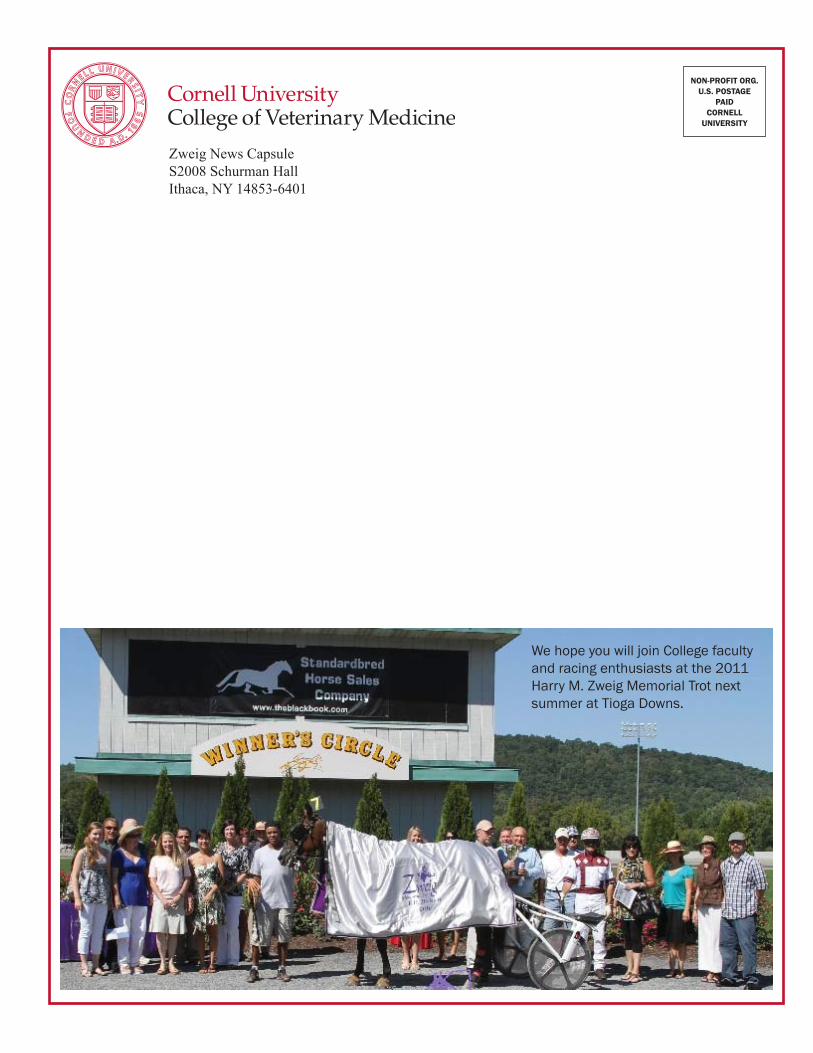
Zweig News CapsuleS2008 Schurman HallIthaca, NY 14853-6401
NON-PROFIT ORG.U.S. POSTAGE
PAIDCORNELL
UNIVERSITY
We hope you will join College faculty and racing enthusiasts at the 2011 Harry M. Zweig Memorial Trot next summer at Tioga Downs.