2007. epic2. national evidence-based guidelines for preventing healthcare-associated infections in...
DESCRIPTION
nursingTRANSCRIPT

epic2: National Evidence-Based Guidelines forPreventing Healthcare-Associated Infections inNHS Hospitals in England
R.J. Pratta*, C.M. Pellowea, J.A. Wilsona,b, H.P. Lovedaya, P.J. Harpera,
S.R.L.J. Jonesa, C. McDougallb, M.H. Wilcoxc
a Richard Wells Research Centre, Faculty of Health and Human Sciences, Thames Valley University
(London).b Department of Healthcare Associated Infection and Antimicrobial Resistance, Centre for Infections,
Health Protection Agency (London).c Microbiology and Infection Control, Leeds Teaching Hospitals NHS Trust and University of Leeds.
Submitted 23 November 2006
Available online 5 February 2007
Executive Summary National evidence-based guidelines for preventing healthcare-associatedinfections (HCAI) in National Health Service (NHS) hospitals in England were commissioned by theDepartment of Health (DH) and developed during 1998-2000 by a nurse-led multi-professional team ofresearchers and specialist clinicians. Following extensive consultation, they were published in January2001.1 These guidelines describe the precautions healthcare workers should take in three areas: standardprinciples for preventing HCAI, which include hospital environmental hygiene, hand hygiene, the use ofpersonal protective equipment, and the safe use and disposal of sharps; preventing infections associatedwith the use of short-term indwelling urethral catheters; and preventing infections associated withcentral venous catheters.The evidence for these guidelines was identified by multiple systematic reviews of experimental and non-experimental research and expert opinion as reflected in systematically identified professional, nationaland international guidelines, which were formally assessed by a validated appraisal process. In 2003, wedeveloped complementary national guidelines for preventing HCAI in primary and community care onbehalf of the National Collaborating Centre for Nursing and Supportive Care (National Institute for Healthand Clinical Excellence).2
A cardinal feature of evidence-based guidelines is that they are subject to timely review in order thatnew research evidence and technological advances can be identified, appraised and, if shown to beeffective in preventing HCAI, incorporated into amended guidelines. Periodically updating the evidencebase and guideline recommendations is essential in order to maintain their validity and authority.Consequently, the DH commissioned a review of new evidence published following the last systematicreviews. We have now updated the evidence base for making infection prevention and controlrecommendations. A critical assessment of the updated evidence indicated that the original epicguidelines published in 2001 remain robust, relevant and appropriate but that adjustments need to be
*Corresponding author: Professor Robert J. Pratt, Director, Richard Wells Research Centre, Faculty of Health and Human
Sciences, Thames Valley University, 32-38 Uxbridge Road, London W5 2BS.
Telephone: +44 (0)20 8280 5142; email: [email protected]
Journal of Hospital Infection (2007) 65S, S1–S64
Available online at www.sciencedirect.com
www.elsevierhealth.com/journals/jhin

1 Introductory section
1.1 Guideline Development Team
• Professor Robert J. Pratt (Project Director) –Professor of Nursing and Director, Richard WellsResearch Centre, Faculty of Health and HumanSciences, Thames Valley University (London).
• Dr. Carol M. Pellowe (Project Manager) –Deputy Director, Richard Wells ResearchCentre, Faculty of Health and Human Sciences,Thames Valley University (London).
• Heather P. Loveday, Principal Lecturer(Research), Richard Wells Research Centre,Faculty of Health and Human Sciences, ThamesValley University (London).
• Dr. Peter J. Harper, Senior Lecturer (Research),Richard Wells Research Centre, Faculty ofHealth and Human Sciences, Thames ValleyUniversity (London).
• Simon R.L.J. Jones, Lecturer (Research),Richard Wells Research Centre, Faculty ofHealth and Human Sciences, Thames ValleyUniversity (London).
• Jennie A. Wilson, Research Fellow, RichardWells Research Centre, Faculty of Health andHuman Sciences, Thames Valley University(London), and Programme Lead for the SurgicalSite Infection Surveillance Service, Departmentof Healthcare Associated Infection andAntimicrobial Resistance, Centre for Infections,Health Protection Agency (London).
• Christine McDougall, Surveillance Manager,Surgical Site Infection, Department ofHealthcare Associated Infection andAntimicrobial Resistance, Centre for Infections,Health Protection Agency (London).
• Professor Mark H. Wilcox, Professor of MedicalMicrobiology, Leeds Teaching Hospitals NHSTrust and Institute of Molecular and CellularBiology, University of Leeds.
1.2 Guideline Advisory Group
• Daphne Colpman, Continence Advisor, UniversityCollege London Hospitals NHS Foundation Trust.
• Andrew Jackson, Nurse Consultant (IntravenousTherapy), Rotherham District General Hospital,South Yorkshire.
• Royal College of Nursing Intravenous TherapyForum.
• Liz Simcock, Clinical Nurse Specialist forCentral Venous Access, Cancer Services,University College London Hospitals NHSFoundation Trust.
• Dr. Godfrey W Smith, Consultant, RoyalLiverpool and Broadgreen University HospitalsNHS Trust and the University of Liverpool.
1.3 Acknowledgements
We would like to acknowledge the assistance wereceived from The Liverpool Reviews andImplementation Group (University of Liverpool)who shared with us data from their HealthTechnology Assessment focused on the clinical andcost effectiveness of central venous catheterstreated with antimicrobial agents in preventingbloodstream infections. We are also indebted tothe Infection Control Nurses Association and theHospital Infection Society for their input into the
S2 R.J. Pratt et al.
made to some guideline recommendations following a synopsis of the evidence underpinning theguidelines.These updated national guidelines (epic2) provide comprehensive recommendations for preventing HCAIin hospitals and other acute care settings based on the best currently available evidence. Because thisis not always the best possible evidence, we have included a suggested agenda for further research ineach section of the guidelines. National evidence-based guidelines are broad principles of best practicewhich need to be integrated into local practice guidelines. To monitor implementation, we havesuggested key audit criteria for each section of recommendations.Clinically effective infection prevention and control practice is an essential feature of protectingpatients. By incorporating these guidelines into routine daily clinical practice, patient safety can beenhanced and the risk of patients acquiring an infection during episodes of healthcare in NHS hospitalsin England can be minimised.

development of these guidelines and to otherassociations, learned societies, professionalorganisations, Royal Colleges and patient groupswho took an active role in the external review ofthe guidelines. We would also like to acknowledgethe support we received from Sally Wellsteed andCarole Fry in the Chief Medical Officer’s Team atthe Department of Health (England) and fromProfessor Brian Duerden, Inspector of Microbiologyand Infection Control, Department of Health(England).
1.4 Source of Funding
The Department of Health (England)
1.5 Conflict of Interest
None
1.6 Relationship of Author(s) with Sponsor
The Department of Health (England) commissionedthe authors to update the evidence and guidelinerecommendations previously developed by themand published as the epic guidelines in the Journal
of Hospital Infection in 2001.
1.7 Responsibility for Guidelines
The views expressed in this publication are thoseof the authors and, following extensive consul-tation, have been endorsed by the Department ofHealth (England).
1.8 Summary of Guidelines
Standard Principles for preventing healthcare-associated infections in hospital and otheracute care settingsThis guidance is based on the best criticallyappraised evidence currently available. The typeand class of supporting evidence explicitly linkedto each recommendation is described. All recom-mendations are endorsed equally and none isregarded as optional. These recommendations are
not detailed procedural protocols and need to beincorporated into local guidelines.
This guidance on infection control precautionsshould be applied by all healthcare practitionersto the care of every patient. Job descriptionsshould reflect this and annual appraisal evidenceshould be available to support continuingengagement of each member of staff. Therecommendations are divided into four distinctinterventions:
1. Hospital environmental hygiene;2. Hand hygiene;3. The use of personal protective equipment; and4. The safe use and disposal of sharps.
These guidelines do not address the additionalinfection control requirements of specialist settings,such as the operating department.
Hospital environmental hygiene
SP1 The hospital environment must be visibly Class C
clean, free from dust and soilage and
acceptable to patients, their visitors and staff.
SP2 Increased levels of cleaning should be Class D
considered in outbreaks of infection where
the pathogen concerned survives in the
environment and environmental contamination
may be contributing to spread.
SP3 The use of hypochlorite and detergent Class D
should be considered in outbreaks of
infection where the pathogen concerned
survives in the environment and environmental
contamination may be contributing to spread.
SP4 Shared equipment used in the clinical Class D
environment must be decontaminated
appropriately after each use.
SP5 All healthcare workers need to be aware Class D
of their individual responsibility for
maintaining a safe care environment for
patients and staff. Every healthcare worker
needs to be clear about their specific
responsibilities for cleaning equipment and
clinical areas (especially those areas in
close proximity to patients).They must be
educated about the importance of ensuring
that the hospital environment is clean and
that opportunities for microbial contamination
are minimised.
Hand hygiene
SP6 Hands must be decontaminated Class C
immediately before each and every
episode of direct patient contact/care and
after any activity or contact that potentially
results in hands becoming contaminated.
epic2: Guidelines for Preventing Healthcare-Associated Infections in NHS Hospitals S3

SP7 Hands that are visibly soiled or potentially Class A
grossly contaminated with dirt or organic
material (i.e. following the removal of
gloves) must be washed with liquid soap
and water.
SP8 Hands should be decontaminated between Class A
caring for different patients or between
different care activities for the same patient.
For convenience and efficacy an alcohol-based
handrub is preferable unless hands are visibly
soiled. Local infection control guidelines may
advise an alternative product in some outbreak
situations.
SP9 Hands should be washed with soap and Class D/GPP
water after several consecutive
applications of alcohol handrub.
SP10 Before a shift of clinical work begins, all Class D
wrist and ideally hand jewellery should
be removed. Cuts and abrasions must be
covered with waterproof dressings.
Fingernails should be kept short, clean and
free from nail polish. False nails and nail
extensions must not be worn by clinical staff.
SP11 An effective handwashing technique Class D
involves three stages: preparation, washing
and rinsing, and drying. Preparation requires
wetting hands under tepid running water
before applying the recommended amount
of liquid soap or an antimicrobial preparation.
The handwash solution must come into
contact with all of the surfaces of the hand.
The hands must be rubbed together
vigorously for a minimum of 10-15 seconds,
paying particular attention to the tips of the
fingers, the thumbs and the areas between
the fingers. Hands should be rinsed
thoroughly prior to drying with good quality
paper towels.
SP12 When decontaminating hands using an Class D
alcohol-based handrub, hands should be free
of dirt and organic material. The handrub
solution must come into contact with all
surfaces of the hand. The hands must be
rubbed together vigorously, paying particular
attention to the tips of the fingers, the
thumbs and the areas between the fingers,
until the solution has evaporated and
the hands are dry.
SP13 Clinical staff should be aware of the Class D
potentially damaging effects of hand
decontamination products. They should be
encouraged to use an emollient hand cream
regularly, for example, after washing hands
before a break or going off duty and when
off duty, to maintain the integrity of
the skin.
SP14 If a particular soap, antiseptic hand wash Class D
or alcohol-based product causes skin
irritation, review methods as described in
Recommendation SP11 and 12 before
consulting the occupational health team.
SP15 Near patient alcohol-based hand rub Class D
should be made available in all healthcare
facilities.
SP16 Hand hygiene resources and individual Class D
practice should be audited at regular
intervals and the results fed back to
healthcare workers.
SP17 Education and training in risk assessment, Class D
effective hand hygiene and glove use
should form part of all healthcare workers’
annual updating.
The use of personal protection equipment
SP18 Selection of protective equipment Class D/H&S
must be based on an assessment of the
risk of transmission of microorganisms to
the patient or to the carer, and the risk
of contamination of the healthcare
practitioners’ clothing and skin by
patients’ blood, body fluids, secretions
or excretions.
SP19 Everyone involved in providing care Class D/H&S
should be educated about standard
principles and trained in the use of
protective equipment.
SP20 Adequate supplies of disposable plastic Class D/H&S
aprons, single use gloves and face
protection should be made available
wherever care is delivered. Gowns should
be made available when advised by the
infection control team.
SP21 Gloves must be worn for invasive Class D/H&S
procedures, contact with sterile sites,
and non-intact skin or mucous membranes,
and all activities that have been assessed
as carrying a risk of exposure to blood,
body fluids, secretions and excretions;
and when handling sharp or contaminated
instruments.
SP22 Gloves must be worn as single use Class D/H&S
items. They are put on immediately
before an episode of patient contact or
treatment and removed as soon as the
activity is completed. Gloves are changed
between caring for different patients, or
between different care/treatment
activities for the same patient.
SP23 Gloves must be disposed of as clinical Class D/H&S
waste and hands decontaminated, ideally
by washing with liquid soap and water
after the gloves have been removed.
S4 R.J. Pratt et al.

SP24 Gloves that are acceptable to Class D/H&S
healthcare personnel and CE marked
must be available in all clinical areas.
SP25 Sensitivity to natural rubber latex in Class D/H&S
patients, carers and healthcare personnel
must be documented and alternatives to
natural rubber latex must be available.
SP26 Neither powdered nor polythene Class C/H&S
gloves should be used in health care
activities.
SP27 Disposable plastic aprons must be worn Class D/H&S
when close contact with the patient,
materials or equipment are anticipated
and when there is a risk that clothing
may become contaminated with pathogenic
microorganisms or blood, body fluids,
secretions or excretions, with the exception
of perspiration.
SP28 Plastic aprons/gowns should be worn Class D/H&S
as single-use items, for one procedure
or episode of patient care, and then
discarded and disposed of as clinical
waste. Non-disposable protective clothing
should be sent for laundering.
SP29 Full-body fluid-repellent gowns must Class D/H&S
be worn where there is a risk of
extensive splashing of blood, body fluids,
secretions or excretions, with the
exception of perspiration, onto the skin
or clothing of healthcare personnel (for
example when assisting with childbirth).
SP30 Face masks and eye protection must Class D/H&S
be worn where there is a risk of blood,
body fluids, secretions or excretions
splashing into the face and eyes.
SP31 Respiratory protective equipment, Class D/H&S
i.e., a particulate filter mask, must be
correctly fitted and used when
recommended for the care of patients
with respiratory infections transmitted
by airborne particles.
The safe use and disposal of sharps
SP32 Sharps must not be passed directly Class D/H&S
from hand to hand and handling should
be kept to a minimum.
SP33 Needles must not be recapped, bent Class D/H&S
broken or disassembled after use.
SP34 Used sharps must be discarded into a Class D/H&S
sharps container (conforming to UN3291
and BS 7320 standards) at the point of
use by the user. These must not be
filled above the mark that indicates
the bin is full.
SP35 All sharps bins should be positioned Class D/H&S
out of the reach of children at a height
that enables safe disposal by all members
of staff. They should be secured to avoid
spillage.
SP36 All staff both clinical and non Class D/H&S /GPP
clinical must be educated about the
safe use and disposal of sharps.
SP37 Consider the use of needlestick- Class B/H&S
prevention devices where there are
clear indications that they will provide
safe systems of working for healthcare
practitioners.
SP38 Conduct a rigorous evaluation of Class D
needlestick-prevention devices to
determine their effectiveness,
acceptability to practitioners, impact
on patient care and cost benefit prior
to widespread introduction.
Guidelines for preventing infections associatedwith the use of short-term indwelling urethralcathetersThis guidance is based on the best criticallyappraised evidence currently available. The typeand class of supporting evidence explicitly linkedto each recommendation is described. All recom-mendations are endorsed equally and none isregarded as optional. These recommendations arenot detailed procedural protocols and need to beincorporated into local guidelines.
These guidelines apply to adults and childrenaged 1 year and older and should be read inconjunction with the guidance on StandardPrinciples. The recommendations are divided intofive distinct interventions:
1. Assessing the need for catheterisation;2. Selection of catheter type and system;3. Catheter insertion;4. Catheter maintenance; and5. Education of patients, relatives and healthcare
workers.
Assessing the need for catheterisation
UC1 Only use indwelling urethral catheters Class D/GPP
after considering alternative methods
of management.
UC2 Document the need for catheterisation, Class D/GPP
catheter insertion and care.
UC3 Review regularly the patient’s clinical Class D/GPP
need for continuing urinary catheterisation
and remove the catheter as soon as possible.
Selection of Catheter Type
UC4 Choice of catheter material will depend Class D
on clinical experience, patient assessment
and anticipated duration of catheterisation.
epic2: Guidelines for Preventing Healthcare-Associated Infections in NHS Hospitals S5
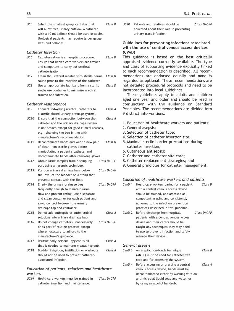
UC5 Select the smallest gauge catheter that Class D
will allow free urinary outflow. A catheter
with a 10 ml balloon should be used in adults.
Urological patients may require larger gauge
sizes and balloons.
Catheter Insertion
UC6 Catheterisation is an aseptic procedure. Class D
Ensure that health care workers are trained
and competent to carry out urethral
catheterisation.
UC7 Clean the urethral meatus with sterile normal Class D
saline prior to the insertion of the catheter.
UC8 Use an appropriate lubricant from a sterile Class D
single use container to minimise urethral
trauma and infection.
Catheter Maintenance
UC9 Connect indwelling urethral catheters to Class A
a sterile closed urinary drainage system.
UC10 Ensure that the connection between the Class A
catheter and the urinary drainage system
is not broken except for good clinical reasons,
e.g., changing the bag in line with
manufacturer’s recommendation.
UC11 Decontaminate hands and wear a new pair Class D
of clean, non-sterile gloves before
manipulating a patient’s catheter and
decontaminate hands after removing gloves.
UC12 Obtain urine samples from a sampling Class D/GPP
port using an aseptic technique.
UC13 Position urinary drainage bags below Class D/GPP
the level of the bladder on a stand that
prevents contact with the floor.
UC14 Empty the urinary drainage bag Class D/GPP
frequently enough to maintain urine
flow and prevent reflux. Use a separate
and clean container for each patient and
avoid contact between the urinary
drainage tap and container.
UC15 Do not add antiseptic or antimicrobial Class A
solutions into urinary drainage bags.
UC16 Do not change catheters unnecessarily Class D/GPP
or as part of routine practice except
where necessary to adhere to the
manufacturer’s guidance.
UC17 Routine daily personal hygiene is all Class A
that is needed to maintain meatal hygiene.
UC18 Bladder irrigation, instillation or washouts Class A
should not be used to prevent catheter-
associated infection.
Education of patients, relatives and healthcare
workers
UC19 Healthcare workers must be trained in Class D/GPP
catheter insertion and maintenance.
UC20 Patients and relatives should be Class D/GPP
educated about their role in preventing
urinary tract infection.
Guidelines for preventing infections associatedwith the use of central venous access devices(CVAD)This guidance is based on the best criticallyappraised evidence currently available. The typeand class of supporting evidence explicitly linkedto each recommendation is described. All recom-mendations are endorsed equally and none isregarded as optional. These recommendations arenot detailed procedural protocols and need to beincorporated into local guidelines.
These guidelines apply to adults and childrenaged one year and older and should be read inconjunction with the guidance on StandardPrinciples. The recommendations are divided into9 distinct interventions:
1. Education of healthcare workers and patients;2. General asepsis;3. Selection of catheter type;4. Selection of catheter insertion site;5. Maximal sterile barrier precautions during
catheter insertion;6. Cutaneous antisepsis;7. Catheter and catheter site care;8. Catheter replacement strategies; and9. General principles for catheter management.
Education of healthcare workers and patients
CVAD 1 Healthcare workers caring for a patient Class D
with a central venous access device
should be trained, and assessed as
competent in using and consistently
adhering to the infection prevention
practices described in this guideline.
CVAD 2 Before discharge from hospital, Class D/GPP
patients with a central venous access
device and their carers should be
taught any techniques they may need
to use to prevent infection and safely
manage their device.
General asepsis
CVAD 3 An aseptic non-touch technique Class B
(ANTT) must be used for catheter site
care and for accessing the system.
CVAD 4 Before accessing or dressing a central Class A
venous access device, hands must be
decontaminated either by washing with an
antimicrobial liquid soap and water, or
by using an alcohol handrub.
S6 R.J. Pratt et al.

CVAD 5 Hands that are visibly soiled or Class A
contaminated with dirt or organic
material must be washed with liquid
soap and water before using an alcohol
handrub.
CVAD 6 Following hand antisepsis, clean gloves Class D
and an ANTT, or sterile gloves should be
used when changing the insertion site
dressing, line manipulation or intravenous
drug administration.
Selection of Catheter Type
CVAD 7 Use a single-lumen catheter unless Class A
multiple ports are essential for the
management of the patient.
CVAD 8 If a multilumen catheter is used, Class D/GPP
identify and designate one port
exclusively for hyperalimentation to
administer parenteral nutrition.
CVAD 9 Use a tunnelled or implanted central Class A
venous access device (one with a
subcutaneous port) for patients in whom
long-term (more than 3-4 weeks) vascular
access is anticipated.
CVAD 10 Consider the use of an antimicrobial Class A
impregnated central venous access device
for adult patients who require short-term
(1 to 3 weeks) central venous
catheterisation and who are at high risk
for catheter-related bloodstream infection
(CR-BSI) if rates of CR-BSI remain high
despite implementing a comprehensive
strategy to reduce rates of CR-BSI.
Selection of Catheter Insertion Site
CVAD 11 In selecting an appropriate insertion Class D/GPP
site, assess the risks for infection
against the risks of mechanical
complications.
CVAD 12 Unless medically contraindicated, use Class C
the subclavian site in preference to
the jugular or femoral sites for
nontunnelled catheter placement.
CVAD 13 Use implantable access devices for Class C
patients who require long-term,
intermittent vascular access. For patients
requiring regular or continuous access,
a tunnelled central venous access
device is preferable.
Maximal Sterile Barrier Precautions during
Catheter Insertion
CVAD 14 Use maximal sterile barriers, including Class C
a sterile gown, sterile gloves, and a
large sterile drape, for the insertion of
central venous access devices.
Cutaneous Antisepsis
CVAD 15 Decontaminate the skin site with a Class A
single patient use application of alcoholic
chlorhexidine gluconate solution
(preferably 2% chlorhexidine gluconate
in 70% isopropyl alcohol) prior to the
insertion of a central venous access device.
CVAD 16 Use a single patient use application Class D/GPP
of alcoholic povidone-iodine solution
for patients with a history of
chlorhexidine sensitivity. Allow the
antiseptic to dry before inserting the
central venous access device.
CVAD 17 Do not apply organic solvents, Class D/GPP
e.g., acetone, ether, to the skin
before the insertion of a central
venous access device.
CVAD 18 Do not routinely apply antimicrobial Class D/GPP
ointment to the catheter placement
site prior to insertion.
Catheter and Catheter Site Care
CVAD 19 Preferably, a sterile, transparent, Class D
semi-permeable polyurethane dressing
should be used to cover the catheter
insertion site.
CVAD 20 Transparent dressings should be changed Class D
every 7 days, or sooner if they are no
longer intact or moisture collects under
the dressing.
CVAD 21 If a patient has profuse perspiration Class D/GPP
or if the insertion site is bleeding or
oozing, a sterile gauze dressing is
preferable to a transparent,
semi-permeable dressing.
CVAD 22 The need for a gauze dressing should Class D/GPP
be assessed daily and changed when
inspection of the insertion site is
necessary or when the dressing becomes
damp, loosened or soiled. A gauze
dressing should be replaced by a
transparent dressing as soon as possible.
CVAD 23 Dressings used on tunnelled or Class D
implanted catheter insertion sites
should be replaced every 7 days until
the insertion site has healed, unless
there is an indication to change them sooner.
CVAD 24 An alcoholic chlorhexidine gluconate Class A
solution (preferably 2% chlorhexidine
gluconate in 70% isopropyl alcohol)
should be used to clean the catheter
insertion site during dressing changes, and
allowed to air dry. An aqueous solution of
chlorhexidine gluconate should be used if
the manufacturer’s recommendations
prohibit the use of alcohol with their product.
epic2: Guidelines for Preventing Healthcare-Associated Infections in NHS Hospitals S7

CVAD 25 Individual single use sachets of Class D/GPP
antiseptic solution or individual
packages of single use antiseptic-
impregnated swabs or wipes should
be used to disinfect the insertion site.
CVAD 26 Do not apply antimicrobial ointment Class D/GPP
to catheter insertion sites as part of
routine catheter site care.
CVAD 27 Healthcare workers should ensure that Class D/GPP
catheter-site care is compatible with
catheter materials (tubing, hubs, injection
ports, luer connectors and extensions)
and carefully check compatibility with
the manufacturer’s recommendations.
Catheter Replacement Strategies
CVAD 28 Do not routinely replace catheters Class A
as a method to prevent catheter-
related infection.
CVAD 29 Use guide wire assisted catheter Class A
exchange to replace a malfunctioning
catheter, or to exchange an existing
catheter only if there is no evidence of
infection at the catheter site or proven
catheter-related bloodstream infection.
CVAD 30 If catheter-related infection is suspected, Class A
but there is no evidence of infection at
the catheter site, remove the existing
catheter and insert a new catheter over
a guide wire; if tests reveal catheter-related
infection, the newly inserted catheter
should be removed and, if still required,
a new catheter inserted at a different site.
CVAD 31 Do not use guide wire assisted catheter Class A
exchange for patients with catheter-
related infection. If continued vascular
access is required, remove the implicated
catheter, and replace it with another
catheter at a different insertion site.
CVAD 32 Replace all fluid administration Class D/GPP
tubing and connectors when the
central venous access device is replaced.
General Principles for Catheter Management
CVAD 33 A single patient use application of Class D/GPP
alcoholic chlorhexidine gluconate
solution (preferably 2% chlorhexidine
gluconate in 70% isopropyl alcohol)
should be used and allowed to dry
when decontaminating the injection
port or catheter hub before and after
it has been used to access the system,
unless contraindicated by the manufacturer’s
recommendations, in which case either
aqueous chlorhexidine gluconate or aqueous
povidone iodine should be used.
CVAD 34 In-line filters should not be used Class D
routinely for infection prevention
purposes.
CVAD 35 Antibiotic lock solutions should not Class D
be used routinely to prevent
catheter-related bloodstream infections.
CVAD 36 Do not routinely administer intranasal Class A
or systemic antimicrobials before
insertion or during the use of a central
venous access device to prevent catheter
colonisation or bloodstream infection.
CVAD 37 Preferably, a single-lumen catheter Class D
should be used to administer parenteral
nutrition. If a multilumen catheter is
used, one port must be exclusively
dedicated for hyperalimentation and all
lumens must be handled with the same
meticulous attention to aseptic technique.
CVAD 38 Preferably, sterile 0.9 percent sodium Class A
chloride for injection should be used
to flush and lock catheter lumens that
are in frequent use.
CVAD 39 When recommended by the manufacturer, Class D
implanted ports or opened-ended
catheter lumens should be flushed and
locked with heparin sodium flush solutions.
CVAD 40 Systemic anticoagulants should not be Class D
used routinely to prevent catheter-related
bloodstream infection.
CVAD 41 The introduction of new intravascular Class D/GPP
devices that include needle-free
devices should be monitored for an
increase in the occurrence of device
associated infection. If an increase in
infection rates is suspected, this
should be reported to the Medicines
and Healthcare products Regulatory Agency
[http://www.mhra.gov.uk]
CVAD 42 If needle-free devices are used, the Class D/GPP
manufacturer’s recommendations
for changing the needle-free
components should be followed.
CVAD 43 When needle-free devices are used, Class D/GPP
healthcare workers should ensure
that all components of the system
are compatible and secured, to
minimise leaks and breaks in the system.
CVAD 44 When needle-free devices are used, Class D
the risk of contamination should be
minimised by decontaminating the access
port before and after use with a single
patient use application of alcoholic
chlorhexidine gluconate solution
(preferably 2% chlorhexidine gluconate
in 70% isopropyl alcohol) unless
contraindicated by the manufacturer’s
S8 R.J. Pratt et al.

recommendations, in which case
aqueous povidone iodine should be used.
CVAD 45 In general, solution administration Class A
sets in continuous use need not be
replaced more frequently than at 72 hour
intervals unless they become disconnected
or a central venous access device is replaced.
CVAD 46 Administration sets for blood and Class D
blood components should be changed
when the transfusion episode is complete
or every 12 hours (whichever is sooner),
or according to the manufacturer’s
recommendations.
CVAD 47 Administration sets used for total Class D
parenteral nutrition infusions should
generally be changed every 24 hours. If
the solution contains only glucose and
amino acids, administration sets in
continuous use do not need to be replaced
more frequently than every 72 hours.
1.9 Introduction – the epic2 Guidelines
National evidence-based guidelines for preventinghealthcare-associated infections (HCAI) in NHShospitals were commissioned by the Departmentof Health (England) (DH) and developed during1998-2000 by a nurse-led multi-professional teamof researchers and specialist clinicians. They wereintended to provide reliable best evidence for thedevelopment of local infection prevention andcontrol guidelines and protocols and facilitateclinically effective practice. Having been developedwithin the ‘epic initiative’ in the Richard WellsResearch Centre at Thames Valley University, theybecame known as the ‘epic’ guidelines. Followingextensive consultation, they were published inJanuary 2001.1 Two years later, under the auspicesof the National Institute for Health and ClinicalExcellence (NICE), a complementary set ofnational evidence-based guidelines were developedby the epic initiative, focused on preventing HCAIin primary and community care.2
An evidence review in 2004 indicated thenecessity to amend and update some of theoriginal epic guideline recommendations to ensurethat they continue to reflect new and emergingevidence, remain relevant to infection control andprevention practice and enjoy the confidence ofpractitioners and patients.3,4
Additional updating systematic reviews wereconducted in 2005 and the original epic guidelineshave now been revised. They are referred to inthis publication as the epic2 infection prevention
guidelines, which now replace the original 2001guidelines.
What are national evidence-based guidelines?These are systematically developed broadstatements (principles) of good practice. They aredriven by practice need, based on evidence andsubject to multi-professional debate, timely andfrequent review, and modification. Nationalguidelines are intended to inform the developmentof detailed operational protocols at local level andcan be used to ensure that these incorporate themost important principles for preventing HCAI inNHS hospitals and other acute care health services.
Why do we need national guidelines forpreventing healthcare-associated infections?During the past two decades, HCAI have become asignificant threat to patient safety. Thetechnological advances made in the treatment ofmany diseases and disorders are often underminedby the transmission of infections within healthcaresettings, particularly those caused by anti-microbial-resistant strains of disease-causingmicroorganisms that are now endemic in manyhealthcare environments. The financial andpersonal cost of these infections, in terms of theeconomic consequences to the NHS and thephysical, social and psychological costs to patientsand their relatives, have increased both govern-ment and public awareness of the risks associatedwith healthcare interventions, especially that ofacquiring a new infection.
Although not all HCAI can be prevented, manycan. Clinical effectiveness, i.e., using preventionmeasures that are based on reliable evidence ofefficacy, is a core component of an effectivestrategy designed to protect patients from the riskof infection.
What is the purpose of the guidelines?These guidelines describe clinically effectivemeasures that are used by healthcare workers forpreventing infections in hospital and other acutecare health services.
What is the scope of the guidelines?Three sets of guidelines were originally developedand have now been updated. They include:
• Standard infection control principles includebest practice recommendations for hospitalenvironmental hygiene, effective handhygiene, the appropriate use of personalprotective equipment, and the safe use anddisposal of sharps;
epic2: Guidelines for Preventing Healthcare-Associated Infections in NHS Hospitals S9

• Guidelines for preventing infections associatedwith the use of short-term indwelling urethralcatheters; and
• Guidelines for preventing infectionsassociated with the use of central venousaccess devices.
What is the evidence for these guidelines?The evidence for these guidelines was identifiedby multiple systematic reviews of experimentaland non-experimental research. In addition, evi-dence from expert opinion as reflected in syste-matically identified professional, national andinternational guidelines was considered followingformal assessment using a validated appraisalprocess.5,6 All evidence was critically appraised forits methodological rigour and clinical practiceapplicability and the best available evidenceinfluenced the guideline recommendations.
Who developed these guidelines?The epic2 guidelines were developed by a nurse-led team of researchers, senior infection controlnurses and a Director of Microbiology and InfectionPrevention and Control in a large NHS TeachingHospital Trust (see 1.1).
Who are these guidelines for?These guidelines can be appropriately adaptedand used by all hospital practitioners. They willinform the development of more detailed localprotocols and ensure that important standardprinciples for infection prevention are incorpora-ted. Consequently, they are aimed at hospitalmanagers, members of hospital infection controlteams, and individual health care practitioners. Atan individual level, they are intended to influencethe quality and clinical effectiveness of infectionprevention decision-making. The dissemination ofthese guidelines also help patients understand thestandard infection prevention precautionsrecommended to protect them from HCAI.
How are these guidelines structured?Each set of guidelines follows an identical format,which consists of:
• a resume of the systematic review process;
• the intervention heading;• a headline statement describing the key issues
being addressed;• a synthesis of the related evidence;• an economic opinion, where appropriate;• guideline recommendation(s) classified
according to the strength of the underpinningevidence.
Finally, at the end of each section there is adescription of areas for further research andsuggested audit criteria. All evidence is referencedin section 5.
How frequently are the guidelines reviewedand updated?A cardinal feature of evidence-based guidelines isthat they are subject to timely review in orderthat new research evidence and technologicaladvances can be identified, appraised and, ifshown to be effective in preventing HCAI, incor-porated into amended guidelines. The evidencebase for these guidelines will be reviewed in twoyears (2009) and the guidelines will be updatedapproximately four years after publication (2011).
How can these guidelines be used to improveyour clinical effectiveness?In addition to informing the development ofdetailed local operational protocols, these guide-lines can be used as a benchmark for determiningappropriate infection prevention decisions and, aspart of reflective practice, to assess clinical effec-tiveness. They also provide a baseline for clinicalaudit, evaluation and education, and facilitateongoing quality improvements.
How much will it cost to implement theseguidelines?Significant additional costs are not anticipated inimplementing these guidelines. However, wherecurrent equipment or resources do not facilitatethe implementation of the guidelines, or wherestaff levels of adherence to current guidance arepoor, there may be an associated increase in costs.Given the social and economic costs of HCAI, theconsequences associated with not implementingthese guidelines would be unacceptable to bothpatients and health care professionals.
Consultation processThese guidelines have been subject to extensiveexternal consultation with key stakeholders,including Royal Colleges, professional societiesand organisations, including patients, and tradesunions (Appendix A.1).
1.10 Guideline Development Methodology
The guidelines were developed using a systematicreview process (Appendix A.2). In each set ofguidelines a resume of the relevant guidelinedevelopment methodology is provided.
S10 R.J. Pratt et al.

Search ProcessElectronic databases were searched for nationaland international guidelines and research studiespublished during the period 01 January 1999 to 31August 2005. A two-stage search process was used.
Stage 1: Identification of systematic reviews and
guidelines
For each set of epic guidelines, an electronicsearch was conducted for systematic reviews ofrandomised controlled trials and current nationaland international guidelines. The following databases were searched:
• Cochrane Library;• National Guideline Clearinghouse;• National Electronic Library of Health;• National Institute for Health and Clinical
Excellence.
Guidelines were retrieved and subjected tocritical appraisal using the AGREE Instrument,6 anevaluation method used in Europe for assessingthe methodological quality of clinical practiceguidelines.
Following appraisal, accepted guidelines wereincluded as part of the evidence base supportingguideline development. They were also used toverify professional consensus and in someinstances, as the primary source of evidence.
Stage 2: Systematic search for additional
evidence
Review questions for the systematic reviews of theliterature were then developed for each set ofepic guidelines following recommendations fromexpert advisors.
Searches were constructed using relevant MeSH(medical subject headings) and free-text terms.On completion of the main search, an economicfilter was applied. The following databases weresearched:
• Medline;• Cumulated Index of Nursing and Allied Health
Literature;• Embase;• The Cochrane Library.
Abstract review – identifying studies forappraisalSearch results were downloaded into a ReferenceManager™ database and titles and abstractsprinted for preliminary review. Reviewers identi-fied and retrieved all studies where the title orabstract: addressed one or more of the review
questions; identified primary research or systema-tically conducted secondary research; indicated atheoretical/clinical/ in use study. No researchdesigns were specifically excluded but whereverpossible, in use rather than in vitro studies wereretrieved.
Where no abstract was available and the titleindicated one or more of the above criteria, thestudy was retrieved. Due to the limited resourcesavailable for this review, foreign language studieswere not reviewed.
All full-text studies retrieved were indepen-dently assessed by two experienced reviewers whoidentified those studies meeting the aboveinclusion criteria for critical appraisal.
Quality Assessment and Data ExtractionIncluded studies were appraised using an adapteddata extraction process based on systemsdeveloped by the Scottish IntercollegiateGuideline Network for study quality assessment.7
Due to the limited resources available for thisreview, studies were not double-blind appraised.However, all studies were appraised and dataextracted by one experienced reviewer and thenchecked by a second experienced reviewer. Anydisagreement between reviewers was resolvedthrough discussion. Evidence tables wereconstructed from the quality assessments and thestudies summarised in the evidence reports. Theevidence was classified using methods adopted bythe National Institute for Health and ClinicalExcellence (NICE) from The Scottish IntercollegiateGuideline Network (SIGN) (Table 1).8,9 This systemdiffers from that used in the previous epic andNICE infection prevention guidelines.1,2
The evidence tables and reports were presentedto the advisors for discussion. At this stage, expertadvice derived from seminal works and appraisednational and international guidelines wereconsidered. Following extensive discussion theguidelines were drafted.
Factors influencing the guideline recommen-dations included:
• the nature of the evidence;• the applicability of the evidence to practice;• costs and knowledge of healthcare systems.
The classification scheme adapted by NICE fromSIGN was used to define the strength ofrecommendation (Table 2).8,9
The complete series of evidence tables areposted on the epic website at: [http://www.epic.tvu.ac.uk].
epic2: Guidelines for Preventing Healthcare-Associated Infections in NHS Hospitals S11

1.11 Consultation Process
The draft guidelines were circulated tostakeholders for comment (see Appendix A.1). Thelist of stakeholders included all those consultedfor the phase 1 guidelines and others agreed bythe DH (England).
Comments were requested on:
• the format;• the content;• practice applicability of the guidelines;• specific sections or recommendations.
All comments received were collated in an MSWord™ table for consideration by the guideline
developers and advisors who agreed any changesto the draft recommendations.
2 Standard Principles for preventinghealthcare-associated infections inhospital and other acute care settings
2.1 Introduction
This guidance is based on the best criticallyappraised evidence currently available. The typeand class of supporting evidence explicitly linkedto each recommendation is described. All recom-
S12 R.J. Pratt et al.
Table 1. Levels of Evidence for Intervention Studies8
Level of evidence Type of evidence
1++ High-quality meta-analyses, systematic reviews of randomised controlled trials (RCT), or RCT with a
very low risk of bias
1+ Well-conducted meta-analyses, systematic reviews of RCT, or RCT with a low risk of bias
1- Meta-analyses, systematic reviews of RCT, or RCT with a high risk of bias*
2++ High-quality systematic reviews of case–control or cohort studies
High-quality case–control or cohort studies with a very low risk of confounding, bias or chance and a
high probability that the relationship is causal
2+ Well-conducted case–control or cohort studies with a low risk of confounding, bias or chance and a
moderate probability that the relationship is causal
2- Case–control or cohort studies with a high risk of confounding bias, or chance and a significant risk
that the relationship is not causal*
3 Non-analytic studies (for example, case reports, case series)
4 Expert opinion, formal consensus
*Studies with a level of evidence ‘-’ should not be used as a basis for making a recommendation
Table 2. Classification of Recommendations8
Class Evidence
A At least one meta-analysis, systematic review, or randomised controlled trial (RCT) that is rated as 1++, and is directly applicable to the target population, or
A systematic review of RCT or a body of evidence that consists principally of studies rated as 1+, is directly applicable to the target population and demonstrates overall consistency of results
Evidence drawn from a NICE technology appraisalB A body of evidence that includes studies rated as 2++, is directly applicable to the target population
and demonstrates overall consistency of results, orExtrapolated evidence from studies rated as 1++ or 1+
C A body of evidence that includes studies rated as 2+, is directly applicable to the target population and demonstrates overall consistency of results, or
Extrapolated evidence from studies rated as 2++D Evidence level 3 or 4, or
Extrapolated evidence from studies rated as 2+, orFormal consensus
D (GPP) A good practice point (GPP) is a recommendation for best practice based on the experience of the Guideline Development Group
IP: Recommendation from NICE Interventional Procedures guidance

mendations are endorsed equally and none isregarded as optional. These recommendations arenot detailed procedural protocols and need to beincorporated into local guidelines.
Standard infection control precautions need tobe applied by all healthcare practitioners to thecare of all patients, i.e., adults, children andneonates. The recommendations are divided intofour distinct interventions:
1. Hospital environmental hygiene;2. Hand hygiene;3. The use of personal protective equipment; and4. The safe use and disposal of sharps.
These guidelines do not address the additionalinfection control requirements of specialistsettings, such as the operating department or foroutbreak situations.
2.2 Systematic Review Process
We have previously described the systematic reviewprocess in Section 1.10. For detailed descriptions ofprevious systematic reviews which have contributedto the evidence base underpinning these guidelines,readers should consult the original guidelines,1 theguidelines for the prevention of health-careassociated infections in primary and communitycare2 and our interim report in 2004 on changes inthe evidence base.3 Search questions weredeveloped from advice received from our specialistadvisors and the results of the searches are found inSection 2.10. The process outlined in Section 2.10refers only to the most recent systematic review ofthe literature undertaken in 2005.
Following our reviews, guidelines were draftedwhich described 38 recommendations within thebelow intervention categories:
1. Hospital environmental hygiene;2. Hand hygiene;3. Personal protective equipment; and4. Safe use and disposal of sharps.
2.3 Hospital Environmental Hygiene
Good hospital hygiene is an integral andimportant component of a strategy for preventinghealthcare-associated infections in hospitalsThis section discusses the evidence upon whichrecommendations for hospital environmental
hygiene are based. Hospital environmentalhygiene encompasses a wide range of routineactivities including: cleaning and decontamina-tion; laundry and housekeeping; safe collectionand disposal of general and clinical waste; andkitchen and food hygiene. Guidelines are providedhere for:
• cleaning the general hospital environment;• cleaning items of shared equipment; and• education and training of staff.
Maintain a clean hospital environmentOur initial systematic review concluded that therewas little research evidence of an acceptablequality upon which to base guidance related to themaintenance of hospital environmental hygiene.1
However, there was a body of clinical evidence,derived from case reports and outbreak investiga-tions, which suggested an association betweenpoor environmental hygiene and the transmissionof microorganisms causing healthcare-associatedinfections in hospital.10,11
Attention had been drawn to perceived fallingstandards in the cleanliness of hospitals since theintroduction of compulsory comprehensivetendering and the internal market. This concernwas addressed by the Infection Control NursesAssociation and the Association of DomesticManagers, resulting in the adoption and publicationby the Department of Health of quality standardsfor hospital cleanliness12,13 and more recently theNHS Healthcare Cleaning Manual.14 In addition,existing regulations,15-17 specialist advice,18,19 andclinical governance guidance,20 all provide aframework within which hospital environmentalhygiene can be improved and monitored. The NHSCode of Practice on the Prevention and Control ofHealthcare Associated infection came into effectin October 2006.21 The purpose of this Code ofPractice is to help NHS bodies plan and implementstrategies for the prevention and control of HCAI.It sets out criteria by which managers of NHSorganisations and other healthcare providersshould ensure that patients are cared for in aclean environment, where the risk of HCAI is keptas low as possible. Failure to comply with the Codemay result in an Improvement Notice being issuedor other measures.
There is new evidence highlighting that thehospital environment can become contaminatedwith microorganisms responsible for HCAI.22-27
Transmission of microorganisms from theenvironment to patients may occur through directcontact with contaminated equipment, or indirectlyas a result of touching by hands. Meticillin
epic2: Guidelines for Preventing Healthcare-Associated Infections in NHS Hospitals S13

resistant Staphylococcus aureus (MRSA) and otherpathogens have been recovered from a range ofsurfaces commonly touched, such as doorhandles,23,28 computer keyboards,29 soapdispensers,30,31 and sink taps,22,26,30 and sites wheredust is allowed to accumulate.24,32 However, whilstthe presence of the same strain of microorganismin the environment as those infecting/colonisingpatients demonstrates that the environmentbecomes contaminated with microorganisms frompatients, it does not provide confirmation that theenvironment is responsible for contamination ofpatients. Evidence suggesting that contaminationof the environment is responsible for the trans-mission of HCAI is therefore not conclusive. Never-theless, the evidence that pathogens responsiblefor HCAI can be widely found in the hospitalenvironment and hence readily acquired on handsby touching surfaces, does demonstrate theimportance of decontaminating hands beforeevery patient contact.
Many microorganisms recovered from thehospital environment do not cause HCAI. Cleaningwill not completely eliminate microorganismsfrom environmental surfaces and reductions intheir numbers will be transient.24 There is someevidence that improved cleaning regimens areassociated with the control of outbreaks of HCAI.In one study, the control of an outbreak of anepidemic strain of MRSA was linked with increasedcleaning hours and an emphasis on the removal ofdust.32 However, often a range of interventions areintroduced in order to control an outbreak and itis difficult to clearly distinguish the effect of asingle component such as cleaning.
Some evidence suggests that routine cleaningmethods may not be sufficient to eliminatesurface contamination with MRSA.26,32 Disinfectantshave been recommended for cleaning of thehospital environment but a systematic reviewfailed to confirm a link between disinfection andthe prevention of HCAI, though contamination ofdetergent and inadequate disinfection strengthcould have been an important confounder.33 Theuse of hypochlorite for cleaning has beenassociated with a reduction in incidence ofClostridium difficile infection in one study but thiswas in the absence of a detectable change inenvironmental contamination when either deter-gent or hypochlorite was used.25 In laboratory testsa combination of cleaning with detergent followedby hypochlorite was required to consistentlyeliminate norovirus from surfaces and preventcross contamination.23 Dusting and cleaning usingdetergent was reported to have no effect on thenumber of MRSA isolated from the hospital
environment, but the organism was virtuallyeliminated by exposure to hydrogen peroxidevapour.26
Indicators of cleanliness based on levels ofmicrobial or adenosine triphosphate (ATP) conta-mination have been proposed but are based onarbitrary standards of acceptable contaminationand do not distinguish between normal environ-mental flora and pathogens responsible forHCAI.22,34 The relationship between these proposedstandards and the risk of acquiring infectionthrough contact with the environment have notbeen established. Since cleaning will only have atransient effect on the numbers of micro-organisms, regular cleaning of hospital surfaceswill not guarantee complete elimination. Handdecontamination before every patient contact istherefore required to ensure that pathogensacquired by touch are not transferred to patients.
SP1 The hospital environment must be visibly Class C
clean, free from dust and soilage and
acceptable to patients, their visitors and staff.
SP2 Increased levels of cleaning should be Class D
considered in outbreaks of infection where
the pathogen concerned survives in the
environment and environmental contamination
may be contributing to spread.
SP3 The use of hypochlorite and detergent Class D
should be considered in outbreaks of
infection where the pathogen concerned
survives in the environment and environmental
contamination may be contributing to spread.
Shared equipment must be decontaminatedafter useThere is some evidence demonstrating that sharedclinical equipment becomes contaminated withpathogens. One study found that more than 50% ofcommodes tested were contaminated withClostridium difficile.25 A systematic reviewidentified a number of studies demonstrating thatpathogens can be recovered from a range of non-invasive clinical equipment, including stetho-scopes, lifting equipment, and ultrasound probes.None of these studies demonstrated a link betweenthe contamination and infection in a patient.22
Shared clinical equipment used to deliver carein the clinical environment comes into contactwith intact skin and is therefore unlikely tointroduce infection. However it can act as avehicle by which microorganisms are transferredbetween patients, which may result in infection.35
This equipment should therefore be appropriatelydecontaminated after each use with detergent
S14 R.J. Pratt et al.

and water. In some outbreak situations hypo-chlorite and detergent should be considered.
SP4 Shared equipment used in the clinical Class D
environment must be decontaminated
appropriately after each use.
Hospital hygiene is everybody’s businessThree studies in a systematic review of healthcareworkers’ knowledge about MRSA and/or frequencyof cleaning practices indicated that staff were notutilising appropriate cleaning practices withsufficient frequency to ensure minimisation ofMRSA contamination of personal equipment.22
Staff education was lacking on optimal cleaningpractices in the clinical areas. Knowledge deficitsmay hinder the application of cleaning practicesand monitoring and evaluation was indicated. Thisis further reinforced by an observational studywhich noted that lapses in adhering to thecleaning protocol were linked with an increase inenvironmental contamination with isolates ofAcinetobacter baumannii.24 A second systematicreview of four cohort studies comparing the use ofdetergents and disinfectants on microbialcontaminated hospital environmental surfacessuggested that a lack of effectiveness was, inmany instances due inadequate strengths of disin-fectants, probably resulting from a lack of know-ledge.33 A national blended e-learning programmeon preventing HCAI is available for all healthcareworkers.36
SP5 All healthcare workers need to be aware Class D
of their individual responsibility for
maintaining a safe care environment for
patients and staff. Every healthcare worker
needs to be clear about their specific
responsibilities for cleaning equipment and
clinical areas (especially those areas in
close proximity to patients).They must be
educated about the importance of ensuring
that the hospital environment is clean and
that opportunities for microbial contamination
are minimised.
2.4 Hand Hygiene
The following section provides the evidence forrecommendations concerning hand hygiene prac-tice. The difficulty in designing and conductingrobust, ethical, randomised controlled trials in thefield of hand hygiene means that recommen-dations in these areas are based on evidence from
non-randomised controlled trials (NRCT), quasi-experimental studies and expert opinion derivedfrom systematically retrieved and appraisedprofessional, national and international guide-lines. The areas discussed include:
• assessment of the need to decontaminatehands;
• the efficacy of hand decontamination agentsand preparations;
• the rationale for choice of handdecontamination practice;
• technique for hand decontamination;• care required to protect hands from the
adverse effects of hand decontaminationpractice;
• promoting adherence to hand hygieneguidelines.
Why is hand decontamination crucial to theprevention of healthcare-associated infection?Cross-transmission, the transfer of microorganismsbetween humans, which occurs directly via hands,or indirectly via an environmental source, such asa commode or wash-bowl, occurs all the time inhospitals. It is the antecedent factor to cross-infection that can result in severe clinicaloutcomes. Overviews of epidemiological evidenceconclude that hand-mediated cross-transmission isa major contributing factor in the currentinfection threats to hospital in-patients.1 Cross-transmission via hands has been identified ascontributing to hospitals outbreaks involving bothmeticillin-sensitive and meticillin-resistantStaphylococcus aureus (MRSA/MSSA), multi-resistant Gram-negative microorganisms, such asAcinetobacter spp and vancomycin resistantenterococci (VRE).1
Hand-mediated cross-transmission from residentflora (microorganisms that are present on the handsmost of the time) and transient flora (micro-organisms that are acquired during healthcareactivity and without hand hygiene can be depositeddirectly on to vulnerable patients) presents a directclinical threat to patients. When these micro-organisms are cross transmitted onto susceptiblesites, such as surgical wounds, endo-tracheal tubesduring pulmonary ventilation, intravascular cannu-lation sites, enteral feeding systems or urinarycatheter drainage systems, etc., serious life-threatening infections can arise. Even the cross-transmission to non-vulnerable sites can still leavea patient colonised with more pathogenic andresistant hospital microorganisms which may, ifopportunity arises, result in a healthcare associatedinfection at sometime in the future.
epic2: Guidelines for Preventing Healthcare-Associated Infections in NHS Hospitals S15

Current evidence-based guidelines concludethat in both outbreak and non-outbreak situationscontaminated hands are responsible for cross-transmission of microorganisms and that effectiveand effective hand decontamination can signifi-cantly reduce both cross-transmission and cross-infection rates for the majority of HCAI in allhealthcare settings.1
A recent case control study, conducted duringan outbreak of Klebsiella pneumoniae in a neo-natal intensive care unit, demonstrated anassociation between being cared for by a nursewith positive hand cultures for the outbreak strainand infants developing infection or colonisation.37
Descriptive studies of the dynamics of bacterialhand contamination demonstrate an associationbetween patient care activities that involvedirect patient contact and hand contamina-tion.38,39 In an observational study of handcontamination during routine patient care in alarge teaching hospital, high levels of handcontamination were associated with directpatient contact, respiratory care and handlingbody fluids.38 A further descriptive study ofhealthcare workers’ hand contamination duringroutine neonatal care demonstrated that handsbecome increasingly contaminated and thatgloves do not fully protect healthcare workers’hands from becoming contaminated.39
The association between hand decontaminationand reductions in infection have been confirmedby two additional clinically-based trials40,41 andtwo descriptive studies.42,43 A NRCT introducing theuse of alcohol-based hand gel to a long termelderly care facility, demonstrated a reduction of30% in HCAI over a period of 34 months whencompared with the control unit.40 A further NRCT,demonstrated a 45% reduction in respiratoryillness in the post-intervention period followingthe introduction of a hand washing programme.41
One descriptive study conducted over a four yearperiod during which alcohol-based handrub wasintroduced for routine hand hygiene demonstrateda reduction in HCAI from 16.9% to 9.9%.42 A secondstudy that compared rates of HCAI caused byMRSA, vancomycin-resistant enterococci (VRE) andClostridium difficile (C. difficile) in the threeyears prior to the introduction of alcohol-basedhandrub showed reductions of 21% in MRSA and 41%decrease in VRE. Rates of C. difficile remainedunchanged throughout the intervention period.43
Current national and international guidanceconsistently identify that effective hand decon-tamination results in significant reductions in thecarriage of potential pathogens on the hands andlogically decreases the incidence of preventable
HCAI leading to a reduction in patient morbidityand mortality.1,44
When must you decontaminate your hands inrelation to patient care?Decontamination refers to a process for thephysical removal of blood, body fluids, and theremoval or destruction of microorganisms from thehands,44 Current national and internationalguidance suggests that in deciding when it isnecessary to decontaminate hands prior to patientcontact, four key factors need to be considered:1,44
• the level of the anticipated contact withpatients or objects;
• the extent of the contamination that mayoccur with that contact;
• the patient care activities being performed;• the susceptibility of the patient.
Patients are put at risk of developing a HCAIwhen informal carers or healthcare workers caringfor them have contaminated hands. Hands must bedecontaminated before every episode of care thatinvolves direct contact with patients’ skin, theirfood, invasive devices or dressings. Current expertopinion recommends that hands need to bedecontaminated after completing an episode ofpatient care and following the removal of glovesto minimise cross contamination of the environ-ment.1,44
SP6 Hands must be decontaminated Class C
immediately before each and every
episode of direct patient contact/care and
after any activity or contact that potentially
results in hands becoming contaminated.
Is any one hand cleaning preparation betterthan another?Current national and international guidelines1,44
consider the effectiveness of various preparationsfor the decontamination of hands using liquid soapand water, antiseptic handwash agents, andalcohol-based handrubs. Overall there is nocompelling evidence to favour the general use ofantiseptic handwashing agents over soap, or oneantiseptic agent over another.1,44
Systematic reviews conducted to underpinguidelines for community and primary care andupdate the 2001 epic guidance2,3 identified nine-teen studies comparing hand hygiene preparationsincluding alcohol-based handrubs and gels, anti-septic hand washes and liquid soap. Fiverandomised controlled trials (RCT) were conductedin clinical settings and compared the use of
S16 R.J. Pratt et al.

alcohol-based preparations with other agents.45-49
Four RCTs demonstrated alcohol-based prepara-tions to be a more effective hand hygiene agentthan non-medicated soap and antiseptic hand-washing agents,45-48 while a fifth study found nostatistical difference between the use of alcohol-based preparations and antiseptic soap.49 A clinicalcrossover trial conducted over 11 months within aneonatal intensive care unit demonstrated nostatistical difference between infection ratesduring the hand washing and handrub phases ofthe trial.50 Three clinically based, quasi-experi-mental studies51-53 and nine controlled laboratoryexperiments54-62 also demonstrated an associationbetween reductions in microbiological flora andthe use of alcohol-based preparations. Thesestudies underpin a growing trend to adopt the useof alcohol-based handrubs and gels in clinicalpractice. However, two of the above laboratorystudies highlight the need for continued evalua-tion of the use of alcohol-based handrubs withinthe clinical environment to ensure staff adherenceto guidelines and effective hand decontamina-tion.61,62 The first study, using European Union (EU)reference standards raises the possibility thatalcohol-based gels may not be as effective ashandrubs for short durations of use.61 The secondlaboratory study, comparing 14 different handhygiene agents used for a ‘clinically realistic’ 10second hand hygiene episode, suggests that somealcohol-based handrubs may lose efficacy after 10consecutive uses.62 One clinically-based quasi-experimental study compared the use of 4% chlor-hexidine gluconate and 1% triclosan antiseptichandwash preparations in reducing MRSA handcarriage in a specialist surgical ward.63 Bothpreparations effectively reduced total handbacterial counts but 1% triclosan was moreeffective at eliminating MRSA.
Choice of decontamination: is it always necessaryto wash hands to achieve decontamination?Choosing the method of decontaminating handswill depend upon the assessment of what isappropriate for the episode of care, the availableresources, what is practically possible and, tosome degree, personal preferences based on theacceptability of preparations or materials.
In general, effective handwashing with a liquidsoap will remove transient microorganisms andrender the hands socially clean. This level ofdecontamination is sufficient for general socialcontact and most clinical care activities.1,3,44 Theuse of a liquid soap preparation that contains anantiseptic will reduce both transient micro-
organisms and resident flora.1,44 The effective useof alcohol-based handrubs will also successfullyremove transient microorganisms and substantiallyreduce resident microorganisms. However, alcoholis not effective against some microorganisms suchas C. difficile, will not remove dirt and organicmaterial and may not be effective in someoutbreak situations.43,63 When deciding which handdecontamination preparation to use, thepractitioner must consider the need to removetransient and/or resident hand flora. Preparationscontaining certain antiseptics that exert a residualeffect on skin flora can be useful in situationswhere prolonged reduction in microbial flora onthe skin is required e.g. surgery and some invasiveprocedures. They are not normally necessary foreveryday clinical practice but may be used inoutbreak situations.
National and international guidelines suggestthat the acceptability of agents and techniques isan essential criterion for the selection ofpreparations for hand hygiene.1,44 Acceptability ofpreparations is dependent upon the ease withwhich the preparation can be used in terms oftime and access together with their dermato-logical effects. Due to their efficacy and ease ofuse, alcohol-based handrubs are recommended forroutine use and offer a practical and acceptablealternative to handwashing when hands are notgrossly soiled.44
There are no robust economic evaluations of thecomparative costs associated with different handhygiene agents and rates of HCAI. In anunpublished study of the potential cost savingsassociated with a national hand hygiene campaignthe cost of a single HCAI is estimated at over£3,000. The authors hypothesise that even a smallreduction in infections through the use of alcohol-based handrubs, would result in a cost saving.64
SP7 Hands that are visibly soiled or potentially Class A
grossly contaminated with dirt or organic
material (i.e. following the removal of
gloves) must be washed with liquid soap
and water.
SP8 Hands should be decontaminated between Class A
caring for different patients or between
different care activities for the same patient.
For convenience and efficacy an alcohol-based
handrub is preferable unless hands are visibly
soiled. Local infection control guidelines may
advise an alternative product in some outbreak
situations.
SP9 Hands should be washed with soap and Class D/GPP
water after several consecutive
applications of alcohol handrub.
epic2: Guidelines for Preventing Healthcare-Associated Infections in NHS Hospitals S17

Is hand decontamination technique important?Investigations into the technique of handdecontamination are limited and observational indesign. Two studies were identified that focusedon different aspects of hand hygiene tech-nique.37,65 The first study proposes that there is anassociation between effective hand decontaminationand the wearing of rings by healthcare staff forclinical care.65 It suggests that the removal of ringsshould result in decreased frequency of handcarriage of pathogens before and after theperformance of hand hygiene. In a case controlstudy, conducted during an outbreak of Klebsiella
pneumoniae in a neonatal intensive care unit,investigators suggest an association between beingcared for by a nurse who wore false nails and hadpositive hand cultures for the outbreak strain, andinfants developing infection or colonisation.37
Systematic reviews conducted to underpinguidelines for community and primary care andupdate the 2001 epic guidance2,3 identified oneRCT comparing different durations of handwashingand handrubbing on bacterial reduction that foundno significant differences between the two studygroups.45 In addition a laboratory study conductedfollowing a period of clinical observation of handhygiene technique identified that practitioners onaverage applied a product for 11.6 seconds andconcluded that some alcohol-based handrubsbecome less effective following 10 consecutivehand hygiene episodes. The authors suggest thatperiodic decontamination of hands, using liquidsoap and water, is advisable throughout a shift.62
Two small-scale laboratory studies investigatingmethods of hand drying were identified. Onefound no statistically significant differencesbetween the four methods studied66 and the othersuggests that warm air drying, when the hands arenot rubbed simultaneously, may be more effectiveat reducing the numbers of bacteria on the handsfollowing hand washing than the use of papertowels.67
Due to the methodological limitations of theabove studies, recommendations continue to bebased on existing expert opinion that the durationof hand decontamination, the exposure of allaspects of the hands and wrists to the preparationbeing used, the use of vigorous rubbing to createfriction, thorough rinsing in the case of hand-washing, and ensuring that hands are completelydry are key factors in effective hand hygiene andthe maintenance of skin integrity.1,44
SP10 Before a shift of clinical work begins, all Class D
wrist and ideally hand jewellery should
be removed. Cuts and abrasions must be
covered with waterproof dressings.
Fingernails should be kept short, clean and
free from nail polish. False nails and nail
extensions must not be worn by clinical staff.
SP11 An effective handwashing technique Class D
involves three stages: preparation, washing
and rinsing, and drying. Preparation requires
wetting hands under tepid running water
before applying the recommended amount
of liquid soap or an antimicrobial preparation.
The handwash solution must come into
contact with all of the surfaces of the hand.
The hands must be rubbed together
vigorously for a minimum of 10-15 seconds,
paying particular attention to the tips of the
fingers, the thumbs and the areas between
the fingers. Hands should be rinsed
thoroughly prior to drying with good quality
paper towels.
SP12 When decontaminating hands using an Class D
alcohol-based handrub, hands should be free
of dirt and organic material. The handrub
solution must come into contact with all
surfaces of the hand. The hands must be
rubbed together vigorously, paying particular
attention to the tips of the fingers, the
thumbs and the areas between the fingers,
until the solution has evaporated and
the hands are dry.
Does hand decontamination damage skin?Expert opinion concludes that skin damage isgenerally associated with the detergent base ofthe preparation and/or poor handwashing tech-nique.1 However, the frequent use of hand hygieneagents may cause damage to the skin and alternormal hand flora. Excoriated hands areassociated with increased colonisation of poten-tially pathogenic microorganisms and increase therisk of infection.1,44 In addition, the irritant anddrying effects of hand preparations have beenidentified as one of the reasons why healthcarepractitioners fail to adhere to hand hygieneguidelines.1,44
Systematic reviews conducted to underpinguidelines for community and primary care andupdate the 2001 epic guidance2,3 identified tenstudies of which four were RCT conducted inclinical settings.46,47,50,68 They compared the use ofalcohol-based preparations with liquid soap andwater using self-assessment of skin condition bynurses. In these studies a greater level of irritationwas associated with the use of soap. Three furtherstudies, one clinically-based quasi-experimentalstudy, one descriptive clinical study and one non-clinical experimental study concluded that
S18 R.J. Pratt et al.

alcohol-based handrubs caused less skinirritation.53,69,70 In addition, one longitudinal studyof the introduction and subsequent use of alcohol-based handrub over a seven year period observedno reports of irritant and contact dermatitisassociated with the use of alcohol-basedhandrubs.42 A laboratory study demonstrated astrong relationship between the frequency ofhandwashing with a chlorhexidine preparation anddermatitis.71
Current national and international guidancesuggests that skin care, through the appropriateuse of hand lotion or moisturizers added to handhygiene preparations, is an important factor inmaintaining skin integrity, encouraging adherenceto hand decontamination practices and assuringthe health and safety of healthcarepractitioners.1,44
SP13 Clinical staff should be aware of the Class D
potentially damaging effects of hand
decontamination products. They should be
encouraged to use an emollient hand cream
regularly, for example, after washing hands
before a break or going off duty and when
off duty, to maintain the integrity of
the skin.
SP14 If a particular soap, antiseptic hand wash Class D
or alcohol-based product causes skin
irritation, review methods as described in
Recommendation SP11 and 12 before
consulting the occupational health team.
How can adherence to hand hygiene guidancebe promoted?National and international guidelines emphasise theimportance of adherence with hand hygieneguidance and provide an overview of the barriers andfactors that impact on hand hygiene compliance.1,44
In a systematic review of 21 studies ofinterventions to improve hand hygiene compliancereviewers concluded that:
• Single interventions have a short-terminfluence on hand hygiene;
• Reminders have a modest but sustained effect;• Feedback increases rates of hand hygiene but
must be regular;• Near patient alcohol-based preparations
improve the frequency with which healthcareworkers clean their hands;
• Multi-faceted approaches have a more markedeffect on hand hygiene and rates of HCAI.72
Recent observational studies of multimodalinterventions involving the introduction of
alcohol-based handrubs support findings that theuse of near patient alcohol-based handrub isconsistently associated with greater complianceby healthcare staff.42,73-77
However, observational studies identify thatstaff fail to assess risk appropriately and thereforemake inappropriate choices in relation to handhygiene and glove use.78-82 One study suggests thatthe use of motivational strategies, for examplefeedback may be beneficial.81 There is someevidence from small-scale observational studiesthat providing patient information and activelyinvolving patients in hand hygiene improvementprogrammes has a positive effect on hand hygienecompliance.73,83,84 In addition, a national blendede-learning programme on preventing HCAI isavailable for all healthcare workers.36
SP15 Near patient alcohol-based hand rub Class D
should be made available in all healthcare
facilities.
SP16 Hand hygiene resources and individual Class D
practice should be audited at regular
intervals and the results fed back to
healthcare workers.
SP17 Education and training in risk assessment, Class D
effective hand hygiene and glove use
should form part of all healthcare workers’
annual updating.
2.5 Personal Protective Equipment
This section discusses the evidence and associatedrecommendations for the use of personal protec-tive equipment (PPE) by healthcare workers ingeneral care settings and includes the use ofaprons, gowns, gloves, eye protection and facemasks. Where appropriate, in addition to theclassification of the evidence underpinning therecommendations, there is an indication of aHealth and Safety (H&S) requirement.
Infection control dress code – protect yourpatients and yourself!Expert opinion suggests that the primary uses ofPPE are to protect staff and reduce opportunitiesfor transmission of microorganisms in hospitals.1,18,85
A trend to eliminate the inappropriate wearing ofaprons, gowns and masks in general care settingshas evolved over the past twenty years due to theabsence of evidence that they are effective inpreventing HCAI.1,85
The decision to use or wear personal protectiveequipment must be based upon an assessment of
epic2: Guidelines for Preventing Healthcare-Associated Infections in NHS Hospitals S19
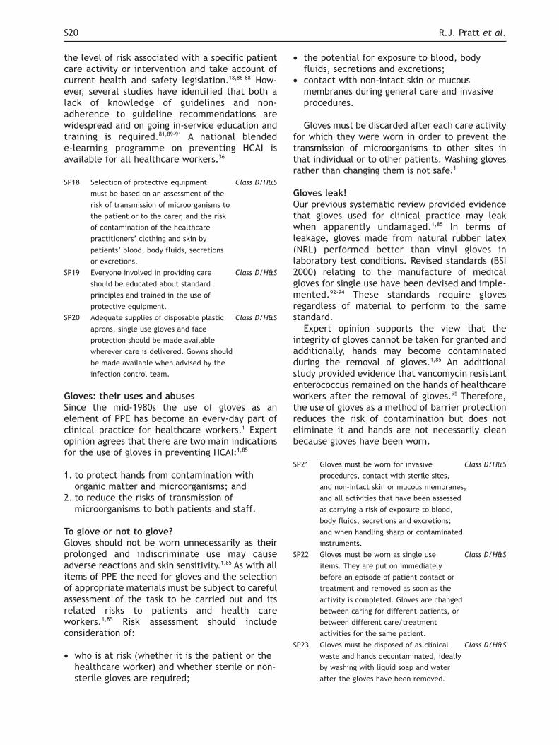
the level of risk associated with a specific patientcare activity or intervention and take account ofcurrent health and safety legislation.18,86-88 How-ever, several studies have identified that both alack of knowledge of guidelines and non-adherence to guideline recommendations arewidespread and on going in-service education andtraining is required.81,89-91 A national blendede-learning programme on preventing HCAI isavailable for all healthcare workers.36
SP18 Selection of protective equipment Class D/H&S
must be based on an assessment of the
risk of transmission of microorganisms to
the patient or to the carer, and the risk
of contamination of the healthcare
practitioners’ clothing and skin by
patients’ blood, body fluids, secretions
or excretions.
SP19 Everyone involved in providing care Class D/H&S
should be educated about standard
principles and trained in the use of
protective equipment.
SP20 Adequate supplies of disposable plastic Class D/H&S
aprons, single use gloves and face
protection should be made available
wherever care is delivered. Gowns should
be made available when advised by the
infection control team.
Gloves: their uses and abusesSince the mid-1980s the use of gloves as anelement of PPE has become an every-day part ofclinical practice for healthcare workers.1 Expertopinion agrees that there are two main indicationsfor the use of gloves in preventing HCAI:1,85
1. to protect hands from contamination withorganic matter and microorganisms; and
2. to reduce the risks of transmission ofmicroorganisms to both patients and staff.
To glove or not to glove?Gloves should not be worn unnecessarily as theirprolonged and indiscriminate use may causeadverse reactions and skin sensitivity.1,85 As with allitems of PPE the need for gloves and the selectionof appropriate materials must be subject to carefulassessment of the task to be carried out and itsrelated risks to patients and health careworkers.1,85 Risk assessment should includeconsideration of:
• who is at risk (whether it is the patient or thehealthcare worker) and whether sterile or non-sterile gloves are required;
• the potential for exposure to blood, bodyfluids, secretions and excretions;
• contact with non-intact skin or mucousmembranes during general care and invasiveprocedures.
Gloves must be discarded after each care activityfor which they were worn in order to prevent thetransmission of microorganisms to other sites inthat individual or to other patients. Washing glovesrather than changing them is not safe.1
Gloves leak!Our previous systematic review provided evidencethat gloves used for clinical practice may leakwhen apparently undamaged.1,85 In terms ofleakage, gloves made from natural rubber latex(NRL) performed better than vinyl gloves inlaboratory test conditions. Revised standards (BSI2000) relating to the manufacture of medicalgloves for single use have been devised and imple-mented.92-94 These standards require glovesregardless of material to perform to the samestandard.
Expert opinion supports the view that theintegrity of gloves cannot be taken for granted andadditionally, hands may become contaminatedduring the removal of gloves.1,85 An additionalstudy provided evidence that vancomycin resistantenterococcus remained on the hands of healthcareworkers after the removal of gloves.95 Therefore,the use of gloves as a method of barrier protectionreduces the risk of contamination but does noteliminate it and hands are not necessarily cleanbecause gloves have been worn.
SP21 Gloves must be worn for invasive Class D/H&S
procedures, contact with sterile sites,
and non-intact skin or mucous membranes,
and all activities that have been assessed
as carrying a risk of exposure to blood,
body fluids, secretions and excretions;
and when handling sharp or contaminated
instruments.
SP22 Gloves must be worn as single use Class D/H&S
items. They are put on immediately
before an episode of patient contact or
treatment and removed as soon as the
activity is completed. Gloves are changed
between caring for different patients, or
between different care/treatment
activities for the same patient.
SP23 Gloves must be disposed of as clinical Class D/H&S
waste and hands decontaminated, ideally
by washing with liquid soap and water
after the gloves have been removed.
S20 R.J. Pratt et al.

Making choicesExpert opinion is quite clear about when glovesmust be used by healthcare workers in generalclinical practice.1,85 Having decided that glovesshould be used for a healthcare activity, thehealthcare worker must make a choice betweenthe use of:
• sterile or non-sterile gloves, based on contactwith susceptible sites or clinical devices;
• surgical or examination gloves, based on theaspect of care or treatment to beundertaken.
NHS Trusts need to provide gloves that conformto European Standard, and which are acceptableto health care practitioners.1,85 Gloves areavailable in a variety of materials, the mostcommon being natural rubber latex (NRL) andsynthetic materials. NRL remains the material ofchoice due to its efficacy in protecting againstbloodborne viruses and properties that enable thewearer to maintain dexterity.1,85 The problem ofpatient or health care practitioner sensitivity toNRL proteins must be considered when deciding onglove materials.
Synthetic materials are generally moreexpensive than NRL and due to certain propertiesmay not be suitable for all purposes.1 Nitrile gloveshave the same chemical range as NRL and may alsolead to sensitivity problems. Vinyl gloves made toEuropean Standards provide the same level ofprotection as NRL.1 Polythene gloves are notsuitable for clinical use due to their permeabilityand tendency to damage easily.1 A study comparingthe performance of nitrile, latex, copolymer andvinyl gloves under stressed and unstressedconditions found that nitrile gloves had the lowestfailure rate, adding further evidence that nitrilegloves are a suitable alternative to latex,providing there are no sensitivity issues.Importantly, the study noted variation inperformance of the same type of glove producedby different manufacturers and propose a test andrating system to assist healthcare workers.96
SP24 Gloves that are acceptable to Class D/H&S
healthcare personnel and CE marked
must be available in all clinical areas.
SP25 Sensitivity to natural rubber latex in Class D/H&S
patients, carers and healthcare personnel
must be documented and alternatives to
natural rubber latex must be available.
SP26 Neither powdered nor polythene Class C/H&S
gloves should be used in health care
activities.
Aprons or gowns?We identified four small scale observationalstudies that investigated the potential for uni-forms to become contaminated during clinicalcare. However none of these studies establishedan association between contaminated uniformsand HCAI.97-99 A further study demonstrated highlevels of contamination of gowns, gloves andstethoscopes with vancomycin-resistant entero-cocci (VRE) following examination of patientsknown to be infected.100
A systematic review of eight studies reportingoutcomes of 3,811 babies to assess the effects ofwearing and gowning by attendants and visitors innewborn nurseries found no evidence to suggestthat over gowns are effective in reducingmortality, clinical infection or bacterial coloni-sation in infants admitted to newborn nurseries.101
One quasi-experimental study investigated the useof gowns and gloves as opposed to gloves only inpreventing the acquisition of VRE in a medicalintensive care unit setting.102 A further prospectiveobservational study investigated the use of asimilar intervention in a medical intensive careunit.103 These studies suggest that the use ofgloves and gowns may minimise the transmissionof VRE when colonisation pressure is high.
National and international guidelines recommendthat protective clothing should be worn by allhealthcare workers when close contact with thepatient, materials or equipment may lead tocontamination of uniforms or other clothing withmicroorganisms or, when there is a risk ofcontamination with blood, body fluids, secretions,or excretions (with the exception of perspi-ration).1,85,104 Disposable plastic aprons are recom-mended for general clinical use.1,85,104 However,unused aprons need to be stored in an appropriatearea away from potential contamination.97 Fullbody gowns need only be used where there is thepossibility of extensive splashing of blood, bodyfluids, secretions or excretions and should be fluidrepellent.1,85,104
SP27 Disposable plastic aprons must be worn Class D/H&S
when close contact with the patient,
materials or equipment are anticipated
and when there is a risk that clothing
may become contaminated with pathogenic
microorganisms or blood, body fluids,
secretions or excretions, with the exception
of perspiration.
SP28 Plastic aprons/gowns should be worn Class D/H&S
as single-use items, for one procedure
or episode of patient care, and then
discarded and disposed of as clinical
epic2: Guidelines for Preventing Healthcare-Associated Infections in NHS Hospitals S21

waste. Non-disposable protective clothing
should be sent for laundering.
SP29 Full-body fluid-repellent gowns must Class D/H&S
be worn where there is a risk of
extensive splashing of blood, body fluids,
secretions or excretions, with the
exception of perspiration, onto the skin
or clothing of healthcare personnel (for
example when assisting with childbirth).
When is a facemask, respiratory protection andeye protection necessary?Healthcare workers (and sometimes patients) mayuse standard surgical facemasks to preventrespiratory droplets from the mouth and nosebeing expelled into the environment. Facemasksare also used, often in conjunction with eyeprotection, to protect the mucous membranes ofthe wearer from exposure to blood and/or bodyfluids when splashing may occur. Our previoussystematic review failed to reveal any robustexperimental studies that demonstrated thathealthcare workers wearing surgical facemasksprotected patients from HCAI during routine wardprocedures, such as wound dressing or invasivemedical procedures.1
Facemasks are also used to protect the wearerfrom inhaling minute airborne respiratoryparticles. As surgical facemasks are not effectivein filtering out such small respiratory particles,specialised respiratory protective equipment isrecommended for the care of patients with certainrespiratory diseases, e.g. active multiple drug-resistant pulmonary tuberculosis,105 Severe AcuteRespiratory Syndrome (SARS), pandemic influenza.The filtration efficiency of these masks(sometimes called ‘respirators’) will protect thewearer from inhaling small respiratory particlesbut to be effective, they must fit closely to theface to minimise leakage around the mask.1,106,107
Although the advice to use particulate filter masksis based on expert opinion, there is evidence fromone study that staff exposed to patients with SARSacquired the infection when they did not useparticulate filter masks.108 Another studydemonstrated a lack of knowledge about guidanceon using particulate respirator masks among staffcaring for patients with SARS and suggests thatfocused training on the use of personal protectiveequipment and the transmission risk of SARS isrequired.109
Our previous systematic review indicated thatdifferent protective eyewear offered protectionagainst physical splashing of infected substancesinto the eyes (although not on all occasions) butthat compliance was poor.1 Expert opinion
recommends that face and eye protection reducethe risk of occupational exposure of healthcareworkers to splashes of blood, body fluids,secretion or excretions.1,85,104
SP30 Face masks and eye protection must Class D/H&S
be worn where there is a risk of blood,
body fluids, secretions or excretions
splashing into the face and eyes.
SP31 Respiratory protective equipment, Class D/H&S
i.e., a particulate filter mask, must be
correctly fitted and used when
recommended for the care of patients
with respiratory infections transmitted
by airborne particles.
2.6 The Safe Use and Disposal of Sharps
This section discusses the evidence and associatedrecommendations for the safe use and disposal ofsharps in general care settings and includeminimising the risks associated with sharps use anddisposal, and the use of needle protection devices.Where appropriate, in addition to the classifi-cation of evidence underpinning the recommen-dations, there is an indication of a Health andSafety (H&S) legislation requirement.
Sharps injuries – what’s the problem?The safe handling and disposal of needles andother sharp instruments forms part of an overallstrategy of clinical waste disposal to protect staff,patients and visitors from exposure to bloodbornepathogens.110 In 2003 the National Audit Officefound that needlestick injuries ranked alongsidemoving and handling, falls, trips and exposure tohazardous substances as the main types ofaccidents experienced by NHS staff.111 In 2001 theRoyal College of Nursing (RCN) launched its Be
Sharp Be Safe campaign aimed at reducing sharpsinjuries. A component of the campaign issurveillance using the software EPINet™. Fifteensites contributed to the RCN 2002 survey andreported a total of 1,445 injuries.112 Although manyinjuries (52.6%) were superficial, 44.6% (n = 626)ranked moderate, including some bleeding, and2.8% (n = 39) were severe. Nurses were the groupwith the highest proportion of sharps injuries,accounting for 41.2% of all reported injuries.
A new report in 2006 from the Health ProtectionAgency confirms that healthcare workers are stillbeing exposed to bloodborne virus infections, eventhough such exposures are largely preventable.The number of reported occupational exposures
S22 R.J. Pratt et al.
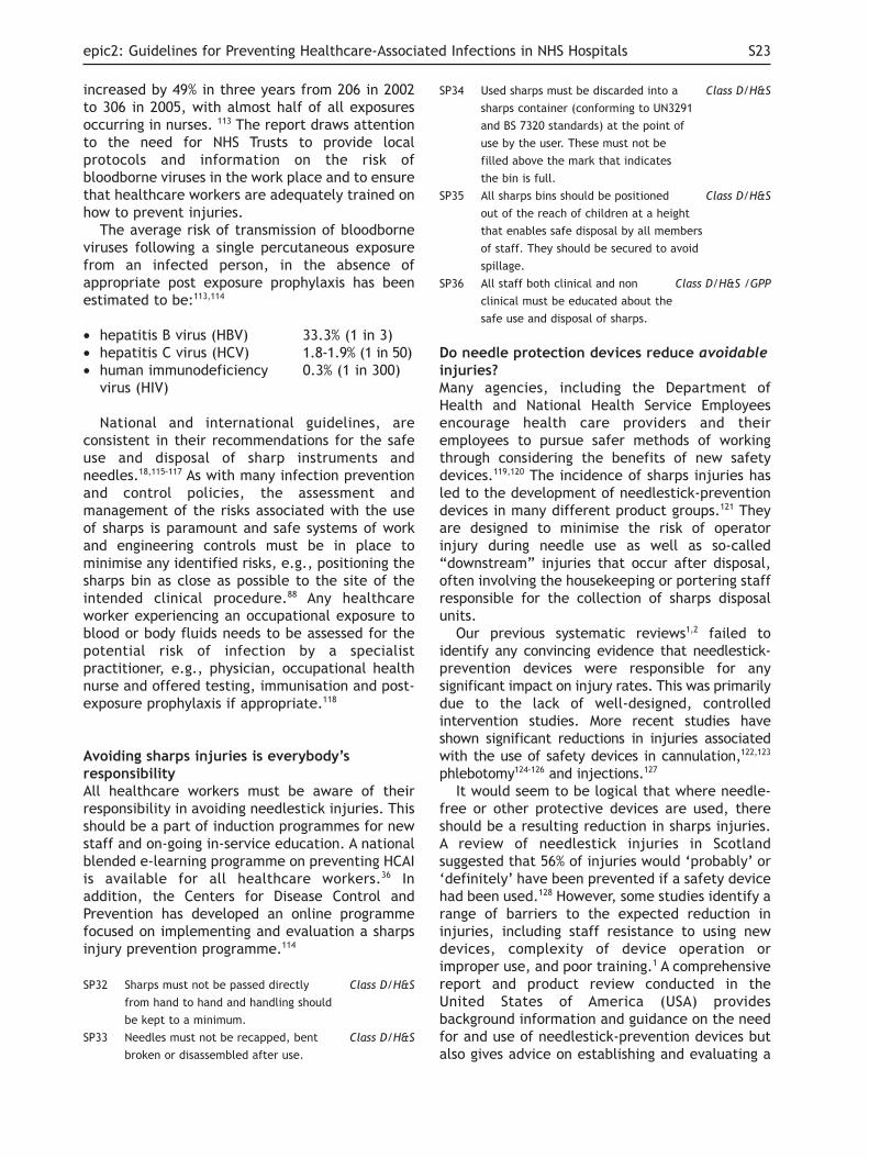
increased by 49% in three years from 206 in 2002to 306 in 2005, with almost half of all exposuresoccurring in nurses. 113 The report draws attentionto the need for NHS Trusts to provide localprotocols and information on the risk ofbloodborne viruses in the work place and to ensurethat healthcare workers are adequately trained onhow to prevent injuries.
The average risk of transmission of bloodborneviruses following a single percutaneous exposurefrom an infected person, in the absence ofappropriate post exposure prophylaxis has beenestimated to be:113,114
• hepatitis B virus (HBV) 33.3% (1 in 3)• hepatitis C virus (HCV) 1.8-1.9% (1 in 50)• human immunodeficiency 0.3% (1 in 300)
virus (HIV)
National and international guidelines, areconsistent in their recommendations for the safeuse and disposal of sharp instruments andneedles.18,115-117 As with many infection preventionand control policies, the assessment andmanagement of the risks associated with the useof sharps is paramount and safe systems of workand engineering controls must be in place tominimise any identified risks, e.g., positioning thesharps bin as close as possible to the site of theintended clinical procedure.88 Any healthcareworker experiencing an occupational exposure toblood or body fluids needs to be assessed for thepotential risk of infection by a specialistpractitioner, e.g., physician, occupational healthnurse and offered testing, immunisation and post-exposure prophylaxis if appropriate.118
Avoiding sharps injuries is everybody’sresponsibilityAll healthcare workers must be aware of theirresponsibility in avoiding needlestick injuries. Thisshould be a part of induction programmes for newstaff and on-going in-service education. A nationalblended e-learning programme on preventing HCAIis available for all healthcare workers.36 Inaddition, the Centers for Disease Control andPrevention has developed an online programmefocused on implementing and evaluation a sharpsinjury prevention programme.114
SP32 Sharps must not be passed directly Class D/H&S
from hand to hand and handling should
be kept to a minimum.
SP33 Needles must not be recapped, bent Class D/H&S
broken or disassembled after use.
SP34 Used sharps must be discarded into a Class D/H&S
sharps container (conforming to UN3291
and BS 7320 standards) at the point of
use by the user. These must not be
filled above the mark that indicates
the bin is full.
SP35 All sharps bins should be positioned Class D/H&S
out of the reach of children at a height
that enables safe disposal by all members
of staff. They should be secured to avoid
spillage.
SP36 All staff both clinical and non Class D/H&S /GPP
clinical must be educated about the
safe use and disposal of sharps.
Do needle protection devices reduce avoidableinjuries?Many agencies, including the Department ofHealth and National Health Service Employeesencourage health care providers and theiremployees to pursue safer methods of workingthrough considering the benefits of new safetydevices.119,120 The incidence of sharps injuries hasled to the development of needlestick-preventiondevices in many different product groups.121 Theyare designed to minimise the risk of operatorinjury during needle use as well as so-called“downstream” injuries that occur after disposal,often involving the housekeeping or portering staffresponsible for the collection of sharps disposalunits.
Our previous systematic reviews1,2 failed toidentify any convincing evidence that needlestick-prevention devices were responsible for anysignificant impact on injury rates. This was primarilydue to the lack of well-designed, controlledintervention studies. More recent studies haveshown significant reductions in injuries associatedwith the use of safety devices in cannulation,122,123
phlebotomy124-126 and injections.127
It would seem to be logical that where needle-free or other protective devices are used, thereshould be a resulting reduction in sharps injuries.A review of needlestick injuries in Scotlandsuggested that 56% of injuries would ‘probably’ or‘definitely’ have been prevented if a safety devicehad been used.128 However, some studies identify arange of barriers to the expected reduction ininjuries, including staff resistance to using newdevices, complexity of device operation orimproper use, and poor training.1 A comprehensivereport and product review conducted in theUnited States of America (USA) providesbackground information and guidance on the needfor and use of needlestick-prevention devices butalso gives advice on establishing and evaluating a
epic2: Guidelines for Preventing Healthcare-Associated Infections in NHS Hospitals S23

sharps injury prevention program.121 The reportidentifies that all devices have limitations inrelation to cost, applicability and/or effective-ness. Some of the devices available are moreexpensive than standard devices, may not becompatible with existing equipment, and may beassociated with an increase in bloodstreaminfection rates.129
In the USA, the Occupational Safety HealthAdministration (OSHA) and the National Institutefor Occupational Safety and Health (NIOSH)suggest that a thorough evaluation of any device isessential before purchasing decisions aremade.117,130 Similarly in the United Kingdom, theNational Health Service Purchasing and SupplyAgency identifies that meaningful evaluations areparamount in assessing user acceptability andclinical applicability of any needle safetydevices.131 The evaluation should ensure that thesafety feature works effectively and reliably, thatthe device is acceptable to health carepractitioners and that it does not adversely affectpatient care.
SP37 Consider the use of needlestick- Class B/H&S
prevention devices where there are
clear indications that they will provide
safe systems of working for healthcare
practitioners.
SP38 Conduct a rigorous evaluation of Class D
needlestick-prevention devices to
determine their effectiveness,
acceptability to practitioners, impact
on patient care and cost benefit prior
to widespread introduction.
2.7 Areas for Further Research
Adherence / behaviour changeAction research studies to explore the use ofbehavioural and quality management sciences toimprove adherence of health care professionals toinfection prevention guidelines, specifically inrelation to:
• Hand hygiene;• The effect of different products, e.g., gels,
foams and lotions on improving adherence torecommended hand hygiene regimens;
• Standard principles for the prevention of thetransmission of bloodborne pathogens;
• Cleanliness of the hospital environment;• Trials of the effectiveness of different educational
methods to increase adherence to guidelines;• Development and evaluation of appropriate
strategies for auditing adherence to infectionprevention guidelines.
Staffing• Investigate the relationship between health
care workers’ staffing levels, workload andskill mix and risk for nosocomial infections.
Surveillance• Develop appropriate and realistic methods and
tools to facilitate local surveillance of hospital-acquired infections.
• The role of screening for HCAI microorganismsas a means of controlling HCAI.
• Further research on community MRSAcolonisation and its impact on acute care.
Needle Safety Devices• Studies to establish the cost-effectiveness,
acceptability and efficacy of needle safetydevices.
Organisational change• Studies to link improvement in infection
control practice, patient outcome and culturalchange;
• Studies to assess performance monitoring ofmandatory infection control standards linkedto government improvement practice
• The role of inter ward and inter hospitaltransfers on spread of HCAI
S24 R.J. Pratt et al.
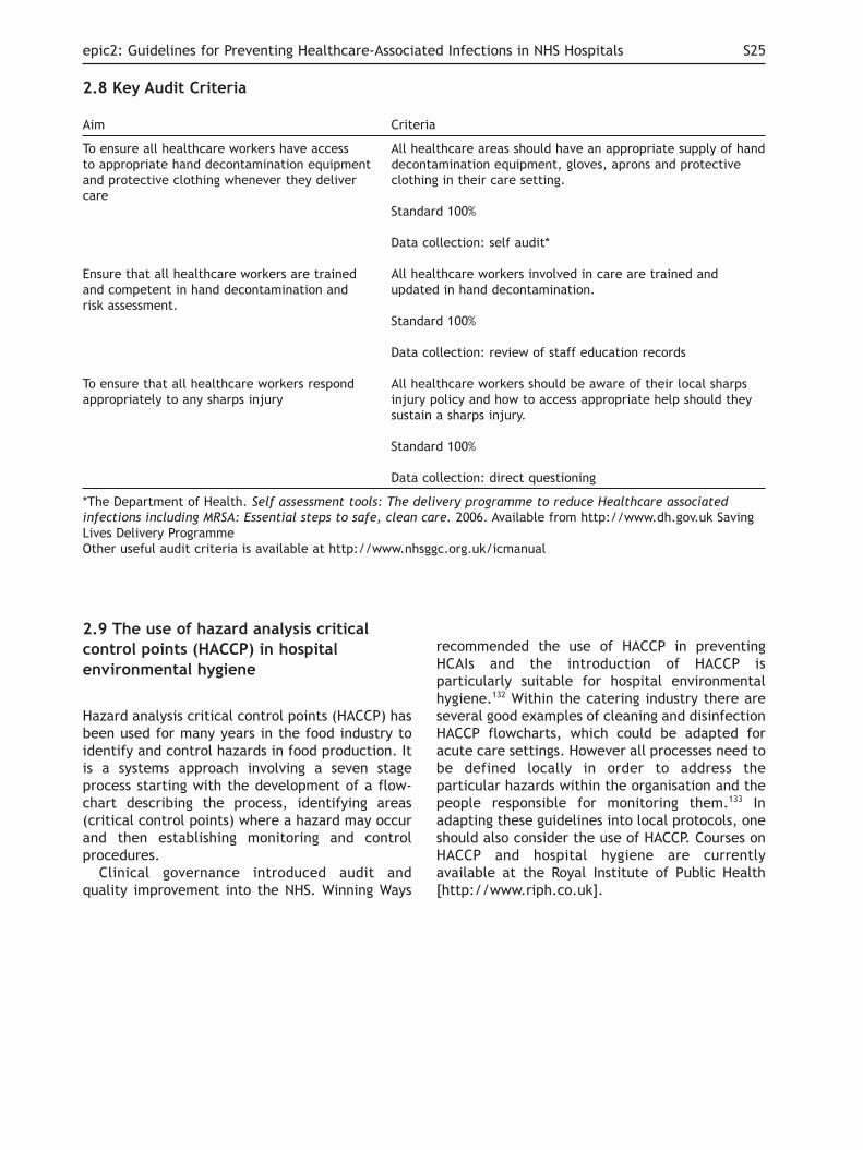
2.8 Key Audit Criteria
2.9 The use of hazard analysis critical
control points (HACCP) in hospital
environmental hygiene
Hazard analysis critical control points (HACCP) hasbeen used for many years in the food industry toidentify and control hazards in food production. Itis a systems approach involving a seven stageprocess starting with the development of a flow-chart describing the process, identifying areas(critical control points) where a hazard may occurand then establishing monitoring and controlprocedures.
Clinical governance introduced audit andquality improvement into the NHS. Winning Ways
recommended the use of HACCP in preventingHCAIs and the introduction of HACCP isparticularly suitable for hospital environmentalhygiene.132 Within the catering industry there areseveral good examples of cleaning and disinfectionHACCP flowcharts, which could be adapted foracute care settings. However all processes need tobe defined locally in order to address theparticular hazards within the organisation and thepeople responsible for monitoring them.133 Inadapting these guidelines into local protocols, oneshould also consider the use of HACCP. Courses onHACCP and hospital hygiene are currentlyavailable at the Royal Institute of Public Health[http://www.riph.co.uk].
epic2: Guidelines for Preventing Healthcare-Associated Infections in NHS Hospitals S25
Aim Criteria
To ensure all healthcare workers have access All healthcare areas should have an appropriate supply of handto appropriate hand decontamination equipment decontamination equipment, gloves, aprons and protective and protective clothing whenever they deliver clothing in their care setting.care
Standard 100%
Data collection: self audit*
Ensure that all healthcare workers are trained All healthcare workers involved in care are trained and and competent in hand decontamination and updated in hand decontamination.risk assessment.
Standard 100%
Data collection: review of staff education records
To ensure that all healthcare workers respond All healthcare workers should be aware of their local sharps appropriately to any sharps injury injury policy and how to access appropriate help should they
sustain a sharps injury.
Standard 100%
Data collection: direct questioning
*The Department of Health. Self assessment tools: The delivery programme to reduce Healthcare associated
infections including MRSA: Essential steps to safe, clean care. 2006. Available from http://www.dh.gov.uk SavingLives Delivery ProgrammeOther useful audit criteria is available at http://www.nhsggc.org.uk/icmanual

2.10 Standard Principles Systematic
Review Process
Hospital Hygiene - Systematic Review Process Hand hygiene - Systematic Review Process
S26 R.J. Pratt et al.
Systematic Review Questions1. What is the evidence that inadequate hospital hygiene contributes to rates of
hospital acquired infection?2. What are the most effective methods and agents for: -terminal disinfection,
decontamination of spills of blood or other, potentially infectious bodysubstances?
3. Is there any evidence that routine use of disinfectants as opposed todetergents for environmental cleaning is more efficacious at preventinghealthcare associated infection?
4. How effective is hydrogen peroxide vapour for environmental or terminalcleaning?
5. Is there any cost effectiveness evidence relating to the above?6. What are the training and education implications for staff and patients?
Databases and Search Terms UsedDATABASESMEDLINE, CUMULATED INDEX OF NURSING AND ALLIED HEALTHLITERATURE (CINAHL), EMBASE, NELH GUIDELINE FINDER,NATIONAL INSTIRURE FOR HEALTH AND CLINICAL EXCELLENCE,THE COCHRANE LIBRARY, US GUIDLEINES CLEARNING HOUSEMeSH TERMSinfection control; cross infection; universal precautions, equipmentcontamination; disease transmission; disinfection; disinfectants; soaps; anti-infective agents; surface-active agents; hospital housekeepingTHESAURUS AND FREE TEXT TERMSRisk assessment; occupational accident; hospital hygiene; hospitalhousekeeper
Search ResultsTotal number of articles located = 7830
Sift 1 CriteriaAbstract indicates that the article: relates to infections associated withhospital hygiene, is written in English, is primary research or a systematicreview or a meta-analysis, and appears to inform one or more of the reviewquestions.
Articles RetrievedTotal number of articles retrieved from sift 1 = 34
Sift 2 CriteriaFull Text confirms that the article relates to infections associated withhospital hygiene is written in English, is primary research or a systematicreview or a meta-analysis, and informs one or more of the review questions.
Articles Selected for AppraisalTotal number of articles selected for appraisal during sift 2 = 5
Critical AppraisalAll articles which described primary research, a systematic review or, ameta-analysis and met the sift 2 criteria were independently criticallyappraised by two appraisers. Consensus and grading was achieved throughdiscussion.
Accepted and Rejected EvidenceTotal number of articles accepted after critical appraisal = 5Total number of articles rejected after critical appraisal = 0
Evidence TablesEvidence tables for accepted and rejected studies were generated andused to create evidence summary reports. The summary reports were,in turn, used as the basis for guideline writing.
Systematic Review Questions1. What is the evidence that contaminated hands are a cause of healthcare
associated infection?2. Which hand disinfection agents are the most effective at removing /
reducing organisms responsible for healthcare associated infection?3. When must hands be disinfected in relation to patient care activities?4. What is the most effective hand washing technique for removing / reducing
organisms responsible for healthcare associated infection?5. Which hand disinfection agents are least toxic to users?6. Which hand disinfection agents are associated with greatest compliance
among healthcare workers?7. Is there any cost effectiveness evidence relating to the above?
8. What are the training and education implications for staff and patients?
Databases and Search Terms UsedDATABASESMEDLINE, CUMULATED INDEX OF NURSING AND ALLIED HEALTHLITERATURE (CINAHL), EMBASE, NELH GUIDELINE FINDER,NATIONAL INSTIRURE FOR HEALTH AND CLINICAL EXCELLENCE,THE COCHRANE LIBRARY, US GUIDLEINES CLEARNING HOUSE
MeSH TERMSinfection control; cross infection; universal precautions, equipmentcontamination; disease transmission; disinfectants; chlorhexidine; soaps;anti-infective agents; surface active agents
THESAURUS AND FREE TEXT TERMS Handwashing; skin; epidermis; nails; antisepsis; decontamination
Search ResultsTotal number of articles located = 32,088
Sift 1 CriteriaAbstract indicates that the article: relates to infections associated withhand hygiene, is written in English, is primary research or a systematicreview or a meta-analysis, and appears to inform one or more of the reviewquestions.
Articles RetrievedTotal number of articles retrieved from sift 1 = 194
Sift 2 CriteriaFull Text confirms that the article relates to infections associated withhand hygiene is written in English, is primary research or a systematicreview or a meta-analysis, and informs one or more of the review questions.
Articles Selected for AppraisalTotal number of articles selected for appraisal during sift 2 = 31
Critical AppraisalAll articles which described primary research, a systematic review or, ameta-analysis and met the sift 2 criteria were independently criticallyappraised by two appraisers. Consensus and grading was achieved throughdiscussion.
Accepted and Rejected EvidenceTotal number of articles accepted after critical appraisal = 25 Total number of articles rejected after critical appraisal = 6
Evidence TablesEvidence tables for accepted and rejected studies were generated andused to create evidence summary reports. The summary reports were,in turn, used as the basis for guideline writing.

Personal Protective Equipment - SystematicReview Process Sharps - Systematic Review Process
epic2: Guidelines for Preventing Healthcare-Associated Infections in NHS Hospitals S27
Systematic Review Questions1. Which glove materials are least toxic to health care workers (HCWs) for
general use?2. What is the evidence that hands need to be disinfected following the use of
gloves?3. What is the evidence that HCWs use gloves appropriately, as a part of
Standard Principles?4. What is the evidence that the uniforms / clothes of HCWs are a source of
healthcare-associated infection?5. What is the evidence that the use of protective clothing reduces the
incidence of healthcare-associated infection?6. What is the evidence that gowns are more beneficial than aprons?7. Is there any cost effectiveness evidence relating to the above?8. What are the training and education implications for staff and patients?
Databases and Search Terms UsedDATABASESMEDLINE, CUMULATED INDEX OF NURSING AND ALLIED HEALTHLITERATURE (CINAHL), EMBASE, NELH GUIDELINE FINDER,NATIONAL INSTITUTE FOR HEALTH AND CLINICAL EXCELLENCE,THE COCHRANE LIBRARY, US GUIDELINES CLEARINGHOUSE.MeSH TERMSinfection control; cross infection; universal precautions; equipmentcontamination; disease transmission; protective clothing; disposableequipment; masks; gloves, protective; eye protective devices.THESAURUS AND FREE TEXT TERMSInfection; contamination; antisepis; universal precaution; disease transmission;disinfection; sterilisation; decontamination; disposable equipment; masks;gloves; face shield; goggles; apron; uniform; gown; protective clothes; visor;hood; eye protection devices
Search ResultsTotal number of articles located = 17,966
Sift 1 CriteriaAbstract indicates that the article: relates to infections associated withprotective clothing, is written in English, is primary research or a systematicreview or a meta-analysis, and appears to inform one or more of the reviewquestions.
Articles RetrievedTotal number of articles retrieved from sift 1 = 112
Sift 2 CriteriaFull Text confirms that the article relates to infections associated withprotective clothing is written in English, is primary research or a systematicreview or a meta-analysis, and informs one or more of the reviewquestions.
Articles Selected for AppraisalTotal number of articles selected for appraisal during sift 2 = 14
Critical AppraisalAll articles which described primary research, a systematic review or, ameta-analysis and met the sift 2 criteria were independently criticallyappraised by two appraisers. Consensus and grading was achieved throughdiscussion.
Accepted and Rejected EvidenceTotal number of articles accepted after critical appraisal = 10Total number of articles rejected after critical appraisal = 4
Evidence TablesEvidence tables for accepted and rejected studies were generated andused to create evidence summary reports. The summary reports were,in turn, used as the basis for guideline writing.
Systematic Review Questions1. What is the evidence that recommended modes of use and disposal of
sharps reduce the incidence of sharps injury in health care workers?2. What is the evidence that education and training interventions improve
health care workers adherence to recommended modes of practice?3. What is the evidence that the use of needle-free devices reduce
occupational exposure to bloodborne pathogens?4. Is there any cost effectiveness evidence relating to the above?5. What are the training and education implications for staff and patients?
Databases and Search Terms UsedDATABASESMEDLINE, CUMULATED INDEX OF NURSING AND ALLIED HEALTHLITERATURE (CINAHL), EMBASE, NELH GUIDELINE FINDER,NATIONAL INSTIRURE FOR HEALTH AND CLINICAL EXCELLENCE,THE COCHRANE LIBRARY, US GUIDLEINES CLEARNING HOUSEMeSH TERMSinfection control; cross infection; universal precautions, equipmentcontamination; disease transmission; needlestick injuriesTHESAURUS AND FREE TEXT TERMS Needles; syringes; occupational accident; occupational exposure; medicalwaste disposal; bloodborne pathogens; exposure prone procedures; sharps;punctures; percutaneous injuries; resheathing
Search ResultsTotal number of articles located = 8130
Sift 1 CriteriaAbstract indicates that the article: relates to infections associated withsharps, is written in English, is primary research or a systematicreview or a meta-analysis, and appears to inform one or more of the reviewquestions.
Articles RetrievedTotal number of articles retrieved from sift 1 = 49
Sift 2 CriteriaFull Text confirms that the article relates to infections associated withsharps is written in English, is primary research or a systematicreview or a meta-analysis, and informs one or more of the review questions.
Articles Selected for AppraisalTotal number of articles selected for appraisal during sift 2 = 19
Critical AppraisalAll articles which described primary research, a systematic review or, ameta-analysis and met the sift 2 criteria were independently criticallyappraised by two appraisers. Consensus and grading was achieved throughdiscussion.
Accepted and Rejected EvidenceTotal number of articles accepted after critical appraisal = 12 Total number of articles rejected after critical appraisal = 7
Evidence TablesEvidence tables for accepted and rejected studies were generated andused to create evidence summary reports. The summary reports were,in turn, used as the basis for guideline writing.

3 Guidelines for preventing infectionsassociated with the use of short-termindwelling urethral catheters
3.1 Introduction
This guidance is based on the best criticallyappraised evidence currently available. The typeand class of supporting evidence explicitly linkedto each recommendation is described. Allrecommendations are endorsed equally and noneis regarded as optional. These recommendationsare not detailed procedural protocols and need tobe incorporated into local guidelines.
These guidelines apply to adults and childrenaged one year and older and should be read inconjunction with the guidance on StandardPrinciples. The recommendations are divided intofive distinct interventions:
1. Assessing the need for catheterisation;2. Selection of catheter type and system;3. Catheter insertion;4. Catheter maintenance; and5. Education of patients, relatives and healthcare
workers.
Background and context of the GuidelinesCatheter associated urinary tract infection(CAUTI) is the most common nosocomial infectionin hospitals. Most bacteria causing infectionassociated with catheterisation gain access to theurinary tract either extraluminally or intra-luminally. Extraluminal contamination may occuras the catheter is inserted, by contamination ofthe catheter from the health care worker’s handsor from the patient’s own colonic or perineal flora.Extraluminal contamination is also thought tooccur by microorganisms ascending from theperineum. Intraluminal contamination occurs byreflux of bacteria from a contaminated urinedrainage bag.
Bacteria quickly develop into colonies known asbiofilms which adhere to the catheter surface anddrainage bag. A biofilm forms when bacteriaattach to a surface and subsequently encasethemselves in an exopolymeric material. Suchbacteria are morphologically and physiologicallydifferent from free-living planktonic bacteria.Bacteria in biofilms have considerable survivaladvantages over free-living microorganisms, beingextremely resistant to antibiotic therapy.
These biofilms cause further problems if thebacteria produce the enzyme urease, such as
Proteus mirabilis. The urine then becomesalkaline, causing the crystallisation of calcium andmagnesium phosphate within the urine, whichthen is incorporated into the biofilm resulting inencrustation of the catheter over a period of time.Encrustation is generally associated with long-term catheterisation, since it has a directrelationship with the length of catheterisation.
3.2 Systematic Review Process
We have previously described the systematic reviewprocess in Section 1.10. For detailed descriptionsof previous systematic reviews which havecontributed to the evidence base underpinningthese guidelines readers should consult theoriginal guidelines,1 the guidelines for theprevention of healthcare associated infections inprimary and community care2 and our interimreport.3 Search questions were developed fromadvice received from our specialist advisors andthe results of the searches are found in Section3.10. The process outlined in Section 3.10 refersonly to the most recent systematic review of theliterature undertaken in 2005.
3.3 Assessing the Need for Catheterisation
Catheterising patients places them insignificant danger of acquiring a urinary tractinfection. The longer a catheter is in place, thegreater the dangerThere is consistent evidence that a significantnumber of healthcare-associated infections inhospital are related to urinary catheterisa-tion.115,134-136 The risk of infection is associatedwith the method and duration of catheterisation,the quality of catheter care and host suscepti-bility. Urinary catheterisation is a frequentintervention during clinical care in hospitalaffecting a significant number of patients at anyone time. The highest incidence of infection isassociated with indwelling urethral catheterisa-tion.137 The per day risk of the development ofbacteriuria appears comparable throughoutcatheterisation (3-6 percent) but the cumulativerisk increases with duration of catheterisation.137-139
Consequently, around 50 percent of hospitalisedpatients catheterised longer than 7-10 dayscontract bacteriuria.137 Although frequentlyasymptomatic, 20-30 percent of patients withcatheter-associated bacteriuria will develop
S28 R.J. Pratt et al.
symptoms of CAUTI.137 Many of these infections areserious and lead to significant morbidity andmortality. Of patients with a CAUTI, 1-4 percentdevelops bacteraemia and, of these, 13-30die.137,140 Duration of catheterisation is stronglyassociated with risk of infection, i.e., the longerthe catheter is in place, the higher the incidenceof urinary tract infection.137,140
Advice from best practice emphasises theimportance of documenting all proceduresinvolving the catheter or drainage system in thepatient’s records and providing patients withadequate information in relation to the need forcatheterisation and details of the insertion,maintenance and removal of their catheter.115,141
There is some evidence to suggest that computermanagement systems improve documentation andin so doing reduce the length of time catheters arein situ.142
UC1 Only use indwelling urethral catheters Class D/GPP
after considering alternative methods
of management.
UC2 Document the need for catheterisation, Class D/GPP
catheter insertion and care.
UC3 Review regularly the patient’s clinical Class D/GPP
need for continuing urinary catheterisation
and remove the catheter as soon as possible.
3.4 Selection of Catheter Type
Is one catheter better than another?Current evidence–based guidelines1 identifiedthree experimental studies that compared the useof latex with silicone catheters.143-145 No significantdifference in the incidence of bacteriuria wasfound. Four studies compared the use of silvercoated (silver alloy or silver oxide) catheters withsilicone, hydrogel or Teflon latex.146-149 A syste-matic review and meta-analysis of these and otherstudies found that silver alloy (but not silveroxide) catheters were associated with a lowerincidence of bacteriuria.140,150
New evidence related to the efficacy of usingurinary catheters coated or impregnated withantiseptic or antimicrobial agents has emergedsince our original review in 2000. Two subsequentreviews,2,3 together with the current updatereview undertaken by us, have identified foursystematic reviews and one meta-analysis thathave examined this issue.150-154 In general, all ofthese five studies suggest antiseptic impregnatedor antimicrobial-coated urinary catheters can
significantly prevent or delay the onset of CAUTIwhen compared to standard untreated urinarycatheters. The consensus in these five reviews ofevidence however, is that the individual studiesreviewed are generally of poor quality; forinstance in one case only 8 studies out of 117 metthe inclusion criteria and in another, of the sixreports describing 7 trials included, only onescored 5 in the quality assessment the other fivescored only 1.150,154
Studies investigating a wide range of coated orimpregnated catheters are explored in the newevidence including: catheters coated or impreg-nated with: silver alloy150,151,154-161; silver oxide150;gendine162; gentamicin163 and silver-hydrogel164-166;minocycline167; rifampicin167; chlorhexidine–silversulfadiazine166; chlorhexidine-sulfadiazine-triclo-san166; nitrofurazone166; and nitrofuroxone.168
New evidence suggests that catheters coatedwith silver alloy are clinically effective in reducingthe incidence of CAUTI, but many studies are ofpoor methodological quality. Consequently thereremains inconclusive evidence to recommend theiruse in preference to other types of catheter at thistime. Despite their unit cost, there is a suggestionthat these devices might be a cost-effectiveoption if overall numbers of infections aresignificantly reduced through their use. The fewstudies that have explored the cost benefit/effectiveness of using these devices have,however, also been inconclusive.157,159,161,165
Evidence from best practice indicates that theincidence of CAUTI in patients catheterised for ashort time (up to one week) is not influenced byany particular type of catheter material.136,169
However, many practitioners have strong prefer-ences for one type of catheter over another. Thispreference is often based on clinical experience,patient assessment, and which materials inducethe least allergic response. Smaller gaugecatheters with a 10 ml balloon minimise urethraltrauma, mucosal irritation and residual urine inthe bladder, all factors that predispose toCAUTI.135,170 However, in adults that have recentlyundergone urological surgery, larger gauge cathe-ters may be indicated to allow for the passage ofblood clots.
UC4 Choice of catheter material will depend Class D
on clinical experience, patient assessment
and anticipated duration of catheterisation.
UC5 Select the smallest gauge catheter that Class D
will allow free urinary outflow. A catheter
with a 10 ml balloon should be used in adults.
Urological patients may require larger gauge
sizes and balloons.
epic2: Guidelines for Preventing Healthcare-Associated Infections in NHS Hospitals S29

3.5 Catheter Insertion
Catheterisation is a skilled aseptic procedureDespite evidence from one systematic review153
which suggests that the use of aseptic techniquehas not demonstrated a reduction in the rate ofCAUTI, principles of good practice, clinicalguidance115,134 and expert opinion135-137,171-174,together with findings from another systematicreview140 agree that urinary catheters must beinserted using sterile equipment and an aseptictechnique.
Expert opinion indicates that there is noadvantage in using antiseptic preparations forcleansing the urethral meatus prior to catheterinsertion.153,173 Urethral trauma and discomfortwill be minimised by using an appropriate sterile,single-use lubricant or anaesthetic gel. Ensuringhealthcare practitioners are trained andcompetent in the insertion of urinary catheterswill minimise trauma, discomfort and thepotential for CAUTI.115,135,173,174
UC6 Catheterisation is an aseptic procedure. Class D
Ensure that health care workers are trained
and competent to carry out urethral
catheterisation.
UC7 Clean the urethral meatus with sterile normal Class D
saline prior to the insertion of the catheter.
UC8 Use an appropriate lubricant from a sterile Class D
single use container to minimise urethral
trauma and infection.
3.6 Catheter Maintenance
Leave the closed system alone!Maintaining a sterile, continuously closed urinarydrainage system is central to the prevention ofCAUTI.115,134,135,173,175,176 The risk of infectionreduces from 97 percent with an open system to8-15 percent when a sterile closed system isemployed.136,174,177 Breaches in the closed systemsuch as unnecessary emptying of the urinarydrainage bag or taking a urine sample, willincrease the risk of catheter-related infection andshould be avoided.115,136,178 Hands must be decon-taminated and clean, non-sterile gloves wornbefore manipulation. A systematic review suggeststhat sealed (e.g., taped, pre-sealed) drainagesystems contribute to preventing bacteriuria.153
There is limited evidence as to how oftencatheter bags should be changed. One studyshowed higher rates of symptomatic and asympto-matic CAUTI were associated with a three day
urinary drainage bag change regimen whencompared to no routine change regimen.179 Bestpractice suggests changing only when necessary,i.e., according to either the manufacturers’recommendations or the patient’s clinicalneed.115,134 Reflux of urine is associated withinfection and consequently, drainage bags shouldbe positioned in a way that prevents back-flow ofurine.115,135 It is also recommended that urinarydrainage bags should be hung on an appropriatestand that prevents contact with the floor.136
A number of studies have investigated theaddition of disinfectants and antimicrobials todrainage bags as a way of preventing CAUTI.140
Three acceptable studies from our originalsystematic review demonstrated no reduction inthe incidence of bacteriuria following the additionof hydrogen peroxide or chlorhexidine to urinarydrainage bags.1,180-182 A systematic review supportsthese findings in that it suggests that addingbacterial solutions to drainage bags has no effecton catheter associated infection.153
UC9 Connect indwelling urethral catheters to Class A
a sterile closed urinary drainage system.
UC10 Ensure that the connection between the Class A
catheter and the urinary drainage system
is not broken except for good clinical reasons,
e.g., changing the bag in line with
manufacturer’s recommendation.
UC11 Decontaminate hands and wear a new pair Class D
of clean, non-sterile gloves before
manipulating a patient’s catheter and
decontaminate hands after removing gloves.
UC12 Obtain urine samples from a sampling Class D/GPP
port using an aseptic technique.
UC13 Position urinary drainage bags below Class D/GPP
the level of the bladder on a stand that
prevents contact with the floor.
UC14 Empty the urinary drainage bag Class D/GPP
frequently enough to maintain urine
flow and prevent reflux. Use a separate
and clean container for each patient and
avoid contact between the urinary
drainage tap and container.
UC15 Do not add antiseptic or antimicrobial Class A
solutions into urinary drainage bags.
UC16 Do not change catheters unnecessarily Class D/GPP
or as part of routine practice except
where necessary to adhere to the
manufacturer’s guidance.
S30 R.J. Pratt et al.

Appropriate maintenance minimises infections
Meatal cleansing with antiseptic solutions is
unnecessary
Our original systematic review considered sixacceptable studies that compared meatal clean-sing with a variety of antiseptic/antimicrobialagents or soap and water.1 No reduction wasdemonstrated in bacteriuria when using any ofthese preparations for meatal care compared withroutine bathing or showering.183-188 Our subsequentreviews2,3 revealed two studies153,189 that supportthese findings in that the outcomes indicate thatthe use of antiseptics provides no benefit inrespect of meatal/peri-urethral hygiene.
Expert opinion134-136 and another systematicreview140 support the view that vigorous meatalcleansing is not necessary and may increase therisk of infection and that daily routine bathing orshowering is all that is needed to maintain meatalhygiene.
UC17 Routine daily personal hygiene is all Class A
that is needed to maintain meatal hygiene.
Irrigation, instillation and washout do not
prevent infection
None of our systematic review evidence demon-strates any beneficial effect of bladder irrigation,instillation or washout with a variety of antisepticor antimicrobial agents in preventing CAUTI.1,140,190-199
Three studies, however, suggest that an acidwashout solution (Suby G) is effective in reducingcatheter encrustation.196,198,200
Evidence from best practice supports thefindings in respect of bladder irrigation,instillation and washout and indicates that theintroduction of such agents may have local toxiceffects and contribute to the development ofresistant microorganisms. However, continuous orintermittent bladder irrigation may be indicatedduring urological surgery or to manage catheterobstruction.115,134-136,140
UC18 Bladder irrigation, instillation or washouts Class A
should not be used to prevent catheter-
associated infection.
3.7 Education of Patients, Relatives and
Healthcare Workers
Given the frequency of urinary catheterisation inhospital patients and the associated risk of urinarytract infection, it is important that patients, theirrelatives and healthcare workers responsible forcatheter insertion and management are educatedabout infection prevention. All those involvedmust be aware of the signs and symptoms ofurinary tract infection and how to access experthelp when difficulties arise. Healthcareprofessionals must be confident and proficient inprocedures associated with preventing CAUTI.
UC19 Healthcare workers must be trained in Class D/GPP
catheter insertion and maintenance.
UC20 Patients and relatives should be Class D/GPP
educated about their role in preventing
urinary tract infection.
3.8 Areas for Further Research
In developing the recommendations we identifiedseveral areas that were inadequately addressed inthe literature. We recommend further research inthe following areas.
Intervention 1: Assessing the need forcatheterisationEpidemiological studies of the prevalence andincidence of bacteriuria/urinary tract infectionduring short-term catheterisation in differentpopulations and different care settings. Theseshould at least encompass the predominantpopulations, i.e. older people and those under-going surgery. There needs to be clear definition ofthe ‘cases’ and the populations from which theyare drawn.
Intervention 2: Selection of catheter typeRandomised controlled trials of the efficacy ofantiseptic/antimicrobial coated/impregnatedurethral catheters for short-term use. These needto be high quality studies, using the hospital’sactual catheter-associated UTI prevalence ratherthan national data, and appropriate follow-up.
Intervention 4: Catheter maintenanceRandomised controlled trials of strategies toestablish how often catheters and catheter bagsneed to be changed.
epic2: Guidelines for Preventing Healthcare-Associated Infections in NHS Hospitals S31

3.9 Key Audit Criteria
S32 R.J. Pratt et al.
Aim Criteria
Identify all patients with indwelling urinary All patients should have a patient record that documents the catheters, their clinical need for catheterisation, reason for catheterisation, type of catheter, catheter assessed and documented. insertion, changes and care.
Standard 100%
Data collection: review of patient notes
Ensure that all healthcare workers are trained Healthcare workers receive training and updates in the and competent in urinary catheterisation. management of urinary catheters.
Standard 100%
Data collection: review of staff education records
To prevent catheter-associated urinary tract All healthcare workers decontaminate their hands and wear a infections (CAUTI) new pair of non-sterile gloves before manipulating the system.
Standard 100%
Data collection: observation/ self audit
To reduce the incidence of CAUTI by All catheters must be connected to a sterile closed drainage maintaining a closed system. system or valve.
Standard 100%
Data collection: observation
To ensure patients and relatives are informed All patients and carers are aware of the need to:and educated about catheter management • Decontaminate their hands;
• Keep the system closed.
Standard 100%
Data collection: direct patient questioning of patients and carers.

3.10 Urinary Catheter Systematic Review
Process4 Guidelines for preventing infectionsassociated with the use of centralvenous access devices (CVAD)
4.1 Introduction
This guidance is based on the best criticallyappraised evidence currently available. The typeand class of supporting evidence explicitly linkedto each recommendation is described. All recom-mendations are endorsed equally and none isregarded as optional. These recommendations arenot detailed procedural protocols and need to beincorporated into local guidelines.
Background and context to the GuidelinesBloodstream infections associated with the inser-tion and maintenance of central venous accessdevices (CVAD) are among the most dangerouscomplications of healthcare that can occur,worsening the severity of the patient’s underlyingill health, prolonging the period of hospitalisationand increasing the cost of care.201-204 Approxi-mately 3 in every 1000 patients admitted tohospital in the UK acquires a bloodstream infec-tion, and nearly one third of these infections arerelated to central venous access devices.205
Catheter related blood stream infection (CR-BSI)involves the presence of systemic infection andevidence implicating the CVAD as its source, i.e.,the isolation of the same microorganism from bloodcultures as that shown to be significantly colonisingthe CVAD of a patient with clinical features ofbacteraemia. Catheter colonization refers to asignificant growth of microorganisms on either theendoluminal or the external catheter surfacebeneath the skin in the absence of systemicinfection.206-208
The microorganisms that colonise catheter hubsand the skin adjacent to the insertion site are thesource of most CR-BSI. Coagulase-negative staphy-lococci, particularly Staphylococcus epidermidis,are the most frequently implicated micro-organisms associated with CR-BSI. Other micro-organisms commonly involved include Staphylo-
coccus aureus, Candida species and enterococci.208
CR-BSI is generally caused either by skinmicroorganisms at the insertion site thatcontaminate the catheter during insertion andmigrate along the cutaneous catheter track, ormicroorganisms from the hands of healthcareworkers that contaminate and colonise thecatheter hub during care interventions.206 Infusatecontamination or haematogenous seeding fromsite of infection elsewhere in the body is morerarely implicated as a cause of CR-BSI.
epic2: Guidelines for Preventing Healthcare-Associated Infections in NHS Hospitals S33
Systematic Review Questions1. Which material should be used for short term catheters?2. How effective are impregnated catheters?3. What is the evidence that use of lubricating gel prior to insertion of a
catheter prevents/reduces the risk of infection?4. Should antiseptics/disinfectants be added to drainage bags?5. Is continuous/intermittent irrigation/washout effective or harmful?6. Is meatal cleansing effective or harmful?7. How often should catheters be changed?8. Does the use of antibiotic prophylaxis at the time of insertion and/or the time
of changing catheters reduce symptomatic infection?9. Is there any cost effectiveness evidence relating to the above?10. What are the training and education implications for staff and patients?
Databases and Search Terms UsedDATABASESMEDLINE, CUMULATED INDEX OF NURSING AND ALLIED HEALTHLITERATURE (CINAHL), EMBASE, NELH GUIDELINE FINDER,NATIONAL INSTIRURE FOR HEALTH AND CLINICAL EXCELLENCE,THE COCHRANE LIBRARY, US GUIDLEINES CLEARNING HOUSEMeSH TERMSinfection control; cross infection; disease transmission; urinary tract infections;urinary catheterization; indwelling catheters; irrigation; biofilms; hydrogen ionconcentration; proteus infections; morganella.THESAURUS AND FREE TEXT TERMSUrinary catheterisation; urinary tract infection; cross infection; diseasetransmission; bacteriuria; pyuria; bladder irrigation; washout; installagel;lubrication.
Search ResultsTotal number of articles located = 6203
Sift 1 CriteriaAbstract indicates that the article: relates to infections associated withshort-term indwelling urinary catheters, is written in English, is primaryresearch or a systematic review or a meta-analysis, and appears to informone or more of the review questions.
Articles RetrievedTotal number of articles retrieved from sift 1 = 46
Sift 2 CriteriaFull Text confirms that the article relates to infections associated withshort-term indwelling urinary catheters, is written in English, is primary research or a systematic review or a meta-analysis, and informs one or more of the review questions.
Articles Selected for AppraisalTotal number of articles selected for appraisal during sift 2 = 12
Critical AppraisalAll articles which described primary research, a systematic review or, ameta-analysis and met the sift 2 criteria were independently criticallyappraised by two appraisers. Consensus and grading was achieved throughdiscussion.
Accepted and Rejected EvidenceTotal number of articles accepted after critical appraisal = 8 Total number of articles rejected after critical appraisal = 4
Evidence TablesEvidence tables for accepted and rejected studies were generated andused to create evidence summary reports. The summary reports were,in turn, used as the basis for guideline writing.

What is the evidence for these guidelines?Evidence upon which practice can be based isderived from a range of sources and throughvarying processes. These guidelines are primarilybased upon an expert review of evidence-basedguidelines for preventing intravascular device-related infections developed at the Centers forDisease Control and Prevention (CDC) in theUnited States of America by the HealthcareInfection Control Practices Advisory Committee(HICPAC)208 which were updated in 2002.209 Using avalidated guideline appraisal instrumentdeveloped by the AGREE collaboration,6 threeexperienced appraisers independently reviewedthe updated guidelines, taking into considerationsupplementary information provided by HICPAC atour request. We concluded that the developmentprocesses were valid and that the guidelines wereevidence-based, categorised to the strength of theevidence examined, reflective of current conceptsof best practice, and acknowledged as the mostauthoritative reference guidelines currentlyavailable. They were subsequently used by us asthe principal source of evidence for updating thefirst version of the epic guidelines.1
4.2 Systematic review process
Following our expert review, we systematicallysearched, retrieved and appraised additionalsupporting evidence published since the 2002HICPAC guidelines were developed. Previously, wehad updated the systematic review we conductedin 2000 for the first version of the epic guidelines1
for the development of complementary nationalevidence-based guidelines for preventing HCAI inprimary and community care (published in 2003 bythe National Institute for Health and ClinicalExcellence),2 and again in 2004.3,4 Comprehensivedescriptions of the methodologies for the abovesystematic reviews can be found in the originalguidelines which are downloadable from the epicwebsite [http://www.epic.tvu.ac.uk].
In preparing the epic2 guidance, we conducteda final updating systematic review which isdescribed in Section 4.14.
This search was confined to elements ofinfection prevention where expert members of theGuideline Advisory Group indicated new develop-ments or changes in technology had occurred, orwhere pertinent new experimental trials orsystematic reviews had been published.
Following our reviews, guidelines were draftedwhich described 47 recommendations within the 9intervention categories listed below:
1. Education of healthcare workers and patients;2. General asepsis;3. Selection of catheter type;4. Selection of catheter insertion site;5. Maximal sterile barrier precautions during
catheter insertion;6. Cutaneous antisepsis;7. Catheter and catheter site care;8. Catheter replacement strategies; and9. General principles for catheter management.
These guidelines apply to caring for all adultsand children over the age of 1 year in NHS acutecare settings with a CVAD which is being used forthe administration of fluids, medications, bloodcomponents and/or total parenteral nutrition(TPN). They should be used in conjunction withthe recommendations on Standard Principles forPreventing HCAI previously described in theseguidelines.
Although these recommendations describegeneral principles of best practice that apply to allpatients in hospital in which a CVAD is being used,they do not specifically address the moretechnical aspects of the care of infants under theage of 1 year or those children or adults receivinghaemodialysis, who will generally have their CVADmanaged in dialysis centres.
Because these recommendations describe broadgeneral statements of best practice, they need tobe adapted and incorporated into local practiceguidelines.
4.3 Education of Healthcare Workers and
Patients
To improve patient outcomes and reduce health-care costs, it is essential that everyone involved incaring for patients with CVAD is educated aboutinfection prevention. Healthcare workers inhospitals need to be confident and proficient ininfection prevention practices and to be aware ofthe signs and symptoms of clinical infection. Well-organised educational programmes that enablehealthcare worker to provide, monitor, andevaluate care and to continually increase theircompetence are critical to the success of anystrategy designed to reduce the risk of infection.Evidence reviewed by HICPAC consistentlydemonstrated that the risk of infection declines
S34 R.J. Pratt et al.

following the standardisation of aseptic care andincreases when the maintenance of intravascularcatheters is undertaken by inexperiencedhealthcare workers.209 Additional evidencedemonstrates that relatively simple educationprogrammes focused on training healthcareworkers to adhere to local evidence-based CVADprotocols may decrease the risk to patients of CR-BSI.210-214
CVAD 1 Healthcare workers caring for a patient Class D
with a central venous access device
should be trained, and assessed as
competent in using and consistently
adhering to the infection prevention
practices described in this guideline.
CVAD 2 Before discharge from hospital, Class D/GPP
patients with a central venous access
device and their carers should be
taught any techniques they may need
to use to prevent infection and safely
manage their device.
4.4 General Asepsis
Good standards of hand hygiene and antiseptictechnique can reduce the risk of infectionBecause the potential consequences of catheter-related infections (CR-infections) are so serious,enhanced efforts are needed to reduce the risk ofinfection to the absolute minimum. For thisreason, hand antisepsis and proper aseptic non-touch technique (ANTT) are required for changingcatheter dressings and for accessing thesystem.44,209
Hand antisepsis can be achieved by washinghands with an antimicrobial liquid soap and wateror by using an alcohol-based handrub.44 Whenhands are visibly dirty or contaminated withorganic material, such as blood and other bodyfluids or excretions, they must first be washedwith liquid soap and water if alcohol-basedhandrubs are going to be used to achieve handantisepsis.
Appropriate ANTT does not necessarily requiresterile gloves; a new pair of disposable non-sterilegloves can be used in conjunction with a non-touch technique, for example, in changingcatheter site dressings.209 The Standard Principlesfor Preventing HCAI previously described in theseguidelines gives additional advice on handdecontamination, the use of gloves and otherprotective equipment.
CVAD 3 An aseptic non-touch technique Class B
(ANTT) must be used for catheter site
care and for accessing the system.
CVAD 4 Before accessing or dressing a central Class A
venous access device, hands must be
decontaminated either by washing with an
antimicrobial liquid soap and water, or
by using an alcohol handrub.
CVAD 5 Hands that are visibly soiled or Class A
contaminated with dirt or organic
material must be washed with liquid
soap and water before using an alcohol
handrub.
CVAD 6 Following hand antisepsis, clean gloves Class D
and an ANTT, or sterile gloves should be
used when changing the insertion site
dressing, line manipulation or intravenous
drug administration.
4.5 Selection of Catheter Type
Selecting the right catheter for the rightpatient can minimise the risk of infectionDifferent types of CVAD are available, i.e.:
• made of different materials;• have one or more lumens;• coated or impregnated with antimicrobial or
antiseptic agents or heparin-bonded;• cuffed and designed to be tunnelled;• having totally implantable ports.
The selection of the most appropriate CVAD foreach individual patient can reduce the risk ofsubsequent CR-related infection (CR-infection).
Catheter materialAlthough catheter material may be an importantdeterminant of CR-infection, evidence available toHICPAC when developing their guidelines wasinconclusive and they were unable to draw anyspecific conclusions about the contribution ofcatheter material to CR-infections.209,215
Teflon® and polyurethane catheters have beenassociated with fewer infections than cathetersmade of polyvinyl chloride or polyethylene. Thereis no additional evidence that demonstratesconclusively that CR-infection rates vary withdifferent materials.206 In England, short-term CVADare almost always made of polyurethane and long-term tunnelled catheters are usually made ofsilicone.
epic2: Guidelines for Preventing Healthcare-Associated Infections in NHS Hospitals S35

Number of catheter lumensClinicians often prefer multi-lumen CVAD becausethey permit the concurrent administration ofvarious fluids and medications, hyperalimentation,and haemodynamic monitoring among critically illpatients. HICPAC examined several randomisedcontrolled trials and other studies which suggestedthat multi-lumen catheters were associated with ahigher risk of infection than were single lumencatheters.208,216-220 However, other studiesexamined by HICPAC failed to demonstrate adifference in the rates of CR-BSI.221,222
HICPAC noted that multi-lumen catheterinsertion sites may be particularly prone toinfection because of increased trauma at theinsertion site or because multiple ports increasethe frequency of CVAD manipulation.218,219 HICPACalso noted that although patients with multi-lumencatheters tend to be more ill than those withoutsuch catheters, the infection risk observed withthese catheters may have been independent of thepatient’s underlying disease severity.220
Two additional studies were identified from oursystematic reviews. A systematic review andquantitative meta-analysis focused on determiningthe risk of CR-BSI and catheter colonisation inmultilumen catheters compared with single-lumencatheters.223 Reviewers reported that although CR-BSI was more common in patients with multilumenwhen compared with single-lumen catheters,when confined to high quality studies that controlfor patient differences, there is no significantdifference in rates of CR-BSI. This analysis suggeststhat multilumen catheters are not a significantrisk factor for increased CR-BSI or local cathetercolonisation compared with single-lumen CVAD.
Another systematic review and quantitativemeta-analysis tested whether single versusmultilumen CVAD had an impact on cathetercolonisation and CR-BSI.224 Study authors concludedthat there is some evidence from 5 randomisedcontrolled trials (RCTs) with data on 530 CVAD,that for every 20 single-lumen catheters inserted,one CR-BSI will be avoided which would haveoccurred had multi-lumen catheters been used. Asauthors were only able to analyze a limitednumber of trials, further large RCTs of adequatepower and rigour are needed to confirm thesefindings. In the meantime, it may be reasonablefor patients who need a CVAD to choose a single-lumen catheter whenever there is no indicationfor a multi-lumen catheter.
CVAD 7 Use a single-lumen catheter unless Class A
multiple ports are essential for the
management of the patient.
CVAD 8 If a multilumen catheter is used, Class D/GPP
identify and designate one port
exclusively for hyperalimentation to
administer parenteral nutrition.
Tunnelled and totally implantable portsSurgically implanted (tunnelled) CVAD, e.g.,Hickman® catheters, are commonly used to providevascular access (and stable anchorage) to patientsrequiring long-term intravenous therapy. Alterna-tively, totally implantable intravascular devices,e.g., Port-A-Cath,® are also tunnelled under theskin but have a subcutaneous port or reservoirwith a self-sealing septum that is accessible byneedle puncture through intact skin.
In developing their 1996 guidelines, HICPACexamined multiple studies that compared theincidence of infection associated with long-termtunnelled CVAD and/or totally implantable intra-vascular devices with that from percutaneously(non-tunnelled) inserted CVAD.208 Although ingeneral most studies reported a lower rate ofinfection in patients with tunnelled CVAD,225-233
some studies (including one randomised controlledtrial) found no significant difference in the rate ofinfection between tunnelled and non-tunnelledcatheters.234,235 However, most studies examinedby HICPAC concluded that totally implantabledevices had the lowest reported rates of CR-BSIcompared to either tunnelled or non-tunnelledCVAD.236-246
Additional evidence was obtained from studiesof efficacy of tunnelling to reduce CR-infections inpatients with short-term CVAD. One randomisedcontrolled trial demonstrated that subcutaneoustunnelling of short-term CVAD inserted into theinternal jugular vein reduced the risk for CR-BSI.247
In a later randomised controlled trial, the sameinvestigators failed to show a statisticallysignificant difference in the risk for CR-BSI forsubcutaneously tunnelled femoral vein catheters.248
An additional meta-analysis of randomisedcontrolled trials focused on the efficacy oftunnelling short-term CVAD to prevent CR-infections.249 Data synthesis demonstrated thattunnelling decreased catheter colonisation by 39%and decreased CR-BSI by 44% in comparison withnon-tunnelled placement. The majority of thebenefit in the decreased rate of catheter-sepsiscame from one trial of CVAD inserted at theinternal jugular site. The reduction in risk was notsignificant when pooled with data from fivesubclavian catheter trials. Tunnelling was notassociated with increased risk of mechanicalcomplications from placement or technicaldifficulties during placement; these outcomes
S36 R.J. Pratt et al.

were not rigorously evaluated. This meta-analysisconcluded that tunnelling decreased CR-infections. However, a synthesis of the evidence inthis meta-analysis does not support routinesubcutaneous tunnelling of short-term subclavianvenous catheters and this cannot be recommendedunless efficacy is evaluated at different placementsites and relative to other interventions.
Neither we nor HICPAC identified any additionalevidence in updating our systematic reviews.
CVAD 9 Use a tunnelled or implanted central Class A
venous access device (one with a
subcutaneous port) for patients in whom
long-term (more than 3-4 weeks) vascular
access is anticipated.
Antimicrobial impregnated Catheters and CuffsSome catheters and cuffs are marketed as anti-infective and are coated or impregnated with anti-microbial or antiseptic agents, e.g., chlorhexidine/silver sulfadiazine, minocycline/rifampin, platinum/silver, and ionic silver in subcutaneous collagencuffs attached to CVAD. Evidence reviewed byHICPAC indicated that the use of antimicrobial orantiseptic-impregnated CVAD in adults whosecatheter is expected to remain in place for morethan 5 days can decrease the risk for CR-BSI.250-260
This may be cost-effective in high risk patients(intensive care, burn and neutropenic patients)and in other patient populations in which the rateof CR-BSI exceeds 3.3 per 1,000 catheter daysdespite implementing a comprehensive strategy toreduce rates of CR-BSI.250
A more recent meta-analysis analysed 23 RCTspublished between 1988-1999 and which includeddata on 4,660 catheters (2,319 anti-infective and2,341 control).261 Eleven of the trials in this meta-analysis were conducted in Intensive Care Unitsettings; 4 among oncologic patients, 2 amongsurgical patients; 2 among patients receiving TPN;4 among other patient populations. Study authorsconcluded that antibiotic and chlorhexidine-silversulfadiazine coatings are anti-infective for short(approximately 1 week) insertion time. For longerinsertion times, there are no data on antibioticcoating, and there is evidence of lack of effect forfirst generation chlorhexidine-silver sulfadiazinecoating. For silver-impregnated collagen cuffs,there is evidence of lack of effect for both short-and long-term insertion.
Second generation chlorhexidine/silver sulfa-diazine catheters with chlorhexidine coating boththe internal and external luminal surfaces are nowavailable. The external surface of these cathetershas three times the amount of chlorhexidine and
extended release of the surface bound antisepticsthan that in the first generation catheters (whichare coated with chlorhexidine/silver sulfadiazineonly on the external luminal surface). Earlystudies indicated that the prolonged anti-infectiveactivity associated with the second generationcatheters improved efficacy in preventinginfections.262
The most recent appraisal of all of the evidencefor the clinical and cost-effectiveness of CVADtreated with antimicrobial agents in preventingCR-BSI is a systematic review and economicevaluation recently conducted by the LiverpoolReviews and Implementation Group (LRiG).263
Study authors conclude that rates of CR-BSI arestatistically significantly reduced when anantimicrobial CVAD was used. Studies report thebest effect when catheters were treated withminocycline/rifampin, or internally and externallytreated with silver or chlorhexidine/silver sulfa-diazine. A trend to statistical significance was seenin catheters only extraluminally coated. Investi-gation of other antibiotic treated catheters islimited to single studies with non-significant results.
HICPAC guidelines recommend the use of anantimicrobial or antiseptic-impregnated CVAD inadults whose catheter is expected to remain inplace for more than 5 days if, after implementinga comprehensive strategy to reduce rates ofCR-BSI, the CR-BSI rate remains above the goal setby the individual institution based on benchmarkrates and local factors.209
CVAD 10 Consider the use of an antimicrobial Class A
impregnated central venous access device
for adult patients who require short-term
(1 to 3 weeks) central venous
catheterisation and who are at high risk
for catheter-related bloodstream infection
(CR-BSI) if rates of CR-BSI remain high
despite implementing a comprehensive
strategy to reduce rates of CR-BSI.
4.6 Selection of Catheter Insertion Site
Selecting the best insertion site for the patientcan minimise the risk of infectionSeveral factors need to be assessed when deter-mining the site of CVAD placement, including:
• patient-specific factors (e.g., pre-existingCVAD, anatomic deformity, bleeding diathesis,some types of positive pressure ventilation);
epic2: Guidelines for Preventing Healthcare-Associated Infections in NHS Hospitals S37

• relative risk of mechanical complications (e.g.,bleeding, pneumothorax, thrombosis);
• the risk of infection.
HICPAC concluded that the site at which a CVAD isplaced can influence the subsequent risk of CR-infection because of variation in both the densityof local skin flora and risk of thrombophlebitis.CVAD are generally inserted in the subclavian,jugular or femoral veins, or peripherally insertedinto the superior vena cava by way of the majorveins of the upper arm, i.e., the cephalic andbasilar veins of the antecubital space.
Subclavian, jugular and femoral placementsMultiple studies examined by HICPAC concludedthat CVAD inserted into subclavian veins had alower risk for CR-infection than those inserted ineither jugular or femoral veins, but none of thesewas a randomised controlled trial. HICPAC statedthat internal jugular insertion sites may pose agreater risk for infection because of theirproximity to oropharyngeal secretions and becauseCVAD at this site are difficult to immobilise. Theynoted, however, that mechanical complicationsassociated with catheterisation might be lesscommon with internal jugular than with subclavianvein insertion.
HICPAC noted that no RCT satisfactorily hascompared CR-infection rates for catheters placedin jugular, subclavian, and femoral sites. However,both previous and new evidence examined byHICPAC demonstrated that catheters inserted intoan internal jugular vein have been associated withhigher risks for CR-infection than those insertedinto a subclavian or femoral vein.252,264,265 Femoralcatheters have been demonstrated to haverelatively high colonization rates when used inadults and should be avoided because they arepresumed to be associated with a higher risk ofdeep vein thrombosis (DVT) and CR-infection thanare internal jugular or subclavian catheters.266-271
Thus, in adult patients, a subclavian site ispreferred for infection control purposes, althoughother factors, e.g., the potential for mechanicalcomplications, risk for subclavian vein stenosis,and catheter-operator skill, should be consideredwhen deciding where to place the catheter.HICPAC cited a meta-analysis of 8 studies andguidelines from the National Institute for Healthand Clinical Excellence (NICE) indicate that theuse of bedside ultrasound for the placement ofCVAD substantially reduced mechanical complica-tions compared with the standard landmarkplacement technique.272,273 Consequently, the useof ultrasound may indirectly reduce the risk of
infection by facilitating mechanically uncomplica-ted subclavian placement.
Antecubital placementPeripherally inserted CVAD (PICC) may be used asan alternative to subclavian or jugular veincatheterisation. These are inserted into thesuperior vena cava by way of the major veins ofthe upper arm. HICPAC stated that they are lessexpensive, associated with fewer mechanicalcomplications, e.g., thrombosis, haemothorax,infiltration and phlebitis, and easier to maintainthan short peripheral venous catheters, i.e., areduced need for frequent site rotation.Additionally, previous evidence examined byHICPAC suggested that PICC are associated with alower rate of infection than that associated withother non-tunnelled CVAD, perhaps because theskin at the antecubital fossa is less moist and oilyand colonised by fewer microorganisms than thechest and neck.234,274,275 HICPAC also noted that anantecubital placement removes the catheter awayfrom endotracheal and nasal secretions. Finally,they noted that further studies were needed toadequately determine how long PICC could besafely left in place and to determine whetherroutine replacement influenced the risk ofassociated infection.
Systematic Review EvidenceWe examined a prospective cohort study usingdata from two randomized trials and a systematicreview published in 2005.276 In the review theauthors reported a rate of PICC-BSI of 2.1 per1,000 PICC-days. This was comparable to the ratesreported in their prospective cohort study (2.1 to3.5 per 1,000 catheter-days) and similar to thatreported with prospectively studied, short-termnon-cuffed CVAD placed percutaneously in theinternal jugular, subclavian or femoral veins ininpatients (approximately 2.3 per 1,000 days).Investigators concluded that PICC used in high-riskhospitalised patients are associated with a rate ofCR-BSI similar to conventional CVAD placed in theinternal jugular or subclavian veins (2 to 5 per1,000 catheter-days). This rate is much higherthan with PICC used exclusively in the outpatientsetting (approximately 0.4 per 1,000 catheter-days). They question whether the growing trend inmany hospital haematology and oncology servicesto switch from the use of cuffed and tunnelledCVAD to PICC is justified, particularly since PICCare more vulnerable to thrombosis and dislodg-ment, and are less useful for drawing bloodspecimens. Moreover, PICC are not advisable inpatients with renal failure and impending need for
S38 R.J. Pratt et al.

dialysis, in whom preservation of upper-extremityveins is needed for fistula or graft implantation.Furthermore: ‘…the assumption that PICC aresafer than conventional CVAD with regard to therisk of infection is in question and should beassessed by a larger, adequately poweredrandomized trial that assesses peripheral veinthrombo-phlebitis, PICC-related thrombosis, andpremature dislodgment, as well as CR-BSI.’
CVAD 11 In selecting an appropriate insertion Class D/GPP
site, assess the risks for infection
against the risks of mechanical
complications.
CVAD 12 Unless medically contraindicated, use Class C
the subclavian site in preference to
the jugular or femoral sites for
nontunnelled catheter placement.
CVAD 13 Use implantable access devices for Class C
patients who require long-term,
intermittent vascular access. For patients
requiring regular or continuous access,
a tunnelled central venous access
device is preferable.
4.7 Maximal Sterile Barrier Precautions
during Catheter Insertion
Using maximal sterile barrier precautions duringCVAD placement will significantly reduce therisk of infectionThe primacy of strict adherence to handdecontamination and aseptic technique as thecornerstone for preventing CR-infection is widelyaccepted. Although this is considered adequate forpreventing infections associated with the insertionof short peripheral venous catheters, it isrecognised that central venous catheterisationcarries a significantly greater risk of infection.However, the level of barrier precautions neededto prevent infection during CVAD insertion wascontroversial at the time of the development ofthe HICPAC guidelines.208
Studies examined by HICPAC concluded that ifmaximal sterile barrier precautions (MSB) wereused during CVAD insertion, cathetercontamination and subsequent CR-infections couldbe significantly minimised.264,277-279
One of these studies was a prospectiverandomised trial that tested the efficacy ofmaximal sterile barriers to reduce infectionsassociated with long-term nontunnelled subclaviansilicone catheters.279 When MSB were compared
with routine procedures, they significantlydecreased the risk of CR-BSI.279
MSB involve wearing sterile gloves and gown, acap, mask and using a large sterile drape duringinsertion of the catheter as opposed to routineinfection prevention procedures that involvewearing only sterile gloves and the use of a smalldrape. However, there is no specific evidence thatwearing a facemask or cap during catheterinsertion is important in preventing CR-BSI.
It has been generally assumed that CVADinserted in the operating theatre posed a lowerrisk of infection than did those inserted oninpatient wards or other patient care areas.208
Data examined by HICPAC from two prospectivestudies suggests that the difference in risk ofinfection depended largely on the magnitude ofbarrier protection used during catheter insertion,rather than the surrounding environment, i.e., wardversus operating room.264,279
Previous expert reviewers who have examinedthe above evidence agree that maximal sterilebarrier precautions are essential during CVADplacement to reduce the risk of infection.115,207,280-282
Systematic Review EvidenceA systematic review published in 2004 aimed todetermine the value of MSB to prevent CVAD-related infection.283 MSB were defined as: personinserting the CVAD wear a head cap, facemask,sterile body gown, and sterile gloves and uses afull-size sterile drape. Their search identified 95articles discussing the prevention of CVAD-relatedinfections. The majority of these articles werereview articles or consensus statements. Onlythree primary research studies comparinginfection outcomes using MSB with less stringentbarrier techniques were identified and included inthe review. Authors identified no additionalunpublished or ongoing primary studies. All threestudies included in the review concluded that theuse of MSB resulted in a reduction in catheter-related infections. The studies differed notably intheir patient populations, research designs, andhealthcare settings. Study authors concluded thatusing MSB has been found to decrease transmissionof microorganisms, to delay colonization, and toreduce the rate of hospital-acquired infections.They suggest that biological plausibility and theavailable evidence support using MSB duringroutine insertion of a CVAD to minimise the risk ofinfection. They recommend that given the lack ofadverse patient reactions, the relatively low costof MSB, and the high cost of CR-BSI, it is probablethat MSB will prove to be a cost-effective or evena cost-saving intervention.
epic2: Guidelines for Preventing Healthcare-Associated Infections in NHS Hospitals S39

CVAD 14 Use maximal sterile barriers, including Class C
a sterile gown, sterile gloves, and a
large sterile drape, for the insertion of
central venous access devices.
4.8. Cutaneous Antisepsis
Appropriate preparation of the insertion site willreduce the risk of catheter-related infectionMicroorganisms that colonise catheter hubs andthe skin surrounding the CVAD insertion site arethe cause of most CR-BSIs.206,260,284 The risk ofinfection increases with the density of micro-organisms around the insertion site. Skin cleansing/antisepsis of the insertion site is therefore one ofthe most important measures for preventing CR-infection.208 An important prospective randomisedtrial of agents used for cutaneous antisepsisdemonstrated that 2% aqueous chlorhexidine wassuperior to either 10% povidone-iodine or 70%alcohol for preventing central venous and arterialCR-infections.285 An additional study has sinceconfirmed the superior efficacy of 2% aqueouschlorhexidine compared to povidone iodine insubstantially reducing central venous cathetercolonisation.286
Direct comparisons of aqueous versus alcoholicsolutions of chlorhexidine have not beenundertaken in relation to cutaneous antisepsis forpreventing CR-infections. However, an alcoholicsolution of chlorhexidine combines the benefits ofrapid action and excellent residual activity.287
The application of organic solvents, such asacetone or ether, to ‘defat’ (remove skin lipids)the skin before catheter insertion and duringroutine dressing changes had been a standardcomponent of many hyperalimentation protocols.However, there was no evidence available toHICPAC to show that the use of these agentsprovided any protection against CR-infection andtheir use could greatly increase local inflammationand patient discomfort.208
Several studies were examined that focused onthe application of antimicrobial ointments to thecatheter site at the time of catheter insertion, orduring routine dressing changes, to reducemicrobial contamination of catheter insertionsites.284 Reported efficacy in preventing CR-infections by this practice yielded contradictoryfindings.288-293 There was also concern that the useof polyantibiotic ointments that were notfungicidal could significantly increase the rate ofcolonisation of the catheter by Candida
species.292,294
Systematic Review EvidenceA meta-analysis published in 2004 assessed studiesthat compared the risk for CR-BSI followinginsertion-site skin care with either any type ofchlorhexidine gluconate (CHG) solution versuspovidone iodine (PI) solution.295 This analysisindicated that the use of CHG rather than PI canreduce the risk for CR-BSI by approximately 49%(risk ratio, 0.51 [CI, 0.27 to 0.97]) in hospitalisedpatients who require short-term catheterisation,i.e., for every 1000 catheter sites disinfected withCHG rather than PI, 71 episodes of cathetercolonization and 11 episodes of CR-BSI would beprevented. In this analysis, several types of CHGsolutions were used in the individual trials,including 0.5 percent or 1 percent CHG alcoholsolution and 0.5 percent or 2 percent CHG aqueoussolution. All of these solutions provided aconcentration of CHG that is higher than theminimal inhibitory concentration (MIC) for mostnosocomial bacteria and yeasts. Subset analysis ofaqueous and non-aqueous solutions showed similareffect sizes, but only the subset analysis of thefive studies that used alcoholic CHG solutionproduced a statistically significant reduction inCR-BSI. Because few studies used CHG aqueoussolution, the lack of a significant difference seenfor this solution compared with PI solution may bea result of inadequate statistical power.
A prospective randomised trial in Germany andpublished in 2004 investigated the optimaldisinfection regimen at the time of catheterinsertion to avoid catheter colonisation, comparingskin disinfection performed with either povidone-iodine 10% (PVP-iodine), chlorhexidine 0.5%propanol 70%, or chlorhexidine 0.5% propanol 70%followed by PVP-iodine 10%.296 Investigators foundthat significantly fewer catheter tips werecolonized following skin disinfection of theinsertion site with propanol/chlorhexidinefollowed by PVP-iodine (p = 0.006). Study authorsconcluded that skin disinfection with sequentialapplication of propanol/chlorhexidine followed byPVP-iodine was superior in the prevention ofmicrobial CVAD colonisation compared to either ofthe regimens alone.
A randomised prospective multiple unit cross-over trial conducted in France and published in2004 compared the effectiveness in preventingcentral venous catheter colonization and infectionof two protocols for pre-insertion cutaneousantisepsis using aqueous 10% povidone-iodine(PVP-I) or a solution of 5% PVP-I in 70% ethanol.297
Investigators found that the incidence of cathetercolonization was significantly lower in thealcoholic PVP-I solution protocol than in the
S40 R.J. Pratt et al.
aqueous PVP-I solution protocol (relative risk,0.38: 95% confidence interval, 0.22-0.65, p < 0.001),and so was the incidence of CR-infection (relativerisk, 0.34: 95% confidence interval, 0.13-0.91,p < 0.04). Study authors concluded that the use ofalcoholic PVP-I rather than aqueous PVP-I cansignificantly reduce the incidence of catheter-tipcolonization and nosocomial catheter-relatedinfection in intensive care units.This study wasdesigned to demonstrate the superiority ofalcoholic PVP-I over aqueous PVP-I in preventingCVAD colonization. However, the weight ofevidence in the majority of studies appraised inour review favours alcoholic chlorhexidine for pre-insertion cutaneous antisepsis.
CVAD 15 Decontaminate the skin site with a Class A
single patient use application of alcoholic
chlorhexidine gluconate solution
(preferably 2% chlorhexidine gluconate
in 70% isopropyl alcohol) prior to the
insertion of a central venous access device.
CVAD 16 Use a single patient use application Class D/GPP
of alcoholic povidone-iodine solution
for patients with a history of
chlorhexidine sensitivity. Allow the
antiseptic to dry before inserting the
central venous access device.
CVAD 17 Do not apply organic solvents, Class D/GPP
e.g., acetone, ether, to the skin
before the insertion of a central
venous access device.
CVAD 18 Do not routinely apply antimicrobial Class D/GPP
ointment to the catheter placement
site prior to insertion.
4.9. Catheter and Catheter Site Care
Infections can be minimised by good catheterand insertion site careThe safe maintenance of a CVAD and relevant careof the insertion site are essential components of acomprehensive strategy for preventing CR-infections. This includes good practice in caringfor the patient’s catheter hub and connectionport, the use of an appropriate CVAD site dressingregimen, and using flush solutions to maintain thepatency of the CVAD.
Choose the right dressing for insertion sites tominimise infectionFollowing CVAD placement, a dressing is used toprotect the insertion site. Because occlusivedressings trap moisture on the skin, and provide an
ideal environment for the rapid growth of localmicroflora, dressings for insertion sites must bepermeable to water vapour.206 The two mostcommon types of dressings used for insertion sitesare sterile, transparent, semi-permeable polyure-thane dressings coated with a layer of an acrylicadhesive (‘transparent dressings’), and gauze andtape dressings. Transparent dressings, e.g.,Opsite® IV3000, Tegaderm IV®, are permeable towater vapour and oxygen, and impermeable tomicroorganisms.
HICPAC reviewed the evidence related to whichtype of dressing provided the greatest protectionagainst infection and found little difference.209 Theyconcluded that the choice of dressing can be a matterof preference. If blood is oozing from the catheterinsertion site, a gauze dressing might be preferred.
Gauze dressings are not waterproof and requirefrequent changing in order to inspect the cathetersite. They are rarely useful in patients with long-term CVAD. Sterile transparent, semi-permeablepolyurethane dressings have become a popularmeans of dressing catheter insertion sites. Theyreliably secure the CVAD, permit continuous visualinspection of the catheter site, allow patients tobathe and shower without saturating the dressing,and require less frequent change than thatrequired for standard gauze and tape dressings,thus saving personnel time.
Systematic Review EvidenceA Cochrane Review of gauze and tape versustransparent polyurethane dressings for CVADconcluded that there was no evidence demonstratingany difference in the incidence of CR-relatedinfections between any of the dressing typescompared in this review.298 Each of thesecomparisons was based on no more than 2 studiesand all of these studies reported data from a smallpatient sample. Therefore it is probable that thefindings of no difference between dressing types isdue to the lack of adequate data. They furtherconcluded that because there is a high level ofuncertainty regarding the risk of infection associatedwith the CVAD dressings included in this review, atthis stage it appears that the choice of dressing forCVAD can be based on patient preference.
CVAD 19 Preferably, a sterile, transparent, Class D
semi-permeable polyurethane dressing
should be used to cover the catheter
insertion site.
CVAD 20 Transparent dressings should be changed Class D
every 7 days, or sooner if they are no
longer intact or moisture collects under
the dressing.
epic2: Guidelines for Preventing Healthcare-Associated Infections in NHS Hospitals S41

CVAD 21 If a patient has profuse perspiration Class D/GPP
or if the insertion site is bleeding or
oozing, a sterile gauze dressing is
preferable to a transparent,
semi-permeable dressing.
CVAD 22 The need for a gauze dressing should Class D/GPP
be assessed daily and changed when
inspection of the insertion site is
necessary or when the dressing becomes
damp, loosened or soiled. A gauze
dressing should be replaced by a
transparent dressing as soon as possible.
CVAD 23 Dressings used on tunnelled or Class D
implanted catheter insertion sites
should be replaced every 7 days until
the insertion site has healed, unless
there is an indication to change them sooner.
Use an appropriate antiseptic agent fordisinfecting the catheter insertion site duringdressing changesHICPAC described compelling evidence that aqueouschlorhexidine 2% was superior to either 10%povidone iodine or 70% alcohol in lowering CR-BSIrates when used for skin antisepsis prior to CVADinsertion.209,285 They made no recommendation forthe use of any disinfectant agent for cleaning theinsertion site during dressing changes.
Studies focused on the use of antimicrobialointment applied under the dressing to thecatheter insertion site to prevent CVAD-relatedinfection do not clearly demonstrate efficacy.289,294
Systematic Review EvidenceA recent meta-analysis assessed studies thatcompared the risk for CR-BSI following insertion-site skin care with either any type of chlorhexidinegluconate (CHG) solution versus povidone iodine(PI) solution.295 This analysis indicated that the useof CHG rather than PI can reduce the risk forCR-BSI by approximately 49% (risk ratio, 0.51 [CI,0.27 to 0.97]) in hospitalised patients who requireshort-term catheterisation, i.e., for every 1000catheter sites disinfected with CHG rather than PI,71 episodes of catheter colonization and 11episodes of CR-BSI would be prevented. In thisanalysis, several types of CHG solutions were usedin the individual trials, including 0.5 percent or 1percent CHG alcohol solution and 0.5 percent or 2percent CHG aqueous solution. All of thesesolutions provided a concentration of CHG that ishigher than the minimal inhibitory concentration(MIC) for most nosocomial bacteria and yeasts.Subset analysis of aqueous and non-aqueoussolutions showed similar effect sizes, but only thesubset analysis of the five studies that used
alcoholic CHG solution produced a statisticallysignificant reduction in CR-BSI. Because fewstudies used CHG aqueous solution, the lack of asignificant difference seen for this solutioncompared with PI solution may be a result ofinadequate statistical power.
Most modern CVAD and other catheter materialsare generally alcohol-resistant, i.e., they are notdamaged by contact with alcohol. However,alcohol and other organic solvents and oil-basedointments and creams may damage some types ofpolyurethane and silicon CVAD tubing, e.g., somecatheters used in haemodialysis. The manufac-turer’s recommendations for only using disin-fectants that are compatible with specificcatheter materials must be followed.
CVAD 24 An alcoholic chlorhexidine gluconate Class A
solution (preferably 2% chlorhexidine
gluconate in 70% isopropyl alcohol)
should be used to clean the catheter
insertion site during dressing changes, and
allowed to air dry. An aqueous solution of
chlorhexidine gluconate should be used if
the manufacturer’s recommendations
prohibit the use of alcohol with their product.
CVAD 25 Individual single use sachets of Class D/GPP
antiseptic solution or individual
packages of single use antiseptic-
impregnated swabs or wipes should
be used to disinfect the insertion site.
CVAD 26 Do not apply antimicrobial ointment Class D/GPP
to catheter insertion sites as part of
routine catheter site care.
CVAD 27 Healthcare workers should ensure that Class D/GPP
catheter-site care is compatible with
catheter materials (tubing, hubs, injection
ports, luer connectors and extensions)
and carefully check compatibility with
the manufacturer’s recommendations.
4.10 Catheter Replacement Strategies
When and how catheters are replaced caninfluence the risk of infectionA catheter replacement strategy is composed oftwo elements; the frequency and the method ofcatheter replacement.
Frequency
HICPAC noted that with short peripheral venouscatheters, the risk of phlebitis and cathetercolonisation, both associated with CR-infection,could be reduced by catheter replacement and
S42 R.J. Pratt et al.

site rotation every 48-72 hours.208 However,decisions regarding the frequency of CVAD replace-ment were more complicated. They consideredevidence that showed duration of catheterisationto be a risk factor for infection and whichadvocated routine replacement of CVAD atspecified intervals as a measure to reduceinfection.222,265,299,300 Other studies, however,suggested that the daily risk of infection remainsconstant and showed that routine replacement ofCVAD, without a clinical indication, does notreduce the rate of catheter colonisation or therate of CR-BSI.301,302 Conclusions from a systematicreview agree that exchanging catheters by anymethod every three days was not beneficial inreducing infections, compared with catheterreplacement on an as-needed basis.303
Methods
Two methods are used for replacing CVAD; placinga new catheter over a guide wire at the existingsite, or percutaneously inserting a new catheter atanother site. Guide wire insertion has been theaccepted technique for replacing a malfunctioningcatheter (or exchanging a pulmonary arterycatheter for a CVAD when invasive monitoring wasno longer needed) as they are associated with lessdiscomfort and a significantly lower rate ofmechanical complications than those percu-taneously inserted at a new site. Studies of therisks for infection associated with guide wire inser-tions examined by HICPAC yielded conflictingresults. One prospective study showed a signifi-cantly higher rate of CR-BSI associated withcatheters replaced over a guide wire comparedwith catheters inserted percutaneously.301 How-ever, three prospective studies (two randomised)showed no significant difference in infection ratesbetween catheters inserted percutaneously andthose inserted over a guide wire.302,304,305 Sincethese studies suggest that the insertion of the newcatheter at a new site does not alter the rate ofinfectious complications per day but does increasethe incidence of mechanical complications, guidewire exchange is recommended. Most studiesexamined by HICPAC concluded that, in caseswhere the catheter being removed is known to beinfected, guidewire exchange is contra-indicated.302,304-307
A systematic review concluded that, comparedwith new site replacement, guidewire exchangewas associated with a trend toward a higher rateof subsequent catheter colonisation, regardless ofwhether patients had a suspected infection at thetime of replacement. Guidewire exchange wasalso associated with trends toward a higher rate of
catheter exit-site infection and CR-BSI. However,guidewire exchange was associated with fewermechanical complications relative to new-sitereplacement.303
Methods are available and techniques have beendescribed which allow a diagnosis of CR-BSI to bemade without the need for catheter removal.308
Such approaches could be used prior to thereplacement of a new catheter over a guide wirein order to reduce the subsequent risk of CR-infection.308,309
CVAD 28 Do not routinely replace catheters Class A
as a method to prevent catheter-
related infection.
CVAD 29 Use guide wire assisted catheter Class A
exchange to replace a malfunctioning
catheter, or to exchange an existing
catheter only if there is no evidence of
infection at the catheter site or proven
catheter-related bloodstream infection.
CVAD 30 If catheter-related infection is suspected, Class A
but there is no evidence of infection at
the catheter site, remove the existing
catheter and insert a new catheter over
a guide wire; if tests reveal catheter-related
infection, the newly inserted catheter
should be removed and, if still required,
a new catheter inserted at a different site.
CVAD 31 Do not use guide wire assisted catheter Class A
exchange for patients with catheter-
related infection. If continued vascular
access is required, remove the implicated
catheter, and replace it with another
catheter at a different insertion site.
CVAD 32 Replace all fluid administration Class D/GPP
tubing and connectors when the
central venous access device is replaced.
4.11 General Principles for Catheter
Management
Aseptic technique is important when accessingthe systemHICPAC considered evidence demonstrating thatcontamination of the catheter hub is an importantcontributor to intraluminal microbial colonisationof catheters, particularly long-term catheters.310-316
In a relatively recent overview, additionalevidence from a prospective cohort study sugges-ted that frequent catheter hub manipulationincreases the risk for microbial contamination.260,317
During prolonged catherisation, catheter hubs areaccessed more frequently, increasing the likeli-
epic2: Guidelines for Preventing Healthcare-Associated Infections in NHS Hospitals S43

hood of a CR-BSI emanating from a colonisedcatheter hub rather than the insertion site.316
Consequently, the reviewer commented that hubsand sampling ports should be disinfected beforethey are accessed and noted that both povidone-iodine and chlorhexidine are effective.250,318,319
Systematic Review EvidenceIn a recent randomized prospective clinical trialconducted in England, the microbial contamina-tion rate of luers of CVAD with either PosiFlow®
needleless connectors or standard caps attachedwas investigated.320 The efficacy of: chlorhexidinegluconate 0.5% w/v in industrial methylated spirit(IMS) BP 70% w/w spray (Hydrex DS®); Sterileisopropyl alcohol (IPA) 70% w/w spray (Spiriclens®);and 10% (w/v) aqueous povidone-iodine (Betadine®)was assessed for the disinfection of intravenousconnections. Patients were designated to receivechlorhexidine/alcohol, isopropyl alcohol orpovidone–iodine for pre-CVAD insertion skinpreparation and disinfection of the connections.After 72 h in situ the microbial contamination rateof 580 luers, 306 with standard caps and 274 withneedleless connectors attached, was determined.The microbial contamination rate of the externalcompression seals of 274 needleless connectorswas also assessed to compare the efficacy of thethree disinfectants. The internal surfaces of 55 outof 306 (18%) luers with standard caps werecontaminated with microorganisms, whilst only 18out of 274 (6.6%) luers with needleless connectorswere contaminated (p < 0.0001). Of thoseneedleless connectors disinfected with isopropylalcohol, 69.2% were externally contaminated withmicroorganisms compared with 30.8% disinfectedwith chlorhexidine/alcohol (p < 0.0001) and 41.6%with povidone–iodine (p < 0.0001). These resultssuggest that the use of needleless connectors mayreduce the microbial contamination rate of CVADluers compared with the standard cap. Further-more, disinfection of needleless connectors witheither chlorhexidine/alcohol or povidone–iodinesignificantly reduced external microbial contami-nation. Both these strategies may reduce the riskof catheter-related infections acquired via theintraluminal route.
Although now generally alcohol-resistant, someCVAD and catheter hub materials may bechemically incompatible with alcohol or iodineand the manufacturer’s recommendations must becomplied with.
CVAD 33 A single patient use application of Class D/GPP
alcoholic chlorhexidine gluconate
solution (preferably 2% chlorhexidine
gluconate in 70% isopropyl alcohol)
should be used and allowed to dry
when decontaminating the injection
port or catheter hub before and after
it has been used to access the system,
unless contraindicated by the manufacturer’s
recommendations, in which case either
aqueous chlorhexidine gluconate or aqueous
povidone iodine should be used.
Inline filters do not help prevent infectionsAlthough in-line filters reduce the incidence ofinfusion-related phlebitis, HICPAC could find noreliable evidence to support their efficacy inpreventing infections associated withintravascular catheters and infusion systems.Infusate-related BSI is rare and HICPAC concludedthat filtration of medications or infusates in thepharmacy is a more practical and less costly wayto remove the majority of particulates. Further-more, in-line filters might become blocked,especially with certain solutions, e.g., dextran,lipids, mannitol, thereby increasing the number ofline manipulations and decreasing the availabilityof administered drugs.209 In our systematic reviewwe found no additional good quality evidence tosupport their use for preventing infusate-relatedCR-BSI. However, there may be a role for the useof in-line filtration of parenteral nutritionsolutions for reasons other than the prevention ofinfection but these are beyond the scope of theseguidelines.
CVAD 34 In-line filters should not be used Class D
routinely for infection prevention
purposes.
Antibiotic lock solutions have limited uses inpreventing infectionAntibiotic lock prophylaxis, i.e., flushing and thenfilling the lumen of the CVAD with an antibioticsolution and leaving it to dwell in the lumen of thecatheter, is sometimes used in specialcircumstances to prevent CR-BSI, e.g., in treatinga patient with a long-term cuffed or tunnelledcatheter or port who has a history of multiple CR-BSI despite optimal maximal adherence to aseptictechnique. Evidence reviewed by HICPACdemonstrated the effectiveness of this type ofprophylaxis in neutropenic patients with long-termCVAD.209 However, they found no evidence thatroutinely using this procedure in all patients withCVAD reduced the risk of CR-BSI and may lead to
S44 R.J. Pratt et al.

an increase in antimicrobial resistant micro-organisms.
CVAD 35 Antibiotic lock solutions should not Class D
be used routinely to prevent
catheter-related bloodstream infections.
Systemic antibiotic prophylaxis does notreliably prevent CR-BSINo studies appraised by HICPAC demonstrated thatoral or parenteral antibacterial or antifungal drugsmight reduce the incidence of CR-BSI amongadults. However, among low birth weight infants,two studies reviewed by HICPAC had assessedvancomycin prophylaxis; both demonstrated areduction in CR-BSI but no reduction in mortality.They noted that because the prophylactic use ofvancomycin is an independent risk factor for theacquisition of vancomycin-resistant enterococci(VRE), the risk for acquiring VRE probablyoutweighs the benefit of using prophylacticvancomycin.209
Systematic Review EvidenceA Cochrane Review published in 2003 concludedthat prophylactic antibiotics or catheter flushingwith vancomycin and heparin may help cancerpatients at high risk of catheter-related infec-tions.321 Patients with cancer often need to begiven drugs and other treatments intravenously, soare frequently fitted with long-term tunnelledCVAD. Infections sometimes occur. Clinical trialevidence shows it may be useful to give prophy-lactic antibiotics prior to inserting a tunnelledCVAD or to flush the catheter with combinedvancomycin and heparin, but microbial resistancemay occur unless this practice is limited to high-risk patients.
CVAD 36 Do not routinely administer intranasal Class A
or systemic antimicrobials before
insertion or during the use of a central
venous access device to prevent catheter
colonisation or bloodstream infection.
A dedicated catheter lumen is needed forparenteral nutritionHICPAC reviewed evidence from a prospectiveepidemiologic study examining the risk for CR-BSIin patients receiving Total Parenteral Nutrition(TPN). They concluded that either using a singlelumen catheter or a dedicated port in a multi-lumen catheter for TPN would reduce the risk ofinfection.209
CVAD 37 Preferably, a single-lumen catheter Class D
should be used to administer parenteral
nutrition. If a multilumen catheter is
used, one port must be exclusively
dedicated for hyperalimentation and all
lumens must be handled with the same
meticulous attention to aseptic technique.
Maintaining CVAD patency and preventingcatheter thrombosis may help preventinfectionsIndwelling central venous and pulmonary arterycatheters are thrombogenic. Thrombus forms onthese catheters in the first few hours followingplacement and may serve as a nidus for microbialcolonization of intravascular catheters.322,323
Thrombosis of large vessels occurs after long-termcatheterisation in 35 to 65% of patients.324-328
Prophylactic heparin and warfarin have beenwidely used to prevent catheter thrombus for-mation and catheter related complications, suchas deep venous thrombosis (DVT).209,329
Two types of heparin can be used: unfraction-ated (standard) heparin and low molecular weightheparins. Although more expensive, low molecularweight heparins have a longer duration of actionthan unfractionated heparin and are generallyadministered by subcutaneous injection oncedaily. The standard prophylactic regimen of lowmolecular weight heparins are at least as effectiveand as safe as unfractionated heparin inpreventing venous thrombo-embolism and doesnot require laboratory monitoring.330
Systemic AnticoagulationA meta-analysis of randomised controlled trialsevaluating the benefit of infused prophylacticheparin through the catheter, given subcu-taneously or bonded to the catheter in patientswith CVAD found that prophylactic heparin:
• was associated with a strong trend for reducingcatheter thrombus (RR, 0.66; 95% confidenceinterval [CI], 0.42, 1.05). The test forheterogeneity of variance was not significant(p = 0.681);
• significantly decreased central venouscatheter-related venous thrombosis by 57%(RR, 0.43; 95% CI, 0.23, 0.78). The test forheterogeneity of variance was not significant(p = 0.526). Significant reduction of deepvenous thrombosis was still present afterexcluding one trial of heparin-bondedcatheters (RR, 0.44; 95% CI, 0.22, 0.87);
• significantly decreased bacterial colonisationof the catheter (RR, 0.18; 95% CI, 0.06, 0.60).
epic2: Guidelines for Preventing Healthcare-Associated Infections in NHS Hospitals S45

The test for heterogeneity of variance was notsignificant (p = 0.719). The significant benefitfor heparin remained after excluding one trialof heparin-bonded catheters (RR, 0.19; 95% CI,0.04, 0.86).
• showed a strong trend for a reduction in CR-BSI(RR, 0.26; 95% CI, 0.07, 1.03). The test forheterogeneity of variance was not significant(p = 0.859); This trend decreased when onetrial of heparin-bonded catheters was excluded(RR,0.33; 95% CI, 0.07, 1.56).329
The authors of this meta-analysis concludedthat heparin administration effectively reducesthrombus formation and may reduce catheter-related infections in patients who have centralvenous and pulmonary artery catheters in place.They suggest that various doses of subcutaneousand intravenous unfractionated and low molecularweight heparins and new methods of heparinbonding need further comparison to determine themost cost-effective strategy for reducingcatheter-related thrombus and thrombosis.
There are many different preparations androutes of administration of heparin, and as yetthere is no definite evidence that heparin reducesthe incidence of CR-BSI, but this may reflect theheterogeneity of heparin and its administration.
Warfarin has also been evaluated as a means forreducing catheter-related thrombosis. A controlledtrial of 82 patients with solid tumours wererandomised to receive or not to receive low-dosewarfarin (1 mg a day) beginning 3 days prior tocatheter insertion and continuing for 90 days.Warfarin was shown to be effective in reducingcatheter-related thrombosis.331 In this study,warfarin was discontinued in 10% of patients dueto prolongation of the prothrombin time.
Heparin versus Normal Saline IntermittentFlushesAlthough many clinicians use low doseintermittent heparin flushes to fill the lumens ofCVAD locked between use in an attempt to preventthrombus formation and to prolong the duration ofcatheter patency, the efficacy of this practice isunproven. Despite its beneficial antithromboticeffects, decreasing unnecessary exposure toheparin is important to minimise adverse effectsassociated with heparin use, e.g., autoimmune-mediated heparin-induced thrombocytopenia,allergic reactions and the potential for bleedingcomplications following multiple, unmonitoredheparin flushes.332 The risks of these adverseeffects can be avoided by using 0.9 percentsodium chloride injection instead of heparin
flushes. A systematic review and meta-analysis ofrandomised controlled trials evaluating the effectof heparin on duration of catheter patency and onprevention of complications associated with theuse of peripheral venous and arterial cathetersconcluded that heparin at doses of 10 U/ml forintermittent flushing is no more beneficial thanflushing with normal saline alone.333 This findingwas in agreement with two other meta-analyses.334,335 Manufacturers of implanted ports oropened-ended catheter lumens may recommendheparin flushes for maintaining catheter patencyand many clinicians feel that heparin flushes areappropriate for flushing CVAD that areinfrequently accessed.
HICPAC reviewed all of the evidence forintermittent heparin flushes and systemic heparinand warfarin prophylaxis and concluded that nodata demonstrated that their use reduces theincidence of CR-BSI and did not recommend themfor infection prevention purposes.209,322-329,331-335
Although their use for preventing CR-BSI remainscontroversial, patients who have CVAD may alsohave risk factors for DVT and systemic anti-coagulants may be prescribed for DVT prophylaxis.In addition, heparin flush solutions may be usefulin helping to maintain patency in catheter lumensthat are infrequently accessed and may also berecommended by manufacturers of implantableports and for CVAD used for blood processing, e.g.,haemodialysis or apheresis.
We did not identify and further new evidencewhen updating our systematic review.
CVAD 38 Preferably, sterile 0.9 percent sodium Class A
chloride for injection should be used
to flush and lock catheter lumens that
are in frequent use.
CVAD 39 When recommended by the manufacturer, Class D
implanted ports or opened-ended
catheter lumens should be flushed and
locked with heparin sodium flush solutions.
CVAD 40 Systemic anticoagulants should not be Class D
used routinely to prevent catheter-related
bloodstream infection.
Needle-free devices require vigilanceNeedle-free infusion systems have been widelyintroduced into clinical practice to reduce theincidence of sharp injuries and the potential forthe transmission of bloodborne pathogens tohealthcare worker. HICPAC examined evidence thatthese devices may increase the risk for CR-BSI andconcluded that when they are used according tothe manufacturers’ recommendations, they do notsubstantially affect the incidence of CR-BSI.209
S46 R.J. Pratt et al.

Some of the devices available are moreexpensive than standard devices, may not becompatible with existing equipment, and may beassociated with an increase in bloodstreaminfection rates.129
CVAD 41 The introduction of new intravascular Class D/GPP
devices that include needle-free
devices should be monitored for an
increase in the occurrence of device
associated infection. If an increase in
infection rates is suspected, this
should be reported to the Medicines
and Healthcare products Regulatory Agency
[http://www.mhra.gov.uk]
CVAD 42 If needle-free devices are used, the Class D/GPP
manufacturer’s recommendations
for changing the needle-free
components should be followed.
CVAD 43 When needle-free devices are used, Class D/GPP
healthcare workers should ensure
that all components of the system
are compatible and secured, to
minimise leaks and breaks in the system.
CVAD 44 When needle-free devices are used, Class D
the risk of contamination should be
minimised by decontaminating the access
port before and after use with a single
patient use application of alcoholic
chlorhexidine gluconate solution
(preferably 2% chlorhexidine gluconate
in 70% isopropyl alcohol) unless
contraindicated by the manufacturer’s
recommendations, in which case
aqueous povidone iodine should be used.
Change intravenous administration setsappropriatelyThe optimal interval for the routine replacementof intravenous (IV) solution administration sets hasbeen examined in three well-controlled studiesreviewed by HICPAC. Data from each of thesestudies reveal that replacing administration setsno more frequently than 72 hours after initiationof use is safe and cost-effective. When a fluid thatenhances microbial growth is infused, e.g., lipidemulsions, blood products, more frequent changesof administration sets are indicated as theseproducts have been identified as independent riskfactors for CR-BSI.209
CVAD 45 In general, solution administration Class A
sets in continuous use need not be
replaced more frequently than at 72 hour
intervals unless they become disconnected
or a central venous access device is replaced.
CVAD 46 Administration sets for blood and Class D
blood components should be changed
when the transfusion episode is complete
or every 12 hours (whichever is sooner),
or according to the manufacturer’s
recommendations.
CVAD 47 Administration sets used for total Class D
parenteral nutrition infusions should
generally be changed every 24 hours. If
the solution contains only glucose and
amino acids, administration sets in
continuous use do not need to be replaced
more frequently than every 72 hours.
4.12 Areas for Further Research
This is a well researched area and few realisticresearch needs were identified in developingthese guidelines. The following investigations,along with a health economic assessment, mayinform future clinical practice.
Current issuesThe effectiveness of subcutaneous low molecularweight heparins or low dose warfarin to preventcatheter thrombus, colonisation and CR-BSI.
The infection risks associated with the use ofperipherally inserted central catheters (PICC).
The impact of nurse consultants (intravenoustherapy) and/or intravenous therapy teams onhospital CR-BSI rates.
Emerging TechnologiesThe efficacy and cost-effectiveness of anti-microbial impregnated CVAD to provide sustainedprotection against CRBSI in hospital patients withlong term catherisation.
The efficacy and cost-effectiveness of anti-microbial impregnated catheter site dressings inpreventing catheter colonisation and CR-BSI.
epic2: Guidelines for Preventing Healthcare-Associated Infections in NHS Hospitals S47

4.13 Key Audit Criteria
S48 R.J. Pratt et al.
Aim Criteria
Identify all patients with central venous All patients should have a patient record that documents the catheters. reason for CVAD placement, type of catheter, catheter
insertion site, catheter replacements and care.
Standard 100%
Data collection: Review of patient notes
Ensure that all healthcare workers are trained All healthcare worker involved in the care of people with CVAD to implement these guidelines and assessed receive training and updates in the management of CVAD.as competent.
Standard 100%Support healthcare workers to consistently adhere to guideline recommendations. Data collection: Review of staff education records/direct
observation/self-audit
Assess the need for continuing venous access Evidence of regular and frequent assessment of the need for on a regular basis and remove a CVAD as soon CVAD and catheter discontinuation rates when the catheter is as clinically possible in order to reduce the no longer essential for medical management.risk for infection.
Standard 100%
Data collection: Review of patient notes

4.14 Central Venous Access Device
Systematic Review Process5 References
1. Pratt RJ, Pellowe C, Loveday HP, Robinson N, Smith GW,
and the epic guideline development team; Barrett S,
Davey P, Harper P, Loveday C, McDougall C, Mulhall A,
Privett S, Smales C, Taylor L, Weller B, Wilcox M. The epic
Project: Developing National Evidence-based Guidelines
for Preventing Healthcare associated Infections. Phase 1:
Guidelines for Preventing Hospital-acquired Infections.
Journal of Hospital Infection 2001;47(Supplement):
S1–S82.
2. Pellowe CM, Pratt RJ, Harper P, Loveday HP, Robinson N,
Jones S, MacRae ED, and the Guideline Development
Group: Mulhall A, Smith G, Bray J, Carroll A, Chieveley
Williams S, Colpman D, Cooper L, McInnes E, McQuarrie I,
Newey JA, Peters J, Pratelli N, Richardson G, Shah PJR,
Silk D, Wheatley C. Infection Control: Prevention of
healthcare-associated infection in primary and community
care. Journal of Hospital Infection 2003;55(Supplement
2):1–127 and British Journal of Infection Control 2003;4
(Supplement):1–100.
3. Pellowe CM, Pratt RJ, Loveday HP, Harper P, Robinson N,
Jones SRLJ. The epic project. Updating the evidence-base
for national evidence-based guidelines for preventing
healthcare-associated infections in NHS hospitals in
England: a report with recommendations. British Journal
of Infection Control 2004;5:10–15.
4. Pellowe CM, Pratt RJ, Loveday HP, Harper P, Robinson N,
Jones SRLJ. The epic project. Updating the evidence-base
for national evidence-based guidelines for preventing
healthcare-associated infections in NHS hospitals in
England: a report with recommendations [letter]. Journal
of Hospital Infection 2005;59:373–374.
5. Centers for Disease Control and Prevention. CDC
Guidelines: Improving the Quality. Atlanta: Centers for
Disease Control and Prevention; 1996.
6. The Agree Collaboration. Appraisal of Guidelines for
Research and Evaluation (AGREE) Instrument. St Georges
Hospital Medical School; June 2001 [http://www.
agreecollaboration.org].
7. SIGN. A guideline developers’ handbook (SIGN 50).
Scottish Intercollegiate Guideline Network. 2001 (last
updated May 2004) [http://www.sign.ac.uk/guidelines/
fulltext/50/index.html].
8. NICE. Guideline Development Methods: Information for
National Collaborating Centres and Guideline Developers.
National Institute for Health and Clinical Excellence
February 2004 (updated 2005) [ www.nice.org.uk].
9. Harbour R, Miller JA. A new system for grading recom-
mendations in evidence based guidelines. British Medical
Journal 2001;323:334–336.
10. Dancer SJ. Mopping up hospital infection. Journal of
Hospital Infection 1999;43:85–100.
11. Garner JS, Favero MS. CDC Guideline for Handwashing and
Hospital Environmental Control, 1985. Infection Control
1986;7:231–235.
12. Working Party for Standards for Environmental Cleanliness
in Hospitals. Standards for Environmental Cleanliness in
Hospitals. Infection Control Nurses Association and the
Association of Domestic Management; 1999.
13. NHS Estates. Standards for environmental cleanliness in
hospitals. London: The Stationery Office; 2000.
14. NHS Estates. The NHS Healthcare Cleaning Manual.
London: Department of Health; 2004.
15. Health and Safety Executive. Safe Disposal of Clinical
Waste. London: HSE; 1999.
epic2: Guidelines for Preventing Healthcare-Associated Infections in NHS Hospitals S49
Systematic Review QuestionsSearch questions:1. What type of catheter is most effective at preventing catheter-related
infections, including catheter-related bloodstream infection (CR-BSI), e.g.,number of lumens, tunnelled and implanted devices, antimicrobial/antisepticimpregnated/coated catheters (N.B. HTA done by RIG University ofLiverpool), peripherally-inserted central venous catheters
2. Which catheter insertion site provides the least risk of CR-infection?3. Has placement of catheters using ultrasound affected infection rates?4. Should the catheter insertion site be protected by a dressing and if so,
which type of dressing should be used and how frequently should it bechanged?
5. Which antiseptic/disinfectant is best for: preparation of the insertion site? cleansing the entry site once the catheter is in place? disinfecting the catheter hub and/or injection ports?
6. Should the catheter be routinely flushed and if so which solution should beused and how often?
7. Will low-dose systemic anticoagulation reduce the risk of CR-BSI?8. Do stopcocks, three-way taps, needle-free devices increase the risk of
catheter colonisation and /or blood stream infection?9. Does the use of inline filters help prevent CR-BSI?10. How frequently and by which method should catheters be changed?11. How frequently should the intravenous catheter administration set be
changed?12. Does the prophylactic administration of systemic antimicrobials reduce the
incidence of CR-BSI?13. Is there any cost effectiveness evidence relating to the above?14. What are the education and training implications for staff?
Databases and Search Terms UsedDATABASESMEDLINE, CUMULATED INDEX OF NURSING AND ALLIED HEALTHLITERATURE (CINAHL), EMBASE, NELH GUIDELINE FINDER,NATIONAL INSTIRURE FOR HEALTH AND CLINICAL EXCELLENCE,THE COCHRANE LIBRARY, US GUIDLEINES CLEARNING HOUSEMeSH TERMSinfection control; cross infection; disease transmission; universal precautions;central venous catheter; bacteremia; chlorhexidine; povidone-iodine;anticoagulants; sepsis; sterilisation; .THESAURUS AND FREE TEXT TERMS
PICC; TPN; catheter hub; implantable catheter; catheter port; needle-freedevices
Search ResultsTotal number of articles located = 5273
Sift 1 CriteriaAbstract indicates that the article: relates to infections associated with centralvenous access devices, is written in English, is primary research or a systematic reviewor a meta-analysis, and appears to inform one or more of the review questions.
Articles RetrievedTotal number of articles retrieved from sift 1 = 169
Sift 2 CriteriaFull Text confirms that the article relates to infections associated with centralvenous access devices, is written in English, is primary research or a systematicreview or a meta-analysis, and informs one or more of the review questions.
Articles Selected for AppraisalTotal number of articles selected for appraisal during sift = 25
Critical AppraisalAll articles which described primary research, a systematic review or, ameta-analysis and met the sift 2 criteria were independently criticallyappraised by two appraisers. Consensus and grading was achieved throughdiscussion.
Accepted and Rejected EvidenceTotal number of articles accepted after critical appraisal = 20
Total number of articles rejected after critical appraisal = 5
Evidence TablesEvidence tables for accepted and rejected studies were generated andused to create evidence summary reports. The summary reports were,in turn, used as the basis for guideline writing.

16. National Health Service Executive. Hospital Laundry
Arrangements for Used and Infected Linen. HSG (95)18
Leeds: NHSE; 1995.
17. National Health Service Executive. Hospital Catering,
Delivering a Quality Service. London: HMSO; 1989.
18. Expert Advisory group on AIDS and the Advisory group on
Hepatitis. Guidance for clinical health care workers:
Protection against infection with bloodborne viruses.
London: Department of Health; 1998.
19. Microbiology Advisory Committee. Decontamination of
equipment, linen or other surfaces contaminated with
Hepatitis B and/or human immunodeficiency viruses.
HC(91) 33 London: Department of Health; 1991.
20. National Health Service Executive. A First Class Service:
Quality in the new NHS. Leeds: Department of Health;
1998.
21. Department of Health. The Health Act 2006: Code of
Practice for the Prevention and Control of Health Care
Associated Infections. London: Department of Health;
2006.
22. Griffiths R, Fernandez R, Halcomb E. Reservoirs of MRSA in
the acute hospital setting: A systematic review.
Contemporary Nurse 2002;13:38–49.
23. Barker J, Vipond IB, Bloomfield SF. Effects of cleaning and
disinfection in reducing the spread of Norovirus
contamination via environmental surfaces. Journal of
Hospital Infection 2004;58:42–49.
24. Denton M, Wilcox MH, Parnell P, Green D, Keer V, Hawkey
PM, Evans I, Murphy P. Role of environmental cleaning in
controlling an outbreak of Acinetobacter baumannii on a
neurosurgical unit. Journal of Hospital Infection 2004;56:
106–110.
25. Wilcox MH, Fawley WN, Wigglesworth N, Parnell P, Verity
P, Freeman J. Comparison of the effect of detergent
versus hypochlorite on environmental contamination and
incidence of Clostridium difficile infection. Journal of
Hospital Infection 2003;54:109–114.
26. French GL, Otter JA, Shannon KP, Adams NMT, Watling D,
Parks MJ. Tackling contamination of the hospital environ-
ment by methicillin-resistant Staphylococcus aureus
(MRSA): a comparison between conventional terminal
cleaning and hydrogen peroxide vapour decontamination.
Journal of Hospital Infection 2004;57:31–37.
27. Boyce JM, Potter-Bynoe G, Chenevert C, King T.
Environmental contamination due to methicillin-resistant
Staphylococcus aureus: possible infection control
implications. Infection Control and Hospital Epidemiology
1997;18:622–627.
28. Oie S, Hosokawa I, Kamiya A. Contamination of room door
handles by methicillin – sensitive/methicillin-resistant
Staphylococcus aureus. Journal of Hospital Infection
2002;51:140–143.
29. Schultz M, Gill J, Zubairi S, Huber R, Gordin F. Bacterial
contamination of computer keyboards in a teaching
hospital. Infection Control and Hospital Epidemiology
2003;24:302–303.
30. Griffith CJ, Cooper RA, Gilmore J, Davies C, Lewis M. An
evaluation of hospital cleaning regimes and standards.
Journal of Hospital Infection 2000;45:19–28.
31. Brooks SE, Walczak MA, Hameed R. Chlorhexidine
resistance in antibiotic resistant bacteria isolated from
the surfaces of dispensers of soap containing
chlorhexidine. Infection Control and Hospital
Epidemiology 2002;23:692–695.
32. Rampling A, Wiseman S, Davis L, Hyett AP, Walbridge AN,
Payne GC, Cornaby AJ. Evidence that hospital hygiene is
important in the control of methicillin-resistant
Staphylococcus aureus. Journal of Hospital Infection
2001;49:109–116.
33. Dettenkofer M, Wenzler S, Amthor S, Antes G, Motschall E,
Daschner FD. Does disinfection of environmental surfaces
influence nosocomial infection rates? A systematic review.
American Journal of Infection Control 2004;32:84–89.
34. Malik RE, Cooper RA, Griffith CJ. Use of audit tools to
evaluate the efficacy of cleaning systems in hospitals.
American Journal of Infection Control 2003;31:181–187.
35. Microbiological Advisory Committee to the Department of
Health. Sterilisation, disinfection and cleaning of medical
equipment. London: Department of Health; 2006. ISBN 1-
85-839518-6.
36. Available free of charge at: http://www.infectioncontrol.
nhs.uk
37. Gupta A, Della-Latta P, Todd B, San Gabriel P, Haas J, Wu
F, Rubenstien D, Saiman L. Outbreak of extended beta
lactamase-producing Klebsiella pneumoniae in a neonatal
intensive care unit linked to artificial nails. Infection
Control and Hospital Epidemiology 2004;25:210–215.
38. Pittet D, Dharan S, Touveneau S, Sauvan V, Perneger T.
Bacterial contamination of the hands of hospital staff
during routine patient care. Archives of Internal Medicine
1999;159:821–826.
39. Pessoa-Silva CL, Dharan S, Hugonnet S, Touveneau S,
Posfay-Barbe K, Pfister R, Pittet D. Dynamics of bacterial
hand contamination during routine neonatal care.
Infection Control and Hospital Epidemiology 2004;25:
187–188.
40. Fendler EJ, Ali Y, Hammond BS, Lyons MK, Kelley MB,
Vowell NA. The impact of alcohol hand sanitizer use on
infection rates in an extended care facility. American
Journal of Infection Control 2002;30:226–233.
41. Ryan MAK, Christian RS, Wohlrabe J. Handwashing and
respiratory illness among young adults in military training.
American Journal of Preventative Medicine 2001;21:79–83.
42. Pittet D, Hugonnet S, Harbarth S, Mourouga P, Sauvan V,
Touveneau S. Effectiveness of a hospital wide programme
to improve compliance with hand hygiene. Lancet 2000;
356:1307–1312.
43. Gordin FM, Schulz ME, Huber RA, Gill JA. Reduction in
nosocomial transmission of drug resistant bacteria after
introduction of an alcohol-based hand rub. Infection
Control and Hospital Epidemiology 2005;26:650–653.
44. Centers for Disease Control and Prevention. Guidelines for
hand hygiene in health-care settings. Recommendations
of the Healthcare Infection Control Practices Advisory
Committee and the HICPAC/SHEA/APIC/IDSA Hand
Hygiene Task Force. Morbidity and Mortality Weekly
Report 2002;51(No.RR-16):1-45 [http://www.cdc.gov/
mmwr/PDF/rr/rr5116.pdf]
45. Lucet J-C, Rigaud M-P, Mentre F, Kassis N, Deblangy C,
Andremont A Bouvet E. Hand contamination before and
after different hand hygiene techniques: a randomised
clinical trial. Journal of Hospital Infection 2002;50:276–280.
46. Winnefeld M, Richard MA, Drancourt M, Grob JJ. Skin
tolerance and effectiveness of two hand decontamination
procedures in everyday hospital use. British Journal of
Dermatology 2000;143:546–550.
47. Larson E, Aiello AE, Bastyr J, Lyle C, Stahl J, Cronquist A,
et al. Assessment of two hand hygiene regimens for
intensive care unit personnel. Critical Care Medicine
2001;29:944–951.
48. Girou E, Loyeau S, Legrand P, Oppein F, Brun-Buisson C.
Efficacy of handrubbing with alcohol based solution versus
standard handwashing with antiseptic soap: randomised
clinical trial. British Medical Journal 2002;325:362–365.
S50 R.J. Pratt et al.
49. Zaragoza M, Salles M, Gomez J, Bayas JM, Trilla A. Hand-
washing with soap or alcoholic solutions? A randomized
clinical trial of its effectiveness. American Journal of
Infection Control 1999;27:258–261.
50. Larson EL, Cimiotti J, Haas J, Parides M, Nesin M, Della-
Latta P, Saiman L. Effect of antiseptic handwashing vs
alcohol sanitizer on health care- associated infections in
neonatal intensive care units. Archives of Paediatric and
Adolescent Medicine 2005;159:377–383.
51. Herruzo-Cabrera R, Garcia-Caballero J, Martin-Moreno JM,
Graciani-Perez-Regadera MA, Perez-Rodriguez J. Clinical
assay of N-duopropenide alcohol solution on hand
application in newborn and pediatric intensive care units:
Control of an outbreak of multiresistant Klebsiella
pneumoniae in a newborn intensive care unit with this
measure. American Journal of Infection Control 2001;29:
162–167.
52. Herruzo-Cabrera R, Garcia-Caballero J, Fernandez-
Acenero MJ. A new alcohol solution (N-duopropenide) for
hygienic (or routine) hand disinfection is more useful than
classic handwashing: in vitro and in vivo studies in burn
and other intensive care units. Burns 2001;27:747–752.
53. Larson E, Silberger M, Jakob K, Whittier S, Lai L, Latta PD,
et al. Assessment of alternative hand hygiene regimens to
improve skin health among neonatal intensive care unit
nurses. Heart and Lung 2000;29:136–142.
54. Kramer A, Rudolph P, Kampf G, Pittet D. Limited efficacy
of alcohol-based hand gels. Lancet 2002;359:1489–1490.
55. Moadab A, Rupley KF, Wadhams P. Effectiveness of a non-
rinse alcohol-free antiseptic hand wash. Journal of the
American Podiatric Medical Association 2001;91:288–293.
56. Guilhermetti M, Hernandes SED, Fukushigue Y, Garcia LB,
Cardoso CL. Effectiveness of hand-cleansing agents for
removing Methicillin-Resistant Staphylococcus aureus
from contaminated hands. Infection Control and Hospital
Epidemiolgy 2001;22:105–108.
57. Paulson DS, Fendler EJ, Dolan MJ, Williams RA. A close
look at alcohol gel as an antimicrobial sanitizing agent.
American Journal of Infection Control 1999;27:332–338.
58. Cardoso CL, Pereira HH, Zequim JC, Guilhermetti M.
Effectiveness of hand-cleansing agents for removing
Acinetobacter baumannii strain from contaminated
hands. American Journal of Infection Control 1999;27:
327–331.
59. Kampf G, Jarosch R, Ruden H. Limited effectiveness of
chlorhexidine based hand disinfectants against
Methicillin-Resistant Staphylococcus aureus (MRSA).
Journal of Hospital Infection 1998;38:297–303.
60. Dyer DL, Gerenraich KB, Wadhams PS. Testing a new
alcohol-free hand santitizer to combat infection. AORN
Journal 1998;68:239–251.
61. Dharan S, Hugonnet S, Sax H, Pittet D. Comparison of
waterless hand antisepsis agents at short application
times: raising the flag of concern. Infection Control and
Hospital Epidemiology 2003;24:160–164.
62. Sickbert-Bennett EE, Weber DJ, Gergen-Teague MF, Sobsey
MD, Samsa GP, Rutala WA. Comparative efficacy of hand
hygiene agents in the reduction of bacteria and viruses.
American Journal of Infection Control 2005;33:67–77.
63. Faoagali JL, George N, Fong J, Davy J, Dowser M.
Comparison of the antibacterial efficacy of 4%
chlorhexidine gluconate and 1% triclosan handwash
products in an acute clinical ward. American Journal of
Infection Control 1999;27:320–326.
64. Storr J, Bowler I. Hand Hygiene Project. Unpublished
document. Acknowledgement, Julie Storr, National
Patient Safety Agency. July 2002.
65. Trick WE, Vernon MO, Hayes RA. Impact of ring wearing on
hand contamination and comparison of hand hygiene
agents in a hospital. Clinical Infectious Diseases 2003;36:
1383–1390.
66. Gustafson DR, Vetter EA, Larson DR, Ilstrup DM, Maker MD,
Thompson RL, et al. Effects of 4 hand-drying methods for
removing bacteria from washed hands: A randomised trial.
Mayo Clinic Proceedings 2000;75:705–708.
67. Yamaoto Y, Kazuhiro U, Takahashi Y. Efficiency of hand
drying for removing bacteria from washed hands:
comparison of paper towel drying with warm air drying.
Infection Control and Hospital Epidemiology 2005;26:
316–320.
68. Boyce JM, Kelliher S, Vallande N. Skin irritation and
dryness associated with two hand-hygiene regimens:
Soap-and-water hand washing versus hand antisepsis with
an alcoholic hand gel. Infection Control and Hospital
Epidemiology 2000;21:442–448.
69. Forrester BG, Roth VS. Hand dermatitis in intensive care
units. Journal of Environmental Medicine 1998;40:881–885.
70. Kampf G, Muscatiello M. Dermal tolerance of sterillium, a
propanol-based hand rub. Journal of Hospital Infection
2003;55:295–298.
71. Pietsch H. Hand antiseptics: rubs versus scrubs, alcohol
solutions versus alcoholic gels. Journal of Hospital
Infection 2001;48(Supplement A):S33–S36.
72. Naikoba S, Hayward A. The effectiveness of interventions
aimed at increasing handwashing in healthcare workers –
a systematic review. Journal of Hospital Infection 2001;
47:173–180.
73. Hilburn J, Hammond BS, Fendler EJ, Groziak PA. Use of
alcohol hand sanitizer as an infection control strategy in
an acute care facility. American Journal of Infection
Control 2003;31:109–116.
74. Rosenthal VD. Guzman S, Safdar N. Reduction in
nosocomial infection with improved hand hygiene in
intensive care units of a tertiary care hospital in
Argentina. American Journal of Infection Control 2005;
33:392–397.
75. Rosenthal VD, McCormick RD, Guzman S, Villamayor C,
Orellano PW. Effect of education and performance
feedback on handwashing: the benefit of administrative
support in Argentinean hospitals. American Journal of
Infection Control 2003;31:85–92.
76. Won SP, Chou HC, Hsieh WS, Chen CY Huang SM, Tsou KI,
Tsao PN. Handwashing programme for the prevention of
nosocomial infections in a neonatal intensive care unit.
Infection Control and Hospital Epidemiology 2004;25:
742–746.
77. Macdonald A, Dinah F, Mackenzie D, Wilson A. Performance
feedback of hand hygiene, using alcohol hand gel as the
skin decontaminant, reduces the number of inpatients
newly affected by MRSA and antibiotic costs. Journal of
Hospital Infection 2004;56:56–63.
78. Wendt C, Knautz D, Baum H. Differences in hand hygiene
behaviour related to the contamination risk of healthcare
activities in different groups of healthcare workers.
Infection Control and Hospital Epidemiology 2004;25:
203–206.
79. Cohen B, Saiman L, Cimmiotti J, Larson E. Factors
associated with hand hygiene practices in two neonatal
intensive care units. The Paediatric Infectious Disease
Journal 2003;22:494–498.
80. Brown SM Lubimova AV, Khrustalyeva NM, Shulaeva SV,
Tekhova I, Zueva LP, Goldmann D, O’Rourke EJ. Use of an
alcohol-based hand rub and quality improvement
interventions to improve hand hygiene in a Russian
epic2: Guidelines for Preventing Healthcare-Associated Infections in NHS Hospitals S51

neonatal intensive care unit. Infection Control and
Hospital Epidemiology 2003;24:172–179.
81. Kuzu N, Ozer F, Aydemir S et al Compliance with hand
hygiene and glove use in a university-affiliated hospital.
Infection Control and Hospital Epidemiology 2005;26:
312–315.
82. Kim PW, Roghmann M, Perencevich EN et al Rates of hand
disinfection associated with glove use, patient isolation,
and changes between exposure to various body sites.
American Journal of Infection Control 2003;31:97–103.
83. McGuckin M, Taylor A, Martin V, Porten L, Salcido R.
Evaluation of a patient education model for increasing hand
hygiene compliance in an inpatient rehabilitation unit.
American Journal of Infection Control 2004;32:235–238.
84. McGuckin M. Waterman R. Storr J. Bowler ICJ. Ashby M.
Topley K. Porten L. Evaluation of a patient-empowering
hand hygiene programme in the UK. Journal of Hospital
Infection 2001;48:222–227.
85. Clark L, Smith W, Young L. Protective Clothing; Principles
and Guidance. London: Infection Control Nurses
Association; 2002.
86. Health and Safety Executive. Management of Health and
Safety at Work Regulations. London: HSE Books. 1999.
87. Health and Safety Executive. Personal Protective
Equipment at Work Regulations: Guidance on Regulations.
London: HSE Books. 1992.
88. Health and Safety Commission. Control of Substances
Hazardous to Health Regulations 2002. Approved Codes of
Practice. London: HSE Books. 2002.
89. Sax H, Perneger T, Hugonnet S, Herrault P, Chraiti M, Pittet
D. Knowledge of standard and isolation precautions in a
large teaching hospital. Infection Control and Hospital
Epidemiology 2005;26:298–304.
90. Trim JC, Adams D, Elliot TSJ. Healthcare workers’
knowledge of inoculation injuries and glove use. British
Journal of Nursing 2003;12:215–221.
91. Ferguson KJ, Waitzkin H, Beekmann SE, Doebbeling BN.
Critical incidents of nonadherence with standard
precautions guidelines among community hospital-based
health care workers. Journal of General Internal Medicine
2004;19:726–730.
92. British Standards Institution. Medical Gloves for Single
Use Part 1: Specification for freedom from holes. BS-EN
455-1 London: BSI 2000.
93. British Standards Institution. Medical Gloves for Single
Use Part 2: Specification for physical properties. BS-EN
455-2 London: BSI 2000.
94. British Standards Institution. Medical Gloves for Single
Use Part 3: Requirements and testing for biological
evaluation. BS-EN 455-3 London: BSI 2000.
95. Tenorio AR, Badri SM, Sahgal NB, Hota B, Matushek M,
Hayden MK, Trenholme GM, Weinstein RA. Effectiveness of
gloves in the prevention of hand carriage of vancomycin-
resistant enterococcus species by health care workers after
patient care. Clinical Infectious Diseases 2001;32:826–829.
96. Korniewicz DM, El-Masri M, Broyles JM. To determine the
effects of gloves stress, type of material (vinyl, nitrile,
copolymer, latex) and manufacturer on the barrier
effectiveness of medical examination gloves. American
Journal of Infection Control 2002;30:133–138.
97. Callaghan I. Bacterial contamination of nurses’ uniforms:
a study. Nursing Standard 1998;13:37–42.
98. Perry C, Marshall R, Jones E. Bacterial contamination of
uniforms. Journal of Hospital Infection 2001;48:238–241.
99. Huntley DE, Campbell J. Bacterial contamination of scrub
jackets during dental hygiene procedures. Journal of
Dental Hygiene 1998;72:19–23.
100. Zachary KC, Bayne PS, Morrison VJ, Ford DS, Silver LC,
Hooper DC. Contamination of gowns, gloves, and stetho-
scopes with vancomycin-resistant enterococci. Infection
Control and Hospital Epidemiology 2001;22:560–564.
101. Webster J, Pritchard MA. Gowning by attendants and
visitors in newborn nurseries for prevention of neonatal
morbidity and mortality (Review). The Cochrane Database
of Systematic Reviews 2003;2:1–23.
102. Puzniak LA, Leet T, Mayfield J, Kollef M, Mundy LM. To
gown or not to gown: The effect on acquisition of
vancomycin-resistant enterococci. Clinical Infectious
Diseases 2002;35:18–25.
103. Srinivasan A, Song X, Ross T, Merz W, Brower R, Perl TM. A
prospective study to determine whether cover gowns in
addition to gloves decrease nosocomial transmission of
vancomycin-resistant enterococci in an intensive care
unit. Infection Control and Hospital Epidemiology 2002;
23:424–428.
104. Garner JS, Hospital Infection Control Practices Advisory
Committee. Guideline for isolation precautions in
hospitals. Infection Control and Hospital Epidemiology
1996;17:53-80 and American Journal of Infection Control
1996;24:24–52.
105. National Collaborating Centre for Chronic Conditions for
the National Institute for Health and Clinical Excellence.
Tuberculosis: Clinical diagnosis and management of
tuberculosis, and measures for its prevention and
control. London: Royal College of Physicians 2006.
106. The Interdepartmental Working Group on Tuberculosis.
The Prevention and Control of Tuberculosis in the United
Kingdom: UK Guidance on the prevention and control of
transmission of 1. HIV-related tuberculosis 2.
Drug–resistant, including multiple drug-resistant,
tuberculosis. London: Department of Health 1998.
107. Health and Safety Commission. Control of Substances
Hazardous to Health Regulations 1999. Approved Codes of
Practice. HSE Books; 1999.
108. Seto WH, Tsang D, Yung RWH, Ching TY, Ng TK, Ho M et al.
Effectiveness of precautions against droplets and contact
in prevention of nosocomial transmission of severe acute
respiratory syndrome (SARS). Lancet 2003;361:1519–1520.
109. Chia SE, Koh, Fones C et al. Appropriate use of personal
protective equipment among healthcare workers in public
sector hospitals and primary healthcare polyclinics during
the SARS outbreak in Singapore. Occupational Environ-
mental Medicine 2004;62:473–477.
110. Health Services Advisory Committee. Safe Disposal of
Clinical Waste. Sheffield: Health and Safety Executive;
1999.
111. National Audit Office. A Safer Place to Work: improving
the Management of Health and Safety Risks to Staff in
NHS Trusts. London: The Stationery Office; 2003.
112. Watterson L. Monitoring sharps injuries: EPINet™
surveillance results. Nursing Standard 2004:19;33–38.
113. Health Protection Agency. Eye of the Needle: United
Kingdom Surveillance of Significant Occupational
Exposure to Bloodborne Viruses in Healthcare Workers.
Centre for Infections; London: Health Protection Agency;
November 2006 [http://www.hpa.org.uk/infections/
topics_az/bbv/s_report.htm].
114. Center for Disease Control and Prevention. Workbook for
Designing, Implementing and Evaluating a Sharps Injury
Prevention Program. 2006 [http://www.cdc.gov/
sharpssafety/workbook.html]
115. Ward V, Wilson J, Taylor L, Cookson B, Glynn A. Preventing
Hospital-acquired Infection: Clinical Guidelines. London:
Public Health Laboratory Service; 1997.
S52 R.J. Pratt et al.

116. Centers for Disease Control. Update. Universal
Precautions for prevention of transmission of human
immunodeficiency virus, hepatitis B virus and other blood
borne pathogens in health care settings. Morbidity and
Mortality Weekly Report 1988;37:24.
117. Occupational Safety and Health Administration.
Enforcement procedures for the occupational exposure to
bloodborne pathogens. Directive number CPL2-2.44D; 1999.
118. Expert Advisory Group on AIDS. HIV Post-exposure
Prophylaxis. London: Department of Health; 2000
[http://www.infectioncontrol.nhs.uk].
119. Department of Health. An organisation with a memory.
London: Stationery Office; 2000.
120. NHS Employees. The Management of Health Safety and
Welfare: Issues for NHS staff. London: Stationery Office;
2005.
121. ECRI. Sharps safety and needlestick prevention. 2nd
edition. Plymouth: 2003 [http://www.ecri.org].
122. Asai T, Hidaka I, Kawashima A, Miki T, Inada K, Kawachi S.
Efficacy of catheter needles with safeguard mechanisms.
Anaesthesia 2002;57:572–577.
123. Sohn S, Eagan J, Sepkowitz, K A., Zuccotti G. Effect of
implementing safety- engineered devices on percutaneous
injury epidemiology. Infection Control and Hospital
Epidemiology 2004;25:536–542.
124. Alvarado-Ramy F, Beltrami EM, Short LJ, Srivastava PU,
Henry K, Mendleson M, Gerberding JL, Delclos GL,
Campbell S, Solomon R, Fahrner R, Culver DH, Bell D,
Cardo DM, Chamberland ME. A comprehensive approach to
percutaneous injury prevention during phlebotomy:
Results of a multicenter study, 1993-1995. Infection
Control and Hospital Epidemiology 2003; 24:97–104.
125. Rogues A M, Verdun-Esquer C, Buisson-Valles I, Laville MF,
Lasheras A, Sarrat A, Beaudelle H, Brochard P, Gachie JP.
Impact of safety devices for preventing percutaneous
injuries related to phlebotomy procedures in health care
workers. American Journal of Infection Control 2004;32:
441–444.
126. Mendelson MH, Lin-Chen BY, Solomon R, Bailey E, Kogan G,
Goldbold J. Evaluation of a safety resheathable winged
steel needle for prevention of percutaneous injuries
associated with intravascular-access procedures among
healthcare workers. Infection Control and Hospital
Epidemiology 2003;24:105–112.
127. Adams D, Elliott TSJ. A comparative user evaluation of
three needle-protective devices. British Journal of
Nursing 2003;12:470–474.
128. Cullen BL, Genasi F, Symington I, Bagg J, McCreadie M,
Taylor A, Henry M, Hutchinson SJ, Goldberg D. Potential for
reported needlestick injury prevention among healthcare
workers through safety devices usage and improvement of
guideline adherence: expert panel assessment. Journal of
Hospital Infection 2006;63:445–451.
129. Centers for Disease Control. Evaluation of safety devices
for preventing percutaneous injuries among health care
workers during phlebotomy procedures – Minneapolis-St
Paul, New York City, and San Francisco, 1993-1995.
Morbidity and Mortality Weekly Report 1997;46:21–25.
130. National Institute for Occupational Safety and Health.
Preventing Needlestick Injuries in Health Care Settings.
Cincinnati: DHHS (NIOSH); 1999.
131. NHS Purchasing and Supply Agency. Sharps and needlestick
interactive website 2006. [http://www.pasa.doh.gov.uk]
132. Department of Health. Winning Ways: Working together
to reduce Healthcare Associated Infection in England.
Report from the Chief Medical Officer. London:
Department of Health; 2002.
133. Baird DR, Henry M, Liddell KG, Mitchell CM, Sneddon JG.
Post-operative endophthalmitis: the application of hazard
analysis critical control points (HACCP) to an infection
control problem. Journal of Hospital Infection 2001;49:
14–22.
134. Wong ES, Hooton TM. Guideline for prevention of
catheter-associated urinary tract infections. American
Journal of Infection Control 1983;11:28–36.
135. Dieckhaus KD, Garibaldi RA. Prevention of Catheter-
Associated Urinary Tract Infections. In: Abrutytn E,
Goldmann DA, Scheckler WE (eds). Saunders Infection
Control Reference Service. Philadelphia: W.B. Saunders
Co.; 1998: 169–174.
136. Kunin CM. Urinary Tract Infections: Detection, Preven-
tion, and Management 5th ed. Baltimore MD: Williams and
Wilkins; 1997.
137. Stamm WE. Urinary Tract Infections. In: Bennett JV,
Brachman PS (eds). Hospital Infection 4th ed. Phila-
delphia: Lippincott-Raven; 1998: 477–485.
138. Garibaldi RA, Burke JP, Britt MR, Miller WA, Smith CB.
Meatal colonisation and catheter associated bacteriuria.
New England Journal of Medicine 1980;303:316–18.
139. Garibaldi RA, Mooney BR, Epstein BJ, Britt MR. An
evaluation of daily bacteriologic monitoring to identify
preventable episodes of catheter-associated urinary tract
infections. Infection Control 1982;3:466.
140. Saint S, Lipsky BA. Preventing Catheter-Related Bacteriuria:
Should We? Can We? How? Archive of Internal Medicine
1999;159:800–808.
141. Dougherty L, Lister S (eds). The Royal Marsden Manual of
Clinical Nursing Procedures 6th ed. Oxford: Blackwell
Publishing; 2004: 339.
142. Cornia PB, Amory JK, Shelagh F, Saint S, and Lipsky BA.
Computer-based order entry decreases duration of
indwelling urinary catheterisation in hospitalised patients.
American Journal of Medicine 2003;114:404–407.
143. Lopez-Lopez G, Pascual A, Martinez-Martinez L, Perea EJ.
Effect of a siliconized latex urinary catheter on bacterial
adherence and human neutrophil activity. Diagnostic
Microbiology and Infectious Disease 1991;14:1–6.
144. Nickel JC, Feero P, Costerton JW, Wilson E. Incidence and
importance of bacteriuria in postoperative short-term
urinary catheterisation. Canadian Journal of Surgery
1989;32:131–132.
145. Talja M, Korpela A, Jarvi K. Comparison of urethral
reaction to full silicone, hydrogen-coated and siliconised
latex catheters. British Journal of Urology 1990;66:
652–657.
146. Riley DK, Classen DC, Stevens LE, Burke JP. A large
randomised clinical trial of a silver-impregnated urinary
catheter: lack of efficacy and staphylococcal super-
infection. American Journal of Medicine 1995;98:
349–356.
147. Johnson JR, Roberts PL, Olsen RJ, Moyer KA, Stamm WE.
Prevention of catheter-associated urinary tract infection
with a silver oxide-coated urinary catheter: clinical and
microbiologic correlates. Journal of Infectious Diseases
1990;162:1145–150.
148. Liedberg H, Lundeberg T, Ekman P. Refinements in the
coating of urethral catheters reduces the incidence of
catheter-associated bacteriuria. An experimental and
clinical study. European Urology 1990;17:236–240.
149. Liedberg H, Lundeberg T. Silver alloy coated catheters
reduce catheter-associated bacteriuria. British Journal of
Urology 1990;65:379–381.
150. Saint S, Elmore JG, Sullivan SD, Emerson SS, Koepsell TD.
The efficacy of sliver alloy-coated urinary catheters in
epic2: Guidelines for Preventing Healthcare-Associated Infections in NHS Hospitals S53

preventing urinary tract infection: A meta-analysis.
American Journal of Medicine 1998;105:236–241.
151. Brosnahan J, Jull A, Tracy C. Types of urethral catheters
for management of short-term voiding problems in
hospitalised adults. (Cochrane Review). The Cochrane
Library (Issue 1). Chichester: John Wiley and Sons; 2004.
152. Johnson JR, Kuskowski MA, Wilt TJ. Systematic Review:
Antimicrobial Urinary Catheters To Prevent Catheter-
Associated Urinary Tract Infection in Hospitalized
Patients. Annals of Internal Medicine 2006;144:116–126.
153. Dunn S, Pretty L, Reid H, Evans D. Management of short
term indwelling urethral catheters to prevent urinary
tract infections. A Systematic Review. Australia: The
Joanna Briggs Institute for Evidence Based Nursing and
Midwifery; 2000.
154. Niel-Weise BS, Arend SM, van den Broek PJ. Is there
evidence for recommending silver-coated urinary
catheters in guidelines? Journal of Hospital Infection
2002;52:81–87.
155. Verleyen P, De Ridder D, Van Poppel H, Baert L. Clinical
application of the Bardex IC Foley Catheter. European
Urology 1998;36:240–246.
156. Karchmer TB, Giannetta ET, Muto CA, Strain BA, Farr BM.
A Randomized Crossover Study of Silver-Coated Urinary
Catheters in Hospitalized Patients. Archives Internal
Medicine 2000;160,3294–3298.
157. Saint S, Veenstra DL, Sullivan SD, Chenoweth C, Fendrick
M. The potential clinical and economic benefits of silver
alloy urinary catheters in preventing urinary tract
infection. Archives Internal Medicine 2000;160:2670–2675.
158. Newton T, Still JM, Law E. A comparison of the effect of
early insertion of standard latex and silver impregnated
latex Foley catheters on urinary tract infections in burn
patients. Infection Control and Hospital Epidemiology
2002;23:217–218.
159. Gentry H, Cope S. Using silver to reduce catheter-
associated urinary tract infections, Nursing Standard
2005;19:51–54.
160. Madeo M, Davies D, Johnson G, Owen E, Wadsworth P,
Martin CR. The impact of using silver alloy urinary
catheters in reducing the incidence of urinary tract
infections in the critical care setting. British Journal of
Infection Control 2004;5:21–24.
161. Rupp ME, Fitzgerald T, Marion N, Helget V, Puumala S,
Anderson JR, Fey PD. Effect of silver-coated urinary
catheters: efficacy, cost-effectiveness, and antimicrobial
resistance. American Journal of Infection Control 2004;
32:445–50.
162. Chaiban G, Hanna H, Dvorak T, Raad I. A rapid method of
impregnating endotracheal tubes and urinary catheters
with gendine: a novel antiseptic agent. Journal of
Antimicrobial Chemotherapy 2000;55:51–56.
163. Cho YW, Park JH, Kim SH, Cho Y H, Choi JM, Shin HJ, Bae
YH, Chung H, Jeong SY, Kwon IC. Gentamicin-releasing
urethral catheter for short-term catheterization. Journal
of Biomaterials Science, Polymer Edition 2003;14:963–972.
164. Thibon P, Le Coutour X, Leroyer R, Fabry J. Randomized
multi-centre trial of the effects of a catheter coated with
hydrogel and silver salts on the incidence of hospital-
acquired urinary tract infections. Journal of Hospital
Infection 2000;45:117–124.
165. Lai KK. Use of silver-hydrogel urinary catheters on the
incidence of catheter-associated urinary tract infections
in hospitalized patients. American Journal of Infection
Control 2002;30:221–225.
166. Gaonkar TA, Sampath LA, Modak SM. Evaluation of the
antimicrobial efficacy of urinary catheters impregnated
with antiseptics in an in vitro urinary tract model.
Infection Control and Hospital Epidemiology 2003;24:
506–513.
167. Darouche RO, Smith JA, Hanna H, Dhabuwala CB, Steiner
MS, Babaian RJ, Boone TB, Scardino PT, Thornby JI, Raad
II. Efficacy of antimicrobial-impregnated bladder
catheters in reducing catheter-associated bacteriuria: a
prospective, randomized, multicenter clinical trial.
Urology 1999;54:976–981.
168. Al-Habdan I, Sadat-Ali M, Corea JR, Al-Othman A, Kamal
BA, Shriyan DS. Assessment of nosocomial urinary tract
infections in orthopaedic patients: a prospective and
comparative study using two different catheters.
International Surgery 2003; 88(3):152–154.
169. Pomfret IJ. Continence clinic. Catheters: design, selection
and management. British Journal of Nursing 1996;5:
245–251.
170. Roe BH, Brocklehurst JC. Study of patients with indwelling
catheters, Journal of Advanced Nursing 1987;12:713–718.
171. Kass EH, Schneiderman LJ. Entry of bacteria into the
urinary tract of patients with inlying catheters. New
England Journal of Medicine 1957;256:556–557.
172. Desautels RF, Walter CW, Graves RC, Harrison JH.
Technical advances in the prevention of urinary tract
infection. Journal of Urology 1962;87:487–490.
173. Kunin CM, McCormack RC. Prevention of catheter-induced
urinary tract infections by sterile closed drainage. New
England Journal of Medicine 1966;274:1155–1162.
174. Garibaldi RA, Burke JP, Dickman ML, Smith CB. Factors
predisposing to bacteriuria during indwelling urethral
catheterisation. New England Journal of Medicine 1974;
291:215–218.
175. Thornton GF, Andriole VT. Bacteriuria during indwelling
catheter drainage: II. Effect of a closed sterile draining
system. Journal of the American Medical Association
1970;214:339.
176. Gillespie WA, Simpson RA, Jones JE, Nashef L, Teasdale C,
Speller DC. Does the addition of disinfectant to urine
drainage bags prevent infection in catheterised patients?
Lancet 1983;1:1037–1039.
177. Gillespie WA, Lennon GG, Linton KB, Slade N. Prevention
of urinary infections in gynaecology. British Medical
Journal 1964;2:423–425.
178. Platt R, Murdock B, Polk BF, Rosner B. Reduction of
mortality associated with nosocomial urinary tract
infection. Lancet 1983;1:893–897.
179. Keerasuntonpong A, Thearawiboon W, Panthawanan A.,
Judaeng T, Kachintorn K, Jintanotaitavorn D, Suddhisanont
L, Waitayapiches S, Tiangrim S, and Thamlikitkul V.
Incidence of urinary tract infections in patients with short-
term indwelling urethral catheters: a comparison between a
3-day urinary drainage bag change and no change regimens.
American Journal of Infection Control 2003;31:9–12.
180. Maizels M, Shaeffer AJ. Decreased incidence of bacteriuria
associated with periodic instillations of hydrogen peroxide
into the urethral catheter drainage bag. Journal of
Urology 1980;123:841–845.
181. Sweet DE, Goodpasture HC, Holl K, Smart S, Alexander H,
Hedari A. Evaluation of H2O2 prophylaxis of bacteriuria
inpatients with long-term indwelling Foley catheters: a
randomised controlled study. Infection Control 1985;6:
263–266.
182. Thompson RL, Haley CE, Searcy MA, Guenthner SM, Kaiser
DL. Catheter-associated bacteriuria. Failure to reduce
attack rates using periodic instillations of a disinfectant
into urinary drainage systems. Journal of the American
Medical Association 1984;251:747–751.
S54 R.J. Pratt et al.

183. Cleland V, Cox F, Berggren H, MacInnis MR. Prevention of
bacteriuria in female patients with indwelling catheters.
Nursing Research 1971;20:309–318.
184. Burke JP, Garibaldi RA, Britt MR. Prevention of catheter-
associated urinary tract infections. Efficacy of daily
meatal care regimens. The American Journal of Medicine
1981;70:655–658.
185. Burke JP, Jacobson JA, Garibaldi RA, Conti MT, Alling DW.
Evaluation of daily meatal care with poly-antibiotic
ointment in the prevention of urinary catheter-associated
bacteriuria. Journal of Urology 1983;129;331–334.
186. Classen DC, Larsen RA, Burke JP, Stevens LE. Prevention of
catheter-associated bacteriuria: clinical trial of methods
to block three known pathways of infection. American
Journal of Infection Control 1991;19:136–142.
187. Classen DC, Larsen RA, Burke JP, Alling DW, Stevens LE.
Daily meatal care for the prevention of catheter-
associated bacteriuria: results using frequent applications
of poly-antibiotic cream. Infection Control and Hospital
Epidemiology 1991;12:157–162.
188. Huth TS, Burke JP, Larsen RA, Classen DC, Stevens LE.
Randomized trial of meatal care with silver sulfadiazine
cream for the prevention of catheter-associated
bacteriuria. Journal of Infectious Diseases 1992;165:14–18.
189. Webster J, Hood RH, Burridge CA, Doidge ML, Phillips KM,
George N. Water or antiseptic for periurethral cleaning
before urinary catheterization: A randomized controlled
trial. American Journal of Infection Control 2001;29:
389–394.
190. Davies AJ, Desai HN, Turton S, Dyas A. Does instillation of
chlorhexidine into the bladder of catheterized geriatric
patients help reduce bacteriuria? Journal of Hospital
Infection 1987;9:72–75.
191. Ball AJ, Carr TW, Gillespie WA, Kelly M, Simpson RA, Smith
PJ. Bladder irrigation with chlorhexidine for the preven-
tion of urinary infection after transurethral operations: a
prospective controlled study. Journal of Urology 1987;
138:491–494.
192. Jones MA, Hasan A. Controlled trial of intravesical
noxythiolin in the prevention of infection following
outflow tract surgery. British Journal of Urology 1988;
62:311–314.
193. Schneeberger PM, Vreede RW, Bogdanowicz JF, van Dijk
WC. A randomised study on the effect of bladder irrigation
with povidone-iodine before removal of an indwelling
catheter. Journal of Hospital Infection 1992;21:223–229.
194. Warren JW, Platt R, Thomas RJ, Rosner B, Kass EH.
Antibiotic irrigation and catheter-associated urinary-tract
infections. New England Journal of Medicine 1978;299:
570–573.
195. Muncie HLJ, Hoopes JM, Damron DJ, Tenney JH, Warren
JW. Once-daily irrigation of long-term urethral catheters
with normal saline. Lack of benefit. Archives of Internal
Medicine 1989;149:441–443.
196. Kennedy AP, Brocklehurst JC, Robinson JM, Faragher EB.
Assessment of the use of bladder washouts/instillations in
patients with long-term indwelling catheters. British
Journal of Urology 1992;70:610–615.
197. Cox F, Smith RF, Elliott JP, Quinn EL. Neomycin-polymyxin
prophylaxis of urinary-tract infection associated with
indwelling catheters. Antimicrobial Agents and Chemo-
therapy 1966;6:165–168.
198. Getliffe KA. The Use of Bladder Wash-Outs to Reduce
Urinary Catheter Encrustation. British Journal of Urology
1994;73:696–700.
199. Kennedy AP, Brocklehurst JC. Assessment of the Use of
Bladder Washouts/Instillations in Patients with Long-term
Indwelling Catheters. British Journal of Urology 1992;70:
610–615.
200. Getliffe KA, Hughes SC, Le Claire M. The dissolution of
urinary catheter encrustation. British Journal of Urology
International 2000;85:60–64.
201. Emmerson AM, Enstone JE, Griffin M, Kelsey MC, Smyth
ETM. The second national prevalence survey of infection
in hospital – overview of results. Journal of Hospital
Infection 1996;32:175–190.
202. Pittet D, Tarara D, Wenzel RP. Nosocomial bloodstream
infection in critically ill patients: excess length of stay,
extra costs, and attributable mortality. Journal of the
American Medical Association 1994;271:1598–1601.
203. Plowman R, Graves N, Griffin MAS, Roberts JA, Swan AV,
Cookson B, Taylor L The rate and cost of hospital-acquired
infections occurring in patients admitted to selected
specialities of a district general hospital in England and
the national burden imposed. Journal of Hospital
Infection 2001;47:198–209.
204. Wenzel RP, Edmond MB The impact of hospital-acquired
bloodstream infections. Emerging Infectious Diseases
2001;7:174–177.
205. Coello R, Charlett A, Ward V, Wilson J, Pearson A,
Sedgwick J, Borriello P. Device-related sources of
bacteraemia in English hospitals – opportunities for the
prevention of hospital-acquired bacteraemia. Journal of
Hospital Infection 2003;53:46–57,
206. Fletcher SJ, Bodenham AR. Catheter-related sepsis: an over-
view – Part 1. British Journal of Intensive Care 1999;9:46–53.
207. Farr B. Nosocomial Infections Related to Use of
Intravascular Devices Inserted for Short-Term Vascular
Access. In: Mayhall CG, editor. Hospital Epidemiology and
Infection Control 2nd ed. Philadelphia: Lippincott
Williams and Wilkins; 1999: p.157.
208. Pearson ML. Hospital Infection Control Practices Advisory
Committee. Guideline for Prevention of Intravascular-
device-related Infections. Infection Control and Hospital
Epidemiology 1996;17:438–473.
209. Centers for Disease Control and Prevention. Guidelines for
the prevention of intravascular-catheter-related infections.
Morbidity and Mortality Weekly Report 2002;51(No. RR-
10):1-29 [http://www.cdc.gov/mmwr/PDF/rr/rr5110.pdf].
210. Eggimann P, Hugonnet S, Sax H, Harbarth S, Chevrolet JC,
Pittet D. Long-term Reduction of Vascular Access-
Associated Bloodstream Infection. Annals of Internal
Medicine 2005;42:875–876.
211. Warren, DK, Zack JE, Mayfield JL, Chen A, Prentice D,
Fraser VJ, Kollef MH. The effect of an education
programme on the incidence of central venous catheter-
associated bloodstream infection in a medical ICU. CHEST
November 2004;126:1612–1618.
212. East D, Jacoby K. The Effect of a nursing staff education
program on compliance with central line care policy in the
cardiac intensive care unit. Pediatric Nursing 2005;31:
182–184.
213. Lobo RD, Levin AS, Brasileiro Gomes LM, Cursino R, Park M,
Figueiredo VB, Taniguchi L, Polido CG, Costa SF. Impact of
an educational program and policy changes on decreasing
catheter-associated bloodstream infections in a medical
intensive care unit in Brazil. American Journal of
Infection Control 2005;33:83–87.
214. Rosenthal VD, Guzman S, Pezzotto SM, Crnich CJ. Effect of
an infection control program using education and
performance feedback on rates of intravascular device-
associated bloodstream infections in intensive care units
in Argentina. American Journal of Infection Control 2003;
31:405–409.
epic2: Guidelines for Preventing Healthcare-Associated Infections in NHS Hospitals S55

215. Tebbs SE, Sawyer A, Elliot TSJ. Influence of surface
morphology on in vitro bacterial adherence to central
venous catheters. British Journal of Anaesthesia 1994;72:
587–591.
216. Pemberton LB, Lyman B, Lander V, Covinsky J. Sepsis from
triple- vs single-lumen catheters during total parenteral
nutrition in surgical or critically ill patients. Archives of
Surgery 1986;121:591–594.
217. McCarthy MC, Shives JK, Robison RJ, Broadie TA.
Prospective evaluation of single and triple lumen
catheters in total parenteral nutrition. Journal of
Parenteral and Enteral Nutrition 1987;11:259–262.
218. Hilton E, Haslett TM, Borenstein MT, Tucci V, Isenberg HD,
Singer C. Central catheter infections; single- versus triple-
lumen catheters. Influence of guide wires on infection
rates when used for replacement of catheters. American
Journal of Medicine 1988;84:667–672.
219. Yeung C, May J, Hughes R. Infection rate for single-lumen
vs triple-lumen subclavian catheters. Infection Control
and Hospital Epidemiology 1988;9:154–159.
220. Clark-Christoff N, Watters VA, Sparks W, Snyder P, Grant
JP. Use of triple-lumen subclavian catheters for
administration of total parenteral nutrition. Journal of
Parenteral and Enteral Nutrition 1992;16:403–407.
221. Farkas JC, Liu N, Bleriot JP, Chevret S, Goldstein FW,
Carlet J. Single-versus triple-lumen central catheter-
related sepsis: a prospective randomised study in a
critically ill population. American Journal of Medicine
1992;93:277–282.
222. Gil RT, Kruse JA, Thill-Baharozian MC, Carolson RW. Triple-
vs single-lumen central venous catheters. A prospective
study in a critically ill population. Archives of Internal
Medicine 1989;149:1139–1143.
223. Dezfulian C, Lavelle J, Nallamothu BK, Kaufman SR, Saint,
S. Rates of infection for single-lumen versus multilumen
central venous catheters: A meta-analysis. Critical Care
Medicine 2003:31:2385–2390.
224. Zürcher M, Tramèr M, Walder B. Colonization and
Bloodstream Infection with Single- Versus Multi-Lumen
Central Venous Catheters: A Quantitative Systematic
Review. Anesthesia and Analgesia 2004;99:177–182.
225. Abraham JL, Mullen JL. A prospective study of prolonged
central venous access in leukaemia. Journal of the
American Medical Association 1982;248:2868–2873.
226. Shapiro ED, Wald ER, Nelson KA, Spiegelman KN. Broviac
catheter-related bacteraemia in oncology patients.
American Journal of Diseases in Children 1982;136:679–681.
227. Press OW, Ramsey PG, Larson EB, Fefer A, Hickman RO.
Hickman catheter infections in patients with malig-
nancies. Medicine 1984;63:189–200.
228. Darbyshire PJ, Weightman NC, Speller DC. Problems
associated with indwelling central venous catheters.
Archives of Diseases in Childhood 1985;60:129–134.
229. Pessa ME, Howard RJ. Complications of Hickman-Broviac
catheters. Surgical Gynecology Obstetrics 1985;161:
257–260.
230. Schuman ES, Winters V, Gross GF, Hayes JF. Management of
Hickman catheter sepsis. American Journal of Surgery
1985;149:627–628.
231. Rannem T, Ladefoged K, Tvede M, Lorentzen JE, Jarnum S.
Catheter-related septicemia in patients receiving home
parenteral nutrition. Scandinavavian Journal of
Gastroenterology 1986;21:455–460.
232. Shulman RJ, Smith EO, Rahman S, Gardner P, Reed T,
Mahoney D. Single- vs double-lumen central venous
catheters in pediatric oncology patients. American
Journal of Diseases in Children 1988;142:893–895.
233. Weightman NC, Simpson EM, Speller DC, Mott MG, Oakhill
A. Bactermia related to indwelling central venous
catheters: prevention, diagnosis, and treatment.
European Journal of Clinical Microbiology and Infectious
Diseases 1988;7:125–129.
234. Raad I, Davis S, Becker M, et al. Low infection rate and
long durability of nontunneled silastic catheters. A safe
cost-effective alternative for long-term venous access.
Archives of Internal Medicine 1993;153:1791–1796.
235. Andrivet P, Bacquer A, Ngoc CV, et al. Lack of clinical
benefit from subcutaneous tunnel insertion of central
venous catheters in immunocompromised patients.
Clinical Infectious Diseases 1994;18:199–206.
236. Gyves J, Ensminger W, Niederhuber J, et al. A totally-
implanted injection port system for blood sampling and
chemotherapy administration. Journal of the American
Medical Association 1984;251:2538–2541.
237. Lokich JJ, Bothe AJ, Benotti P, Moore C. Complications and
management of implanted venous access catheters.
Journal of Clinical Oncology 1985;3:710–717.
238. Khoury MD, Lloyd LR, Burrows J, Berg R, Yap J. A totally
implanted venous access system for the delivery of
chemotherapy. Cancer 1985;56:1231–1234.
239. McDowell HP, Hart CA, Martin J. Implantable subcutaneous
venous catheters. Archives of Diseases in Childhood 1986;
61:1037–1038.
240. Brickner H, Saeter G. Fifty-five patient years’ experience
with a totally implanted system for intravenous chemo-
therapy. Cancer 1986;57:1124–1129.
241. Wurzel CL, Halom K, Feldman JG, Rubin LG. Infection
rates of Broviac-Hickman catheters and implantable
venous devices. American Journal of Diseases in Children
1988;142:536–540.
242. Kappers-Klunne MC, Degener JE, Stijnen T, Abels J.
Complications from long-term indwelling central venous
catheters in hematologic malignancy patients with special
reference to infection. Cancer 1989;64:1747–1752.
243. Carde P, Cosset-Delaigue MF, Laplanche A, Chareau I.
Classical external indwelling central venous catheter
versus total implanted venous access systems for
chemotherapy administration. A randomised trial in 100
patients with solid tumors. European Journal of Cancer
and Clinical Oncology 1989;25:939–944.
244. Pegues D, Axelrod P, McClarren C, et al. Comparison of
infections in Hickman and implanted port catheters in
adult solid tumor patients. Journal of Surgical Oncology
1992;49:156–162.
245. van der Pijl H, Frissen PH. Experience with a totally
implantable venous access device (Port-A-Cath) in
patients with AIDS. AIDS 1992;6:709–713.
246. Groeger JS, Lucas AB, Thaler HT, et al. Infectious
morbidity associated with long-term use of venous access
devices in patients with cancer. Annals of Internal
Medicine 1993;119:1168–1174
247. Timsit JF, Sebille V, Farkas JC, Misset B, Martin JB, Chevret
S, et al. Effects of subcutaneous tunneling on internal
jugular catheter-related sepsis in critically ill patient: a
prospective randomised multicenter study. Journal of the
American Medical Association 1996;276:1416–1420.
248. Timsit JF, Bruneel F, Cheval C, Mamzer MF, Garrouster-
Orgeas M, Wolff M, et al. Use of tunneled femoral
catheters to prevent catheter-related infection: a
randomised controlled trial. Annals of Internal Medicine
1999;130:729–735.
249. Randolph A, Cook D, Gonzales C, Brun-Buisson C.
Tunneling short-term central venous catheters to prevent
catheter-related infection: A meta-analysis of randomized
S56 R.J. Pratt et al.
controlled trials. Critical Care Medicine 1998;26:
1452–1457.
250. Maki DG, Stolz SM, Wheeler S, Mermel LA. Prevention of
central venous catheter–related bloodstream infection by
use of an antiseptic-impregnated catheter. A randomized
controlled trial. Annals of Internal Medicine 1997;
127:257–266.
251. Logghe C, Van Ossel Ch, D’Hoore W, Ezzedine H, Wauters
G, Haxhe JJ. Evaluation of chlorhexidine and silver-
sulfadiazine impregnated central venous catheters for the
prevention of bloodstream infection in leukaemic
patients: a randomized controlled trial. Journal of
Hospital Infection 1997;37:145–156.
252. Heard SO, Wagle M, Vijayakumar E, McLean S,
Brueggemann A, Napolitano LM, Edwards LP, O’Connell
FM, Puyana JC, Doern G. Influence of triple-lumen central
venous catheters coated with chlorhexidine and sliver
sulfadiazine on the incidence of catheter-related
bacteremia. Archives of Internal Medicine 1998;158:
81–87.
253. Haxhe JJ, D’Hoore W. A meta-analysis dealing with the
effectiveness of chlorhexidine and silver-sulfadiazine
impregnated central venous catheters. Journal of Hospital
Infection 1998;40:166-168.
254. Oda T, Hamasaki J, Kanda N, Mikami K. Anaphylactic shock
induced by an antiseptic-coated central venous catheter.
Anesthesiology 1997;87:1242–1244.
255. Veenstra DL, Saint S, Sullivan S. Cost-effectiveness of
antiseptic-impregnated central venous catheters for the
prevention of catheter-related bloodstream infection.
Journal of the American Medical Association 1999;282:
554–560.
256. Raad I, Darouiche R, Dupuis J, Abi-Said D, Gabrielli A,
Hachem R, Harris R, Jones J, Buzaid A, Robertson C,
Shenaq S, Curling P, Burke T, Ericsson C, the Texas Medical
Center Catheter Study Group. Central venous catheters
coated with minocycline and rifampin for the prevention
of catheter-related colonization and bloodstream
infections. A randomized, double-blind trial. Annals of
Internal Medicine 1997;127:267–274.
257. Colin GR. Decreasing catheter colonization through the
use of an antiseptic-impregnated catheter: a continuous
quality improvement project. Chest 1999;115:1632–1640.
258. Darouiche RO, Raad I, Heard S, Thornby JL, Wenker OC,
Gabrielli A, Berg J, Khardori N, Hanna H, Henchem R,
Harris R, Mayhall G. A comparison of two antimicrobial-
impregnated central venous catheters. New England
Journal of Medicine 1999;340:1–8.
259. Veenstra DL, Saint S, Saha S, Lumley T, Sullivan S. Efficacy
of antiseptic-impregnated central venous catheters in
preventing catheter–related bloodstream infection: a
meta-analysis. Journal of the American Medical
Association 1999;281:261–267.
260. Mermel LA. Prevention of intravascular catheter-related
infections. Annals of Internal Medicine 2000;132:391–402.
261. Walder B, Pittet D, Tramer M. Prevention of bloodstream
infections with central venous catheters treated with
anti-infective agents depends on catheter type and
insertion time: evidence from a meta-analysis. Infection
Control and Hospital Epidemiology 2002;23:748–756.
262. Bassetti S, Hu J, D’Agostino RB Jr, Sherertz RJ. Prolonged
antimicrobial activity of a catheter containing chlor-
hexidine/silver sulfadiazine extends protection against
catheter infections in vivo. Antimicrobial Agents and
Chemotherapy 2001;45:1535–1538.
263. Hockenhull JC, Dwan K, Boland A, Smith G, Bagust A,
Dickson R, Dundar Y, Gambol C, McCleod C, Walley T. The
clinical and cost effectiveness of central venous catheters
treated with anti-microbial agents in preventing
bloodstream infections: a systematic review and
economic evaluation. Health Technology Assessment 2006
[http://www.hta.nhsweb.nhs.uk/]
264. Mermel LA, McCormick RD, Springman SR, Maki DG. The
pathogenesis and epidemiology of catheter-related
infection with pulmonary artery Swan-Ganz catheters: a
prospective study utilizing molecular subtyping. American
Journal of Medicine 1991;91(suppl 3B):S197–S205.
265. Richet H, Hubert B, Nitemberg G, et al. Prospective
multicenter study of vascular-catheter-related
complications and risk factors for positive central-
catheter cultures in intensive care unit patients. Journal
of Clinical Microbiology 1990;28:2520–2525.
266. Goetz AM, Wagener MM, Miller JM, Muder RR. Risk of
infection due to central venous catheters: effect of site of
placement and catheter type. Infection Control and
Hospital Epidemiology 1998;19:842–845.
267. Trottier SJ, Veremakis C, O’Brien J, Auer AI. Femoral deep
vein thrombosis associated with central venous
catheterization: results from a prospective, randomized
trial. Critical Care Medicine 1995;23:52–59.
268. Merrer J, De Jonghe B, Golliot F, et al. Complications of
femoral and subclavian venous catheterization in
critically ill patients: a randomized controlled trial.
Journal of the American Medical Association 2001;286:
700–707.
269. Joynt GM, Kew J, Gomersall CD, Leung VY, Liu EK. Deep
venous thrombosis caused by femoral venous catheters in
critically ill adult patients. Chest 2000;117:178–183.
270. Mian NZ, Bayly R, Schreck DM, Besserman EB, Richmand D.
Incidence of deep venous thrombosis associated with
femoral venous catheterization. Academic Emergency
Medicine 1997;4:1118–1121.
271. Durbec O, Viviand X, Potie F, Vialet R, Albanese J, Martin
C. A prospective evaluation of the use of femoral venous
catheters in critically ill adults. Critical Care Medicine
1997;25:1986–1989.
272. Randolph AG, Cook DJ, Gonzales CA, Pribble CG.
Ultrasound guidance for placement of central venous
catheters: a meta-analysis of the literature. Critical Care
Medicine 1996;24:2053–2058.
273. National Institute for Clinical Excellence. Guidance on the use
of ultrasound locating devices for placing central venous
catheters (No. 49). September 2002 [http://www.nice.org.uk]
274. Ryder MA. Peripheral access options. Surgical Oncology
Clinics of North America 1995;4;395–427.
275. Noble WC. Skin microbiology: coming of age. Journal of
Medical Microbiology 1984;17:1–12.
276. Safdar N, Maki DG. Risk of catheter-related bloodstream
infection with peripherally inserted central venous
catheters used in hospitalized patients. Chest 2005;128:
489–495.
277. Maki DG. Yes, Virginia, aseptic technique is very
important: maximal barrier precautions during insertion
reduce the risk of central venous catheter-related
bacteremia. Infection Control and Hospital Epidemiology
1994;15:227–230.
278. Bull DA, Neumayer LA, Hunter GC, et al. Improved sterile
technique diminishes the incidence of positive line
cultures in cardiovascular patients. Journal of Surgical
Research 1992;52:106–110.
279. Raad I, Hohn DC, Gilbreath BJ, et al. Prevention of central
venous catheter-related infections by using maximal
sterile barrier precautions during insertion. Infection
Control and Hospital Epidemiology 1994;15:231–238.
epic2: Guidelines for Preventing Healthcare-Associated Infections in NHS Hospitals S57

280. Maki DG, Mermel LA. Infections due to Infusion Therapy.
In: Bennett JV, Brachman PS, editors. Hospital Infections
4th ed. Philadelphia: Lippincott-Raven Publishers;1998:
709–710.
281. Raad I, Umphrey J. Catheter-Related Bloodstream
Infections: Evaluation of CDC Guidelines. In: Abrutyn EA,
Goldman DA, Scheckler WE, editors. Saunders Infection
Control Reference Services. Philadelphia: WB Saunders
Co.; 1998: 191–194.
282. Fletcher SJ, Bodenham AR. Catheter-related sepsis: an
overview – Part 2. British Journal of Intensive Care 1999;
9:74–80.
283. Hu KK, Lipsky BA, Veenstra DL, Saint S. Using maximal
sterile barriers to prevent central venous catheter-related
infection: A systematic evidence-based review. American
Journal of Infection Control 2004;32:142–146.
284. Mimoz O, Pieroni L, Lawrence C, Edouard A, Costa Y, Samii
K, Brun-Buisson C. Prospective, randomized trial of two
antiseptic solutions for prevention of central venous or
arterial catheter colonization and infection in intensive
care unit patients. Critical Care Medicine 1996;24:
1818–1823.
285. Maki DG, Ringer M, Alvarado CJ. Prospective randomized
trial of povidone-iodine, alcohol, and chlorhexidine for
prevention of infection associated with central venous
and arterial catheters. Lancet 1991;338:339–343.
286. Mimoz O, Pieroni L, Lawrence C, Edouard A, Costa Y, Samii
K, Brun-Buisson C. Prospective, randomized trial of two
antiseptic solutions for prevention of central venous or
arterial catheter colonization and infection in intensive
care unit patients. Critical Care Medicine 1996;24:
1818–1823.
287. Larson E. Guideline for use of topical antimicrobial
agents. American Journal of Infection Control 1988;16:
253–266.
288. Rannem T, Ladefoged K, Hegnhoj F, Hylander Moller E,
Bruun B, Farnum S. Catheter-related sepsis in long-term
parenteral nutrition with Broviac catheters. An evaluation
of different disinfectants. Clinical Nutrition 1990;9:
131–136.
289. Prager RL, Silva J. Colonization of central venous
catheters. Southern Medical Journal 1984;77:458–461.
290. Moran JM, Atwood RP, Rowe MI. A clinical and bacteriologic
study of infections associated with venous cutdowns. New
England Journal of Medicine 1965;272:554–560.
291. Norden CW. Application of antibiotic ointment to the site
of venous catheterization: a controlled trial. Journal of
Infectious Diseases 1969;120:611–615.
292. Zinner SH, Denny-Brown BC, Braun P, Burke JP, Toala P,
Kass EH. Risk of infection with indwelling intravenous
catheters: effect of application of antibiotic ointment.
Journal of Infectious Diseases 1969;120:616–619.
293. Jarrad MM, Freeman JB. The effects of antibiotic ointments
and antiseptics on the skin flora beneath subclavian
catheter dressings during intravenous hyperalimentation.
Journal of Surgical Research 1977;22:520–526.
294. Maki DG, Band JD. A comparative study of polyantibiotic
and iodophor ointment in prevention of vascular catheter-
related infection. American Journal of Medicine 1981;
70:739–744.
295. Chaiyakunapruk N, Veenstra DL, Lipsky BA, Saint S.
Chlorhexidine compared with povidone-iodine solution for
vascular catheter-site care: A meta-analysis. Annals of
Internal Medicine 4 June 2002;136:792–801.
296. Langgartner J, Linde, HJ, Lehn N, Reng M, Schölmerich J,
Gluck. Combined skin disinfection with chlorhexidine/
propanol and aqueous povidone-iodine reduces bacterial
colonisation of central venous catheters. Intensive Care
Medicine 2004;30:1081–1088.
297 Parienti JJ, du Cheyron D, Ramakers M, Malbruny B, Leclercq
R, Le Coutour X, Charbonneau P, (for) Members of the NACRE
Study Group. Alcoholic povidone-iodine to prevent central
venous catheter colonization: a randomized unit-crossover
study. Critical Care Medicine 2004;32:708–13.
298. Gillies D, O’Riordan L, Carr D, Frost J, Gunning R, O’Brien
I. Gauze and tape and transparent polyurethane dressings
for central venous catheters (Review). The Cochrane
Database of Systematic Reviews 2003;3:1–18.
299. Miller JJ, Venus B, Mathru M. Comparison of the sterility
of long-term central venous catheterisation using single-
lumen, triple-lumen, and pulmonary artery catheters.
Critical Care Medicine 1984;12:634–637.
300. Ullman RF, Gurevich I, Schoch PE, Cunha BA. Colonization
and bacteremia related to duration of triple-lumen
intravascular catheter placement. American Journal of
Infection Control 1990;18:201–207.
301. Cobb DK, High KP, Sawyer RG, et al. A controlled trial of
scheduled replacement of central venous and pulmonary
artery catheters. New England Journal of Medicine 1992;
237:1062–1068.
302. Michel LA, Bradpiece HA, Randour P, Pouthier F. Safety of
central venous catheter change over a guide wire for
suspected catheter-related sepsis: a prospective
randomised trial. International Surgery 1988;73:180–186.
303. Cook D, Randolph A, Kemerman P, Cupido C, King D,
Soukup C, Brun-Buisson C. Central venous catheter
replacement strategies: A systematic review of the
literature. Critical Care Medicine 1997;25:1417–1424.
304. Armstrong CW, Mayhall CG, Miller KB, et al. Prospective
study of catheter replacement and other risk factors for
infection of hyperalimentation catheters. Journal of
Infectious Diseases 1986;154:808–816.
305. Synder RH, Archer FJ, Endy T, et al. Catheter infection. A
comparison of two catheter maintenance techniques.
Annals of Surgery 1988;208:651–653.
306. Pettigrew RA, Lang SDR, Haydock DA, Parry BR, Bremner
DA, Hill GL. Catheter-related sepsis in patients on
intravenous nutrition: a prospective study of quantitative
catheter cultures and guide wire changes for suspected
sepsis. British Journal of Surgery 1985;72:52–55.
307. Newsome HH Jr, Armstrong CW, Mayhall CG, et al.
Mechanical complications from insertion of subclavian
venous feeding catheters: comparison of de novo
percutaneous venipuncture to change of catheter over
guide wire. Journal of Parenteral and Enteral Nutrition
1984;8:560–562.
308. Farr BM. Accuracy and cost-effectiveness of new tests for
diagnosis of catheter-related bloodstream infections.
Lancet 1999;354:1487–1488.
309. Siegman-Igra Y, Anglim AM, Shapiro D, Adal KA, Strain B,
Farr MB. Diagnosis of vascular catheter-related
bloodstream infection: a meta-analysis. Journal of
Clinical Microbiology 1997;35:928–936.
310. deCicco M, Chiaradia V, Veronesi A, et al. Source and route
of microbial colonization of parenteral nutrition
catheters. Lancet 1982;2:1258–1261.
311. Capell S, Linares J, Sitges-Serra A. Catheter sepsis due to
coagulase-negative staphylococci in patients on total
parenteral nutrition. European Journal of Clinical
Microbiology and Infectious Diseases1986:5:40–42.
312. Salzman MB, Isenberg HD, Shapiro JF, Lipsitz PJ, Rubin LG. A
prospective study of the catheter hub as the portal of entry
for microorganisms causing catheter-related sepsis in
neonates. Journal of Infectious Diseases 1993;167:487–490.
S58 R.J. Pratt et al.

313. Peters G, Locci R, Pulverer G. Adherence and growth of
coagulase-negative staphylococci on surfaces of
intravenous catheters. Journal of Infectious Diseases
1982;146:479–482.
314. Linares J, Sitges-Serra A, Garau J, Perez JL, Martin R.
Pathogenesis of catheter sepsis: a prospective study with
quantitative and semiquantitative cultures of catheter
hub and segments. Journal of Clinical Microbiology 1985;
21:357–360.
315. Sitges-Serra A, Linares J, Perez JL, Jaurrieta E, Lorente L.
A randomized trial on the effect of tubing changes on hub
contamination and catheter sepsis during parenteral
nutrition. Journal of Parenteral and Enteral Nutrition
1985;9:322–325.
316. Raad I, Costerton W, Sabharwal U, Sacilowski M, Anaissie
E, Bodey GP. Ultrastructural analysis of indwelling
vascular catheters: a quantitative relationship between
luminal colonization and duration of placement. Journal
of Infectious Diseases 1993;168:400–407.
317. Weist K, Sperber A, Dietz E, Ruden H. Contamination of
stopcocks mounted in administration sets for central
venous catheters with replacement at 24 hrs versus 72
hrs: a prospective cohort study. Infection Control and
Hospital Epidemiology 1997;18:24.
318. Salzman MB, Isenberg HD, Rubin LG. Use of disinfectants to
reduce microbial contamination of hubs of vascular
catheters. Journal of Clinical Microbiology 1993;31:475–479.
319. Rushman KL, Fulton JS. Effectiveness of disinfectant
techniques on intravenous tubing latex injection ports.
Journal of Intravenous Nursing 1993;16:304–308.
320. Casey AL, Worthington T, Lambert PA, Quinn D, Faroqui
MH, Elliott TSJ. A randomized, prospective clinical trial to
assess the potential infection risk associated with the
PosiFlow® needleless connector. Journal of Hospital
Infection 2003;54:288–293.
321. Van de Wetering MD, van Woensel JBM. Prophylactic
antibiotics for preventing early central venous catheter
Gram positive infections in oncology patients (Review).
The Cochrane Database of Systematic Reviews 2003;1:
1–16.
322. Hoar PF, Wilson RM, Managano DT, et al. Heparin bonding
reduces thrombogenicity of pulmonary-artery catheters.
New England Journal of Medicine 1981;305:993–995.
323. Raad II,Luna M, Kahlil SA, Costerton JW, Lam C, Bradley GP.
The relationship between the thrombotic and infectious
complications of central venous catheters. Journal of the
American Medical Association 1994;271:1014–1016.
324. Chastre J., Comud F, Bouchama A, et al. Thrombosis as a
complication of pulmonary-artery catheterisation within
the internal jugular vein. New England Journal of
Medicine 1982;306:278–281.
325. Valerio D, Hussey JK, Smith FW. Central venous thrombosis
associated with intravenous feeding: a prospective study.
Journal of Parenteral and Enteral Nutrition 1981;5:
240–242.
326. Andrew M, Marzinotto V, Pencharz P, et al. A cross-
sectional study of catheter-related thrombosis in children
receiving total parenteral nutrition at home. Journal of
Pediatrics 1995;126:358–363.
327. Krafte-Jacobs B, Sivit CJ, Mejia R, et al. Catheter-related
thrombosis in critically ill children: comparison of
catheters with and without heparin bonding. Journal of
Pediatrics 1995;126:50–54.
328. Talbott GA, Winters WD, Bratton SL, et al. A prospective
study of femoral catheter-related thrombosis in children.
Archives of Pediatric and Adolescent Medicine 1995;
149:288–289.
329. Randolph AG, Cook DJ, Gonzales CA, Andrew M. Benefit of
Heparin in Central Venous and Pulmonary Artery
Catheters: A Meta-analysis of Randomized Controlled
Trials. Chest January 1998;113:165–171.
330. Mehta DK, executive editor. British National Formulary
No. 51. London: British Medical Association and the Royal
Pharmaceutical Society of Great Britain; 2006:119–126.
331. Bern MM, Lokich JJ, Wallach SR, Bothe A Jr, Benotti PN,
Arkin CF, et al. Very low doses of warfarin can prevent
thrombosis in central venous catheters. A randomized
prospective trial. Annals of Internal Medicine 1990;112:
423–428.
332. Passannante A, Macik BG. Case report: the heparin flush
syndrome: a cause of iatrogenic hemorrhage. American
Journal of Medical Science July 1998;296:71–73.
333. Randolph AG, Cook DJ, Gonzales CA, Andrew M. Benefit of
heparin in peripheral venous and arterial catheters:
systematic review and meta-analysis of randomised
controlled trials. British Medical Journal 1998;316:
969–975.
334. Goode CJ, Titler M, Rakel B, Ones DS, Kleiber C, Small S,
et al. A meta-analysis of effects of heparin flush and
saline flush: quality and cost implications. Nursing
Research 1991;40:324-330.
335. Peterson FY, Kirchoff KT. Analysis of the research about
heparinized versus nonheparinized intravascular lines.
Heart and Lung 1991;20:631–640.
epic2: Guidelines for Preventing Healthcare-Associated Infections in NHS Hospitals S59