2004 canadian cardiovascular society consensus conference ... · cations such as precipitated heart...
TRANSCRIPT

Can J Cardiol Vol 21 Suppl B September 2005 9B
1University of British Columbia; 2St Paul’s Hospital, Vancouver, British Columbia; 3University of Montreal; 4Montreal Heart Institute, Montreal,Quebec; 5McMaster University; 6Hamilton General Hospital, Hamilton, Ontario; 7Memorial University; 8Health Sciences Centre, St John’s,Newfoundland; 9University of Toronto; 10Sunnybrook and Women’s College Health Sciences Centre; 11St Michael’s Hospital, Toronto, Ontario;12Libin Cardiovascular Institute of Alberta; 13University of Calgary, Calgary, Alberta; 14Toronto General Hospital, Toronto; 15University ofWestern Ontario; 16London Health Sciences Centre, London, Ontario; 17Sacré-Coeur Hospital, Montreal, Quebec; 18Canadian CardiovascularSociety, Ottawa; 19Queen’s University; 20Kingston General Hospital, Kingston; 21University of Ottawa; 22Children’s Hospital of EasternOntario, Ottawa; 23Hospital for Sick Children, Toronto, Ontario; 24McGill University, Montreal, Quebec; 25Ottawa Heart Institute, Ottawa,Ontario; 26Royal Jubilee Hospital, Victoria, British Columbia; 27University of Alberta, Edmonton, Alberta; 28University of Pennsylvania,Philadelphia, Pennsylvania, USA; 29Family Practice, Fraser Lake, British Columbia; 30University of Manitoba, Winnipeg, Manitoba
Correspondence: Dr Denis Roy, Department of Medicine, University of Montreal, Montreal Heart Institute, 5000 rue Belanger, Montreal,Quebec H1T 1C8. Telephone 514-376-3330, fax 514-593-2540, e-mail [email protected]
BACKGROUND AND CONSENSUS PROCESS
Atrial fibrillation (AF) affects approximately 200,000 to250,000 Canadians and is associated with many common clin-ical conditions such as aging, thromboembolism, hypertension,valvular heart disease and heart failure. In Canada, AF is like-wise responsible for substantial morbidity and increased mor-tality. Increased mortality rates are mainly due to strokes, withAF being a major independent risk factor (up to 15% of allstrokes are due to AF). Even still, the clinical impact of AF isprobably underestimated because this arrhythmia is frequent-ly asymptomatic and can be the unrecognized cause of compli-cations such as precipitated heart failure or stroke.Consequently, AF places a tremendous burden on our healthcare resources. Therefore, the management of AF is complexand has far-ranging implications that make it an importantchallenge for treating physicians.
WHY UPDATE THE AF CONSENSUS
CONFERENCE?
AF was the topic of the 1994 Consensus Conference of theCanadian Cardiovascular Society (CCS). The subsequent
publication of the Consensus in the January 1996 issue of TheCanadian Journal of Cardiology provided the first NorthAmerican recommendations regarding the management of AF(1). At the time, however, the Chair of the Consensus initia-tive, Charles R Kerr, indicated in his introductory remarks thatmany of the recommendations were based on clinical judge-ment with little firm scientific evidence.
In the intervening decade, much of our knowledge about themanagement of AF has been solidified or modified by the enor-mous amount of research being performed on this disease.Unfortunately, many issues remain for which there is little or noscientific evidence to guide clinical practice. Other organiza-tions have reported practice guidelines on AF, the most recentand comprehensive of which was from the 2001 AmericanCollege of Cardiology/American Heart Association/EuropeanSociety of Cardiology Board Task Force (2), but the CCSthought it worthwhile to revisit the topic for two important rea-sons. First, since the publication of the 2001 Task Force report,a number of major randomized clinical trials on the subjecthave been completed, and as will be evident in the ensuingpapers, Canadian physicians, researchers, nurses and patients
2004 CCS CONSENSUS CONFERENCE: ATRIAL FIBRILLATION
©2005 Pulsus Group Inc. All rights reserved
Chairs:
Charles R Kerr1,2,
Denis Roy3,4
Primary panelists:
Stuart J Connolly5,6,
Sean P Connors7,8,
Eugene Crystal9,10,
Paul Dorian9,11,
Anne M Gillis12,13,
Peter G Guerra3,4,
Louise Harris9,14,
Brett G Heilbron1,2,
George J Klein15,16,
L Brent Mitchell12,13,
Pierre Pagé3,17,
John H Parker18,
Christopher S Simpson19,20,
Allan C Skanes15,16,
Mario Talajic3,4,
D George Wyse12,13
Nonpanelist authors:
Robert M Gow21,22,
Joel A Kirsh9,23,
Samuel C Siu9,14
Secondary panel members:
Denis Bouchard4,
Davy CH Cheng15,
Kenneth M Flegel24,
Martin S Green21,25,
Paul J Hendry21,
Malcolm Hing21,
Daniel W Howes20,
Jane Irvine9,
Richard A Leather26,
Heather Kertland11,
Paul Khairy4,
Shane Kimber27,
Andrew Krahn15,
Francis Marchlinski28,
John Pawlovich29,
Steve Shalansky2,
Kevin Wolfe30,
Raymond Yee15
2004 Canadian Cardiovascular Society Consensus Conference: Atrial Fibrillation
Roy_intro.qxd 8/22/2005 11:35 AM Page 9

Kerr et al
Can J Cardiol Vol 21 Suppl B September 200510B
have been at the forefront of these studies. Second, Canadianpractices for dealing with AF differ somewhat from those ofour American and European counterparts, particularly inareas such as antiarrhythmic drug use, health care access andcosts.
The present Consensus Conference was developed to incor-porate these new data and to update AF practice recommenda-tions in the context of Canadian standards of practice and theCanadian health care system.
ORGANIZATION OF THE CONSENSUS
Following a recommendation of the CCS ConsensusConference Committee, the cochairs were appointed bycouncil in June 2003 and, subsequently, they identified the16 clinical and scientific experts of the primary panel. Majorareas of interest were selected and assigned to the experts onthe panel. The primary panelists prepared documents thatwere circulated, and recommendations were then debated,revised and voted on during a face-to-face meeting inFebruary 2004. A secondary panel of physicians, cardiologistsand arrhythmia experts reviewed the manuscripts during thespring of 2004. Following these revisions, the documents werethen reviewed by the entire CCS membership through e-mailsand postings on the CCS Web site. The final text and recom-mendations were presented at the Annual Meeting of theCanadian Cardiovascular Congress in Calgary, Alberta, inOctober 2004.
RECOMMENDATIONS AND RULES OF
EVIDENCE
Recommendations are expressed in the standard AmericanCollege of Cardiology/American Heart Association/EuropeanSociety of Cardiology Board format:
Class I: Conditions for which there is evidence forand/or general agreement that the procedure ortreatment is useful and effective.
Class II: Conditions for which there is conflicting evidence
and/or a divergence of opinion about theusefulness/efficacy of a procedure or treatment.
Class IIa:The weight of evidence or opinion is in favourof the procedure or treatment.
Class IIb:Usefulness/efficacy is less well established byevidence or opinion.
Class III: Conditions for which there is evidence and/orgeneral agreement that the procedure ortreatment is not useful/effective and in somecases may be harmful.
Evidence supporting the recommendations is ranked as:
A (highest): When the data were derived frommultiple randomized clinical trialsinvolving a large number ofindividuals.
B (intermediate): When the data were derived from alimited number of randomized trials,nonrandomized studies or observationalregistries.
C (lowest): When the primary basis for therecommendation was expert consensus.
REFERENCES
1. Canadian Cardiovascular Society Consensus Conference onAtrial Fibrillation. Can J Cardiol 1996;12(Suppl A):1A-61A.
2. Fuster V, Ryden LE, Asinger RW, et al; American College ofCardiology/American Heart Association/European Society ofCardiology Board. ACC/AHA/ESC guidelines for themanagement of patients with atrial fibrillation: Executivesummary. A Report of the American College of Cardiology/American Heart Association Task Force on Practice Guidelinesand the European Society of Cardiology Committee for PracticeGuidelines and Policy Conferences (Committee to DevelopGuidelines for the Management of Patients With AtrialFibrillation): Developed in collaboration with the NorthAmerican Society of Pacing and Electrophysiology. J Am CollCardiol 2001;38:1231-66.
These recommendations reflect emerging clinical and scientific advances as of the date issued and are subject to change. These consensus conference statements are intended to assist practitioners in clinical decision-making by describing a range of generallyacceptable approaches for the diagnosis, management, or prevention of specific diseases or conditions. The information is not to beconstrued as dictating an exclusive course of treatment or procedure to be followed and variations may be appropriate. Each cardiovascular specialist must exercise his or her own professional judgment in determining the proper course of action in eachpatient’s differing circumstances. The CCS assumes no responsibility or liability arising from any error or omission in or from the useof any information contained herein.
Roy_intro.qxd 8/22/2005 11:35 AM Page 10

Can J Cardiol Vol 21 Suppl B September 2005 11B
Etiology and initial investigation
Allan C Skanes MD FRCPC1, Paul Dorian MD FRCPC2
1University of Western Ontario, London; 2University of Toronto, Toronto, OntarioCorrespondence: Dr Allan C Skanes, Department of Medicine, University of Western Ontario, London Health Sciences Centre,
PO Box 5339, Station B, London, Ontario N6A 5A5. Telephone 519-663-3746, fax 519-434-3278, e-mail [email protected]
RECOMMENDATIONSClass I
1)Baseline history, appropriate laboratory tests, 12-leadelectrocardiogram (ECG) and echocardiography shouldbe obtained in all patients to identify potential etiologyand other comorbidities, and to stratify for risk of stroke.Details are highlighted in Tables 1 and 2 (level ofevidence C).
2)Underlying causes or precipitating factors includingunderlying hypertension should be identified,eliminated or treated (level of evidence C).
Class IIa
1)Other ancillary tests should be considered under specificcircumstances. Details are highlighted in Table 1 (levelof evidence C).
INITIAL EVALUATIONThe initial evaluation of a patient with atrial fibrillation (AF)should include a comprehensive review of historical factors, aphysical examination and initial investigations. This evalua-tion has many important purposes including developing a ther-apeutic strategy for symptom relief, assessing and managingthromboembolic risks, and identifying underlying etiology.
This evaluation should also review management of risk factorsfor overall cardiovascular morbidity and its treatment.
It is incumbent upon the physician to document AF in atleast one ECG lead. A perception of ‘rapid irregular palpitations’may be reported during a multitude of rhythms including atrialtachycardia or atrial flutter with variable ventricular response,and occasionally during sinus tachycardia with or withoutectopic beats. The approach to treatment and the thromboem-bolic risks differ significantly for these alternate rhythms.
The predominant pattern of AF should be determined:The following points are based on references 1 and 2.
• First detected AF;
• Paroxysmal: AF is self-terminating within seven days ofrecognized onset;
• Persistent: AF is not self-terminating within seven daysor is terminated electrically or pharmacologically; or
• Permanent: AF in which cardioversion has failed or inwhich clinical judgment has led to a decision not topursue cardioversion.
One may not be able to identify the pattern of AF at the timeof initial presentation and the pattern may change over time.An assessment of the severity of symptoms and impact on
2004 CCS CONSENSUS CONFERENCE: ATRIAL FIBRILLATION
©2005 Pulsus Group Inc. All rights reserved
AC Skanes, P Dorian. Etiology and initial investigation. Can J
Cardiol 2005;21(Suppl B):11B-14B.
The initial evaluation of a patient with atrial fibrillation should include
a comprehensive review of historical factors, a physical examination
and initial investigations. This evaluation has many important purpos-
es, including the development of a therapeutic strategy for symptom
relief, the assessment and management of thromboembolic risks, and
the identification of underlying etiology. This evaluation should also
review management of risk factors for overall cardiovascular morbidity
and the treatment of atrial fibrillation. Baseline history, appropriate lab-
oratory tests, and 12-lead electrocardiogram and echocardiography
results should be obtained in all patients to identify the potential etiol-
ogy and other comorbidities, and to stratify for risk of stroke.
Key Words: Ablation; Arrhythmia; Atrial fibrillation; Cardioversion;
Electrophysiology
L’étiologie et l’exploration initiale de lafibrillation auriculaire
L’évaluation initiale de la fibrillation auriculaire (FA) devrait comprendre
une anamnèse complète des antécédents, un examen physique et une
première série d’examens. L’évaluation vise trois objectifs importants :
élaborer une stratégie de traitement pour atténuer les symptômes, évaluer
et traiter le risque de thrombo-embolie et rechercher la cause sous-jacente.
L’évaluation devrait également tenir compte des facteurs de risque de
l’ensemble des maladies cardiovasculaires et de leur traitement. Enfin, il
faudrait procéder à une anamnèse de départ, à des examens de laboratoire
appropriés ainsi qu’à une électrocardiographie à 12 dérivations et à une
échocardiographie chez tous les patients atteints de FA pour rechercher la
cause possible et l’existence d’autres maladies concomitantes et pour
évaluer le risque d’accident vasculaire cérébral.
Skanes.qxd 8/22/2005 11:36 AM Page 11

Skanes and Dorian
Can J Cardiol Vol 21 Suppl B September 200512B
quality of life should be performed. Symptoms associated withAF are highly variable, with some patients being truly asymp-tomatic and others having highly disruptive symptoms. Theimpact of these symptoms on lifestyle as well as a record ofemergency room visits, hospital admissions and cardioversionsshould be made.
Symptoms at the termination of paroxysms should besought and, if present, a symptom-rhythm correlation shouldbe made using an ambulatory ECG (Holter, event recorder orloop recorder) if possible. Patients with tachycardia-bradycardia(sick sinus) syndrome often have sinus pauses, especially at thetermination of AF. Symptomatic pauses may require pacing.Asymptomatic pauses may limit the use of rate- or rhythm-controlling agents in these patients.
Both paroxysmal supraventricular tachycardia (PSVT) andatrial flutter can cause a tachycardia-induced tachycardia anddegenerate into AF. Successful ablation of the underlyingPSVT can eliminate both the supraventricular tachycardia andthe associated AF (3-6). Therefore, it is important to elicit andinvestigate any history of regular palpitation. This can be furtherexplored by ambulatory ECG recording during symptoms, espe-cially at the onset.
The underlying etiology and associated factors should alsobe determined. Specifically, efforts should be made to deter-mine ‘potentially reversible’ causes such as excessive alcoholconsumption that can trigger AF. Likewise alcohol withdrawal –the ‘holiday heart syndrome’ is also recognized as a potentialtrigger. Thyroid disease can be a potentially reversible andimportant cause of underlying AF. This may be particularly dif-ficult to diagnose in the elderly. Hypertension is likely the mostcommon cause of AF. Careful blood pressure assessment andinvestigation for underlying hypertension should be undertaken.It is incumbent upon the physician to make careful blood pres-sure determinations as outlined by the Canadian HypertensionSociety for the diagnosis of underlying hypertension (7,8).AF may be the first presentation of an otherwise untreated
TABLE 1Initial investigation of atrial fibrillation (AF)
Routine investigation
History and physical examination
Establish pattern (first detected, paroxysmal, persistent, permanent)
Establish severity including impact on quality of life
Identify potential etiology
Consider hypertension, alcohol abuse, thyroid disease and sleep apnea
Determine underlying thromboembolic risk
Develop a treatment strategy based on clinical risk factors
Evaluate likelihood of other arrhythmia – PSVT/atrial flutter
Document prior pharmacological therapies aimed at rhythm and
rate control, including effectiveness and adverse effects
Twelve-lead electrocardiogram
Document presence of AF
Assess for left atrial abnormality/left ventricular hypertrophy/
conduction disease/pre-excitation/sinus node disease
Assess for myocardial infarction
Measure baseline intervals (eg, QT interval) that may be affected
by pharmacological therapy
Transthoracic echocardiography
Assess for chamber size and ventricular function
Assess valvular function
Assess for hypertrophy
Complete blood count, electrolytes, renal function
Thyroid function
Additional investigations of potential value
Chest radiography
When two-dimensional echocardiography is unavailable or difficult to obtain
If specific pulmonary abnormalities are anticipated
Ambulatory electrocardiogram monitoring
This includes 24 h Holter monitor, event recorder or loop monitor
Document arrhythmia and establish symptom-rhythm correlation –
AF or alternative contributing arrhythmia (PSVT/flutter)
Assess rate control with activity during AF
Assess for bradycardia that may limit the use of specific
rate- or rhythm-controlling agents
Help determine pattern if unclear from history
Treadmill exercise test
Only in those who have an intermediate or high risk for coronary disease
To evaluate rate control
Transesophageal echocardiography
Assess left atrial size
Rule out left atrial thrombus
Facilitate direct current cardioversion (with respect to stroke risk)
Investigate specific underlying etiological factors, especially
left ventricular hypertrophy associated with hypertension
Electrophysiological study
Documented or suspected underlying PSVT
Consider atrial flutter ablation in those where this forms a
substantial part of the symptom burden
PSVT Paroxysmal supraventricular tachycardia
TABLE 2Etiology of atrial fibrillation (AF)
Cardiovascular causes
Hypertension
Valvular disease
Coronary artery disease with prior myocardial infarction
Cardiomyopathy
Dilated
Hypertrophic
Restrictive
Pericardial disease
Electrical/senescence
Bradycardia-tachycardia (sick sinus) syndrome
Frequent/prolonged episodes of AF may cause electrical and
structural remodelling of the atria, promoting further AF
Genetic/familial
Postoperative
Congenital heart disease
Noncardiovascular causes
Autonomically mediated (vagal)
Toxin – eg, alcohol
Endocrine – eg, thyroid disease
Pulmonary disease – chronic obstructive pulmonary disease, pneumonia,
sleep apnea
Neurological – usually associated with myopathic muscle diseases
Idiopathic
Occult hypertension
Occult genetic causes
Skanes.qxd 8/22/2005 11:36 AM Page 12

hypertensive patient. It is important to note that home bloodpressure and 24 h blood pressure monitoring devices are greatlyinfluenced by the variable rate during AF and, therefore, arenot useful during this condition. Nonetheless, during sinusrhythm, these devices may add to the diagnostic yield of hyper-tension (see Canadian Hypertension Society Guidelines [7,8]).Other clues to an underlying etiology of hypertension includethe presence of left ventricular hypertrophy detected usingECG or echocardiography.
Coronary artery disease with prior myocardial infarctionand valvular disease are obvious etiologies of AF. Moreover,the left ventricular function will influence choices of therapyfor both rate control and rhythm control. Some patients havea strong family history of AF, which may have a genetic basis inthese cases (9).
Screening evaluation for obstructive and nonobstructivesleep apnea should be performed and investigation consideredif suspected. Obstructive sleep apnea may be associated withobesity and hypertension, and may lead to AF. Moreover, man-agement of the obstructive sleep apnea may facilitate controlof hypertension and underlying AF. While not well investigated,it is likely that wide swings in autonomic tone associated withsleep apnea may facilitate development of AF in thesepatients. Holter monitoring for nocturnal bradycardia may be auseful screening test in these patients. In some patients, a formalsleep study may be required. Other forms of pulmonary disease,(eg, chronic obstructive pulmonary disease) are associatedwith AF. Investigations such as chest x-ray and pulmonaryfunction tests should be performed as appropriate.
It is also important at the initial evaluation to determinethromboembolic risk in each patient. Large clinical trials havedetermined clinical risk factors for stroke associated with AF(10-14). Recommendations for anticoagulation therapy usethese clinical risk factors to determine the use of acetylsalicylicacid versus warfarin therapy (see the American College of ChestPhysicians recommendations for anticoagulation therapy in AF[Connolly and Gillis, pages 71B-73B]). It is important to high-light that these recommendations do not distinguish betweenpatterns of AF. Paroxysmal AF in large clinical trials results in asimilar risk for stroke as persistent or permanent AF (15).
It is important to document which rhythm- and rate-controlling agents have been used and discontinued in thepast and the reason for discontinuation, such as perceived inef-ficacy or adverse effects.
The physical findings suggestive of AF include an irregularpulse (that may or may not be rapid in rate), an irregularjugular venous pulse and variation in the loudness of the firstheart sound. Confirmation using ECG should be made if pos-sible. The ventricular response during AF and the associatedblood pressure should be noted (see above). The physicalexamination may also uncover associated valvular disease,myocardial disease or congestive heart failure.
A number of routine investigations are warranted in allpatients presenting with a history of AF (Table 1). An ECGis useful both in sinus rhythm and AF. Evidence of left atrialenlargement, left ventricular hypertrophy, pre-excitation,underlying conduction disease or clues as to the underlying eti-ology of AF should be sought. A transthoracic echocardiogramshould be performed in all patients. This will evaluate left ven-tricular function, which is useful for determination of specifictherapies for rate control and antiarrhythmic drugs. Left atrialsize should be noted, as well as any evidence of left atrial or left
atrial appendage thrombus; however, this is rarely observedwith the transthoracic echocardiogram. Depending on practicepatterns, prompt echocardiography may not be easily attained.Under these circumstances, a posteroanterior and lateral chestx-ray may replace the echocardiography to screen for car-diomegaly or left atrial enlargement. Otherwise, chest x-rayshould not be routinely performed unless a specific underlyingdiagnosis is sought.
Routine bloodwork should be performed. Specifically, acomplete blood count should be performed at least once. Ureaand creatinine should be performed as a screen for renal func-tion. This will influence choices and dose of drugs as well aspotentially highlight end-organ damage of hypertension oradverse effects of other cardiovascular drugs. In the case of ahistory of excessive ethanol use, liver enzymes should be deter-mined. A lipid profile is recommended in most patients as partof an overall assessment of cardiovascular risk. Thyroid func-tion is not routinely measured due to cost. However, in elderlypatients or those with clinical suspicion of hyperthyroidism,thyroid function should be measured. The yield of routinescreening is likely to be low. Nonetheless, the impact ofuntreated hyperthyroidism can be significant and hyperthy-roidism is frequently occult and difficult to diagnose from clin-ical presentation in the elderly population (16).
Ambulatory ECG monitoring is not routinely performedbut has a number of important purposes. Holter monitoring,transient event recordings or loop recordings may documentthe underlying AF. It is also useful in an attempt to uncoverPSVT or atrial flutter contributing to the AF (see above).Holter monitoring is very useful for assessment of rate controlduring activities of daily living and exercise. It may be alsouncover associated bradycardia either caused by pharmacolog-ical agents or associated tachycardia-bradycardia (sick sinus)syndrome. It may be useful to determine the pattern of AF inminimally symptomatic patients who are unable to determinewhether intervening sinus rhythm occurs. Such determinationmay have influence on plans for antiarrhythmic agents anddirect current cardioversion.
The routine use of treadmill exercise testing is not recom-mended. It is best reserved for assessment of underlying func-tional capacity in patients during persistent or permanent AFas well as determination of the adequacy of rate control withexercise. Because myocardial ischemia is a rare cause of AF,treadmill exercise testing is not routinely recommended forpatients without a history of ischemic symptoms. Patients whopresent with AF and associated chest pain may require assess-ment for underlying ischemic disease. Likewise, routine cardiactroponin evaluations should not be performed in patients withacute presentation of AF in whom there is a low likelihood ofunderlying ischemic disease.
Transesophageal echocardiography is not routinelyrequired. It has become part of a useful strategy to rule out thepresence of left atrial appendage thrombus and to facilitatedirect current cardioversion in selected clinical scenarios(17-19).
Electrophysiological studies should be considered inpatients with idiopathic AF at a young age, especially in thosewith documented regular supraventricular tachycardias or ahistory suggesting an underlying supraventricular tachycardia(ie, a history of regular palpitations preceding irregular palpi-tations). Underlying PSVT, due to accessory pathway-mediatedtachycardia, AV node re-entry tachycardia or focal atrial
Etiology and initial investigation of AF
Can J Cardiol Vol 21 Suppl B September 2005 13B
Skanes.qxd 8/22/2005 11:36 AM Page 13

tachycardia, both inside and outside of the pulmonary veins,may cause AF as a tachycardia-induced tachycardia. Successfulablation of such PSVTs may also eliminate AF in youngpatients with these substrates (3-6). In addition, electrophysi-ological study and catheter ablation of underlying atrial fluttershould be considered when atrial flutter forms a substantialpart of the symptom burden. The best results occur when atrialflutter is the predominant or sole rhythm disturbance. A morethorough discussion of this topic is presented in Guerra andSkanes, pages 31B-34B, and Simpson et al, pages 67B-70B.
Skanes and Dorian
Can J Cardiol Vol 21 Suppl B September 200514B
7. Hemmelgarn BR, Zarnke KB, Campbell NR, et al; CanadianHypertension Education Program, Evidence-Based RecommendationsTask Force. The 2004 Canadian Hypertension Education Programrecommendations for the management of hypertension: Part I – bloodpressure measurement, diagnosis and assessment of risk. Can J Cardiol2004;20:31-40.
8. Khan NA, McAlister FA, Campbell NR, et al; CanadianHypertension Education Program. The 2004 Canadianrecommendations for the management of hypertension: Part II –Therapy. Can J Cardiol 2004;20:41-54.
9. Brugada R, Tapscott T, Czernuszewicz GZ, et al. Identification of agenetic locus for familial atrial fibrillation. N Engl J Med1997;336:905-11.
10. Petersen P, Boysen G, Godtfredsen J, Andersen ED, Andersen B.Placebo-controlled, randomised trial of warfarin and aspirin forprevention of thromboembolic complications in chronic atrialfibrillation. The Copenhagen AFASAK study. Lancet 1989;1:175-9.
11. Singer DE, Hughes RA, Gress DR, et al. The effect of aspirin on therisk of stroke in patients with nonrheumatic atrial fibrillation: The BAATAF Study. Am Heart J 1992;124:1567-73.
12. Connolly SJ, Laupacis A, Gent M, Roberts RS, Cairns JA, Joyner C.Canadian Atrial Fibrillation Anticoagulation (CAFA) Study. J Am Coll Cardiol 1991;18:349-55.
13. Stroke Prevention in Atrial Fibrillation Study. Final results.Circulation 1991;84:527-39.
14. Preliminary report of the Stroke Prevention in Atrial Fibrillation Study.N Engl J Med 1990;322:863-8.
15. Hart RG, Pearce LA, Rothbart RM, McAnulty JH, Asinger RW,Halperin JL. Stroke with intermittent atrial fibrillation: Incidence andpredictors during aspirin therapy. Stroke Prevention in AtrialFibrillation Investigators. J Am Coll Cardiol 2000;35:183-7.
16. Krahn AD, Klein GJ, Kerr CR, et al. How useful is thyroid functiontesting in patients with recent-onset atrial fibrillation? The CanadianRegistry of Atrial Fibrillation Investigators. Arch Intern Med1996;156:2221-4.
17. Klein AL, Grimm RA, Murray RD, et al; Assessment of CardioversionUsing Transesophageal Echocardiography Investigators. Use oftransesophageal echocardiography to guide cardioversion in patientswith atrial fibrillation. N Engl J Med 2001;344:1411-20.
18. Klein AL, Murray RD, Grimm RA. Role of transesophagealechocardiography-guided cardioversion of patients with atrialfibrillation. J Am Coll Cardiol 2001;37:691-704.
19. Tejan-Sie SA, Murray RD, Black IW, et al; ACUTE Investigators.Spontaneous conversion of patients with atrial fibrillation scheduledfor electrical cardioversion: An ACUTE trial ancillary study. J Am Coll Cardiol 2003;42:1638-43.
REFERENCES1. Fuster V, Ryden LE, Asinger RW, et al; American College of
Cardiology/American Heart Association/European Society ofCardiology Board. ACC/AHA/ESC guidelines for themanagement of patients with atrial fibrillation: Executivesummary. A Report of the American College of Cardiology/American Heart Association Task Force on Practice Guidelinesand the European Society of Cardiology Committee for PracticeGuidelines and Policy Conferences (Committee to DevelopGuidelines for the Management of Patients with AtrialFibrillation): Developed in collaboration with the NorthAmerican Society of Pacing and Electrophysiology. J Am CollCardiol 2001;38:1231-66.
2. McNamara RL, Brass LM, Drozda JP, et al. ACC/AHA key dataelements and definitions for measuring the clinical managementand outcomes of patients with atrial fibrillation. A report of theACC/AHA task force on clinical data standards. Circulation2004;3223-43.
3. Chen PS, Pressley JC, Tang AS, Packer DL, Gallagher JJ,Prystowsky EN. New observations on atrial fibrillation before andafter surgical treatment in patients with the Wolff-Parkinson-White syndrome. J Am Coll Cardiol 1992;19:974-81.
4. Fischell TA, Stinson EB, Derby GC, Swerdlow CD. Long-termfollow-up after surgical correction of Wolff-Parkinson-Whitesyndrome. J Am Coll Cardiol 1987;9:283-7.
5. Haissaguerre M, Fischer B, Labbe T, et al. Frequency of recurrentatrial fibrillation after catheter ablation of overt accessorypathways. Am J Cardiol 1992;69:493-7.
6. Sharma AD, Klein GJ, Guiraudon GM, Milstein S. Atrialfibrillation in patients with Wolff-Parkinson-White syndrome:Incidence after surgical ablation of the accessory pathway.Circulation 1985;72:161-9.
Skanes.qxd 8/22/2005 11:36 AM Page 14

Can J Cardiol Vol 21 Suppl B September 2005 15B
Rate control versus rhythm control – Decision making
D George Wyse MD PhD1, Christopher S Simpson MD FRCPC2
1University of Calgary, Calgary, Alberta; 2Queen’s University, Kingston, OntarioCorrespondence: Dr D George Wyse, Department of Cardiac Science,Libin Cardiovascular Institute, University of Calgary,
G0009 Health Sciences Centre, 3330 Hospital Drive Northwest, Calgary, Alberta T2N 4N1. Telephone 403-220-2025, fax 403-283-5594,e-mail [email protected].
RECOMMENDATIONSThe following recommendations apply to recurrent atrialfibrillation (AF) outside the setting of reversible causes. Anti-coagulation therapy should be used according to the subsequentsections of the present supplement, regardless of whether a ratecontrol or rhythm control approach is used. The recommenda-tions are based on a primarily pharmacological approach.
Class I
1)There is no evidence that rhythm control or ratecontrol is superior to the other, and both arerecommended as acceptable initial approaches, with theexception of permanent AF, for which rate control isrecommended (level of evidence A).
Class IIa
1)The choice of rate control or rhythm control for initialtherapy should be individualized and is determined by anumber of factors (Table 1) such as classification of AFand degree of symptoms (level of evidence C).
Class IIb
1)Crossover to the alternative strategy, return to the initialstrategy and nonpharmacological therapies should beconsidered when therapy fails due to adverse effects orfailure to improve symptoms (level of evidence C).
ORIGIN OF THE RATE VERSUS RHYTHM
QUESTIONThere are two accepted general strategies for arrhythmia man-agement in AF. The first is to control the heart rate withoutany specific attempt to restore and maintain sinus rhythm (ratecontrol strategy). The second is to restore and attempt tomaintain sinus rhythm, including repeated cardioversion forrecurrences (rhythm control strategy). Of note, rhythm man-agement, however accomplished, is accompanied by a con-current strategy for the reduction of thromboembolism risk.Antithromboembolic therapy usually consists of permanent anti-coagulation therapy for high-risk patients, acetylsalicylic acid orno therapy for low-risk patients, and episodic anticoagulation
2004 CCS CONSENSUS CONFERENCE: ATRIAL FIBRILLATION
©2005 Pulsus Group Inc. All rights reserved
DG Wyse, CS Simpson. Rate control versus rhythm control –
Decision making. Can J Cardiol 2005;21(Suppl B):15B-18B.
The present review examines the data and presents recommendations
concerning the selection of rate-control or rhythm-control strategies,
as opposed to the selection of specific therapies for rate control or
rhythm control. There are several trials completed and others in
progress that address issues surrounding the comparison of the two
strategies, primarily using pharmacological therapies. The main results
and some subanalyses of these trials are briefly reviewed. Gaps in the
available data are identified. On the basis of the data, there is no clear
advantage of one strategy over the other, although each seems to have
potential advantages in different subsets of patients. Accordingly, the
main recommendations are that either approach is acceptable, and
that selection of a rhythm-management strategy should be individual-
ized. This recommendation is based on a primarily pharmacological
approach because that is currently the most common form of therapy
used for rhythm management and because the evidence base is com-
posed of comparisons of drug therapies. A number of clinical factors
are identified to help individualize therapy and, included in these, is
patient preference. It is also recommended that treating physicians be
prepared to cross over from one strategy to another or change to non-
pharmacological therapies when treatment goals are not achieved or
adverse effects prevail.
Key Words: Atrial fibrillation; Rate control; Rhythm control; Rhythm
management
Maîtrise de la fréquence cardiaque ou maîtrisedu rythme cardiaque? Voilà la question
Le présent article passe en revue des données et présente des
recommandations sur le choix de la stratégie entre la maîtrise de la
fréquence cardiaque et la maîtrise du rythme cardiaque, par opposition à
un choix de traitements particuliers pour la maîtrise de la fréquence
cardiaque ou pour la maîtrise du rythme cardiaque. Plusieurs essais sont
déjà terminés et d’autres sont en cours sur la comparaison de ces deux
stratégies de traitement, principalement médicamenteuses. Nous faisons
un bref survol des principaux résultats et de certaines analyses secondaires
de ces essais. Nous relevons également certaines lacunes dans les données
existantes. D’après les éléments recueillis, aucune stratégie ne semble
vraiment supérieure à l’autre, même si chacune semble avoir des avantages
potentiels dans des sous-groupes différents de patients. Par conséquent, les
principales recommandations sont que les deux approches sont valables et
que la stratégie de traitement du rythme devrait être individualisée. La
recommandation repose sur une approche essentiellement
médicamenteuse parce qu’il s’agit là de la forme la plus courante de
traitement des troubles du rythme et que la base de données se compose de
comparaisons de traitements médicamenteux. Un certain nombre de
facteurs cliniques peuvent aider à individualiser le traitement, notamment
la préférence du patient. Il est également recommandé que les médecins
traitants soient disposés à passer d’une stratégie de traitement à l’autre ou
à une stratégie de traitement non médicamenteux lorsque les objectifs
visés ne sont pas atteints ou que les effets indésirables l’emportent sur les
bienfaits recherchés.
wyse_ch2.qxd 8/22/2005 11:37 AM Page 15

Wyse and Simpson
Can J Cardiol Vol 21 Suppl B September 200516B
therapy for cardioversion in all patients, which will be furtherdiscussed in the present supplement (see Talajic and Roy, pages19B-25B, and Connolly and Gillis, pages 71B-73B).
Historically, the rate control approach came first with theintroduction of digitalis glycosides over 200 years ago. However,with the advent of effective antiarrhythmic drugs and electricalcardioversion almost 50 years ago, rhythm control became pre-ferred by physicians based on a logical but unproven rationale(better relief of symptoms; reduced risk of thromboembolism andneed for anticoagulation therapy; lower risk of death; increasedfunctional capacity; better quality of life; better ventricular func-tion; etc). However, approximately 15 years ago, the primarystatus of the rhythm control strategy began to be questioned.
The basis for questioning the primacy of the rhythm controlstrategy was twofold. First, the major therapeutic modality forheart rhythm control in AF was antiarrhythmic drugs, andthese drugs were found to have poor efficacy (1) and a signifi-cant potential for toxicity, including death (2,3). Second, themajor morbidity attributable to AF was due to thromboem-bolism, and antithrombotic therapy had a clear evidence basein the reduction of this problem (4). The juxtaposition of thesetwo points led many to question whether the rhythm controlstrategy and selective anticoagulation therapy should indeedbe the primary approach to AF arrhythmia management com-pared with the heart rate control strategy and anticoagulationtherapy (5). This has also led to several recent randomized trials,some of which have been completed.
REVIEW OF CURRENT AND PENDING TRIALS
WITH RESPECT TO THE FORMULATION OF
GUIDELINESThere have been five major trials that have been completedand published concerning the rate versus rhythm question (6),and two more are in progress (7,8). The findings of the pub-lished trials have been summarized in a recent review (6).Briefly, these trials have not demonstrated any major advan-tage of the rhythm control strategy and have elevated the ratecontrol strategy to the status of a primary therapy that is atleast equivalent to the rhythm control strategy. With respect tothe primary and secondary endpoints in these trials, two of thetrials (9,10) that administered a six-minute walk test foundthat there was a small advantage (approximately 10% differ-ence in distance walked; unblinded evaluation) favouring therhythm control strategy. This difference might be more clini-cally significant in highly symptomatic patients. However, inall of the other important measures of morbidity or mortality,
there was either no difference between the two strategies, orthe trend actually favoured the rate control strategy. Adversedrug effects and hospitalization (important determinants ofcost) were more frequent in the rhythm control strategy.Furthermore, the need for continued antithrombotic therapyin high-risk patients despite the apparent maintenance of sinusrhythm was underscored. However, when formulating guide-lines, it is helpful to delve a little deeper into the results of thesetrials.
Which AF patients were enrolled in the trials?The first issue requiring examination involves the characteris-tics of the patients enrolled in these trials because the resultscannot be generalized to patients that were not enrolled or wereenrolled in small numbers. Close examination of patient char-acteristics in the major trials leads to several observations thathave a direct impact on the interpretation of these trials in thecontext of clinical guidelines. For example, the patientsenrolled in the completed trials were largely elderly patientswith recurrent, persistent AF who had risk factors for stroke.Few had severely impaired systolic function and advanced con-gestive heart failure. The Atrial Fibrillation Follow-upInvestigation of Rhythm Management (AFFIRM) trial (11)was the only one of these trials that allowed enrollment ofpatients following their first episode of AF. Thirty-six per centof the patients enrolled in AFFIRM were enrolled after theirfirst documented episode of AF, but these patients were highlyselected and far from typical of all patients who have had a firstdocumented episode of AF (12). Indeed, the AFFIRM investi-gators were instructed only to enroll such patients when theythought there was a high risk of recurrence of AF. In registriesof patients who presented with their first documented episode ofAF, particularly lone AF or paroxysmal AF, one of the keyobservations was that in many such patients, it may be monthsor years before AF recurs (13,14). Thus, one might argue thatthe addition of long-term antiarrhythmic therapy to optimaltherapy for underlying problems such as hypertension shouldnot be undertaken until AF is recurrent. The results of the rateversus rhythm control trials clearly apply only to patients withrecurrent AF or those with a high likelihood of recurrence.Because there is no accepted method to quantify symptoms ofAF, and because the enrolling physicians had to think that apatient was eligible for both strategies (due to the bias thathighly symptomatic patients require rhythm control), it canalso be surmised that an unknown but probably low proportionof patients in four of the trials had disabling symptoms duringAF. One trial was an exception to the other four. TheParoxysmal Atrial Fibrillation 2 (PAF 2) trial enrolled onlypatients with highly symptomatic paroxysmal AF who hadfailed medical therapy. All patients had an atrioventricularjunction ablation and permanent pacemaker implantation andthen were randomly assigned to receive or not receive anti-arrhythmic drug therapy (15).
How were patients in these trials managed?The second issue requiring examination involves the types oftherapy that were used in these trials because the results cannotbe generalized to include therapies that were infrequently used.Rhythm control was largely attempted with antiarrhythmicdrugs and amiodarone was the drug most commonly used, oftenafter failure of other antiarrhythmic drugs. Only a handful ofpatients were treated with newer, nonpharmacological therapies.
TABLE 1Rate control versus rhythm control
Favours rate control Favours rhythm control
Persistent atrial fibrillation Paroxysmal atrial fibrillation
Recurrent atrial fibrillation First episode of atrial fibrillation
Less symptomatic More symptomatic
≥65 years of age <65 years of age
Hypertension No hypertension
No history of congestive heart failure History of congestive heart failure
Previous antiarrhythmic drug failure No previous antiarrhythmic drug failure
Patient preference Patient preference
wyse_ch2.qxd 8/26/2005 10:02 AM Page 16

Drug therapy was also the main means of controlling heart rate,and only approximately 5% of those randomly assigned to thisapproach went on to have atrioventricular junction ablationand a permanent pacemaker. Again, PAF 2 was an exceptionin this regard because all of the patients enrolled had an atrio-ventricular junction ablation and pacemaker (15). Thus, theresults of the trials apply most specifically to AF arrhythmiamanagement with drug therapy.
Information from additional analysesThere are some ancillary analyses that are also pertinent to thepresent discussion. The first is the analysis of the prespecifiedsubgroups in AFFIRM with respect to the primary endpoint oftotal mortality. In this analysis (16), two subgroups showed aclear advantage in favour of the rate control strategy – those65 years of age or older and those without a history of conges-tive heart failure. In the Rate Control versus ElectricalCardioversion for Persistent Atrial Fibrillation (RACE) trial(17), the subgroups that showed a clear advantage for the ratecontrol strategy with respect to their composite primary end-point were women and those with a history of hypertension.A subgroup analysis on the basis of age and history of heartfailure was not presented for RACE. Those same trends (ratecontrol favourable in women and hypertensive patients) werealso seen in AFFIRM but were not found to be significant. InAFFIRM, however, the analysis was confined to mortality,which was only one element of the composite endpoint usedin RACE.
A second analysis of AFFIRM that is pertinent to the presentdiscussion is an analysis of the reasons for abandonment ofeither of the two strategies (18). In this analysis, a duration ofAF longer than two days was associated with failure (crossoverto rate control) of the rhythm control strategy and converselyassociated with successful rate control. These analyses are pri-marily hypothesis-generating in nature, but they do suggestthat there are groups who may do better with one approachcompared with the other, and underscore the point that a singleapproach for all patients is probably inappropriate.
REVIEW OF EXISTING GUIDELINESOver the years, a number of organizations, including theCanadian Cardiovascular Society, have formulated guidelinesconcerning the treatment of patients with AF. As evidence con-tinues to accumulate, each guideline supersedes the precedingedition. With respect to current guidelines of major organiza-tions to be considered in the present discussion of rate controlversus rhythm control, there are two – those of the AmericanCollege of Cardiology/American Heart Association/EuropeanSociety of Cardiology (ACC/AHA/ESC) (19) and those of theAmerican Academy of Family Practice/American College ofPhysicians (AAFP/ACP) (20).
The ACC/AHA/ESC guidelines were published before thepublication of the results of the major rate control versusrhythm control trials. There is only brief mention of the ratecontrol versus rhythm control issue in the ACC/AHA/ESCguidelines (19). In the context of the ACC/AHA/ESC guide-lines and the rate control versus rhythm control issue, AF issubdivided into “first documented episode”, “recurrent parox-ysmal” and “recurrent persistent” categories. In all cases,however, the recommendation is that the rhythm controlstrategy is the preferred initial approach for patients presentingwith ‘disabling symptoms’ during AF. The problem with this
of course, is that no definition of ‘disabling symptoms’ is pro-vided and, as mentioned previously, there is no widelyaccepted schema for the quantification of the symptoms ofAF. Thefore, the decision about what constitutes ‘disablingsymptoms’ is left entirely to the judgment of the treatingphysician.
The AAFP/ACP guidelines were published after the resultsof the major trials of rate control versus rhythm control wereavailable. This set of guidelines was aimed at newly detected AFin the primary care setting. The AAFP/ACP guidelines recom-mend rate control (and anticoagulation therapy) for the majorityof such patients, with rhythm control as a secondary option onthe basis of special considerations, such as patient symptoms,exercise tolerance and patient preference. However, the obser-vation that some types of AF may not recur for years after thefirst episode (13,14) suggests that decisions about rate controlversus rhythm control may be deferred until the problem isrecurrent. The restoration of sinus rhythm without specificmaintenance therapy other than optimal treatment of anyunderlying cardiac condition may be preferable for the firstepisode. Another advantage of restoring sinus rhythm with thefirst episode is that it allows the practitioner to make an assess-ment of symptoms during AF by asking the patient to comparesymptoms before and after the restoration of sinus rhythm. Inthose who have an insidious and apparently asymptomatic onsetof their AF, it is not uncommon for the patient to retrospectivelyrecognize that they were quite symptomatic. Recall that symp-toms during AF play a major role in determining which approachwill be used.
GAPS IN THE AVAILABLE DATAOne major deficiency in the available data is the examinationof the rate control versus rhythm control question in othersubsets of patients that are commonly plagued by AF. Patientswith reduced systolic function and congestive heart failure areone such group, and they are being investigated in an ongoingtrial (7). The largest remaining populations in which AF iscommonly encountered are the subset of patients with AF andisolated diastolic dysfunction and the subset of patients withparoxysmal AF, but who are otherwise healthy. The other defi-ciency in the available database is the examination of thisquestion using some of the more recent, nonpharmacologicaltherapies. This type of study has a number of methodologicalissues that need to be resolved before they can provide reliable,unbiased data. The other major trial in progress (8) is currentlyevaluating different drugs than those used in the European andNorth American studies; nevertheless, it is still primarily anevaluation of drug therapies.
Rate control versus rhythm control
Can J Cardiol Vol 21 Suppl B September 2005 17B
REFERENCES 1. Miller MR, McNamara RL, Segal JB, et al. Efficacy of agents for
pharmacologic conversion of atrial fibrillation and subsequentmaintenance of sinus rhythm: A meta-analysis of clinical trials. J Fam Pract 2000;49:1033-46.
2. Coplen SE, Antman EM, Berlin JA, Hewitt P, Chalmers TC.Efficacy and safety of quinidine therapy for maintenance of sinusrhythm after cardioversion. A meta-analysis of randomized controltrials. Circulation 1990;82:1106-16. (Erratum in 1991;83:714).
3. Flaker GC, Blackshear JL, McBride R, Kronmal RA, Halperin JL,Hart RG. Antiarrhythmic drug therapy and cardiac mortality inatrial fibrillation. The Stroke Prevention in Atrial FibrillationInvestigators. J Am Coll Cardiol 1992;20:527-32.
4. Connolly SJ. Preventing stroke in patients with atrial fibrillation:Current treatments and new concepts. Am Heart J 2003;145:418-23.
wyse_ch2.qxd 8/22/2005 11:37 AM Page 17

Wyse and Simpson
Can J Cardiol Vol 21 Suppl B September 200518B
5. The National Heart, Lung, and Blood Institute Working Group on Atrial Fibrillation. Atrial fibrillation: Currentunderstandings and research imperatives. J Am Coll Cardiol1993;22:1830-4.
6. Wyse DG. Rhythm versus rate control trials in atrial fibrillation. J Cardiovasc Electrophysiol 2003;14(Suppl 9):S35-9.
7. Rationale and design of a study assessing treatment strategies ofatrial fibrillation in patients with heart failure: The AtrialFibrillation and Congestive Heart Failure (AF-CHF) Trial. Am Heart J 2002;144:597-607.
8. Yamashita T, Ogawa S, Aizawa Y, et al; J-RHYTHM Investigators.Investigation of the optimal treatment strategy for atrial fibrillationin Japan. Circ J 2003;67:738-41.
9. Hohnloser SH, Kuck KH, Lilienthal J. Rhythm or rate control in atrial fibrillation – Pharmacologic Intervention in AtrialFibrillation (PIAF): A randomized trial. Lancet 2000;356:1789-94.
10. The AFFIRM Investigators. Effect of rate-control versus rhythm-contol strategies on the functional status of patients in the AtrialFibrillation Follow-up Investigation of Rhythm Management(AFFIRM) Study. J Am Coll Cardiol 2005. (In press)
11. The Planning and Steering Committees of the AFFIRM study forthe NHLBI AFFIRM investigators. Atrial fibrillation follow-upinvestigation of rhythm management – the AFFIRM study design.Am J Cardiol 1997;79:1198-202.
12. AFFIRM Investigators. Atrial Fibrillation Follow-up Investigationof Rhythm Management. Baseline characteristics of patients withatrial fibrillation: The AFFIRM Study. Am Heart J 2002;143:991-1001.
13. Rostagno C, Bacci F, Martelli M, Naldoni A, Bertini G, Gensini G.Clinical course of lone atrial fibrillation since first symptomaticarrhythmic episode. Am J Cardiol 1995;76:837-9.
14. Kerr CR, Humphries KH, Talajic M, et al. Progression to chronicatrial fibrillation after the initial diagnosis of paroxysmal atrialfibrillation: Results from the Canadian Registry of AtrialFibrillation. Am Heart J 2005;149:489-96.
15. Brignole M, Menozzi C, Gasparini M, et al; PAF 2 StudyInvestigators. An evaluation of the strategy of maintenance of sinusrhythm by antiarrhythmic drug therapy after ablation and pacingtherapy in patients with paroxysmal atrial fibrillation. Eur Heart J2002;23:892-900.
16. Curtis AB, Gersh BJ, Corley SD, et al; AFFIRM Investigators.Clinical factors that influence response to treatment strategies inatrial fibrillation: The Atrial Fibrillation Follow-up Investigation of Rhythm Management (AFFIRM) study. Am Heart J2005;149:645-9
17. Van Gelder IC, Hagens VE, Bosker HA, et al; Rate Control versusElectrical Cardioversion for Persistent Atrial Fibrillation StudyGroup. A comparison of rate control and rhythm control inpatients with recurrent persistent atrial fibrillation. N Engl J Med2002;347:1834-40.
18. Curtis AB, Seals AA, Safford RE, et al. Clinical factors associatedwith abandonment of a rate-control or a rhythm-control strategyfor the management of atrial fibrillation in the AFFIRM study. Am Heart J 2005;149:304-8.
19. Fuster V, Ryden LE, Asinger RW, et al; American College ofCardiology; American Heart Association; European Society ofCardiology; North American Society of Pacing and Electrophysiology.ACC/AHA/ESC guidelines for the management of patients withatrial fibrillation. A report of the American College ofCardiology/American Heart Association Task Force on PracticeGuidelines and the European Society of Cardiology Committee forPractice Guidelines and Policy Conferences (Committee to developguidelines for the management of patients with atrial fibrillation)developed in collaboration with the North American Society ofPacing and Electrophysiology. Eur Heart J 2001;22:1852-923.
20. Snow V, Weiss KB, LeFevre M, et al; AAFP Panel on AtrialFibrillation; ACP Panel on Atrial Fibrillation. Management ofnewly detected atrial fibrillation: A clinical practice guideline fromthe American Academy of Family Physicians and the AmericanCollege of Physicians. Ann Intern Med 2003;139:1009-17.
wyse_ch2.qxd 8/22/2005 11:37 AM Page 18

Can J Cardiol Vol 21 Suppl B September 2005 19B
Drug therapy for termination of atrial fibrillation andmaintenance of sinus rhythm
Mario Talajic MD FRCPC1, Denis Roy MD FRCPC2
Montreal Heart Institute, Montreal, QuebecCorrespondence: Dr Mario Talajic, Department of Medicine, University of Montreal, Montreal Heart Institute,
5000 rue Belanger, Montreal, Quebec H1T 1C8. Telephone 514-376-3330, fax 514-376-1355, e-mail [email protected]
RECOMMENDATIONSConversion of atrial fibrillationClass I
1)Electrical or pharmacological conversion should beconsidered in patients with atrial fibrillation (AF) whoare hemodynamically stable (level of evidence C).
2) Immediate conversion to sinus rhythm is recommendedin patients with AF who are hemodynamically unstable.Electrical cardioversion is more effective and ispreferred over pharmacological conversion in thesepatients (level of evidence C).
Class IIA
1)Rate control with anticoagulation therapy alone isacceptable while awaiting spontaneous conversion inpatients with AF of less than 48 h duration (level ofevidence B).
2)Pharmacological agents may be used to accelerateconversion of AF in patients with AF of less than 48 hduration (level of evidence B). See Table 1 for drugrecommendations.
3)Antiarrhythmic drugs may be used to pretreat patientsbefore electrical cardioversion (to decrease earlyrecurrence of AF and to enhance cardioversionefficacy) (level of evidence B).
Class IIB
1)Blockade of the angiotensin-renin system may beconsidered in combination with amiodarone beforeelectrical cardioversion to decrease the recurrence rateof AF (level of evidence B).
Maintenance of sinus rhythm in patients with AFClass I
1)Oral antiarrhythmic drugs may be used in patients withrecurrent AF in whom long-term maintenance of sinusrhythm is desired and in whom a reversible cause of AFis not identified (level of evidence B).
2)The choice of an antiarrhythmic drug should be basedon the safety profile of the different agents, taking intoaccount the clinical characteristics of the patient (levelof evidence B). Recommendations regarding specificagents are listed in Table 2.
Class IIA
1) In patients without risk factors for proarrhythmia,antiarrhythmic drugs may be initiated as outpatients(level of evidence B).
2) In patients with structural heart disease (including thosewith left ventricular [LV] dysfunction) amiodarone maybe initiated as outpatients (level of evidence B).
2004 CCS CONSENSUS CONFERENCE: ATRIAL FIBRILLATION
©2005 Pulsus Group Inc. All rights reserved
M Talajic, D Roy. Drug therapy for termination of atrial
fibrillation and maintenance of sinus rhythm. Can J Cardiol
2005;21(Suppl B):19B-25B.
Antiarrhythmic drug therapy to maintain sinus rhythm has not been
demonstrated in randomized clinical trials to improve prognosis or
prevent thromboembolic complications in patients with atrial fibrilla-
tion (AF). Therefore, drug therapy to restore and maintain sinus
rhythm should be limited to those patients who have a greater symp-
tomatic burden of AF. Patients with AF may be completely unaware of
their arrhythmia or may present with palpitations, poor exercise toler-
ance or symptoms of congestive heart failure. In general, younger
patients with paroxysmal arrhythmia and patients with decreased left
ventricular compliance tend to be more symptomatic. The present
article outlines the mechanisms of action of antiarrhythmic drugs in
AF. Drugs that are recommended and frequently used to convert AF
and maintain sinus rhythm are reviewed, and the toxicity of antiar-
rhythmic drug toxicity is discussed.
Key Words: Antiarrythmic agents; Arrythmia; Atrial fibrillation;
Cardioversion; Drugs
La pharmacothérapie de la suppression de lafibrillation auriculaire et du maintien durythme sinusal
Les antiarythmiques utilisés pour maintenir le rythme sinusal n’ont pas
montré, dans des essais cliniques menés avec hasardisation, leur efficacité
à améliorer le pronostic ou à prévenir les complications thrombo-
emboliques chez les patient atteints de fibrillation auriculaire (FA). La
pharmacothérapie visant à rétablir et à maintenir le rythme sinusal devrait
donc être limitée aux patients qui manifestent le plus de symptômes. Dans
certains cas, la FA peut être complètement asymptomatique, tandis que,
dans d’autres, elle peut causer des palpitations, une faible tolérance à
l’effort et même des symptômes d’insuffisance cardiaque. En général, les
jeunes qui présentent de l’arythmie paroxystique et les patients qui ont une
diminution de la compliance ventriculaire gauche ont tendance à
présenter davantage de symptômes. Le présent article donne un aperçu des
mécanismes d’action des antiarythmiques utilisés dans le traitement de la
FA. Nous passerons en revue les médicaments recommandés et souvent
prescrits pour réduire la FA et maintenir le rythme sinusal et nous
traiterons également de la toxicité des antiarythmiques.
Talajic_ch3.qxd 8/22/2005 11:37 AM Page 19

Talajic and Roy
Can J Cardiol Vol 21 Suppl B September 200520B
3)An atrioventricular (AV) nodal blocking agent isrecommended in patients treated with a class ICantiarrhythmic drug (level of evidence B).
Class IIB
1)Patients treated with sotalol or dofetilide should bereassessed if QTc exceeds 480 ms (level of evidence C).
Class III
1)Sotalol should not be used for rate control alone inpatients with permanent AF (level of evidence C).
INTRODUCTIONAntiarrhythmic drug therapy to maintain sinus rhythm has notbeen demonstrated in randomized clinical trials to improveprognosis or prevent thromboembolic complications inpatients with AF. Therefore, drug therapy to restore and main-tain sinus rhythm should be limited to those patients who havea greater symptomatic burden of AF. Patients with AF may becompletely unaware of their arrhythmia or may present withpalpitations, poor exercise tolerance or symptoms of conges-tive heart failure. In general, younger patients with paroxysmalarrhythmia and patients with decreased LV compliance tend tobe more symptomatic. Uncommonly, uncontrolled AF with arapid ventricular response rate may cause LV dysfunction,which is reversible after rhythm reversion or control of theventricular response.
MECHANISMS OF ACTION OF
ANTIARRHYTHMIC DRUGS IN AFAF is due to the coexistence of multiple reentrant atrialwavelets which are often initiated by arrhythmogenic focilocated within the pulmonary veins (1-3). During AF, electricalremodelling of atrial myocytes occurs as a defense mechanismagainst excessive calcium overloading. This results in a short-ened atrial action potential duration and refractory period,thus favouring reentry (4,5). In addition, underlying heart dis-ease, renin-angiotensin system activation and persistentarrhythmia may lead to atrial structural changes also favouringintra-atrial reentry (6).
The primary action of class I drugs is blockade of sodiumchannels and, therefore, slowing of atrial conduction, especiallyat pivot points of reentrant circuits (7). In addition, thesedrugs suppress automaticity and increase atrial refractory periodsat faster rates (8). As a result, these drugs increase the size offunctional reentrant circuits and increase the probability thata circulating wavelet encounters refractory tissue, thus extin-guishing itself (9,10).
Class III drugs such as dofetilide and sotalol prolong atrialaction potential and the refractory period by blocking repolar-izing potassium currents (11). These effects may prevent pre-mature atrial complexes from initiating AF (12) and may causeconversion of AF by prolonging refractoriness sufficientlywithout affecting conduction velocity.
Amiodarone has multiple effects including slowing of atrialconduction (as described for class I drugs) and classic class IIIproperties. Unlike other antiarrhythmic drugs, amiodaronemay reverse the electrophysiological and biochemical remod-elling associated with AF (13).
DRUG CONVERSION OF AFDrug therapy may be used for conversion in patients withhemodynamically stable AF in whom long-term maintenanceof sinus rhythm is desired. Therapy to control the ventricularrate response to AF should be initiated before or simultaneouslywith therapy to convert the arrhythmia.
Before attempting drug conversion, patients should beadequately anticoagulated to prevent postconversion throm-boembolic complications (see Connolly and Gillis, pages71B-73B). Because thromboembolism is associated with thereturn of mechanical atrial contraction after conversion, therisk of thromboembolic complications after cardioversion issimilar whether conversion is achieved electrically or withdrugs.
DRUG EFFICACY FOR AF CONVERSIONRecent-onset AF (less than 48 h duration) terminates spon-taneously in approximately 50% of cases. Commonly useddrugs for rate control (digoxin, calcium channel blockers and
TABLE 1Recommended drugs for the conversion of atrial fibrillation
Class I Ibutilide (level of evidence A)
Flecainide (level of evidence A)
Procainamide (level of evidence B)
Propafenone (level of evidence A)
Class IIA Chronic oral amiodarone (level of evidence B)
Class III Sotalol (level of evidence B)
TABLE 2Chronic antiarrhythmic drug selection
Patients with structurally normal hearts
First choices Propafenone
Flecainide
Sotalol*
Second choice Amiodarone
Alternative choices Disopyramide
Dofetilide†
Patients with structurally abnormal hearts
Coronary artery disease with normal ventricular function
First choice Sotalol*
Second choice Amiodarone
Additional choices Dofetilide†
Propafenone
Left ventricular dysfunction (with or without congestive heart failure)
First choice Amiodarone
Second choice Dofetilide†
Hypertension with left ventricular hypertrophy
First choices Sotalol*
Amiodarone
Propafenone
Flecainide
*Contraindicated in women older than 65 years of age taking diuretics;†Dofetilide is available in Canada through Health Canada’s special accessprogram
Talajic_ch3.qxd 8/22/2005 11:37 AM Page 20

beta-blockers) are no better than placebo for AF conversion.The decision to await spontaneous conversion (while activelycontrolling rate) versus pursuing pharmacological or electricalconversion depends on the duration of AF and the sympto-matic status of the patient. In general, pharmacological con-version will accelerate AF conversion.
If AF persists beyond 48 h, spontaneous termination is lesscommon and active therapy is recommended. Drug conver-sion, although less effective than electrical cardioversion,avoids the need for general anaesthesia and may reduce theearly recurrence of AF (30% to 40% of patients electricallycardioverted).
Table 3 summarizes the reported efficacy of antiarrhythmicdrugs to convert AF. The details of individual trials may befound in the references listed or in systematic reviews of thesubject (14).
Many trials include patients with atrial flutter. Because theconversion rate for atrial flutter is greater for sotalol and ibu-tilide, the reported efficacy rates for AF are probably over-estimated. Trials also excluded patients with known sick sinussyndrome and intraventricular conduction delays. As a result,the incidence of bradycardia complicating drug conversionmay be underestimated and antiarrhythmic drugs must be usedwith caution.
Several clinical trials have shown that the duration of AF isthe main determinant of the efficacy of antiarrhythmic drugs inconverting AF. Only 20% to 30% of patients with AF lastingmore than 48 h will convert with currently available oral orintravenous antiarrhythmic agents.
CLASS I DRUGSOral quinidine has been used for many years for AF conversion(15-22). Its use has been largely abandoned because of a highincidence of gastrointestinal side effects and a risk of torsade depointes ventricular arrhythmia (particularly after AF conver-sion). Procainamide continues to be used in a large number ofcentres and is more effective than placebo (23-25).Comparative studies have shown it to be inferior to ibutilideand flecainide (26-29).
Class IC agents such as flecainide and propafenone termi-nate recent onset AF in 50% to 80% of patients (15-17,30-44).Most studies have used single oral doses and have excludedpatients with LV dysfunction and intraventricular conductionabnormalities. Conversion rates increase up to 24 h afteradministration. In general these drugs were well tolerated.
CLASS III DRUGSStudies (20,21,45-47) of sotalol for conversion of AF suggest aconversion rate of 20% to 30%. In comparative trials (47), it hasbeen found to be inferior to quinidine and ibutilide and no moreefficacious than placebo. As a result, sotalol is not recom-mended for acute conversion of AF. Ibutilide is a newer intra-venous class III medication that converts AF to sinus rhythm in30% to 50% of cases (26,27,45,48). It has been demonstrated tobe superior to procainamide and sotalol in comparative studies.Its main limitation is the occurrence of torsade de pointes ven-tricular arrhythmia in 2% to 3% of patients.
Studies (18,19,49-61) of amiodarone to convert AF have hadvariable results. It has moderate efficacy (30% to 40%) inpatients with persistent AF when treated with prolonged oralloading regimens (three to four weeks) (49,55,56). However,intravenous amiodarone has been shown to be of limited value insome but not all acute conversion studies (51-53,57-59,61). Forthis reason, it should not be used routinely for conversion of AF.
DRUG PRETREATMENT BEFORE ELECTRICAL
CARDIOVERSIONThe majority of recurrences of AF occur within one month ofelectrical cardioversion and frequently occur within the firsthour after conversion (62).
Antiarrhythmic drugs may be useful as pretreatment beforeelectrical cardioversion to increase the success rate of the pro-cedure and to prevent early recurrences of AF (63).Conflicting data exist concerning the utility of calcium chan-nel blockers to prevent early recurrences of AF after electricalcardioversion and, for this reason, it cannot be recommendedat this time (64-66). Two randomly assigned studies (67,68)have shown that blockade of the renin-angiotensin systemimproves the proportion of amiodarone-treated patientsremaining in sinus rhythm after electrical cardioversion. Thisapproach is promising but needs further confirmatory studies.
DRUG THERAPY FOR MAINTENANCE OF
SINUS RHYTHMThis section is summarized in Table 4. In the absence of areversible cause, AF is usually recurrent. Placebo-controlledtrials have shown that the one-year recurrence rate of AF inthe absence of an antiarrhythmic drug is approximately 75%.Antiarrhythmic drug therapy is usually necessary to decreasethe number of episodes in patients with paroxysmal AF and toprevent recurrence in patients with persistent AF.
Drug therapy for termination of AF and maintenance of sinus rhythm
Can J Cardiol Vol 21 Suppl B September 2005 21B
TABLE 3Frequently used drugs to convert atrial fibrillation
Drug Dose Efficacy Risks Cost (dose)*
Class IA
Procainamide 15 mg/kg to 17 mg/kg iv ++ 5% hypotension $6.28 (1 g)
Class IC
Propafenone 600 mg orally +++ Hypotension, 1:1 flutter $1.74 (600 mg)
Flecainide 300 mg to 400 mg orally +++ Hypotension, 1:1 flutter $3.09 (300 g)
Class III
Amiodarone combined iv and oral loading (1.0 g iv for 24 h + Hypotension, phlebitis, $78.00 (1 g iv)
and 400 mg bid for one week) gastrointestinal
Ibutilide 1 mg to 2 mg iv ++ 2% to 3% TdP $262.50 (1 mg)
*Based on actual costs in one Canadian hospital pharmacy. iv Intravenously; TdP Torsade de pointes
Talajic_ch3.qxd 8/22/2005 11:37 AM Page 21

The dosages, efficacy and side effects of different antiarrhyth-mic drugs are summarized in Tables 4 and 5 (69-83). Of thepresently available oral antiarrhythmic drugs, amiodarone hasbeen demonstrated in comparative studies to be more effica-cious than other drugs (75,76). However, it also has significantnoncardiac side effects limiting its widespread use as an agentof first choice. Other agents have the potential for significantproarrhythmia when given to patients with underlying heartdisease (69,77,78). As a result, the choice of a chronic anti-arrhythmic drug in an individual patient is usually guided bythe safety profile of the drug with respect to the clinical char-acteristics of the patient (Table 2).
Patients without underlying heart disease can be treatedinitially with sotalol, propafenone or flecainide. These drugs,while less effective than amiodarone, have fewer side effects inthis population. While no clear advantages are apparentamong sotalol, propafenone or flecainide, individual patientsmay respond more favourably to one agent over another. Forexample, patients in whom physical activity frequently precip-itates AF may respond better to a pure beta-blocker or sotalol.Occasionally, patients who experience AF during intense vagalreactions may respond to disopyramide. In the AtrialFibrillation Follow-up Investigation of Rhythm Management(AFFIRM) trial (81), in which serial drug selection andcardioversion was performed as needed, sinus rhythm wasmaintained in 82% and 73% of patients after one and threeyears, respectively.
The overall goal of antiarrhythmic drug therapy is to suppresssymptoms due to AF. Patients may have occasional break-through arrhythmia without excessive symptoms. In thesepatients, therapy should not be considered a failure and shouldbe continued. If significant arrhythmia does recur, dose increasesor an alternative agent should be considered. Throughout theclinical course of an individual patient, the relevance of anti-arrhythmic drug therapy should be reassessed. In some patients,AF will recur with minimal symptoms due to adequate rate con-trol. In others, the arrhythmia will recur despite multiple drugtrials. Leaving the patient in permanent AF with adequate ratecontrol and anticoagulation is an appropriate therapy at thisstage. If the patient remains too symptomatic, then a non-pharmacological form of therapy should be considered.
ANTIARRHYTHMIC DRUG TOXICITY
This section is summarized in Table 5. All antiarrhythmic drugshave potentially serious side effects, which may limit therapy.Class IA and class III drugs may cause torsade de pointes ven-tricular arrhythmia in 1% to 3% of cases (this arrhythmia rarelyoccurs with amiodarone). Risk factors for torsade de pointesinclude hypokalemia, hypomagnesemia, a prolonged baselineQT interval, being female, LV dysfunction and renal failure (inthe case of sotalol and dofetilide) (82,83). To minimize the riskof torsade de pointes, serum potassium, magnesium and renalfunction should be measured periodically. Periodic electrocar-diograms should be performed and the antiarrhythmic drugshould be reassessed if excessive QT prolongation occurs (QTgreater than 480 ms). Patients taking a class IA or class III drugshould avoid other medications which may prolong the QTinterval. These include domperidone, erythromycin, clar-ithromycin and some antipsychotic medications. Complete listsare available at <http://www.torsades.org>.
All drugs may aggravate bradycardia due to coexisting sinusnode dysfunction or AV block. Drug discontinuation orimplantation of a permanent pacemaker may become neces-sary in these patients.
Atrial flutter frequently coexists in these patients or canoccur because of antiarrhythmic drug transformation of AF.This occurs most frequently with class IC drugs. Because thesedrugs slow atrial conduction, the atrial rate is often much slowerthan that observed with classic atrial flutter, thus allowing thepossibility of 1:1 AV conduction (82). To prevent this compli-cation, a negative dromotropic drug (digoxin, beta-blocker,diltiazem or verapamil) is recommended as adjunctive therapywhen class IC drugs are used.
Talajic and Roy
Can J Cardiol Vol 21 Suppl B September 200522B
TABLE 5Toxicity of antiarrhythmic drugs
Drug Side effects
Class IA
Disopyramide Congestive heart failure
Torsade de pointes
Dry mouth, blurred vision, urinary retention
Bradycardia
Class IC (propafenone Congestive heart failure
and flecainide) Ventricular tachycardia
Bradycardia
Atrial proarrhythmia (1:1 flutter)
Class III
Sotalol Bradycardia
Torsade de pointes
Beta-blocker side effects
Amiodarone Photosensitivity
Bradycardia
Gastrointestinal upset
Thyroid dysfunction
Phlebitis
Hepatic toxicity
Neuropathy
Pulmonary toxicity
Torsade de pointes (rare)
Dofetilide* Torsade de pointes
*Available in Canada through Health Canada’s special access program
TABLE 4Drugs frequently used to maintain sinus rhythm
Cost per monthDrug Dosage (mg/day) Efficacy (%)* (dose)†
Class IA
Disopyramide 400–750 50 $54 (250 mg bid)
Class IC
Propafenone 450–900 50 $59 (150 mg tid)
Flecainide 100–300 50 $73 (100 mg bid)
Class III
Sotalol 80–320 50 $74 (80 mg tid)
Amiodarone 100–400 70 $53 (200 mg daily)
Dofetilide‡ 0.5–1 60–70 –
*Efficacy is defined by the absence of atrial fibrillation one year after initiatingtherapy; †As provided by a commercial pharmacy in Montreal, Quebec;‡Dofetilide is available in Canada through Health Canada’s special accessprogram. bid Twice daily; tid Three times daily
Talajic_ch3.qxd 8/26/2005 10:02 AM Page 22

Class I drugs may exacerbate congestive heart failure and,therefore, should not be administered to patients with LV dys-function. They may also provoke ventricular arrhythmias inthese patients and are associated with an increased risk of suddendeath (77,78,82). Class I drugs are also proarrhythmic duringexperimental episodes of acute myocardial ischemia. As a result,they should be used with caution in patients with stable coro-nary artery disease, even in those with normal LV function.
Amiodarone may aggravate bradycardia and rarely causestorsade de pointes (when it occurs it is usually in associationwith severe bradycardia). In general, noncardiac toxicity (aslisted in Table 5) limits its use. To minimize these effects,patients should use adequate sun protection when outdoors.Clinical history, hepatic enzymes and thyroid functionshould be monitored periodically. Patients should be ques-tioned for new pulmonary symptoms and, if present, furtherpulmonary evaluation is indicated to exclude possible pul-monary toxicity.
INPATIENT VERSUS OUTPATIENT INITIATION
OF ANTIARRHYTHMIC DRUG THERAPYPatients with no underlying heart disease have a low risk ofproarrhythmia. As a result, antiarrhythmic drug initiation canbe generally started as an outpatient if sinus node dysfunction orAV conduction disturbances are not present. Special cautionshould be taken in patients currently in AF because underlyingsinus node function may be unknown. Dofetilide has specificdosing and labelling requirements necessitating inhospital
initiation in all patients. Sotalol may be initiated as an out-patient in an individual without risk factors for torsade depointes.
Patients with underlying heart disease have a higher risk ofproarrhythmia. Drugs should be initiated inhospital with elec-trocardiogram monitoring if a drug other than amiodarone isused. Amiodarone has been shown to be safe even when givenas an outpatient in patients with LV dysfunction. Inhospitalinitiation should be considered if underlying conductionabnormalities are present.
CONCOMITANT MEDICAL THERAPY IN
PATIENTS WITH AFAppropriate treatment of coexisting cardiovascular conditionsis important, especially hypertension and LV dysfunction. Forexample, in the AFFIRM study (81), hypertension was the pre-dominant cardiovascular condition in 50% of patients and wasprevalent in 70% of patients. Several lines of evidence suggestthat blockade of the angiotensin-renin system may have salu-tary effects in patients with AF. Angiotensin-convertingenzyme (ACE) inhibition attenuates electrical (84) and struc-tural (85) remodelling in experimental models of AF. Clinicaldata (86,87) suggest that treatment with an ACE inhibitorreduces the incidence of recurrent AF in patients with LV dys-function. In addition, as mentioned earlier, pretreatment withangiotensin receptor blockers or an ACE inhibitor reduces theoccurrence of AF after electrical cardioversion in amiodarone-treated patients (67,68).
Drug therapy for termination of AF and maintenance of sinus rhythm
Can J Cardiol Vol 21 Suppl B September 2005 23B
REFERENCES 1. Moe GK, Abildskov JA. Atrial fibrillation as a self-sustaining
arrhythmia independent of focal discharge. Am Heart J 1959;58:59-70.
2. Allessie MA, Konings KT, Kirchhof CJ. Mapping of atrialfibrillation. In: Olsson SB, Allessie MA, Campbell RW, eds. AtrialFibrillation: Mechanisms and Therapeutic Strategies. Armonk,New York: Futura Publishing, 1994:37-49.
3. Morillo CA, Klein GJ, Jones DL, Guiraudon CM. Chronic rapidatrial pacing. Structural, functional, and electrophysiologicalcharacteristics of a new model of sustained atrial fibrillation.Circulation 1995;91:1588-95.
4. Wijffels MC, Kirchhof CJ, Dorland R, et al. Atrial fibrillationbegets atrial fibrillation: A study in awake chronically instrumentedgoats. Circulation 1995;92:1954-68.
5. Allessie M, Ausma J, Schotten U. Electrical, contractile andstructural remodeling during atrial fibrillation. Cardiovasc Res2002;54:230-46.
6. Haissaguerre M, Jais P, Shah DC, et al. Spontaneous initiation ofatrial fibrillation by ectopic beats originating in the pulmonaryveins. N Engl J Med 1998;339:659-66.
7. Danse PW, Garratt CJ, Allessie MA. Flecainide widens theexcitable gap at pivot points of premature turning wavefronts inrabbit ventricular myocardium. J Cardiovasc Electrophysiol2001;12:1010-7.
8. Wang J, Bourne GW, Wang Z, Villemaire C, Talajic M, Nattel S.Comparative mechanisms of antiarrhythmic drug action inexperimental atrial fibrillation: Importance of use-dependent effectson refractoriness. Circulation 1993;88:1030-44.
9. Wijffels MC, Dorland R, Mast F, Allessie MA. Widening of the excitable gap during pharmacological cardioversion of atrial fibrillation in the goat: Effects of cibenzoline, hydroquinidine, flecainide, and d-sotalol. Circulation2000;102:260-7.
10. Kawase A, Ikeda T, Nakazawa K, et al. Widening of the excitablegap and enlargement of the core of reentry during atrial fibrillationwith a pure sodium channel blocker in canine atria. Circulation2003;107:905-10.
11. Singh BN, Ahmed R, Sen L. Prolonging cardiac repolarization asan evolving antiarrhythmic principle. In: Escande D, Standen N, eds. K+ Channels in Cardiovascular Medicine. Paris: Springer-Verlag, 1993:247-72.
12. Derakhchan K, Villemaire C, Talajic M, Nattel S. The class IIIantiarrhythmic drugs dofetilide and sotalol prevent AF inductionby atrial premature complexes at doses that fail to terminate AF.Cardiovasc Res 2001;50:75-84.
13. Shinagawa K, Shiroshita-Takeshita A, Schram G, Nattel S. Effectsof antiarrhythmic drugs on fibrillation in the remodeled atrium.Insights into the mechanism of the superior efficacy of amiodarone.Circulation 2003;107:1440-6.
14. Slavik RS, Tisdale JE, Borzak S. Pharmacologic conversion of atrialfibrillation: A systematic review of available evidence. Prog Cardiovasc Dis 2001;44:121-52.
15. Borgeat A, Goy JJ, Maendly R, Kaufmann U, Grbic M, Sigwart U.Flecainide versus quinidine for conversion of atrial fibrillation tosinus rhythm. Am J Cardiol 1986;58:496-8.
16. Capucci A, Boriani G, Rubino I, Della Casa S, Sanguinetti M,Magnani B. A controlled study on oral propafenone versus digoxinplus quinidine in converting recent onset atrial fibrillation to sinusrhythm. Int J Cardiol 1994;43:305-13.
17. Di Benedetto S. Quinidine versus propafenone for conversion ofatrial fibrillation to sinus rhythm. Am J Cardiol 1997;80:518-9.
18. Kerin NZ, Faitel K, Naini M. The efficacy of intravenousamiodarone for the conversion of chronic atrial fibrillation.Amiodarone vs quinidine for conversion of atrial fibrillation. Arch Intern Med 1996;156:49-53.
19. Zehender M, Hohnloser S, Muller B, Meinertz T, Just H. Effects ofamiodarone versus quinidine and verapamil in patients withchronic atrial fibrillation: Results of a comparative study and a 2-yearfollow-up. J Am Coll Cardiol 1992;19:1054-9.
20. Hohnloser SH, van de Loo A, Baedeker F. Efficacy andproarrhythmic hazards of pharmacologic cardioversion of atrialfibrillation: Prospective comparison of sotalol versus quinidine. J Am Coll Cardiol 1995;26:852-8.
21. Halinen MO, Huttunen M, Paakkinen S, Tarssanen L. Comparison
Talajic_ch3.qxd 8/22/2005 11:37 AM Page 23

Talajic and Roy
Can J Cardiol Vol 21 Suppl B September 200524B
of sotalol with digoxin-quinidine for conversion of acute atrialfibrillation to sinus rhythm (the Sotalol-Digoxin-Quinidine Trial).Am J Cardiol 1995;76:495-8.
22. Capucci A, Villani GQ, Aschieri D, et al. Safety of oralpropafenone in the conversion of recent onset atrial fibrillation: A prospective parallel placebo-controlled multicentre study. Int JCardiol 1999;68:187-96. (Erratum in 1999;70:213).
23. Kochiadakis GE, Igoumenidis NE, Solomou MC, et al. Conversionof atrial fibrillation to sinus rhythm using acute intravenousprocainamide infusion. Cardiovasc Drugs Ther 1998;12:75-81
24. Halpern SW, Ellrodt G, Singh BN, Mandel WJ. Efficacy ofintravenous procainamide infusion in converting atrial fibrillationto sinus rhythm. Relation to left atrial size. Br Heart J 1980;44:589-95.
25. Fenster PE, Comess KA, Marsh R, Katzenburg C, Hager WD.Conversion of atrial fibrillation to sinus rhythm by acuteintravenous procainamide infusion. Am Heart J 1983;106:501-4.
26. Stambler BS, Wood MA, Ellenbogen KA. Antiarrhythmic actionsof intravenous ibutilide compared with procainamide during humanatrial flutter and fibrillation: Electrophysiological determinants ofenhanced conversion efficacy. Circulation 1997;96:4298-306.
27. Volgman AS, Carberry PA, Stambler B, et al. Conversion efficacyand safety of intravenous ibutilide compared with intravenousprocainamide in patients with atrial flutter or fibrillation. J AmColl Cardiol 1998;31:1414-9.
28. Madrid AH, Moro C, Marin-Huerta E, Mestre JL, Novo L, Costa A.Comparison of flecainide and procainamide in cardioversion ofatrial fibrillation. Eur Heart J 1993;14:1127-31.
29. Mattioli AV, Lucchi GR, Vivoli D, Mattioli G. Propafenone versusprocainamide for conversion of atrial fibrillation to sinus rhythm.Clin Cardiol 1998;21:763-66.
30. Vita JA, Friedman PL, Cantillon C, Antman EM. Efficacy ofintravenous propafenone for the acute management of atrialfibrillation. Am J Cardiol 1989;63:1275-8.
31. Barroffio R, Tisi G, Guzzini F, Milvio E, Annoni P. A randomisedstudy comparing digoxin and propafenone in the treatment ofrecent onset atrial fibrillation. Clin Drug Invest 1995;9:277-83.
32. Boriani G, Capucci A, Lenzi T, Sanguinetti M, Magnani B.Propafenone for conversion of recent-onset atrial fibrillation. A controlled comparison between oral loading dose andintravenous administration. Chest 1995;108:355-8.
33. Boriani G, Biffi M, Capucci A, et al. Oral propafenone to convertrecent-onset atrial fibrillation in patients with and withoutunderlying heart disease. A randomized, controlled trial. Ann Intern Med 1997;126:621-5.
34. Fresco C, Proclemer A, Pavan A, et al. Intravenous propafenone inparoxysmal atrial fibrillation: A randomized, placebo-controlled,double-blind, multicenter clinical trial. Paroxysmal Atrial FibrillationItalian Trial (PAFIT)-2 Investigators. Clin Cardiol 1996;19:409-12.
35. Stroobandt R, Stiels B, Hoebrechts R. Propafenone for conversionand prophylaxis of atrial fibrillation. Propafenone Atrial FibrillationTrial Investigators. Am J Cardiol 1997;79:418-23.
36. Bellandi F, Cantini F, Pedone T, Palchetti R, Bamoshmoosh M,Dabizzi RP. Effectiveness of intravenous propafenone for conversionof recent-onset atrial fibrillation: A placebo-controlled study. Clin Cardiol 1995;18:631-4.
37. Bianconi L, Mennuni M, Lukic V, Castro A, Chieffi M, Santini M.Effects of oral propafenone administration before electricalcardioversion of chronic atrial fibrillation: A placebo-controlledstudy. J Am Coll Cardiol 1996;28:700-6.
38. Bertini G, Conti A, Fradella G, et al. Propafenone versusamiodarone in field treatment of primary atrial tachydysrhythmias.J Emerg Med 1990;8:15-20.
39. Weiner P, Ganam R, Ganem R, Zidan F, Rabner M. Clinical courseof recent-onset atrial fibrillation treated with oral propafenone.Chest 1994;105:1013-6.
40. Capucci A, Boriani G, Rubino I, Della Casa S, Sanguinetti M,Magnani B. A controlled study on oral propafenone versus digoxinplus quinidine in converting recent onset atrial fibrillation to sinusrhythm. Int J Cardiol 1994;43:305-13.
41. Azpitarte J, Alvarez M, Baun O, et al. Value of single oral loadingdose of propafenone in converting recent-onset atrial fibrillation.Results of a randomized, double-blind, controlled study. Eur Heart J1997;18:1649-54.
42. Baldi N, Russo VA, Lenti V, et al. Relation between plasma levelsand efficacy of flecainide and propafenone for treatment of atrialfibrillation of recent onset. New Trends Arrhythmias 1993;9:899-906.
43. Capucci A, Lenzi T, Boriani G, et al. Effectiveness of loading oralflecainide for converting recent-onset atrial fibrillation to sinusrhythm in patients without organic heart disease or with onlysystemic hypertension. Am J Cardiol 1992;70:69-72.
44. Suttorp MJ, Kingma JH, Jessurun ER, Lie-A-Huen H, van HemelNM, Lie KI. The value of class IC antiarrhythmic drugs for acuteconversion of paroxysmal atrial fibrillation or flutter to sinusrhythm. J Am Coll Cardiol 1990;16:1722-7.
45. Vos MA, Golitsyn SR, Stangl K, et al. Superiority of ibutilide (a new class III agent) over DL-sotalol in converting atrial flutterand atrial fibrillation. The Ibutilide/Sotalol Comparator StudyGroup. Heart 1998;79:568-75.
46. Sung RJ, Tan HL, Karagounis L, et al. Intravenous sotalol for thetermination of supraventricular tachycardia and atrial fibrillationand flutter: A multicenter, randomized, double-blind, placebo-controlled study. Sotalol Multicenter Study Group. Am Heart J1995;129:739-48.
47. Singh S, Saini RK, DiMarco J, Kluger J, Gold R, Chen YW.Efficacy and safety of sotalol in digitalized patients with chronicatrial fibrillation. The Sotalol Study Group. Am J Cardiol1991;68:1227-30.
48. Ellenbogen KA, Stambler BS, Wood MA, et al. Efficacy ofintravenous ibutilide for rapid termination of atrial fibrillation andatrial flutter: A dose-response study. J Am Coll Cardiol1996;28:130-6.(Erratum in 1996;28:1082).
49. Tieleman RG, Gosselink AT, Crijns HJ, et al. Efficacy, safety, anddeterminants of conversion of atrial fibrillation and flutter with oralamiodarone. Am J Cardiol 1997;79:53-7.
50. Opolski G, Stanislawska J, Gorecki A, Swiecicka G, Torbicki A,Kraska T. Amiodarone in restoration and maintenance of sinusrhythm in patients with chronic atrial fibrillation afterunsuccessful direct-current cardioversion. Clin Cardiol1997;20:337-40.
51. Donovan KD, Power BM, Hockings BE, Dobb GJ, Lee KY.Intravenous flecainide versus amiodarone for recent-onset atrialfibrillation. Am J Cardiol 1995;75:693-7.
52. Bertini G, Conti A, Fradella G, et al. Propafenone versusamiodarone in field treatment of primary atrial tachydysrhythmias.J Emerg Med 1990;8:15-20.
53. Peuhkurinen K, Niemela M, Ylitalo A, Linnaluoto M, Lilja M,Juvonen J. Effectiveness of amiodarone as a single oral dose forrecent-onset atrial fibrillation. Am J Cardiol 2000;85:462-5.
54. Noc M, Stajer D, Horvat M. Intravenous amiodarone versusverapamil for acute conversion of paroxysmal atrial fibrillation tosinus rhythm. Am J Cardiol 1990;65:679-80.
55. Vardas PE, Kochiadakis GE, Igoumenidis NE, Tsatsakis AM,Simantirakis EN, Chlouverakis GI. Amiodarone as a first-choicedrug for restoring sinus rhythm in patients with atrial fibrillation: A randomized, controlled study. Chest 2000;117:1538-45.
56. Kochiadakis GE, Igoumenidis NE, Solomou MC, Kaleboubas MD,Chlouverakis GI, Vardas PE. Efficacy of amiodarone for the terminationof persistent atrial fibrillation. Am J Cardiol 1999;83:58-61.
57. Hou ZY, Chang MS, Chen CY, et al. Acute treatment of recent-onset atrial fibrillation and flutter with a tailored dosing regimen ofintravenous amiodarone. A randomized, digoxin-controlled study.Eur Heart J 1995;16:521-8.
58. Galve E, Rius T, Ballester R, et al. Intravenous amiodarone intreatment of recent-onset atrial fibrillation: Results of arandomized, controlled study. J Am Coll Cardiol 1996;27:1079-82.
59. Hilleman DE, Spinler SA. Conversion of recent-onset atrialfibrillation with intravenous amiodarone: A meta-analysis ofrandomized controlled trials. Pharmacotherapy 2002;22:66-74.
60. Letelier LM, Udol K, Ena J, Weaver B, Guyatt GH. Effectiveness ofamiodarone for conversion of atrial fibrillation to sinus rhythm:A meta-analysis. Arch Intern Med 2003;163:777-85.
61. Cotter G, Blatt A, Kaluski E, et al. Conversion of recent onsetparoxysmal atrial fibrillation to normal sinus rhythm: The effect ofno treatment and high-dose amiodarone. A randomized, placebo-controlled study. Eur Heart J 1999;20:1833-42.
62. Van Noord T, Van Gelder IC, Crijns HJ. How to enhance acuteoutcome of electrical cardioversion by drug therapy: Importance ofimmediate reinitiation of atrial fibrillation. J CardiovascElectrophysiol 2002;13:822-5.
63. Timmermans C, Rodriguez LM, Smeets JL, et al. Immediatereinitiation of atrial fibrillation following internal atrialdefibrillation. J Cardiovasc Electrophysiol 1998;9:122-8.
Talajic_ch3.qxd 8/22/2005 11:37 AM Page 24

64. Van Noord T, Van Gelder IC, Tieleman RG, et al. VERDICT: The Verapamil versus Digoxin Cardioversion Trial: A randomizedstudy on the role of calcium lowering for maintenance of sinusrhythm after cardioversion of persistent atrial fibrillation. JCardiovasc Electrophysiol 2001;12:766-9.
65. Villani GQ, Piepoli MF, Terracciano C, Capucci A. Effects ofdiltiazem pretreatment on direct-current cardioversion in patientswith persistent atrial fibrillation: A single-blind, randomized,controlled study. Am Heart J 2000;140:437-43.
66. De Simone A, Stabile G, Vitale DF, et al. Pretreatment withverapamil in patients with persistent or chronic atrial fibrillation whounderwent electrical cardioversion. J Am Coll Cardiol 1999;34:810-4.
67. Madrid AH, Bueno MG, Rebollo JM, et al. Use of irbesartan tomaintain sinus rhythm in patients with long-lasting persistent atrialfibrillation: A prospective and randomized study. Circulation2002;106:331-6.
68. Ueng KC, Tsai TP, Yu WC, et al. Use of enalapril to facilitate sinusrhythm maintenance after external cardioversion of long-standingpersistent atrial fibrillation. Results of a prospective and controlledstudy. Eur Heart J 2003;24:2090-8.
69. Coplen SE, Antman EM, Berlin JA, et al. Efficacy and safety ofquinidine therapy for maintenance of sinus rhythm aftercardioversion. A meta-analysis of randomized control trials.Circulation 1990;82:1106-16. (Erratum in 1991;83:714).
70. Juul-Moller S, Edvardsson N, Rehnqvist-Ahlberg N. Sotalol versusquinidine for the maintenance of sinus rhythm after direct currentconversion of atrial fibrillation. Circulation 1990;82:1932-9.
71. Van Gelder IC, Crijns HJ, Van Gilst WH, Van Wijk LM, Hamer HP, Lie KI. Efficacy and safety of flecainide acetate in themaintenance of sinus rhythm after electrical cardioversion ofchronic atrial fibrillation or atrial flutter. Am J Cardiol1989;64:1317-21.
72. Gosselink AT, Crijns HJ, Van Gelder IC, Hillige H, Wiesfeld AC,Lie KI. Low-dose amiodarone for maintenance of sinus rhythm aftercardioversion of atrial fibrillation or flutter. JAMA 1992;267:3289-93.
73. Anderson JL, Gilbert EM, Alpert BL, et al. Prevention ofsymptomatic recurrences of paroxysmal atrial fibrillation in patientsinitially tolerating antiarrhythmic therapy. A multicenter, double-blind, crossover study of flecainide and placebo withtranstelephonic monitoring. Flecainide SupraventricularTachycardia Study Group. Circulation 1989;80:1557-70.
74. Clementy J, Dulhoste MN, Laiter C, Denjoy I, Dos Santos P.Flecainide acetate in the prevention of paroxysmal atrialfibrillation: A nine-month follow-up of more than 500 patients.Am J Cardiol 1992;70:44A-9A.
75. Roy D, Talajic M, Dorian P, et al. Amiodarone to preventrecurrence of atrial fibrillation. Canadian Trial of Atrial FibrillationInvestigators. N Engl J Med 2000;342:913-20.
76. Kochiadakis GE, Igoumenidis NE, Marketou ME, et al. Low doseamiodarone and sotalol in the treatment of recurrent, symptomaticatrial fibrillation: A comparative, placebo controlled study. Heart2000;84:251-7.
77. Echt DS, Liebson PR, Mitchell LB, et al. Mortality and morbidityin patients receiving encainide, flecainide, or placebo. TheCardiac Arrhythmia Suppression Trial. N Engl J Med1991;324:781-8.
78. Teo KK, Yusuf S, Furberg CD. Effects of prophylacticanitarrhythmic drug therapy in acute myocardial infarction. An overview of results from randomized controlled clinical trials.JAMA 1993;270:1589-95.
79. Julian DG, Camm AJ, Frangin G, et al. Randomised trial of effectof amiodarone on mortality in patients with left-ventriculardysfunction after recent myocardial infarction: EMIAT. EuropeanMyocardial Infarct Amiodarone Trial Investigators. Lancet1997;349:667-74. (Erratum in 1997;349:1180; and in1997;349:1776).
80. Cairns JA, Connolly SJ, Roberts R, Gent M. Randomised trial ofoutcome after myocardial infarction in patients with frequent orrepetitive ventricular premature depolarisations: CAMIAT.Canadian Amiodarone Myocardial Infarction Arrhythmia TrialInvestigators. Lancet 1997;349:675-82. (Erratum in1997;349:1776).
81. Wyse DG, Waldo AL, DiMarco JP, et al; Atrial Fibrillation Follow-upInvestigation of Rhythm Management (AFFIRM) Investigators. A comparison of rate control and rhythm control in patients withatrial fibrillation. N Engl J Med 2002;347:1825-33.
82. Roden DM. Mechanisms and management of proarrhythmia. Am J Cardiol 1998;82:49I-57I.
83. Lehmann MH, Hardy S, Archibald D, quart B, MacNeil DJ. Sexdifference in risk of torsade de pointes with d,l-sotalol. Circulation1996;94:2535-41.
84. Nakashima H, Kumagai K, Urata H, et al. Angiotensin IIantagonist prevents electrical remodeling in atrial fibrillation.Circulation 2000;101:2612-7.
85. Li D, Shinagawa K, Pang L, et al. Effects of angiotensin-convertingenzyme inhibition on the development of the atrial fibrillationsubstrate in dogs with ventricular tachypacing-induced congestiveheart failure. Circulation 2001;104:2608-14.
86. Pedersen OD, Bagger H, Kober L, Torp-Pedersen C. Trandolaprilreduces the incidence of atrial fibrillation after acute myocardialinfarction in patients with left ventricular dysfunction. Circulation1999;100:376-80.
87. Vermes E, Tardif JC, Bourassa MG, et al. Enalapril decreases theincidence of atrial fibrillation in patients with left ventriculardysfunction: Insight from the Studies Of Left VentricularDysfunction (SOLVD) trials. Circulation 2003;107:2926-31.
Drug therapy for termination of AF and maintenance of sinus rhythm
Can J Cardiol Vol 21 Suppl B September 2005 25B
Talajic_ch3.qxd 8/22/2005 11:37 AM Page 25

Can J Cardiol Vol 21 Suppl B September 200526B
Pharmacological and nonpharmacological methods for rate control
Paul Dorian MD MSc1, Sean P Connors MD PhD2
1University of Toronto, Toronto, Ontario; 2Memorial University, St John’s, NewfoundlandCorrespondence: Dr Paul Dorian, St Michael’s Hospital, 30 Bond Street, 6-050Q, Toronto, Ontario M5B 1W8.
Telephone/fax 416-864-5104, e-mail [email protected]
RECOMMENDATIONS
Class I
1)Rate control should be undertaken for improvement ofsymptoms and control of ventricular rate (level ofevidence C).
2)Administer nondihydropyridine calcium channelblocking agents (ie, diltiazem, verapamil) or beta-blocking agents as initial rate-slowing therapy in activeand younger patients (level of evidence B).
3)Administer beta-blocking agents combined withdigoxin to control ventricular rate in patients withheart failure (level of evidence C).
4)Consider pacemaker implantation and atrioventricular(AV) nodal ablation for patients with persistingsymptoms due to rapid or irregular ventricular rate, inwhom oral drug therapy is ineffective or not tolerated(level of evidence A).
5) In patients with a rapid ventricular rate associated withpre-excitation over an accessory bypass tract (Wolff-Parkinson-White syndrome), administer intravenousprocainamide or ibutilide or perform direct currentcardioversion if unstable (level of evidence B).
Class IIa
1)Assess ventricular rate at rest and during exercise andmodify target rates depending on patients’ symptoms(level of evidence C).
2)Administer digoxin as the initial therapy in elderlyand inactive patients (level of evidence C) or asadjunctive therapy to calcium channel blocking orbeta-blocking agents in younger and active patients(level of evidence C).
RATE CONTROL IN ATRIAL FIBRILLATIONIn addition to the loss of AV synchrony, many patients with per-sistent or paroxysmal atrial fibrillation (AF) develop symptomsattributable to a usually rapid and irregular ventricular rate.
The present article will discuss the detailed management of‘rate control’ therapy intended to slow ventricular rate response.
Even in patients for whom the ‘rhythm control’ strategy isselected, AF may still recur in a substantial minority (1,2). Ifthe drug used for rhythm control does not have independentAV nodal blocking properties (drugs such as flecainide,propafenone, quinidine and disopyramide), additional AVnodal blocking drugs are often useful to control ventricularresponse in the case that AF recurs. Although drugs such aspropafenone and flecainide modestly prolong AV nodal refrac-toriness, their use as monotherapies may be associated with noslowing of ventricular response, or even a markedly more rapidventricular rate in case of atrial arrhythmia recurrence; this lattersituation may arise if the atrial arrhythmia becomes ‘organized’and is slowed by the antiarrhythmic drug, thus permitting 1:1AV conduction, a type of proarrhythmia most often observedduring exercise (3). Beta-blockers or calcium channel blockersshould be used in the prevention of rapid ventricular rate ifthis proarrhythmia occurs.
2004 CCS CONSENSUS CONFERENCE: ATRIAL FIBRILLATION
©2005 Pulsus Group Inc. All rights reserved
P Dorian, SP Connors. Pharmacological and nonpharmacological
methods for rate control. Can J Cardiol 2005;21(Suppl B):
26B-30B.
In many patients with atrial fibrillation, the most appropriate strategy
is ‘rate control’, designed to slow down the rapid ventricular rates
often seen with atrial fibrillation. Based on the hypothesis that symp-
toms, especially palpitations and exercise intolerance, are due to rapid
ventricular rates with activity, optimum rate control usually requires
reducing ventricular rates at rest and during activity. Beta-blockers
and nondihydropyridine calcium channel blockers are likely more
effective than digoxin alone, and the adequacy of rate control is best
assessed with heart rate measurement during activity or with ambula-
tory electrocardiographic monitoring. Taking a patient’s symptoms into
account, reasonable target ventricular rates are less than 80 beats/min at
rest and less than 100 beats/min, on average, over 24 h.
Key Words: Atrial fibrillation; Atrial fibrillation therapy; Ventricular
rate control
Traitement médicamenteux et nonmédicamenteux de la fibrillation auriculairepour la maîtrise de la fréquence cardiaque
Chez de nombreux patients atteints de fibrillation auriculaire (FA), le
traitement le plus approprié est la maîtrise de la fréquence cardiaque (FC),
qui consiste à ralentir la réponse ventriculaire rapide, qui accompagne
souvent la FA. D’après l’hypothèse selon laquelle les symptômes, en
particulier les palpitations et l’intolérance à l’effort, sont dus à la réponse
ventriculaire rapide durant les activités, la maîtrise optimale de la FC exige
en général une réduction de la réponse ventriculaire au repos et à l’effort.
Les bêta-bloquants et les inhibiteurs calciques non dihydropyridiniques
sont probablement plus efficaces que la digoxine seule, et la meilleure
façon d’évaluer le degré de maîtrise de la FA est de mesurer la FC durant
une activité ou de procéder à un ECG ambulatoire. Compte tenu des
symptômes du patient, il est raisonnable de viser une fréquence
ventriculaire inférieure à 80 battements/min au repos et à 100 battements/min,
en moyenne, sur 24 h.
Dorian_ch4.qxd 8/22/2005 11:31 AM Page 26

Methods for rate control
Can J Cardiol Vol 21 Suppl B September 2005 27B
For patients treated with sotalol or amiodarone using therhythm-control strategy, additional AV nodal blocking agentsmay not be necessary, but treatment should be individualizedbased on the ventricular response in cases of documentedrecurrence.
Ventricular response patterns during AFIn most patients not on antiarrhythmic drugs, ventricular ratesare rapid and irregular during AF. The irregularity is probablycaused by variable degrees of concealed conduction of AFwavefronts that reach the anterior or posterior inputs to theAV node and are either conducted to the ventricle or areblocked but cause relative refractoriness of the AV node forsubsequent impulses. The ventricular rate is a complex result ofthe frequency and orientation of atrial wavefronts reaching theAV node, the intrinsic refractory properties of the AV node,and autonomic modulation (via vagal and sympathetic influ-ences) of AV nodal behaviour. As a result, ventricular ratescan be extremely rapid but not irregular (‘pseudo regulariza-tion’) in patients with very short AV nodal refractory periods,especially in young patients who are under conditions ofadrenergic stress. On the other hand, in some patients, partic-ularly the elderly, ventricular rates may be in the normal phys-iological range, or even relatively slow in the absence of anyAV nodal blocking drugs. Ventricular rates can be markedlyinfluenced by patient activity and setting, such that a patientcan have marked bradycardia at rest or at night and then havemarked tachycardia during daytime activities.
Clinical significance of ventricular response ratesThe rapid and irregular rates during AF, rather than the loss ofAV synchrony, primarily contribute to the preponderance ofsymptoms (4,5). Rapid rates during AF can also cause or con-tribute to left ventricular (LV) systolic dysfunction (oftencalled ‘tachycardiomyopathy’), cause or worsen myocardialischemia, and possibly increase the risk of ventricular tachy-cardia or fibrillation in predisposed individuals.
The severity of symptoms attributable to AF and the pre-vailing ventricular rate are not always well correlated. In somepatients, LV systolic function improves after ventricular ratecontrol, and longstanding rapid ventricular rates may con-tribute to the future risk of development of heart failure; how-ever, the degree of ‘harm’ associated with rapid ventricular ratesduring AF is not known. In patients with minimal symptomsand normal LV systolic function, even if they have rapid ven-tricular rates, it is speculative that the benefits of rate controloutweigh the risks, or that rate control increases the qualityand/or quantity of life.
Therapeutic benefit from rate controlOnly a few published studies (1,2,6-8) of rate control in AFhave systematically evaluated the effect of rate control onquality of life or patient-related symptoms. There are no studiesthat attempt to correlate the extent of rate control with theextent of symptom improvement: such a study would verify theimplicit hypothesis that tighter rate control (ie, ventricularrates in a desirable range) results in better symptom improve-ment than less-than-stringent rate control. Most studies assessingthe effectiveness of drugs to control ventricular rate during AFfocus on the heart rate itself, rather than the quality of life orsymptoms. In a study by Steeds et al (9), sotalol and atenololresulted in a median 10 mm improvement of symptom severity
on a 0 mm to 100 mm visual analog scale, with no improve-ment in general health quality using the Nottingham HealthSurvey. In the Pharmacological Intervention in AtrialFibrillation (PIAF) study (7), 80% of patients reported‘improvement’, with a similar improvement in patientstreated with diltiazem, titrated to achieve a resting heart rateof 85 beats/min or less, compared with amiodarone with car-dioversion (the rhythm control strategy). In a randomizedstudy (6) of AV nodal blocking drugs versus AV junction abla-tion, there was a similar improvement in exercise tolerance,symptoms attributable to AF and the quality of life in both thepharmacological rate control arm and the AV node ablationarm (15 of 16 patients received calcium channel blockers forrate control).
There are a large number of randomized studies (1,6,10-22)comparing beta-blockers or calcium channel blockers withdigoxin and/or placebo, both with respect to rate control andexercise tolerance in persistent AF. Most studies of calciumblocking agents resulted in no change in maximum exercisetolerance, although a few showed an improvement. Moststudies of beta-blockers indicate that they are highly effectivealone or in combination with digoxin at controlling ventricularresponse, but no study showed an increased exercise tolerance,and only some studies showed decreased exercise tolerance anddecreased myocardial O2 consumption (9,16-19,23,24). In theAtrial Fibrillation Follow-up Investigation of RhythmManagement (AFFIRM) rate control substudy (25), signifi-cantly more patients achieved ‘successful’ rate control usingbeta-blockers than using calcium channel blockers, althoughthe comparison was not randomized. Digoxin was the least effec-tive drug at controlling ventricular response in this substudy. Ina crossover study of beta-blockers, digoxin and calcium channelblockers for rate control, Farshi et al (14) found that the beta-blocker plus digoxin combination was the most effective at ratecontrol compared with combination or individual drugs.However, circadian variability was most inhibited by this drugcombination, and there was no difference in maximum exercisetolerance among any of the agents or combinations used.
Digoxin as a monotherapy is less effective at slowing ven-tricular rates than calcium channel blockers or beta-blockers(3,5,15,26-28). Digoxin, in combination with either beta-blockers or calcium channel blockers, enhances the effectivenessof the latter agents (14,16,17).
The data are sparse on the use of amiodarone as a rate-controlling agent. Due to its calcium channel blocking andantiadrenergic effects, amiodarone can slow the ventricularrate during AF. Because of its side effects and toxicity, how-ever, amiodarone should rarely be used to control ventricularrate, and should be reserved for patients in whom other rate-controlling strategies are ineffective or unfeasible.
In summary, digoxin is only moderately effective atrestoring ventricular rates to the physiological range. Bothcalcium channel blockers and beta-blockers are effective atreducing ventricular response, in particular when combinedwith digoxin. Although beta-blockers may be somewhatmore effective at reducing ventricular rates than calcium channelblockers, they are more likely to be associated with a reduc-tion in exercise tolerance. There are more data to support thebenefit of calcium channel blockers in improving exercisetolerance and patient well-being than there are for beta-blockers. Suggested dose ranges for ventricular rate-slowingdrugs are illustrated in Table 1.
Dorian_ch4.qxd 8/22/2005 11:31 AM Page 27

Most studies of rate control have excluded patients withheart failure or severe LV dysfunction and, thus, the risk-benefitrelationship of either beta-blockers or calcium channel blockersfor rate control in patients with heart failure is unknown.Because beta-blockers are independently indicated in patientswith a prior myocardial infarction or symptomatic heart failure,beta-blockers seem to be a logical treatment of choice in thisclinical setting.
In patients with LV dysfunction likely caused by‘tachycardiomyopathy’, restoration and maintenance of sinusrhythm is associated with improved ventricular function (29).Following AV junction ablation and permanent ventricularpacing, LV systolic function improves in patients with LV dys-function (4,30). Acute rate control with intravenous digoxin ordiltiazem leads to an increase in LV ejection fraction measuredby a radionuclide vest (portable radionuclide detector) (31). Inpatients with AF, ventricular function seems to decline as theheart rate increases, suggesting that systolic function improvesas the heart rate slows below a patient-specific range (32).
Target end points for rate controlThere are no controlled studies systematically assessing the rel-ative benefits of varying degrees of rate control. The AmericanCollege of Cardiology/American Heart Association/EuropeanSociety of Cardiology (33) recommendations for heart ratecontrol suggest measuring heart rate response both at rest andduring exercise and reducing the rate to the ‘physiologicalrange’; this range is undefined. In the largest study with protocol-specified recommendations for target rates in persistent AF, theAFFIRM study (25) recommended administering digoxin,beta-blockers, calcium channel blockers, or a combination toachieve a resting heart rate less than 80 beats/min and a heartrate during a 6 min walk of less than 110 beats/min, or an averageheart rate of less than 100 beats/min on a 24 h Holter monitor.In this study of 2027 patients, the resting heart rate was reducedto the desired range 75% of the time with beta-blockers and66% of the time with calcium channel blockers. Followingexercise, rates were in the target range 85% of the time withbeta-blockers and 72% of the time with calcium channelblockers. However, the patients were not administered drugsrandomly, and there were systematic differences in the patient
clinical profiles in groups treated with varying rate controldrugs (specific doses and types of beta-blockers and calciumchannel blockers were not detailed). Five per cent of patientseventually required AV nodal ablation and permanent pacingbecause of the inability to control ventricular rates with drugtherapy alone; and 7% received a pacemaker for bradycardia,presumably caused by or contributed to by the rate-controllingagents used.
Practical considerations in rate control managementResting heart rate is poorly correlated with heart rate duringdaily activities or during a 6 min walk (8) and, thus, usingresting heart rate alone to assess the adequacy of ventricularrate control is inadequate.
In assessing the adequacy of rate control, it is important totake patient symptoms and well-being into consideration.General well-being may improve, remain the same or evenworsen with AV nodal blocking therapy and, as some symptomssuch as palpitations or light-headedness improve, other symptomssuch as fatigue and exercise intolerance may appear or worsen.Patients can develop specific drug-related adverse symptomssuch as ankle swelling (with calcium channel blockers), daytimesleepiness or cold extremities (with beta-blockers). Rate con-trolling agents should usually be commenced at a relatively lowdose, with systematic and gradual up-titration to achieve heartrates in the ranges specified in the AFFIRM study (25), providedthat patient well-being continues to improve and that noadverse events develop during drug up-titration.
In most cases, it is reasonable to begin therapy with either arate-slowing calcium channel blocker (diltiazem or verapamil)or a beta-blocker. Digoxin can subsequently be added asadjunctive therapy if necessary. In patients with a pre-existingindication for a beta-blocker, such as a history of coronaryartery disease with myocardial infarction, LV dysfunction or ahistory of heart failure, beta-blockers are indicated and, there-fore, should be used preferentially. In patients with heart failure,beta-blockers should be initiated at low doses to avoid acuteexacerbation of heart failure symptoms. In patients withabsolute or relative contraindications to beta-blockers, andpossibly in young or active patients in whom beta-blockeradverse effects may be the most bothersome, initial therapywith a calcium channel blocker (adding adjunctive digoxin ifnecessary) is reasonable. In older, particularly sedentarypatients, therapy with digoxin alone may be adequate and isrecommended as initial therapy, with dose adjustmentdepending on renal function.
Patient symptoms and well-being should be carefully evalu-ated at every patient visit. If the resting ventricular rate isfaster than the desired maximum rate, AV nodal blockingtherapy may be up-titrated immediately. If the resting heartrate is in the desired range, some measure of ventricular rateduring activity or exercise is still required. A titratable amountof physical activity in the office setting may be considered (eg,a 6 min walk or stair climbing). In most outpatient settings,routinely performing 6 min walks is impractical, and 24 hHolter monitoring may be considered. In some individuals,Holter monitoring reveals nocturnal bradycardia and daytimetachycardia; the average heart rate in these patients may bewithin the target range, even as daytime heart rates are rapidand associated with symptoms. Such individuals may requirefurther up-titration of AV nodal blocking therapy, paying spe-cial attention to the potential risk of symptomatic nocturnal
Dorian and Connors
Can J Cardiol Vol 21 Suppl B September 200528B
TABLE 1Drugs used to control ventricular rate during atrialfibrillation
Common total Drug daily dose (mg) Comments
Beta-blockers
Bisoprolol 2.5–10 Higher doses can be
considered in selected cases
Metoprolol 25–200 Higher doses can be
considered in selected cases
Atenolol 25–100 Higher doses can be
considered in selected cases
Calcium channel blockers
Verapamil 180–480 Less data supporting long-term
(sustained release) efficacy than for diltiazem
Diltiazem 120–480 Different formulations are
(extended release) available
Digoxin 0.125–0.25 Less effective as monotherapy
Dorian_ch4.qxd 8/26/2005 10:03 AM Page 28

Methods for rate control
Can J Cardiol Vol 21 Suppl B September 2005 29B
bradycardia. Importantly, asymptomatic nocturnal bradycardiadoes not reflect an indication for permanent pacing, even ifthere are nocturnal pauses of 3 s or more. Discontinuingdigoxin in patients with marked nocturnal bradycardia can beconsidered.
The role of AV nodal ablation and pacing in rate controlfor AFA number of well-controlled studies have indicated that inselected patients, AV nodal ablation and pacing effectivelycontrols ventricular rates and results in a symptomatic andfunctional improvement, as well as an improvement in thequality of life. In a meta-analysis of studies of AV nodal ablationand pacing, Wood et al (30) showed improvements in symp-toms and exercise tolerance, both in patients with pre-existingnormal and poor LV function. One smaller study (34) has sug-gested AV nodal ablation and pacing results in improved LVfunction and possibly symptom improvement, even in patientswith ‘drug-controlled’ ventricular rates during AF.
However, studies (6,8,35) that randomly assigned patientsto AV nodal ablation versus rate-slowing drug therapy werenot able to show significantly better results with AV nodalablation on any of the end points of exercise tolerance, symp-tomatic improvement or improvements in LV function. Inaddition, AV nodal ablation and pacing results in permanentpacemaker dependency, requires two independent procedures,each with a low risk of complication, and has been associatedwith a small but not negligible risk of sudden death, presum-ably related to the sudden slowing of heart rate following the
procedure (33). Although a large retrospective series ofpatients subjected to AV nodal ablation versus rate control didnot demonstrate a significantly higher sudden death rate thanexpected for age and underlying cardiac disease following AVnodal ablation, Ozcan et al (36) reported a 2.1% sudden deathrate following AV nodal ablation; some deaths were possiblyrelated to the procedure. Importantly, patients are obligatorilypaced 100% of the time following AV nodal ablation. Recentstudies have (37) suggested that long-term right ventricularapical pacing may be associated with a deterioration of LVfunction and, thus, the possible long-term risks of continuousright ventricular apical pacing following AV nodal ablationneed to be considered. Finally, not all patients improve symp-tomatically following AV nodal ablation, and some patientshave unexpected functional deterioration, even though LVfunction is maintained or improved. A recent randomized trial(38) of biventricular versus right ventricular pacing in con-junction with AV node ablation for AF suggests that there maybe less frequent deterioration of LV function in the groupassigned to biventricular paced. Biventricular pacing can beconsidered with AV node ablation, especially if there is pre-existing LV dysfunction (38).
Thus, although AV nodal ablation and pacing can be ben-eficial in select patients with symptomatic AF, the procedureshould be reserved for those who have relatively severe symp-toms and who cannot be effectively treated with existingdrug treatments for rate control. Patients should be fullyinformed of the risks and benefits of AV node ablation andpacing.
REFERENCES1. Hohnloser SH, Kuck KH, Lilienthal J. Rhythm or rate control in
atrial fibrillation – Pharmacological Intervention in AtrialFibrillation (PIAF): A randomised trial. Lancet 2000;356:1789-94.
2. Wyse DG, Waldo AL, DiMarco JP, et al; Atrial Fibrillation Follow-upInvestigation of Rhythm Management (AFFIRM) Investigators. A comparison of rate control and rhythm control in patients withatrial fibrillation. N Engl J Med 2002;347:1825-33.
3. Peters NS, Schilling RJ, Kanagaratnam P, Markides V. Atrial fibrillation: Strategies to control, combat, and cure. Lancet 2002;359:593-603.
4. Verma A, Newman D, Geist M, et al. Effects of rhythmregularization and rate control in improving left ventricularfunction in atrial fibrillation patients undergoing atrioventricularnodal ablation. Can J Cardiol 2001;17:437-45.
5. Falk RH. Atrial fibrillation. N Engl J Med 2001;344:1067-78.6. Levy T, Walker S, Mason M, et al. Importance of rate control or rate
regulation for improving exercise capacity and quality of life inpatients with permanent atrial fibrillation and normal left ventricularfunction: A randomised controlled study. Heart 2001;85:171-8.
7. Gronefeld GC, Lilienthal J, Kuck KH, Hohnloser SH;Pharmacological Intervention in Atrial Fibrillation (PIAF) Studyinvestigators. Impact of rate versus rhythm control on quality of lifein patients with persistent atrial fibrillation. Results from aprospective randomized study. Eur Heart J 2003;24:1430-6.
8. Dorian P, Philippon F, Gardner MJ, Talajic M, Sterns L. A randomized pilot study of pacing and ablation vs rate control.Can J Cardiol 2003;19:229A. (Abst)
9. Steeds RP, Birchall AS, Smith M, Channer KS. An open label,randomised, crossover study comparing sotalol and atenolol in thetreatment of symptomatic paroxysmal atrial fibrillation. Heart 1999;82:170-5.
10. Lang R, Klein HO, Di Segni E, et al. Verapamil improves exercisecapacity in chronic atrial fibrillation: Double-blind crossover study.Am Heart J 1983;105:820-5.
11. Lundstrom T, Ryden L. Ventricular rate control and exerciseperformance in chronic atrial fibrillation: Effects of diltiazem andverapamil. J Am Coll Cardiol 1990;16:86-90.
12. Botto GL, Bonini W, Broffoni T. Modulation of ventricular rate inpermanent atrial fibrillation: Randomized, crossover study of theeffects of slow-release formulations of gallopamil, diltiazem, orverapamil. Clin Cardiol 1998;21:837-40.
13. Atwood JE, Myers JN, Sullivan MJ, Forbes SM, Pewen WF,Froelicher VF. Diltiazem and exercise performance in patients withchronic atrial fibrillation. Chest 1988;93:20-5.
14. Farshi R, Kistner D, Sarma JS, Longmate JA, Singh BN.Ventricular rate control in chronic atrial fibrillation during dailyactivity and programmed exercise: A crossover open-label study offive drug regimens. J Am Coll Cardiol 1999;33:304-10.
15. Segal JB, McNamara RL, Miller MR, et al. The evidence regardingthe drugs used for ventricular rate control. J Fam Pract 2000;49:47-59.
16. Koh KK, Kwon KS, Park HB, et al. Efficacy and safety of digoxinalone and in combination with low-dose diltiazem or betaxolol tocontrol ventricular rate in chronic atrial fibrillation. Am J Cardiol1995;75:88-90.
17. Koh KK, Song JH, Kwon KS, et al. Comparative study of efficacyand safety of low-dose diltiazem or betaxolol in combination withdigoxin to control ventricular rate in chronic atrial fibrillation:Randomized crossover study. Int J Cardiol 1995;52:167-74.
18. Lewis RV, McMurray J, McDevitt DG. Effects of atenolol,verapamil, and xamoterol on heart rate and exercise tolerance indigitalised patients with chronic atrial fibrillation. J CardiovascPharmacol 1989;13:1-6.
19. Matsuda M, Matsuda Y, Yamagishi T, et al. Effects of digoxin,propranolol, and verapamil on exercise in patients with chronicisolated atrial fibrillation. Cardiovasc Res 1991;25:453-7.
20. Pomfret SM, Beasley CR, Challenor V, Holgate ST. Relativeefficacy of oral verapamil and digoxin alone and in combination forthe treatment of patients with chronic atrial fibrillation. Clin Sci(Lond) 1988;74:351-7.
21. Roth A, Harrison E, Mitani G, Cohen J, Rahimtoola SH, Elkayam U.Efficacy and safety of medium- and high-dose diltiazem alone andin combination with digoxin for control of heart rate at rest andduring exercise in patients with chronic atrial fibrillation.Circulation 1986;73:316-24.
Dorian_ch4.qxd 8/22/2005 11:31 AM Page 29

Dorian and Connors
Can J Cardiol Vol 21 Suppl B September 200530B
22. Steinberg JS, Katz RJ, Bren GB, Buff LA, Varghese PJ. Efficacy oforal diltiazem to control ventricular response in chronic atrialfibrillation at rest and during exercise. J Am Coll Cardiol1987;9:405-11.
23. DiBianco R, Morganroth J, Freitag JA, et al. Effects of nadolol onthe spontaneous and exercise-provoked heart rate of patients withchronic atrial fibrillation receiving stable dosages of digoxin. Am Heart J 1984;108:1121-7.
24. Zoble RG, Brewington J, Olukotun AY, Gore R. Comparative effectsof nadolol-digoxin combination therapy and digoxin monotherapy forchronic atrial fibrillation. Am J Cardiol 1987;60:39D-45D.
25. Olshansky B, Rosenfeld LE, Warner AL, et al; AFFIRMInvestigators. The Atrial Fibrillation Follow-up Investigation ofRhythm Management (AFFIRM) study: Approaches to control ratein atrial fibrillation. J Am Coll Cardiol 2004;43:1201-8.
26. Botker HE, Toft P, Klitgaard NA, Simonsen EE. Influence ofphysical exercise on serum digoxin concentration and heart rate inpatients with atrial fibrillation. Br Heart J 1991;65:337-41.
27. Goldman S, Probst P, Selzer A, Cohn K. Inefficacy of “therapeutic”serum levels of digoxin in controlling the ventricular rate in atrialfibrillation. Am J Cardiol 1975;35:651-5.
28. Rawles JM, Metcalfe MJ, Jennings K. Time of occurrence, duration,and ventricular rate of paroxysmal atrial fibrillation: The effect ofdigoxin. Br Heart J 1990;63:225-7.
29. Grogan M, Smith HC, Gersh BJ, Wood DL. Left ventriculardysfunction due to atrial fibrillation in patients initially believed tohave idiopathic dilated cardiomyopathy. Am J Cardiol 1992;69:1570-3.
30. Wood MA, Brown-Mahoney C, Kay GN, Ellenbogen KA. Clinicaloutcomes after ablation and pacing therapy for atrial fibrillation: A meta-analysis. Circulation 2000;101:1138-44.
31. Pinter A, Dorian P, Paquette M, et al. Left ventricular performanceduring acute rate control in atrial fibrillation: The importance of heartrate and agent used. J Cardiovasc Pharmacol Ther 2003;8:17-24.
32. Rawles JM. What is meant by a “controlled” ventricular rate inatrial fibrillation? Br Heart J 1990;63:157-61.
33. Fuster V, Ryden LE, Asinger RW, et al; American College ofCardiology/American Heart Association Task Force on PracticeGuidelines; European Society of Cardiology Committee for PracticeGuidelines and Policy Conferences (Committee to DevelopGuidelines for the Management of Patients With Atrial Fibrillation);North American Society of Pacing and Electrophysiology.ACC/AHA/ESC Guidelines for the Management of Patients WithAtrial Fibrillation: Executive Summary. A Report of the AmericanCollege of Cardiology/American Heart Association Task Force onPractice Guidelines and the European Society of CardiologyCommittee for Practice Guidelines and Policy Conferences(Committee to Develop Guidelines for the Management of PatientsWith Atrial Fibrillation) Developed in Collaboration With theNorth American Society of Pacing and Electrophysiology.Circulation 2001;104:2118-50.
34. Brignole M, Menozzi C, Gasparini M, et al; PAF 2 StudyInvestigators. An evaluation of the strategy of maintenance of sinusrhythm by antiarrhythmic drug therapy after ablation and pacingtherapy in patients with paroxysmal atrial fibrillation. Eur Heart J2002;23:892-900.
35. Weerasooriya R, Davis M, Powell A, et al. The AustralianIntervention Randomized Control of Rate in Atrial FibrillationTrial (AIRCRAFT). J Am Coll Cardiol 2003;41:1697-702.
36. Ozcan C, Jahangir A, Friedman PA, et al. Sudden death afterradiofrequency ablation of the atrioventricular node in patientswith atrial fibrillation. J Am Coll Cardiol 2002;40:105-10.
37. Wilkoff BL, Cook JR, Epstein AE, et al; Dual Chamber and VVIImplantable Defibrillator Trial Investigators. Dual-chamber pacingor ventricular backup pacing in patients with an implantabledefibrillator: The Dual Chamber and VVI Implantable Defibrillator(DAVID) Trial. JAMA 2002;288:3115-23.
38. Doshi R, Daoud E, Fellows C, Turk K, Duran A, Hamdan M.PAVE: The first prospective randomized study evaluating BV pacingafter ablate and pace therapy. Late Breaking Clinical Trial, ACCAnnual Scientific Session, 2004.
Dorian_ch4.qxd 8/22/2005 11:31 AM Page 30
Can J Cardiol Vol 21 Suppl B September 2005 31B
Catheter ablation therapy for atrial fibrillation
Peter G Guerra MD FRCPC1, Allan C Skanes MD FRCPC2
1Montreal Heart Institute, Montreal, Quebec; 2University of Western Ontario, London, OntarioCorrespondence and reprints: Dr Peter G Guerra, Montreal Heart Institute, 5000 Belanger East, Montreal, Quebec H1T 1C8.
Telephone 514-376-3330 ext 3652, fax 514-593-2581, e-mail [email protected]
RECOMMENDATIONS FOR CATHETER
ABLATION FOR RHYTHM CONTROL
Class I
1) In patients with atrial fibrillation (AF) andpreexcitation, catheter ablation of the accessory pathwayis recommended, particularly if associated with syncope,rapid ventricular rates, or if the accessory pathway has ashort refractory period (level of evidence B).
Class IIa
1) In young patients with lone paroxysmal AF, anelectrophysiological study should be considered toexclude a reentrant tachycardia as a potential etiologyfor AF, and if present, curative ablation should beperformed (level of evidence B).
2)Patients with highly symptomatic paroxysmal AFrefractory to medical therapy should be considered foran ablation procedure aimed at maintaining sinusrhythm (level of evidence B).
RECOMMENDATIONS FOR CATHETER
ABLATION FOR RATE CONTROL
Class I
1)Patients with highly symptomatic permanent AF withrapid ventricular rates in whom oral rate-control drugtherapy is insufficiently effective or not tolerated shouldbe considered for atrioventricular (AV) node ablationand pacemaker implantation (level of evidence B).
2)Patients with highly symptomatic paroxysmal AF inwhom attempts at rhythm control have beenabandoned and in whom pharmacological rate controlis insufficiently effective or not tolerated should beconsidered for AV node ablation and pacemakerimplantation (level of evidence B).
INTRODUCTIONOther articles in the present supplement to The CanadianJournal of Cardiology (Wyse and Simpson, pages 15B-18B;
2004 CCS CONSENSUS CONFERENCE: ATRIAL FIBRILLATION
©2005 Pulsus Group Inc. All rights reserved
PG Guerra, AC Skanes. Catheter ablation therapy for atrial
fibrillation. Can J Cardiol 2005;21(Suppl B):31B-34B.
Catheter ablation therapy for the treatment of atrial fibrillation (AF)
has evolved considerably in the past decade. Although the therapy was
initially limited to ablation of the atrioventricular node to ensure ade-
quate rate control for patients with rapid AF, the possibility of
catheter-based rhythm control has now been demonstrated in several
studies. Atrial extrasystoles originating from the pulmonary veins are
now known to be triggers for the initiation of AF. Consequently,
attempts at ablation have focused on the ablation of these triggers or
on electrical isolation of these veins using radiofrequency ablation.
More recently, three-dimensional electroanatomical imaging tech-
niques have allowed for the development of left atrial ablation tech-
niques, whereby long, linear lesions are created around the pulmonary
venous ostia. Both of these techniques have shown interesting success
rates in the treatment of symptomatic paroxysmal AF. The present
article reviews the evolution of these techniques and lists the recom-
mendations for the use of catheter ablation for both rate and rhythm
control of AF.
Key Words: Atrial fibrillation; Atrium; Catheter ablation; Pulmonary
veins
Traitement ablatif de la fibrillation auriculairepar cathéter
Le traitement ablatif de la fibrillation auriculaire (FA) par cathéter a
considérablement évolué au cours de la dernière décennie. Bien qu’à
l’origine, il se soit limité à l’ablation du nœud auriculoventriculaire pour
promouvoir un contrôle adéquat du rythme chez les patients ayant une FA
rapide, la possibilité de contrôler le rythme par cathéter a désormais été
documentée lors de plusieurs études. Les extrasystoles auriculaires dont
l’impulsion émane des veines pulmonaires déclencheraient la FA. Par
conséquent, les mesures d’ablation ont porté sur la neutralisation de ces
déclencheurs ou l’isolement des veines par ablation par radiofréquence.
Plus récemment, les techniques d’imagerie électroanatomique
tridimensionnelle ont permis la mise au point de méthodes d’ablation
auriculaire gauche reposant sur la création de longues stries autour de
l’orifice pulmonaire. Ces deux techniques ont donné lieu à des taux de
succès intéressants pour le traitement de la FA paroxystique
symptomatique. Le présent article passe en revue l’évolution des
techniques et dresse la liste des recommandations d’utilisation de la
technique ablative par cathéter dans le but de contrôler la régularité et la
fréquence dans la FA.
guerra_ch5.qxd 8/22/2005 11:32 AM Page 31
Guerra and Skanes
Can J Cardiol Vol 21 Suppl B September 200532B
Talajic and Roy, pages 19B-25B; Dorian and Connors, pages26B-30B) have outlined some of the issues involved in decid-ing between rate-control and rhythm-control strategies inpatients suffering from AF, and they have explained some ofthe more recent studies suggesting that maintaining a reason-ably controlled ventricular response may be as beneficial asrestoring sinus rhythm. However, an important cohort ofpatients may merit more vigorous attempts to maintain sinusrhythm. In general, patients with paroxysmal rather than per-sistent AF tend to be more symptomatic. They are lessresponsive to rate-slowing agents that frequently cause brady-cardia during sinus rhythm and do not adequately relieve thesymptoms while in AF. As well, younger patients with loneAF have been underrepresented in the Atrial FibrillationFollow-up Investigation of Rhythm Management (AFFIRM)study (1), where the mean age of subjects was 70 years, andwhere only 12% had no other cardiac pathology. Of impor-tance, some of the large trials comparing rate-control andrhythm-control strategies used exclusively pharmacologicalmethods with all of their attendant proarrhythmic potentialto maintain sinus rhythm, thus making it impossible to com-ment on the potential benefits of rhythm control usingcatheter ablative therapies. Catheter-based interventions forthe maintenance of sinus rhythm, despite requiring an invasiveprocedure, may obviate some of the long-term side effects andproarrhythmic risks of antiarrhythmic drug therapy, and theseinterventions are associated with improvements in quality of lifewhen compared with standard antiarrhythmic therapy (2). Thus,in highly symptomatic patients with paroxysmal AF, explorationof nonpharmacological treatment options is warranted.
MECHANISMS OF AF
Attempts at developing curative ablative therapy have tradi-tionally focused on depriving AF of one of its two primarysubstrates: the substrate for the maintenance of AF, or the sub-strate for AF initiation. Early studies by Moe et al (3,4) andAllessie et al (5) described the multiple wandering wavelethypothesis of AF and explained how these wavelets require anadequate extent of contiguous, electrically active tissuethrough which to propagate in order to sustain AF. Persistent,rapid atrial rates lead to a process of electrical remodellingwhereby atrial refractoriness decreases, thereby enhancing thesubstrate for AF maintenance (6,7). Canine models have sug-gested that congestive heart failure can lead to atrial fibrosiscausing heterogeneous conduction in the atria, thus also pro-moting this substrate for AF maintenance (8).
The concept that certain triggers may play a role in the initi-ation of AF was initially evoked by Scherf (9), who proposedthat rapidly firing ectopic foci could entrain the atria into dysyn-chronous activity. The presence of accessory pathways and re-entrant supraventricular tachycardia may also serve to initiateAF, although whether this is through rapid atrial rates degener-ating into AF or through abnormalities in the atrial refractoryperiods is debatable (10-13). Nonetheless, ablating this poten-tial trigger can avoid recurrences not only of AV reentranttachycardia but also of AF. The notion of triggers that specifical-ly induce AF gained prominence when Haissaguerre et al (14)detailed the initiation of AF by atrial ectopic beats that usuallyoriginate in the pulmonary veins. These triggers arise in thesleeve of atrial tissue, which extends from the left atrium for sev-eral centimetres into the vein (15). Although the pulmonaryveins appear to be the source of most of the triggers that initiate
AF, venous structures with similar atrial tissue extensions, suchas the superior vena cava (16), the coronary sinus (17,18) andthe ligament of Marshall (19,20), have also been reported asgenerating ectopic triggers for AF. Other sites of ectopic atrialtachycardia may occasionally initiate AF (21,22).
ABLATION THERAPIES FOR AF
Other articles in the present supplement (Pagé and Skanes,pages 35B-39B, Gillis et al, pages 41B-44B) have described AVnode ablation and pacemaker implantation as a method forrate control in patients with refractory, symptomatic AF, par-ticularly when associated with tachycardia-induced cardiomy-opathy. Even when employing very rigorous pharmacologicalrate-control strategies, a number of patients will still requireAV node ablation, as evidenced by the fact that 5.2% ofpatients in the AFFIRM trial necessitated such an interventionto achieve adequate rate control (1). This strategy has beenshown to significantly improve quality of life and significantlyreduce doctor visits, hospital admissions and antiarrhythmicdrug trials (23). This therapy has also been shown to reducethe number of episodes of congestive heart failure in this groupof patients (23). However, AV node ablation and pacemakerimplantation is likely most beneficial in patients that have amore persistent form of AF, where rate control is the sole objec-tive. Patients with paroxysmal AF can also derive symptomaticrelief from this type of ablation because it will prevent exces-sively rapid rates and irregularity, the two major causes of symp-toms, by switching to a solely ventricular pacing mode duringepisodes of AF. Importantly, though, some patients may remainsymptomatic from these changes in rhythm and pacing mode.
Despite being an effective palliative therapy, there are someconcerns about rendering patients pacemaker-dependent, espe-cially younger patients with no other underlying cardiac diseasewho will, over time, require multiple pacemaker changes.Antithrombotic therapy must also be maintained in this group,as these patients will either remain in AF or have recurrences,with a significant proportion progressing to permanent AF.Nonetheless, because of its high efficacy at reducing symptoms,AV nodal ablation and pacemaker implantation play an impor-tant role in the management of highly symptomatic patients.
ABLATING THE SUBSTRATE
THAT MAINTAINS AF
Initial attempts at developing truly curative ablation strategiesfor AF centred around the same basic premise as surgical thera-pies: segment the atria so as to deprive the multiple wanderingwavelets of an adequate spatial extent through which to propa-gate, thus targeting the substrate for the maintenance of AF. Inelectrophysiology, this was done by extending and connectingthe natural barriers to conduction, such as the crista terminalis,the vena cava, the mitral and tricuspid valves and the pulmonaryveins, by creating linear lesions between these structures usingradiofrequency ablation. Although multiple surgical incisionscan create effective barriers to conduction, when using radiofre-quency ablation, linear lesions can only be created by draggingthe catheter incrementally across the endocardium during energyapplication. This can be challenging, especially when lesionsneed to be applied over a long area, and particularly when work-ing through a transeptal sheath to make lesions in the anatomi-cally complex left atrium. Pathological analyses havedemonstrated the difficulty in achieving complete linear lesionsin the canine model (24-26); while incomplete linear lesions are
guerra_ch5.qxd 8/26/2005 10:04 AM Page 32

ineffective at best, they may also provide a substrate for further ornew atrial re-entrant arrhythmias (27). Early experiences withthese ablations were disappointing, as right atrial lesions alonehad success rates as low as 33%, with some patients still requiringantiarrhythmic treatment (28). The addition of left atrial lesionsaugmented the success rate somewhat, but also resulted in proce-dures that were technically more difficult and more time con-suming. Jais et al (29) subsequently reported their attempts atdeploying a series of right and left atrial linear lesions to treat AF,but they only achieved a 57% success rate and encountered ahigh rate of serious complications such as pericardial effusions,pulmonary embolus, inferior myocardial infarction, transientischemic attack and thrombosis of the left pulmonary veins.These experiences underscore two more potential difficultiesassociated with trying to ablate the substrate for AF mainte-nance: first, that creating complete, contiguous linear lesions canbe challenging, often resulting in ineffective procedures; and sec-ond, that prolonged, repeated energy applications and thelengthy procedure times required to achieve these completelesions can lead to thrombus and embolus formation (30,31),thus explaining some of the encountered complications.
ABLATING THE SUBSTRATE
THAT INITIATES AFThe elucidation of the substrate for the initiation of AF as beingectopic beats originating primarily in the pulmonary veins led tothe important clinical correlate that identification and ablationof these foci could actually prevent AF. In a series of 45 patients,Haissaguerre et al (14) reported that 94% of initiating triggersoriginated in the pulmonary veins, and ablation resulted in along-term success rate of 62%. These patients had initiating trig-gers located up to 4 cm inside the veins. However, infrequentlyfiring ectopics can be difficult to localize in these more distalareas of the pulmonary veins where secondary- and tertiary-levelbranching occurs, and this rendered the task of ablation longand arduous. One report (32) showed that 32% of patients withparoxysmal AF undergoing an attempt at pulmonary vein triggerablation had insufficient ectopy at the time of the study to allowadequate localization of the origin of the arrhythmia. Furthercomplicating these ablations was the fact that multiple initiatingtriggers could originate from different branches of the same pul-monary vein or from the other pulmonary veins (33,34).
Because previous attempts at a catheter-based Maze proce-dure have caused severe pulmonary vein stenosis and pul-monary hypertension (35), there has always been concernabout the possibility of pulmonary vein stenosis complicatingthese AF ablation procedures. Establishing the incidence ofthis particular complication is difficult because the develop-ment of symptoms is often delayed, and imaging modalities arenot particularly sensitive or specific in detecting this problem.Initial case reports (36-38) have suggested that symptoms of thiscomplication could be delayed up to three months following anablation procedure, and that stenosis occurred most frequentlywhen ablating more distally or in smaller caliber veins. In largerseries, the reported incidence has varied from 3% to 8% (32,33).Identifying predictors of stenosis has been difficult (39), but it isgenerally felt that limiting the extent of ablation (both the energyused and the circumferential degree of ablation) (33) andremaining as close to the ostium as possible during applicationsreduces the incidence of this complication.
The challenge of ablating all potential arrhythmogenic trig-gers while reducing the possibility of pulmonary vein stenosis
led to the development of a more anatomically based proce-dure. Because the majority of atrial ectopics responsible for theinitiation of AF originate in the pulmonary veins, a proceduredesigned to electrically isolate these veins could prevent egressof triggering ectopics into the left atrium, thus preventing theinitiation of AF (34,40). The development of a circularcatheter with 10 electrodes of 1 mm each has allowed betteridentification of these exit points between the pulmonaryveins and the left atrium. Using such a catheter, Haissaguerreet al (41) defined the perimetric distribution of pulmonaryvein potentials and, hence, the extension of atrial tissue with-in 162 pulmonary veins. Pulmonary vein isolation wasachieved by ablating the site of earliest activation within thepulmonary vein, and AF was eliminated in 71% of patients.Thus, the end point for the procedure became the electricalisolation of the potentially arrhythmogenic pulmonary veinsfrom the left atrium, limiting the potential for more distal pul-monary vein stenosis by maintaining the application ofradiofrequency energy to the ostium of the veins.
THE EVOLUTION OF CURATIVE
ABLATION TECHNIQUESNew developments have focused on newer tools and techniquesdesigned to make the ablative procedure simpler and more effi-cacious. The use of catheters that deliver larger lesions appearto decrease procedural time and improve success rates (40). Inparticular, irrigated-tip catheters may decrease some of thecomplications associated with pulmonary vein isolation (42).Intracardiac ultrasound also appears to enhance the facilitywith which these procedures are performed by allowing bettervisualization of the left atrium and the pulmonary veins duringthe ablation and also by allowing titration of energy deliveryand monitoring of pulmonary vein stenosis (43-45).
The advent of three-dimensional electroanatomic mappingsystems has permitted the development of different ablationtechniques. One such technique, demonstrated in studies byPappone et al (46,47), involves encircling a wide berth aroundthe pulmonary veins. In those studies, circumferential lesions inthe left atrium at a distance from the pulmonary veins weremade around each ostium, or in some cases around two adjacentostia. This technique provided a success rate of 80% at10 months. Interestingly, recurrence rates were less in patientsthat had a larger ablation area (47), suggesting that the elimi-nation of pulmonary vein triggers combined with the reductionof effective atrial conducting tissue mass provided additionalbenefit to standard pulmonary vein isolation. More recently, acomparison of pulmonary vein isolation and this technique ofencircling them by performing a left atrial ablation (48)demonstrated a six-month success rate of 67% for patients inthe pulmonary vein isolation group compared with 88% forthose in the left atrial ablation group. This increased successrate might be explained by the fact that this ablation techniquetargets multiple mechanisms of AF: isolating the foci of triggersin the pulmonary veins, containing microreentry circuits andeliminating the substrate for macroreentry.
CONCLUSIONSUntil recently, AV node ablation was the only catheter-basedoption for patients with AF and failed attempts at medical man-agement. Subsequent refinements in technique are makingcatheter-based, rhythm-control strategies more viable by helping
Catheter ablation therapy for atrial fibrillation
Can J Cardiol Vol 21 Suppl B September 2005 33B
guerra_ch5.qxd 8/22/2005 11:32 AM Page 33

to improve procedural success rates and reduce complications.Furthermore, the modification in the substrates achieved bymore extensive left atrial ablation lesions may extend the indica-tions to those with persistent AF and significant structural heartdisease, where the predominant problem appears to be the sub-strate for the maintenance of AF. Should further studies confirmthe latest efficacy and safety results of these ablation techniques,then ablation may become a potential first-line treatment forpatients with lone paroxysmal AF. Thus, catheter ablation has animportant role to play as either an adjunctive or an alternative tostandard pharmacological therapy for both the rate-control andrhythm-control strategies for the management of AF.
Guerra and Skanes
Can J Cardiol Vol 21 Suppl B September 200534B
REFERENCES 1. Wyse DG, Waldo AL, DiMarco JP, et al. A comparison of rate control and
rhythm control in patients with atrial fibrillation. N Engl J Med2002;347:1825-33.
2. Pappone C, Rosanio S, Augello G, et al. Mortality, morbidity, and qualityof life after circumferential pulmonary vein ablation for atrial fibrillation:Outcomes from a controlled nonrandomized long-term study. J Am CollCardiol 2003;42:185-97.
3. Moe GK. On the multiple wavelet hypothesis of atrial fibrillation. Arch IntPharmacodyn Ther 1962;140:183-8.
4. Moe GK, Rheinholdt WC, Abildskov JA. A computer model of atrialfibrillation. Am Heart J 1964;67:200-20.
5. Allessie M, Lammers WJEP, Bonke FI, Hollen J. Experimental evaluation ofMoe’s multiple wavelet hypothesis of atrial fibrillation. In: Zipes D, Jalife J,eds. Cardiac electrophysiology and arrhythmias. New York: Grune andStratton, 1985:265-75.
6. Wijffels M, Kirchhof C, Dorland R, Allessie M. Atrial fibrillation begetsatrial fibrillation. A study in awake chronically instrumented goats.Circulation 1995;92:1954-68.
7. Yue L, Feng J, Gaspo R, Li GR, Wang Z, Nattel S. Ionic remodelingunderlying action potential changes in a canine model of atrial fibrillation.Circ Res 1997;81:512-25.
8. Li D, Fareh S, Leung TK, Nattel S. Promotion of atrial fibrillation by heartfailure in dogs: Atrial remodeling of a different sort. Circulation1999;100:87-95.
9. Scherf D. Studies on auricular tachycardia caused by aconitineadministration. Proc Exp Biol Med 1947;64:233-40.
10. Sharma AD, Klein GJ, Guiraudon GM, Milstein S. Atrial fibrillation inpatients with Wolff-Parkinson-White syndrome: Incidence after surgicalablation of the accessory pathway. Circulation 1985;72:161-9.
11. Della Bella P, Brugada P, Talajic M, et al. Atrial fibrillation in patients withan accessory pathway: Importance of the conduction properties of theaccessory pathway. J Am Coll Cardiol 1991;17:1352-6.
12. Fujimura O, Klein GJ, Yee R, Sharma AD. Mode of onset of atrialfibrillation in the Wolff-Parkinson-White syndrome: How important is theaccessory pathway? J Am Coll Cardiol 1990;15:1082-6.
13. Iesaka Y, Yamane T, Takahashi A, et al. Retrograde multiple and multifiberaccessory pathway conduction in the Wolff-Parkinson-White syndrome:Potential precipitating factor of atrial fibrillation. J CardiovascElectrophysiol 1998;9:141-51.
14. Haissaguerre M, Jais P, Shah DC, et al. Spontaneous initiation of atrial fibrillation by ectopic beats originating in the pulmonary veins. N Engl J Med 1998;339:659-66.
15. Nathan H, Eliakim M. The junction between the left atrium and thepulmonary veins. An anatomic study of human hearts. Circulation1966;34:412-22.
16. Tsai CF, Tai CT, Hsieh MH, et al. Initiation of atrial fibrillation by ectopicbeats originating from the superior vena cava: Electrophysiologicalcharacteristics and results of radiofrequency ablation. Circulation2000;102:67-74.
17. Katritsis D, Ioannidis JP, Giazitzoglou E, Korovesis S, Anagnostopoulos CE,Camm AJ. Conduction delay within the coronary sinus in humans:Implications for atrial arrhythmias. J Cardiovasc Electrophysiol2002;13:859-62.
18. Oral H, Ozaydin M, Chugh A, et al. Role of the coronary sinus inmaintenance of atrial fibrillation. J Cardiovasc Electrophysiol2003;14:1329-36.
19. Hwang C, Karagueuzian HS, Chen PS. Idiopathic paroxysmal atrialfibrillation induced by a focal discharge mechanism in the left superiorpulmonary vein: Possible roles of the ligament of Marshall. J CardiovascElectrophysiol 1999;10:636-48.
20. Wu TJ, Ong JJ, Chang CM, et al Pulmonary veins and ligament of marshallas sources of rapid activations in a canine model of sustained atrialfibrillation. Circulation 2001;103:1157-63.
21. Lesh MD, Van Hare GF, Epstein LM, et al. Radiofrequency catheterablation of atrial arrhythmias – results and mechanisms. Circulation1994;89:1074-89.
22. Haissaguerre M, Marcus FI, Fischer B, Clementy J. Radiofrequency catheter
ablation in unusual mechanisms of atrial fibrillation: Report of threecases. J Cardiovasc Electrophysiol 1994;5:743-51.
23. Fitzpatrick AP, Kourouyan HD, Siu A, et al. Quality of life andoutcomes after radiofrequency His-bundle catheter ablation andpermanent pacemaker implantation: Impact of treatment in paroxysmaland established atrial fibrillation. Am Heart J 1996;131:499-507.
24. Mitchell MA, McRury ID, Haines DE. Linear atrial ablations in acanine model of chronic atrial fibrillation: Morphological andelectrophysiological observations. Circulation 1998;97:1176-85.
25. Mitchell MA, McRury ID, Everett TH, Li H, Mangrum JM, Haines DE.Morphological and physiological characteristics of discontinuous linearatrial ablations during atrial pacing and atrial fibrillation. J CardiovascElectrophysiol 1999;10:378-86.
26. Packer D, Johnson S. Origin of unablated discontinuities in linear lesionsin the canine atrium. J Am Coll Cardiol 1998;31:254A. (Abst)
27. Avitall B, Helms R, Chiang W, Periman B. Nonlinear atrialradiofrequency lesions are arrhythmogenic: a study of skipped lesions inthe normal atria. Circulation 1995;92:I-265.
28. Haissaguerre M, Jais P, Shah D, et al. Right and left atrial radiofrequencycatheter ablation of paroxysmal atrial fibrillation. J CardiovascElectrophyiolo 1996;7:1132-44.
29. Jais P, Shah DC, Haissaguerre M, et al. Efficacy and safety of septal andleft-atrial linear ablation for atrial fibrillation. Am J Cardiol1999;84:139R-46R.
30. Schwartzman D, Michele JJ, Trankiem CT, Ren JF. Electrogram-guidedradiofrequency catheter ablation of atrial tissue comparison withthermometry-guide ablation: Comparison with thermometry-guideablation. J Interv Card Electrophysiol 2001;5:253-66.
31. Khairy P, Chauvet P, Lehmann J, et al. Lower incidence of thrombusformation with cryoenergy versus radiofrequency catheter ablation.Circulation 2003;107:2045-50.
32. Gerstenfeld EP, Guerra P, Sparks PB, Hattori K, Lesh MD. Clinical outcome after radiofrequency catheter ablation of focal atrialfibrillation triggers. J Cardiovasc Electrophysiol 2001;12:900-8.
33. Haissaguerre M, Jais P, Shah DC, et al. Electrophysiological End Pointfor Catheter Ablation of Atrial Fibrillation Initiated From MultiplePulmonary Venous Foci. Circulation 2000;101:1409-17.
34. Hocini M, Haissaguerre M, Shah D, et al. Multiple sources initiatingatrial fibrillation from a single pulmonary vein identified by acircumferential catheter. Pacing Clin Electrophysiol 2000;23:1828-31.
35. Robbins IM, Colvin EV, Doyle TP, et al. Pulmonary vein stenosis aftercatheter ablation of atrial fibrillation. Circulation 1998;98:1769-75.
36. Sohn RH, Schiller NB. Left upper pulmonary vein stenosis 2 months afterradiofrequency catheter ablation of atrial fibrillation. Circulation2000;101:E154-5.
37. Scanavacca MI, Kajita LJ, Vieira M, Sosa EA. Pulmonary vein stenosiscomplicating catheter ablation of focal atrial fibrillation. J Cardiovasc Electrophysiol 2000;11:677-81.
38. Moak JP, Moore HJ, Lee SW, et al. Case report: Pulmonary vein stenosisfollowing RF ablation of paroxysmal atrial fibrillation: Successfultreatment with balloon dilation. J Interv Card Electrophysiol2000;4:621-31.
39. Yu WC, Hsu TL, Tai CT, et al. Acquired pulmonary vein stenosis afterradiofrequency catheter ablation of paroxysmal atrial fibrillation. J Cardiovasc Electrophysiol 2001;12:887-92.
40. Marrouche NF, Dresing T, Cole C, et al. Circular mapping and ablationof the pulmonary vein for treatment of atrial fibrillation: Impact ofdifferent catheter technologies. J Am Coll Cardiol 2002;40:464-74.
41. Haissaguerre M, Shah DC, Jais P, et al. Electrophysiologicalbreakthroughs from the left atrium to the pulmonary veins. Circulation2000;102:2463-5.
42. Macle L, Jais P, Weerasooriya R, et al. Irrigated-tip catheter ablation ofpulmonary veins for treatment of atrial fibrillation. J CardiovascElectrophysiol 2002;13:1067-73.
43. Marrouche NF, Martin DO, Wazni O, et al. Phased-array intracardiacechocardiography monitoring during pulmonary vein isolation inpatients with atrial fibrillation: Impact on outcome and complications.Circulation 2003;107:2710-6.
44. Martin RE, Ellenbogen KA, Lau YR, et al. Phased-array intracardiacechocardiography during pulmonary vein isolation and linear ablation for atrial fibrillation. J Cardiovasc Electrophysiol 2002;13:873-9.
45. Ren JF, Marchlinski FE, Callans DJ, Zado ES. Intracardiac Dopplerechocardiographic quantification of pulmonary vein flow velocity: Aneffective technique for monitoring pulmonary vein ostia narrowingduring focal atrial fibrillation ablation. J Cardiovasc Electrophysiol2002;13:1076-81.
46. Pappone C, Rosanio S, Oreto G, et al. Circumferential radiofrequencyablation of pulmonary vein ostia: A new anatomic approach for curingatrial fibrillation. Circulation 2000;102:2619-28.
47. Pappone C, Oreto G, Rosanio S, et al. Atrial electroanatomic remodelingafter circumferential radiofrequency pulmonary vein ablation: Efficacy ofan anatomic approach in a large cohort of patients with atrial fibrillation.Circulation 2001;104:2539-44.
48. Oral H, Scharf C, Chugh A, et al. Catheter ablation for paroxysmal atrialfibrillation: Segmental pulmonary vein ostial ablation versus left atrialablation. Circulation 2003;108:2355-60.
guerra_ch5.qxd 8/22/2005 11:32 AM Page 34

Can J Cardiol Vol 21 Suppl B September 2005 35B
Surgical treatment of atrial fibrillation
Pierre Pagé MD1, Allan C Skanes MD2
1University of Montreal, Montreal, Quebec; 2University of Western Ontario, London, OntarioCorrespondence and reprints: Dr Pierre Pagé, University of Montreal, Sacré-Coeur Hospital, 5400 boulevard Gouin Ouest, Montreal, Quebec
H4J 1C5. Telephone 514-338-2222 ext 3180, fax 514-338-2694, e-mail [email protected]
RECOMMENDATIONSClass I
1)Patients undergoing intraoperative ablation of atrialfibrillation (AF) should be anticoagulatedpostoperatively unless they have a strongcontraindication to oral anticoagulation therapy (level of evidence C).
Class IIa
1) In patients undergoing mitral valve replacement orrepair with a history of symptomatic persistent orparoxysmal AF, concomitant intraoperative AF ablationshould be considered to increase the likelihood of therestoration of sinus rhythm (level of evidence B).
Class IIb
1)Patients with symptomatic persistent or paroxysmal AFundergoing other cardiac surgery (eg, coronary arterybypass grafting, aortic valve replacement or both) may
be considered for intraoperative AF ablation (level ofevidence C).
2)Patients with refractory, symptomatic AF not associatedwith organic heart disease and without comorbiditiesmay be considered for surgical ablation when othernonpharmacological procedures have failed (level ofevidence C).
3)Patients who have undergone intraoperative AFablation should be re-evaluated for anticoagulationtherapy after three months of follow-up according tothe general recommendations made after valvularsurgery (level of evidence C).
INTRODUCTIONEarlier attempts at surgical therapy of AF targeted the reductionof rapid ventricular rate by interrupting the nodohisian pathwayand inserting a permanent pacemaker (1). Soon after thesefirst attempts at ventricular rate control, Scheinman et al (2)
2004 CCS CONSENSUS CONFERENCE: ATRIAL FIBRILLATION
©2005 Pulsus Group Inc. All rights reserved
P Pagé, AC Skanes. Surgical treatment of atrial fibrillation. Can
J Cardiol 2005;21(Suppl B):35B-39B.
Surgery aims to eliminate atrial fibrillation (AF) through direct mod-
ification of the arrhythmogenic substratum. The Maze procedure,
developed two decades ago, has proven to be clearly effective in
restoring sinus rhythm in AF patients with or without associated
organic cardiac disorders. Indications for surgery may be tailored to
the clinical situation involved. In patients with continuous AF associ-
ated with structural heart disease (eg, valvular, congenital or coronary
artery disease), the performance of a concomitant AF ablation proce-
dure proven to add minimal morbidity to the operation may be highly
beneficial to patient outcome. It is likely, although not entirely
proven, that the restoration and maintenance of sinus rhythm after
mitral valve surgery promotes survival by preventing tachycardia-
induced cardiomyopathy and stroke. Novel strategies for AF surgery
involve the use of alternate energy sources to create the lines of block
in the atria and the simplification of the lesion pattern compared with
the earlier Cox-Maze procedure. Published clinical data support the
contention that left atrial ablation techniques performed concomi-
tantly with valvular and/or coronary artery bypass surgery are likely to
result in a 70% to 90% cure rate of AF in patients with preoperatively
documented AF. Despite the lack of evidence for long-term outcome
benefit, intraoperative pulmonary vein ablation, feasible with minimal
morbidity, clearly appears to be an improvement over simply ignoring
AF in patients undergoing cardiac surgery. Left atrial appendectomy
appears warranted in patients with chronic persistent AF.
Key Words: Arrhythmia surgery; Atrial fibrillation surgery; Left atrial
appendage; Radiofrequency; Stroke; Valvular atrial fibrillation
Le traitement chirurgical de la fibrillationauriculaire
La chirurgie vise à mettre fin à la fibrillation auriculaire (FA) par une
modification directe du substrat arythmogène. La technique du labyrinthe,
mise au point il y a une vingtaine d’années, s’est montrée vraiment efficace
pour rétablir le rythme sinusal chez des patients atteints de FA, associée ou
non à des troubles cardiaques organiques. Les indications de la chirurgie
peuvent être adaptées à différentes situations cliniques. Dans les cas de FA
permanente, associée à une cardiopathie structurale (lésion valvulaire,
congénitale ou coronarienne), la réalisation d’une intervention
concomitante de suppression de la FA, qui augmente de très peu le risque
de morbidité associé à l’opération peut s’avérer grandement bénéfique. Par
ailleurs, il est probable, mais non confirmé encore, que le rétablissement et
le maintien du rythme sinusal après une opération valvulaire mitrale
puissent améliorer la survie en prévenant la myocardiopathie d’origine
tachycardique et les accidents vasculaires cérébraux. Des techniques
chirurgicales novatrices utilisent des sources nouvelles d’énergie pour créer
les lignes de segmentation des oreillettes tout en simplifiant la forme des
lésions par rapport à celles produites par l’intervention originelle de Cox,
en labyrinthe. Des données cliniques publiées étayent le point de vue selon
lequel les techniques d’ablation tissulaire de l’oreillette gauche, pratiquées
en même temps qu’une intervention chirurgicale valvulaire ou un pontage
coronarien sont susceptibles de se solder par un taux de guérison de 70 %
à 90 % chez les patients souffrant de FA avérée en phase préopératoire.
Malgré l’absence de données sur les résultats à long terme, l’ablation
peropératoire des veines pulmonaires, intervention réalisable tout en étant
associée à une très faible morbidité, paraît comme une nette amélioration
par rapport au simple traitement abstentionniste de la FA chez les patients
subissant une opération cardiaque. Enfin, l’auriculectomie gauche semble
justifiée chez les patients présentant de la FA chronique.
page_ch6.qxd 8/22/2005 11:34 AM Page 35

Pagé and Skanes
Can J Cardiol Vol 21 Suppl B September 200536B
introduced the concept of catheter-based ablation techniques.However, these procedures led to permanent pacemakerdependency. To overcome this problem, surgical proceduresaimed at preserving the patient’s own sinus nodal functionwere introduced (3-5). In spite of the relative success in sup-pressing most of the symptoms associated with uncontrolledventricular response, these procedures hardly offered anyadvantage over radiofrequency catheter ablation of thenodohisian pathway (6).
Further developments in arrhythmia surgery were aimed atthe elimination of AF through direct modification of thearrhythmogenic substrate. Varying degrees of surgical modifi-cation may be tailored to the clinical situation involved (7). Inthe case of occasional paroxysmal ‘lone’ AF, where symptomsmay be the main treatment objective, the invasiveness of surgicaltherapy has a major impact on decision making. However, inpatients with structural heart disease (eg, valvular or congen-ital) or associated coronary artery disease, the performance of aconcomitant AF ablation procedure, proven to add minimalmorbidity to the operation, may be highly beneficial to patientoutcome.
INDICATIONS FOR SURGICAL THERAPYThe physiological objectives of AF surgery are to preventsymptoms associated with rapid and irregular heart beat, toavoid blood stasis in the atrium and the attendant risk ofthromboembolic events, and to preserve atrial function, whichensures optimal cardiac performance. However, the clinicalvalue of surgical AF ablation procedures needs to be assessedwith respect to the rate of freedom from AF recurrence, therate of freedom from thromboembolic events, and theimprovement of long-term survival.
The need for AF surgery during mitral valve operationsThere is no large-scale randomized clinical trial that demon-strates the long-term benefits of a concomitant Maze proce-dure or any of its later modifications. However, inferences canbe made based on a historical comparison of the results ofvalvular operations. The maintenance of sinus rhythm aftersuccessful cardioversion promotes both atrioventricular syn-chrony and active diastolic ventricular filling (8). The restora-tion and maintenance of sinus rhythm after mitral valvesurgery may improve survival by preventing tachycardia-induced cardiomyopathy and stroke. AF occurs in 30% to 50%of patients undergoing mitral valve surgery (9-13). These studieshave also demonstrated that preoperative AF is a strong deter-minant of postoperative AF (9-13). According to Jessurun et al(14), preoperative sinus rhythm, preoperative paroxysmal AFand preoperative chronic continuous AF would confer a rela-tive risk of AF recurrence of 1.18 (95% CI 54±79), 2.35 (95% CI18±54) and 19.2 (95% CI 0.8±12), respectively. In the samestudy, AF continued in 94% of the cases with preoperativechronic AF, whereas sinus rhythm persisted in 86% of patientsundergoing mitral valve surgery with preoperative sinusrhythm (14). Similarly, paroxysmal AF persisted after surgeryin 95% of the patients with preoperative AF, despite anti-arrhythmic drugs. At the end of follow-up, more patients withpreoperative chronic AF had died than those with preopera-tive sinus rhythm or paroxysmal AF, and AF persisting aftersurgery tended to determine survival (P=0.05). Other studies(13,15) also showed that patients with AF had a worse survivalrate after mitral valve repair than patients in sinus rhythm.
Results of the Maze-III procedureInterpretation of published results from the classic Maze-III pro-cedure is confounded by the inconsistency of patient selectionregarding the severity of symptoms, the type and duration of AF,and whether the operation was performed for ‘lone’ or ‘valvular’AF. Reports (16-20) of the Cox-Maze operation have demon-strated a long-term elimination of AF in 84% to 98% of cases.Only a few studies have demonstrated a long-term benefit of theMaze procedure in reducing late morbidity and improving func-tional outcomes. Bando et al (21) showed that the addition of aMaze procedure to mitral valve operations did not increase mor-bidity in the immediate postoperative period. Their results indi-cated that a combined Maze procedure restored sinus rhythm in84% of patients at three years after surgery, whereas only 6% ofpatients with mitral valve replacement alone avoided recurrentAF. Although survival benefit from the Maze procedure is notyet fully demonstrated, a number of reports (16-26), mainly withcase-matched comparisons, have shown that the procedure ishighly effective in restoring sinus rhythm compared with valvularsurgery alone. Of further interest, 97% of the patients in theseries by Bando et al (21) were free from late stroke at five yearsafter surgery, compared with only 79% of patients who under-went mitral valve replacement alone. That study also showedthat, according to multivariate analysis, the omission of a Mazeprocedure was the most significant risk factor for the develop-ment of late stroke. However, their selection criteria for actuallyperforming a Maze procedure may have introduced a significantbias, suggesting that more diseased atria are more important forpatient outcome than the arrhythmia itself.
Some surgeons suggest avoiding the Maze procedure inaddition to mitral valve replacement with mechanical valves,arguing that these patients receive permanent systemic anti-coagulation therapy anyway. However, in the study by Bandoet al (21), most of the late strokes among patients receivingwarfarin occurred in patients with mechanical valves. Thesefindings confirmed that the restoration of sinus rhythm aftermitral valve surgery by a Maze procedure is the most effectivemeans of preventing late strokes, even for patients withmechanical valves. This propensity to reduce stroke rate maybe related to the resection of the left atrial appendage and tothe preserved atrial transport function. Several groups havereported their results with Doppler echocardiography afterAF surgery. Most of them claimed a 75% to 90% incidence ofbiatrial contraction (27-32). However, atrial function is likelyto remain abnormal in such patients who already have dam-aged atria (28). The resection or obliteration of the left atrialappendage may have contributed to the low incidence of latestroke. Although controversial, left atrial appendectomy isconsidered to have a high potential for stroke rate reduction inseveral studies (33-36). However, at least two reports (36,37)suggest that incomplete obliteration of the left atrialappendage may, in fact, promote stroke.
In summary, the Maze procedure has proven effective inrestoring sinus rhythm in AF patients with or without associatedorganic cardiac disorders. Although attempts to restore post-operative sinus rhythm at the time of mitral valve surgery mayappear worthwhile, the definitive proof of a long-term survivalbenefit has yet to be determined in a randomized study.
CHANGING CONCEPTS IN AF SURGERYFor arrhythmias associated with stable macroreentrant circuits(eg, Wolff-Parkinson-White syndrome), the ablation process
page_ch6.qxd 8/22/2005 11:34 AM Page 36

follows the classic paradigm of arrhythmia surgery that is basedon the identification of the arrhythmia mechanism, theanatomical localization of the arrhythmogenic substrate andthe design of effective surgical technique ablating the deter-mined target (38,39). The same paradigm linked to the use ofradiofrequency energy led to the development of percutaneousprocedures that have proven to be highly effective for arrhyth-mias due to discrete anatomical substrates (40). AF, however,remains the most complex and least understood among thesupraventricular arrhythmias, despite the significant researchadvances that have taken place in the past few years (41).Nevertheless, nearly two decades ago, a group at WashingtonUniversity (St Louis, Missouri, USA) (42-44) came up with theidea that multiple lesions created in both atria aimed at sup-pressing all possible AF mechanisms might lead to an effectiveprocedure. The two major principles of the operation were thefragmentation of the atria into smaller myocardial segmentsnot able to withstand microreentrant circuits, and the creationof connecting lines of block to the mitral and tricuspid valveannuli to prevent macroreentry in the left or right atria.Although these concepts aimed for the suppression of the main-tenance mechanism of AF, they inadvertently affected the trig-gering mechanisms through the isolation of the pulmonary
veins. New information on the electrophysiological triggers ofAF came well after the development phase of the Cox-Mazeprocedure. A major step in the understanding of AF camemore recently when Haissaguerre et al (45) found that ectopicactivity initiating AF originates in the pulmonary vein ostia.This work had a remarkable impact on the clinical manage-ment of AF (40), and not only paved the way to catheter-basedinterventions, but also contributed to the design of new opera-tive approaches. Although it appears quite clear that ectopicfoci within or near the pulmonary vein orifices are the majorarrhythmogenic event in patients with paroxysmal AF, intra-operative mapping studies in patients with mitral valve diseaseare rather scarce (46-51). Moreover, in most series, the successrate achieved by Cox et al (16) could hardly be duplicated, asthe rate of freedom from AF recurrence varied between 70% and90% (17-19,21-25). The Cox-Maze procedure was also per-formed at the expense of long pump runs, prolonged cardio-plegic arrest, increased risk of bleeding, increased risk ofpostoperative fluid retention and poor atrial function (24).The new knowledge regarding the role of pulmonary vein-leftatrial junction, combined with the low adoption rate of theMaze procedure in the surgical community, led several groupsto evaluate novel strategies based on the following changes:the use of alternate energy sources replacing cutting and sewingto create the lines of block (Table 1); and the simplificationof the lesion pattern consisting of the omission of some of theMaze-III incisions (Table 2 and Figure 1).
The first attempts at replacing the Maze incisions werebased on radiofrequency energy (52-60). Sie et al (53) andGüden et al (54) used an irrigated tip monopolar radio-frequency catheter, with which lesions can be drawn on theendocardium with a pen-like action. Actuarial freedom fromAF was 80% and 70% at three and five years, respectively.Raman et al (32) used a dry monopolar coil to create right andleft atrial lesions, and they reported 100% freedom from AF at12 months. Clinical data suggest that left atrial ablation alonecan afford the same results as biatrial ablation (54,56-58,60).
Surgical treatment of atrial fibrillation
Can J Cardiol Vol 21 Suppl B September 2005 37B
TABLE 1Energy sources for atrial fibrillation surgical ablation
Radiofrequency Nonirrigated monopolar coil
Irrigated monopolar coil
Irrigated tip
Nonirrigated bipolar jaws
Irrigated, malleable bipolar jaws
Microwave Rigid linear probe
Flexible catheter
Cryoablation Malleable linear coil
Rigid focal probes
Laser Rigid linear probe*
High-frequency ultrasounds Flexible catheter*
*Not yet commercially available in Canada
TABLE 2Summary of atrial fibrillation (AF) surgical approaches
Procedure Advantages Disadvantages
Cox-Maze-III High success rate Increased operative
(Cut-and-sew) (freedom from AF); difficulty/time;
low TEE rate reduced atrial function
Full Maze-III Easier to learn; Risk of left atrial flutter;
design using proven efficacy unknown effect on late
ablation energy (85% early, 75% late survival
freedom from AF)
Left atrial RF Easy to perform with Does not address right
ablation including valvular surgery (75% atrial flutter; unknown
LAA resection to 88% freedom from effect on late survival
AF after five years)
Pulmonary veins Easier to perform; Does not address right
‘en bloc’ isolation amenable to minimally atrial flutter; unknown
invasive approaches effect on late TEE rate
and late survival
LAA Left atrial appendage; RF Radiofrequency; TEE Thromboembolism
RPV
LPV
rLAA
MV
Figure 1) Left atrial lesion pattern currently used for atrial fibrillationablation. Inside view of the left atrium from an atriotomy in theWaterston groove (standard for mitral valve [MV] surgery). Pulmonaryveins are encircled in pairs. Connecting lesions are made between pul-monary veins, the mitral valve annulus and the left atrial appendage,which is resected or tightly closed. LPV Left pulmonary veins; rLAAResected left atrial appendage; RPV Right pulmonary veins
page_ch6.qxd 8/22/2005 11:34 AM Page 37
Although variable, reported results indicate success rates rangingbetween 70% and 90% (52-60). Mohr et al (58) achieved an85% cure rate with a continuous lesion connecting all fourpulmonary veins to the mitral valve annulus. Smaller seriesusing left atrial ablation patterns with microwave energy orcryosurgery have also reported acceptable results (61,62). Thedifficulty in drawing any firm conclusion from these datacomes from the fact that these series are not comparable interms of incidence of type of AF, duration of AF before theoperation, the definition of success, the left atrial diameter andthe lesion pattern itself. Many series indicate that large quies-cent atria and an AF duration of more than 15 years wouldhamper the expected benefit of the procedure (18-24). Figure 1shows the most commonly used left atrial lesion pattern. Thispattern, created by means of either irrigated tip or coiledmonopolar radiofrequency devices, only takes approximately14 min to complete (55). Despite this apparent simplicity,damage to adjacent structures (eg, esophageal perforation) mayoccur, particularly with the use of dry monopolar coil radio-frequency devices (63). Finally, the feasibility of left atrial abla-tion on the closed heart would be of particular interest inpatients with aortic valve diseases or coronary bypass proce-dures. This will be made possible by the refinement of newerdevices (eg, bipolar radiofrequency clamps) (61,64).
Although these comments seem to favour the use of intra-operative AF ablation devices in patients with documented AFpreoperatively (see recommendations), we believe that inpatients undergoing mitral valve surgery without documented
AF before surgery, there is no evidence to date supporting pro-phylactic intraoperative ablation. Elderly patients (over 80 yearsof age) or patients with a high risk of perioperative morbidity/mortality with few or no AF symptoms should not undergointraoperative ablation therapy for AF. Younger patients withdocumented asymptomatic AF undergoing mitral valve surgeryshould be considered for the AF ablation procedure only incentres with considerable experience in AF surgery, given thefact that the proposed procedure should not add any risk to theprimary operation.
APPROACHES TO ‘LONE’ AFApproaches to the patient with ‘lone’ AF should be entirelydifferent from that of the patient with concomitant cardiacpathologies. In the former case, epicardial ablation withmicrowave technology with open chest, thoracoscopic orrobotic techniques is currently under development (61,65). Todate, the role of these procedures as a stand-alone alternativeor combined with catheter-based techniques is not known.
In summary, published clinical data suggest that left atrialablation performed concomitantly with valvular and/or coro-nary artery bypass surgery is likely to afford a 70% to 90% curerate in patients with preoperatively documented AF. Despite thelack of evidence of its long-term outcome benefit, intraoperativepulmonary vein ablation, feasible with minimal morbidity,appears to be a promising procedure to reduce postoperative AFin patients undergoing cardiac surgery. Left atrial appendectomyappears warranted in patients with chronic persistent AF.
Pagé and Skanes
Can J Cardiol Vol 21 Suppl B September 200538B
REFERENCES 1. Sealy WC, Gallagher JJ, Kasell J. His bundle interruption for
control of inappropriate ventricular responses to atrial arrhythmias.Ann Thorac Surg 1981;32:429-38.
2. Scheinman MM, Morady F, Hess DS, Gonzalez R. Catheter-induced ablation of the atrioventricular junction to controlrefractory supraventricular arrhythmias. JAMA 1982;248:851-5.
3. Williams JM, Ungerleider GK, Lofland GK, Cox JL. Left atrialisolation: A new technique for the treatment of supraventriculararrhythmias. J Thorac Cardiovasc Surg 1980;80:373-80.
4. Sueda T, Nagata H, Shikata H, et al. Simple left atrial procedurefor chronic atrial fibrillation associated with mitral valve disease. Ann Thorac Surg 1996;62:1796-800.
5. Leitch JW, Klein G, Yee R, Guiraudon G. Sinus nodeatrioventricular node isolation: Long-term results with the “corridor”operation for atrial fibrillation. J Am Coll Cardiol 1991;17:970-5.
6. van Hemel NM, Defauw JJ, Kingma JH, et al. Long-term results of the corridor operation for atrial fibrillation. Br Heart J1994;71:170-6.
7. Cox JL. Atrial fibrillation I: A new classification system. J ThoracCardiovasc Surg 2003;126:1686-92.
8. Gosselink AT, Crijns HJ, Hamer HP, Hillege H, Lie KI. Changes inleft and right atrial size after cardioversion of atrial fibrillation: Role of mitral valve disease. J Am Coll Cardiol 1993;22:1666-72.
9. Chua YL, Schaff HV, Orszulak TA, Morris JJ. Outcome of mitralvalve repair in patients with preoperative atrial fibrillation. Shouldthe maze procedure be combined with mitral valvuloplasty? J Thorac Cardiovasc Surg 1994;107:408-15.
10. Brodell GK, Cosgrove D, Schiavone W, Underwood DA, Loop FD.Cardiac rhythm and conduction disturbances in patientsundergoing mitral valve surgery. Cleve Clin J Med 1991;58:397-9.
11. Hansen JF, Andersen E, Olesen KH, et al. DC-conversion of atrialfibrillation after mitral valve operation. An analysis of the long-term results. Scand J Thorac Cardiovasc Surg 1979;13:267-70.
12. Flugelman MY, Hasin Y, Katznelson N, Kriwisky M, Shefer A,Gotsman MS. Restoration and maintenance of sinus rhythm aftermitral valve surgery for mitral stenosis. Am J Cardiol 1984;54:617-9.
13. Obadia JF, el Farra M, Bastien OH, Lievre M, Martelloni Y,Chassignolle JF. Outcome of atrial fibrillation after mitral valverepair. J Thorac Cardiovasc Surg 1997;114:179-85.
14. Jessurun ER, van Hemel NM, Kelder JC, et al. Mitral valve surgeryand atrial fibrillation: Is atrial fibrillation surgery also needed? Eur J Cardiothorac Surg 2000;17:530-7.
15. Lim E, Barlow CW, Hosseinpour AR, et al. Influence of atrialfibrillation on outcome following mitral valve repair. Circulation2001;104(Suppl 1):I59-63.
16. Cox JL, Ad N, Palazzo T, et al. Current status of the Mazeprocedure for the treatment of atrial fibrillation. Semin ThoracCardiovasc Surg 2000;12:15-9.
17. Handa N, Schaff HV, Morris JJ, Anderson BJ, Kopecky SL,Enriquez-Sarano M. Outcome of valve repair and the Cox mazeprocedure for mitral regurgitation and associated atrial fibrillation. J Thorac Cardiovasc Surg 1999;118:628-35.
18. Schaff HV, Dearani JA, Daly RC, Orszulak TA, Danielson GK.Cox-Maze procedure for atrial fibrillation: Mayo Clinic experience.Semin Thorac Cardiovasc Surg 2000;12:30-7.
19. Kosakai Y. Treatment of atrial fibrillation using the Maze procedure:The Japanese experience. Semin Thorac Cardiovasc Surg2000;12:44-52.
20. Prasad SM, Maniar HS, Camillo CJ, et al. The Cox maze IIIprocedure for atrial fibrillation: Long-term efficacy in patientsundergoing lone versus concomitant procedures. J ThoracCardiovasc Surg 2003;126:1822-8.
21. Bando K, Kobayashi J, Kosakai Y, et al. Impact of Cox mazeprocedure on outcome in patients with atrial fibrillation and mitralvalve disease. J Thorac Cardiovasc Surg 2002;124:575-83.
22. Raanani E, Albage A, David TE, Yau TM, Armstrong S. The efficacyof the Cox/maze procedure combined with mitral valve surgery: A matched control study. Eur J Cardiothorac Surg 2001;19:438-42.
23. Jatene MB, Marcial MB, Tarasoutchi F, Cardoso RA, Pomerantzeff P,Jatene AD. Influence of the maze procedure on the treatment ofrheumatic atrial fibrillation – evaluation of rhythm control andclinical outcome in a comparative study. Eur J Cardiothorac Surg2000;17:117-24.
24. Kawaguchi AT, Kosakai Y, Sasako Y, Eishi K, Nakano K,Kawashima Y. Risks and benefits of combined maze procedure foratrial fibrillation associated with organic heart disease. J Am CollCardiol 1996;28:985-90.
page_ch6.qxd 8/22/2005 11:34 AM Page 38

Surgical treatment of atrial fibrillation
Can J Cardiol Vol 21 Suppl B September 2005 39B
25. Guang Y, Zhen-jie C, Yong LW, Tong L, Ying L. Evaluation ofclinical treatment of atrial fibrillation associated with rheumaticmitral valve disease by radiofrequency ablation. Eur J CardiothoracSurg 2002;21:249-54.
26. Cox JL, Ad N, Palazzo T. Impact of the maze procedure on thestroke rate in patients with atrial fibrillation. J Thorac CardiovascSurg 1999;118:833-40.
27. Itoh T, Okamoto H, Nimi T, et al. Left atrial function after Cox’smaze operation concomitant with mitral valve operation. Ann Thorac Surg 1995;60:354-60.
28. Kondo H, Scalia G, Stugaard M, Vargo RL, Stewart WJ,McCarthy PM. Preserved volume change with long axis shortening of left atrium after MAZE surgery. Circulation1996;94(Suppl I):I-316.
29. Ueshima K, Hashimoto K, Chiba M, et al. Recovery of atrialfunction after combined treatment with surgical repair for organicheart disease and maze procedure for atrial fibrillation. J ThoracCardiovasc Surg 1997;113:214-5.
30. Albirini A, Scalia GM, Murray RD, et al. Left and right atrialtransport function after the Maze procedure for atrial fibrillation:An echocardiographic Doppler follow-up study. J Am SocEchocardiogr 1997;10:937-45.
31. Yashima N, Nasu M, Kawazoe K, Hiramori K. Serial evaluation ofatrial function by Doppler echocardiography after the maze procedurefor chronic atrial fibrillation. Eur Heart J 1997;18:496-502.
32. Raman J, Ishikawa S, Storer MM, Power JM. Surgicalradiofrequency ablation of both atria for atrial fibrillation: Results of a multicenter trial. J Thorac Cardiovasc Surg2003;126:1357-65.
33. Blackshear JL, Odell JA. Appendage obliteration to reduce strokein cardiac surgical patients with atrial fibrillation. Ann Thorac Surg1996;61:755-9.
34. Johnson WD, Ganjoo AK, Stone CD, Srivyas RC, Howard M. The left atrial appendage: Our most lethal human attachment!Surgical implications. Eur J Cardiothorac Surg 2000;17:718-22.
35. Halperin JL, Gomberg-Maitland M. Obliteration of the left atrialappendage for prevention of thromboembolism. J Am Coll Cardiol2003;42:1259-61.
36. Garcia-Fernandez M, Perez-David E, Quiles J, et al. Role of the leftatrial appendage obliteration in stroke reduction in patients withmitral valve prosthesis: A transesophageal echocardiographic study.J Am Coll Cardiol 2003;42:1253-8.
37. Pennec PY, Jobic Y, Blanc JJ, Bezon E, Barra JA. Assessment ofdifferent procedures for surgical left atrial appendage exclusion.Ann Thorac Surg 2003;76:2168-9.
38. Pagé PL. Surgery for cardiac arrhythmias. In: Zipes DP, Jalife J, eds.Cardiac Electrophysiology: From Cell to Bedside. Philadelphia:WB Saunders Co, 2004:Chapter 120:1104-15.
39. Schuessler RB. Do we need a map to get through the maze? J Thorac Cardiovasc Surg 2004;127:627-8.
40. Morady F. Catheter ablation of supraventricular arrhythmias: State of the art. Pacing Clin Electrophysiol 2004;27:125-42.
41. Nattel S. New ideas about atrial fibrillation 50 years on. Nature2002;415:219-26.
42. Cox JL, Schuessler RB, D’Agostino HJ Jr, et al. The surgicaltreatment of atrial fibrillation. III. Development of a definitivesurgical procedure. J Thorac Cardiovasc Surg 1991;101:569-83.
43. Cox JL. Atrial fibrillation II: Rationale for surgical treatment. J Thorac Cardiovasc Surg 2003;126:1693-9.
44. Cox JL. Cardiac surgery for arrhythmias. Pacing Clin Electrophysiol2004;27:266-82.
45. Haissaguerre M, Jais P, Shah DC, et al. Spontaneous initiation ofatrial fibrillation by ectopic beats originating in the pulmonaryveins. N Engl J Med 1998;339:659-66.
46. Cox JL, Canavan TE, Schuessler RB, et al. The surgical treatmentof atrial fibrillation. II. Intraoperative electrophysiologic mapping
and description of the electrophysiologic basis of atrial flutter andatrial fibrillation. J Thorac Cardiovasc Surg 1991;101:406-26.
47. Harada A, Sasaki K, Fukushima T, et al. Atrial activation duringchronic atrial fibrillation in patients with isolated mitral valvedisease. Ann Thorac Surg 1996;61:104-12.
48. Nitta T, Ishii Y, Miyagi Y, Ohmori H, Sakamoto S, Tanaka S.Concurrent multiple left atrial focal activations with fibrillatoryconduction and right atrial focal or reentrant activation as themechanism in atrial fibrillation. J Thorac Cardiovasc Surg2004;127:770-8.
49. Konings KT, Kirchhof CJ, Smeets JR, Wellens HJ, Penn OC,Allessie MA. High-density mapping of electrically induced atrialfibrillation in humans. Circulation 1994;89:1665-80.
50. Nitta T, Ohmori H, Sakamoto S, Miyagi Y, Kanno S, Shimizu K.Map-guided surgery for atrial fibrillation. J Thorac Cardiovasc Surg2005;129:291-9.
51. Yamauchi S, Ogasawara H, Saji Y, Bessho R, Miyagi Y, Fujii M.Efficacy of intraoperative mapping to optimize the surgical ablationof atrial fibrillation in cardiac surgery. Ann Thorac Surg2002;74:450-7.
52. Chiappini B, Martìn-Suàrez S, LoForte A, Arpesella G, Di Bartolomeo R, Marinelli G. Cox/Maze III operation versusradiofrequency ablation for the surgical treatment of atrialfibrillation: A comparative study. Ann Thorac Surg 2004;77:87-92.
53. Sie HT, Beukema WP, Misier AR, et al. Radiofrequency modifiedmaze in patients with atrial fibrillation undergoing concomitantcardiac surgery. J Thorac Cardiovasc Surg 2001;122:249-56.
54. Güden M, Akpinar B, Sanisoglu I, Sagbas E, Bayindir O.Intraoperative saline-irrigated radiofrequency modified Mazeprocedure for atrial fibrillation. Ann Thorac Surg 2002;74:S1301-6.
55. Pasic M, Bergs P, Müller P, et al. Intraoperative radiofrequency mazeablation for atrial fibrillation: The Berlin modification. Ann Thorac Surg 2001;72:1484-91.
56. Melo J, Adragao P, Neves J, et al. Surgery for atrial fibrillation usingradiofrequency catheter ablation: Assessment of results at one year.Eur J Cardiothorac Surg 1999;15:851-5.
57. Benussi S, Nascimbene S, Agricola E, et al. Surgical ablation ofatrial fibrillation using the epicardial radiofrequency approach:Mid-term results and risk analysis. Ann Thorac Surg 2002;74:1050-7.
58. Mohr FW, Fabricius A, Falk V, et al. Curative treatment of atrialfibrillation with intraoperative radiofrequency ablation: Short-termand midterm results. J Thorac Cardiovasc Surg 2002;123:919-27.
59. Sie HT, Beukema WP, Elvan A, Ramdat Misier AR. Long-termresults of irrigated radiofrequency modified maze procedure in 200 patients with concomitant cardiac surgery: Six yearsexperience. Ann Thorac Surg 2004;77:512-7.
60. Deneke T, Khargi K, Grewe PH, et al. Left atrial versus bi-atrialMaze operation using intraoperative cooled-tip radiofrequencyablation in patients undergoing open-heart surgery: Safety andefficacy. J Am Coll Cardiol 2002;39:1644-50.
61. Maessen JG, Nijs JF, Smeets JL, Vainer J, Mochtar B. Beating-heartsurgical treatment of atrial fibrillation with microwave ablation.Ann Thorac Surg 2002;74:S1307-11.
62. Doll N, Kiaii BB, Fabricius AM, et al. Intraoperative left atrialablation (for atrial fibrillation) using a new argon cryocatheter:Early clinical experience. Ann Thorac Surg 2003;76:1711-5.
63. Doll N, Borger MA, Fabricius A, et al. Esophageal perforationduring left atrial radiofrequency ablation: Is the risk too high? J Thorac Cardiovasc Surg 2003;125:836-42.
64. Bonanomi G, Schwartzman D, Francischelli D, Hebsgaard K,Zenati MA. A new device for beating heart bipolarradiofrequency atrial ablation. J Thorac Cardiovasc Surg2003;126:1859-66.
65. Saltman AE, Rosenthal LS, Francalancia NA, Lahey SJ. A completely endoscopic approach to microwave ablation for atrialfibrillation. Heart Surg Forum 2003;6:E38-41.
page_ch6.qxd 8/22/2005 11:34 AM Page 39

Can J Cardiol Vol 21 Suppl B September 2005 41B
Pacing for the prevention of atrial fibrillation
Anne M Gillis MD1, Charles R Kerr MD2, Eugene Crystal MD3
1University of Calgary, Calgary, Alberta; 2University of British Columbia, Vancouver, British Columbia; 3University of Toronto, Toronto, Ontario.Correspondence: Dr Anne M Gillis, Department of Cardiac Science, Libin Cardiovascular Institute, University of Calgary,
Health Sciences Centre, 3330 Hospital Drive Northwest, Calgary, Alberta T2N 4N1. Telephone 403-220-6841, fax 403-270-0313, e-mail [email protected]
RECOMMENDATIONSClass IIa
1)Atrial pacing (with or without a ventricular lead)should be considered in patients with symptomaticbradycardia to decrease the probability of developingatrial fibrillation (AF) and progressing to permanentAF (level of evidence A).
2)The proportion of the time the ventricles are pacedshould be minimized in patients with intrinsicatrioventricular (AV) conduction to reduce theincidence of AF (level of evidence B).
3)Temporary atrial pacing should be considered followingheart surgery to reduce the incidence of perioperativeAF (level of evidence B).
Class III
1)Atrial pacing for the prevention of AF in the absenceof symptomatic bradycardia is not recommended (levelof evidence B).
AF is very common in patients with a pacemaker, particu-larly in those patients with a sinus node dysfunction as theprimary indication for cardiac pacing (1-3). Emerging evidence
suggests that atrial pacing can prevent AF in select patientpopulations (3).
MECHANISMS FOR THE PREVENTION OF AF The potential mechansims by which atrial pacing might preventAF include:
1)Maintenance of AV synchrony prevents retrogradeventriculoatrial conduction and prevents the developmentof mitral and/or tricuspid valvular regurgitation thatleads to stretch-induced changes in atrial repolarization,a potential electrophysiological substrate for AF (4-6).
2)Elimination of bradycardia-induced dispersion of atrialrepolarization, a potential electrophysiological substratefor AF (2,7).
3)Overdrive suppression of atrial premature beats, atrigger for AF (8,9).
4)Continuous atrial pacing at selected sites may change atrialactivation patterns and prevent the development of intra-atrial re-entry if an atrial premature beat occurs (9,10).
5)Pacing in the ventricle may induce ventricular dysfunctionand secondarily increase the risk of developing AF (11).
2004 CCS CONSENSUS CONFERENCE: ATRIAL FIBRILLATION
©2005 Pulsus Group Inc. All rights reserved
AM Gillis, CR Kerr, E Crystal. Pacing for the prevention of
atrial fibrillation. Can J Cardiol 2005;21(Suppl B):41B-44B.
Multiple randomized clinical trials have demonstrated that atrial or
dual-chamber pacing prevents paroxysmal and permanent atrial fibril-
lation (AF) in patients with symptomatic bradycardia as the primary
indication for cardiac pacing. The benefit of atrial pacing for the pre-
vention of AF is observed predominantly in patients with sinus node
dysfunction. Emerging evidence also suggests that the risk of AF is
directly linked to the proportion of time that ventricular pacing
occurs. Consequently, pacemakers should be programmed to minimize
the amount of ventricular pacing in patients with intrinsic atrioven-
tricular conduction. Temporary atrial pacing following heart surgery
may be of benefit for the prevention of perioperative AF. Atrial pacing
has not been shown to prevent AF in patients without symptomatic
bradycardia. In addition, selective pacing algorithms designed to pre-
vent AF have minimal or no incremental benefits for the prevention
of AF. At present, the role of selective atrial lead site(s) for the pre-
vention of AF remains uncertain.
Key Words: Atrial fibrillation; Atrial pacing; Dual-chamber pacing
La prévention de la fibrillation auriculaire parla stimulation cardiaque
Selon de nombreux essais cliniques avec hasardisation, la stimulation
auriculaire et la stimulation bicavitaire préviennent la fibrillation
auriculaire (FA) paroxystique ou permanente chez les patients présentant
de la bradycardie symptomatique comme principale indication de
stimulation cardiaque. L’avantage de la stimulation auriculaire pour la
prévention de la FA s’observe surtout chez les patients atteints d’un
dysfonctionnement du nœud sinusal. D’après des données récentes, le
risque de FA serait directement lié au temps où il y a stimulation
ventriculaire. Aussi les stimulateurs cardiaques devraient-ils être
programmés de manière à réduire le plus possible la stimulation
ventriculaire chez les patients ayant une conduction auriculo-ventriculaire
intrinsèque. La stimulation auriculaire temporaire après une intervention
chirurgicale cardiaque peut s’avérer utile pour prévenir l’apparition de FA
en phase périopératoire. Par contre, on n’a pas réussi à montrer que la
stimulation auriculaire prévenait la FA chez les patients ne présentant pas
de bradycardie symptomatique. De plus, les algorithmes sélectifs de
stimulation, conçus pour prévenir la FA offrent un avantage minime, voire
aucun avantage supplémentaire, quant à la prévention de la FA. Jusqu’à
maintenant, le rôle des sièges sélectifs d’implantation des électrodes
auriculaires pour la prévention de la FA reste incertain.
Gillis_ch7.qxd 8/22/2005 11:31 AM Page 41
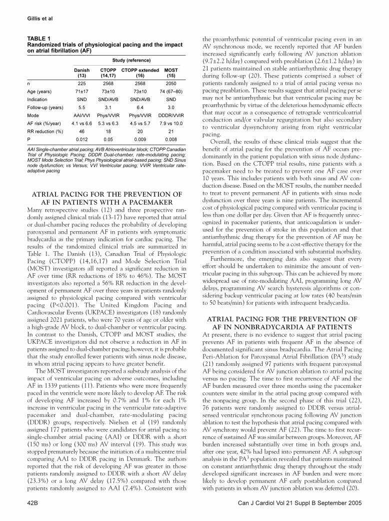
Gillis et al
Can J Cardiol Vol 21 Suppl B September 200542B
ATRIAL PACING FOR THE PREVENTION OF
AF IN PATIENTS WITH A PACEMAKERMany retrospective studies (12) and three prospective ran-domly assigned clinical trials (13-17) have reported that atrialor dual-chamber pacing reduces the probability of developingparoxysmal and permanent AF in patients with symptomaticbradycardia as the primary indication for cardiac pacing. Theresults of the randomized clinical trials are summarized inTable 1. The Danish (13), Canadian Trial of PhysiologicPacing (CTOPP) (14,16,17) and Mode Selection Trial(MOST) investigators all reported a significant reduction inAF over time (RR reductions of 18% to 46%). The MOSTinvestigators also reported a 56% RR reduction in the devel-opment of permanent AF over three years in patients randomlyassigned to physiological pacing compared with ventricularpacing (P<0.001). The United Kingdom Pacing andCardiovascular Events (UKPACE) investigators (18) randomlyassigned 2021 patients, who were 70 years of age or older witha high-grade AV block, to dual-chamber or ventricular pacing.In contrast to the Danish, CTOPP and MOST studies, theUKPACE investigators did not observe a reduction in AF inpatients assigned to dual-chamber pacing; however, it is probablethat the study enrolled fewer patients with sinus node disease,in whom atrial pacing appears to have greater benefit.
The MOST investigators reported a substudy analysis of theimpact of ventricular pacing on adverse outcomes, includingAF in 1339 patients (11). Patients who were more frequentlypaced in the ventricle were more likely to develop AF. The riskof developing AF increased by 0.7% and 1% for each 1%increase in ventricular pacing in the ventricular rate-adaptivepacemaker and dual-chamber, rate-modulating pacing(DDDR) groups, respectively. Nielsen et al (19) randomlyassigned 177 patients who were candidates for atrial pacing tosingle-chamber atrial pacing (AAI) or DDDR with a short(150 ms) or long (300 ms) AV interval (19). This study wasstopped prematurely because the initiation of a multicentre trialcomparing AAI to DDDR pacing in Denmark. The authorsreported that the risk of developing AF was greater in thosepatients randomly assigned to DDDR with a short AV delay(23.3%) or a long AV delay (17.5%) compared with thosepatients randomly assigned to AAI (7.4%). Consistent with
the proarrhythmic potential of ventricular pacing even in anAV synchronous mode, we recently reported that AF burdenincreased significantly early following AV junction ablation(9.7±2.2 h/day) compared with preablation (2.6±1.2 h/day) in21 patients maintained on stable antiarrhythmic drug therapyduring follow-up (20). These patients comprised a subset ofpatients randomly assigned to a trial of atrial pacing versus nopacing preablation. These results suggest that atrial pacing per semay not be antiarrhythmic but that ventricular pacing may beproarrhythmic by virtue of the deleterious hemodynamic effectsthat may occur as a consequence of retrograde ventriculoatrialconduction and/or valvular regurgitation but also secondaryto ventricular dyssynchrony arising from right ventricularpacing.
Overall, the results of these clinical trials suggest that thebenefit of atrial pacing for the prevention of AF occurs pre-dominantly in the patient population with sinus node dysfunc-tion. Based on the CTOPP trial results, nine patients with apacemaker need to be treated to prevent one AF case over10 years. This includes patients with both sinus and AV con-duction disease. Based on the MOST results, the number neededto treat to prevent permanent AF in patients with sinus nodedysfunction over three years is nine patients. The incrementalcost of physiological pacing compared with ventricular pacing isless than one dollar per day. Given that AF is frequently unrec-ognized in pacemaker patients, that anticoagulation is under-used for the prevention of stroke in this population and thatantiarrhythmic drug therapy for the prevention of AF may beharmful, atrial pacing seems to be a cost-effective therapy for theprevention of a condition associated with substantial morbidity.
Furthermore, the emerging data also suggest that everyeffort should be undertaken to minimize the amount of ven-tricular pacing in this subgroup. This can be achieved by morewidespread use of rate-modulating AAI, programming long AVdelays, programming AV search hysteresis algorithms or con-sidering backup ventricular pacing at low rates (40 beats/minto 50 beats/min) for patients with infrequent bradycardia.
ATRIAL PACING FOR THE PREVENTION OF
AF IN NONBRADYCARDIA AF PATIENTSAt present, there is no evidence to suggest that atrial pacingprevents AF in patients with frequent AF in the absence ofdocumented significant sinus bradycardia. The Atrial PacingPeri-Ablation for Paroxysmal Atrial Fibrillation (PA3) study(21) randomly assigned 97 patients with frequent paroxysmalAF being considered for AV junction ablation to atrial pacingversus no pacing. The time to first recurrence of AF and theAF burden measured over three months using the pacemakercounters were similar in the atrial pacing group compared withthe nonpacing group. In the second phase of this trial (22),76 patients were randomly assigned to DDDR versus atrial-sensed ventricular synchronous pacing following AV junctionablation to test the hypothesis that atrial pacing compared withAV synchrony would prevent AF (22). The time to first recur-rence of sustained AF was similar between groups. Moreover, AFburden increased substantially over time in both groups and,after one year, 42% had lapsed into permanent AF. A subgroupanalysis in the PA3 population revealed that patients maintainedon constant antiarrhythmic drug therapy throughout the studydeveloped significant increases in AF burden and were morelikely to develop permanent AF early postablation comparedwith patients in whom AV junction ablation was deferred (20).
TABLE 1Randomized trials of physiological pacing and the impacton atrial fibrillation (AF)
Study (reference)
Danish CTOPP CTOPP extended MOST(13) (14,17) (16) (15)
n 225 2568 2568 2050
Age (years) 71±17 73±10 73±10 74 (67–80)
Indication SND SND/AVB SND/AVB SND
Follow-up (years) 5.5 3.1 6.4 3.0
Mode AAI/VVI Phys/VVIR Phys/VVIR DDDR/VVIR
AF risk (%/year) 4.1 vs 6.6 5.3 vs 6.3 4.5 vs 5.7 7.9 vs 10.0
RR reduction (%) 46 18 20 21
P 0.012 0.05 0.009 0.008
AAI Single-chamber atrial pacing; AVB Atrioventricular block; CTOPP CanadianTrial of Physiologic Pacing; DDDR Dual-chamber, rate-modulating pacing;MOST Mode Selection Trial; Phys Physiological atrial-based pacing; SND Sinusnode dysfunction; vs Versus; VVI Ventricular pacing; VVIR Ventricular rate-adaptive pacing
Gillis_ch7.qxd 8/22/2005 11:31 AM Page 42

SELECTIVE PACING ALGORITHMS DESIGNED
TO PREVENT AFA number of selective pacing algorithms have been developedto prevent AF (3). These algorithms have been designed toprevent pauses following atrial premature beats, to overdrivesuppress premature beats or to promote a consistent atrial acti-vation sequence. The Atrial Dynamic Overdrive Pacing Trial(ADOPT) investigators (23) randomly assigned 399 patientswith sinus node dysfunction and paroxysmal AF to DDDR orDDDR plus dynamic atrial overdrive pacing. Patients were fol-lowed for one, three and six months following pacemakerinsertion. The investigators reported a very modest but statisti-cally significant reduction in symptomatic AF during follow-up.However, the absolute risk reduction for AF diminished overtime (1.25% at one month compared with 0.36% at sixmonths). Both groups experienced a significant reduction insymptomatic AF over time. The AF Therapy Investigators(24) reported that several atrial pacing algorithms in theVitatron Selection device (Viatron, Netherlands) significantlyreduced AF burden over time (24). Other studies have notconfirmed these benefits. The Atrial Septal Pacing ClinicalEfficacy Trial (ASPECT) Investigators (25) randomly assigned298 patients with symptomatic bradycardia and AF to septal orright atrial appendage (RAA) pacing sites. Following a one-month stabilization period, patients were randomly assigned toAF prevention algorithms ON or OFF and followed for threemonths. Patients were then crossed over to the alternate pacingstrategy for an additional three months. The combined AFprevention algorithms did not significantly reduce AF burden.The Pacing in Prevention of AF (PIPAF) Study investigators(26) randomly assigned 192 patients with bradycardia and AFto a trial of three AF prevention algorithms in a six-monthcrossover design. The primary outcome measure (total modeswitch duration) was similar when the AF prevention algo-rithms were programmed ON (11.9±27.7 days) compared withwhen they were programmed OFF (11.6±26.5 days; not statisti-cally significant). The Atrial Therapy Efficacy and Safety Trial(ATTEST) Investigators (27) randomly assigned 370 patients ina parallel study design to a comparison of atrial antitachycardiapacing (ATP) plus three AF pace prevention algorithms withDDDR pacing. Over three months of follow-up, more than15,000 episodes of atrial tachycardia were treated with atrialATP therapy. Over 40% of the episodes were classified as beingeffectively terminated by the pacemaker; however, no significant
reduction in AF burden was observed in the group randomlyassigned to the AF prevention treatment arm.
Overall, these studies suggest that current AF pace preven-tion algorithms in implantable devices have minimal or noincremental benefit for the prevention of AF. Without ques-tion, atrial ATP therapy is of benefit in terminating atrialtachycardia or atrial flutter in select patients (28,29). Whetherother subgroups that are likely to benefit can be identifiedrequires further study.
SITE-SPECIFIC ATRIAL PACING FOR
PREVENTION OF AFA number of experimental and clinical studies (30-32) havereported that septal pacing, dual-site right atrial pacing or biatrialpacing shorten total atrial activation time and reduce overalldispersion of atrial refractoriness. In cardiac surgery popula-tions, multiple small randomly assigned trials have reportedthat right atrial, dual-site right atrial, left atrial and biatrialpacing prevent perioperative AF (33). Biatrial pacing may bemore effective than right atrial pacing alone.
A number of clinical trials have evaluated the effect of var-ious atrial pacing sites for the prevention of AF in the pace-maker population. Pacing at Bachmann’s bundle comparedwith pacing at the RAA has been reported to prevent thedevelopment of permanent AF (47% versus 75%, respectively;P<0.05) (34). A significant reduction in AF burden has beenreported in patients randomly assigned to septal pacing nearthe triangle of Koch (47±84 min/day) compared with patientsrandomly assigned to RAA pacing (140±217 min/day; P<0.05)(35). However, this result was not confirmed by a larger ran-domly assigned trial (25) of RAA pacing versus septal pacing.Dual-site right atrial pacing (RAA and coronary sinus os leadlocation) offers a modest benefit for the prevention of AF com-pared with RAA pacing (36). Biatrial pacing has been reported(37) to prevent paroxysmal and permanent AF in patientswith markedly delayed intra-atrial conduction. At present, therole of selective atrial lead site(s) for the prevention of AF inthe pacemaker population remains uncertain. Given the com-plexity and added expense of additional leads, a single-sitelocation would be preferable.
ACKNOWLEDGEMENTS: Dr Gillis is a Medical Scientist ofthe Alberta Heritage Foundation for Medical Research.
Pacing for the prevention of atrial fibrillation
Can J Cardiol Vol 21 Suppl B September 2005 43B
REFERENCES1. Gillis AM, Morck M. Atrial fibrillation after DDDR pacemaker
implantation. J Cardiovasc Electrophysiol 2002;13:542-7.2. Gillis AM. Pacing to prevent atrial fibrillation. Cardiol Clin
2000;18:25-36.3. Gillis AM. Clinical trials of pacing for maintenance of sinus
rhythm. J Interv Card Electrophysiol 2004;10(Suppl 1):55-62.4. Willems R, Gillis AM. New indications for pacing. Curr Cardiol
Rep 2003;5:369-74.5. Nazir SA, Lab MJ. Mechanoelectric feedback and atrial
arrhythmias. Cardiovasc Res 1996;32:52-61.6. Morgan DE, Norman R, West RO, Burggraf G. Echocardiographic
assessment of tricuspid regurgitation during ventricular demandpacing. Am J Cardiol 1986;58:1025-9. (Erratum in 1987;59:A12).
7. Attuel P, Pellerin D, Mugica J, Coumel P. DDD pacing: An effective treatment modality for recurrent atrial arrhythmias.Pacing Clin Electrophysiol 1988;11:1647-54.
8. Murgatroyd FD, Nitzsche R, Slade AK, et al. A new pacingalgorithm for overdrive suppression of atrial fibrillation. ChorusMulticentre Study Group.Pacing Clin Electrophysiol 1994;17:1966-73.
9. Mehra R, Hill MR. Prevention of atrial fibrillation/flutter by pacingtechniques. In: Saksena S, Lüderitz B, eds. Interventional Electrophysiology: A Textbook, 2nd edn. Armonk: Futura,1996:521-40.
10. Lau CP, Tse HF, Yu CM, et al; New Indication for PreventivePacing in Atrial Fibrillation (NIPP-AF) Investigators. Dual-site atrial pacing for atrial fibrillation in patients without bradycardia.Am J Cardiol 2001;88:371-5.
11. Sweeney MO, Hellkamp AS, Ellenbogen KA, et al; MOdeSelection Trial Investigators. Adverse effect of ventricular pacingon heart failure and atrial fibrillation among patients with normalbaseline QRS duration in a clinical trial of pacemaker therapy forsinus node dysfunction. Circulation 2003;107:2932-7.
12. Connolly SJ, Kerr C, Gent M, Yusuf S. Dual-chamber versusventricular pacing. Critical appraisal of current data. Circulation1996;94:578-83.
13. Andersen HR, Nielsen JC, Thomsen PE, et al. Long-term follow-upof patients from a randomised trial of atrial versus ventricularpacing for sick-sinus syndrome. Lancet 1997;350:1210-6.
Gillis_ch7.qxd 8/22/2005 11:31 AM Page 43
Gillis et al
Can J Cardiol Vol 21 Suppl B September 200544B
14. Connolly SJ, Kerr CR, Gent M, et al. Effects of physiologic pacingversus ventricular pacing on the risk of stroke and death due tocardiovascular causes. Canadian Trial of Physiologic PacingInvestigators. N Engl J Med 2000;342:1385-91.
15. Lamas GA, Lee KL, Sweeney MO, et al; Mode Selection Trial inSinus-Node Dysfunction. Ventricular pacing or dual-chamberpacing for sinus-node dysfunction. N Engl J Med 2002;346:1854-62.
16. Kerr CR, Connolly S, Abdollah H, et al. Canadian Trial ofPhysiological Pacing: Effects of physiological pacing during long-term follow-up. Circulation 2004;109:357-62.
17. Skanes A, Krahn AD, Yee R, et al; Canadian Trial of PhysiologicPacing. Progression to chronic atrial fibrillation after pacing: The Canadian Trial of Physiologic Pacing. CTOPP Investigators. J Am Coll Cardiol 2001;38:167-72.
18. Toff WD; UK PACE trial. Presented at the American College ofCardiology Scientific Sessions. Chicago: March 30-April 2, 2003.
19. Nielsen JC, Kristensen L, Andersen HR, Mortensen PT, Pedersen OL,Pedersen AK. A randomized comparison of atrial and dual-chamberpacing in 177 consecutive patients with sick sinus syndrome:Echocardiographic and clinical outcome. J Am Coll Cardiol2003;42:614-23.
20. Willems R, Wyse DG, Gillis AM; for the Atrial Pacing Periablationfor Paroxysmal Atrial Fibrillation (PA3) Study Investigators. Totalatrioventricular nodal ablation increases atrial fibrillation burden inpatients with paroxysmal atrial fibrillation despite continuation ofantiarrhythmic drug therapy. J Cardiovasc Electrophysiol2003;14:1296-301.
21. Gillis AM, Connolly SJ, Lacombe P, et al. Randomized crossovercomparison of DDDR versus VDD pacing after atrioventricularjunction ablation for prevention of atrial fibrillation. The atrialpacing peri-ablation for paroxysmal atrial fibrillation (PA (3)) studyinvestigators. Circulation 2000;102:736-41.
22. Gillis AM, Wyse DG, Connolly SJ, et al. Atrial pacing periablation forprevention of paroxysmal atrial fibrillation. Circulation 1999;99:2553-8.
23. Carlson MD, Ip J, Messenger J, et al; Atrial Dynamic OverdrivePacing Trial (ADOPT) Investigators: A new pacemaker algorithmfor the treatment of atrial fibrillation: Results of the Atrial DynamicOverdrive Pacing Trial (ADOPT). J Am Coll Cardiol 2003;42:627-33.
24. Camm J; for the AF Therapy Investigators. AF Therapy Study:Prevention pacing for paroxysmal atrial fibrillation. Pacing ClinElectrophysiol 2002;24:554.
25. Padeletti L, Purerfellner H, Adler SW, et al; Worldwide ASPECTInvestigators. Combined efficacy of atrial septal lead placement andatrial pacing algorithms for prevention of paroxysmal atrialtachyarrhythmia. J Cardiovasc Electrophysiol 2003;14:1189-95.
26. Mabo P, Funck R, De Roy L, Poezevara Y, Anselme F, Di Donna P.Impact of ventricular pacing on atrial fibrillation prevention.Pacing Clin Electrophysiol 2002;24:II-621.
27. Lee MA, Weachter R, Pollak S, et al; ATTEST Investigators. The effect of atrial pacing therapies on atrial tachyarrhythmiaburden and frequency: Results of a randomized trial in patients with bradycardia and atrial tachyarrhythmias. J Am Coll Cardiol2003;41:1926-32.
28. Gillis AM, Morck M, Fitts S. Antitachycardia pacing therapies andarrhythmia monitoring diagnostics for the treatment of atrialfibrillation. Can J Cardiol 2002;18:992-5.
29. Gillis AM, Morck M, Willems R, et al. Atrial antitachycardiapacing therapy and reduction in atrial tachyarrhythmia burden.Pacing Clin Electrophysiol 2003;26:11-927
30. Yu WC, Chen SA, Tai CT, Feng AN, Chang MS. Effects of different atrial pacing modes on atrial electrophysiology: Implicating the mechanism of biatrial pacing in prevention of atrial fibrillation. Circulation1997;96:2992-6.
31. Prakash A, Delfaut P, Krol RB, Saksena S. Regional right and leftatrial activation patterns during single- and dual-site atrial pacingin patients with atrial fibrillation. Am J Cardiol 1998;82:1197-204.
32. Papageorgiou P, Anselme F, Kirchhof CJ, et al. Coronary sinuspacing prevents induction of atrial fibrillation. Circulation1997;96:1893-8.
33. Crystal E, Connolly SJ, Sleik K, Ginger TJ, Yusuf S. Interventions on prevention of postoperative atrial fibrillation inpatients undergoing heart surgery: A meta-analysis. Circulation 2002;106:75-80.
34. Bailin SJ, Adler S, Giudici M. Prevention of chronic atrialfibrillation by pacing in the region of Bachmann’s bundle: Results of a multicenter randomized trial. J CardiovascElectrophysiol 2001;12:912-7.
35. Padeletti L, Porciani MC, Michelucci A, et al. Interatrial septumpacing: A new approach to prevent recurrent atrial fibrillation. J Interv Card Electrophysiol 1999;3:35-43.
36. Saksena S, Prakash A, Ziegler P, et al; DAPPAF Investigators.Improved suppression of recurrent atrial fibrillation with dual-siteright atrial pacing and antiarrhythmic drug therapy. J Am CollCardiol 2002;40:1140-52.
37. D’Allonnes GR, Pavin D, Leclercq C, et al. Long-term effects ofbiatrial synchronous pacing to prevent drug-refractory atrialtachyarrhythmia: A nine-year experience. J CardiovascElectrophysiol 2000;11:1081-91.
Gillis_ch7.qxd 8/22/2005 11:31 AM Page 44

Can J Cardiol Vol 21 Suppl B September 2005 45B
Atrial fibrillation following cardiac surgery
L Brent Mitchell MD FRCPC1, Eugene Crystal MD FRCPC2, Brett Heilbron MD FRCPC3, Pierre Pagé MD FRCPS4
1Libin Cardiovascular Institute of Alberta, Calgary, Alberta; 2University of Toronto, Toronto, Ontario; 3University of British Columbia,Vancouver, British Columbia; 4University of Montreal, Montreal, Quebec
Correspondence: Dr L Brent Mitchell, Department of Cardiac Sciences, Libin Cardiovascular Institute, Calgary Health Region and University of Calgary,1403 29th Street Northwest, Calgary, Alberta T2N 2T9. Telephone 403-944-1683, fax 403-944-2906, e-mail [email protected]
RECOMMENDATIONS FOR THE PREVENTION
AND TREATMENT OF ATRIAL FIBRILLATION
FOLLOWING CARDIAC SURGERYClass I
1)Patients who have been receiving a beta-blocker beforecardiac surgery should have that therapy continuedthrough the operative period in the absence of thedevelopment of a new contraindication (level ofevidence A).
2)Temporary ventricular epicardial pacing electrodewires should be placed at the time of cardiac surgeryto allow for backup pacing as necessary (level ofevidence C).
3)Postoperative atrial fibrillation (AF) with a rapidventricular response rate should be treated with a beta-blocker, a nondihydropyridine calcium antagonist or
amiodarone to establish ventricular rate control. In theabsence of a specific contraindication, the order ofchoice is as listed (level of evidence B).
Class IIa
1)Patients who have not been receiving a beta-blockerbefore cardiac surgery should be considered forprophylactic therapy to prevent postoperative AF witha beta-blocker or amiodarone (level of evidence A) orwith atrial pacing or magnesium (level of evidence B).
2)Postoperative AF may be appropriately treated witheither a ventricular response rate-control strategy or arhythm-control strategy (level of evidence A).
3)Consideration should be given to anticoagulationtherapy if postoperative AF persists for more than 48 h(level of evidence C).
2004 CCS CONSENSUS CONFERENCE: ATRIAL FIBRILLATION
©2005 Pulsus Group Inc. All rights reserved
LB Mitchell, E Crystal, B Heilbron, P Pagé. Atrial fibrillation
following cardiac surgery. Can J Cardiol 2005;21(Suppl B):
45B-50B.
Atrial tachyarrhythmias, usually atrial fibrillation or atrial flutter, are the
most common complications of cardiac surgery. Atrial tachyarrhythmias
are associated with patient discomfort/anxiety, hemodynamic deterio-
ration, cognitive impairment, thromboembolic events (including
stroke), exposure to the risks of antiarrhythmic treatments, longer
hospital stays and increased costs. Many approaches to the prevention
of postoperative atrial tachyarrhythmias have been studied. Of these,
studies using perioperative beta-blocking agents or amiodarone provide
level A evidence of efficacy and, in properly selected patients, have
shown a high degree of safety. Less convincing, level B evidence exists
for the use of postoperative temporary atrial pacing and for perioperative
intravenous magnesium treatment. The treatment of postoperative
atrial tachyarrhythmias is similar to those occurring in other settings
and includes excluding other potential causes of atrial tachyarrhythmias,
antithrombotic or anticoagulation therapy, control of the ventricular
response rate and consideration of restoring/maintaining sinus rhythm.
The selection of therapies to achieve these goals should consider the
sympathetic nervous system discharge state of the postoperative envi-
ronment and the natural history of postoperative atrial fibrillation,
which includes spontaneous resolution of the arrhythmogenic tendency
after approximately six weeks. The Canadian Cardiovascular Society
Consensus Conference recommendations for the prevention of atrial
tachyarrhythmias after cardiac surgery and for the treatment of atrial
tachyarrhythmias that occur after cardiac surgery are presented along
with evidence that supports these recommendations.
Key Words: Atrial fibrillation; Cardiac surgery; Consensus guidelines;
Postoperative
La fibrillation auriculaire après uneintervention chirurgicale cardiaque
Les tachyarythmies auriculaires (TA), habituellement la fibrillation
auriculaire ou le flutter auriculaire, sont les complications les plus fréquentes
de la chirurgie cardiaque. Ces troubles du rythme sont associés à divers
inconvénients : malaises et anxiété, détérioration hémodynamique,
déficience cognitive, accidents thrombo-emboliques (y compris l’accident
vasculaire cérébral), exposition aux risques des traitements
antiarythmiques, prolongement du séjour à l’hôpital et augmentation des
coûts. De nombreuses interventions visant à prévenir les TA
postopératoires ont fait l’objet d’étude. Parmi celles-ci, l’administration de
bêta-bloquants ou d’amiodarone en phase périopératoire a été associée à
des données d’efficacité de niveau A et, chez les patients bien sélectionnés,
à un degré élevé d’innocuité. La stimulation auriculaire temporaire
postopératoire et l’administration intraveineuse de magnésium en phase
périopératoire donnent des résultats moins probants (niveau B). Le
traitement des TA postopératoires est le même que celui qui est appliqué
dans d’autres contextes et il consiste en l’exclusion d’autres causes
possibles de TA, en l’administration d’un traitement anticoagulant ou
antithrombotique, en la maîtrise de la fréquence ventriculaire et dans le
rétablissement et le maintien possibles du rythme sinusal. Le choix des
traitements pour atteindre les objectifs visés devrait tenir compte de l’état
de décharge du système nerveux sympathique après une opération et de
l’évolution naturelle de la fibrillation auriculaire postopératoire,
notamment de sa tendance à disparaître spontanément au bout de six
semaines environ. Sont présentées dans l’article les recommandations
consensuelles de la Société canadienne de cardiologie concernant la
prévention et le traitement des TA après une intervention chirurgicale
cardiaque, ainsi que les données à l’appui de ces recommandations.
mitchell_ch8.qxd 8/22/2005 11:34 AM Page 45

Mitchell et al
Can J Cardiol Vol 21 Suppl B September 200546B
4)When anticoagulation therapy, rate-control therapy,and/or rhythm control therapy has been prescribed forpostoperative AF, formal reconsideration of the ongoingneed for such therapy should be undertaken six to eightweeks later (level of evidence B).
POSTOPERATIVE ATRIAL
TACHYARRHYTHMIASIncidence of postoperative atrial tachyarrhythmiasGiven that AF and atrial flutter are facilitated by atrial trauma,atrial stretch, atrial ischemia, epicardial inflammation, hypoxia,acidosis, electrolyte disturbances and the refractorinesschanges that accompany sympathetic nervous system dis-charge, and given that all of these factors are often presentimmediately after cardiac surgical procedures, it is not surprisingthat AF and atrial flutter are frequent complications of theseprocedures. Indeed, atrial tachyarrhythmias are the mostcommon postoperative complication of cardiac surgery thatrequires intervention or prolongs intensive care unit and totalhospital stay (1-10). The incidence of AF and atrial flutterafter cardiac surgery ranges from approximately 30% for patientsundergoing isolated coronary artery bypass graft (CABG) sur-gery to approximately 40% for patients undergoing valve replace-ment or repair, and this incidence increases to approximately50% for patients undergoing both procedures (11). Furthermore,there is evidence that the incidence of postoperative AF andatrial flutter is increasing because older individuals with ahigher prevalence of atrial tachyarrhythmia risk factors aremore commonly having these surgeries (2,8).
The peak incidence of these atrial tachyarrhythmias isbetween postoperative days 2 and 4. Of the patients who developan atrial tachyarrhythmia, 70% do so before the end of postopera-tive day 4, and 94% do so before the end of posthospital day 6 (8).
Risk factors for postoperative atrial tachyarrhythmiasIndependent patient characteristics that predict the occur-rence of atrial tachyarrhythmias after cardiac surgery includeolder age, being male, history of hypertension, requirement foran intraoperative balloon pump, requirement for prolongedventilation (greater than 24 h), and withdrawal of beta-blockertherapy (2-8,12,13). As in the general population (14), age hasthe greatest predictive value. Operative variables reported tobe atrial tachyarrhythmia risk factors include the procedureperformed (isolated CABG, valve repair/replacement or both),the number of bypass grafts, the duration of the surgery and theaortic cross-clamp time (2,15-17).
Consequences of postoperative atrial tachyarrhythmiasPostcardiac surgery atrial tachyarrhythmias may be transientand cause little morbidity. However, for some patients thesetachyarrhythmias have important consequences includingpatient discomfort/anxiety, hemodynamic deterioration, cog-nitive impairment, thromboembolic events including stroke,exposure to the risks of arrhythmia treatments, longer hospitalstay and increased health care costs (2,4,8,9,18-23). Linearregression models indicate that postoperative atrial tachy-arrhythmias are independently associated with an increase inhealth care costs and the duration of hospital stay (8,24).
Prophylaxis against postoperative atrial tachyarrhythmiasMany therapies have been evaluated for the prevention ofpostoperative AF and atrial flutter after cardiac surgery
(17,25-74). In the section that follows, published meta-analysisdata are provided and referenced when current. When pub-lished meta-analysis data are either unavailable or are not cur-rent, individual study data were meta-analyzed using arandom-effects model and are provided without a reference.
Seven randomized trials (17,25-30) have evaluated digoxintherapy in 709 patients. One trial showed a significant advan-tage for digoxin (26), one showed a significant disadvantage(28), and the other five showed no difference between treat-ment and control group outcomes (17,25,27,29,30). The ORfor the postoperative incidence of atrial tachyarrhythmia inour weighted meta-analysis of prophylactic digoxin therapystudies is 0.91 (95% CI 0.59 to 1.40, P = not significant [NS]).
Three randomized trials (31-33) have evaluated verapamilin 432 patients. No trial showed a significant advantage or dis-advantage to having therapy. The OR in our weighted meta-analysis of prophylactic verapamil therapy studies is 0.94 (95%CI 0.56 to 1.58, P=NS).
Twenty-seven randomized trials (34) evaluated beta-blockerprophylaxis in 3840 patients. Sixteen of the 27 trials showed asignificant advantage for beta-blocker therapy. No beta-blockertrial showed a significant disadvantage to having therapy. TheOR in this meta-analysis (34) of prophylactic beta-blockertherapy studies was 0.39 (95% CI 0.28 to 0.52, P<0.0001).Sotalol is a beta-blocker that also has important class III anti-arrhythmic drug effects. Eight randomized trials (34) evaluatedsotalol prophylaxis in 1294 patients. One of these trials pro-duced a neutral result and the other seven trials reported a statis-tically significant benefit from sotalol therapy. The OR in thatmeta-analysis (34) of prophylactic sotalol drug therapy studieswas 0.35 (95% CI 0.26 to 0.49, P<0.0001). Four trials (34) com-pared sotalol prophylaxis with that of other beta-blockers in900 patients. One of these trials produced a neutral result andthe other three trials reported a statistically significant benefitfrom sotalol therapy. Compared with other beta-blocker drugs,the OR in that meta-analysis (34) of prophylactic sotalol drugtherapy studies was 0.50 (95% CI 0.34 to 0.74, P<0.0001).However, one trial comparing sotalol with metoprolol in dosesconsidered to provide equivalent beta-blockade reported ahigher prevalence of postoperative bradyarrhythmias withsotalol prophylaxis (35).
Fourteen randomized trials (36-49) evaluated amiodaroneprophylaxis in 2823 patients. Eight (37,38,40,43,45,47-49) ofthe 14 trials showed a significant advantage for amiodaronetherapy. No amiodarone trial showed a significant disadvantageto having therapy. The OR in our meta-analysis of prophylacticamiodarone therapy studies is 0.59 (95% CI 0.50 to 0.69,P<0.001). In a recent study involving 600 patients (49), theprophylactic effect of amiodarone was consistent in subgroupanalyses of young and older patients, patients undergoing iso-lated CABG, undergoing valve repair/replacement with orwithout concomitant CABG, and patients also receiving or notreceiving beta-blocker therapy.
Thirteen randomized trials (30,42,50-60) evaluated intra-venous magnesium prophylaxis in 2009 patients. Two of the13 trials showed a significant advantage for magnesium therapy(53,54). No magnesium trial showed a significant disadvan-tage to having therapy. The OR in our meta-analysis of pro-phylactic magnesium therapy studies is 0.83 (95% CI 0.65 to1.06, P=NS).
Two randomized trials (61,62) evaluated procainamideprophylaxis in 146 patients. Neither showed a significant
mitchell_ch8.qxd 8/22/2005 11:34 AM Page 46

advantage for procainamide therapy. The OR in our meta-analysis of these underpowered prophylactic procainamidetherapy studies is 0.47 (95% CI 0.22 to 0.99, P=0.05). Thewell-documented hazards of class I antiarrhythmic drug thera-pies in patients with structural heart disease have precludedacceptance of this form of postoperative atrial tachy-arrhythmia prophylaxis.
Finally, 12 randomized trials (40,63-73) evaluated atrial pacingprophylaxis in 1708 patients. Three of the 12 trials showed a sig-nificant advantage for atrial pacing therapy (64,72,73). Noatrial pacing trial showed a significant disadvantage to havingtherapy. The OR in our meta-analysis of prophylactic atrialpacing therapy studies is 0.67 (95% CI 0.54 to 0.84, P<0.0001).
In summary, published clinical trial evidence supports thecontention that beta-blockers, amiodarone, atrial pacing and(perhaps) magnesium therapy prevent postoperative AF andatrial flutter after cardiac surgery (Table 1). To determine ifsuch prophylactic therapy was associated with a reduction inany of the presumed adverse consequences of postoperativeatrial tachyarrhythmias, a meta-analysis (74) of 13 of the trialssummarized above that also reported on length of hospital staywas performed. That analysis suggested that prophylactic therapyis associated with a significant reduction of 1.0±0.2 days(P<0.001) in the length of hospital stay, and with a nearly sig-nificant reduction of US$1287±673 (P=0.056) in hospitalcosts. Although there was a directional trend in the reductionof the incidence of postoperative cerebrovascular accidentswith an OR of 0.50 (95% CI 0.22 to 1.17), this trend was notstatistically significant. Of note, two completed trials (49,75)were powered to specifically detect a reduction in the length ofhospital stay in patients receiving prophylactic therapy forprevention of atrial tachyarrhythmias. A large beta-blocker
trial (75) did not identify a reduction in hospital stay. A largeamiodarone trial (49) demonstrated a trend toward a reductionin total hospital stay duration. Furthermore, a previous meta-analysis limited to amiodarone trials (34) found a significantreduction in the length of hospital stay in amiodarone-treatedpatients.
Treatment of postoperative atrial tachyarrhythmiasThe treatment options for AF and atrial flutter that occur aftercardiac surgery are similar to those of AF and atrial flutter thatoccur in other settings. The therapeutic goals that are consid-ered include prevention of thromboembolic events, slowingthe ventricular response rate, conversion to sinus rhythm andmaintenance of sinus rhythm. Nevertheless, the postoperativesetting does have features that may favour some strategies overothers. First, the natural history of postoperative AF and flutterafter cardiac surgery is dominated by self-terminating, butfrequently recurrent, tachyarrhythmia episodes and resolutionof the tachyarrhythmia propensity in six to eight weeks,regardless of the treatment approach used (76-78). Second, theadrenergic discharge in the postoperative state lessens theeffectiveness of therapies that do not include beta-blockade.
Several studies have demonstrated associations between AFafter cardiac surgery and cerebrovascular events (2,3,8,20,21)and cognitive impairment (19). Accordingly, in the absence ofa specific contraindication, anticoagulation therapy is recom-mended for patients with prolonged (greater than 48 h) AF.Once initiated, anticoagulation therapy is usually continuedfor at least six weeks.
In the postoperative setting, therapy for ventricular ratecontrol for atrial tachyarrhythmias is usually required. Becausethe postsurgical state includes adrenergic discharge, beta-blocker
Atrial fibrillation following cardiac surgery
Can J Cardiol Vol 21 Suppl B September 2005 47B
TABLE 1Prophylactic therapies for the prevention of postoperative atrial tachyarrhythmias
Therapy Dose* OR† Cautions Adverse effects
Preoperative Any usual therapeutic dose (ie, metoprolol 0.39 (0.28 to 0.52) Reactive airways disease, Sinus bradycardia,
beta-blocker 50 mg po q12h or q8h for at least 2 preoperative decompensated CHF AV block,
days, day of surgery, and at least 6 postoperative hypotension,
days) bronchospasm
Preoperative 10 mg/kg/day (rounded to nearest 100 mg) 0.61 (0.50 to 0.74) 30% to 50% reduction in the doses Sinus bradycardia,
amiodarone divided into two daily po dosages for of other drugs with antiarrhythmic AV block hypotension,
6 preoperative days, day of surgery, and or sinus/AV nodal effects and torsade de pointes VT (rare),
6 postoperative days‡ warfarin will be required pulmonary toxicity (rare)
Postoperative 900 mg to 1200 mg IV over 24 h beginning 0.53 (0.39 to 0.71) 30% to 50% reduction in the doses Sinus bradycardia,
amiodarone within 6 h of surgery, then 400 mg po of other drugs with antiarrhythmic AV block hypotension,
three times daily each of the next 4 days§ or sinus/AV nodal effects and torsade de pointes VT (rare),
warfarin will be required pulmonary toxicity (rare)
Magnesium 1.5 g IV over 4 h first preoperative day, 0.83 (0.65 to 1.06) Renal failure Hypotension (rare),
sulfate immediately postoperatively and next sedation (very rare),
4 postoperative days¶. Other trials have respiratory depression
omitted the preoperative dosage (very rare)
Atrial pacing Right, left or biatrial pacing for 3 to 4 days 0.67 (0.54 to 0.84) May increase atrial Diaphragmatic stimulation,
postoperatively**. Rate set to overdrive tachyarrhythmias if pacing increased myocardial
sinus rate either manually or using continues in setting of sensing oxygen requirements,
sensing algorithms malfunction possible increased infection rate
*Doses used in the randomized studies vary widely and the optimal doses for this indication have not been established. The doses provided are those used in thelargest positive trial of that therapy and are referenced to that study; †The ORs provided are from meta-analyses of the studies of each prophylactic approach (notfor the single study referenced for dose). For further information on doses, see references 49‡, 40§, 57¶ and 72**. Comparisons of the efficacies of various prophy-lactic approaches require randomized trials, which, for the most part, have not been performed. Accordingly, comparisons among the ORs provided in the Tableshould be avoided. AV Atrioventricular; CHF Congestive heart failure; IV Intravenous; po By mouth; q Every; VT Ventricular tachycardia
mitchell_ch8.qxd 8/22/2005 11:34 AM Page 47
therapy is often very effective. When beta-blocker therapy isineffective, poorly tolerated or contraindicated, the other ther-apeutic options for ventricular response rate control include anondihydropyridine calcium antagonist (eg, diltiazem or vera-pamil) or amiodarone. In the postoperative state, therapy withdigoxin is usually insufficient for adequate control of the ven-tricular response rate. Specific information regarding the doses,efficacies and adverse effects of these rate-control therapies areprovided in Dorian and Connors, pages 26B-30B.
The general considerations for the advisability of conversionof a sustained atrial tachyarrhythmia in the postoperative settingare similar to those in other settings. However, because earlyrecurrence of the atrial tachyarrhythmia is the rule rather thanthe exception, pharmacological cardioversion or direct currentcardioversion after the initiation of pharmacological therapy toprevent atrial tachyarrhythmia recurrences are preferred overisolated direct current cardioversion provided that time is not ofthe essence. Intravenous ibutilide has been studied as a rapidlyacting approach for pharmacological cardioversion of atrialtachyarrhythmias after cardiac surgery (79). In that study, ibu-tilide infusion was associated with conversion to sinus rhythm in48% of patients. Success rates were higher in patients with atrialflutter than in patients with AF. The major adverse effect of ibu-tilide administration was the precipitation of torsade de pointesventricular tachycardia in approximately 2% to 5% of those whoreceive it in this setting (79). Specific information regarding thedoses, efficacies and adverse effects of rhythm-control therapiesare provided in Talajic and Roy, pages 19B-25B.
Because the vast majority of patients who experience apostoperative atrial tachyarrhythmia in the absence of a his-tory of preoperative atrial tachyarrhythmias will lose theirpropensity to atrial tachyarrhythmia recurrences within sixweeks, a rate-control strategy without resort to a rhythm-control strategy has appeal because it does not expose thepatient (who by definition has structural heart disease) to therisks of class I or class III antiarrhythmic drugs. To date, nolarge randomized clinical trial has specifically evaluated theadvantages and disadvantages of the rate-control strategyversus the rhythm-control strategy for AF or atrial flutter thatoccurs early after cardiac surgery. One small randomized pilotstudy (77) addressing this question found a statistically signif-icant reduction in the duration of the postsurgical hospitalstay in patients developing postoperative AF who wereassigned to a rhythm-control approach versus a rate-controlapproach to treatment. On the other hand, a retrospectiveevaluation (80) of the question has suggested a statisticallysignificant reduction in the duration of postsurgical hospitalstay in patients discharged in AF (after ventricular responserate control and anticoagulation therapy) compared withpatients discharged in sinus rhythm. Accordingly, the relativeadvantages, disadvantages and risk of a rate-control versus arhythm-control approach to the treatment of atrial tachy-arrhythmia that occurs after cardiac surgery have not beendetermined. Nevertheless, all agree that therapy provided forpostoperative AF and atrial flutter can usually be withdrawnafter six to eight weeks.
Mitchell et al
Can J Cardiol Vol 21 Suppl B September 200548B
REFERENCES 1. Andrews TC, Reimold SC, Berlin JA, Antman EM. Prevention of
supraventricular arrhythmias after coronary artery bypass surgery: A meta-analysis of randomized control trials. Circulation1991;84(Suppl 5):III236-44.
2. Creswell LL, Schuessler RB, Rosenbloom M, Cox JL. Hazards ofpostoperative atrial arrhythmias. Ann Thorac Surg 1993;56:539-49.
3. Fuller JA, Adams GG, Buxton B. Atrial fibrillation after coronaryartery bypass grafting. Is it a disorder of the elderly? J ThoracCardiovasc Surg 1989;97:821-5.
4. Leitch JW, Thomson D, Baird DK, Harris PJ. The importance ofage as a predictor of atrial fibrillation and flutter after coronaryartery bypass grafting. J Thorac Cardiovasc Surg 1990;100:338-42.
5. Crosby LH, Pifalo WB, Woll KR, Burkholder JA. Risk factors foratrial fibrillation after coronary artery bypass grafting. Am J Cardiol1990;66:1520-2.
6. Hashimoto K, Ilstrup DM, Schaff HV. Influence of clinical andhemodynamic variables on risk of supraventricular tachycardia aftercoronary artery bypass. J Thorac Cardiovasc Surg 1991;101:56-65.
7. Frost L, Mølgaard H, Christiansen EH, Hjortholm K, Paulsen PK,Thomsen PE. Atrial fibrillation and flutter after coronary arterybypass surgery: Epidemiology, risk factors and preventative trials.Int J Cardiol 1992;36:253-61.
8. Aranki SF, Shaw DP, Adams DH, et al. Predictors of atrialfibrillation after coronary artery surgery: Current trends and impactof hospital resources. Circulation 1996;94:390-7.
9. Khaja F, Parker JO. Hemodynamic effects of cardioversion inchronic atrial fibrillation. Special reference to coronary arterydisease. Arch Intern Med 1972;129:433-40.
10. Wong DT, Cheng DC, Kustra R, et al. Risk factors of delayedextubation, prolonged length of stay in the intensive care unit, andmortality in patients undergoing CABG with fast track cardiacanesthesia: A new cardiac risk score. Anesthesiology 1999;91:936-44.
11. Mitchell LB. Incidence, timing and outcome of atrialtachyarrhythmias after cardiac surgery, In: Steinberg JS, ed. AtrialFibrillation After Cardiac Surgery. Norwell, Massachusetts: KluwerAcademic Publishers, 2000:37-50.
12. Abel RM, van Gelder HM, Pores IH, Liguori J, Gielchinsky I,Parsonnet V. Continued propranolol administration following
coronary bypass surgery: Antiarrhythmic effects. Arch Surg1983;118:727-31.
13. Silverman NA, Wright R, Levitsky S. Efficacy of low-dosepropranolol in preventing postoperative supraventriculartachyarrhythmias. Ann Surg 1982;196:194-7.
14. Kannel WB, Abbott RD, Savage DD, McNamara PM.Epidemiologic features of chronic atrial fibrillation: The Framingham Study. N Engl J Med 1982;306:1018-22.
15. Caretta Q, Mercanti CA, De Nardo D, et al. Ventricularconduction defects after coronary artery bypass grafting:Multivariate analysis of preoperative, intra-operative andpostoperative variables. Eur Heart J 1991;12:1107-11.
16. Roffman JA, Fieldman A. Digoxin and propranolol in theprophylaxis of supraventricular tachydysrhythmias after coronaryartery bypass surgery. Ann Thorac Surg 1981;31:496-501.
17. Ormerod OJ, McGregor CG, Stone DL, Wisbey C, Petch MC.Arrhythmias after coronary bypass surgery. Br Heart J 1984;51:618-21.
18. Orlando JR, van Herick R, Aronow WS, Olson HG.Hemodynamics and echocardiograms before and after cardioversion of atrial fibrillation to normal sinus rhythm. Chest1979;76:521-6.
19. Stanley TO, Mackensen GB, Grocott HP, et al; NeurologicalOutcome Research Group; CARE Investigators of the Duke HeartCenter. The impact of post-operative atrial fibrillation onneurocognitive outcome after coronary artery bypass graft surgery.Anesth Analg 2002;94:290-5.
20. Taylor GJ, Malik SA, Colliver JA, et al. Usefulness of atrialfibrillation as a predictor of stoke after isolated coronary arterybypass grafting. Am J Cardiol 1987;60:905-7.
21. Lynn GM, Stefanko K, Reed JF III, Gee W, Nicholas G. Riskfactors for stoke after coronary artery bypass. J Thorac Cardiovasc Surg 1992;104:1518-23.
22. Coplen SE, Antman EM, Berlin JA, Hewitt P, Chalmers TC.Efficacy and safety of quinidine therapy for maintenance of sinusrhythm after cardioversion: A meta-analysis of randomized controltrials. Circulation 1990;82:1106-16. (Erratum in 1991;83:714).
23. Flaker GC, Blackshear JL, McBride R, et al. Antiarrhythmic drugtherapy and cardiac mortality in atrial fibrillation. The Stroke
mitchell_ch8.qxd 8/22/2005 11:34 AM Page 48

Atrial fibrillation following cardiac surgery
Can J Cardiol Vol 21 Suppl B September 2005 49B
Prevention in Atrial Fibrillation Investigators. J Am Coll Cardiol1992;20:527-32.
24. Kowey PR, Stanowski A, Schnoor E. Impact of atrial fibrillation onduration of hospital stay and cost of coronary artery bypass surgery.Clin Res 1992;40:A365. (Abst)
25. Rubin DA, Nieminski KE, Reed GE, Herman MV. Predictors,prevention, and long-term prognosis of atrial fibrillation aftercoronary bypass graft operations. J Thorac Cardiovasc Surg1987;94:331-5.
26. Johnson LW, Dickstein RA, Fruehan CT, et al. Prophylacticdigitalization for coronary artery bypass surgery. Circulation1976;53:819-22.
27. Weiner B, Rheinlander HF, Decker EL, Cleveland RJ. Digoxinprophylaxis following coronary artery bypass surgery. Clin Pharm1986;5:55-8.
28. Tyras DH, Stothert JC Jr, Kaiser GC, Barner HB, Codd JE,Willman VL. Supraventricular tachyarrhythmias after myocardialrevascularization: A randomized trial of prophylactic digitalization.J Thorac Cardiovasc Surg 1979;77:310-4.
29. Yazicioglu L, Eryilmaz S, Sirlak M, et al. The effect of preoperativedigitalis and atenolol combination on postoperative atrialfibrillation incidence. Eur J Cardiothorac Surg 2002;22:397-401.
30. Bert AA, Reinert SE, Singh AK. A beta-blocker, not magnesium, is effective prophylaxis for atrial tachyarrhythmias after coronaryartery bypass graft surgery. J Cardiothorac Vasc Anesth2001;15:204-9.
31. Davison R, Hartz R, Kaplan K, Parker M, Feiereisel P, Michaelis L.Prophylaxis of supraventricular tachyarrhythmia after coronarybypass surgery with oral verapamil: A randomized, double-blindtrial. Ann Thorac Surg 1985;39:336-9.
32. Williams DB, Misbach GA, Kruse AP, Ivey TD. Oral verapamil forprophylaxis of supraventricular tachycardia after myocardialrevascularization. A randomized trial. J Thorac Cardiovasc Surg1885;90:592-6.
33. Smith EE, Shore DF, Monro JL, Ross JK. Oral verapamil fails toprevent supraventicular tachycardia following coronary arterysurgery. Int J Cardiol 1985;9:37-44.
34. Crystal E, Connolly SJ, Sleik K, Ginger TJ, Yusuf S. Interventionson prevention of postoperative atrial fibrillation in patientsundergoing heart surgery: A meta-analysis. Circulation2002;106:75-80.
35. Nystrom U, Edvardsson N, Berggren H, Pizzarelli GP, Radegran K.Oral sotalol reduces the incidence of atrial fibrillation aftercoronary artery bypass surgery. Thorac Cardiovasc Surg 1993;41:34-7.
36. Butler J, Harriss DR, Sinclair M, Westaby S. Amiodaroneprophylaxis for tachycardias after coronary artery surgery: A randomized, double-blind, placebo controlled trial. Br Heart J1993;70:56-60.
37. Lee SH, Chang CM, Lu MJ, et al. Intravenous amiodarone forprevention of atrial fibrillation after coronary artery bypass grafting.Ann Thorac Surg 2000;70:157-61.
38. Guarnieri T, Nolan S, Gottlieb SO, Dudek A, Lowry DR.Intravenous amiodarone for the prevention of atrial fibrillationafter open heart surgery: the Amiodarone Reduction in CoronaryHeart (ARCH) trial. J Am Coll Cardiol 1999;34:343-7.
39. Dörge H, Schoendube FA, Schoberer M, et al. Intraoperativeamiodarone as prophylaxis against atrial fibrillation after coronaryoperations. Ann Thorac Surg 2000:69:1358-62.
40. White CM, Caron MF, Kalus JS, et al; Atrial FibrillationSuppression Trial II. Intravenous plus oral amiodarone, atrial septalpacing, or both strategies to prevent post-cardiothoracic surgeryatrial fibrillation: The Atrial Fibrillation Suppression Trial II(AFIST II). Circulation 2003;108(Suppl 1):II200-6.
41. Hohnloser SH, Meinertz T, Dammbacher T, et al.Electrocardiographic and antiarrhythmic effects of intravenousamiodarone: Results of a prospective, placebo-controlled study. Am Heart J 1991;121:89-95.
42. Treggiari-Venzi MM, Waeber JL, Perneger TV, et al. Intravenousamiodarone or magnesium sulphate is not cost-beneficialprophylaxis for atrial fibrillation after coronary artery bypasssurgery. Br J Anaesth 2000;85:690-5.
43. Giri S, White CM, Dunn AB, et al. Oral amiodarone forprevention of atrial fibrillation after open heart surgery, the AtrialFibrillation Suppression Trial (AFIST): A randomised placebo-controlled trial. Lancet 2001;357:830-6.
44. Redle JD, Khurana S, Marzan R, et al. Prophylactic oralamiodarone compared with placebo for prevention of atrialfibrillation after coronary artery bypass surgery. Am Heart J1999;138:144-50.
45. Daoud EG, Strickberger SA, Man KC, et al. Preoperativeamiodarone as prophylaxis against atrial fibrillation after heartsurgery. N Engl J Med 1997;337:1785-91.
46. Maras D, Boskovic SD, Popovic Z, et al. Single-day loading dose ofamiodarone for the prevention of new-onset atrial fibrillation aftercoronary artery bypass surgery. Am Heart J 2001;141:E8
47. Tokmakoglu H, Kandemir O, Gunaydin S, Catav Z, Yorgancioglu C,Zorlutuna Y. Amiodarone versus digoxin and metoprololcombination for the prevention of postcoronary bypass atrialfibrillation. Eur J Cardiothorac Surg 2002;21:401-5.
48. Yazigi A, Rahbani P, Zeid HA, Madi-Jebara S, Haddad F, Hayek G.Postoperative oral amiodarone as prophylaxis against atrialfibrillation after coronary artery surgery. J Cardiothorac VascAnesth 2002;16:603-6.
49. Mitchell LB, Exner DV, Wyse DG, et al. Prophylactic amiodaronefor the prevention of arrhythmias that begin early afterrevascularization, valve repair, or replacement (PAPABEAR):Preliminary results. Circulation 2003;108:2723. (Abst)
50. Fanning WJ, Thomas CS Jr, Roach A, Tomichek R, Alford WC,Stoney WS Jr. Prophylaxis of atrial fibrillation with magnesiumsulfate after coronary artery bypass grafting. Ann Thorac Surg1991;52:529-33.
51. England MR, Gordon G, Salem M, Chernow B. Magnesiumadministration and dysrhythmias after cardiac surgery. A placebo-controlled, double-blind, randomised trial. JAMA 1992;268:2395-402.
52. Parikka H, Toivonen L, Pellinen T, Verkkala K, Järvinen A,Nieminen MS. The influence of intravenous magnesium sulphateon the occurrence of atrial fibrillation after coronary artery by-passoperation. Eur Heart J 1993;14:251-8.
53. Colquhoun IW, Berg GA, el-Fiky M, Hurle A, Fell GS, Wheatley DJ. Arrhythmia prophylaxis after coronary artery surgery.A randomised controlled trial of intravenous magnesium chloride.Eur J Cardiothorac Surg 1993;7:520-3.
54. Karmy-Jones R, Hamilton A, Dzavik V, Allegreto M, Finegan BA,Koshal A. Magnesium sulphate prophylaxis after cardiac operations.Ann Thorac Surg 1995;59:502-7.
55. Nurozler F, Tokgozoglu L, Pasaoglu I, Boke E, Ersoy U, Bozer AY.Atrial fibrillation after coronary artery bypass surgery: Predictorsand the role of MgSO4 replacement. J Card Surg 1996;11:421-7.
56. Solomon AJ, Berger AK, Trivedi KK, Hannan RL, Katz NM. The combination of propranolol and magnesium does not preventpostoperative atrial fibrillation. Ann Thorac Surg 2000;69:126-9.
57. Toraman F, Karabulut EH, Alhan HC, Dagdelen S, Tarcan S.Magnesium infusion dramatically decreases the incidence of atrialfibrillation after coronary artery bypass surgery. Ann Thorac Surg2001;72:1256-62.
58. Yeatman M, Caputo M, Narayan P, et al. Magnesium-supplementedwarm blood cardioplegia in patients undergoing coronary arteryrevascularization. Ann Thorac Surg 2002;73:112-08.
59. Kaplan M, Kut MS, Icer UA, Demirtas MM. Intravenousmagnesium sulphate prophylaxis for atrial fibrillation after coronaryartery bypass surgery. J Thorac Cardiovasc Surg 2003;125:344-52.
60. Hazelrigg SR, Boley TM, Cetindag IB, et al. The efficacy ofsupplemental magnesium in reducing atrial fibrillation aftercoronary artery bypass grafting. Ann Thorac Surg 2004;77:824-830.
61. Laub GW, Janeira L, Muralidharan S, et al. Prophylacticprocainamide for prevention of atrial fibrillation after coronarybypass grafting: A prospective, double-blind, randomized, placebo-controlled pilot study. Crit Care Med 1993;21:1474-8.
62. Gold MR, O’Gara PT, Buckley MJ, DeSanctis RW. Efficacy andsafety of procainamide in preventing arrhythmias after coronaryartery bypass surgery. Am J Cardiol 1996;78:975-9.
63. Schweikert RA, Grady TA, Gupta N, et al. Atrial pacing in theprevention of atrial fibrillation after cardiac surgery: results of thesecond postoperative pacing study (POPS-2). J Am Coll Cardiol1998;31:117A. (Abst)
64. Orr W, Tsui S, Stafford P, et al. Synchronized bi-atrial pacing aftercoronary artery by-pass surgery. Pacing Clin Electrophysiol1999;22:755. (Abst)
65. Goette A, Mittag J, Friedl A, et al. Pacing of Bachmann’s bundleafter coronary artery bypass grafting. Pacing Clin Electrophysiol2002;25:1072-8.
mitchell_ch8.qxd 8/22/2005 11:34 AM Page 49

Mitchell et al
Can J Cardiol Vol 21 Suppl B September 200550B
66. Kurz DJ, Naegeli B, Kunz M, et al. Epicardial, biatrial synchronouspacing for prevention of atrial fibrillation after cardiac surgery.Pacing Clin Electrophysiol 1999;22:721-6.
67. Gerstenfeld EP, Hill MR, French SN, et al. Evaluation of rightatrial and biatrial temporary pacing for the prevention of atrialfibrillation after coronary artery bypass surgery. J Am Coll Cardiol1999;33:1981-8.
68. Blommaert D, Gonzalez M, Mucumbitsi J, et al. Effectiveprevention of atrial fibrillation by continuous atrial overdrivepacing after coronary artery surgery. J Am Coll Cardiol2000;35:1411-5.
69. Chung MK, Augostini RS, Asher CR, et al. Ineffectiveness andpotential proarrhythmia of atrial pacing for atrial fibrillationprevention after coronary artery bypass grafting. Ann Thorac Surg2000;69:1057-63.
70. Daoud EG, Dabir R, Archambeau M, et al. Randomized, double-blind trial of simultaneous right and left atrial epicardial pacing forprevention of post-open heart surgery atrial fibrillation. Circulation2000;102:761-5.
71. Fan K, Lee KL, Chiu CS, et al. Effects of biatrial pacing inprevention of postoperative atrial fibrillation after coronary arterybypass surgery. Circulation 2000;102:755-60.
72. Greenberg MD, Katz NM, Iuliano S, Tempesta BJ, Solomon AJ.Atrial pacing for the prevention of atrial fibrillation aftercardiovascular surgery. J Am Coll Cardiol 2000;35:1416-22.
73. Levy T, Fotopoulos G, Walker S, et al. Randomized controlledstudy investigating the effect of biatrial pacing in prevention of
atrial fibrillation after coronary artery bypass grafting. Circulation2000;102:1382-7.
74. Zimmer J, Pezzullo J, Choucair W, et al. Meta-analysis ofantiarrhythmic therapy in the prevention of postoperative atrialfibrillation and the effect on hospital length of stay, costs,cerebrovascular accidents, and mortality in patients undergoingcardiac surgery. Am J Cardiol 2003;91:1137-40.
75. Connolly SJ, Cybulsky I, Lamy A, et al; Beta-Blocker Length OfStay (BLOS) study. Double-blind, placebo-controlled, randomizedtrial of prophylactic metoprolol for reduction of hospital length ofstay after heart surgery: The beta-Blocker Length Of Stay (BLOS)study. Am Heart J 2003;145:226-32.
76. Kowey PR, Stebbins D, Igidbashian, et al. Clinical outcome ofpatients who develop PAF after CABG surgery. Pacing ClinElectrophysiol 2001;24:191-3.
77. Lee JK, Klein GJ, Krahn AD, et al. Rate-control versus conversionstrategy in postoperative atrial fibrillation: A prospective,randomized pilot study. Am Heart J 2000;140:871-7.
78. Danias PG, Caulfield TA, Weigner MJ, et al. Likelihood ofspontaneous conversion of atrial fibrillation to sinus rhythm. J AmColl Cardiol 1998;31:588-92.
79. VanderLugt JT, Mattioni T, Denker S, et al. Efficacy and safety ofibutilide fumarate for the conversion of atrial arrhythmias aftercardiac surgery. Circulation 1999;100:369-75.
80. Solomon AJ, Kouretas PC, Hopkins RA, Katz NM, Wallace RB,Hannan RL. Early discharge of patients with new-onset atrialfibrillation after cardiovascular surgery. Am Heart J 1998;135:557-63.
mitchell_ch8.qxd 8/22/2005 11:34 AM Page 50

Can J Cardiol Vol 21 Suppl B September 2005 51B
Atrial arrhythmias and special circumstances
Louise Harris MBChB FRCPC1, Robert M Gow MB BS FRACP2, Joel A Kirsh BASc MSc MD FRCPC1
George J Klein MD FRCPC3, Samuel C Siu MD SM FRCPC1
1University of Toronto, Toronto; 2University of Ottawa, Ottawa; 3University of Western Ontario, London, OntarioCorrespondence: Dr Louise Harris, Department of Medicine, Toronto General Hospital, PMCC3 562–200 Elizabeth Street, Toronto, Ontario
M5G 2C4. Telephone 416-340-3324, fax 416-595-1811, e-mail [email protected]
RECOMMENDATIONS FOR MANAGEMENT OF
ATRIAL FIBRILLATION IN PATIENTS WITH
HYPERTROPHIC CARDIOMYOPATHYClass I
1)Anticoagulate patients with paroxysmal, persistent orpermanent atrial fibrillation (AF) with warfarin(international normalized ratio 2.0 to 3.0) (level ofevidence B).
Class IIa
1)Strategies to maintain sinus rhythm are generallypreferred over rate control (level of evidence C).
2)Amiodarone is generally the preferred antiarrhythmicagent for maintenance of sinus rhythm (level ofevidence C).
DISCUSSION AF is the most common arrhythmia in hypertrophic car-diomyopathy (HCM), ocurring in 20% to 25% of patients,and is generally associated with increased risk of complica-tions including sudden and nonsudden death, heart failureand stroke (1-9). The results of the Atrial Fibrillation Follow-up Investigation of Rhythm Management (AFFIRM) trialnotwithstanding (10), restoration and maintenance of sinusrhythm has been considered an important priority because ofthe increased morbidity and mortality associated with AF in
HCM (1,11). Although rigorous comparative studies are notavailable, amiodarone has been considered the most effectiveand safest drug for the maintenance of sinus rhythm (12-15).Other antiarrhythmics have been used, but disopyramide hasbeen recommended (16) in the absence of large comparativetrials, possibly due to its reported favourable hemodynamiceffects in patients with obstruction (17). Rate-control, whendesired, is achieved with the usual agents, namely, beta-blockers and calcium channel blockers such as verapamil.Digoxin has theoretical disadvantages in obstruction and isless effective in most contexts. Nonpharmacological thera-pies including operative and catheter ablation have not beenspecifically evaluated in HCM, but their role in managementof AF in this context is likely to increase.
Finally, the high incidence of stroke in HCM (4,11,18) hasled to a prevalent recommendation for anticoagulation withwarfarin (1,16) even in the absence of a large specific trial inHCM. There are as yet insufficient data to recommend use oforal antithrombin agents in this context.
Discrepancies with American College of Cardiology/American Heart Association/European Society ofCardiology guidelinesThese recommendations are comparable with those of theAmerican College of Cardiology/American Heart Association/European Society of Cardiology (ACC/AHA/ESC) (16). We donot, however, recommend disopyramide specifically over other
2004 CCS CONSENSUS CONFERENCE: ATRIAL FIBRILLATION
©2005 Pulsus Group Inc. All rights reserved
L Harris, RM Gow, JA Kirsh, GJ Klein, SC Siu. Atrial
arrhythmias and special circumstances. Can J Cardiol
2005;21(Suppl B):51B-60B.
The present paper discusses the general principles of management ofatrial fibrillation and atrial flutter under special circumstances.Management recommendations, which encompass initial assessment,initial arrhythmia management and chronic pharmacological therapy,are outlined for patients with hypertrophic cardiomyopathy, Wolff-Parkinson-White syndrome and congenital heart disease.Recommendations are also made for pregnant patients and pediatricpatients without congenital heart disease. Discrepancies between theauthors’ recommendations and those from the American College ofCardiology/American Heart Association/European Society ofCardiology are discussed.
Key Words: Atrial arrhythmias; Atrial arrhythmias in a pediatric
population; Congenital heart disease; Hypertrophic cardiomyopathy;
Intra-atrial reentrant tachycardia; Pregnancy; Wolff-Parkinson-White
syndrome
Arythmie auriculaire et situations particulières
Le présent article porte sur les grands principes du traitement de lafibrillation auriculaire et du flutter auriculaire dans des situationsparticulières. On y traitera plus précisément de la prise en chargerecommandée de ce type d’arythmie, comprenant l’évaluation initiale, letraitement de départ et le traitement médicamenteux prolongé, dans lescas de myocardiopathie hypertrophique, de syndrome de Wolff-Parkinson-White et de cardiopathie congénitale. À cela s’ajoutent desrecommandations sur les femmes enceintes et les enfants non porteursd’une cardiopathie congénitale. Enfin, il sera question des différences entreles recommandations des auteurs et celles de l’American College of
Cardiology, de l’American Heart Association et de la Société européenne decardiologie.
harris_ch_9.qxd 8/22/2005 11:32 AM Page 51

Harris et al
Can J Cardiol Vol 21 Suppl B September 200552B
agents due to insufficient data supporting its preferential use.We also recommend a preferential rhythm control strategysupported in the body of the text of the ACC/AHA/ESCguidelines but not listed as a recommendation.
RECOMMENDATIONS FOR MANAGEMENT OF
AF IN THE WOLFF-PARKINSON-WHITE
SYNDROMEClass I
1)Catheter ablation of the accessory pathway isrecommended in symptomatic patients with AF (levelof evidence B).
2)Operative ablation of the accessory pathway isindicated in patients with problematic or life-threatening AF where catheter ablation is not feasible(level of evidence B).
3)Antiarrhythmic therapy with amiodarone, sotalol,disopyramide, flecainide, propafenone, quinidine orprocainamide is recommended when corrective ablationis not feasible (level of evidence C).
4) Immediate electrical cardioversion is recommendedwhere AF occurs with a rapid ventricular response andhypotension (level of evidence B).
5) Intravenous procainamide or ibutilide is recommendedin AF with predominantly preexcited complexes whenthe patient is hemodynamically stable (level ofevidence C).
6)Verapamil, diltiazem or beta-blockers are indicated forrate control when AF occurs without preexcitation(level of evidence C).
Class III
Intravenous beta-blocking agents are not generally useful anddigitalis, diltiazem or verapamil is contraindicated in patientswith a rapid ventricular response related to preexcitation (levelof evidence B).
DISCUSSIONThe unique feature of AF in the Wolff-Parkinson-White(WPW) syndrome is the presence of one or more accessorypathways in addition to the normal atrioventricular (AV)conduction system that may allow conduction to the ventri-cles. Accessory pathways may have extremely short effectiverefractory periods, allowing very rapid ventricular rates in theevent of AF (19). This may result in ventricular fibrillationand sudden death (20). AF in a patient with WPW mayresult from any cause unrelated to the preexcitation, andablation of the accessory pathway in such instances may notprevent subsequent AF (21). However, AF in WPW is mostfrequently related to supraventricular tachycardia which thendegenerates into AF (20). A second unique feature in theWPW syndrome is the nature of the accessory pathway.Unlike the AV node, accessory pathways are composed ofworking muscle fibres. Consequently, drugs that usually pro-long AV node refractoriness such as digitalis, verapamil andbeta-blockers do not prolong refractoriness in accessory path-ways. AV node-blocking drugs are contraindicated in patientswith AF and predominantly preexcited QRS complexesbecause they do not slow the ventricular rate and may be
detrimental. Intravenous verapamil in particular may precip-itate hemodynamic collapse due to its negative inotropiceffect and by accelerating the ventricular rate probably due toa reflex sympathetic effect (22). Intravenous sodium- andpotassium-blocking drugs such as procainamide and ibutilideprolong the refractory period of the accessory pathway andslow the ventricular response in preexcited AF (22,23). Theymay also result in conversion to sinus rhythm. Intravenousamiodarone has not been extensively evaluated for acutetreatment of arrhythmias related to WPW. It has been shownto terminate AV reentrant tachycardia and prolong the effec-tive refractory period of the accessory pathway (23,24). Atbest, it would not be expected to be very useful for acutetreatment because of the slow onset of its antiarrhythmiceffect. Ventricular fibrillation has been reported duringadministration of intravenous amiodarone during preexcitedAF (25).
Catheter ablation of the accessory pathway is currentlythe treatment of choice for symptomatic WPW syndrome.Where catheter ablation is not feasible, surgical ablation of theaccessory pathway is advised in patients with life-threateningAF.
The management of the asymptomatic individual withWPW is only peripherally related to the current guidelines andwill not be discussed in detail. Although an argument can bemade for recommending catheter ablation in such an individ-ual (26,27), the risk of catheter-related complications withablation is at least comparable with the risk of sudden death asan initial presentation (28). Catheter ablation can nonethelessbe offered to the patient who, after a balanced discussion,prefers a small procedural risk to a comparable but more long-term risk related to WPW.
Discrepancies with ACC/AHA/ESC guidelinesThese guidelines do not differ substantively from other guide-lines. Some discrepancies are as follows:
1)Operative therapy is suggested where ablation istechnically not feasible.
2)Medical therapy is recommended where neithercatheter nor operative accessory pathway ablation isfeasible.
3)AV nodal blocking drugs are recommended in patientswith AF and non-preexcited QRS while ACC/AHA/ESC guidelines do not include this in finalrecommendations.
4)While AV node-blocking drugs such as verapamil arecontraindicated in preexcited AF with a rapidventricular response, the rationale cited in the text ofthe ACC/AHA/ESC guidelines for this suggests thatthe mechanism is related to prolonging AV noderefractoriness resulting in preferential accessory pathwayconduction (16). It is our view that the AV node is notrelevant when there is rapid preexcited AF (due torepetitive retrograde concealment into the AV noderelated to rapid preexcited response) and that the majorfactors related to deterioration include the delayresulting from an ineffective intervention combinedwith the negative inotropic effect of verapamil inparticular.
harris_ch_9.qxd 8/22/2005 11:32 AM Page 52

RECOMMENDATIONS FOR THE MANAGEMENT
OF AF IN PREGNANCYClass I
1)Control the rate of ventricular response with digoxin, abeta-blocker or a calcium channel antagonist (level ofevidence C).
2)Perform electrical cardioversion in patients whobecome unstable due to AF (level of evidence C).
3)Administer antithrombotic therapy (anticoagulant oracetylsalicylic acid [ASA]) throughout pregnancy in allpatients with persistent or paroxysmal AF (level ofevidence C).
4) In patients at risk of thromboembolism, administerheparin during the first trimester and after 36 weeks’gestation. Unfractionated heparin may be administeredby intravenous infusion or by twice-a-day subcutaneousinjection (dose adjusted to maintain an activatedpartial thromboplastin time two to three times thecontrol value). Alternately, low molecular weightheparin can be used (dose adjustment guided by anti-Xalevels) (level of evidence C).
5)Administer warfarin or heparin during the secondtrimester to patients with AF and at highthromboembolic risk (level of evidence C).
Class IIa
1)For symptomatic patients or those with poorly toleratedAF, pharmacological or electrical cardioversion may beconsidered (level of evidence C).
DISCUSSIONAF during pregnancy is usually associated with the presence ofmaternal structural heart disease or hyperthyroidism (29-32).A rapid ventricular response to AF can have deleterious effectson both mother and fetus.
In a pregnant women who develops AF, diagnosis andtreatment of the underlying condition causing AF is thefirst priority (33). The ventricular rate should be controlledwith digoxin, a beta-blocker or a calcium channel antagonist(33-35). All currently available antiarrhythmic drugs have thepotential to cross the placenta and to be excreted in breast milk.Sotalol, quinidine, mexilitine, flecanide and amiodarone havebeen used successfully during pregnancy in a small number ofcases (35-39). Amiodarone may impair neonatal thyroid func-tion and should be reserved for situations in which alternativeantiarrhythmic agents are either contraindicated or ineffective(40,41). In the event of hemodynamic instability, electrical car-dioversion can be performed without fetal damage (42).
The optimal antithrombotic regimen for pregnant womenwith AF has not been defined. Because the risk of thromboem-bolism resulting from AF is high in the presence of structuralheart disease, anticoagulation should be administered in preg-nant women with structural heart disease and AF.
Warfarin should be avoided especially in the first trimester(risk of embryopathy) and last month (risk of intracranial hem-orrhage during vaginal delivery) (43). The risk of embryopathymay be dose-dependent; in one study (44), no embryopathy wasreported when the daily warfarin dose was 5 mg or less. Heparin,which does not cross the placenta, is the anticoagulant of choice
at some centres as an extension of its use in pregnant womenwith prosthetic heart valves or venous thromboembolism.However, the relative efficacy of unfractionated heparin, lowmolecular weight heparin, or warfarin in the prevention ofthromboembolism in pregnant women with AF has not beendefined.
AF in the absence of structural heart disease (lone AF) isuncommon during pregnancy. Because serum levels of severalcoagulation factors are increased during pregnancy (45), preg-nant women with lone AF may not be at as low a risk of throm-boembolism as nonpregnant individuals. Decisions for thetreatment of lone AF during pregnancy (no treatment versusASA) will need to be tailored for the individual patient.
RECOMMENDATIONS FOR MANAGEMENT OF
ATRIAL ARRHYTHMIAS IN PATIENTS WITH
CONGENITAL HEART DISEASEAtrial tachycardias (ATs) are being recognized increasingly asan important cause of morbidity in patients with repaired, pal-liated or untreated congenital heart disease. The arrhythmia ismost frequently a macroreentrant AT. Although often labelledas atrial flutter, it is now preferably called intra-atrial reentranttachycardia (IART). However, multiple mechanisms for atrialarrhythmias exist in these patients and AF is also welldescribed. Many of the medical issues that are important inadults with AF are relevant to the patient with congenitalheart disease and AT – the potential for 1:1 AV conduction, apredisposition to thrombus formation and the potential for fur-ther compromise of heart function. Therefore, the more genericterm (AT) will be used for the purpose of the following recom-mendations to encompass these different arrhythmias.
Recommendations for cardioversion of ATClass I
1) Immediately perform electrical cardioversion in patientswith AT who are hemodynamically unstable (level ofevidence C).
Class IIa
1)Electrical cardioversion for the early restoration of sinusrhythm is advisable in patients with newly diagnosedAT after appropriate anticoagulation. For patients withpacemakers, cardioversion may also be accomplished byoverdrive pace termination of AT (level of evidence C).
2)All patients with congenital heart disease and ATshould be managed as patients with AF and structuralheart disease with respect to anticoagulation (level ofevidence C).
3) In addition, all patients with complex heart lesionsrequire a transesophageal echocardiogram beforeelective cardioversion if no prior anticoagulation or ifanticoagulation is subtherapeutic, independent ofarrhythmia duration. (A complex heart lesion in thissetting is defined as one with excessive atrialenlargement [in particular, right atrial enlargement] andscarring, sluggish blood flow and predisposition to atrialthrombus formation even in sinus rhythm, oftenaccompanied by systemic ventricular dysfunction and/orright to left shunting – as such, it most commonlyapplies to the patient post-Fontan operation but can
Atrial arrhythmias and special circumstances
Can J Cardiol Vol 21 Suppl B September 2005 53B
harris_ch_9.qxd 8/22/2005 11:32 AM Page 53

also be encountered in other clinical situations such asEbstein’s anomaly) (level of evidence C).
4)Strategies to maintain sinus rhythm are generallypreferred over rate control (level of evidence C).
Class IIb
1)Pharmacological cardioversion of AT may be consideredin patients who are hemodynamically stable and whohave a controlled ventricular rate (level of evidence C).
Recommendations for pharmacological therapy to maintainsinus rhythmClass I
1)Patients placed on antiarrhythmic drugs require periodicambulatory monitoring to identify proarrhythmia, inparticular, bradycardia (level of evidence C).
Class IIa
1)Class III drugs (amiodarone and sotalol) are generallythe preferred antiarrhythmic agents for themaintenance of sinus rhythm (level of evidence C).
Class IIb
1) If a class Ic drug is to be used for preventing recurrenceof AT, the concomitant administration of drugs tomodify AV node conduction is advised. Considerationshould be given to commencing antiarrhythmic drugtherapy under electrocardiographic monitoring inhospital (level of evidence C).
Recommendations for heart rate controlClass I
1)Patients with persistent or permanent AT should haveheart rate control assessed at rest and with exercise(level of evidence C).
2)Beta-blockers or calcium channel blockers are to beadministered to slow the ventricular response rate inpatients with persistent or permanent AT with rapidventricular response not requiring immediate electricalcardioversion (level of evidence C).
Class IIa
1)Adjunctive therapy with digoxin may be used tocontrol the ventricular rate. Use of digoxin alone is notrecommended (level of evidence C).
Recommendations for long-term antithrombotic management in patients with congenital heart disease and ATClass I
1)Anticoagulation with adjusted-dose warfarin is advisedin patients with complex high-risk lesions who havehad an episode of AT (level of evidence C).
Class IIa
1)Anticoagulation with adjusted-dose warfarin should beconsidered in all other patients with congenital heartdisease and recurrent episodes of AT (level ofevidence C).
Class IIb
1)The usefulness of anticoagulation or ASA in patientswith congenital heart disease who have minimalresidual lesions and who have experienced a singleepisode of AT is uncertain. The decision to initiateanticoagulation with adjusted-dose warfarin should thenbe based on conventional risk factors (see Connolly andGillis, pages 71B-73B) (level of evidence C).
Recommendations for the nonpharmacological managementof patients with congenital heart disease and ATClass I
1)Cardiac pacing should be considered in patients withsinus node or AV node conduction disorders whenpharmacological management causes symptomatic orhemodynamically relevant bradycardia (level ofevidence C).
2)Ablation therapy directed at the arrhythmia substrate isbeneficial and should be considered in selected patientswith recurrent episodes of AT in whom there is areasonable expectation of success (level of evidence C).
Class IIa
1)Ablation therapy directed at the arrhythmia substratecan be useful in patients with recurrent AT followingthe Fontan operation (level of evidence C).
Recommendations for surgery in patients with congenitalheart disease and recurrent ATClass I
1)All patients presenting with AT require full clinicalassessment and investigation to evaluate anatomicallycorrectable abnormalities (level of evidence C).
2)Concomitant atrial arrhythmia surgery is recommendedin patients with symptomatic, recurrent AT who will beundergoing an operative procedure to correctanatomical abnormalities (level of evidence C).
Class IIa
1)Arrhythmia mapping and surgery as primary indicationsfor surgery are reasonable and may be considered inpatients with arrhythmias refractory to medical andablation therapy without a coexistinganatomical/hemodynamic indication for surgery (levelof evidence C).
DISCUSSIONDescription of mechanism of IARTThe mechanism for the atrial arrhythmias in congenital heartdisease has been well studied and new insights have arisen withthe advent of current sophisticated mapping techniques. Ingeneral, IART is considered to be a macroreentrant tachycar-dia that is dependent on both functional and fixed barrierssuch as scar tissue, suture lines and anatomical structures such asthe crista terminalis (46-48). Typical atrial flutter may coexistwith IART (49). Similarly, AF is also well described and hasbeen shown to occur in nearly one-third of patients beingreferred for electrical cardioversion (50). While IART isencountered in both children and adults with congenital heart
Harris et al
Can J Cardiol Vol 21 Suppl B September 200554B
harris_ch_9.qxd 8/22/2005 11:32 AM Page 54

disease, AF is uncommon in children. Risk factors for thedevelopment of AF appear to be residual left-sided disease andlong-term palliation.
Description of electrocardiography findings of IARTIART is an atrial arrhythmia that is independent of the AVconducting system. The marked variability of the reentrantcircuits makes the electrocardiographic appearance quite vari-able. In general, there is discernable atrial activity with slowerrates than are seen with typical atrial flutter. The cycle lengthsare usually more than 260 ms and less than 450 ms. There is anincreased likelihood of 1:1 AV conduction with the longeratrial cycle lengths (51).
PrevalenceSignificant atrial arrhythmias occur with different prevalencedepending on the underlying anatomical and surgical sub-strate. Examples of high-risk situations are previousMustard/Senning operation, previous Fontan operation, atrialseptal defect (ASD) repair and Ebstein’s anomaly. Long-termfollow-up data show that IART develops in 30% of patientsfollowing the Mustard/Senning operation (52), in 50% fol-lowing the Fontan operation (53,54) and in 23% followingASD repair (54). Despite previous emphasis on ventriculararrhythmias, patients with repair of tetralogy of Fallot alsoappear to be at increased risk of symptomatic atrial arrhyth-mias (55).
Clinical impact of atrial arrhythmiasThere is a significant clinical consequence of atrial arrhyth-mias in patients with congenital heart disease and IART;reduction in exercise capacity has been well documentedand, in part, appears to be due to more rapid AV conductionduring exercise (56). Similarly, overall functional status dete-riorates and is demonstrated by a reduction in Ability Index(57). Important residual hemodynamic abnormalities arefound in over 80% of patients in some studies (57). RecurrentIART is also associated with an increased risk of suddendeath (58).
Thrombus formationInvestigation by transesophageal echocardiogram has shownthrombus formation in up to 33% of patients who are asymp-tomatic after the Fontan operation (59,60). There appears tobe abnormalities of procoagulant and anticoagulant factors, aswell as increased factor VIII levels (58). Some of these havebeen shown to precede surgery (61). These findings haveimplications for the management of patients who may requireelectrical or pharmacological cardioversion. A much higherindex of suspicion needs to exist to suggest that a clot may formduring an atrial arrhythmia, and appropriate steps need to betaken. Thrombi have been identified by transesophagealechocardiogram in up to 42% of patients with congenital heartdisease and nonfibrillation atrial arrhythmias (62). Recentguidelines advise the same anticoagulation management forpatients with atrial flutter and AF (63).
Medical managementThere have been few, if any, controlled trial data looking at theefficacy of different antiarrhythmic agents in converting IARTto sinus rhythm, or looking at efficacy of control of ventricular
rate in chronic IART. Sotalol has been shown in one seriesto convert atrial flutter to sinus rhythm in 85% of children,with the majority occurring within 24 h (64). Long-termmanagement with sotalol showed that only 63% were freefrom recurrence at two years (65). Amiodarone is consideredto be the most effective single agent in some studies (66) butits use is limited by the well-known side effects. Adults withcongenital heart disease appear to be particularly susceptibleto amiodarone-induced thyroid abnormalities, with 36% ofpatients demonstrating dysfunction in one report (67).Because of the younger age of many of the congenitalpatients with IART, and the slower atrial rate, 1:1 AV con-duction may be seen in as many as 50% of episodes (68).Recommendations for control of the ventricular rate includ-ing using beta-blockers, calcium channel blockers anddigoxin are extrapolated from adult studies in AF. Ibutilideand dofetilide have proven to be effective and relatively safefor the conversion of atrial flutter in adults, with conversionrates as high as 63% (69). However, no comprehensive dataare available describing their use in patients with IART andcongenital heart disease.
Pacemaker therapyPacemakers are predominantly inserted for sinus node or AVnode conduction disease, which is common in many postoper-ative patients. Pacemakers may be required in patients who areprescribed antiarrhythmic medication to treat IART. Animprovement in arrhythmia frequency has been observed afterpacing alone in patients in whom the IART appeared to bebradycardia-dependent (70). Initial results with antitachycar-dia pacemakers were encouraging before their withdrawal fromthe market (71). The current generation of antitachycardiapacemakers appears promising as an adjunctive therapy.Overall efficacy is 54%, and there appears to be higher successin Mustard/Senning operation patients (68). One limitation isthat episodes with 1:1 AV conduction are not treated for safetyreasons, and misclassification of arrhythmia is common.Standard permanent pacemakers may be used to successfullyoverdrive IART (72) and success with atrial defibrillators hasbeen described (73).
Catheter-based ablation therapyCatheter techniques have evolved quite rapidly from a diag-nostic to therapeutic tool. They have also been responsiblefor a rapid increase in our understanding of the substratesinvolved in the maintenance of macroreentrant circuitswithin the atria of patients with repaired congenital heartdisease. Essentially, structural (eg, the orifices of the greatveins, crista terminalis, AV valve rings) and surgicallyinduced obstacles (eg, suture lines, scars) provide the path-ways for circuits which are often dependent on narrow path-ways between adjacent obstacles (74). These narrowpathways, or isthmuses, are ideal sites for ablation of criticalparts of the arrhythmia circuit.
The exact location of these critical pathways varies withthe underlying anatomical substrate and the precise nature ofthe surgery performed. Enough knowledge has been gainedthat some prediction is possible and more focused attempts atablation considered. For example, the tricuspid valve region isthe critical region in the majority of patients with previousMustard/Senning operations and in those with repaired con-genital heart disease (eg, tetralogy of Fallot) (75). Patients
Atrial arrhythmias and special circumstances
Can J Cardiol Vol 21 Suppl B September 2005 55B
harris_ch_9.qxd 8/22/2005 11:32 AM Page 55

with previous Fontan operation tend to have IART, which isdependent on the lateral right atrial wall (75).
Initial attempts at ablation using standard fluoroscopicbipolar electrogram techniques showed a reasonable acute suc-cess with a high recurrence rate. The evolution of sophisticatedcomputerized mapping techniques has resulted in improvedacute and long-term success (76,77). Acute, in laboratory suc-cess is as high as 80% to 90% (77). Recurrence rate depends tosome extent on the underlying anatomy. Rates vary from 12%in patients with previous Mustard/Senning operations (78) to62% for patients with previous Fontan operations and multiplecircuits (79).
Overall, catheter ablation techniques are a reasonableoption in patients with recurrent IART in whom a relativelyhigh success rate, with low risk, can be anticipated.Examples are patients with previous ASDs, repaired congen-ital disease with IART or typical atrial flutter (eg, repair oftetralogy of Fallot), and patients with previous Mustard/Senning operations. The decision is less clear in patientswith previous Fontan operation who may have multiple cir-cuits and who have a much higher chance of early recur-rence.
Cardiac surgery and congenital heart disease
Surgery for hemodynamic indications: in the patient withcongenital heart disease, the onset of atrial arrhythmias oftenheralds a change in the hemodynamic status of the patient –for example, worsening pulmonary and/or tricuspid regurgita-tion in the patient with previous tetralogy of Fallot repair. TheAT should therefore not be managed in isolation and it is rec-ommended that all patients presenting with AT undergo fullclinical assessment and investigation to identify anatomicallycorrectable abnormalities.Surgery for management of IART: The surgical approach tothe management of recurrent IART is receiving more atten-tion. Although arrhythmia surgery alone is considered occa-sionally, most reports describe the arrhythmia surgery takingplace with concomitant surgery to improve abnormal hemody-namics or repair structural abnormalities (80-83). The actualapproach may differ with variations of the right atrial Mazeoperation being most common.
Diagnostic electrophysiology and hemodynamic studies areusually performed preoperatively, although this is not univer-sal. The atrial lesions may be created by cryoablation (81),radiofrequency ablation (84) or surgical incisions (83). Right-sided surgery is usually done alone for IART, although the leftside may be included if there is left atrial dilation. Pulmonaryvein isolation or left atrial Maze operation may be performed ifthere is clinical AF. The overall mortality rate may be as low as0% (81,83), and as high as 13% for complex Fontan revision atthe time of arrhythmia surgery (82).
Recurrence rates also vary between 0% and 25%.Although direct comparative data are not available, in gen-eral, the arrhythmia recurrence rate appears to be less thanthat after catheter-based ablation procedures. It has beensuggested that older patients having surgery to correct hemo-dynamic abnormalities should have ‘prophylactic’ right atrialMaze operation (81). There are no data to support this posi-tion. It is recommended that arrhythmia surgery in thispatient population be performed at an appropriately experi-enced centre.
AF AND ATRIAL FLUTTER IN THE PEDIATRIC
PATIENT WITHOUT CONGENITAL HEART
DISEASEThe strength of recommendations is compromised by theabsence of level I and level II studies, making all recommenda-tions level of evidence C.
Recommendations: Acute managementClass I
1)Perform electrical cardioversion if there is severehemodynamic compromise.
2)Unless otherwise contraindicated, anticoagulate withheparin for urgent cardioversion in patients in whomthe duration of arrhythmia is greater than 48 h or isuncertain.
3)Administer beta-blockers, calcium channel blockersand, less frequently, digoxin to achieve acute ratecontrol. Intravenous calcium channel blockers shouldbe avoided in infants who are more susceptible to theirnegative inotropic effects.
4)Consider transesophageal atrial pacing, which has beenshown to be particularly effective in terminatingneonatal atrial flutter.
5) In patients not on anticoagulation or subtherapeuticallyanticoagulated, perform transesophagealechocardiography before cardioversion if arrhythmia hasbeen present for greater than 48 h or is of uncertainduration.
6) In stable patients with duration of AF greater than 48 hor of uncertain duration in whom a decision has beenmade to attempt cardioversion, optimize rate controland anticoagulate to an international normalized ratioof 2.0 to 3.0 for three weeks before cardioversion.
Class IIa
1)Transesophageal echocardiography is recommended inindividuals in whom it is considered that thetransthoracic echocardiogram has not providedsufficient imaging quality to rule out thrombus.
Recommendations: Chronic managementClass IIa
1)Consider drugs with class IC and class III actions aspreferred agents for prevention of recurrence of atrialarrhythmias. AV node blockade should be considered asadjunctive therapy when using class IC drugs.
2)Consider radiofrequency ablation of recurrent atrialflutter.
3)Antithrombotic therapy with ASA, if not contraindicated,may be considered in young patients with recurrentepisodes who are considered low risk of stroke.
InvestigationClass I
1)Echocardiography to rule out cardiomyopathy and/orstructural heart disease is recommended in patients withnewly presenting atrial flutter and AF.
Harris et al
Can J Cardiol Vol 21 Suppl B September 200556B
harris_ch_9.qxd 8/22/2005 11:32 AM Page 56

2)Holter monitoring and exercise testing should beperformed in young patients with chronic atrialarrhythmia because of the increased occurrence of 1:1conduction.
DISCUSSIONThis review focuses on AF and atrial flutter in children, and inyoung adults without congenital heart disease. Atrial arrhyth-mias in children with congenital heart disease have been dis-cussed in conjunction with the adult patient. Most of themanagement issues have been addressed in detail elsewhere inthis consensus report. AF is rarely seen in the pediatric patientwithout congenital heart disease, and the largest series, pub-lished nearly 30 years ago, was able to identify only 35 casesover a 22-year period (85). Other pediatric studies have com-bined AF and atrial flutter, making it difficult to ascertain thefrequency of fibrillation in many conditions.
PREDISPOSING FACTORSStructurally normal heartChildren with structurally normal hearts may have atrial flut-ter or AF. This is particularly true for fetal and neonatal atrialflutter which is infrequently associated with congenital heartdisease (86). In older children, however, an underlying cardiacabnormality is the norm. Lone AF was seen in only one patientin Radford and Izukawa’s study (85), and a collaborative studyshowed that only 8% of 380 children with atrial flutter had anormal heart (60). Familial AF has been described (87), withfetal presentation being documented in one case (88).
WPWChildren with WPW syndrome may develop AF, although thisis much more common in adults. Clinical AF was found duringfollow-up in only three of 105 children with WPW syndrome(89), and has only rarely been described in young children(90,91). The inducibility of AF during an electrophysiologystudy in children depends on the clinical circumstances. AFwas induced in 12 of 14 patients who presented with syncopeand preexcitation (92), and in more than 90% of those whopresented with a life-threatening event or clinical AF (93). AFis induced infrequently in children younger than six years ofage, and most frequently in children older than 12 years of age(94). There were two other important observations from thepaper by Bromberg et al (93). First, a slow ventricular responseduring induced AF was not observed. Second, there was a largediscrepancy between the shortest preexcited RR intervals dur-ing induced AF and the shortest preexcited intervals duringrapid atrial pacing. This last observation suggests that usingpacing techniques alone (such as esophageal pacing) may notaccurately risk-stratify children with an antegradely conduct-ing accessory pathway. It is still advisable, however, to attemptto define the conduction properties of an accessory pathway ascompletely as possible.
CardiomyopathyAF occurs in children with various cardiomyopathies. Isolatedcase reports have included cases of restrictive cardiomyopathy(95); however, HCM and dilated cardiomyopathy are the mostimportant in this patient population. McKenna et al (96)found an 8% incidence of nonsustained AT in 53 childrenwith HCM. One other child had pre-excitation and recurrent
AV reentry tachycardia and one developed AF during follow-up.Invasive electrophysiological studies have helped to definethe mechanisms of supraventricular arrhythmia in HCM. In55% of patents with clinical AF the arrhythmia could beinduced, whereas it was induced in only 7% of those withoutthe clinical arrhythmia.
There has been concern that AF heralds a poor prognosisand that symptomatic deterioration occurs with the onset ofrapid atrial rates. Indeed, Stafford et al (97) reported a youthwho developed ventricular fibrillation as a consequence of rapidventricular conduction during AF. Overall, the current evi-dence supports the observations of increased complications,including sudden death, in patients with HCM who developAF. Therefore, the preferred management is for rhythm ratherthan rate control as per these and other guidelines (1). Carefulassessment of hemodynamics is important after establishingsinus rhythm.
Although a variety of supraventricular arrhythmias havebeen described in dilated cardiomyopathy, AF is particularlyfrequent and occurs in 10% to 20% of patients. Atrial arrhyth-mias were diagnosed in 22% of the pediatric patients describedby Friedman et al (98). AF and atrial flutter were found in 70%of these (16% of all patients), but were not predictive of a poorprognosis. Ventricular arrhythmias were found in 24% of allpatients, with ventricular tachycardia being documented in46% of these. Other studies (99) have shown that the presenceof arrhythmias (atrial and ventricular combined) is a risk fac-tor and that AF was common. However, atrial arrhythmiashave not been shown to be an independent indicator of a pooroutcome in children. Although it is theoretically important tomaintain sinus rhythm in any child with dilated cardiomyopathyfor as long as possible, there is no data to support this strategyas being superior to rate control if AF develops. In adults, theAFFIRM trial (10) found that a rhythm control strategy wasassociated with a higher risk of death in patients with conges-tive heart failure.
Patients with some neuromuscular disorders may manifestcardiac disease (100). AF has been described in patients withcardiac involvement in Emery-Dreifuss muscular dystrophy(101), fascioscapulohumeral muscular dystrophy (102) andmuscular dystrophy (103). Atrial flutter and AF were associatedwith a worse outcome in pediatric patients following hearttransplant (104).
Miscellaneous factorsAF occurs in up to 40% of adults with rheumatic mitral valvedisease, and although progressive mitral valve disease mayoccur in childhood, the incidence of AF is less than 5% (105).Hyperthyroidism has only rarely been implicated in causingAF in children (106). In one study (107), none of 92 hyper-thyroid patients younger than 40 years of age developed AF.Methylprednisolone pulse therapy has caused AF in children(108). Other causes that have been implicated are electricinjury (109) and alcohol ingestion (110). Although pericardi-tis has been suggested as an independent cause of AF (111),this is disputed (112).
MANAGEMENTThe general principles of management of AF and atrial flutterfor children do not differ from those for adult patients.Common ground will only be touched on, while any specialissues relating to children will be dealt with in more detail. As
Atrial arrhythmias and special circumstances
Can J Cardiol Vol 21 Suppl B September 2005 57B
harris_ch_9.qxd 8/22/2005 11:32 AM Page 57

stated previously the strength of recommendations in thispatient group is compromised by lack of level I and level IIstudies. Thus, the recommendations can only be classified aslevel of evidence C.
Initial assessmentThe immediate evaluation includes a clinical assessment of thehemodynamic impact of the arrhythmia, electrocardiographicdocumentation and basic blood work as suggested for adultpatients. Esophageal or epicardial wire studies, with or withoutadenosine, can be used to sort out difficult diagnoses. Thistechnique is particularly useful in neonates who often haverapid (300 beats/min) reentrant supraventricular tachycardiasthat may be difficult to differentiate from atrial flutter by rou-tine electrocardiography (113). A stable patient with atrial flut-ter and AF who is being admitted for assessment should bemonitored with a 24 h Holter tape to assess periods of rapidconduction that may not be apparent. Recommendations forechocardiography are as for adult patients. Young childrenwith excellent transthoracic imaging may not require trans-esophageal echocardiography.
Initial arrhythmia managementImmediate cardioversion may be required in a hemodynami-cally compromised patient with rapidly conducting atrialflutter or AF. Consideration should be given to initiatinganticoagulation before cardioversion. In more stable patients,control of AV conduction can be achieved with intravenouscalcium channel blockers, or beta-blockers (114).Intravenous calcium channel blockers should be avoided ininfants younger than one year of age because they are moresensitive to their negative inotropic and vasodilator effects.Intravenous digoxin may be used in young patients.Esophageal pacing may also be used to terminate atrial flut-ter, and is particularly effective in neonates (113). If trans-esophageal pacing is not effective and rapid conduction
continues, cardioversion is the treatment of choice. Olderchildren can be managed with initial rate control and elec-tive cardioversion. Children with AF who require anticoagu-lation need AV rate control, and the principles do not differfrom those for adult patients.
Chronic pharmacological therapyThe question of using pharmacological therapy to maintainsinus rhythm following successful reversion of AF in chil-dren/adolescents has not been studied. It seems appropriateto follow the adult management guidelines. AF due toreversible causes (such as alcohol ingestion) does not requireongoing therapy. Neonatal atrial flutter tends not to recur,and ongoing therapy is usually not indicated. The choice ofrate or rhythm control depends to some extent on patientpreference, and whether there is a significant compromise ofquality of life during recurrence of AF. Appropriate attentionto rate control in the younger population who is more likelyto have rapid ventricular rates during exercise is required.Analysis of the adult data indicates a preference for atenolol,metoprolol, diltiazem or verapamil (115). Digoxin shouldonly be used as a secondary medication (116). The choice ofpharmacological therapy to maintain sinus rhythm is unclearbecause no controlled studies have been performed in chil-dren. Based on adult studies the type III drugs (sotalol oramiodarone) could be considered the preferred initial pro-phylactic drug therapy (114). Type IC agents (flecainide andpropafenone) may be used. The role of radiofrequency abla-tion in this group of patients (excluding those with WPW)has not been defined. Chronic anticoagulation with warfarinmay be required in some young patients but ASA can be con-sidered if they do not have risk factors for thromboembolism.Unfortunately, these factors have not been as clearly delin-eated in the young patient as in the adult. Anticoagulation inpatients with congenital heart disease is discussed in therelevent section.
Harris et al
Can J Cardiol Vol 21 Suppl B September 200558B
REFERENCES1. Maron BJ, McKenna WJ, Danielson GK, et al. American College of
Cardiology/European Society of Cardiology clinical expert consensusdocument on hypertrophic cardiomyopathy. A report of theAmerican College of Cardiology Foundation Task Force on ClinicalExpert Consensus Documents and the European Society ofCardiology Committee for Practice Guidelines. J Am Coll Cardiol2003;42:1687-713.
2. Favale S, Pappone C, Nacci F, Fino F, Resta F, Dicandia CD. Suddendeath due to atrial fibrillation in hypertrophic cardiomyopathy: A predictable event in a young patient. Pacing Clin Electrophysiol2003;26:637-9.
3. Boriani G, Rapezzi C, Biffi M, Branzi A, Spirito P. Atrial fibrillationprecipitating sustained ventricular tachycardia in hypertrophiccardiomyopathy. J Cardiovasc Electrophysiol 2002;13:954.
4. Olivotto I, Cecchi F, Casey SA, Dolara A, Traverse JH, Maron BJ.Impact of atrial fibrillation on the clinical course of hypertrophiccardiomyopathy. Circulation 2001;104:2517-24.
5. Doi Y, Kitaoka H. Hypertrophic cardiomyopathy in the elderly:Significance of atrial fibrillation. J Cardiol 2001;37(Suppl 1):133-8.
6. Lopez Gil M, Arribas F, Cosio FG. Ventricular fibrillation induced byrapid atrial rates in patients with hypertrophic cardiomyopathy.Europace 2000;2:327-32.
7. Suzuki M, Hirayama T, Marumoto K, Okayama H, Iwata T.Paroxysmal atrial fibrillation as a cause of potentially lethalventricular arrhythmia with myocardial ischemia in hypertrophiccardiomyopathy – a case report. Angiology 1998;49:653-7.
8. Shigematsu Y, Hamada M, Mukai M, Matsuoka H, Sumimoto T,Hiwada K. Mechanism of atrial fibrillation and increased incidence of thromboembolism in patients with hypertrophic cardiomyopathy.Jpn Circ J 1995;59:329-36.
9. Albanesi Filho FM, Girardi JM, Castier MB, Ginefra P. [Influence ofatrial fibrillation in the natural history of hypertrophiccardiomyopathy.] Arq Bras Cardiol 1994;62:337-41.
10. Wyse DG, Waldo AL, DiMarco JP, et al. A comparison of ratecontrol and rhythm control in patients with atrial fibrillation. N Engl J Med 2002;347:1825-33.
11. Maron BJ, Olivotto I, Bellone P, et al. Clinical profile of stroke in900 patients with hypertrophic cardiomyopathy. J Am Coll Cardiol2002;39:301-7.
12. McKenna WJ, Harris L, Rowland E, et al. Amiodarone for long-termmanagement of patients with hypertrophic cardiomyopathy. Am JCardiol 1984;54:802-10.
13. Sugrue DD, Dickie S, Myers MJ, Lavender JP, McKenna WJ. Effect ofamiodarone on left ventricular ejection and filling in hypertrophiccardiomyopathy as assessed by radionuclide angiography. Am JCardiol 1984;54:1054-8.
14. Robinson K, Frenneaux MP, Stockins B, Karatasakis G, Poloniecki JD,McKenna WJ. Atrial fibrillation in hypertrophic cardiomyopathy: A longitudinal study. J Am Coll Cardiol 1990;15:1279-85.
15. Spirito P, Seidman CE, McKenna WJ, Maron BJ. The managementof hypertrophic cardiomyopathy. N Engl J Med 1997;336:775-85.
16. Fuster V, Ryden LE, Asinger RW, et al. ACC/AHA/ESC Guidelinesfor the Management of Patients With Atrial Fibrillation: ExecutiveSummary. A Report of the American College ofCardiology/American Heart Association Task Force on PracticeGuidelines and the European Society of Cardiology Committee forPractice Guidelines and Policy Conferences (Committee to DevelopGuidelines for the Management of Patients With Atrial Fibrillation)Developed in Collaboration With the North American Society ofPacing and Electrophysiology. Circulation 2001;104:2118-50.
17. Pollick C, Kimball B, Henderson M, Wigle ED. Disopyramide inhypertrophic cardiomyopathy. I. Hemodynamic assessment afterintravenous administration. Am J Cardiol 1988;62:1248-51.
18. Maron BJ. Hypertrophic cardiomyopathy: A systematic review.JAMA 2002;287:1308-20.
harris_ch_9.qxd 8/22/2005 11:32 AM Page 58
19. Wellens HJ, Durrer D. Wolff-Parkinson-White syndrome and atrialfibrillation. Relation between refractory period of accessory pathwayand ventricular rate during atrial fibrillation. Am J Cardiol1974;34:777-82.
20. Klein GJ, Bashore TM, Sellers TD, Pritchett EL, Smith WM,Gallagher JJ. Ventricular fibrillation in the Wolff-Parkinson-Whitesyndrome. N Engl J Med 1979;301:1080-5.
21. Hamada T, Hiraki T, Ikeda H, et al. Mechanisms for atrial fibrillationin patients with Wolff-Parkinson-White syndrome. J Cardiovasc Electrophysiol 2002;13:223-9.
22. Gulamhusein S, Ko P, Carruthers SG, Klein GJ. Acceleration of theventricular response during atrial fibrillation in the Wolff-Parkinson-White syndrome after verapamil. Circulation 1982;65:348-54.
23. Kuga K, Yamaguchi I, Sugishita Y. Effect of intravenous amiodaroneon electrophysiologic variables and on the modes of termination ofatrioventricular reciprocating tachycardia in Wolff-Parkinson-Whitesyndrome. Jpn Circ J 1999;63:189-95.
24. Soult JA, Munoz M, Lopez JD, Romero A, Santos J, Tovaruela A.Efficacy and safety of intravenous amiodarone for short-termtreatment of paroxysmal supraventricular tachycardia in children.Pediatr Cardiol 1995;16:16-9.
25. Boriani G, Biffi M, Frabetti L, et al. Ventricular fibrillation afterintravenous amiodarone in Wolff-Parkinson-White syndrome withatrial fibrillation. Am Heart J 1996;131:1214-6.
26. Pappone C, Santinelli V, Manguso F, et al. A randomized study ofprophylactic catheter ablation in asymptomatic patients with theWolff-Parkinson-White syndrome. N Engl J Med 2003;349:1803-11.
27. Pappone C, Santinelli V, Rosanio S, et al. Usefulness of invasiveelectrophysiologic testing to stratify the risk of arrhythmic events inasymptomatic patients with Wolff-Parkinson-White pattern: Resultsfrom a large prospective long-term follow-up study. J Am CollCardiol 2003;41:239-44.
28. Todd DM, Klein GJ, Krahn AD, Skanes AC, Yee R. AsymptomaticWolff-Parkinson-White syndrome: Is it time to revisit guidelines? J Am Coll Cardiol 2003;41:245-8.
29. Mendelsohn CL. Disorders of the heartbeat during pregnancy. Am JObstet Gynecol 1956;72:1268-301.
30. Siu SC, Sermer M, Harrison DA, et al. Risk and predictors forpregnancy-related complications in women with heart disease.Circulation 1997;96:2789-94.
31. Silversides CK, Colman JM, Sermer M, Siu SC. Cardiac risk inpregnant women with rheumatic mitral stenosis. Am J Cardiol2003;91:1382-5.
32. Forfar JC, Miller HC, Toft AD. Occult thyrotoxicosis: A correctable cause of “idiopathic” atrial fibrillation. Am J Cardiol1979;44:9-12.
33. Fuster V, Ryden LE, Asinger RW, et al. ACC/AHA/ESC guidelinesfor the management of patients with atrial fibrillation: Executivesummary. A Report of the American College of Cardiology/American Heart Association Task Force on Practice Guidelines andthe European Society of Cardiology Committee for PracticeGuidelines and Policy Conferences (Committee to DevelopGuidelines for the Management of Patients With Atrial Fibrillation):Developed in Collaboration With the North American Society ofPacing and Electrophysiology. J Am Coll Cardiol 2001;38:1231-66.
34. Page RL. Treatment of arrhythmias during pregnancy. Am Heart J1995;130:871-6.
35. Chow T, Galvin J, McGovern B. Antiarrhythmic drug therapy inpregnancy and lactation. Am J Cardiol 1998;82:58I-62I.
36. Lownes HE, Ives TJ. Mexiletine use in pregnancy and lactation. Am J Obstet Gynecol 1987;157:446-7.
37. Wagner X, Jouglard J, Moulin M, Miller AM, Petitjean J, Pisapia A.Coadministration of flecainide acetate and sotalol during pregnancy:Lack of teratogenic effects, passage across the placenta, and excretionin human breast milk. Am Heart J 1990;119:700-2.
38. Ovadia M, Brito M, Hoyer GL, Marcus FI. Human experience withamiodarone in the embryonic period. Am J Cardiol 1994;73:316-7.
39. Magee LA, Downar E, Sermer M, Boulton BC, Allen LC, Koren G.Pregnancy outcome after gestational exposure to amiodarone inCanada. Am J Obstet Gynecol 1995;172:1307-11.
40. Bartalena L, Bogazzi F, Braverman LE, Martino E. Effects ofamiodarone administration during pregnancy on neonatal thyroidfunction and subsequent neurodevelopment. J Endocrinol Invest2001;24:116-30.
41. Blomstrom-Lundqvist C, Scheinman MM, Aliot EM, et al.ACC/AHA/ESC guidelines for the management of patients withsupraventricular arrhythmias – executive summary. A report of theAmerican College of Cardiology/American Heart Association TaskForce on Practice Guidelines and the European Society of CardiologyCommittee for Practice Guidelines (writing committee to developguidelines for the management of patients with supraventriculararrhythmias) developed in collaboration with NASPE-Heart RhythmSociety. J Am Coll Cardiol 2003;42:1493-531.
42. Leung CY, Brodsky MA. Cardiac arrhythmias and pregnancy. In: Elkayam U, Gleicher N, eds. Cardiac Problems in Pregnancy.New York: Wiley-Liss, 1998:155-74.
43. Ginsberg JS, Greer I, Hirsh J. Use of antithrombotic agents duringpregnancy. Chest 2001;119:122S-31S.
44. Vitale N, De Feo M, De Santo LS, Pollice A, Tedesco N, Cotrufo M.Dose-dependent fetal complications of warfarin in pregnant womenwith mechanical heart valves. J Am Coll Cardiol 1999;33:1637-41.
45. Ozanne P, Linderkamp O, Miller FC, Meiselman HJ. Erythrocyteaggregation during normal pregnancy. Am J Obstet Gynecol1983;147:576-83.
46. Rodefeld MD, Bromberg BI, Schuessler RB, Boineau JP, Cox JL,Huddleston CB. Atrial flutter after lateral tunnel construction in themodified Fontan operation: A canine model. J Thorac CardiovascSurg 1996;111:514-26.
47. Gandhi SK, Bromberg BI, Rodefeld MD, et al. Spontaneous atrialflutter in a chronic canine model of the modified Fontan operation. J Am Coll Cardiol 1997;30:1095-103.
48. Love BA, Collins KK, Walsh EP, Triedman JK. Electroanatomiccharacterization of conduction barriers in sinus/atrially paced rhythmand association with intra-atrial reentrant tachycardia circuitsfollowing congenital heart disease surgery. J Cardiovasc Electrophysiol2001;12:17-25.
49. Akar JG, Kok LC, Haines DE, DiMarco JP, Mounsey JP. Coexistenceof type I atrial flutter and intra-atrial re-entrant tachycardia inpatients with surgically corrected congenital heart disease. J Am CollCardiol 2001;38:377-84.
50. Kirsh JA, Walsh EP, Triedman JK. Prevalence of and risk factors foratrial fibrillation and intra-atrial reentrant tachycardia among patientswith congenital heart disease. Am J Cardiol 2002;90:338-40.
51. Muller GI, Deal BJ, Strasburger JF, Benson DW Jr.Electrocardiographic features of atrial tachycardias after operation forcongenital heart disease. Am J Cardiol 1993;71:122-4.
52. Gelatt M, Hamilton RM, McCrindle BW, et al. Arrhythmia andmortality after the Mustard procedure: A 30-year single-centerexperience. J Am Coll Cardiol 1997;29:194-201.
53. Bink-Boelkens MT, Velvis H, van der Heide JJ, Eygelaar A,Hardjowijono RA. Dysrhythmias after atrial surgery in children. Am Heart J 1983;106:125-30.
54. Gelatt M, Hamilton RM, McCrindle BW, et al. Risk factors for atrialtachyarrhythmias after the Fontan operation. J Am Coll Cardiol1994;24:1735-41.
55. Roos-Hesselink J, Perlroth MG, McGhie J, Spitaels S. Atrialarrhythmias in adults after repair of tetralogy of Fallot. Correlationswith clinical, exercise, and echocardiographic findings. Circulation1995;91:2214-9.
56. Li W, Somerville J, Gibson DG, Henein MY. Effect of atrial flutteron exercise tolerance in patients with grown-up congenital heart(GUCH). Am Heart J 2002;144:173-9.
57. Li W, Somerville J. Atrial flutter in grown-up congenital heart(GUCH) patients. Clinical characteristics of affected population. Int J Cardiol 2000;75:129-37.
58. Garson A Jr, Bink-Boelkens M, Hesslein PS, et al. Atrial flutter inthe young: A collaborative study of 380 cases. J Am Coll Cardiol1985;6:871-8.
59. Balling G, Vogt M, Kaemmerer H, Eicken A, Meisner H, Hess J.Intracardiac thrombus formation after the Fontan operation. J Thorac Cardiovasc Surg 2000;119:745-52.
60. Odegard KC, McGowan FX Jr, Zurakowski D, et al. Procoagulant andanticoagulant factor abnormalities following the Fontan procedure:Increased factor VIII may predispose to thrombosis. J Thorac Cardiovasc Surg 2003;125:1260-7.
61. Odegard KC, McGowan FX Jr, Dinardo JA, et al. Coagulationabnormalities in patients with single-ventricle physiology precede theFontan procedure. J Thorac Cardiovasc Surg 2002;123:459-65.
62. Feltes TF, Friedman RA. Transesophageal echocardiographicdetection of atrial thrombi in patients with nonfibrillation atrialtachyarrhythmias and congenital heart disease. J Am Coll Cardiol1994;24:1365-70.
63. Blomstrom-Lundqvist C, Scheinman MM, Aliot EM, et al.ACC/AHA/ESC guidelines for the management of patients withsupraventricular arrhythmias – executive summary: A report of theAmerican College of Cardiology/American Heart Association TaskForce on Practice Guidelines and the European Society ofCardiology Committee for Practice Guidelines (Writing Committeeto Develop Guidelines for the Management of Patients WithSupraventricular Arrhythmias). Circulation 2003;108:1871-909.
64. Beaufort-Krol GC, Bink-Boelkens MT. Effectiveness of sotalol foratrial flutter in children after surgery for congenital heart disease. Am J Cardiol 1997;79:92-4.
65. Beaufort-Krol GC, Bink-Boelkens MT. Sotalol for atrial tachycardiasafter surgery for congenital heart disease. Pacing Clin Electrophysiol1997;20:2125-9.
Atrial arrhythmias and special circumstances
Can J Cardiol Vol 21 Suppl B September 2005 59B
harris_ch_9.qxd 8/22/2005 11:32 AM Page 59
66. Kanter RJ, Garson A Jr. Atrial arrhythmias during chronic follow-upof surgery for complex congenital heart disease. Pacing ClinElectrophysiol 1997;20:502-11.
67. Thorne SA, Barnes I, Cullinan P, Somerville J. Amiodarone-associated thyroid dysfunction: Risk factors in adults with congenitalheart disease. Circulation 1999;100:149-54.
68. Stephenson EA, Casavant D, Tuzi J, et al. Efficacy of atrialantitachycardia pacing using the Medtronic AT500 pacemaker inpatients with congenital heart disease. Am J Cardiol 2003;92:871-6.
69. Gallik D, Altamirano J, Singh BN. Restoring sinus rhythm in patientswith atrial flutter and fibrillation: Pharmacologic or electricalcardioversion? J Cardiovasc Pharmacol Ther 1997;2:135-44.
70. Silka MJ, Manwill JR, Kron J, McAnulty JH. Bradycardia-mediatedtachyarrhythmias in congenital heart disease and responses tochronic pacing at physiologic rates. Am J Cardiol 1990;65:488-93.
71. Fukushige J, Porter CB, Hayes DL, McGoon MD, Osborn MJ,Vlietstra RE. Antitachycardia pacemaker treatment of postoperativearrhythmias in pediatric patients. Pacing Clin Electrophysiol1991;14:546-56.
72. Chiu CC, McCrindle BW, Hamilton RM, Griffiths JE, Gow RM.Clinical use of permanent pacemaker for conversion of intraatrialreentry tachycardia in children. Pacing Clin Electrophysiol2001;24:950-6.
73. Timmermans C, Rodriguez LM, Reuter D, et al. Management of atrialarrhythmias secondary to severe congenital heart disease with theAtrioverter. Pacing Clin Electrophysiol 2000;23:1181-3.
74. Nakagawa H, Shah N, Matsudaira K, et al. Characterization ofreentrant circuit in macroreentrant right atrial tachycardia aftersurgical repair of congenital heart disease: Isolated channels betweenscars allow “focal” ablation. Circulation 2001;103:699-709.
75. Collins KK, Love BA, Walsh EP, Saul JP, Epstein MR, Triedman JK.Location of acutely successful radiofrequency catheter ablation ofintraatrial reentrant tachycardia in patients with congenital heartdisease. Am J Cardiol 2000;86:969-74.
76. de Groot NM, Schalij MJ, Zeppenfeld K, Blom NA, Van der Velde ET,Van der Wall EE. Voltage and activation mapping: How therecording technique affects the outcome of catheter ablationprocedures in patients with congenital heart disease. Circulation2003;108:2099-106.
77. Triedman JK, Alexander ME, Love BA, et al. Influence of patientfactors and ablative technologies on outcomes of radiofrequencyablation of intra-atrial re-entrant tachycardia in patients withcongenital heart disease. J Am Coll Cardiol 2002;39:1827-35.
78. Kanter RJ, Papagiannis J, Carboni MP, Ungerleider RM, SandersWE, Wharton JM. Radiofrequency catheter ablation ofsupraventricular tachycardia substrates after mustard and senningoperations for d-transposition of the great arteries. J Am Coll Cardiol2000;35:428-41.
79. Deal BJ, Mavroudis C, Backer CL, Buck SH, Johnsrude C.Comparison of anatomic isthmus block with the modified right atrialmaze procedure for late atrial tachycardia in Fontan patients.Circulation 2002;106:575-9.
80. Mavroudis C, Backer CL, Deal BJ, Johnsrude CL. Fontan conversionto cavopulmonary connection and arrhythmia circuit cryoblation. J Thorac Cardiovasc Surg 1998;115:547-56.
81. Deal BJ, Mavroudis C, Backer CL. Beyond Fontan conversion:Surgical therapy of arrhythmias including patients with associatedcomplex congenital heart disease. Ann Thorac Surg 2003;76:542-53.
82. Vignati G, Crupi G, Vanini V, Iorio FS, Borghi A, Giusti S. Surgicaltreatment of arrhythmias related to congenital heart diseases. AnnThorac Surg 2003;75:1194-9.
83. Theodoro DA, Danielson GK, Porter CJ, Warnes CA. Right-sidedmaze procedure for right atrial arrhythmias in congenital heartdisease. Ann Thorac Surg 1998;65:149-53.
84. Kopf GS, Mello DM, Kenney KM, Moltedo J, Rollinson NR, Snyder CS. Intraoperative radiofrequency ablation of the atrium:Effectiveness for treatment of supraventricular tachycardia incongenital heart surgery. Ann Thorac Surg 2002;74:797-804.
85. Radford DJ, Izukawa T. Atrial fibrillation in children. Pediatrics1977;59:250-6.
86. Casey FA, McCrindle BW, Hamilton RM, Gow RM. Neonatal atrialflutter: Significant early morbidity and excellent long-term prognosis.Am Heart J 1997;133:302-6.
87. Brugada R, Tapscott T, Czernuszewicz GZ, et al. Identification of agenetic locus for familial atrial fibrillation. N Engl J Med1997;336:905-11.
88. Tikanoja T, Kirkinen P, Nikolajev K, Eresmaa L, Haring P. Familialatrial fibrillation with fetal onset. Heart 1998;79:195-7.
89. Gillette P, Blair H, Crawford F. Preexcitation syndromes. In: Gillette P,Garson A Jr, eds. Pediatric Arrhythmias: Electrophysiology andPacing. Philadelphia: WB Saunders Company, 1990:360-79.
90. Belhassen B, Pauzner D, Blieden L, et al. Intrauterine and postnatal
atrial fibrillation in the Wolff-Parkinson-White syndrome.Circulation 1982;66:1124-8.
91. Pagis B, Villain E, Hidden-Lucet F, Frank R, Sidi D. Wolff-Parkinson-White syndrome in the child. A case report with associated atrialfibrillation. Arch Pediatr 2003;10:38-41.
92. Paul T, Guccione P, Garson A Jr. Relation of syncope in youngpatients with Wolff-Parkinson-White syndrome to rapid ventricularresponse during atrial fibrillation. Am J Cardiol 1990;65:318-21.
93. Bromberg BI, Lindsay BD, Cain ME, Cox JL. Impact of clinicalhistory and electrophysiologic characterization of accessory pathwayson management strategies to reduce sudden death among childrenwith Wolff-Parkinson-White syndrome. J Am Coll Cardiol1996;27:690-5.
94. Vignati G, Balla E, Mauri L, Lunati M, Figini A. Clinical andelectrophysiologic evolution of the Wolff-Parkinson-White syndromein children: Impact on approaches to management. Cardiol Young2000;10:367-75.
95. Miyazaki A, Ichida F, Suzuki Y, Okada T. Long-term follow-up of achild with idiopathic restrictive cardiomyopathy. Heart Vessels Suppl1990;5:74-6.
96. McKenna WJ, Franklin RC, Nihoyannopoulos P, Robinson KC,Deanfield JE. Arrhythmia and prognosis in infants, children andadolescents with hypertrophic cardiomyopathy. J Am Coll Cardiol1988;11:147-53.
97. Stafford WJ, Trohman RG, Bilsker M, Zaman L, Castellanos A,Myerburg RJ. Cardiac arrest in an adolescent with atrial fibrillationand hypertrophic cardiomyopathy. J Am Coll Cardiol 1986;7:701-4.
98. Friedman RA, Moak JP, Garson A Jr. Clinical course of idiopathicdilated cardiomyopathy in children. J Am Coll Cardiol 1991;18:152-6.
99. Lewis AB, Chabot M. Outcome of infants and children with dilatedcardiomyopathy. Am J Cardiol 1991;68:365-9.
100. Sachdev B, Elliott PM, McKenna WJ. Cardiovascular Complicationsof Neuromuscular Disorders. 2002;4:171-9.
101. Boriani G, Gallina M, Merlini L, et al. Clinical relevance of atrialfibrillation/flutter, stroke, pacemaker implant, and heart failure inEmery-Dreifuss muscular dystrophy: A long-term longitudinal study.Stroke 2003;34:901-8.
102. Stevenson WG, Perloff JK, Weiss JN, Anderson TL.Facioscapulohumeral muscular dystrophy: Evidence for selective,genetic electrophysiologic cardiac involvement. J Am Coll Cardiol1990;15:292-9.
103. Lazarus A, Varin J, Babuty D, Anselme F, Coste J, Duboc D. Long-term follow-up of arrhythmias in patients with myotonic dystrophytreated by pacing: A multicenter diagnostic pacemaker study. J AmColl Cardiol 2002;40:1645-52.
104. Collins KK, Thiagarajan RR, Chin C, et al. Atrial tachyarrhythmiasand permanent pacing after pediatric heart transplantation. J HeartLung Transplant 2003;22:1126-33.
105. Okubo S, Nagata S, Masuda Y, Kawazoe K, Atobe M, Manabe H.Clinical features of rheumatic heart disease in Bangladesh. Jpn Circ J1984;48:1345-9.
106. Perry LW, Hung W. Atrial fibrillation and hyperthyroidism in a 14-year-old body. J Pediatr 1971;79:668-71.
107. Iwasaki T, Naka M, Hiramatsu K, et al. Echocardiographic studies onthe relationship between atrial fibrillation and atrial enlargement inpatients with hyperthyroidism of Graves’ disease. Cardiology1989;76:10-7.
108. Ueda N, Yoshikawa T, Chihara M, Kawaguchi S, Niinomi Y, Yasaki T. Atrial fibrillation following methylprednisolone pulsetherapy. Pediatr Nephrol 1988;2:29-31.
109. Arya KR, Taori GK, Khanna SS. Electrocardiographic manifestationsfollowing electric injury. Int J Cardiol 1996;57:100-1.
110. Thornton JR. Atrial fibrillation in healthy non-alcoholic people afteran alcoholic binge. Lancet 1984;2:1013-5.
111. Ristic AD, Maisch B, Hufnagel G, et al. Arrhythmias in acutepericarditis. An endomyocardial biopsy study. Herz 2000;25:729-33.
112. Spodick DH. Arrhythmias during acute pericarditis. A prospectivestudy of 100 consecutive cases. JAMA 1976;235:39-41.
113. Dunnigan A, Benson W Jr, Benditt DG. Atrial flutter in infancy:Diagnosis, clinical features, and treatment. Pediatrics 1985;75:725-9.
114. Luedtke SA, Kuhn RJ, McCaffrey FM. Pharmacologic managementof supraventricular tachycardias in children. Part 2: Atrial flutter,atrial fibrillation, and junctional and atrial ectopic tachycardia. Ann Pharmacother 1997;31:1347-59.
115. McNamara RL, Tamariz LJ, Segal JB, Bass EB. Management of atrialfibrillation: Review of the evidence for the role of pharmacologictherapy, electrical cardioversion, and echocardiography. Ann InternMed 2003;139:1018-33.
116. Snow V, Weiss KB, LeFevre M, et al. Management of newly detectedatrial fibrillation: A clinical practice guideline from the AmericanAcademy of Family Physicians and the American College ofPhysicians. Ann Intern Med 2003;139:1009-17.
Harris et al
Can J Cardiol Vol 21 Suppl B September 200560B
harris_ch_9.qxd 8/22/2005 11:32 AM Page 60

Can J Cardiol Vol 21 Suppl B September 2005 61B
Management of atrial fibrillation in the emergencydepartment and following acute myocardial infarction
Brett Heilbron MD FRCPC1, George J Klein MD FRCPC FACC2, Mario Talajic MD3, Peter G Guerra MD3
1University of British Columbia, Vancouver, British Columbia; 2University of Western Ontario, London, Ontario; 3Montreal Heart Institute,Montreal, Quebec
Correspondence: Dr Brett Heilbron, Division of Cardiology, University of British Columbia, #350–1144 Burrard Street, Vancouver, British Columbia V6Z 2A5. Telephone 604-688-5215, fax 604-688-5229, e-mail [email protected]
RECOMMENDATIONS FOR THE MANAGEMENT
OF ATRIAL FIBRILLATION IN THE
EMERGENCY DEPARTMENT
Class I
1) In stable patients with a duration of atrial fibrillation(AF) greater than 48 h or of uncertain duration inwhom a decision has been made to attemptcardioversion, optimize rate control and anticoagulateto an international normalized ratio (INR) of 2.0 to3.0 for three weeks before cardioversion (level ofevidence C).
2) In patients with a duration of AF greater than 48 h or ofuncertain duration who are highly symptomatic afterefforts to achieve adequate rate control, transesophagealechocardiography (TEE) to exclude atrial thrombus canbe considered before cardioversion (level of evidence B).
3)Select a strategy of rate control or rhythm control basedon symptoms and clinical variables (see text; level ofevidence B).
4)When a decision is made to cardiovert patients with anAF duration of less than 48 h, synchronized electricalcardioversion or pharmacological cardioversion may beused. See Talajic and Roy, pages 19B-25B (level ofevidence C).
5)When electrical cardioversion is chosen, use biphasicwaveform when available to increase success and reducecardioversion energy (level of evidence B).
6)After acute conversion of an episode of AF or atrialflutter, long-term antithrombotic therapy should beprescribed based on thromboembolic risk and bleedingrisk from antithrombotic therapy. See Connolly andGillis, pages 71B-73B (level of evidence A).
2004 CCS CONSENSUS CONFERENCE: ATRIAL FIBRILLATION
©2005 Pulsus Group Inc. All rights reserved
B Heilbron, GJ Klein, M Talajic, PG Guerra. Management of
atrial fibrillation in the emergency department and following
acute myocardial infarction. Can J Cardiol 2005;21(Suppl B):
61B-66B.
Atrial fibrillation (AF) is the most common arrhythmia managed by
emergency physicians and there is increasing evidence that selected
patients with acute AF can be safely managed in the emergency depart-
ment without the need for hospital admission. The principles of man-
agement are identification and treatment of precipitating or underlying
causes, hemodynamic stabilization/rate control, reduction of throm-
boembolism risk and the conversion/maintenance of sinus rhythm.
A strategy of rate or rhythm control should be chosen based on the
patient’s clinical status, the duration of AF, the experience of the
treating physician and the status of anticoagulation.
Before either electric or pharmacological cardioversion, anticoagula-
tion should be considered. Most patients should be given heparin or
low molecular weight heparin while preparing for cardioversion. All
patients should be considered for long-term anticoagulation based on
their thromboembolic risk and bleeding risk from antithrombotic
therapy. Following restoration of sinus rhythm, a decision regarding
the use of antiarrhyhmic drugs should be made based on the estimated
frequency of recurrence and degree of symptoms.
In the setting of acute myocardial infarction, beta-blockers should be
administered whenever possible. If beta-blockers are contraindicated,
the rate can be slowed with digoxin or amiodarone. Cardioversion
should be performed if the patient is hemodynamically unstable. Class IC
antiarrhythmic drugs should not be administered in this setting.
Key Words: Anticoagulation; Atrial fibrillation; Electrical cardioversion;
Emergency department; Myocardial infarction
Le traitement de la fibrillation auriculaire auservice d’urgence et après un infarctus aigu dumyocarde
La fibrillation auriculaire (FA) est le trouble du rythme le plus fréquent,
traité par les urgentologues, et, selon des données de plus en plus
nombreuses, il est possible de traiter en toute sécurité des épisodes aigus de
FA au service d’urgence, sans recourir à l’hospitalisation, chez certains
patients. Le traitement repose sur quatre grands principes : la recherche et le
traitement des causes déclenchantes ou des causes sous-jacentes, la
stabilisation hémodynamique et la maîtrise de la fréquence cardiaque, la
diminution du risque de thrombo-embolie, ainsi que le rétablissement et le
maintien du rythme sinusal.
Il faudrait choisir une stratégie de traitement entre la maîtrise de la
fréquence cardiaque et la maîtrise du rythme cardiaque selon l’état clinique
du patient, la durée de la FA, l’expérience du médecin traitant et le degré
d’anticoagulation.
Il faudrait envisager un traitement anticoagulant avant de procéder à la
cardioversion électrique ou médicamenteuse. On devrait administrer à la
plupart des patients soit de l’héparine ordinaire, soit de l’héparine de faible
masse moléculaire pendant la préparation à la cardioversion.
L’anticoagulothérapie devrait être envisagée à long terme chez tous les
patients, compte tenu du risque de thrombo-embolie et du risque de
saignement associé au traitement antithrombotique. Une fois que le rythme
sinusal a été rétabli, il faudrait prendre une décision quant à l’emploi
d’antiarythmiques, selon la fréquence prévue des récidives et l’intensité des
symptômes.
Dans les cas d’infarctus aigu du myocarde, il faudrait administrer des bêta-
bloquants chaque fois que c’est possible. Si les bêta-bloquants sont contre-
indiqués, on peut ralentir la fréquence cardiaque par la digoxine ou
l’amiodarone. La cardioversion est souhaitable chez les patients se trouvant
dans un état instable sur le plan hémodynamique. Enfin, les antiarythmiques
de classe IC ne devraient pas être prescrits dans le présent contexte.
heilbron_ch_10.qxd 8/26/2005 10:04 AM Page 61

Heilbron et al
Can J Cardiol Vol 21 Suppl B September 200562B
7) In patients with AF and pre-excitation, perform urgentcardioversion if the patient is hemodynamically unstable.If stable, consider using class I (eg, procainamide) orclass III (eg, ibutilide) antiarrhythmic agents (level ofevidence C).
8)Hospital admission can be limited to highlysymptomatic patients, those with structural heartdisease, those who have had an embolic event or thoseat high risk for thromboembolism, and those withfailure of rate control in the emergency department(ED) (level of evidence C).
Class IIa
1) Anticoagulation therapy with either unfractionatedheparin (UFH) or low-molecular-weight heparin(LMWH) should be considered for most patientspresenting to the ED with AF. Exceptions include thosealready on warfarin with an INR greater than 2.0, orthose in whom the short-term risk of bleeding onanticoagulation therapy is thought to exceed the risk ofthromboembolism (level of evidence C).
2)After conversion to sinus rhythm has been achieved,decide whether antiarrhythmic drug therapy isindicated based on the estimated probability ofrecurrence and the symptoms during AF (level ofevidence C).
Class III
1)Do not administer digoxin, calcium channel blockingagents or beta-blocking agents alone to patients withpre-excitation during AF (level of evidence B).
2)Do not administer adenosine to attempt rate control orcardioversion during AF (level of evidence B).
RECOMMENDATIONS FOR THE MANAGEMENT
OF PATIENTS WITH AF AND ACUTE
MYOCARDIAL INFARCTION
Class I
1)Use electrical cardioversion for patients requiringurgent restoration of sinus rhythm for hemodynamicreasons (level of evidence C).
2)Administer beta-blockers to slow a rapid ventricularresponse in patients without contraindication to beta-blockers. Diltiazem may be used as an alternative.These agents may be given orally or intravenously,depending on the urgency (level of evidence C).
3)Administer intravenous digitalis or amiodarone toslow a rapid ventricular response in patients withimpaired left ventricular (LV) function (level ofevidence C).
4)Administer heparin for patients with AF and acutemyocardial infarction (MI), unless contraindicated(level of evidence C).
Class III
1)Do not administer class IC antiarrhythmic drugs inpatients with AF in the setting of acute MI (level ofevidence C).
INTRODUCTIONAF accounts for approximately one-third of hospital admis-sions for cardiac arrhythmias, and is the most commonarrhythmia managed by emergency physicians (1). The inci-dence of AF is steadily increasing, likely owing mainly to theincreasing age of the population (2). There is increasing evi-dence that selected patients with acute AF can be safely man-aged in the ED without the need for hospital admission (3,4).
There exists a wide range of management practices regardingpatients with AF, which highlights the need for evidence-basedconsensus guidelines for the management of these patients (5).
CLASSIFICATION OF AFThere is no universal consensus on the classification of AF, buta clinically useful and widely used classification exists in theAmerican College of Cardiology/American Heart Association/European Society of Cardiology practice guidelines for themanagement of AF (6). In this classification, paroxysmal AF isself-terminating, and persistent AF requires treatment for ter-mination. In permanent AF, sinus rhythm cannot be main-tained after cardioversion of AF or a decision has been made toleave the patient in AF.
The term ‘lone AF’ refers to AF in the absence of demon-strable underlying cardiovascular disease (eg, coronary arterydisease, valvular disease, heart failure and cardiomyopathy) ora history of hypertension. Physicians frequently overlookhypertension as a cause of AF, and patients should not belabelled as having ‘lone AF’ in the presence of a history ofhypertension. ‘Lone AF’ occurs in 3% to 35% of AF cases,depending on the population studied (7).
AF occurring in the setting of Wolff-Parkinson-White(WPW) syndrome deserves special mention because rapid atrio-ventricular conduction through the accessory pathway may pre-cipitate ventricular fibrillation (VF). In these patients, drugsthat block atrioventricular conduction (digoxin, beta-blockersand nondihydropyridine calcium channel blockers) are relativelycontraindicated because they do not slow conduction throughthe accessory pathway and, therefore, may precipitate VF.
MANAGEMENT PRINCIPLES IN PATIENTS WHO
PRESENT TO THE ED WITH AFThe principles of management are identification and treat-ment of precipitating or underlying causes, hemodynamic sta-bilization/rate control, reduction of thromboembolism risk,and conversion/maintenance of sinus rhythm.
In all patients presenting to the ED with acute AF, consid-eration should be given to the establishment of an intravenousline, continuous electrocardiographic monitoring and supple-mental oxygen if needed. Most patients should have theirhemoglobin, electrolytes and creatinine checked, with addi-tional tests as indicated by special circumstances.
Identification and treatment of precipitating or underlyingcausesAF may be related to acute temporary causes such as alcoholuse (‘holiday heart syndrome’), surgery, electrocution, MI,myocarditis, pericarditis, pulmonary embolism or other pul-monary diseases, and hyperthyroidism and other metabolic dis-orders (1). Successful treatment of these underlying conditionsmay result in the resolution of the AF.
Other supraventricular tachycardias or WPW syndromemay be associated with AF, and treatment of these dysrhythmias
heilbron_ch_10.qxd 8/22/2005 11:33 AM Page 62

may reduce the frequency of AF recurrence. AF is a commonpostoperative complication of both cardiac and noncardiacsurgery.
The initial evaluation of AF should include characteriza-tion of the arrhythmia as paroxysmal or persistent if possible,determining the cause and defining associated cardiac andextracardiac factors.
Hemodynamic stabilization/rate controlThe heart rate is generally considered controlled when it isbetween 60 beats/min and 80 beats/min at rest (6), but rates ofup to 100 beats/min are usually well tolerated. Overly aggres-sive rate control can risk causing symptomatic bradycardia.Adenosine is only briefly effective for rate control in AF, owingto its very short duration of action of only a few seconds. Itdoes not cardiovert the patient and is associated with a signifi-cant risk of causing serious ventricular arrhythmias in WPWsyndrome patients with AF.
Drugs used for rate control include beta-blockers (eg, meto-prolol, esmolol), calcium channel blockers (eg, diltiazem, ver-apamil), digoxin and amiodarone. The selection of abeta-blocker or calcium channel blocker should be based onthe patient’s clinical condition and the physician’s experience.Beta-blockers are preferable for acute MI, ischemic heart dis-ease and ‘holiday heart syndrome’, but contraindicated forasthma. Beta-blockers and calcium channel blockers are bothrelatively contraindicated for WPW syndrome and decompen-sated heart failure. Beta-blockers may be very effective for ratecontrol in compensated heart failure, although clinical trialsverifying this have not been performed.
Digoxin is often inappropriately used as a first-line agent forrate control despite the fact that it has some important limita-tions (8). These limitations include the fact that it has little orno effect in terminating AF, and that it may promote AF byshortening the atrial refractory period (9). Peak systemic levelsare not achieved for up to 6 h, and there is a delay of the effecton heart rate reduction of at least 1 h in most patients (10-14).Because the effect of digoxin is predominantly mediated byenhanced vagal tone, it is less effective for rate control inpatients with high sympathetic tone. However, digoxin may bea useful adjunctive agent when used in conjunction with beta-blockers or nondihydropyridine calcium channel blockers,allowing lower doses of these drugs to be used (eg, in patientswith LV systolic dysfunction).
Reduction of thromboembolism riskMost patients presenting to the ED with AF should be consid-ered for anticoagulation therapy with either UFH or LMWH.Exceptions include those patients already on warfarin with atherapeutic INR greater than 2.0, or those in whom the short-term bleeding risk from anticoagulation is believed to exceedthe risk of thromboembolism. Anticoagulation therapy shouldbe used regardless of the method (chemical or electrical) usedto restore sinus rhythm (6).
An AF duration of 48 h is considered the point beyondwhich the thromboembolic risk of acute conversion is con-sidered significant. These patients require a minimum ofthree weeks of anticoagulation therapy with warfarin to anINR of greater than 2.0 before attempted cardioversion; forthree weeks before anticoagulation therapy, antiarrhythmicdrugs (eg, amiodarone, sotalol and propafenone) should beavoided. An alternative strategy is the use of TEE to guide
cardioversion (15-20). The absence of atrial thrombus atthe time of TEE does not remove the need for subsequentanticoagulation.
Following cardioversion, most patients should be consid-ered for warfarin anticoagulation therapy (to a goal INR of 2.0 to3.0) for a minimum of one month, and possibly indefinitely.The decision regarding the duration of anticoagulation shouldtake into account patient preference, the risk of AF recur-rence, the risk of thromboembolism and the risk of bleedingfrom anticoagulation therapy.
Following acute cardioversion, the decision to anti-coagulate with UFH or LMWH before attainment of a thera-peutic INR with warfarin is controversial. Warfarin, when usedalone, may theoretically increase the risk of thromboembolismin the first few days following initiation because it antagonizesprotein C. In most patients, however, the risks and inconven-ience of UFH or LMWH therapy for an average of two to sevendays (the general time needed to attain a therapeutic INR)outweigh their benefits, except in patients with a very high riskof thromboembolism.
Conversion/maintenance of sinus rhythmIn hemodynamically unstable patients (eg, acute coronary syn-dromes, hypotension or pulmonary edema), considerationshould be given to acute electrical cardioversion; however, AFseldom causes significant hemodynamic compromise in theabsence of significant underlying cardiac disease, and electricalcardioversion in these patients will usually only be of modestbenefit unless the ventricular rate is particularly fast (greaterthan 140 beats/min) (6). Additionally, cardioversion may beunsuccessful (or only briefly successful) unless the underlyingcardiovascular problem is successfully treated. Patients withsevere underlying cardiovascular disease often have permanentAF, with rapid rates during acute decompensation.
Although the results of two recent large clinical trials(21,22) suggest that many patients with persistent AF are besttreated with a strategy of rate control rather than rhythm con-trol, the optimal initial strategy for patients presenting to theED with acute-onset or recent-onset AF is controversial andhas not been subjected to rigorous clinical trials.
Spontaneous conversion of AF to sinus rhythm within 24 his common, occurring in up to two-thirds of patients (23-25).The decision regarding the timing and method (chemical orelectrical) of cardioversion depends on a number of factors,including patient and physician preference, expertise andavailable facilities.
Chemical cardioversion is simpler but less efficacious thanelectrical cardioversion, being successful in approximately50% to 80% of patients presenting to the ED with recent-onsetAF. Electrical cardioversion is effective in approximately 80%to 90% of similar patients (26-28), but requires intravenoussedation. Patients in whom chemical cardioversion is unsuc-cessful can be considered for electrical cardioversion.
Options for oral chemical cardioversion include propafenone,flecainide and amiodarone. Oral amiodarone is less effectivefor early cardioversion, but may result in cardioversion at alater time. Sotalol has not been shown to be more effectivethan placebo in effecting cardioversion, but has been shown tomaintain sinus rhythm (2). Intravenous options include pro-cainamide, ibutilide and amiodarone.
All patients undergoing electrical or chemical cardio-version require continuous electrocardiographic monitoring
Atrial fibrillation in the ED and following acute MI
Can J Cardiol Vol 21 Suppl B September 2005 63B
heilbron_ch_10.qxd 8/22/2005 11:33 AM Page 63

and temporary pacing capability. Standard drugs used for car-diac resuscitation need to be readily available.
The decision to initiate maintenance antiarrhythmictherapy following conversion to sinus rhythm should involveconsideration of the likelihood of recurrence of AF and therisks of antiarrhythmic drug therapy. Outpatient (rather thaninpatient) initiation of antiarrhythmic drug therapy is con-troversial but may be considered for patients who are asymp-tomatic, have a normal QT interval and have no significantunderlying structural or ischemic cardiovascular diseases(3,4,29-31). Amiodarone, however, appears to be safer thanother antiarrhythmic agents and may be considered for out-patient initiation in patients with structural heart disease,including LV dysfunction (see Talajic and Roy, pages 19B-25B,Drugs for Termination and Maintenance of Sinus Rhythm).Hemodynamically stable patients with WPW syndrome andAF of a duration less than 48 h are best managed with class I(eg, procainamide) or class III (eg, ibutilide) agents.
ELECTRICAL CARDIOVERSIONThe efficacy of cardioversion depends on the nature of theunderlying heart disease and the current density delivered tothe atria. Large paddles result in lower impedance thansmaller ones, but when the paddles are too large, the currentdensity through the cardiac tissue is insufficient to cause cardio-version, whereas smaller paddles may produce too much cur-rent density and cause injury. A paddle or gel-electrodediameter of 8 cm to 12 cm is generally recommended (6). Ifpaddles are used, then firm-chest-wall pressure will maximizethe delivered current and minimize the potential for a cuta-neous electrical burn.
Synchronization of the electrical discharge with the intrinsiccardiac rhythm is necessary to ensure that the vulnerable phaseof the cardiac cycle (80 ms before to 30 ms after the apex of theT wave) is avoided, thus reducing the risk of precipitating ven-tricular tachycardia or VF. Because all currently availableexternal cardioverter/defibrillators revert to the unsynchro-nized mode after each shock, they need to be changed back tosynchronized mode before delivery of the next shock.
The optimum electrode positioning is controversial, but theevidence suggests that the anterior-posterior position is moreeffective for cardioversion, despite the fact that the impedanceis greater with this approach because of the greater distancebetween the electrodes (6,32-34).
When the resting heart rate is relatively slow (less than60 beats/min), atropine can be given before cardioversion toreduce the risk of postprocedural bradycardia; however, thereare no published data to support this practice.
In patients scheduled for elective outpatient electrical cardio-version, frequent INR monitoring to ensure that the INR isconsistently in the therapeutic range (2.0 to 3.0) will reducethe risk of thromboembolism.
Biphasic cardioverters/defibrillators have been shown to bemore efficacious than monophasic devices (35-37). Becausebiphasic devices require delivery of a much lower energy forcardioversion, the potential for cutaneous and cardiac injury isreduced with these devices. As a general rule, biphasic devicesrequire delivery of approximately one-half the energy ofmonophasic devices to effect cardioversion.
There is a general tendency for physicians to underdose thedelivered energy when attempting to cardiovert patients. Thiscan result in higher cumulative doses (if multiple attempts are
required), longer sedation times and more ‘unsuccessful’cardioversions. The literature suggests that the most appropriateinitial dose is 100 joules biphasic (or 200 joules monophasic).Higher initial doses should be considered if a higher dose wasrequired on a previous successful attempt. Lower initial dosesshould be considered for frail, low body weight, elderly andpostoperative patients.
CRITERIA FOR HOSPITAL ADMISSIONHospital admission can be limited to highly symptomaticpatients, those with structural heart disease, those who havehad an embolic event or are at high risk for thromboembolism,and those with failure of rate control in the ED (3,4,30,31).
Inpatient electrocardiographic monitoring may also berequired for high-risk patients (eg, advanced age and renalfailure) who, following cardioversion, are started on oralantiarrhythmic therapy with high proarrhythmia potential(eg, sotalol).
Patients with noncardiac causes of AF (eg, pneumonia)may require admission for investigation and treatment of theunderlying condition.
PATIENTS WITH AF AND ACUTE MIThe incidence of AF with acute MI in the modern era hasbeen reported to be between 10.4% and 22% (38-40). Olderage, higher Killip class, ventricular dysfunction and extent ofischemic burden are generally acknowledged as the major riskfactors for AF (39,41-46). AF is associated with increased mor-tality (39) and a higher stroke rate (40).
Measures directed at reducing infarct size, ischemia and pre-serving LV function according to current standards would beexpected to reduce the incidence of AF, as was demonstratedby angiotensin-converting enzyme inhibition (38). With apaucity of controlled trials evaluating therapy for AF in thesetting of acute MI, current therapy is largely guided by con-sensus and therapeutic strategies recommended for AF in thegeneral context (41).
There is no proven benefit of a rhythm-control strategyover a rate-control strategy; such decisions must be individual-ized. Rate control with beta-blockers is preferable when feasiblebecause of the general benefits of beta-blockers in ischemicsyndromes, but calcium channel blockers and digitalis are alsoacceptable. Intravenous and oral amiodarone are useful for ratecontrol, especially when other rate control agents are relativelyor absolutely contraindicated, such as with bronchospasm orheart failure. Intravenous and oral amiodarone are also usefulfor rhythm control (46,47).
Electrical cardioversion is the treatment of choice whenacute restoration of sinus rhythm is desirable for hemo-dynamic improvement. Ibutilide has shown efficacy in acuterestoration of sinus rhythm with atrial flutter and, to a lesserextent, with AF (48-50). Its use in acute MI has not beenspecifically and extensively evaluated but it is safe and effec-tive in AF of relatively recent onset in critical care settings andafter cardiac surgery. Serious adverse effects have been reportedin patients with congestive heart failure (51). In the future, itis possible that ibutilide will have a greater role in the conver-sion of AF in the setting of acute MI.
The use of class I antiarrhythmics and sotalol were associ-ated with lower unadjusted one year mortality in patientswith AF in the global use of strategies to open occludedcoronary arteries (GUSTO)-III trial (46). Class IC drugs are
Heilbron et al
Can J Cardiol Vol 21 Suppl B September 200564B
heilbron_ch_10.qxd 8/22/2005 11:33 AM Page 64

REFERENCES1. Bialy D, Lehmann MH, Schumacher DN, Steinman RT, Meissner MD.
Hospitalization for arrhythmias in the United States: Importance ofatrial fibrillation. J Am Coll Cardiol 1992;19:41A. (Abst)
2. Furberg CD, Psaty BM, Manolio TA, Gardin JM, Smith VE,Rautaharju PM. Prevalence of atrial fibrillation in elderly subjects(the Cardiovascular Health Study). Am J Cardiol 1994;74:236-41.
3. Maisel WH, Kuntz KM, Reimold SC, et al. Risk of initiatingantiarrhythmic drug therapy for atrial fibrillation in patientsadmitted to a university hospital. Ann Intern Med 1997;127:281-4.
4. Prystowsky EN. Inpatient versus outpatient initiation ofantiarrhythmic drug therapy for patients with supraventriculartachycardia. Clin Cardiol 1994;17(Suppl 2):II7-10.
5. Wakai A, O’Neill JO. Emergency management of atrial fibrillation.Postgrad Med J 2003;79:313-9.
6. Fuster V, Ryden LE, Asinger RW, et al; American College ofCardiology; American Heart Association; European Society ofCardiology; North American Society of Pacing and Electrophysiology.ACC/AHA/ESC guidelines for the management of patients with atrialfibrillation. A report of the American College of Cardiology/AmericanHeart Association Task Force on Practice Guidelines and the EuropeanSociety of Cardiology Committee for Practice Guidelines and PolicyConferences (Committee to develop guidelines for the management ofpatients with atrial fibrillation) developed in collaboration with theNorth American Society of Pacing and Electrophysiology. Eur Heart J2001;22:1852-923.
7. Brand FN, Abbott RD, Kannel WB, Wolf PA. Characteristics andprognosis of lone atrial fibrillation. 30-year follow-up in theFramingham Study. JAMA 1985;254:3449-53.
8. Fang MC, Stafford RS, Ruskin JN, Singer DE. National trends inantiarrhythmic and antithrombotic medication use in atrialfibrillation. Arch Intern Med 2004;164:55-60.
9. Falk RH, Knowlton AA, Bernard SA, Gotlieb NE, Battinelli NJ.Digoxin for converting recent-onset atrial fibrillation to sinusrhythm. A randomized, double-blinded trial. Ann Intern Med1987;106:503-6.
10. Lewis RV, Laing E, Moreland TA, Service E, McDevitt DG. A comparison of digoxin, diltiazem and their combination in thetreatment of atrial fibrillation. Eur Heart J 1988;9:279-83.
11. Jordaens L, Trouerbach J, Calle P, et al. Conversion of atrialfibrillation to sinus rhythm and rate control by digoxin incomparison to placebo. Eur Heart J 1997;18:643-8.
12. Intravenous digoxin in acute atrial fibrillation. Results of arandomized, placebo-controlled multicentre trial in 239 patients.The Digitalis in Acute Atrial Fibrillation (DAAF) Trial Group. Eur Heart J 1997;18:649-54.
13. Koh KK, Kwon KS, Park HB, et al. Efficacy and safety of digoxinalone and in combination with low-dose diltiazem or betaxolol tocontrol ventricular rate in chronic atrial fibrillation. Am J Cardiol1995;75:88-90.
14. Ang EL, Chan WL, Cleland JG, et al. Placebo controlled trial ofxamoterol versus digoxin in chronic atrial fibrillation. Br Heart J1990;64:256-60.
15. Leung DY, Davidson PM, Cranney GB, Walsh WF.Thromboembolic risks of left atrial thrombus detected bytransesophageal echocardiogram. Am J Cardiol 1997;79:626-9.
16. Stollberger C, Chnupa P, Kronik G, et al; ELAT Study Group.Transesophageal echocardiography to assess embolic risk in patientswith atrial fibrillation. Ann Intern Med 1998;128:630-8.
17. The Stroke Prevention in Atrial Fibrillation InvestigatorsCommittee on Echocardiography. Transesophagealechocardiographic correlates of thromboembolism in high-riskpatients with nonvalvular atrial fibrillation. Ann Intern Med1998;128:639-47.
18. Zabalgoitia M, Halperin JL, Pearce LA, Blackshear JL, Asinger RW,Hart RG. Transesophageal echocardiographic correlates of clinical risk of thromboembolism in nonvalvular atrial fibrillation. StrokePrevention in Atrial Fibrillation III Investigators. J Am CollCardiol 1998;31:1622-6.
19. Jones EF, Calafiore P, McNeil JJ, Tonkin AM, Donnan GA. Atrial fibrillation with left atrial spontaneous contrast detected bytransesophageal echocardiography is a potent risk factor for stroke.Am J Cardiol 1996;78:425-9.
20. Leung DY, Black IW, Cranney GB, Hopkins AP, Walsh WF.Prognostic implications of left atrial spontaneous echo contrast innonvalvular atrial fibrillation. J Am Coll Cardiol 1994;24:755-62.
21. Wyse DG, Waldo AL, DiMarco JP, et al; Atrial Fibrillation Follow-upInvestigation of Rhythm Management (AFFIRM) Investigators. A comparison of rate control and rhythm control in patients withatrial fibrillation. N Engl J Med 2002;347:1825-33.
22. Van Gelder IC, Hagens VE, Bosker HA, et al; Rate Control versusElectrical Cardioversion for Persistent Atrial Fibrillation StudyGroup. A comparison of rate control and rhythm control inpatients with recurrent persistent atrial fibrillation. N Engl J Med2002;347:1834-40.
23. Krahn AD, Manfreda J, Tate RB, Mathewson FA, Cuddy TE. The natural history of atrial fibrillation: Incidence, risk factors, andprognosis in the Manitoba Follow-Up Study. Am J Med1995;98:476-84.
24. Segal JB, McNamara RL, Miller MR, et al. Anticoagulants orantiplatelet therapy for non-rheumatic atrial fibrillation and flutter.Cochrane Database Syst Rev 2001:CD001938.
25. Roden DM. Risks and benefits of antiarrhythmic therapy. N Engl JMed 1994;331:785-91.
26. Kim MH, Conlon B, Ebinger M, et al. Clinical outcomes and costsassociated with a first episode of uncomplicated atrial fibrillationpresenting to the emergency room. Am J Cardiol 2001;88:A7,74-6.
27. Michael JA, Stiell IG, Agarwal S, Mandavia DP. Cardioversion ofparoxysmal atrial fibrillation in the emergency department. Ann Emerg Med 1999;33:379-87.
28. Koenig BO, Ross MA, Jackson RE. An emergency departmentobservation unit protocol for acute-onset atrial fibrillation isfeasible. Ann Emerg Med 2002;39:374-81.
29. Crozier I, Melton I, Pearson S. Management of atrial fibrillation inthe emergency department. Intern Med J 2003;33:182-5.
30. Zimetbaum PJ, Schreckengost VE, Cohen DJ, et al. Evaluation ofoutpatient initiation of antiarrhythmic drug therapy in patientsreverting to sinus rhythm after an episode of atrial fibrillation. Am J Cardiol 1999;83:450-2,A9.
31. Mulcahy B, Coates WC, Henneman PL, Lewis RJ. New-onset atrial fibrillation: When is admission medically justified? Acad Emerg Med 1996;3:114-9.
32. Lown B, Perlroth MG, Kaidbey S, Abe T, Harken DE.“Cardioversion” of atrial fibrillation. A report on the treatment of65 episodes in 50 patients. N Engl J Med 1963;269:325-31.
33. Levy S, Lauribe P, Dolla E, et al. A randomized comparison ofexternal and internal cardioversion of chronic atrial fibrillation.Circulation 1992;86:1415-20.
34. Alt E, Ammer R, Schmitt C, et al. A comparison of treatment ofatrial fibrillation with low-energy intracardiac cardioversion andconventional external cardioversion. Eur Heart J 1997;18:1796-804.
35. Page RL, Kerber RE, Russell JK, et al; BiCard Investigators.Biphasic versus monophasic shock waveform for conversion ofatrial fibrillation: The results of an international randomized,double-blind multicenter trial. J Am Coll Cardiol 2002;39:1956-63.
36. Mittal S, Ayati S, Stein KM, et al. Transthoracic cardioversion ofatrial fibrillation: Comparison of rectilinear biphasic versus dampedsine wave monophasic shocks. Circulation 2000;101:1282-7.
Atrial fibrillation in the ED and following acute MI
Can J Cardiol Vol 21 Suppl B September 2005 65B
not generally recommended in ischemic syndromes (52).Sotalol and amiodarone are efficacious for rhythm control,with amiodarone associated with a low proarrhythmia riskand no significant negative inotropic effect. Dofetilide(53,54) is not currently approved in Canada, but has shownsafety and efficacy in rhythm control in patients with ven-tricular dysfunction after MI. Heparin is generally used in AFwith acute MI, and long-term anticoagulation with persist-ent or permanent AF is dictated by the presence of estab-lished risk factors (55).
The recommendations are generally based on consensusand do not differ appreciably from those recommended by theAmerican College of Cardiology/American Heart Association/European Society of Cardiology (41). The recommendationsin the present article are directed at management issues in AFthat are unique to a context of acute MI, with general recom-mendations otherwise applicable.
heilbron_ch_10.qxd 8/22/2005 11:33 AM Page 65

Heilbron et al
Can J Cardiol Vol 21 Suppl B September 200566B
37. Niebauer MJ, Chung MK, Wilkoff BL, et al. Success rate of therectilinear biphasic waveform in atrial cardioversion in a largecohort of patients. Circulation 2000;102:574F. (Abst)
38. Pedersen OD, Bagger H, Kober L, Torp-Pedersen C. Trandolaprilreduces the incidence of atrial fibrillation after acute myocardialinfarction in patients with left ventricular dysfunction. Circulation1999;100:376-80.
39. Rathore SS, Berger AK, Weinfurt KP, et al. Acute myocardialinfarction complicated by atrial fibrillation in the elderly:Prevalence and outcomes. Circulation 2000;101:969-74.
40. Crenshaw BS, Ward SR, Granger CB, Stebbins AL, Topol EJ,Califf RM. Atrial fibrillation in the setting of acute myocardialinfarction: The GUSTO-I experience. Global Utilization ofStreptokinase and TPA for Occluded Coronary Arteries. J Am CollCardiol 1997;30:406-13.
41. Fuster V, Ryden LE, Asinger RW, et al; American College ofCardiology/American Heart Association Task Force on PracticeGuidelines; European Society of Cardiology Committee for PracticeGuidelines and Policy Conferences (Committee to Develop Guidelinesfor the Management of Patients With Atrial Fibrillation); NorthAmerican Society of Pacing and Electrophysiology. ACC/AHA/ESCGuidelines for the Management of Patients With Atrial Fibrillation:Executive Summary A Report of the American College ofCardiology/American Heart Association Task Force on PracticeGuidelines and the European Society of Cardiology Committee forPractice Guidelines and Policy Conferences (Committee to DevelopGuidelines for the Management of Patients With Atrial Fibrillation)Developed in Collaboration With the North American Society ofPacing and Electrophysiology. Circulation 2001;104:2118-50.
42. Goldberg RJ, Yarzebski J, Lessard D, Wu J, Gore JM. Recent trendsin the incidence rates of and death rates from atrial fibrillationcomplicating initial acute myocardial infarction: A community-wide perspective. Am Heart J 2002;143:519-27.
43. Pizzetti F, Turazza FM, Franzosi MG, et al. Incidence and prognosticsignificance of atrial fibrillation in acute myocardial infarction: The GISSI-3 data. Heart 2001;86:527-32.
44. Waldecker B. Atrial fibrillation in myocardial infarctioncomplicated by heart failure: Cause or consequence? Eur Heart J1999;20:710-2.
45. Wong CK, White HD, Wilcox RG, et al. New atrial fibrillationafter acute myocardial infarction independently predicts death: The GUSTO-III experience. Am Heart J 2000;140:878-85.
46. Wong CK, White HD, Wilcox RG, et al; GUSTO-III Investigators.Management and outcome of patients with atrial fibrillation duringacute myocardial infarction: The GUSTO-III experience. Globaluse of strategies to open occluded coronary arteries. Heart2002;88:357-62.
47. Kontoyannis DA, Anastasiou-Nana MI, Kontoyannis SA, Zaga AK, Nanas JN. Intravenous amiodarone decreases theduration of atrial fibrillation associated with acute myocardialinfarction. Cardiovasc Drugs Ther 2001;15:155-60.
48. Zaqqa M, Afshar H, Rasekh A, Khoshnevis R, Vaughn WK,Massumi A. Predictors of conversion to sinus rhythm using ibutilide for atrial fibrillation or flutter. Am J Cardiol 2000;85:112-4,A9.
49. Varriale P, Sedighi A. Acute management of atrial fibrillation andatrial flutter in the critical care unit: Should it be ibutilide? Clin Cardiol 2000;23:265-8.
50. Hennersdorf MG, Perings SM, Zuhlke C, et al. Conversion ofrecent-onset atrial fibrillation or flutter with ibutilide afteramiodarone has failed. Intensive Care Med 2002;28:925-9.
51. Harg P, Madsen S, Amlie JP. [Severe ibutilide-induced arrhythmiain patients with heart failure.] Tidsskr Nor Laegeforen2001;121:2834-5.
52. Preliminary report: Effect of encainide and flecainide on mortalityin a randomized trial of arrhythmia suppression after myocardialinfarction. The Cardiac Arrhythmia Suppression Trial (CAST)Investigators. N Engl J Med 1989;321:406-12.
53. Al-Dashti R, Sami M. Dofetilide: A new class III antiarrhythmicagent. Can J Cardiol 2001;17:63-7.
54. Pedersen OD, Bagger H, Keller N, Marchant B, Kober L, Torp-Pedersen C. Efficacy of dofetilide in the treatment of atrialfibrillation-flutter in patients with reduced left ventricular function:A Danish investigations of arrhythmia and mortality on dofetilide(diamond) substudy. Circulation 2001;104:292-6.
55. Hart RG, Benavente O, McBride R, Pearce LA. Antithrombotictherapy to prevent stroke in patients with atrial fibrillation: A meta-analysis. Ann Intern Med 1999;131:492-501.
heilbron_ch_10.qxd 8/22/2005 11:33 AM Page 66

Can J Cardiol Vol 21 Suppl B August 2005 67B
Similarities and differences between
atrial flutter and atrial fibrillation
Christopher S Simpson MD FRCPC1, L Brent Mitchell MD FRCPC2, George J Klein MD FRCPC3
1Queen’s University, Kingston, Ontario; 2University of Calgary, Libin Cardiovascular Institute of Alberta, Calgary, Alberta; 3University ofWestern Ontario, London, Ontario
Correspondence: Dr Christopher S Simpson, Department of Medicine, Queen’s University, Kingston General Hospital, FAPC Level 3 – 76 Stuart Street,Kingston, Ontario K7L 2V7. Telephone 613-549-6666 ext 3377, fax 613-548-1387, e-mail [email protected]
The electrocardiographic pattern of atrial flutter (AFl) (whichconsists of regular atrial activity at a rate of 240 beats/min
to 340 beats/min usually with no intervening isoelectric periods)reflects the dominant macroreentrant electrophysiologicalmechanism of AFl, and distinguishes AFl from atrial fibrilla-tion (AF). Nevertheless, there is a strong clinical relationshipbetween AFl and AF. A single patient may have, at differenttimes, paroxysms of both AFl and AF (1,2). Antiarrhythmicdrugs are known to facilitate the transformation of disorgan-ized AF into more organized AF (3-5), and patients who haveundergone successful ablation procedures to cure AFl may goon to develop AF (6-10).
The precise nature of the relationship between these tworhythm disturbances is not fully understood. At the time of spon-taneous conversion from AF to AFl, the AF cycle length changesjust before AFl develops (11). AF may induce electrophysiologi-cal remodelling of atrial tissue, facilitating the development andpersistence of the reentrant circuit of typical AFl (12-14). AFl, inturn, may give rise to AF if the cycle length is sufficiently shortenough to provoke fibrillatory conduction (15). This may beparticularly true of atypical AFl (16-19). Ectopic beats from thepulmonary veins have been postulated to be the obligatory trig-gers for activation of the AFl reentrant circuit (20) and have alsobeen implicated in the transition from AFl to AF (21). In somepatients, AFl and AF may even occur simultaneously (22).
Classical AFl (also known as typical or counterclockwiseAFl) is due to a macroreentrant right atrial circuit in which thewavefront proceeds up the interatrial septum, down the rightatrial free wall and through the cavotricuspid isthmus. Thisresults in the classic electrocardiographic pattern of negativesawtooth flutter waves in leads II, III and aVF, along with posi-tive flutter waves in lead V1. Reverse typical AFl (also known asclockwise AFl) uses the same circuit but in the reverse direction.This produces positive flutter waves in leads II, III and aVF,along with negative flutter waves in lead V1. Atypical AFls are
those that have less characteristic macroreentrant wavefrontsand other regular electrocardiographic atrial activation patterns.In the present report, the term ‘AFl’ is used in a generic way thatincludes all of these electrophysiological mechanisms.
RECOMMENDATIONSPharmacological managementClass I
1)When pharmacological management for patients with AFlis selected, either the rate-control strategy or the rhythm-control strategy is appropriate (level of evidence C).
2) The pharmacological agents used for rate control andrhythm control for patients with AFl are the same asthose used for patients with AF (level of evidence C).
3)When a class IC or IA agent is chosen to treat a patientwith AFl, an atrioventricular (AV) node blocking agentshould generally be used concurrently (level ofevidence C).
As with AF, the pharmacological treatment of AFl can bedirected to achieve ventricular rate control (the rate-controlstrategy) or to attempt to restore and maintain sinus rhythm(the rhythm-control strategy). Because rate control can bemore difficult to achieve in patients with AFl versus patientswith AF, the rhythm-control strategy is often the primaryapproach to therapy. Nevertheless, the objective benefits ofthis strategy over effective rate control are unproven.
For macroreentrant AFl, including typical and reversetypical AFl, antiarrhythmic drug therapy that prolongs therefractory period within the reentrant circuit would be expectedto inhibit the advancing wavefront (23-26) and, thereby, pre-vent the initiation and maintenance of AFl. The class III drugs(amiodarone, dofetilide, ibutilide and sotalol) are widely usedboth for the conversion to and maintenance of normal sinus
2004 CCS CONSENSUS CONFERENCE: ATRIAL FIBRILLATION
©2005 Pulsus Group Inc. All rights reserved
CS Simpson, LB Mitchell, GJ Klein. Similarities and differences
between atrial flutter and atrial fibrillation. Can J Cardiol
2005;21(Suppl B):67B-70B.
Atrial flutter and atrial fibrillation have a complex relationship that has
mechanistic, diagnostic, therapeutic and prognostic components. While
thromboembolic risk management and pharmacological strategies
share many similarities, there are important differences. Radiofrequency
ablation should be considered to be an early, first-line alternative to
pharmacological strategies for many patients with atrial flutter.
Key Words: Atrial fibrillation; Atrial flutter; Guidelines; Pharmacological
strategy; Radiofrequency ablation
Les ressemblances et les différences entre leflutter auriculaire et la fibrillation auriculaire
Le flutter auriculaire et la fibrillation auriculaire ont un rapport complexe,
qui comporte à la fois des éléments mécanistes, diagnostiques,
thérapeutiques et pronostiques. Même si le traitement du risque de
thrombo-embolie et la pharmacothérapie ont plusieurs ressemblances, il
existe également des différences importantes. L’ablation par courant de
radiofréquence devrait être considérée comme une première bonne
solution de rechange au traitement médicamenteux chez de nombreux
patients atteints de flutter auriculaire.
simpson_ch_11.qxd 8/22/2005 11:35 AM Page 67

Simpson et al
Can J Cardiol Vol 21 Suppl B August 200568B
rhythm. For the acute termination of AFl, ibutilide appears tobe the most effective (27,28). Antiarrhythmic drugs in class IA(disopyramide, procainamide and quinidine) and those in class IC(flecainide and propafenone) may also be useful for either theconversion to or maintenance of sinus rhythm.
The shortcoming of each of these antiarrhythmic drug ther-apies is antiarrhythmic drug-related proarrhythmia. Class IIIdrugs can produce torsade de pointes in 1% to 4% of patients(29). Class I drugs (particularly class IC) can slow the AFl ratedramatically to the point where the unencumbered AV nodecan conduct the slower AFl to the ventricles in a 1:1 ratio,leading to a substantial increase in the ventricular response rate(30). Accordingly, patients should be treated with an AV node-blocking agent concurrently whenever class I drugs are selected,except when AV node conduction is known to be poor.
When a rate-control strategy is chosen for the treatment ofpatients with AFl, drugs that prolong the refractory period ofthe AV node are effective (beta-blockers, nondihydropyridinecalcium channel blockers and digitalis), just as they are inpatients with AF. Nevertheless, rate control in patients withAFl may be more difficult to achieve than in patients with AF.
RECOMMENDATIONS: THROMBOEMBOLIC
RISK MANAGEMENTClass I
1)As with AF, AFl patients at high risk for systemicemboli should receive chronic oral anticoagulationtherapy (level of evidence B).
2)Patients should have therapeutic internationalnormalized ratio (INR) measurements on warfarin for atleast three weeks before and at least three weeksfollowing the restoration of sinus rhythm (whether bypharmacological therapy, electrical cardioversion orcatheter ablation). Alternatively, cardioversion may beaccomplished without prior long-term anticoagulationtherapy if the atria have been cleared by low-riskfindings on a transesophageal echocardiogram.Following a transesophageal echocardiogram-guidedstrategy, patients should be subsequently anticoagulatedfor at least four weeks (level of evidence C).
There are no randomized controlled trials that have exam-ined the efficacy of any antithrombotic strategy in patients withAFl. However, several lines of evidence suggest that patientswith AFl face an increased risk of thromboembolic events,which can be stratified on the basis of traditional risk factors(age 65 years or older, clinical evidence of left ventricular sys-tolic dysfunction, history of hypertension, history of diabetesmellitus or history of a previous thromboembolic event).
In several series, the risk of thromboembolism has beenfound to be elevated in patients with AFl (30-34), particularlyafter conversion to normal sinus rhythm. One study (35) hassuggested that at least some of this excess risk may be attrib-uted to those patients with AFl who also have paroxysms ofAF. This elevated thromboembolic risk may also be expressedat the time of catheter ablation for cure of AFl (36,37).Patients with AFl and impaired left atrial appendage functionhave also been reported to be at higher risk for thromboembolicevents (38,39). Independent of left atrial appendage function,left atrial thrombus and spontaneous contrast (‘smoke’) appearto occur with higher frequency in AFl patients (40,41). Finally,one patient series (42) reported that all AFl patients who had
suffered a postcardioversion thromboembolic event were eithernot taking warfarin or were suboptimally anticoagulated. Incontrast, none of the patients in this series who were well anti-coagulated suffered a thromboembolic complication. Thesereports and clinical experiences demonstrating that many (per-haps most) patients with AFl also have periods of AF lead to arecommendation that the antithrombotic considerations forpatients with AFl should be similar to those of patients with AF.When warfarin therapy is chosen, the target INR is 2.0 to 3.0.
RECOMMENDATIONS:
CATHETER ABLATION
1)Curative catheter ablation for symptomatic AFl may beoffered as a first-line therapy and presented as areasonable alternative to pharmacological therapy(level of evidence B).
2)AV node ablation with permanent pacing should bereserved for patients with symptomatic AFl despiteoptimal medical therapy when curative ablation is notfeasible (level of evidence C).
Catheter ablation of the cavotricuspid isthmus has emergedas a safe and effective strategy for the management of patientswith isthmus-dependent AFl (43-48). Technologies that allowfor larger atrial myocardial lesions appear to generate the bestresults (49). A large, prospective, randomized trial (50) hasestablished the superiority of isthmus ablation to anti-arrhythmic drug therapy in terms of success rate, quality of lifeand lower recurrence of AFl in follow-up. A single-centrestudy (51) examining catheter ablation in older patients foundit to be safe and efficacious even in the elderly. Given thepotential for electrical remodelling in AFl, similar to that seenin patients with AF (52), a compelling argument can be madefor early intervention with catheter ablation for the treatmentof patients with problematic isthmus-dependent AFl.
Notwithstanding the advantages of catheter ablation forthe treatment of selected patients with AFl, early and lateoccurrences of AF following successful isthmus ablation arecommon (46,53,54). A previous history of AF, the presence ofsignificant mitral regurgitation, and left ventricular systolicdysfunction all predict the occurrence of AF following successfulablation of AFl. Therefore, AFl ablation may not be appropriatefor these patients with a high risk of subsequent AF in theabsence of demonstrated failure of antiarrhythmic drug therapy.Emerging ‘hybrid’ strategies, however, are currently exploringcombinations of pacing, antiarrhythmic drugs, isthmus abla-tion and pulmonary vein isolation; these strategies appearpromising for patients who have both AFl and AF (55).
On occasion, usually after the failure of both pharmacolog-ical therapy and isthmus ablation, patients with problematicAFl may be treated with catheter ablation of the AV node tocreate an iatrogenic complete AV block, with the ventricularrate then governed by a permanent pacemaker.
RECOMMENDATIONS: ELECTRICAL
CARDIOVERSION OF AFlClass I
1)Electrical cardioversion of AFl should be carried out forthe same indications as AF. The technique is the sameas that for cardioversion of AF (level of evidence B).
simpson_ch_11.qxd 8/22/2005 11:35 AM Page 68
Class I

Electrical cardioversion of persistent AFl is a safe, effectiveand economical procedure (56-58). Patients presenting withacute onset AFl who are unstable should be promptly car-dioverted with synchronized direct current energy, as with apatient with AF. More stable patients may undergo elective orsemi-elective cardioversion in the same way as a patient withAF, with similar attention to precardioversion anticoagulation(at least three weeks with therapeutic INRs). Postcardioversionanticoagulation also appears to be important (as it is for thepatient with AF) as discussed in the above thromboembolicrisk management section of the present article.
The only difference between the cardioversion of AFl andAF is the recommended starting energy. Many authorities(59-61) have recommended a starting energy of 50 joules (J).However, these recommendations were based on anecdotalexperience and consensus. Since these recommendations weremade, one trial (53) has reported that an initial energy of 100 Jis superior to an initial energy of 50 J, with 100 J reducing thenumber of shocks required per case and having a first shocksuccess rate of 85% (compared with 70% for a first shock of 50 J).Another study (62) in patients with long-standing AFl that
had been present for more than 30 days reported that an initial100 J shock was successful 68% of the time.
Atypical AFlPatients with substantially disordered atrial electrophysiology,including those with congenital heart disease and previous car-diac surgery, may develop atypical AFl. Interatrial reentrantcircuits can form around atrial septal defects, atriotomy scars,the crista terminalis, AV valves, venae cavae and pulmonaryveins. New mapping technologies permit the delineation ofthese circuits, making catheter ablation feasible for these com-plex arrhythmias (18,62-68).
CONCLUSIONSAFl and AF have a complex relationship that has mech-anistic, diagnostic, therapeutic and prognostic components.Pharmacological management and thromboembolic riskmanagement considerations are similar for the two rhythmabnormalities. Radiofrequency ablation should be consideredas an early alternative for many patients with problematicAFl.
Similarities and differences between AFl and AF
Can J Cardiol Vol 21 Suppl B August 2005 69B
REFERENCES1. Waldo AL. Atrial flutter. In: Podrid PJ, Kowey PR, eds. Cardiac
Arrhythmia: Mechanisms, Diagnosis, and Management, 2nd edn.Baltimore: Williams & Wilkins, 2001:501-16.
2. Biblo LA, Yuan Z, Quan KJ, Mackall JA, Rimm AA. Risk of strokein patients with atrial flutter. Am J Cardiol 2001;87:346-9,A9.
3. Schumacher B, Jung W, Lewalter T, Vahlhaus C, Wolpert C,Luderitz B. Radiofrequency ablation of atrial flutter due toadministration of class IC antiarrhythmic drugs for atrialfibrillation. Am J Cardiol 1999;83:710-3.
4. Huang DT, Monahan KM, Zimetbaum P, Papageorgiou P, Epstein LM,Josephson ME. Hybrid pharmacologic and ablative therapy: A novel and effective approach for the management of atrialfibrillation. J Cardiovasc Electrophysiol 1998;9:462-9.
5. Nabar A, Rodriguez LM, Timmermans C, Smeets JL, Wellens HJ.Radiofrequency ablation of “class IC atrial flutter” in patients withresistant atrial fibrillation. Am J Cardiol 1999;83:785-7,A10.
6. Gilligan DM, Zakaib JS, Fuller I, et al. Long-term outcome ofpatients after successful radiofrequency ablation for typical atrialflutter. Pacing Clin Electrophysiol 2003;26:53-8.
7. Bertaglia E, Zoppo F, Bonso A, et al; Northeastern Italian Study onAtrial Flutter Ablation Investigators. Long term follow up ofradiofrequency catheter ablation of atrial flutter: Clinical courseand predictors of atrial fibrillation occurrence. Heart 2004;90:59-63.
8. Hsieh MH, Tai CT, Chiang CE, et al. Recurrent atrial flutter andatrial fibrillation after catheter ablation of the cavotricuspidisthmus: A very long-term follow-up of 333 patients. J Interv CardElectrophysiol 2002;7:225-31.
9. Philippon F, Plumb VJ, Epstein AE, Kay GN. The risk of atrialfibrillation following radiofrequency catheter ablation of atrialflutter. Circulation 1995;92:430-5.
10. Baszko A, Simon RD, Rinaldi A, Gill JS. Occurrence of atrial fibrillation after flutter ablation: The significance of intra-atrial conduction and atrial vulnerability. J Electrocardiol2003;36:219-25.
11. Roithinger FX, Karch MR, Steiner PR, SippensGroenewegen A,Lesh MD. Relationship between atrial fibrillation and typical atrialflutter in humans: Activation sequence changes during spontaneousconversion. Circulation 1997;96:3484-91.
12. Ortiz J, Niwano S, Abe H, Rudy Y, Johnson NJ, Waldo AL. Mappingthe conversion of atrial flutter to atrial fibrillation and atrial fibrillationto atrial flutter. Insights into mechanisms. Circ Res 1994;74:882-94.
13. Waldo AI, Cooper TB. Spontaneous onset of Type I atrial flutter inpatients. J Am Coll Cardiol 1996;28:707-12.
14. Waldo AL. Inter-relationships between atrial flutter and atrialfibrillation. Pacing Clin Electrophysiol 2003;26:1583-96.
15. Cheng J, Scheinman MM. Acceleration of typical atrial flutter dueto double-wave reentry induced by programmed electricalstimulation. Circulation 1998;97:1589-96.
16. Olgin JE, Jayachandran JV, Engesstein E, Groh W, Zipes DP. Atrial macroreentry involving the myocardium of the coronarysinus: A unique mechanism for atypical atrial flutter. J CardiovascElectrophysiol 1998;9:1094-9.
17. Cheng J, Cabeen WR Jr, Scheinman MM. Right atrial flutter dueto lower loop reentry: Mechanism and anatomic substrates.Circulation 1999;99:1700-5.
18. Yang Y, Cheng J, Bochoeyer A, et al. Atypical right atrial flutterpatterns. Circulation 2001;103:3092-8.
19. Yang Y, Mangat I, Glatter KA, Cheng J, Scheinman MM.Mechanism of conversion of atypical right atrial flutter to atrialfibrillation. Am J Cardiol 2003;91:46-52.
20. Wazni O, Marrouche NF, Martin DO, et al. Randomized studycomparing combined pulmonary vein-left atrial junctiondisconnection and cavotricuspid isthmus ablation versus pulmonaryvein-left atrial junction disconnection alone in patients presentingwith typical atrial flutter and atrial fibrillation. Circulation2003;108:2479-83.
21. Hsieh MH, Tai CT, Tsai CF, et al. Mechanism of spontaneoustransition from typical atrial flutter to atrial fibrillation: Role of ectopicatrial fibrillation foci. Pacing Clin Electrophysiol 2001;24:46-52.
22. Horvath G, Goldberger JJ, Kadish AH. Simultaneous occurrence ofatrial fibrillation and atrial flutter. J Cardiovasc Electrophysiol2000;11:849-58.
23. Crijns HJ, Van Gelder IC, Kingma JH, Dunselman PH, Gosselink AT,Lie KI. Atrial flutter can be terminated by a class III antiarrhythmicdrug but not by a class IC drug. Eur Heart J 1994;15:1403-8.
24. Tai CT, Chen SA, Chiang CE, et al. Characterization of low rightatrial isthmus as the slow conduction zone and pharmacologicaltarget in typical atrial flutter. Circulation 1997;96:2601-11.
25. Singh S, Zoble RG, Yellen L, et al. Efficacy and safety of oraldofetilide in converting to and maintaining sinus rhythm inpatients with chronic atrial fibrillation or atrial flutter: The symptomatic atrial fibrillation investigative research ondofetilide (SAFIRE-D) study. Circulation 2000;102:2385-90.
26. Cosio FG, Delpon E. New antiarrhythmic drugs for atrial flutterand atrial fibrillation: A conceptual breakthrough at last?Circulation 2002;105:276-8.
27. Volgman AS, Carberry PA, Stambler B, et al. Conversion efficacyand safety of intravenous ibutilide compared with intravenousprocainamide in patients with atrial flutter or fibrillation. J Am Coll Cardiol 1998;31:1414-9.
28. Vos MA, Golitsyn SR, Stangl K, et al. Superiority of ibutilide (a new class III agent) over DL-sotalol in converting atrial flutterand atrial fibrillation. The Ibutilide/Sotalol Comparator StudyGroup. Heart 1998;79:568-75.
29. Roden DM. Risks and benefits of antiarrhythmic therapy. N Engl J Med 1994;331:785-91.
simpson_ch_11.qxd 8/22/2005 11:35 AM Page 69

30. Seidl K, Hauer B, Schwick NG, et al. Risk of thromboembolicevents in patients with atrial flutter. Am J Cardiol 1998;82:580-3.
31. Wood KA, Eisenberg SJ, Kalman JM, et al. Risk of thromboembolismin chronic atrial flutter. Am J Cardiol 1997;79:1043-7.
32. Lanzarotti CJ, Olshansky B. Thromboembolism in chronic atrialflutter: Is the risk underestimated? J Am Coll Cardiol 1997;30:1506-11.
33. Hernandez Madrid A, Pena Perez G, Gonzalez Rebollo JM, et al.[Systemic embolism after reversion to sinus rhythm of persistentatrial flutter]. Rev Clin Esp 2003;203:230-5.
34. Mehta D, Baruch L. Thromboembolism following cardioversion of“common” atrial flutter. Risk factors and limitations oftransesophageal echocardiography. Chest 1996;110:1001-3.
35. Schmidt H, von der Recke G, Illien S, et al. Prevalence of leftatrial chamber and appendage thrombi in patients with atrial flutterand its clinical significance. J Am Coll Cardiol 2001;38:778-84.
36. Gronefeld GC, Wegener F, Israel CW, Teupe C, Hohnloser SH.Thromboembolic risk of patients referred for radiofrequencycatheter ablation of typical atrial flutter without prior appropriateanticoagulation therapy. Pacing Clin Electrophysiol 2003;26:323-7.
37. Takami M, Suzuki M, Sugi K, Ikeda T. Time course for resolution ofleft atrial appendage stunning after catheter ablation of chronicatrial flutter. J Am Coll Cardiol 2003;41:2207-11.
38. Narumiya T, Sakamaki T, Sato Y, Kanmatsuse K. Relationshipbetween left atrial appendage function and left atrial thrombus inpatients with nonvalvular chronic atrial fibrillation and atrialflutter. Circ J 2003;67:68-72.
39. Sakurai K, Hirai T, Nakagawa K, et al. Left atrial appendagefunction and abnormal hypercoagulability in patients with atrialflutter. Chest 2003;124:1670-4.
40. Bikkina M, Alpert MA, Mulekar M, Shakoor A, Massey CV,Covin FA. Prevalence of intraatrial thrombus in patients with atrialflutter. Am J Cardiol 1995;76:186-9.
41. Irani WN, Grayburn PA, Afridi I. Prevalence of thrombus,spontaneous echo contrast, and atrial stunning in patientsundergoing cardioversion of atrial flutter. A prospective study usingtransesophageal echocardiography. Circulation 1997;95:962-6.
42. Elhendy A, Gentile F, Khandheria BK, et al. Thromboemboliccomplications after electrical cardioversion in patients with atrialflutter. Am J Med 2001;111:433-8.
43. Fischer B, Haissaguerre M, Garrigues S, et al. Radiofrequencycatheter ablation of common atrial flutter in 80 patients. J Am CollCardiol 1995;25:1365-72.
44. Garcia-Cosio F, Lopez Gil M, Arribas F, Goicolea A, Pastor A,Nunez A. [The ablation of atrial flutter. The long-term results after8 years of experience]. Rev Esp Cardiol 1998;51:832-9.
45. Tai CT, Chen SA, Chiang CE, et al. Long-term outcome ofradiofrequency catheter ablation for typical atrial flutter: Risk prediction of recurrent arrhythmias. J CardiovascElectrophysiol 1998;9:115-21.
46. Blanck Z, Cetta T, Sra J, et al. Catheter ablation of atrial flutterusing radiofrequency current: Cumulative experience in 61patients. WMJ 1998;97:43-8. (Erratum in 1998;97:6).
47. Anselme F, Saoudi N, Poty H, Douillet R, Cribier A.Radiofrequency catheter ablation of common atrial flutter:Significance of palpitations and quality-of-life evaluation inpatients with proven isthmus block. Circulation 1999;99:534-40.
48. Schmieder S, Ndrepepa G, Dong J, et al. Acute and long-termresults of radiofrequency ablation of common atrial flutter and theinfluence of the right atrial isthmus ablation on the occurrence ofatrial fibrillation. Eur Heart J 2003;24:956-62.
49. Marrouche NF, Schweikert R, Saliba W, et al. Use of differentcatheter ablation technologies for treatment of typical atrial flutter:Acute results and long-term follow-up. Pacing Clin Electrophysiol2003;26:743-6.
50. Natale A, Newby KH, Pisano E, et al. Prospective randomizedcomparison of antiarrhythmic therapy versus first-line
radiofrequency ablation in patients with atrial flutter. J Am CollCardiol 2000;35:1898-904.
51. Da Costa A, Zarqane-Sliman N, Romeyer-Bouchard C, et al. Safety and efficacy of radiofrequency ablation of common atrialflutter in elderly patients: A single center prospective study. PacingClin Electrophysiol 2003;26:1729-34.
52. Franz MR, Karasik PL, Li C, Moubarak J, Chavez M. Electricalremodeling of the human atrium: Similar effects in patients withchronic atrial fibrillation and atrial flutter. J Am Coll Cardiol1997;30:1785-92.
53. Loutrianakis E, Barakat T, Olshansky B. Early versus late atrialfibrillation after atrial flutter ablation. J Interv Card Electrophysiol2002;6:173-80.
54. Da Costa A, Romeyer C, Mourot S, et al. Factors associated withearly atrial fibrillation after ablation of common atrial flutter. A single centre prospective study. Eur Heart J 2002;23:498-506.
55. Prakash A, Saksena S, Krol RB, et al. Catheter ablation ofinducible atrial flutter, in combination with atrial pacing andantiarrhythmic drugs (“hybrid therapy”) improves rhythm controlin patients with refractory atrial fibrillation. J Interv CardElectrophysiol 2002;6:165-72.
56. Crijns HJ, Van Gelder IC, Tieleman RG, et al. Long-term outcomeof electrical cardioversion in patients with chronic atrial flutter.Heart 1997;77:56-61.
57. Kerber RE. Transthoracic cardioversion of atrial fibrillation andflutter: Standard techniques and new advances. Am J Cardiol1996;78:22-6.
58. Botkin SB, Dhanekula LS, Olshansky B. Outpatient cardioversionof atrial arrhythmias: Efficacy, safety and costs. Am Heart J2003;145:233-8.
59. Guidelines for cardiopulmonary resuscitation and emergencycardiac care. Emergency Cardiac Care Committee andSubcommittees, American Heart Association. Part III. Adultadvanced cardiac life support. JAMA 1992;268:2199-241. (Erratumin 1994;271:984).
60. Josephson ME, Buxton AE, Marchlinski FE. The tachyarrhythmias.In: Isselbacher KJ, Braunwald E, Wilson JD, Martin JB, Fauci AS,Kasper DL, eds. Harrison’s Principles of Internal Medicine, 13th edn.New York: McGraw-Hill, 1994:1019-36.
61. Olshansky B, Kerber RE. Cardioversion of atrial flutter. In: Waldo AL, Touboul P, eds. Atrial flutter: Advances inMechanisms and Management. New York: Futura PublishingCompany, 1996:387-409.
62. Cosio FG, Pastor A, Nunez A, Montero MA. How to map andablate atrial scar macroreentrant tachycardia of the right atrium.Europace 2000;2:193-200.
63. Tai CT, Lin YK, Chen SA. Atypical atrial flutter involving theisthmus between the right pulmonary veins and fossa ovalis. Pacing Clin Electrophysiol 2001;24:384-7.
64. Triedman JK, Alexander ME, Berul CI, Bevilacqua LM, Walsh EP.Electroanatomic mapping of entrained and exit zones in patientswith repaired congenital heart disease and intra-atrial reentranttachycardia. Circulation 2001;103:2060-5.
65. Nakagawa H, Shah N, Matsudaira K, et al. Characterization ofreentrant circuit in macroreentrant right atrial tachycardia aftersurgical repair of congenital heart disease: Isolated channels betweenscars allow “focal” ablation. Circulation 2001;103:699-709.
66. Dyrud M, Shellaberger H, Nawman R, West G, Kusumoto FM.Radiofrequency catheter ablation of atypical atrial flutter masqueradingas atrial fibrillation. Pacing Clin Electrophysiol 2002;25:1777-9.
67. Cosio FG, Martin-Penato A, Pastor A, Nunez A, Goicolea A.Atypical flutter: A review. Pacing Clin Electrophysiol2003;26:2157-69.
68. Tritto M, De PR, Zardini M, Spadacini G, Salerno-Uriarte JA.Bystander cavo-tricuspid isthmus activation during post-incisionalintra-atrial reentrant tachycardia. Europace 2002;4:91-7.
Simpson et al
Can J Cardiol Vol 21 Suppl B August 200570B
simpson_ch_11.qxd 8/22/2005 11:35 AM Page 70

Can J Cardiol Vol 21 Suppl B September 2005 71B
Therapies for the prevention of stroke and othervascular events in atrial fibrillation and atrial flutter
Stuart J Connolly MD1, Anne M Gillis MD2
1McMaster University, Hamilton, Ontario; 2University of Calgary, Calgary, AlbertaCorrespondence: Dr Stuart J Connolly, Department of Medicine, McMaster University, Hamilton General Hospital, 237 Barton Street East,
Hamilton, Ontario L8L 2X2. Telephone 905-577-8004, fax 905-521-8820, e-mail [email protected]
RECOMMENDATIONS
Class I
1)All patients with atrial fibrillation (AF) or atrial flutter(AFl) should be stratified for risk of stroke and vascularevents, and for risk of bleeding with anticoagulationtherapy (level of evidence A).
2)Patients with AF or AFl at high risk of stroke shouldreceive oral anticoagulation unless there is an excessiverisk of hemorrhage (level of evidence A).
3)Patients with AF or AFl at intermediate risk shouldreceive either oral anticoagulation or acetylsalicylicacid (ASA) (75 mg/day to 325 mg/day), and low-riskpatients may receive ASA unless there is excessive riskof bleeding (level of evidence B).
4)Patients undergoing direct current cardioversion for AFor AFl should receive therapeutic oral anticoagulationfor at least three to four weeks before, and at least threeto four weeks after, the procedure. Low-risk patientsmay undergo cardioversion without oral anticoagulationif performed within 48 h of arrhythmia onset.Transesophageal echocardiography (TEE)-guidedcardioversion (following protocol from the Assessment ofCardioversion Using Transesophageal Echocardiography
[ACUTE] trial) is an acceptable alternative to oralanticoagulation (level of evidence C).
5)When reversal of oral anticoagulation is required (ie, for surgery), therapy should be discontinued five to sixdays beforehand. Consideration should be given to theuse of heparin or low-molecular-weight heparin duringthis period in higher risk patients (level of evidence C).
STROKE, VASCULAR EVENTS AND RISK
STRATIFICATIONThe risk of stroke and vascular events in AF is well documented.AF increases the overall risk of stroke to a rate of 4.5% per year(1). This risk, however, varies by an order of magnitudeaccording to the clinical factors associated with AF. In healthy,young AF patients with no other clinical conditions, the risk ofstroke and other vascular events is less than 1% per year (2).On the other hand, in elderly patients with multiple risk fac-tors (such as prior stroke or hypertension), the risk of vascularevents may exceed 10% per year (3). Therefore, risk stratifica-tion should guide therapy. In 1996, the CanadianCardiovascular Society (CCS) issued guidelines recommend-ing risk stratification according to the proposal from the AFinvestigators (4). Subsequently, in 1998, the AmericanCollege of Chest Physicians issued guidelines that updated this
2004 CCS CONSENSUS CONFERENCE: ATRIAL FIBRILLATION
©2005 Pulsus Group Inc. All rights reserved
SJ Connolly, AM Gillis. Therapies for the prevention of stroke
and other vascular events in atrial fibrillation and atrial flutter.
Can J Cardiol 2005;21(Suppl B):71B-73B.
Stroke is the major clinical cause of atrial fibrillation (AF).
Treatments to reduce the risk of stroke and other vascular events are
an important part of AF management. Oral anticoagulation is effec-
tive against stroke and has an acceptable risk of adverse events (pri-
marily bleeding). Acetylsalicylic acid is less effective than oral
anticoagulants but better tolerated. Anticoagulants are recommended
for AF patients with risk factors for stroke.
Key Words: Acetylsalicylic acid; Anticoagulation; Atrial fibrillation;
Prevention; Stroke
Traitements préventifs de l’AVC et autrescomplications vasculaires associés à lafibrillation et au flutter auriculaire
L’AVC est la principale cause clinique de la fibrillation auriculaire (FA).
Les traitements visant à réduire le risque d’AVC et autres complications
vasculaires sont une partie importante de la prise en charge de la FA.
L’anticoagulation orale est efficace contre l’AVC et comporte un risque
acceptable de réactions indésirables (principalement les saignements).
L’acide acétylsalicylique est moins efficace que les anticoagulants oraux,
mais elle est mieux tolérée. Les anticoagulants sont recommandés chez les
patients qui font de la FA et qui sont exposés à des facteurs de risque
d’AVC.
Connolly_ch_12.qxd 8/22/2005 11:30 AM Page 71

Connolly and Gillis
Can J Cardiol Vol 21 Suppl B September 200572B
approach, incorporating new information regarding the valueof echocardiography. The risk stratification outlined by theAmerican College of Chest Physicians in 1998 is widelyaccepted, comprehensive and recommended (5). Patients aredivided into low, medium and high categories of risk based onclinical parameters. Tables 1 and 2 present this risk stratifica-tion system.
Intermittent AF is associated with a risk of stroke that issimilar to that of permanent AF. In an analysis of patientsinvolved in the Stroke Prevention in Atrial Fibrillation(SPAF) trials (6), the annual risk of stroke was found to be3.2% in patients with intermittent AF and 3.3% in thosewith permanent AF. Patients with a single episode of AF can,in general, be considered to be similar to patients with recur-rent AF. The minimum duration of AF that requiresantithrombotic therapy is unknown. It has recently been rec-ognized that AF recurrence is often not symptomatic, even inpatients with a history of highly symptomatic episodes.Physicians should not rely on symptoms to assess the value ofusing antithrombotic therapy in AF. Although data regardingAFl are sparse, there is considerable overlap between AFl andAF within patients. It appears prudent to manage patientswith AFl in a manner similar to that recommended for AF.
THERAPEUTIC RECOMMENDATIONSThe 1996 CCS guidelines (4) recommended oral anticoagula-tion for patients with AF who were stratified to the high-riskgroup. These recommendations were based on the results ofplacebo-controlled trials of anticoagulation in AF. Several trialspublished in the intervening period have served to solidifythese recommendations. A reanalysis of all currently availableevidence, including a meta-analysis of placebo-controlled trialsand comparative trials (7), showed that oral anticoagulation isa highly effective therapy, resulting in a greater than 60%reduction in stroke. ASA reduces the risk of stroke by approx-imately 20%. ASA therapy has been evaluated at a variety ofdoses, from 50 mg/day to 1200 mg/day in patients with AF,without any clear indication that any particular dose is superior(7). The Antithrombotic Trialists’ Collaboration supports theuse of doses from 75 mg/day to 325 mg/day in a wide variety ofpatient groups (8). Therefore, this dose range of ASA is pre-ferred when ASA is to be used in AF. Compared with ASA,oral anticoagulation is more effective (50% reduction instroke, 30% reduction in vascular events), but confers a higherrisk of bleeding. In the most recent meta-analysis of oral anti-coagulation versus ASA trials (9), oral anticoagulationreduced the risk of stroke by 45% (hazard ratio [HR]=0.55,95% CI 0.43 to 0.71) and reduced the risk of overall cardiovas-cular events by 29% (HR=0.71, 95% CI 0.59 to 0.85).
Compared with ASA, oral anticoagulation increased the riskof major bleeding by 71% (HR=1.71, 95% CI 1.21 to 2.41). In1996, the target international normalized ratio (INR) recom-mended for anticoagulant prophylaxis was between 2.0 to 3.0.Hylek et al (10) have confirmed that this recommendation isappropriate and have emphasized the increased risk of strokeassociated with INR values below 2.0.
Several randomized trials (11,12) of oral anticoagulation inAF have used an extensive screening process to eliminatepatients at increased risk of hemorrhage. These studies esti-mated that these criteria would exclude 15% to 30% ofpatients with AF. The known risk factors for hemorrhageinclude prior stroke, advanced age and underlying malignancy.As has been noted in a number of guidelines, including the1996 CCS consensus conference guidelines, the physician mustbalance the benefits of stroke prevention against the risks ofhemorrhage for each patient in which antithrombotic therapyis to be used. ASA therapy may be appropriate for patients inwhom the risk-to-benefit ratio of hemorrhage versus strokeprevention is high with oral anticoagulation.
Although the benefit of warfarin in preventing thromboem-bolic events in patients with AF has been unquestionablydemonstrated, many patients do not receive the drug because ofthe rigours involved in monitoring the INR and maintaining atherapeutic level. These challenges could lead to underutiliza-tion and either bleeding or thromboembolism if the INR is toohigh or too low, respectively. These drawbacks to warfarin haveled to the investigation of new agents that act on differentphases of the coagulation process, such as direct thrombin inhi-bition (13,14). Other studies have focused on combinations ofantiplatelet agents. The ability to administer drugs without sig-nificant drug or food interaction and with consistent dosingwould improve patient acceptance of antithrombotic therapyand provide consistent therapeutic efficacy.
CONVERSION TO SINUS RHYTHMPatients undergoing electrical cardioversion of AF or AFl face arisk of thromboembolism that has been estimated to be between1% and 5% in case series. The 1996 CCS guidelines recom-mended that patients undergoing electrical cardioversion betreated with therapeutic anticoagulation for at least three to fourweeks before and at least three to four weeks after the procedure.Cardioversion within 48 h of the onset of AF could be per-formed without anticoagulation. Although not based on theresults of clinical trials, this recommendation is widely acceptedin the international cardiology community. It is reasonable tocontinue this approach, although caution is now advised forcardioversion of the high-risk AF patient without oral anticoag-ulation or TEE, even within 48 h of onset. In 2001, the practice
TABLE 1Risk factor stratification
High-risk factors Moderate-risk factors
History of stroke/TIA Age between 65 and 75 years
Hypertension Diabetes
Reduced LV function Coronary artery disease without
Age over 75 years LV dysfunction
Mitral stenosis
Prosthetic heart valve
LV Left ventricular; TIA Transient ischemic attack
TABLE 2Antithrombotic therapy by risk group
Risk factors Recommended therapy
Any high-risk factors or Warfarin (target international normalized
more than one moderate- ratio of 2.5 [range 2.0 to 3.0])
risk factor
One moderate-risk factor Acetylsalicylic acid 75 mg/day to 325 mg/day,
or warfarin (target international normalized
ratio of 2.5 [range 2.0 to 3.0])
No high-risk factors and Acetylsalicylic acid 75 mg/day to 325 mg/day
no moderate-risk factors
Connolly_ch_12.qxd 8/22/2005 11:30 AM Page 72

of using TEE to assess the risk of thromboembolism associatedwith cardioversion was evaluated in the ACUTE study (15), amajor clinical trial in which patients with AF lasting morethan two days or those with AFl with previous AF were ran-domized to a TEE-guided approach or the conventionalapproach. Patients randomly assigned to the TEE-guided strat-egy received anticoagulation with heparin just before TEE, andsubsequent cardioversion was refused if no thrombus was iden-tified in the left atria or its appendage. Oral anticoagulationtherapy was started and continued for four weeks after car-dioversion. Cardioversion was postponed if left atrial thrombuswas identified by TEE. Similar outcomes were observed in thetwo groups for stroke, bleeding and maintenance of sinus rhythmat eight weeks postcardioversion. The TEE-guided approach to
cardioversion provided results that were comparable to thestandard approach. In general, there is no reason to prefer TEE,although it is useful when prompt cardioversion is desired.
TEMPORARY DISCONTINUATION OF ORAL
ANTICOAGULATIONIt is occasionally necessary to discontinue oral anticoagulationfor invasive procedures and elective surgery. In general, INRlevels return to normal within five to six days; therefore, dis-continuation for this period is recommended, as is an evalua-tion of the INR on the morning of surgery. In high-riskpatients, the risk of stroke during this period would justify theuse of heparin or low-molecular-weight heparin until 6 h to12 h before surgery.
The prevention of stroke in atrial fibrillation and atrial flutter
Can J Cardiol Vol 21 Suppl B September 2005 73B
REFERENCES1. Wolf PA, Abbott RD, Kannel WB. Atrial fibrillation as an
independent risk factor for stroke: The Framington Study. Stroke 1991;22:983-8.
2. Kopecky SL, Gersh BJ, McGoon MD, et al. The natural history oflone atrial fibrillation. A population-based study over threedecades. N Engl J Med 1987;317:669-74.
3. Risk factors for stroke and efficacy of antithrombotic therapy inatrial fibrillation. Analysis of pooled data from five randomizedcontrolled trials. Arch Intern Med 1994;154:1449-57.
4. Connolly SJ, Turpie AG. Antithrombotic therapy in atrialfibrillation. Can J Cardiol 1996;12(Suppl A):17A-20A.
5. Laupacis A, Albers G, Dalen J, Dunn MI, Jacobson AK, Singer DE.Antithrombotic therapy in atrial fibrillation. Chest 1998;114:579S-89S.
6. Murgatroyd FD, Gibson SM, Baiyan X, et al. Double-blind placebo-controlled trial of digoxin in symptomatic paroxysmal atrialfibrillation. Circulation 1999;99:2765-70.
7. Hart RG, Benavente O, McBride R, Pearce LA. Antithrombotictherapy to prevent stroke in patients with atrial fibrillation: A meta-analysis. Ann Intern Med 1999;131:492-501.
8. Antithrombotic Trialists’ Collaboration. Collaborative meta-analysisof randomised trials of antiplatelet therapy for prevention of death,myocardial infarction, and stroke in high risk patients. BMJ2002;324:71-86.
9. van Walraven C, Hart RG, Singer DE, et al. Oral anticoagulants vsaspirin in non-valvular atrial fibrillation: An individual patientmeta-analysis. JAMA 2002;288:2441-8.
10. Hylek EM, Skates SJ, Sheehan MA, Singer DE. An analysis of thelowest effective intensity of prophylactic anticoagulation forpatients with nonrheumatic atrial fibrillation. N Engl J Med1996;335:540-6.
11. Feinberg WM, Blackshear JL, Laupacis A, Kronmal R, Hart RG.Prevalence, age distribution, and gender of patients with atrialfibrillation. Analysis and implications. Arch Intern Med1995;155:469-73.
12. Sudlow M, Thomson R, Thwaites B, Rodgers H, Kenny RA.Prevalence of atrial fibrillation and eligibility for anticoagulants inthe community. Lancet 1998;352:1167-71.
13. Olsson SB; Executive Steering Committee on behalf of theSPORTIF III Investigators. Stroke prevention with the oral directthrombin inhibitor ximelagatran compared with warfarin inpatients with non-valvular atrial fibrillation (SPORTIF III):Randomised controlled trial. Lancet 2003;362:1691-8.
14. Cleland JG, Freemantle N, Kaye G, et al. Clinical trials updatefrom the American Heart Association meeting: Omega-3 fattyacids and arrhythmia risk in patients with an implantabledefibrillator, ACTIV in CHF, VALIANT, the Hanover autologousbone marrow transplantation study, SPORTIF V, ORBIT and PADand DEFINITE. Eur J Heart Fail 2004;6:109-15.
15. Klein AL, Grimm RA, Murray RD, et al; Assessment ofCardioversion Using Transesophageal EchocardiographyInvestigators. Use of transesophageal echocardiography to guidecardioversion in patients with atrial fibrillation. N Engl J Med2001;344:1411-20.
Connolly_ch_12.qxd 8/22/2005 11:30 AM Page 73