(2001) pegylation, a method for assessing topological accessibilities in kv1.3.pdf
TRANSCRIPT

8/12/2019 (2001) PEGylation, A Method for Assessing Topological Accessibilities in Kv1.3.pdf
http://slidepdf.com/reader/full/2001-pegylation-a-method-for-assessing-topological-accessibilities-in-kv13pdf 1/14
Pegylation: A Method for Assessing Topological Accessibilities in Kv1.3†
Jianli Lu and Carol Deutsch*
Department of Physiology, UniV ersity of PennsylV ania, Philadelphia, PennsylV ania 19104-6085
ReceiV ed April 16, 2001; ReV ised Manuscript ReceiV ed August 31, 2001
ABSTRACT: Each subunit of a voltage-gated potassium channel (Kv) contains six putative transmembranesegments, S1-S6, and a cytosolic N-terminal recognition domain, T1. Although it is well-establishedthat Kv channels are tetrameric structures, the protein-protein, protein-lipid, and protein-aqueousinterfaces are not precisely mapped. The topological accessibility of specific amino acids may help toidentify these border residues. Toward this end, a variant of the substituted-cysteine-accessibility methodthat relies on mass-labeling of accessible SH groups with a large SH reagent, methoxy-polyethylene glycolmaleimide, and gel shift assay has been used. Pegylation of full-length Kv1.3, as well as Kv1.3 fragments,integrated into microsomal membranes, allows topological characterization of the 12 native cysteines(C1-C12), as well as cysteines engineered into a T1-T1 interface. Cysteines engineered into the T1-T1interface had lower rates of pegylation than cytosolic-facing cysteines, namely, C5 in the T1 domain andC10-C12 in the C terminus.
Voltage-gated potassium (Kv) channels are tetramericstructures (1, 2) embedded in a lipid bilayer. Potential inter-and intrasubunit contact sites for a prokaryotic K+ channelfrom Streptomyces liV idans (KcsA) (3, 4) and for eukaryoticKv channels (5-8 ) have been identified. Additionally,intersubunit interactions between core transmembrane seg-ments have been implicated by several studies of NH2-terminally deleted Kv channels (9-15). Residues directlycontributing to subunit-subunit interactions, or intrasubunitinteractions, will be at protein-protein interfaces. Theremaining residues will be either at protein-lipid interfacesor at protein-aqueous interfaces, including the cytoplasmicand extracellular membrane borders. One strategy for
identifying Kv channel residues at protein-protein interfacesis alanine or tryptophan scanning (16 -18 ). Identifyingresidues at either protein-lipid or protein-aqueous interfacesrequires a different approach.
As an approach to interface identification, we havedeveloped a strategy that is a variant of the substituted-cysteine-accessibility method (19-21). Our strategy relieson the mass-tagging of accessible SH groups with a largeSH reagent, methoxy-polyethylene glycol maleimide (MAL-PEG, MW 5000; Shearwater, Inc). MAL-PEG reagent hasthe following advantageous properties for our purpose. Itdoes not cross membranes even upon incubation at 4 °C for>24 h at high concentration. For every SH group, MAL-
PEG adds a PEG molecule, which shifts the apparentmolecular weight of the parent protein by g10 kDa. Theadduct is stable in water under reducing conditions becausethe newly formed bond is a covalent C-S bond, is stable indetergents including SDS, and is relatively specific for SHgroups (maleimides react ∼1000 times more rapidly withSH groups than with amino groups at neutral pH). Its
limitations are that the number of SH groups that cansimultaneously be assayed depends on the parent proteinmolecular weight (up to 9-10 cysteines in proteins in themolecular weight range from 10 to 200 kDa), and reactionrates are slow compared with smaller maleimides.
In membrane-integrated Kv1.3, cysteines located at protein-aqueous interfaces facing the impermeant MAL-PEG willbe labeled. Cysteines located at the protein-lipid interfacein the membrane and those buried at protein-proteininterfaces will not be labeled unless first exposed using anappropriate detergent. Cytoplasmic-facing cysteines can bedistinguished from lumenal (extracellular)-facing cysteinesby using (i) small, hydrophilic, impermeant or (ii) small,membrane-permeant SH reagents, respectively, in the ab-sence of detergent, to block available cysteines prior topegylation. The covalently bound blockers have too small amolecular weight to be detected on SDS-PAGE gels. Inthis paper we have used pegylation to identify available SHgroups in full-length Kv1.3, as well as in Kv1.3 fragments,to determine their respective orientation and topology inmicrosomal (endoplasmic reticulum, ER) membranes. Ourresults suggest that pegylation will be a powerful tool in thearsenal of techniques used to explore the topology of membrane proteins. A preliminary report of this work hasappeared previously (22).
MATERIALS AND METHODS
Recombinant DNA Techniques. Standard methods of plasmid DNA preparation, restriction enzyme analysis,agarose gel electrophoresis, and bacterial transformation wereused. All isolated fragments were purified with Geneclean(Bio 101 Inc., La Jolla, CA), recircularized using T4 DNAligase, and then used to transform DH5R or JM 109competent cells (Promega, Madison, WI). The nucleotidesequences of all mutants were confirmed by restrictionenzyme analysis or by automated cycle sequencing per-
† Supported by National Institutes of Health Grant GM 52302.* Corresponding author [fax (215) 573-5851; e-mail cjd@
mail.med.upenn.edu; telephone (215) 898-8014].
13288 Biochemistry 2001, 40, 13288-13301
10.1021/bi0107647 CCC: $20.00 © 2001 American Chemical SocietyPublished on Web 10/09/2001

8/12/2019 (2001) PEGylation, A Method for Assessing Topological Accessibilities in Kv1.3.pdf
http://slidepdf.com/reader/full/2001-pegylation-a-method-for-assessing-topological-accessibilities-in-kv13pdf 2/14
formed by the DNA Sequencing Facility at the School of Medicine, University of Pennsylvania, on an ABI 377sequencer using Big dye terminator chemistry (ABI).
Plasmid Constructs. All mutant DNAs were sequencedin the region of the mutation. Additionally, for cysteine-freeand engineered R118C/D126C, the entire open reading frameof the gene was sequenced. pSP/Kv1.3(T1-) was generatedby cutting pSP vector with NcoI/ XbaI, cutting pGEM/Kv1.3(13) with NcoI /SpeI, and ligating the vector and the insert.
Kv1.3(T1-)/C7- was generated from the pSP/Kv1.3(T1-)template using the QuikChange site-directed mutagenesis kit(Stratagene, La Jolla, CA) to make the C250S mutation site.Throughout the text S1-S6 refers to the transmembranesegments 1-6, respectively, in Kv1.3. Clones of pSP/S1 andpSP/S1-S2-S3 were generated using Kv1.3(T1-) as atemplate, a sense oligonucleotide starting at SP6, and theantisense oligonucleotide for S1 and S1-S2-S3, respec-tively. Clone S5-S6-C-terminus was generated by ligationof a 0.5 kb fragment obtained from draIII/ BamHI digestionof pSP/Kv1.3 into a draIII/ BamHI-digested pSP/S5-S6-C-prolactin (23). pSP/Kv1.3/cysteine-free (referred to asC-free in the text and figures) was generated by removal of
all 12 native cysteines from the pSP/Kv1.3 template usingthe QuikChange site-directed mutagenesis kit (Stratagene).The mutations are, in order from the first cysteine in thesequence (C1) to the last (C12), C26S, C31S, C49S, C50S,C71S, C200V, C250S, C265S, C412A, C453S, C504S, andC513S. pSP/Kv1.3/C1-C8- /C9+ /C10-C12- is referred toin the text and figures as C9+. pSP/Kv1.3/C1-C4- /C5+ / C6-C9- /C10-C12+ is referred to as C5+ /C10-C12+ in thetext and figures and contains mutated C6-C9 (C200V,C250S, C265S, and C412A) as well as mutated C1-C4(C26S, C31S, C49S, and C50S).
pSP/Kv1.3/C1-C5- /C6-C12+ /R118C/D126C was gener-ated by ligating a fragment (0.74 kb) from NcoI/ Bst EII
digestion of pSP/Kv1.3 into NcoI/ Bst EII-digested pSP/Kv1.3/ C1-C8- /C9+ /C10-C12-. pSP/Kv1.3/C1-C5- /C6-C9+ / C10-C12- /R118C/D126C/Flag (referred to as R118C/ D126C in the text and figures) was generated by ligating aPst I/ EcoRI-digested fragment from pSP/Kv1.3/C1-C5- / C6-C12+ /R118C/D126C into pSP/Kv1.3/C1-C8- /C9+ / C10-C12- /Flag, which was generated from the pSP/Kv1.3/ cysteine-free/Flag template using the QuikChange site-directed mutagenesis kit. pSP/Kv1.3/C1-C5- /C6-C9+ / C10-C12- /R62C/E64C/Flag (referred to as R62C/E64C inthe text and figures) was generated by ligation of a fragmentobtained from Pst I/ EcoRI digestion of pSP/Kv1.3/C1-C5- / C6-C12+ /R62C/E64C into pSP/Kv1.3/C1-C8- /C9+ /C10-C12- /Flag, made as described above from the pSP/Kv1.3/
cysteine-free/Flag template. pSP/Kv1.3/D126C and pSP/ Kv1.3/R118C, referred to as D126C and R118C, respectively,in the text and legends, were each made from the cysteine-free template using the QuikChange site-directed mutagenesiskit.
In Vitro Translation. Capped cRNA was synthesized invitro from linearized templates using Sp6 RNA polymerase(Promega). Proteins were translated in vitro with [35S]-methionine (2 µL/25 µL translation mixture; ∼10 µCi/ µLexpress, Dupont/NEN Research Products, Boston, MA) for120 min at 30 °C in the presence of canine microsomalmembranes in rabbit reticulocyte lysate according to thePromega Protocol and Application Guide.
Pegylation Assays. Translation product (5-10 µL) wascentrifuged through a sucrose cushion (100 µL; 0.5 Msucrose, 100 mM KCl, 5 mM MgCl2, 50 mM Hepes, 1 mMDTT, pH 7.5) for 5 min at 55000 rpm at 4 °C to isolateonly membrane-integrated protein. The pellet was solubilizedfor 1 h in phosphate-buffered saline (PBS; 50 µL) containing137 mM NaCl, 1.2 mM KH2PO4, 15.3 mM Na2HPO4, 2.7mM KCl, 2 mM EDTA, 1 mM DTT, pH 6.5-7.3, and eitherno detergent or 1% sodium dodecyl sulfate (SDS). When
SDS was used, the pellet was solubilized at room temper-ature; when no detergent was used, the samples weresolubilized at 0-4 °C. Effective resuspension of membranevesicles in the absence of detergent required careful (avoidbubble formation), repetitive pipetting (>100 times). Re-suspended protein was diluted with 50 µL of PBS containing2 mM EDTA, 40 mM MAL-PEG, and no DTT to give afinal MAL-PEG concentration of 20 mM, incubated at 4 °Cfor 15 min or 3, 6, or 16-19 h, and analyzed by SDS-PAGE or LDS-NUPAGE. For some constructs, shorterincubation times at 4 °C and 20 mM reagent were insufficientto label all cysteines. Samples were prepared for SDS-PAGE by adding 100 µL of loading buffer containing 7.2%
SDS, 2 M Tris base, 34% glycerol, 171 mM DTT, and 0.85%bromophenol blue. When precast NUPAGE gels were used,we added 40 µL of NUPAGE loading buffer (4× stock) and16 µL of NUPAGE reducing agent (10× stock) to 100 µLsamples. MAL-PEG was stored as the dry powder at -20°C and made fresh just prior to each experiment. In somecases, small (non-mass-detectable) blocking agents, 4-acetamido-4-maleimidylstilbene-2,2-disulfonic acid (AMS)and N -ethylmaleimide (NEM), were used prior to the sucrosecushion step at the concentrations and times indicated underResults. AMS and NEM were stored as the dry powder at-20 °C and made fresh just prior to each experiment. Thekinetics of pegylation are pH-dependent, the reaction being
faster at alkaline pH. However, the specificity is less stringentand, consequently, the background higher, at alkaline pH.Therefore, a balance between these two conditions must bechosen for each construct. In general, for constructs contain-ing zero to two cysteines, a pH of 6.5-6.8 was used; forconstructs containing three or more cysteines, a pH of 7.0-7.3 was used.
The rate constants for pegylation of select residues weredetermined. C5 in a Kv1.3 construct that contains only thisexposed cysteine (5-7 ) is 8 M-1 s-1 at pH 7.0 and 4 °C(137 mM NaCl, 1.2 mM KH2PO4, 15.3 mM Na2HPO4, 2.7mM KCl, and 2 mM EDTA). The rate constant for NEMlabeling of C5 is 126 M-1 s-1 at pH 7.0 and 4 °C. Undersimilar conditions, the rate constant for pegylation of C12
is 170 M-1 s-1. Pegylation in 1% SDS of C9 in a Kv1.3construct that contains only this cysteine is complete within10-15 min at pH 7.0 and 4 °C (137 mM NaCl, 1.2 mMKH2PO4, 15.3 mM Na2HPO4, 2.7 mM KCl, and 2 mMEDTA) using 1 mM MAL-PEG, thus giving a rate constantof g8 M-1 s-1. Pegylation of β-mercaptoethanol is complete(4,4′-dithiodipyridine absorbance assay at 324 nm (24))within 5 s at pH 8.0 and 4 °C (0.1 M borate buffer and 5mM EDTA) using 2 mM MAL-PEG, thus giving a rateconstant g500 M-1 s-1.
Analysis of Pegylation Ladders. For any given construct,the radioactive bands in SDS-PAGE of protein incubatedwith MAL-PEG were quantitated using PhosphorImaging
Pegylation of Kv1.3 Biochemistry, Vol. 40, No. 44, 2001 13289

8/12/2019 (2001) PEGylation, A Method for Assessing Topological Accessibilities in Kv1.3.pdf
http://slidepdf.com/reader/full/2001-pegylation-a-method-for-assessing-topological-accessibilities-in-kv13pdf 3/14
and the data analyzed as follows. The fraction of total protein,F (i), with a given number of SH pegylated per proteinmolecule, i, was calculated as F (i) ) cpm(i)/ ∑cpm(i), wherei ) 0-imax and cpm are counts per minute in the ith bin. Astatistical comparison between different F (i) distributions wasmade using a Kolmogorov-Smirnov test (25) to yield a p
significance value. The average number of pegylated cys-teines in each construct is X ) ∑iF (i). If each cysteine hasthe same accessibility and is pegylated independently, then
the number of pegylated cysteines will obey a binomialdistribution B(i, N ), where N is the total number of cysteinesin the molecule and the probability of an individual cysteinebeing pegylated, P, is X/ N . P can be used to calculate thebinomial distribution, B(i, N ) ) [(P)i(1 - P) N -i N !/ i!( N -i)!].A statistical comparison of the F (i) and B(i, N ) distributionswas made using a Kolmogorov-Smirnov test. To estimatethe fraction of cysteines unpegylated, singly pegylated, ordoubly pegylated for the data shown in Figure 8, thefollowing equations were used, respectively: P0 ) (1- PR)-(1 - PD); P1 ) 1 - P2 - P0; P2 ) PRPD, where PR and PD
are the fractions of singly labeled cysteines for constructsR118C and D126C, respectively (data shown in Figure 9).
Sucrose Gradients. Twenty-five microliters of translationproduct (containing membranes) was centrifuged through asucrose cushion (100 µL; 0.5 M sucrose, 100 mM KCl, 5mM MgCl2, 50 mM Hepes, and 1 mM DTT, pH 7.5) for 5min at 55000 rpm at 4 °C. The pellet was resuspended inHepes buffer (200-300 µL) containing 200 mM NaCl, 2mM EDTA, 1 mM DTT, 20 mM Hepes, pH 8.0-8.4, and0.05% C12M and kept on ice for 1 h. The solution wascentrifuged at 60000 rpm for 60 min and the supernatantloaded on the top of a 5-20% sucrose Hepes buffer gradientcolumn, spun at 36000 rpm in an SW40T rotor for 20 h at4 °C. Fractions (0.35 mL) were collected and precipitatedwith trichloroacetic acid and analyzed by SDS-PAGE. The
fractional migration was calibrated using molecular weightstandards: carbonic anhydrase (MW ) 29 kDa), bovineserum albumin (MW ) 66 kDa), fumerase (MW ) 206kDa), and catalase (MW ) 250 kDa). The Kv1.3 proteindistributed in the sucrose gradients represents >90% of theprotein in the ER membranes.
Gel Electrophoresis and Fluorography. Electrophoresiswas performed on a C.B.S. Scientific gel apparatus using15% SDS-polyacrylamide gels, depending on the molecularweight of the proteins being assayed. Gels were madeaccording to standard Sigma protocols (Sigma TechnicalBulletin MWM-100). SDS in the sampling buffer, runningbuffer, and gel was 3.6, 0.1, and 0.1%, respectively.Alternatively, some gels were run using the NUPAGE system
Bis-Tris 10%, 12%, or gradient 4-12% gels. Gels weresoaked in Amplify (Amersham Corp., Arlington Heights, IL)to enhance 35S fluorography, dried, and exposed to KodakX-AR film at -70 °C. Typical exposure times were 16-30h. Quantitation of gels was carried out directly using aMolecular Dynamic PhosphorImager (Sunnyvale, CA), whichis very sensitive and detects cpm that are not necessarilyvisualized in autoradiograms exposed for 16-30 h. Thus,some bands, at the level of 5-10% of the protein, are notvisible but are detected by PhosphorImaging.
Oocyte Expression and Electrophysiology. Oocytes wereisolated from Xenopus laeV is females (Xenopus I, Michigan)as described previously (26 ). Stage V-VI oocytes were
selected and microinjected with 0.05-0.5 ng of cRNAencoding for wild-type or mutant Kv1.3. K+ currents fromcRNA-injected oocytes were measured with a two-micro-electrode voltage clamp using an OC-725C oocyte clamp(Warner Instrument Corp., Hamden, CT) after 24-48 h, atwhich time currents were 2-10 µA. Electrodes (<1 MΩ)contained 3 M KCl. The currents were filtered at 1 kHz.The bath Ringer solution contained 116 mM NaCl, 2 mMKCl, 1.8 mM CaCl2, 2 mM MgCl2, and 5 mM Hepes (pH7.6). The holding potential was -100 mV. For experimentsin which inactivation kinetics were determined, we fit thedata at 50 mV using the simplex algorithm (Clampfit, AxonInstruments).
RESULTS
Verification of the Pegylation Method. To demonstrate theability of MAL-PEG to stably label proteins generated byin vitro translation in microsomal membrane vesicles, wetranslated two types of 35S-labeled control proteins andpegylated them with MAL-PEG. The first control protein,bovine preprolactin (Prl; 25 kDa), is a secretory protein with
FIGURE 1: Pegylation of Prl and S1. (A) Prl was translated andlabeled with [35S]methionine in a rabbit reticulocyte lysate contain-ing microsomal membranes. In the absence of detergent (lanes 1and 2), pegylation of Prl reflects the leakiness of the vesicles toPrl or MAL-PEG. SDS (1%) solubilizes the membrane vesicles,as measured by the absence of radioactivity in the pellet from ahigh-speed spin of the solubilized vesicles, exposing lumenal Prlto membrane impermeant MAL-PEG (lanes 3 and 4). Pegylationwas carried out as described under Materials and Methods for thetimes indicated. These data are representative of five experiments.(B) S1 was translated, labeled with [35S]methionine, and pegylated,as described for Prl. These data are representative of five identi-cal experiments. For both parts A and B, the numbers to the left of the gels are MW standards (kDa); the numbers to the right indi-cate unpegylated (0) and multiply pegylated (1-6) protein,respectively.
13290 Biochemistry, Vol. 40, No. 44, 2001 Lu and Deutsch

8/12/2019 (2001) PEGylation, A Method for Assessing Topological Accessibilities in Kv1.3.pdf
http://slidepdf.com/reader/full/2001-pegylation-a-method-for-assessing-topological-accessibilities-in-kv13pdf 4/14
six internal disulfide-linked cysteines. We have shown thatin our experimental in vitro system, 90-95% of this proteinis located in the aqueous compartment of the vesicle lumen(23). The second control protein, S1, is the first trans-membrane segment of Kv1.3. S1, which is integrated intothe vesicle membrane and oriented with its C terminus inthe lumen (23, 27 ), contains one cysteine, located at the Cterminus of the construct. After translation, both Prl and S1should be protected from pegylation because the available
SH groups are in, or close to, the lumen. However, detergenttreatment solubilizes the membrane and should permitlabeling of available SH groups. Figure 1 shows this wasindeed the case.
Prl was translated in the presence of DTT to ensure thatall six cysteines were available for subsequent pegylation.Translated Prl, in the absence of detergent, gave a singleband in the absence or presence of MAL-PEG (Figure 1A,lanes 1 and 2). Pegylation did not occur, indicating that themembranes were not leaky to either Prl or MAL-PEG. Onthe other hand, Prl derived from SDS-solubilized vesiclesgave a ladder of bands (lanes 3) shifted from the unpegylatedparent (lanes 1 and 2), which is indicated by the number“0” on the right of the gel. No unpegylated Prl remainedafter treatment with MAL-PEG. Numbers 1-6 indicateprotein labeled with one to six PEG molecules, respectively.All Prl had at least one pegylated cysteine in the presenceof detergent. The pegylation conditions are reducing, andtherefore all six SH groups were available to form PEGadducts.1 When oxidizing translation conditions (2 mM
oxidized glutathione) were used to translate Prl, subsequenttreatment with MAL-PEG gave only unpegylated Prl (datanot shown).
Translated S1, in the absence of detergent, gave a singleband at ∼9 kDa in the absence and presence (lanes 1 and 2,respectively, in Figure 1B) of MAL-PEG. Pegylation did notoccur. However, S1 derived from SDS-solubilized vesiclesalso gave one band after treatment with MAL-PEG (lane3), but the molecular weight was ∼22 kDa, indicating that
all of the S1 protein was pegylated with one PEG per proteinmolecule. These results are consistent with the experimentallydetermined topology of S1 (23), which locates its onecysteine in the lumen.
We also used these control proteins, Prl and S1, todetermine optimal conditions for non-mass-tagging SHreagents that were included in our assays prior to pegylation.The purpose of these SH reagents was to distinguishcytoplasmic-facing cysteines from lumenal (extracellular)-facing cysteines, using 4-acetamido-4-maleimidylstilbene-2,2-disulfonic acid (AMS), a charged membrane-impermeantmaleimide, and N -ethylmaleimide (NEM), an unchargedmembrane-permeant blocker, respectively. In the absence of
detergent, these small SH reagents will react irreversibly withavailable cysteines prior to pegylation, and the covalentlybound blockers have too small a molecular weight to bedetected on Bis-Tris gels. Therefore, in our experiments wepretreated intact vesicles with AMS or NEM in the absenceof detergent and then solubilized and pegylated them. AMSshould react with only cytosolic SH groups, and NEM shouldreact with both cytosolic and lumenal SH groups. Appropriateconditions were defined in our microsomal membranepreparation as the minimum time and concentration of AMSrequired to completely label cytosolic, but not lumenal, SHgroups, and as the minimum time and concentration of NEMrequired to ensure labeling of all available cytosolic and
1 The hydrodynamic properties of MAL-PEG cause it to run withslower mobility on LDS-NUPAGE gels, i.e., at 10-15 kDa abovethe molecular weight of the parent protein. Moreover, addition of multiple MAL-PEG molecules to a protein results in shielding of someof the charge on the protein, and therefore the gel mobilities of themulti-PEG protein adduct are not linear with the number of MAL-PEG per protein.
FIGURE 2: Block of S1 pegylation by AMS and NEM. S1 was translated, pretreated with either AMS (A), NEM (B), or no blocker for 2h at 4 °C, and then pegylated either in the absence of detergent or in SDS. In the left panels of (A) and (B), the numbers to the left of thegel are MW standards (kDa); the numbers to the right indicate unpegylated (0) and singly pegylated (1) protein, respectively. In the right
panels of (A) and (B), pegylation was quantitated by PhosphorImager analysis of level 0 and 1 bands in each lane and plotted as fractionof protein pegylated for each blocker concentration. Solid circles represent the fraction pegylated; open circles represent the fractionunpegylated. The gels shown in (A) are 12% LDS-NUPAGE gels with MES running buffer; those shown in (B) are 12% LDS-NUPAGEgels with MOPS running buffer.
Pegylation of Kv1.3 Biochemistry, Vol. 40, No. 44, 2001 13291

8/12/2019 (2001) PEGylation, A Method for Assessing Topological Accessibilities in Kv1.3.pdf
http://slidepdf.com/reader/full/2001-pegylation-a-method-for-assessing-topological-accessibilities-in-kv13pdf 5/14
lumenal SH groups. With longer times, small, so-calledimpermeant reagents will cross the membrane and labellumenal SH groups due to time-dependent diffusion and/orincreased membrane permeability. Blocking of S1 becamesignificant at AMS concentrations >5 mM (2 h, 4 °C; Figure2A). For these incubation conditions, the low permeabilityof the membrane prevents AMS (5 mM) inhibition of thelumenal cysteines. Blockage by 5 mM AMS of cytosoliccysteines is complete within 15-30 min, as shown in Figures4-7. Blockage by NEM was largest from 1 to 5 mM andmaximal (g80%) at 20 mM within 2 h (Figure 2B; see alsoFigures 4-7). Therefore, in subsequent experiments we used5 mM AMS and 20 mM NEM in 15-30 min and 2-hpreincubations, respectively.
Pegylation of K V 1.3 Fragments. Because full-length nativeKv1.3 (Figure 3) contains 12 cysteines per monomericsubunit, we first applied the pegylation method to nativecysteines in simpler Kv1.3 fragments from each end of theKv1.3 polypeptide, namely, S1-S2-S3 and S5-S6-C-terminus, and then to Kv1.3(T1-), a construct that lacks thefirst 141 amino acids of the N terminus (13, 14). Figure 3shows cartoon arrangements of Kv1.3 monomer and Kv1.3fragments in an endoplasmic reticulum (ER) membrane,consistent with the experimentally determined topology forKv1.3 and these fragments (23). The native cysteines are
numbered 1-12 from the N terminus to the C terminus. S1-S2-S3 has three native cysteines (C6, C7, and C8) and isefficiently integrated into microsomal membranes (23, 27 ).S5-S6-C-terminus has four native cysteines (C9, C10, C11,and C12) and is also efficiently integrated into microsomalmembranes with the C terminus in the cytosol (23).Kv1.3(T1-) has seven native cysteines (C6-C12) andcompletely integrates into microsomal membranes and formsfunctional channels in vivo (13, 14, 23, 27 ).
In our experiments, S1-S2-S3 protein was translated inmicrosomal membrane vesicles and labeled with [35S]-methionine, incubated with AMS, NEM, or no reagent, andthen pelleted through a sucrose cushion to isolate onlymembrane-integrated protein. Following this treatment,membranes were solubilized either in SDS or in no detergent,then pegylated, and run on SDS-PAGE. Figure 4 showsthe results of pegylating S1-S2-S3. S1-S2-S3 containsa consensus site for N-linked glycosylation and thereforeappears as a doublet of unglycosylated and glycosylatedprotein (27 ). In the presence of MAL-PEG and no detergent,S1-S2-S3 was mostly unpegylated (level 0), with a faintband of singly pegylated species (level 1; lane 1). Pegylation
of SDS-solubilized S1-S2-S3 gave only pegylated protein,the major bands being doubly and triply pegylated protein(lane 2, Figure 4B; Table 1). Negligible unpegylated proteinwas detected at level 0. These results indicate that the threenative cysteines, C6, C7, and C8, in membrane-integratedS1-S2-S3 are mostly inaccessible from the cytosol.
To determine whether C6, C7, and C8 are accessible fromthe lumen, we used AMS and NEM prior to pegylation. AfterAMS pretreatment, only unpegylated S1-S2-S3 was de-tected in the absence of detergent (lane 3). This suggeststhat the faint band detected in lane 1 represents a cysteinethat can be blocked from the cytosol, perhaps C8 (Figure3B). In SDS, AMS pretreatment and pegylation gave mostly
doubly pegylated protein (level 2 is the darkest band; lane4, Figure 4B; Table 1), consistent with two of the three nativecysteines being located in noncytosolic compartments. Werepeated this experiment using NEM, which should react withcysteines in the cytosol and the lumen. Although lipid-facingcysteines could theoretically react with NEM, generation of a reactive thiolate anion in the membrane is rare due to thelow dielectric constant of the environment, and the predictedreaction rate would be extremely slow compared to rates withionized cysteines in an aqueous environment (28 ). Thus,NEM appears to react with SH groups at aqueous interfaces,both cytosolic and lumenal. Preblock with NEM, followedby pegylation, gave only unpegylated S1-S2-S3 in theabsence of detergent (doublet at level 0, lane 5) and only 1pegylated band in SDS (doublet at level 1; lane 6; Table 1).For the experiments shown in Figure 4, the number of pegylated S1-S2-S3 bands was the same after 18 and 50h of pegylation at 4 °C (data not shown). Although S1-S2-S3 is integrated into microsomal membranes and gly-cosylated (i.e., translocated; 23, 27 ), some of the cysteinesin this fragment do reside in, or visit, aqueous compartments.C8 is accessible to blocker from the cytoplasmic side andcan be pegylated, albeit slowly, most likely due to anequilibrium between accessible and inaccessible states at themembrane border. Either C6 or C7 is accessible from thelumenal side of the vesicle and likely resides in an aqueousvestibule. Similar experiments with the S1 segment of Kv1.3
FIGURE 3: Topological representations of Kv1.3 proteins in ERmembranes. (A) Topology of full-length wild-type Kv1.3 in theER membrane, according to ref 23. Twelve native cysteines are inthe NH2 terminus (C1-C5), S1 (C6), S2 (C7, C8), S6 (C9), andthe C terminus (C10-C12), indicated by encircled C1-C12. (B)Topology of fragments S1-S2-S3, S5-S6-C-terminus, andKv1.3(T1-) in the ER membrane, according to ref 23. They contain,respectively, three, four, and seven native cysteines, as indicted.Glycosylation is indicated by a ball-and-stick representation and ispresent in wild-type Kv1.3, S1-S2-S3, and Kv1.3(T1-). S3 isdotted in S1-S2-S3 to reflect the finding that the translocation
efficiency of S3 in this construct is <50% (23).
13292 Biochemistry, Vol. 40, No. 44, 2001 Lu and Deutsch

8/12/2019 (2001) PEGylation, A Method for Assessing Topological Accessibilities in Kv1.3.pdf
http://slidepdf.com/reader/full/2001-pegylation-a-method-for-assessing-topological-accessibilities-in-kv13pdf 6/14
(above, Figure 2) indicate that C6 resides at a protein-aqueous interface in the lumen (Figure 3B).
In summary, the most likely tentative interface assignmentsfor native cysteines in S1-S2-S3 are that C6 and C8 areon opposite sides of the membrane at protein-aqueousinterfaces. C7 is inaccessible in the absence of detergent andmay be at a protein-protein or protein-lipid interface. Theseresults indicate that S1 and S2 each span the membrane,
similar to their proposed topology in the full-length channel.
This has been confirmed using a complementary reportermethod (23) and glycosylation assays (14, 27 ), which showthat the C and N termini, respectively, of S1 and S2 are eachefficiently translocated across the membrane.
S5-S6-C-terminus protein was similarly pegylated, andthe results are shown in Figure 5A,B. In the presence of MAL-PEG and no detergent, no unpegylated protein at level0 was detected (lane 1). Approximately one-third of theprotein is the triply pegylated protein (Figure 5B; Table 1).These results indicate that the three native C-terminalcysteines C10-C12 in membrane-integrated S5-S6-C-terminus are mostly accessible from the cytosol. Pegylationof the SDS-solubilized S5-S6-C-terminus gave pegylationprimarily at levels 3 and 4 (lane 4, Figure 5A,B), consistentwith additional accessibility of C9, which is located on thelumenal side of the membrane. However, pretreatment withAMS or NEM gave pegylated protein that was at most singlypegylated (lanes 2, 3, 5, and 6, Figure 5A; Table 1). AMSand NEM were able to block C10-C12 and C9-C12,respectively, again consistent with the accessibility of C9from the lumen, but not from the cytosol (see also Figures6 and 7). Because S5-S6-C-terminus contains the structuralelements of the K+ channel pore, known to form tetramersin the case of KcsA (3), we determined the oligomeric statusof this fragment in ER membranes. S5-S6-C-terminusforms only monomers in the membrane, as determined by asucrose density centrifugation experiment shown in Figure
FIGURE 4: Pegylated S1-S2-S3 peptide fragment in membranes.(A) S1-S2-S3 was translated and labeled with [35S]methioninein a rabbit reticulocyte lysate system containing microsomalmembranes, incubated with either no blocking reagent (lanes 1 and2), AMS (5 mM, lanes 3 and 4), or NEM (20 mM, lanes 5 and 6),and then centrifuged through a sucrose cushion. The vesicles were
resuspended in either no detergent (lanes 1, 3, and 5) or 1% SDS(lanes 2, 4, and 6), and pegylated. All steps after translation weredone at 4 °C. The gels are standard Bio-Rad 15% Tris-glycinegels. The numbers to the left of the gels are MW standards (kDa);the “0, 1, 2, 3” on the right indicate unpegylated, singly, doubly,and triply pegylated protein, respectively. Unpegylated S1-S2-S3 is a doublet of unglycosylated and glycosylated protein (14,27 ) but appears as a single broad band at higher molecular weights(>∼40 kDa). These data are representative of five experiments.(B) Fraction of S1-S2-S3 protein pegylated in 1% SDS with theindicated number of MAL-PEG per translated S1-S2-S3 pre-blocked with no reagent (black), AMS (hatched), or NEM (white).The fractions of unpegylated, singly, doubly, and triply pegylatedprotein were calculated as F (i) ) cpm(i)/ ∑cpm(i), i ) 0-imax),where i is the number of SH pegylated per protein molecule andcpm(i) is the number of counts per minute in the ith bin. The
distribution histograms are significantly different for AMS or NEMpretreatment compared to control (no pretreatment) ( p < 0.001 and p < 0.001, respectively, Komogorov-Smirnov test). The fractionaldistribution of pegylated S1-S2-S3 in SDS is significantlydifferent from binomality ( p < 0.001, Komogorov-Smirnov test).
Table 1a
n
constructdeter-gent blocker 1 2 3 4 5 6 7
S1-S2-S3 1.0 **b **AMS ** ** **NEM ** ** **
SDS 0.06 0.45 0.49AMS 0.23 0.56 0.21NEM 1.0 ** **
S5-S6-C-terminus 0.36 0.32 0.32 **AMS 1.0 ** ** **NEM 1.0 ** ** **
SDS 0.15 0.17 0.32 0.36AMS 1.0 ** ** **NEM ** ** ** **
Kv1.3(T1-) 0.11 0.22 0.49 0.19 ** ** **AMS 1.0 ** ** ** ** ** **NEM ** ** ** ** ** ** **
SDS 0.02 0.04 0.07 0.11 0.16 0.28 0.33AMS 0.28 0.57 0.15 ** ** ** **NEM 1.0 ** ** ** ** ** **
Kv1.3(T1-)C7- 0.21 0.24 0.31 0.24 ** ** **AMS ** ** ** ** ** ** **NEM ** ** ** ** ** ** **
SDS 0.03 0.05 0.09 0.15 0.29 0.40 **AMS 0.87 0.13 * * ** ** ** **NEM 1.0 ** ** ** ** ** **
C5+ /C10-C12+ 0.15 0.24 0.30 0.32AMS 1.0 ** ** **NEM 1.0 ** ** **
SDS 0.04 0.18 0.35 0.44AMS 1.0 ** ** **NEM 1.0 ** ** **
a Data were obtained by PhosphorImager detection of the gel, notnecessarily visible in a 16-30 h exposed autoradiogram. Each entry isthe fraction of pegylated protein with n pegylated cysteines, calculatedas F (n) ) cpm(n)/ ∑cpm(n), where n )1 to nmax. b Double asterisksindicate cpm are less than background cpm.
Pegylation of Kv1.3 Biochemistry, Vol. 40, No. 44, 2001 13293

8/12/2019 (2001) PEGylation, A Method for Assessing Topological Accessibilities in Kv1.3.pdf
http://slidepdf.com/reader/full/2001-pegylation-a-method-for-assessing-topological-accessibilities-in-kv13pdf 7/14
5C. The peak fractional migration occurred at fractions 5-7,which corresponds to monomer. We have previously shownthat S5-S6-C-terminus is efficiently integrated into themembrane with the correct topology determined for the full-length Kv1.3 channel (23). Thus, our results pertain to themonomer fragment and suggest that in monomeric S5-S6-C-terminus cytosolic C10-C12 are available, whereas C9
is available only from the lumen.The final fragment we analyzed was Kv1.3(T1-), whichcontains a total of seven native cysteines, including C6-C12. C6-C9 are in transmembrane domains, and C10-C12are in the cytosolic C terminus. Although Kv1.3(T1-) ismissing the first 141 amino acids of the cytosolic N terminus,including the so-called “T1” recognition domain, it is fullyfunctional and has electrophysiological and topologicalproperties virtually identical with those of the full-lengthwild-type Kv1.3 (13, 14, 23). As shown in Figure 6A, whenKv1.3(T1-) was pegylated without prior treatment withblocker, four pegylated bands at levels 1-4 (lane 1) weredetected in zero detergent, the darkest bands being at levels2 and 3 (Table 1). We interpret these results as pegylationof C10-12 and partial pegylation of C8. In SDS, pegylationof Kv1.3(T1-) gave a ladder in which all of the protein waspegylated with the darkest bands at levels 5-7 (Figure 6A,lane 4; Table 1; Figure 6B). Pretreatment with AMS gavealmost entirely unpegylated protein (level 0) in zero detergent(lane 2) but three pegylated bands at levels 1-3, the majorone being at level 2 (lane 5; Table 1; Figure 6B) in SDS .
Therefore, all three C-terminal cysteines (C10-C12) andsome of C8 were blocked by AMS. Pretreatment with NEMgave only unpegylated protein (level 0) in zero detergent(lane 3), whereas in SDS, Kv1.3(T1-) was present asunpegylated and singly pegylated protein (lane 6; Table 1;Figure 6B). The relative intensities of individual bands in
each ladder are shifted toward unpegylated protein in thecase of NEM pretreatment, and the distribution histogramsare significantly different for no blocker versus AMS orNEM ( p < 0.001, Kolmogorov-Smirnov test, Figure 6B).These results are consistent with partial blockage of C8 andcomplete blockage of C10-C12 in both AMS and NEM.Additionally, the three remaining core cysteines, C6, C7, and
C9, were partially blocked by NEM, suggesting that inKv1.3(T1-) C8 and C10-C12 are in the cytosol but thatsome of the three core cysteines, C6, C7, and C9, arerelatively more accessible than the others from lumenalaqueous interfaces.
On the basis of the results obtained with S1, S1-S2-S3,and S5-S6-C-terminus, we hypothesized that the mostlikely candidate for the least accessible core cysteine is C7.To test this, we mutated C7 in Kv1.3(T1-). As shown inFigure 6C,D and Table 1, elimination of C7 produces proteinthat has at most six pegylated cysteines in SDS with nopreblock and at most singly pegylated cysteines followingNEM pretreatment, suggesting that C7 is accessible to NEM
and that C6, C8, and C9 are only partially accessible.The truncated Kv1.3(T1-) is less efficient at forming
tetramers than full-length Kv1.3 (14, 27 ). Two hours of translation in membrane vesicles gave maximal protein fora given amount of cRNA but no tetramer (Figure 6E).However, after 16 h, substantial amounts of tetramer weredetected (data not shown). Thus, when Kv1.3(T1-) orKv1.3(T1-)/C7- is translated for 2 h and then pegylated ineither the absence of detergent or the presence of SDS, theresults reflect availability of cysteines in the monomer.However, 40 h post-translation in vivo, Kv1.3(T1-) as-sembles to give functional channels in Xenopus oocytes(Figure 6F).
FIGURE 5: Pegylated S5-S6-C-terminus peptide fragment in membranes. (A) S5-S6-C-terminus was translated and labeled with[35S]methionine in a rabbit reticulocyte lysate system containing microsomal membranes, incubated with either no blocking reagent (lanes1 and 4), AMS (5 mM, lanes 2 and 5), or NEM (20 mM, lanes 3 and 6), and then centrifuged through a sucrose cushion. The vesicles wereresuspended in either no detergent (lanes 1-3) or 1% SDS (lanes 4-6) and pegylated. All steps after translation were done at 4 °C. Thegels are 10% LDS-NUPAGE Bis-Tris gels with MOPS running buffer. The numbers to the left of the gels are MW standards (kDa); thoseon the right indicate the number of cysteines (0-4) pegylated. (B) Fraction of S5-S6-C-terminus protein pegylated in no detergent or 1%SDS after preblock with no reagent (black), AMS (hatched), or NEM (white). The fractions of unpegylated and multipegylated proteinwere calculated as F (i) ) cpm(i)/ ∑cpm(i), i ) 0-imax), where i is the number of SH pegylated per protein molecule and cpm( i) is thenumber of counts per minute in the ith bin. The distribution histograms are significantly different for AMS or NEM pretreatment ( p <0.001, Komogorov-Smirnov test) compared to control (no pretreatment). The fractional distribution of pegylated S5-S6-C-terminus inSDS is significantly different from binomality ( p < 0.001, Komogorov-Smirnov test). (C) Sucrose gradients of S5-S6-C-terminus.Protein was translated and labeled with [35S]methionine in a rabbit reticulocyte lysate system containing microsomal membranes, centrifugedthrough a sucrose cushion, and the vesicles were solubilized in 0.05% C 12M and treated as described under Materials and Methods. Thefractional migration was calibrated using molecular weight standards (see Materials and Methods). The fractional migration is plotted ascpm normalized to the maximum cpm. Predicted fractional migration for S5-S6-C-terminus as a monomer is fractions 5-7.
13294 Biochemistry, Vol. 40, No. 44, 2001 Lu and Deutsch

8/12/2019 (2001) PEGylation, A Method for Assessing Topological Accessibilities in Kv1.3.pdf
http://slidepdf.com/reader/full/2001-pegylation-a-method-for-assessing-topological-accessibilities-in-kv13pdf 8/14
Pegylation of Full-Length K V 1.3. Next, we investigatedthe accessibility of residues in full-length Kv1.3, whichcontains 12 native cysteines, has been fully characterizedelectrophysiologically in oocytes (13, 14, 29), and formsalmost exclusively tetramers in the ER membrane within 2h at 30 °C (22; Figure 7). Full-length Kv1.3 was translatedin vesicles and either pegylated directly in zero detergent orin SDS or first treated with blocker, either AMS or NEM,and then pegylated in zero detergent or in SDS.
According to the results shown in Figure 7B, in theabsence of detergent no unpegylated protein remained anda diffuse ladder could be discerned (lane 2), but notquantitated, beyond four of the cytosolic cysteines (C1-C5,C8, and C10-C12). Pretreatment with AMS or NEM led to
only unpegylated protein (lanes 3 and 4). In SDS, pegylationwas more robust, yielding neither unpegylated Kv1.3 norminority labeling, but rather most cysteines (10-12) werepegylated (lane 5). In this molecular weight range it isdifficult to resolve individual bands, especially as this is agradient gel. Pretreatment with AMS or NEM shifts themajority of pegylated species from 10 to 12 MAL-PEG/ Kv1.3 to e3 (darkest band is level 3, lane 6, Figure 7B;Table 2) or e2 (darkest band is level 1, lane 7, Figure 7B;Table 2), respectively. Both AMS and NEM lead to asignificant decrease of pegylated protein (bands appear atlevels 1 and 0, lanes 6 and 7). NEM produces more blockagethan AMS, consistent with NEM’s ability to block lumenalas well as cytosolic sites. A plausible, yet equivocal,
FIGURE 6: Pegylated Kv1.3(T1-) in membranes. (A) Kv1.3(T1-) was translated and labeled with [35S]methionine in a rabbit reticulocytelysate system containing microsomal membranes, incubated with no reagent (lanes 1 and 4), AMS (lanes 2 and 5), or NEM (lanes 3 and6), then centrifuged through a sucrose cushion. The vesicles were resuspended in either no detergent (lanes 1-3) or 1% SDS (lanes 4-6)and pegylated. All steps after translation were done at 4 °C. The gels are 4-12% LDS-NUPAGE Bis-Tris gels with MOPS running buffer.The numbers to the left of the gels are MW standards (kDa); those on the right indicate the number of cysteines (0 -7) pegylated perKv1.3(T1-). (B) Fraction of total Kv1.3(T1-) protein pegylated in 1% SDS with the indicated number of MAL-PEG per translated Kv1.3preblocked with no reagent (black), AMS (hatched), or NEM (white). The fractions of unpegylated and multipegylated protein were calculatedas F (i) ) cpm(i)/ ∑cpm(i), i ) 0-imax), where i is the number of SH pegylated per protein molecule and cpm(i) is the number of counts perminute in the ith bin. (C, D) For Kv1.3(T1-)/C7-, as above in for parts A and B. For both Kv1.3(T1-) and Kv1.3(T1-)/C7-, the distributionhistograms are significantly different ( p < 0.001, Komogorov-Smirnov test) for AMS or NEM pretreatment compared to control (nopretreatment), and the fractional distribution of pegylated protein in SDS is significantly different from binomality ( p < 0.001, Komogorov-Smirnov test). (E) Sucrose gradients of Kv1.3(T1-)and Kv1.3(T1-)/C7-. Protein was translated and labeled with [35S]methionine in arabbit reticulocyte lysate system containing microsomal membranes, centrifuged through a sucrose cushion, and the vesicles were solubilizedin 0.05% C12M and treated as described under Materials and Methods. The fractional migration was calibrated using molecular weightstandards (see Materials and Methods). The fractional migration is plotted as cpm normalized to the maximum cpm. Predicted fractionalmigrations for Kv1.3(T1-) monomer are fractions 5-7 and for the tetramer, fractions 13-15. (F) Xenopus oocytes were injected withcRNA for Kv1.3(T1-), and recordings were made 48 h postinjection. Peak current at +50 mV was measured to give the current traceshown. The dashed line indicates zero current.
Pegylation of Kv1.3 Biochemistry, Vol. 40, No. 44, 2001 13295

8/12/2019 (2001) PEGylation, A Method for Assessing Topological Accessibilities in Kv1.3.pdf
http://slidepdf.com/reader/full/2001-pegylation-a-method-for-assessing-topological-accessibilities-in-kv13pdf 9/14
FIGURE 7: Pegylated full-length Kv1.3. Four full-length constructs were characterized by pegylation, fractional migration in a sucrose
gradient, and electrophysiology. The constructs are wild type (WT), cysteine-free (C-free), cysteine-free/C9+
(C9+
), and cysteine-free containingC5, C10-C12 (C5+ /C10-C12+). (A) Schematic representation of the four constructs. Only native cysteines are present and are indicatedby encircled C1-C12. (B) WT was translated with membranes, pretreated with no blocker, AMS, or NEM, and centrifuged through asucrose cushion. The vesicles were resuspended in either no detergent (lanes 1-4) or 1% SDS (lanes 5-7) and pegylated. (C, D) Inseparate translations, C-free, C9+, and C5+ /C10-C12+ were generated in membranes, centrifuged, and resuspended in no detergent (D,lanes 1-3) or in 1% SDS (C, lanes 1-5; D, lanes 4-6), pretreated with no blocker, AMS, or NEM and pegylated. All incubations aftertranslation were done at 4 °C. All gels were 4-12% LDS-NUPAGE Bis-Tris gels using a MOPS running buffer. The fractional distributionof pegylated for WT and C5+ /C10-C12+ in SDS is significantly different from binomality ( p < 0.001, Komogorov-Smirnov test). (E)Sucrose gradients of full-length Kv1.3. Protein was translated and labeled with [35S]methionine in a rabbit reticulocyte lysate system containingmicrosomal membranes, centrifuged through a sucrose cushion, and the vesicles were solubilized in 0.05% C12M and treated as describedunder Materials and Methods. The fractional migration was plotted and calibrated as described above. Predicted fractional migrations forKv1.3 monomer are fractions 5-7 and for the tetramer, fractions 17-19. (F) Xenopus oocytes were injected with cRNA for Kv1.3, andrecordings were made 24-48 h postinjection. Peak current at +50 mV was measured to give the current trace shown. For clarity, currentsare normalized. All current amplitudes ranged between 5 and 10 µA. The inactivation time constants were 613, 95, 388, and 113 ms,respectively, for WT, C-free, C9+, and C5+ /C10-C12+. The absence of C9 increases the inactivation time constant, an observation alsoreported for Shaker mutants (55). Zero current is indicated by the ) ‚) line.
13296 Biochemistry, Vol. 40, No. 44, 2001 Lu and Deutsch

8/12/2019 (2001) PEGylation, A Method for Assessing Topological Accessibilities in Kv1.3.pdf
http://slidepdf.com/reader/full/2001-pegylation-a-method-for-assessing-topological-accessibilities-in-kv13pdf 10/14
assignment would be that C6, C7, partially C8, and C9account for the 3.5 pegylated bands in AMS-pretreated SDSpegylation (lane 6), consistent with results for Kv1.3(T1-)(Figure 6) and those described below for other full-lengthconstructs. Further assignment cannot be made withoutadditional mutations. It is possible that the inability to label/ block some cysteines in the absence of detergent, perhapsincluding some in the cytosol, is due to the reactivity as wellas the accessibility of the residue. The latter possibility mayreflect the oligomeric state of protein (see below).
Given the complexity of a 12-cysteine Kv1.3, we mutatedall of the native cysteines to create a cysteine-free Kv1.3(C-free) for further investigation and selective placement of cysteines. C-free Kv1.3 predominantly forms tetramers(Figure 7E), is functional (Figure 7F), and is not pegylated(Figure 7C). Substitution of C9 into the cysteine-free Kv1.3(C9+) also forms mostly tetramers (Figure 7E), is functional(Figure 7F), and yields one pegylated band (Figure 7C, lanes3 and 4), which is blocked by NEM but not by AMS (Figure7C, lanes 5 and 4, respectively). Upon reinsertion of C5 andC10-C12 in the cysteine-free, full-length Kv1.3 (C5+ /C10-C12+), tetramers are primarily formed in ER membranes(Figure 7E), the channels are functional (Figure 7F), and allfour cysteines are pegylated both in the absence of detergentand in SDS (Figure 7D, lanes 1 and 4; Table 1). Moreover,pegylation of all four cysteines is blocked by AMS or NEM,as indicated by Figure 7D, lanes 2, 3, 5, and 6. These resultsindicate that C5 and C10-C12 are accessible in the cytosolin the intact tetramer.
Application of Pegylation To Test PutatiV e Protein-
Protein Interfaces. Can this approach be used to tell us aboutputative protein-protein interfaces? To address this issuewe chose the T1 recognition domain as a target. Kvsubfamilies contain a highly conserved cytosolic N terminusthat constitutes a subfamily-specific recognition domain (30-
32). A crystal structure of the T1 domain of a Shaker -subfamily Kv1.1a was determined at 1.55 Å resolution (5),for Shaker itself (6 ), and for Kv1.2 (7 ). In each case, thetetrameric structure surrounds a narrow pore. Each T1 subunitburies 940 Å2, or 20% of its solvent-accessible surface area,at its two subunit interfaces. Side chains of 15 residues areinvolved in polar intersubunit interactions and are highlyconserved in Kv channels in a subfamily-specific manner.The region crystallized is virtually identical to the T1 region
of Kv1.3. On the basis of the T1 crystal structure, we lookedfor residue pairs that are within 2.4-6 Å of each other atthe T1-T1 interface. If the residues are truly this close withinthe channel tetramer in the ER membrane, then they shouldbe relatively less accessible to pegylation in the absence of detergent.
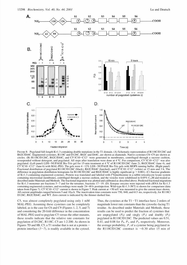
We chose two pairs of residues: R118/D126 and R62/ E64. Each pair was mutated to cysteines in separateconstructs (Figure 8A). As a control, we used C5+ /C10-
C12+, which contains one N-terminal cysteine (C5) in T1,not at the T1-T1 interface but facing the cytosol (5-7 ).The other three cysteines (C10-C12) are cytosolic andreadily available for pegylation (Figures 6 and 7). One pair,R118C/D126C, can be cross-linked to give tetramers usingeither oxidizing conditions or bifunctional cross-linkers,whereas the other pair, R62C/E64C, and the control, C5+ / C10-C12+, cannot be cross-linked (33). The cross-linkingresults with R118C/D126C and R62C/E64C are similar toresults reported by Miller and co-workers for the equivalentpairs of engineered Shaker cysteines in oocyte membranesunder oxidizing conditions (8 ). As a precaution, all sampleswere kept in the continuous presence of DTT (1-2 mM)
and were diluted into solutions pre-degassed with nitrogento avoid any air oxidation of cysteines to form disulfides.This was confirmed by the absence of multimer formationusing LDS-NUPAGE analysis (data not shown).
Each pair forms mostly tetramers (Figure 8C) and isfunctional in oocytes (Figure 8D). Figure 8B shows that theR118C/D126C pair was less accessible than the R62C/E64Cpair, and both pairs of residues were much less accessiblethan the four cysteines in C5+ /C10-C12+. Within 15 min,>20% of the total C5+ /C10-C12+ had all four cysteinespegylated and only a small fraction, e5% of the total C5+ / C10-C12+, remained unpegylated. In contrast, in the same15-min period, R118C/D126C was ∼45% unpegylated and
R62C/E64C was ∼
30% unpegylated. In neither case didincubation as long as 3 h pegylate all of the protein, whereasall of C5+ /C10-C12+ is at least singly pegylated and themajority is at least triply pegylated.2 At 15 min, the averageprobability, P, of a cysteine being pegylated (see Materialsand Methods) is 0.65 for C5+ /C10-C12+ and only 0.34 forR118C/D126C. The probability for R62C/E64C is intermedi-ate, 0.48. The difference between R118C/D126C and R62C/ E64C pegylation histograms is highly significant ( p < 0.001).Our findings indicate that the relative accessibilities of cysteines are C5+ /C10-C12+ . R62C/E64C > R118C/ D126C. These results agree with the cross-linking resultsand are consistent with R118C/D126C and R62C/E64C beingat a protein-protein interface in the T1 tetrameric structure.
To obviate any ambiguities due to undetected cross-linkingbetween cysteine pairs, we mutated R118 and D126 inseparate constructs. Pegylation of these constructs, D126Cand R118C, each containing only one cysteine, is shown inFigure 9. After 15 min, only 23% of D126C and 33% of R118C were pegylated in the presence of 20 mM MAL-PEG, whereas a control construct containing one cysteine,
2 Moreover, R118C/D126C and R62C/E64C also contain trans-membrane native cysteines, C6-C9, which are not labeled during thelonger 3-h incubation, again demonstrating the integrity of themembrane and the protein. Only the relative amounts of unpegylated,singly pegylated, and doubly pegylated protein change, not the numberof pegylated cysteines.
Table 2a
n
contruct detergent blocker 1 2 3 4 5 6 7-12
WT 0.01 0.04 0.09 0.18 0.30 0.39 **b
AMS ** ** ** ** ** ** **NEM ** ** ** ** ** ** **
n
detergent blocker 1 2 3 4 5-12
WT SDS ** ** ** ** 1.0AMS 0.10 0.31 0.37 0.22 **NEM 0.33 0.37 0.30 ** **
a Data were obtained by PhosphorImager detection of the gel, notnecessarily visible in a 16-30 h exposed autoradiogram. Each entry isthe fraction of pegylated protein with n pegylated cysteines, calculatedas F (n) ) cpm(n)/ ∑cpm(n), where n ) 1 to nmax. b Double asterisksindicate cpm are less than background cpm.
Pegylation of Kv1.3 Biochemistry, Vol. 40, No. 44, 2001 13297
8/12/2019 (2001) PEGylation, A Method for Assessing Topological Accessibilities in Kv1.3.pdf
http://slidepdf.com/reader/full/2001-pegylation-a-method-for-assessing-topological-accessibilities-in-kv13pdf 11/14
C5, was almost completely pegylated using only 1 mMMAL-PEG. Assuming these cysteines can be completelylabeled, as is the case for C6 and C9 (Figures 1, 2, 5, and 7)and considering the 20-fold difference in the concentrationof MAL-PEG used to pegylate C5 versus the other mutants,these results indicate that the relative rate constants forpegylation of D126C, R118C, C5 are 1:2:200. As shown inFigures 7D and 8B, C5, a T1 residue that is not at a protein-protein interface (5-7 ), is readily available in the cytosol.
Thus, the cysteines at the T1-T1 interface have 2 orders of magnitude lower rate constants than the cytosolic-facing T1residue. As described under Materials and Methods, theseresults can be used to predict the fraction of cysteines thatare unpegylated (P0) and singly (P1) and doubly (P2)pegylated in R118C/D126C. The predicted values are 0.51,0.41, and 0.08 for P0, P1, and P2, respectively. Moreover,the average probability, P, of a cysteine being pegylated inthe R118C/D126C construct is ∼0.30 after 15 min of
FIGURE 8: Pegylated full-length Kv1.3 containing double mutations in the T1 domain. (A) Schematic representation of R118C/D126C andR62C/E64C. Engineered cysteines, R118C and D126C, R62C and E64C, are shown as diamonds. Native cysteines C6-C9 are shown ascircles. (B) R118C/D126C, R62C/E64C, and C5+ /C10-C12+ were generated in membranes, centrifuged through a sucrose cushion,resuspended without detergent, and pegylated. All steps after translation were done at 4 °C. For comparison, C5+ /C10-C12+ was alsopegylated. (Left panel) LDS-NUPAGE Bis-Tris gel for 15-min treatment at 4 °C of R118C/D126C (lane 2), R62C/E64C (lane 4), andC5+ /C10-C12+ (lane 6) with MAL-PEG. The gels were 4-12% LDS-NUPAGE Bis-Tris gels with MOPS running buffer. (Right panel)Fractional distribution of pegylated R118C/D126C (black), R62C/E64C (hatched), and C5+ /C10-C12+ (white) at 15 min and 3 h. Thedifference in pegylation distribution histograms for R118C/D126C and R62C/E64C is highly significant ( p < 0.001). (C) Sucrose gradientsof Kv1.3 containing engineered cysteines. Protein was translated and labeled with [35S]methionine in a rabbit reticulocyte lysate systemcontaining microsomal membranes, centrifuged through a sucrose cushion, and the vesicles were solubilized in 0.05% C 12M and treated asdescribed under Materials and Methods. The fractional migration was plotted and calibrated as described above. Predicted fractional migrationsfor Kv1.3 monomer are fractions 5-7 and for the tetramer, fractions 17-19. (D) Xenopus oocytes were injected with cRNA for Kv1.3
containing engineered cysteines, and recordings were made 24-48 h postinjection. Wild-type Kv1.3 (WT) is shown for comparison (datataken from Figure 7). C5+ /C10-C12+ current is shown in Figure 7. Peak current at +50 mV was measured to give the current trace shown.All current amplitudes ranged between 5 and 10 µA. The inactivation time constants were 750, 840, and 613 ms, respectively, for R118C/ D126C, R62C/E64C, and WT. Zero current is indicated by the thinner dashed line.
13298 Biochemistry, Vol. 40, No. 44, 2001 Lu and Deutsch

8/12/2019 (2001) PEGylation, A Method for Assessing Topological Accessibilities in Kv1.3.pdf
http://slidepdf.com/reader/full/2001-pegylation-a-method-for-assessing-topological-accessibilities-in-kv13pdf 12/14
pegylation, in agreement with the results calculated from thedata in Figure 8B. Comparison of these predictions, derivedfrom the singly mutated constructs, with experimental datafrom the doubly mutated construct provides no evidence forcooperative pegylation between nearby cysteines.
DISCUSSION
Pegylation Method. Pegylation of cysteines is a promisingbiochemical tool to be used in conjunction with othermethods to define membrane protein topology and interfacesinvolved in oligomeric structures. The strategy of usingcysteines to obtain structure/function information for mem-brane proteins has many precedents, for example, thesubstituted-cysteine-accessibility method (SCAM; 19, 28 ,34), which uses functional manifestations of cysteine modi-fication by methanethiosulfonate derivatives to infer struc-tural information. Yet another example is the use of quaternary amine maleimides with flexible linkers to measuredistances between extracellular loop residues and the poreof Kv channels (35). More direct structural information isobtained from spectroscopic and biochemical methods incombination with SCAM, which permit detection of func-tionally silent cysteine modifications (2, 4, 21, 34, 36 -45).Some of these spectroscopic approaches require largeamounts of protein, purification of the protein and/orreconstitution of the protein into lipid, and spectroscopictools. Biochemical, rather than spectroscopic, methods permit
small quantities (picomoles to femtomoles) to be studiedusing SDS-PAGE techniques. In these cases, conjugatedmaleimides are bound to cysteines to determine the mem-brane topology of, for example, human P-glycoprotein (21),the peripheral benzodiazepine receptor (44), and the R subunit of F1F0ATP synthase from Escherichia coli (45).Furthermore, gel shift assays using small molecular weightSH reagents (e.g., benzophenone-MAL, fluorescein-MAL,and iodoacetylated reagent) have been used to study mem-
brane protein surface accessibility (46 , 47 ). The use of thesereagents is limited to small molecular weight proteins, wherethe resolution on SDS-PAGE permits a detectable gel shiftfor the cysteine adduct. However, larger polytopic proteins,such as Kv1.3, require a larger mass-tag. PEG (5 kDa) servesthis purpose. Recently, an ο-pyridyl disulfide PEG3 has beenused to study a bacterial surface layer protein layered on acell wall, specifically to determine which residues are locatedat the external surface of the S-layer lattice, which are atthe intersubunit interface, and which are within the pores(49). These approaches detect single cysteines, one at a time.The advantage of a mass-tag method such as ours is thataccessibility of multiple cysteines can be evaluated simul-
taneously, and distribution histograms can be used tounderstand relative changes in accessibility. The sequentialuse of blockers (permeant and impermeant) and PEG, andof different detergents (denaturing and non-denaturing) orno detergent, is an effective strategy for determining trans-membrane topology and identifying protein-aqueous, protein-lipid, and protein-protein interfaces. Moreover, our approachpermits multiple native cysteines to be assessed and dynamicchanges in accessibility to be monitored during assembly.In the latter case, multiple reference cysteines can besimultaneously positioned at each interface.
One note of caution is that extrapolation of accessibilitymeasurements in one channel protein, or fragment of the
channel, to another should be done judiciously, taking intoaccount that topology is generated through cooperativeinteractions between multiple topogenic determinants duringassembly (23). Such determinants may differ for peptidefragments versus full-length channel. Another caveat derivesfrom our limitation to quantitate the fraction of protein thatis correctly folded into functional channels in the ERmembrane. A final note of caution is that long pegylationtimes in the absence of detergent can produce high back-ground and aggregation for multi-cysteine constructs. Thus,in the absence of detergent the method works best for shortincubation times (1-3 h) and fewer than five cysteines.
Assessment of cysteine accessibility using covalentlylinked markers depends on the availability of the SH group,its reactivity, and the stability of the adduct. The latterparameter is not an issue in pegylation assays. Rather, theavailability and reactivity operationally define accessibility.Factors governing reactivity are the pK a of the cysteine andthe local pH. Availability may be a function of stericexclusion, the protein-protein interface in a folded state of a protein, whether a protein is buried in a lipid membrane ata protein-lipid interface, or whether a protein is free insolution. Among the possible intermolecular protein-proteininteractions to consider in our experiments is the oligomeric
3 o-Pyridyl disulfide PEG has also been used recently to locate theconstriction in the pore of staphylococcal R -hemolysin (48 ).
FIGURE 9: Pegylated full-length Kv1.3 containing single mutationsin the T1 domain. R118C and D126C were generated in separatetranslations and pegylated as described in Figure 8. All steps aftertranslation were done at 4 °C. For comparison, C5+ ( Bst EII-cutand released from tRNA by puromycin; (33), containing only C5,was also pegylated. (Bottom panel) LDS-NUPAGE Bis-Tris gelfor 15-min treatment at 4 °C of R118C (lane 1), D126C (lane 2),and C5+ (lane 3) with MAL-PEG (20 mM for lanes 1 and 3, 1mM for lane 3). The gels were 4-12% LDS-NUPAGE Bis-Trisgels with MOPS running buffer.
Pegylation of Kv1.3 Biochemistry, Vol. 40, No. 44, 2001 13299

8/12/2019 (2001) PEGylation, A Method for Assessing Topological Accessibilities in Kv1.3.pdf
http://slidepdf.com/reader/full/2001-pegylation-a-method-for-assessing-topological-accessibilities-in-kv13pdf 13/14
state of Kv1.3. Inaccessibility could be due to SH groupsthat are buried at intersubunit interfaces in a tetramer oroligomeric intermediate. Tetramers of Kv1.3 exist in vitroin microsomal membrane vesicles and are retained innonionic detergent (29; Figures 7 and 8). The Kv1.3 proteindistributed in the sucrose gradients represents >90% of theprotein in the ER membrane preparations. Therefore, theresults of pegylation studies represent the major fraction of Kv1.3 protein in any given sample. Pegylation of full-length
Kv1.3 in membrane vesicles in the absence of detergentlikely reflects availability of cysteines in the tetramer,whereas pegylation in SDS primarily reflects pegylation of the denatured monomer. AMS and NEM incubations arecarried out in the intact membrane vesicle in the absence of detergent, and therefore AMS and NEM react with availablecysteines in the tetramer. It is not clear whether all nativecysteines in full-length Kv1.3 can be pegylated or blockedwith AMS or NEM because in the wild-type channel proteinthere are more complicated intra- and intersubunit interac-tions than in the smaller Kv1.3 fragments or mutated full-length channels. Such tertiary and quaternary interactionscould alter the availabilities and reactivities of C1-C12.
The presence of a pegylation ladder in the Bis-Tris gelsfor some multi-cysteine constructs [e.g., S1-S2-S3, S5-S6-C-terminus, Kv1.3(T1-), WT, C5+ /C10-C12+] indi-cates that under the conditions of our experiments, theprobability of individual cysteines being pegylated is <1 forall cysteines in the test protein. Additional information canbe obtained from analysis of these distributions of pegylatedspecies. For instance, neither the truncated Kv1.3 fragments(Figures 4-6) nor the full-length Kv1.3 constructs (Figure7) displays a binomial distribution for the number of pegylated cysteines (Figures 4, 6, and 7) in either zerodetergent or in SDS (in all cases p < 0.001). Binomalityassumes that the cysteines in any given protein have identical
availability/reactivity and that each is pegylated inde-pendently. Because of discrepancies between the observeddistributions and the calculated binomial distributions,regardless of whether detergent is present or not, eithercooperativity of pegylation or differences in availability/ reactivity of the cysteines exist. Our analysis of the data inFigures 8 and 9 provides no evidence for cooperativity inpegylation. Differences in availability/reactivity are expectedwhen the membrane is intact (absence of detergent) as thetopologies of C1-C12 are different. Pegylation differencesin SDS are more likely to be due to differences in reactivitiesrather than availabilities because MAL-PEG likely interca-lates and equilibrates more readily across a detergent micellethan a membrane bilayer. One possible origin of reactivity
differences is suppressed ionization of thiols to thiolate ions(hence, slower reaction with MAL-PEG) of cysteines buriedmore deeply within the hydrophobic micelle interior. Thenegative charges of SDS will also suppress ionization.
Topology and Accessibility of NatiV e Cysteines. On thebasis of the pegylation results, we have made the followingassignments in Kv1.3. C6, near the C-terminal end of S1,faces the lumen at a protein-aqueous interface in fragmentsS1 and S1-S2-S3 but at a protein-protein or protein-lipid interface in channel-forming species such as Kv1.3(T1-)and wild-type Kv1.3. Such a location is consistent withglycosylation and topological determinations of the S1-S2loop in Kv1.3(T1-) (23). We favor assignment of C7, in the
middle of S2, to a protein-protein interface in Kv1.3(T1-),consistent with tryptophan-scanning studies of S2 in Shaker
(16 ). According to the models for Shaker channels (50), thecorresponding cysteine faces S4 and protrudes into theaqueous vestibule of the “gating pore” or S4 channel (51,52). Such a location might influence reagent accessibility toC7. For instance, although AMS is negatively charged andtherefore might be sensitive to the local potential in the S4pore (53), it is somewhat larger than NEM and may have
hindered access to putative crevices (51, 52).C8, near the C-terminal end of S2, is at a protein-aqueous
interface, in equilibrium between exposed and buried statesat the cytosolic membrane border. C9, at the N-terminal endof S6, is at an extracellular (lumenal) location. The crystalstructure of tetrameric KcsA, which contains a pore regionnearly identical to the amino acid sequence of mammalianKv channels, suggests that C9 is in a groove between S5and the bottom of the pore helix. The homologous residuein Shaker was not labeled with 5 µM tetramethylrhodaminemaleimide during a 30-min incubation at 0 °C (54).Nevertheless, we were able to modify C9 using 20 mM NEMfor 2 h at 4 °C. The lack of labeling in Shaker may be due
to low concentration or steric hindrance of the bulkytetramethylrhodamine, short exposure time, or the fact thatcysteines on the extracellular surface of the plasma mem-brane are oxidized or otherwise protected. A kinetic analysisof modification rates should provide more insight into theaccessibility of C9.
Pegylation studies of S5-S6-C-terminus, Kv1.3(T1-),and full-length C5+ /C10-C12+ suggest that the C-terminalcysteines, C10-C12, are in the cytosol at a protein-aqueousinterface. Whereas the first two constructs exist as monomersin our pegylation studies, the latter one, full-length C5+ / C10-C12+, exists as a tetramer. Yet, in both cases, C10-C12 are available and sufficiently reactive for labeling by
MAL-PEG.In contrast, the N-terminal cysteines C1-C5, although alsoin the cytosol (23), are not all available for pegylation. Infull-length tetrameric channels, we only know that C5 isaccessible. The crystal structure of the T1 domain (5-7 )predicts that C5 is exposed to the cytosol and should bepegylated. The N-terminal region containing C1-C4 wasnot included in the peptide used in the crystal structuredetermination, so we cannot speculate about the ability of C1-C4 to be pegylated. However, membrane-targetingstudies (23) suggest that the N terminus of Kv1.3 monomersmay already exist in a folded state, which could include C1-C4.
The availability and reactivity of cysteines may dependon whether the protein is monomeric or multimeric. Someresidues may never be buried at protein-protein interfaces,whereas others may become buried or uncovered as assemblyproceeds. Nonetheless, changes in availability and reactivitymay accompany tetramerization and, therefore, may bemanifest as time-dependent pegylation of Kv1.3. Futurestudies of Kv1.3 containing engineered cysteines will addressthese issues. In the meantime, we have studied one possibilityof putative inaccessibility, that of the N-terminal cytosolicT1-T1 interface. In the intact, detergent-free system, foldedT1 residues shown to be in close proximity (<3.2 Å) at aprotein-protein interface and experimentally cross-linkableinto disulfide bonds (8 , 33) had much lower rates of
13300 Biochemistry, Vol. 40, No. 44, 2001 Lu and Deutsch

8/12/2019 (2001) PEGylation, A Method for Assessing Topological Accessibilities in Kv1.3.pdf
http://slidepdf.com/reader/full/2001-pegylation-a-method-for-assessing-topological-accessibilities-in-kv13pdf 14/14
pegylation than cytosolic-facing cysteines, namely, C5 in T1and C10-C12 in the C terminus (Figures 8 and 9). Thenumber of cysteines pegylated, as well as the relative kineticsof pegylation, can be a useful tool for probing relativetopological accessibilities in membrane proteins.
ACKNOWLEDGMENT
We thank Dr. L. Tu and J. Wang for technical assistanceand Dr. R. Horn for careful reading of the manuscript andassistance with the statistical analyses.
REFERENCES
1. MacKinnon, R. (1991) Nature 350, 232-235.2. Schulteis, C., Nagaya, N., and Papazian, D. (1996) Biochem-
istry 35, 12133-12140.3. Doyle, D. A., Cabral, J. M., Pfuetzner, R. A., Kuo, A., Gulbis,
J. M., Cohen, S. L., Chait, B. T., and MacKinnon, R. (1998)Science 280, 69-76.
4. Perozo, E., Cortes, D. M., and Cuello, L. G. (1998) Nat. Struct. Biol. 5, 459-469.
5. Kreusch, A., Pfaffinger, P. J., Stevens, C. F., and Choe, S.(1998) Nature 392, 945-948.
6. Bixby, K. A., Nanao, M. H., Shen, N. V., Kreusch, A.,Bellamy, H., Pfaffinger, P. J., and Choe, S. (1999) Nat. Struct.
Biol. 6 , 38-43.7. Minor, D. L., Lin, Y. F., Mobley, B. C., Avelar, A., Jan, Y.
N., Jan, L. Y., and Berger, J. M. (2000) Cell 102, 657-670.8. Kobertz, W. R., Williams, C., and Miller, C. (2000) Biochem-
istry 39, 10347-10352.9. Vandongen, A. M. J., Frech, G. C., Drewe, J. A., Joho, R. H.,
and Brown, A. M. (1990) Neuron 4, 433-443.10. Hopkins, W. F., Demas, V., and Tempel, B. L. (1994) J.
Neurosci. 14, 1385-1393.11. Babila, T., Moscucci, A., Wang, H., Weaver, F. E., and Koren,
G. (1994) Neuron 12, 615-626.12. Lee, T. E., Phillipson, L. H., Kuznetsov, A., and Nelson, D.
J. (1994) Biophys. J. 66 , 667-673.13. Tu., L., Santarelli, V., and Deutsch, C. (1995) Biophys. J. 68 ,
147-156.14. Tu, L., Santarelli, V., Sheng, Z.-F., Skach, W., Pain, D., and
Deutsch, C. (1996) J. Biol. Chem. 271, 18904-18911.15. Kobertz, W. R., and Miller, C. (1999) Nat. Struct. Biol. 6 ,
1122-1125.16. Monks, S. A., Needleman, D. J., and Miller, C. (1999) J. Gen.
Physiol. 113, 415-423.17. Li-Smerin, Y., Hackos, D. H., and Swartz, K. J. (2000) Neuron
25, 411-423.18. Li-Smerin, Y., Hackos, D. H., and Swartz, K. J. (2000) J. Gen.
Physiol. 115, 33-50.19. Akabas, M. H., Stauffer, D. A., Xu, M., and Karlin, A. (1992)
Science 258 , 307-310.20. Akabas, M. H., Kaufmann, C., Archdeacon, P., and Karlin,
A. (1994) Neuron 4, 919-927.21. Loo, T. W., and Clarke, D. M. (1998). J. Biol. Chem. 270,
843-848.22. Lu, J., and Deutsch, C. (1999) Biophys. J. 76 , A76.
23. Tu, L., Wang, J., Helm, A., Skach, W. R., and Deutsch, C.(2000) Biochemistry 39, 824-836.
24. Grassetti, D. R., and Murray, J. F., Jr. (1967) Arch. Biochem. Biophys. 119, 41-49.
25. Rao, C. R. (1973) in Linear Statistical Inference and Its Applications, Wiley, New York.
26. Chahine, M., Chen, L.-Q., Barchi, R. L., Kallen, R. G., andHorn, R. (1992) J. Mol. Cell Cardiol. 24, 1231-1236.
27. Sheng, Z., Skach, W., Santarelli, V., and Deutsch, C. (1997) Biochemistry 36 , 15501-15513.
28. Karlin, A., and Akabas, M. H. (1998) Methods Enzymology293, 123-145.
29. Tu, L., and Deutsch, C. (1999) Biophys. J. 76 , 2004-2017.30. Li, M., Jan, Y. N., and Jan, L. Y. (1992) Science 257 , 1225-
1230.31. Shen, N. V., Chen, X., Boyer, M. M., and Pfaffinger, P. (1993)
Neuron 11, 67-76.32. Xu, J., Yu, W., Jan, J. N., Jan, L., and Li, M. (1995) J. Biol.
Chem. 270, 24761-24768.33. Lu, J., Robinson, M. R., Edwards, D., and Deutsch, C. (2001)
Biochemistry 40, 10934-10946.34. Horn, R. (1998) Methods Enzymol. 293, 145-155.35. Blaustein, R. O., Cole, P. A., Williams, C., and Miller, C.
(2000) Nat. Struct. Biol. 7 , 309-311.36. Kaback, H. R., Voss, J., and Wu, J. (1997) Curr. Opin. Struct.
Biol. 7 , 537-542.37. Kaback, H. R., and Wu, J. (1997) Q. ReV . Biophys. 30, 333-
364.38. Hubbell, W. L., and Altenbach, C. (1994) in Site-Directed Spin
Labeling of Membrane Proteins, Oxford University Press, NewYork.
39. Hubbell, W. L., Mchaourab, H. S., Altenbach, C., and Lietzow,M. A. (1996) Structure 4, 779-783.
40. Gross, A., Columbus, L., Hideg, K., Altenbach, C., andHubbell, W. L. (1999) Biochemistry 38 , 10324-10335.
41. Arkin, I. T., MacKenzie, K. R., Fisher, L., Aimoto, S.,Engelman, D. M., and Smith, S. O. (1996) Nat. Struct. Biol.3, 240-243.
42. Arkin, I. T., Adams, P. D., Brunger, A. T., Aimoto, S.,Engelman, D. M., and Smith, S. O. (1997) J. Membr. Biol.155, 199-206.
43. Klein-Seetharaman, J., Hwa, J., Cai, K., Altenbach, C.,Hubbell, W. L., and Khorana, H. G. (1999) Biochemistry 38 ,7938-7944.
44. Joseph-Liauzun, E., Delmas, P., Shire, D., and Ferrara, P.(1998) J. Biol. Chem. 273, 2146-2152.
45. Long, J. C., Wang, S., and Vik, S. B. (1998) J. Biol. Chem.273, 16235-16240.
46. Krishnasastry, M., Walker, B., Braha, O., and Bayley, H.(1994) FEBS Lett. 356 , 66-71.47. Jones, P. C., Sivaprasadarao, A., Wray, D., and Findlay, J. B.
(1996) Mol. Membr. Biol. 13, 53-60.48. Movileanu, L., Cheley, S., Howorka, S., Braha, O., and Bayley,
H. (2001) J. Gen. Physiol. 117 , 239-251.49. Howorka, S., Sara, M., Wang, Y., Kuen, B., Sleytr, U. B.,
Lubitz, W., and Bayley, H. (2000) J. Biol. Chem. 275, 37876-37886.
50. Durell, S. R., Hao, Y., and Guy, R. (1998) J. Struct. Biol.121, 263-284.
51. Yang, N., George, A. L., and Horn, R. (1996) Neuron 16 , 113-122.
52. Larsson, H. P., Baker, O. S., Dhillon, D. S., and Isacoff, E.Y. (1996) Neuron 16 , 387-397.
53. Yang, N., George, A. L. J., and Horn, R. (1997) Biophys. J.
73, 2260-
2268.54. Mannuzzu, L. M., Moronne, M. M., and Isacoff, E. Y. (1996)Science 271, 213-216.
55. Boland, L. M., Jurman, M. E., and Yellen, G. (1994) Biophys. J. 66 , 694-699.
BI0107647
Pegylation of Kv1.3 Biochemistry, Vol. 40, No. 44, 2001 13301